The Imaging of the Orbit
100
The Orbit Thorsang R1 Neuroradiology topic 04.06.2014
-
Upload
fern-ferretie -
Category
Science
-
view
129 -
download
5
description
The imaging of the orbit
Transcript of The Imaging of the Orbit
The Orbit
Thorsang R1 Neuroradiology topic
04.06.2014
Outline
• Orbit overview
• Imaging techniques
• Orbital anatomy
• Diseases of the orbit
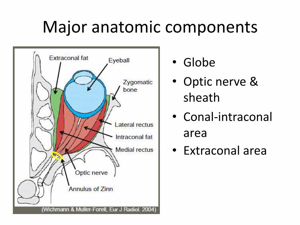
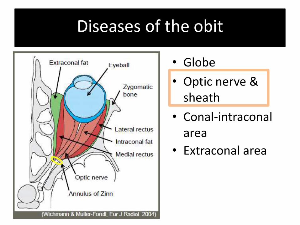
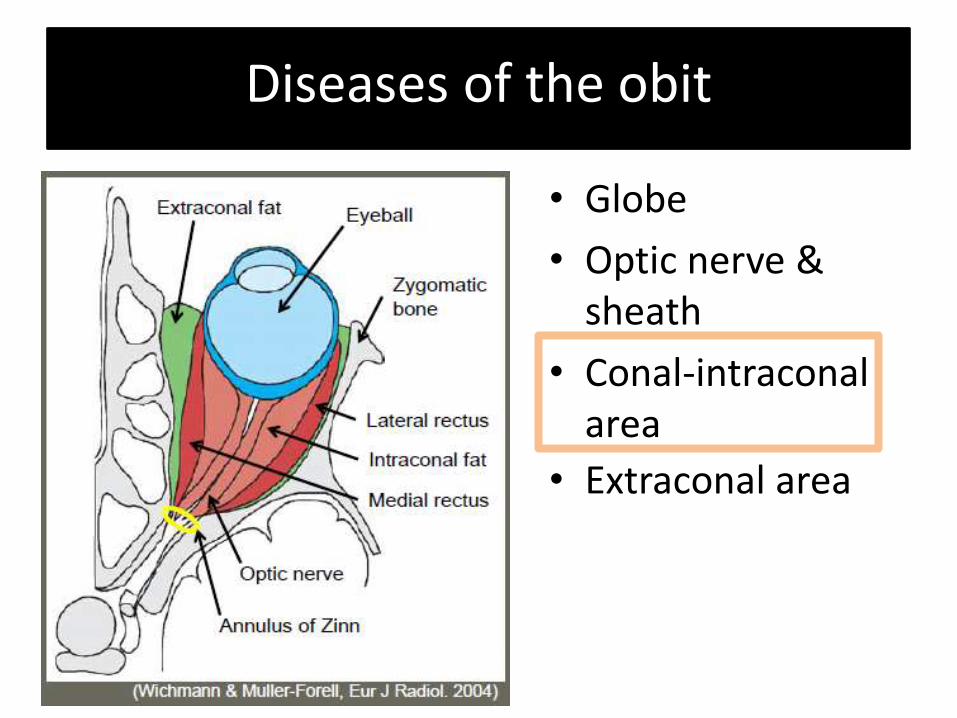
Major anatomic components
• Globe
• Optic nerve & sheath
• Conal-intraconal area
• Extraconal area
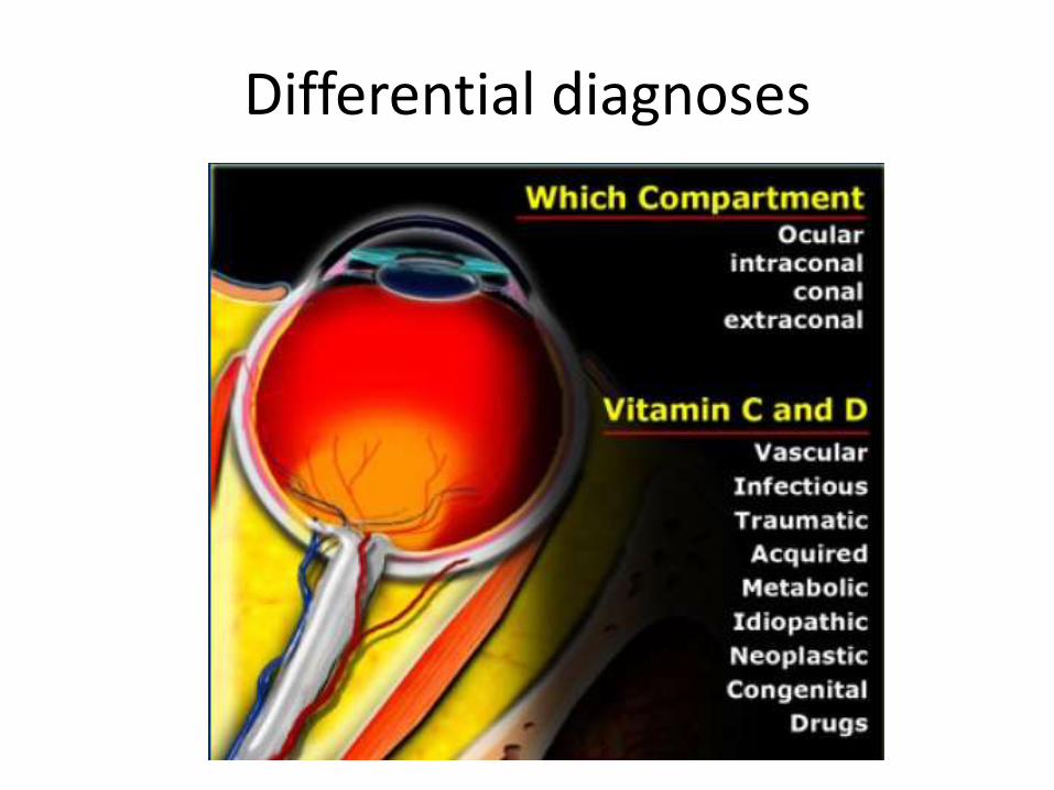
Differential diagnoses
Imaging techniques
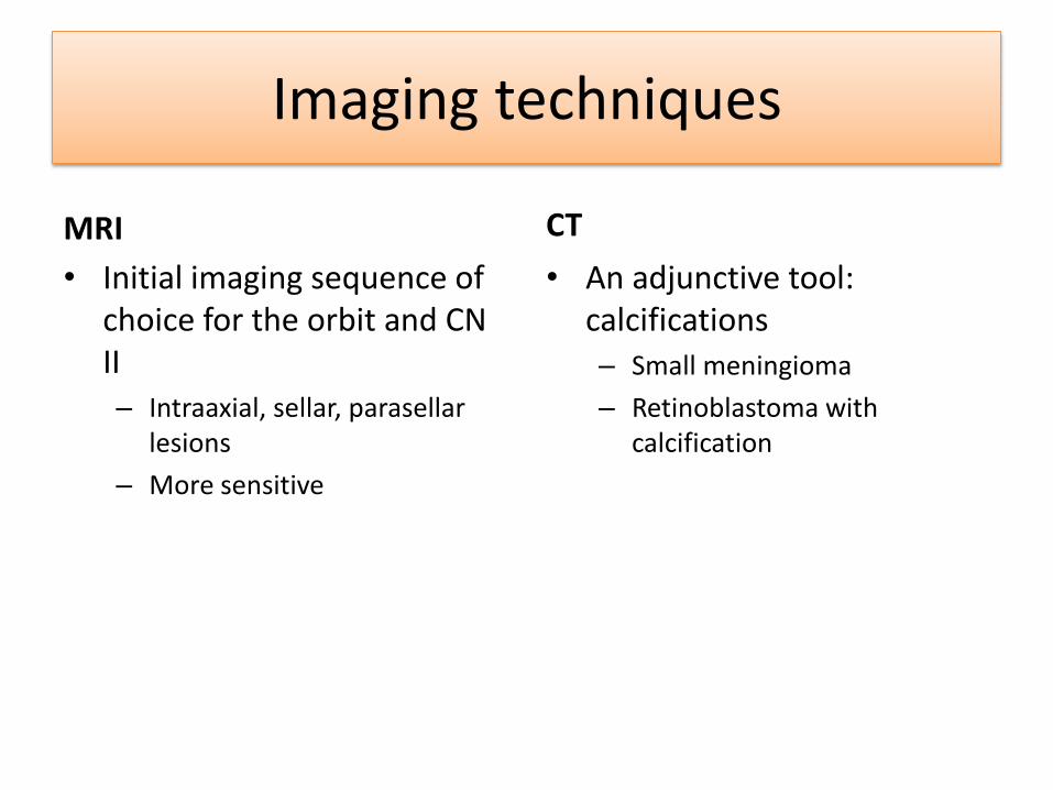
MRI
• Initial imaging sequence of choice for the orbit and CN II – Intraaxial, sellar, parasellar
lesions
– More sensitive
CT
• An adjunctive tool: calcifications
– Small meningioma
– Retinoblastoma with calcification
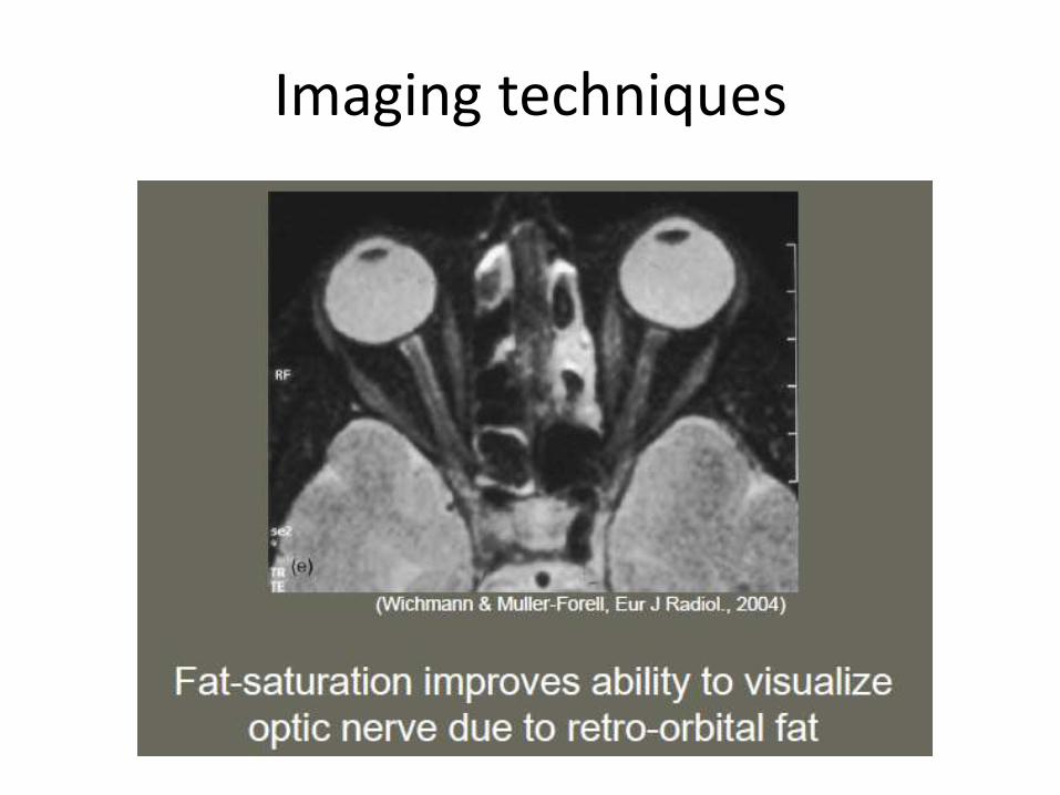
Imaging techniques
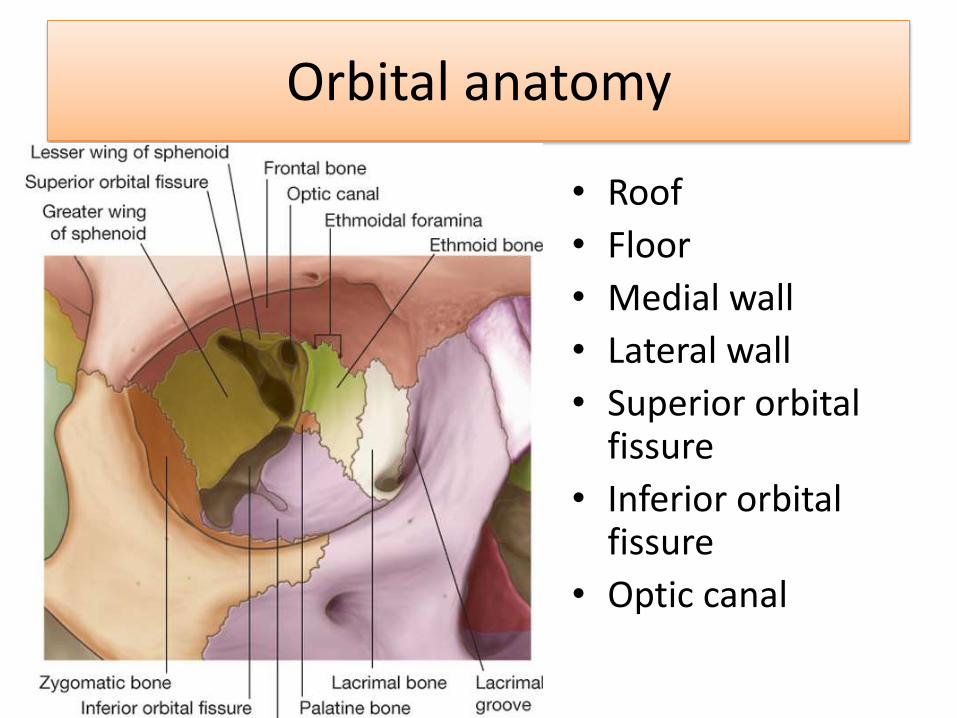
Orbital anatomy
• Roof
• Floor
• Medial wall
• Lateral wall
• Superior orbital fissure
• Inferior orbital fissure
• Optic canal
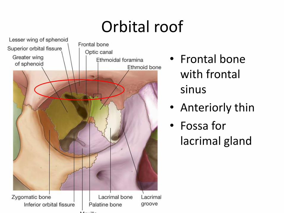
Orbital roof
• Frontal bone with frontal sinus
• Anteriorly thin
• Fossa for lacrimal gland
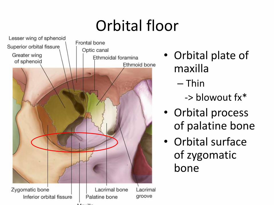
Orbital floor
• Orbital plate of maxilla – Thin
-> blowout fx*
• Orbital process of palatine bone
• Orbital surface of zygomatic bone
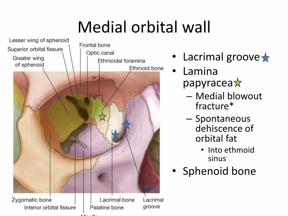
Medial orbital wall
• Lacrimal groove • Lamina
papyracea – Medial blowout
fracture* – Spontaneous
dehiscence of orbital fat • Into ethmoid
sinus
• Sphenoid bone
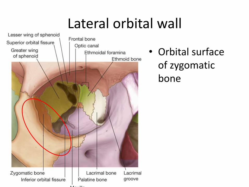
Lateral orbital wall
• Orbital surface of zygomatic bone
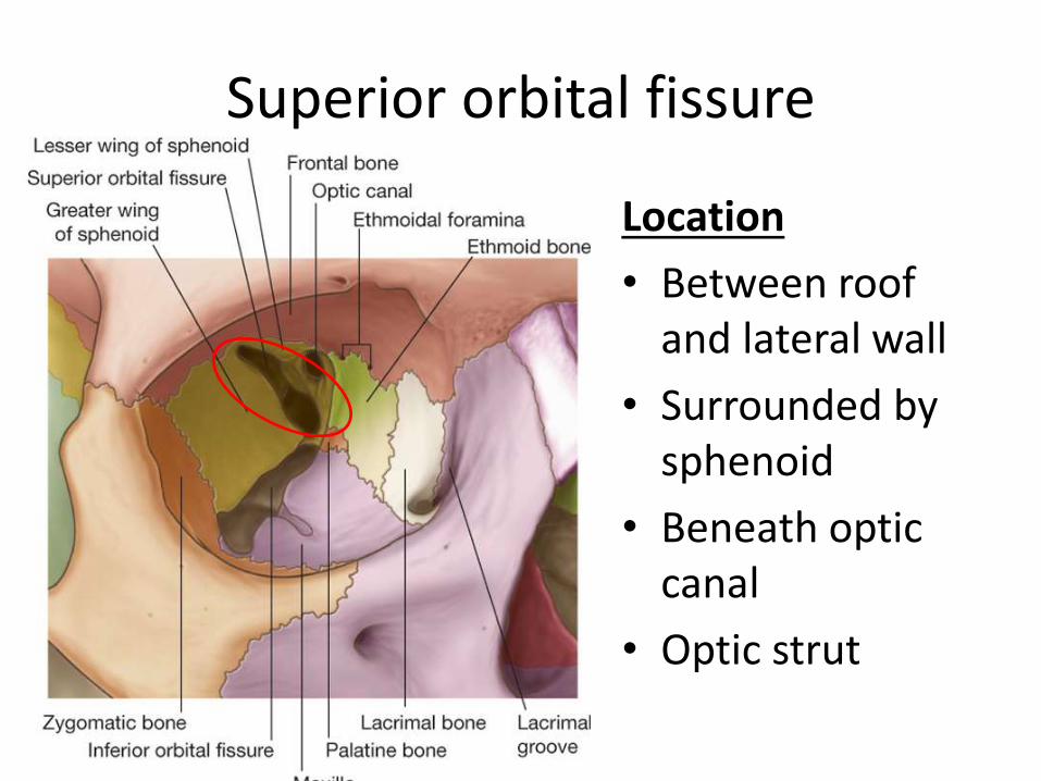
Superior orbital fissure
Location
• Between roof and lateral wall
• Surrounded by sphenoid
• Beneath optic canal
• Optic strut
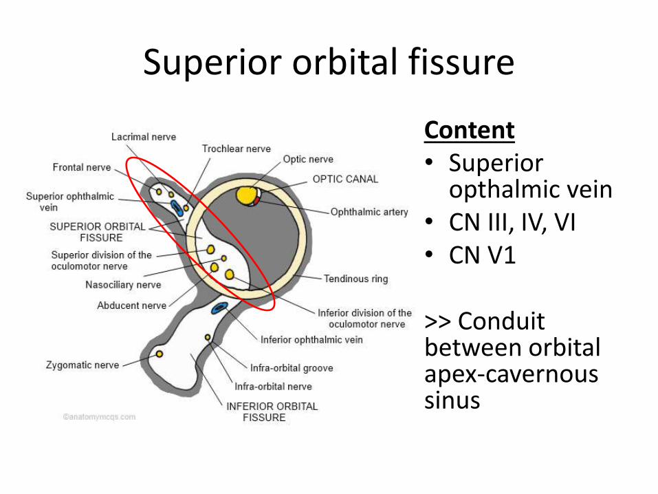
Superior orbital fissure
Content • Superior
opthalmic vein • CN III, IV, VI • CN V1
>> Conduit between orbital apex-cavernous sinus
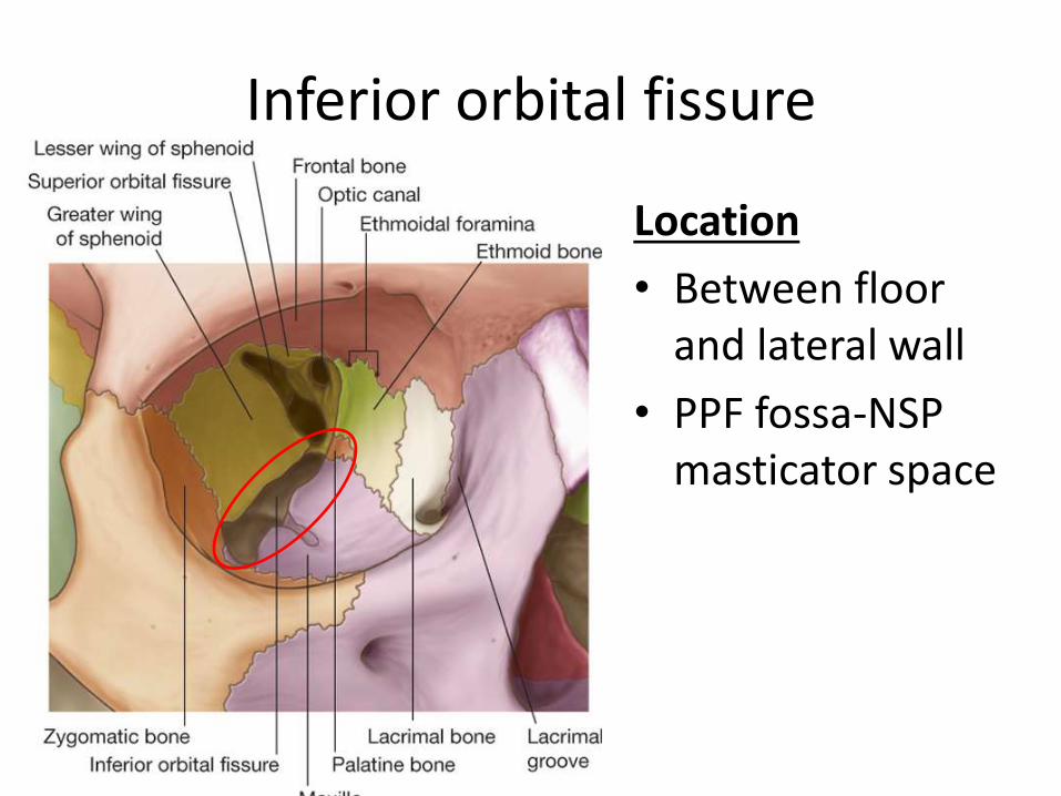
Inferior orbital fissure
Location
• Between floor and lateral wall
• PPF fossa-NSP masticator space
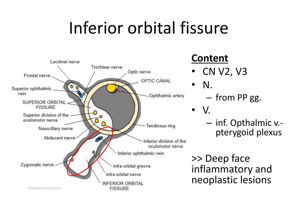
Inferior orbital fissure
Content • CN V2, V3 • N.
– from PP gg.
• V. – inf. Opthalmic v.-
pterygoid plexus
>> Deep face inflammatory and neoplastic lesions
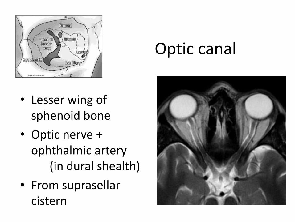
Optic canal
• Lesser wing of sphenoid bone
• Optic nerve + ophthalmic artery (in dural shealth)
• From suprasellar cistern
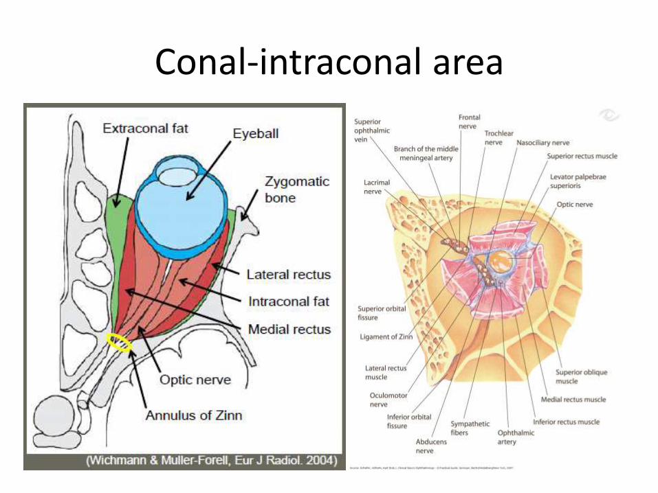
Conal-intraconal area
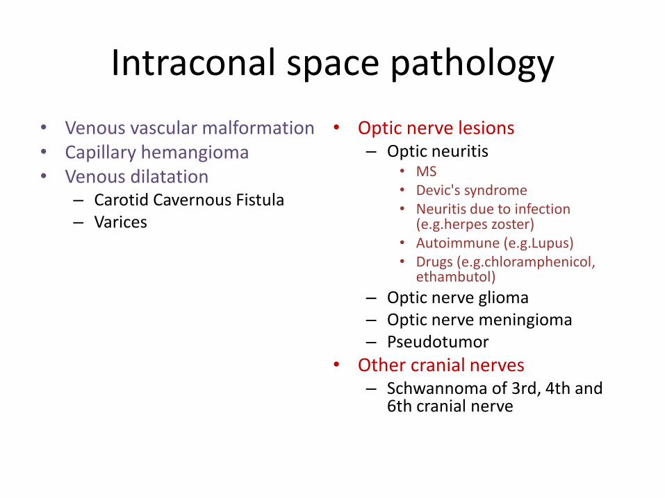
Intraconal space pathology
• Venous vascular malformation • Capillary hemangioma • Venous dilatation
– Carotid Cavernous Fistula – Varices
• Optic nerve lesions – Optic neuritis
• MS • Devic's syndrome • Neuritis due to infection
(e.g.herpes zoster) • Autoimmune (e.g.Lupus) • Drugs (e.g.chloramphenicol,
ethambutol)
– Optic nerve glioma – Optic nerve meningioma – Pseudotumor
• Other cranial nerves – Schwannoma of 3rd, 4th and
6th cranial nerve
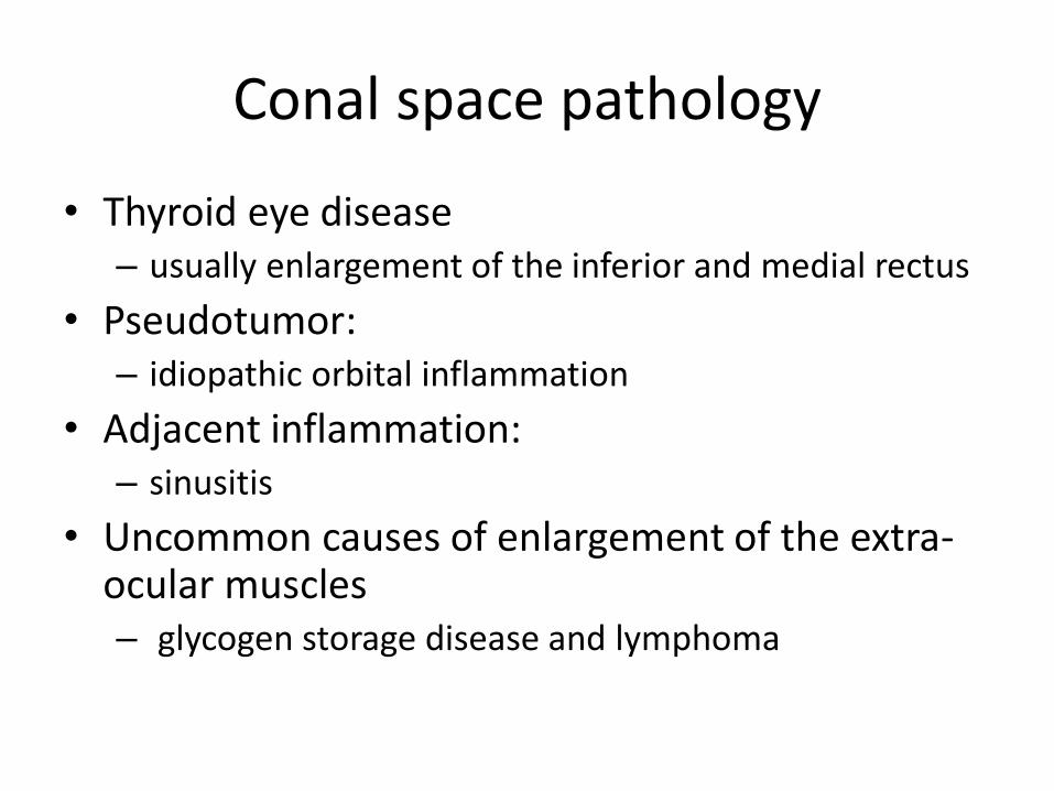
Conal space pathology
• Thyroid eye disease – usually enlargement of the inferior and medial rectus
• Pseudotumor: – idiopathic orbital inflammation
• Adjacent inflammation: – sinusitis
• Uncommon causes of enlargement of the extra-ocular muscles – glycogen storage disease and lymphoma
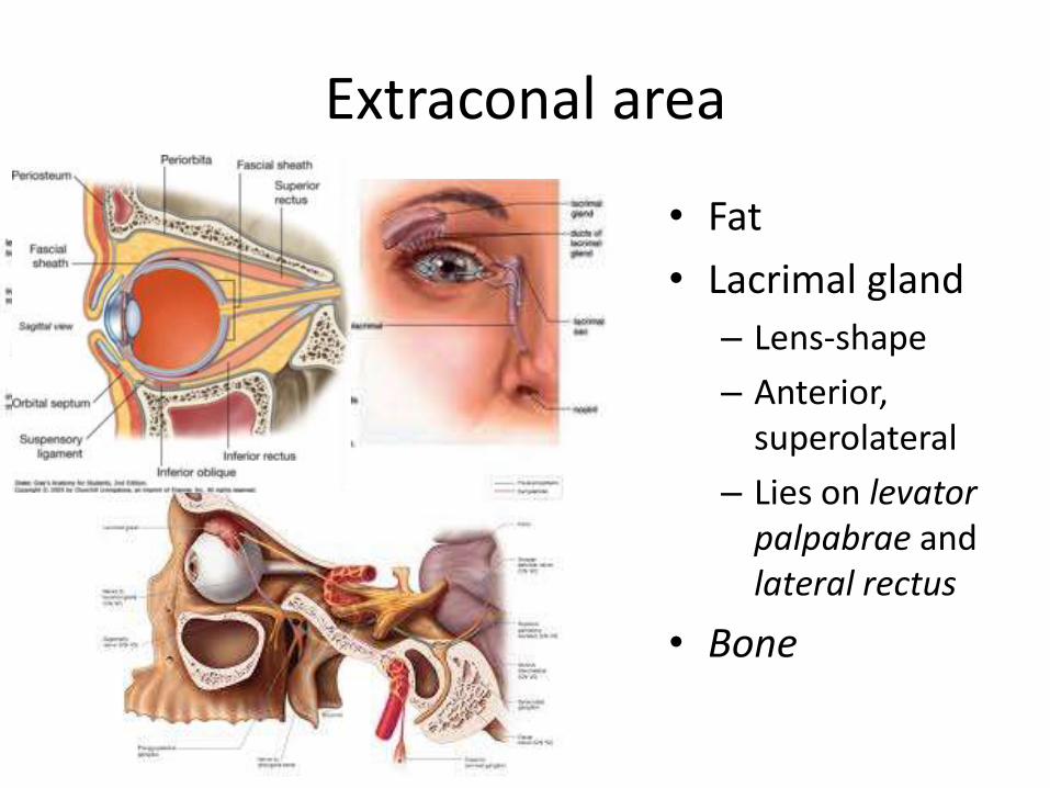
Extraconal area
• Fat
• Lacrimal gland
– Lens-shape
– Anterior, superolateral
– Lies on levator palpabrae and lateral rectus
• Bone
Extraconal pathology
• Abscess due to sinusitis
• Schwannoma of the V1 and V2
• Bone lesions:
– Fibrous dysplasia of the sphenoid wing
– Metastases
– Multiple myeloma
• Diseases of the orbital appendages
Orbital appendages
• Lacrimal gland
– superolaterally in the orbit
– granulomatous, epithelial/glandular, and developmental
• lacrimal sac
• lacrimal duct
– -> inferior terbinate into the nose
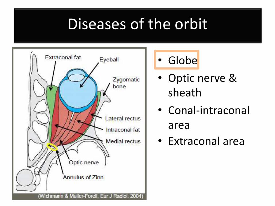
Diseases of the orbit
• Globe
• Optic nerve & sheath
• Conal-intraconal area
• Extraconal area
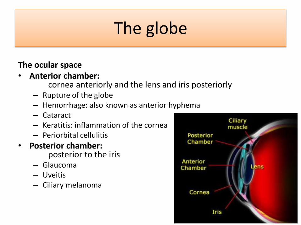
The ocular space • Anterior chamber:
cornea anteriorly and the lens and iris posteriorly – Rupture of the globe – Hemorrhage: also known as anterior hyphema – Cataract – Keratitis: inflammation of the cornea – Periorbital cellulitis
• Posterior chamber: posterior to the iris – Glaucoma – Uveitis – Ciliary melanoma
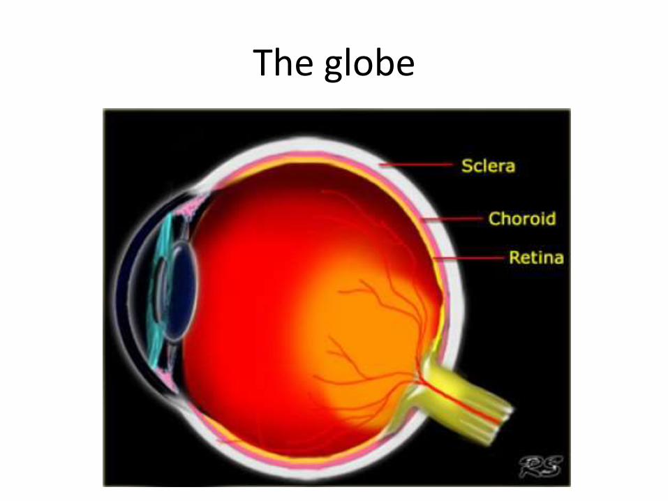
The globe
The globe
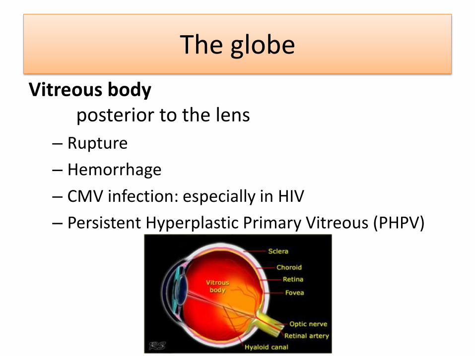
Vitreous body posterior to the lens
– Rupture
– Hemorrhage
– CMV infection: especially in HIV
– Persistent Hyperplastic Primary Vitreous (PHPV)
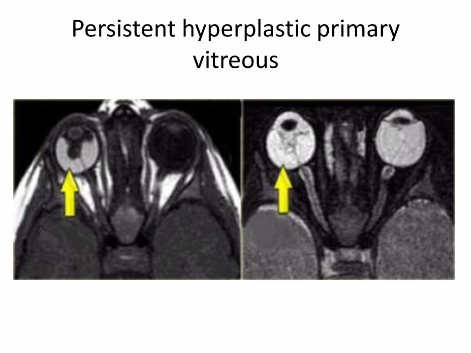
Persistent hyperplastic primary vitreous
• Unilateral leukokoria in male infants
• Persistent hyaloid canal
• Persistence of the primary vascular vitreous
• Hyperplasia of the residual embryonic connective tissue
• PHPV is the second most common cause of leukocoria
• Also develops glaucoma and cataract
• Findings:
– Microphtalmic globe with enhancing increased density in the vitreous humor.
– Unilateral/bilateral tissue density band from back of the lens to the posterior inner globe (Persistent Cloquet’s canal)
– Retinal detachment (occurs in 30-55%)
Persistent hyperplastic primary vitreous
Persistent hyperplastic primary vitreous
The globe
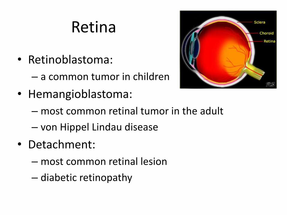
Retina
• Retinoblastoma:
– a common tumor in children
• Hemangioblastoma:
– most common retinal tumor in the adult
– von Hippel Lindau disease
• Detachment:
– most common retinal lesion
– diabetic retinopathy
Retinoblastoma
• Common tumor in the first year of life
• Child < 3 years of age (98%)
• Other presentations: leukokoria, strabismus, decreased visual acuity, family hx, eye pain, proptosis
• Believed to arise from neuroectodermal cells
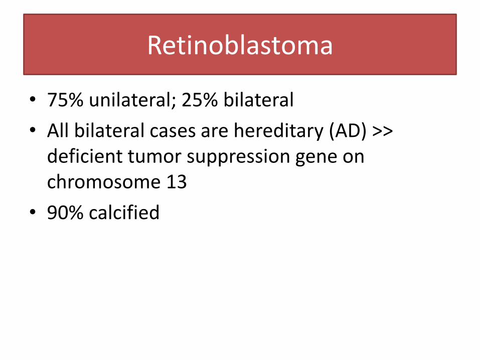
• 75% unilateral; 25% bilateral
• All bilateral cases are hereditary (AD) >> deficient tumor suppression gene on chromosome 13
• 90% calcified
Retinoblastoma
Retinoblastoma
• The other tumors in this age group are
– Neuroblastoma
– Wilm's tumor
– Leukemia
– Teratoma.
Retinoblastoma
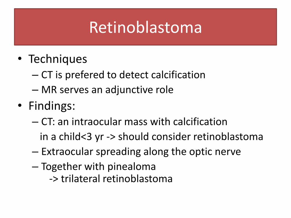
• Techniques – CT is prefered to detect calcification
– MR serves an adjunctive role
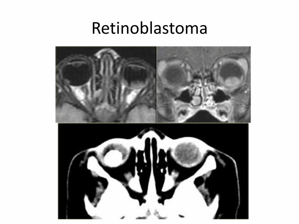
• Findings: – CT: an intraocular mass with calcification
in a child<3 yr -> should consider retinoblastoma
– Extraocular spreading along the optic nerve
– Together with pinealoma -> trilateral retinoblastoma
Retinoblastoma
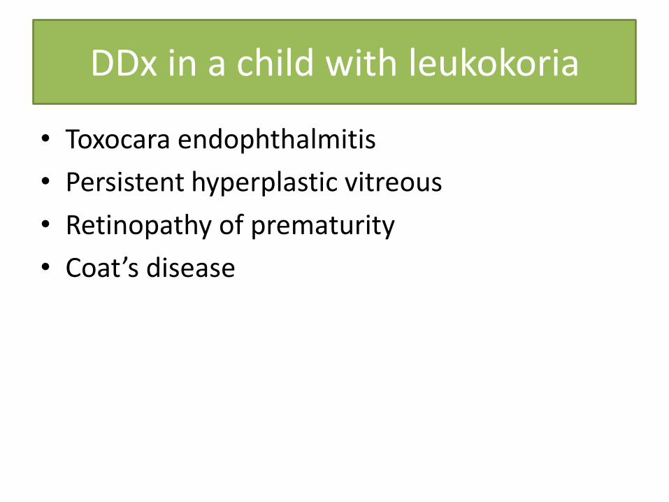
DDx in a child with leukokoria
• Toxocara endophthalmitis
• Persistent hyperplastic vitreous
• Retinopathy of prematurity
• Coat’s disease
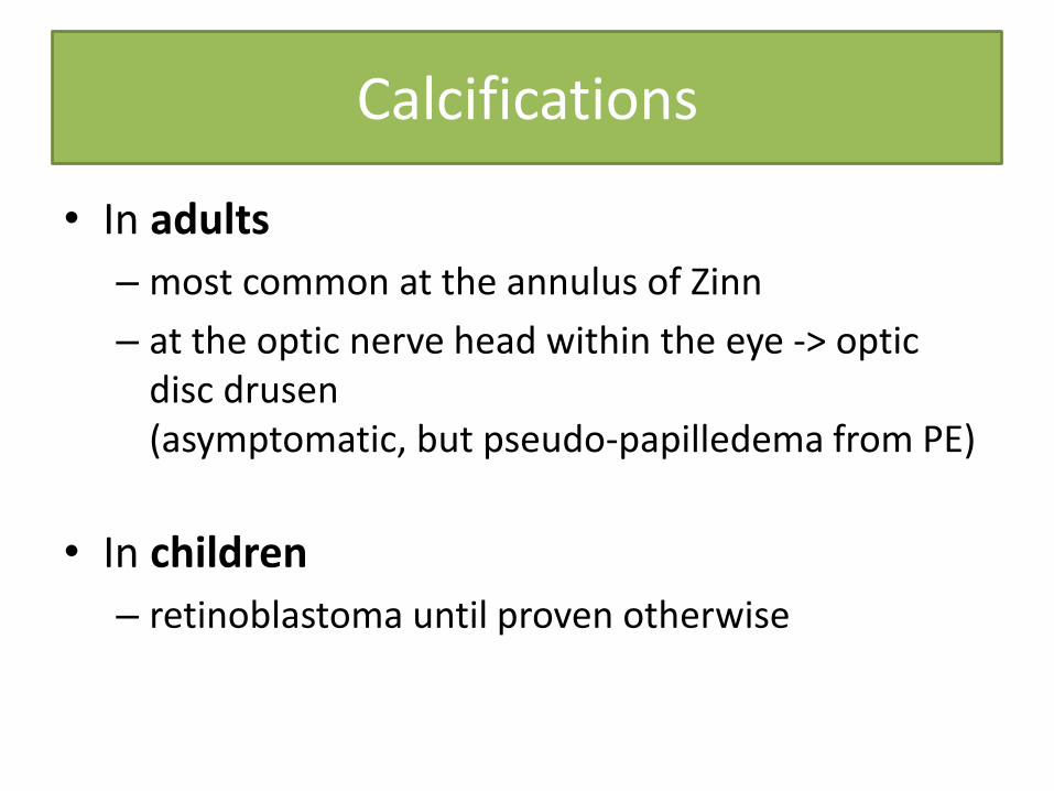
Calcifications
• In adults
– most common at the annulus of Zinn
– at the optic nerve head within the eye -> optic disc drusen (asymptomatic, but pseudo-papilledema from PE)
• In children
– retinoblastoma until proven otherwise
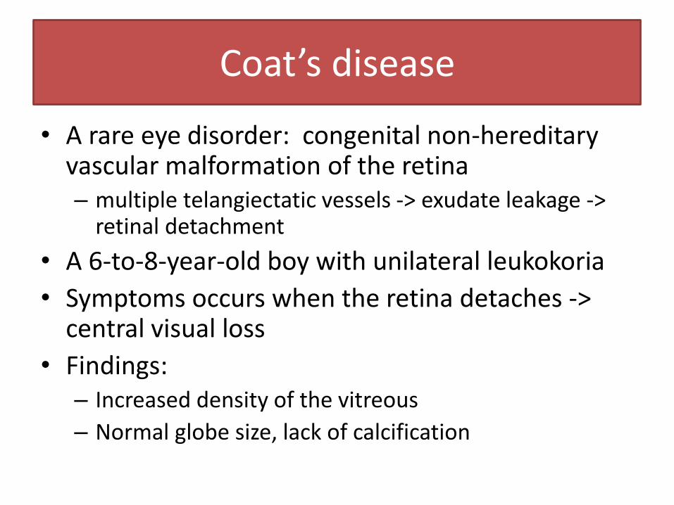
Coat’s disease
• A rare eye disorder: congenital non-hereditary vascular malformation of the retina – multiple telangiectatic vessels -> exudate leakage ->
retinal detachment
• A 6-to-8-year-old boy with unilateral leukokoria
• Symptoms occurs when the retina detaches -> central visual loss
• Findings: – Increased density of the vitreous
– Normal globe size, lack of calcification
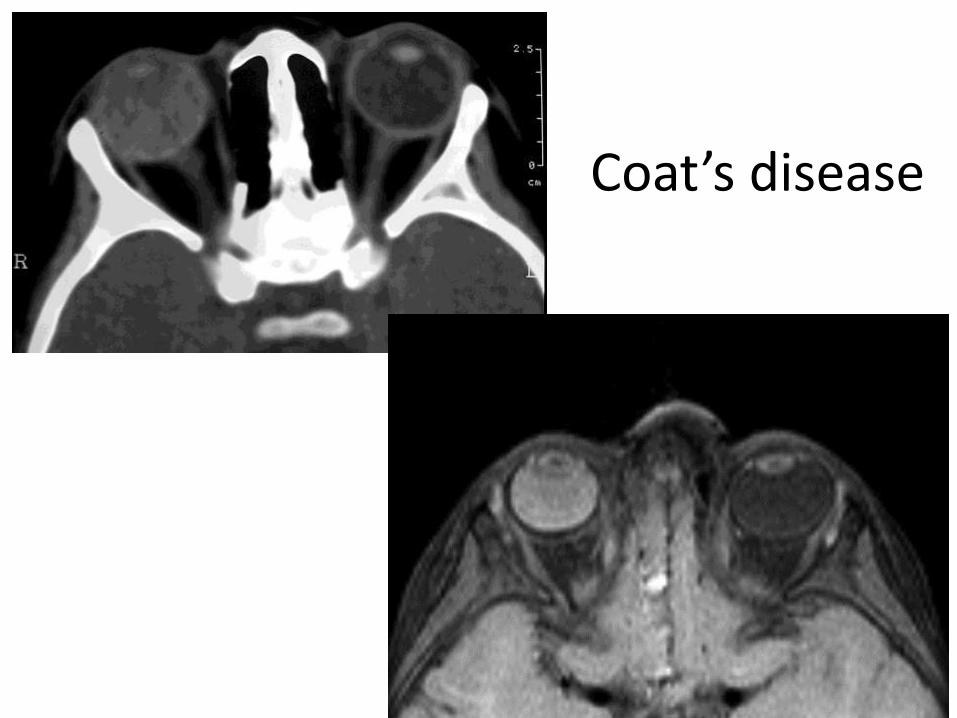
Coat’s disease
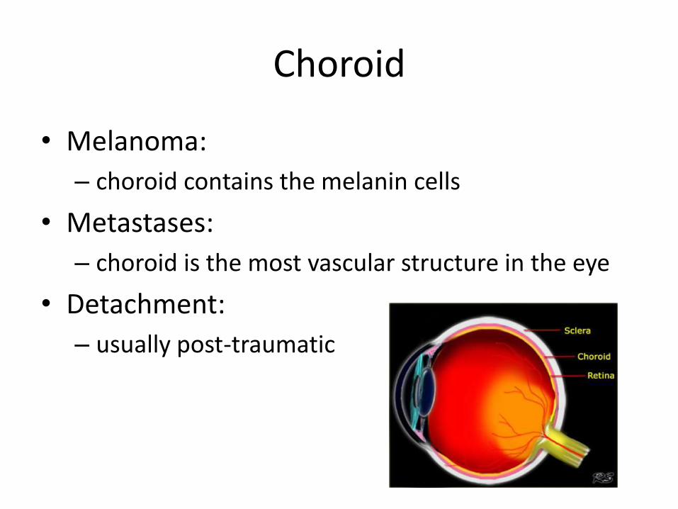
Choroid
• Melanoma:
– choroid contains the melanin cells
• Metastases:
– choroid is the most vascular structure in the eye
• Detachment:
– usually post-traumatic
Uveal melanoma
• The most common primary intraocular malignancy in adult
• A 50-to-70-year old with unilateral ocular complaint
• 85% from choroid, 9% from ciliary body, and 6% from iris
• Dx from PE + U/S
• CT/MR when opaque ocular media prevents a clear view
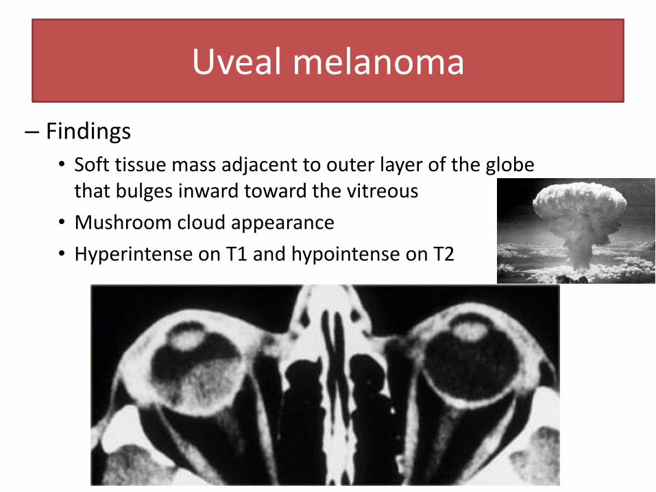
Uveal melanoma
– Findings
• Soft tissue mass adjacent to outer layer of the globe that bulges inward toward the vitreous
• Mushroom cloud appearance
• Hyperintense on T1 and hypointense on T2
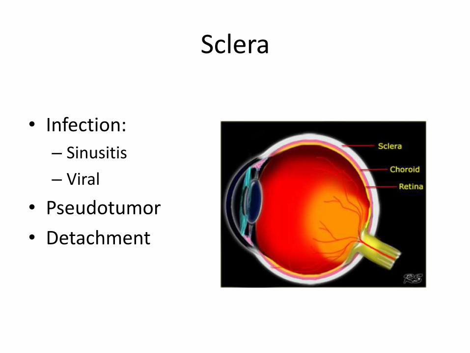
Sclera
• Infection:
– Sinusitis
– Viral
• Pseudotumor
• Detachment
Scleritis
• granulomatous inflammatory disorder
• Erythema and chemosis
• Characteristics – optic disk edema
– exudative retinal detachment
– Choroidal folds and scleral thickening >> elevated mass
• Findings: – CT:
• thickened posterior sclera, may enhanced
• Thickening of extraocular muscles can also be seen
– Magnetic resonance imaging
• nodular elevation into the vitreous
• Iso-to-hyperintense to normal sclera on T1
• Hypointense on T2
• moderate to marked inhomogeneous enhancement with Gd
• A retinal detachment appears as a crescent-shaped area that is hyperintense on both T1 and T2
Scleritis
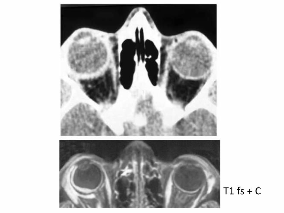
T1 fs + C
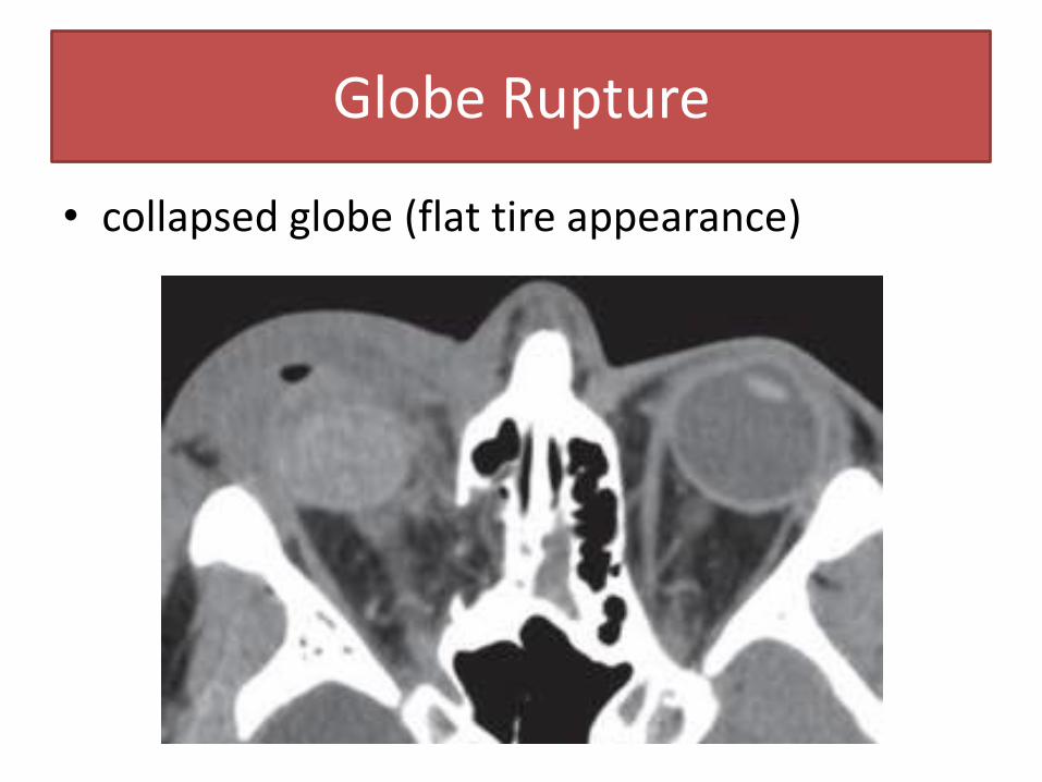
• collapsed globe (flat tire appearance)
Globe Rupture
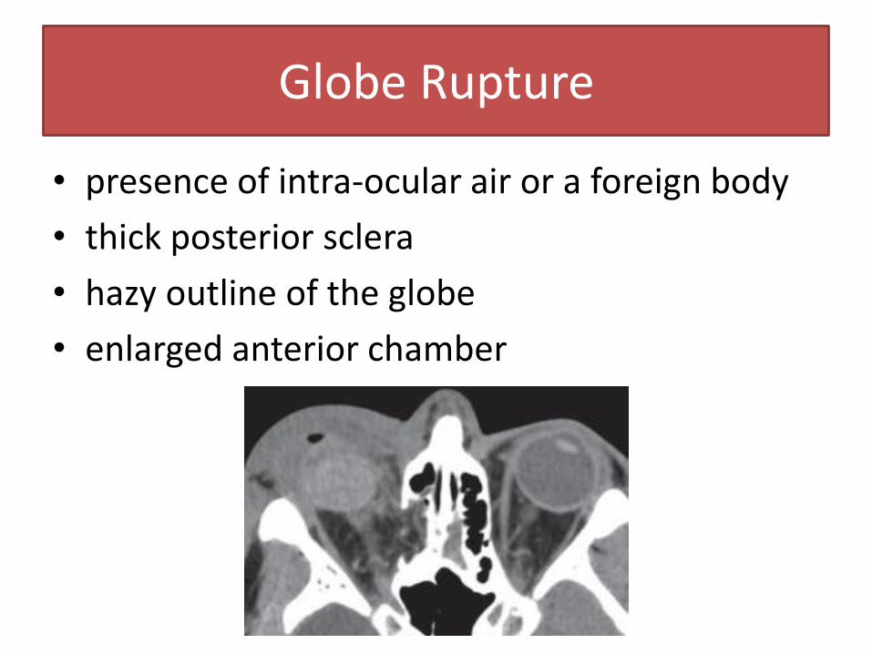
• presence of intra-ocular air or a foreign body
• thick posterior sclera
• hazy outline of the globe
• enlarged anterior chamber
Globe Rupture
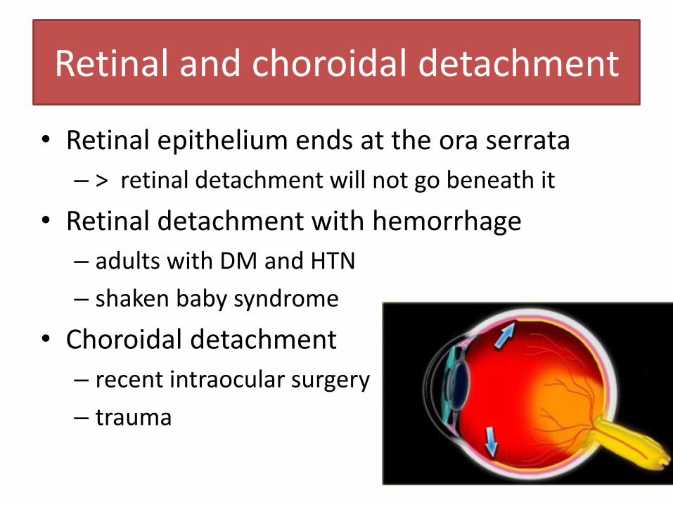
Retinal and choroidal detachment
• Retinal epithelium ends at the ora serrata
– > retinal detachment will not go beneath it
• Retinal detachment with hemorrhage
– adults with DM and HTN
– shaken baby syndrome
• Choroidal detachment
– recent intraocular surgery
– trauma
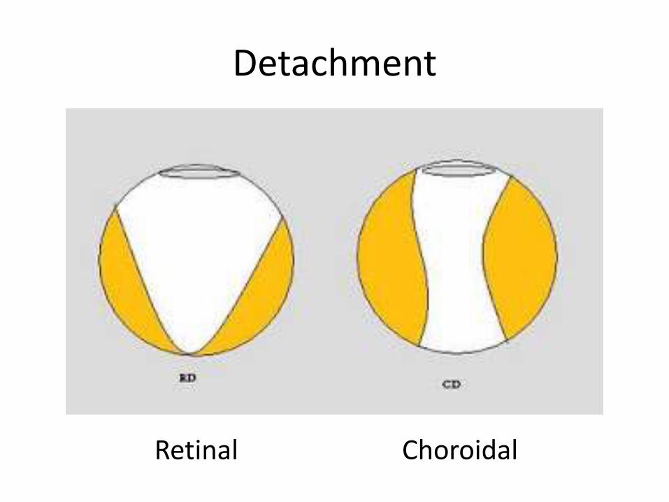
Detachment
Retinal Choroidal
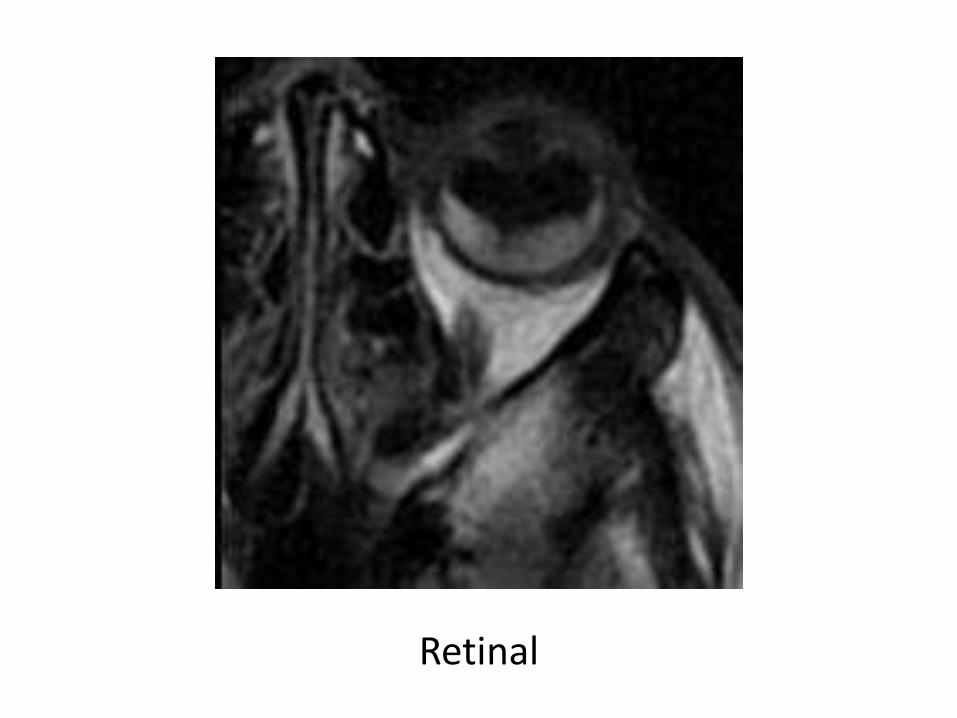
Retinal
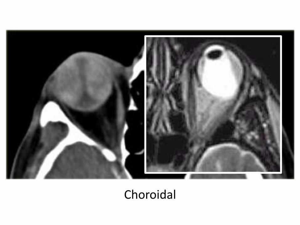
Choroidal
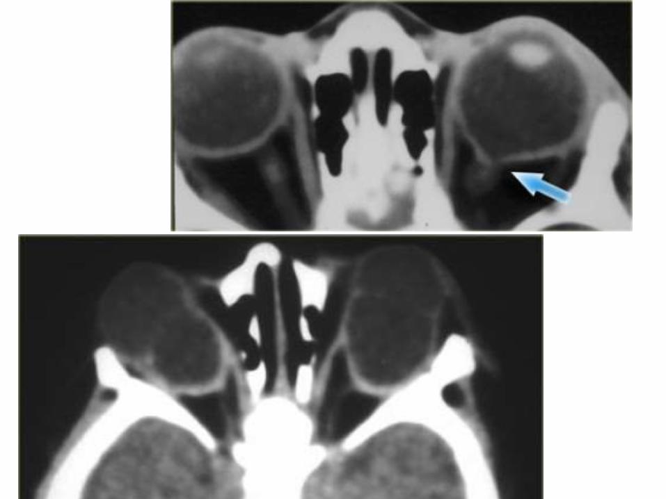
Coloboma
• A congenital globe defect, usually at the optic nerve insertion point
• Often there is microphtalmia and the eye protrudes inferiorly.
• 10% also have other CNS anomalies.
• Findings: – a small globe with a cystic outpouching of vitreous
at the optic nerve attachment site.
– Retroocular cyst
Coloboma
• Coloboma can be part of the CHARGE syndrome: – Coloboma – Heart anomalies – choanal Atresia – Retardation of growth and development – Genital and Ear anomalies.
• Coloboma can also be part of the COACH syndrome:
– Cerebellar vermis hypoplasia, – Oligophrenia (MR) – congenital Ataxia – Coloboma – Hepatic fibrosis.
Diseases of the obit
• Globe
• Optic nerve & sheath
• Conal-intraconal area
• Extraconal area
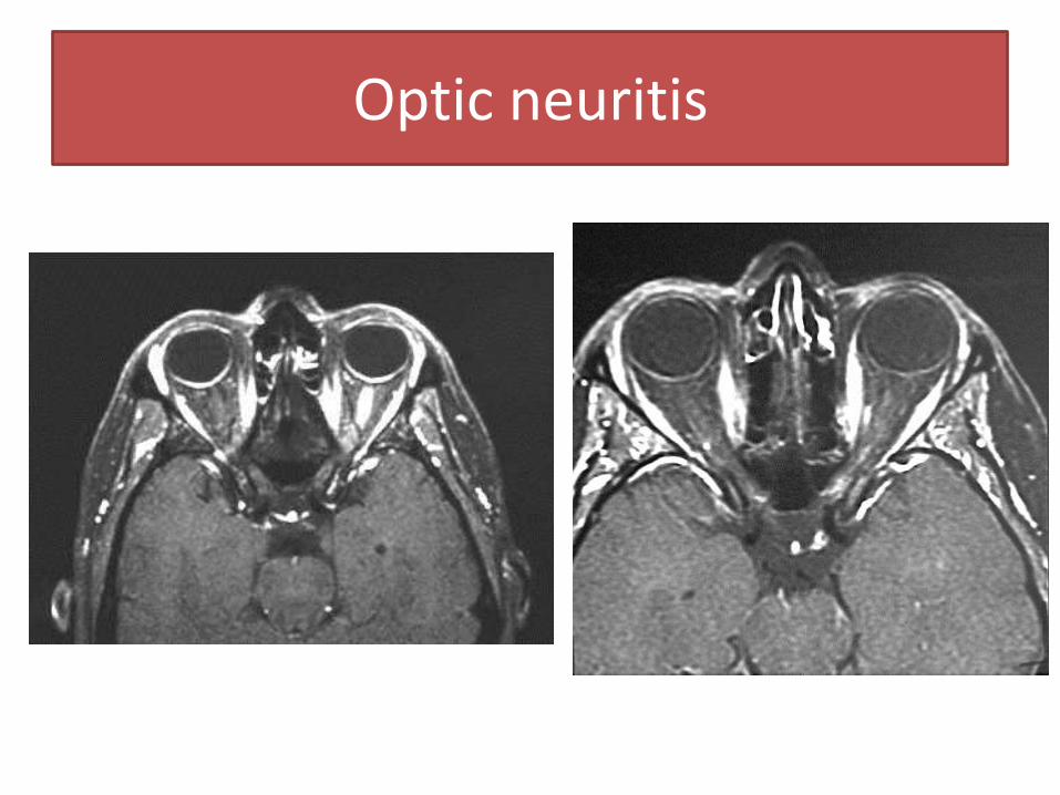
Optic neuritis
• Presentations – Visual loss over hours to days
– Pain on movement and tenderness when pressure applied to globe
• Sporadic/multiple sclerosis – If found, T2 whole brain should be done
• Less common causes – pseudotumor, sarcoidosis, radiation, viral, TB,
syphilis neuritis
Optic neuritis
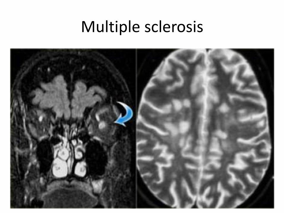
Multiple sclerosis
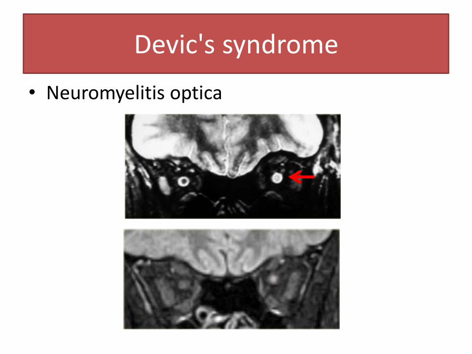
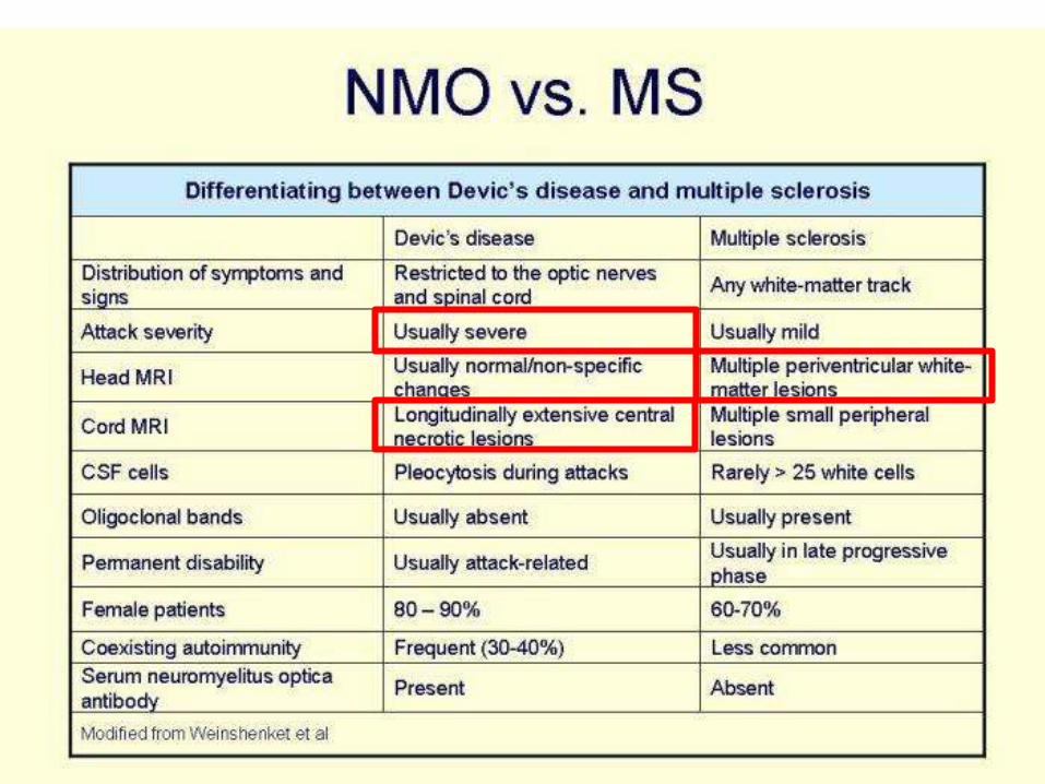
Devic's syndrome
• Neuromyelitis optica
Optic nerve/sheath tumor
• Optic nerve sheath meningioma
• Optic nerve glioma
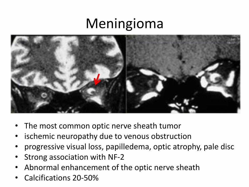
Meningioma
• The most common optic nerve sheath tumor • ischemic neuropathy due to venous obstruction • progressive visual loss, papilledema, optic atrophy, pale disc • Strong association with NF-2 • Abnormal enhancement of the optic nerve sheath • Calcifications 20-50%
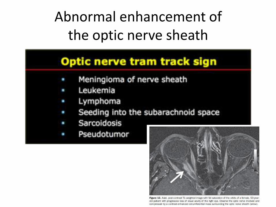
Abnormal enhancement of the optic nerve sheath
Optic nerve glioma
• Juvenile pilocytic astrocytomas WHO type 1 • Anywhere along the optic tract from the occipital
region to the chiasm and the optic nerve
• 50% of optic nerve glioma have NF1. • Only 10% of NF1 have optic nerve glioma. • Less commonly cystic in NF than in non-NF
• Age 4-5 years and only 20% of patients have
visual symptoms.
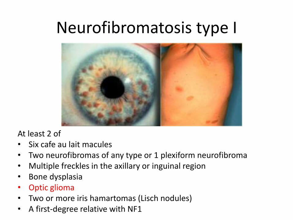
Neurofibromatosis type I
At least 2 of • Six cafe au lait macules • Two neurofibromas of any type or 1 plexiform neurofibroma • Multiple freckles in the axillary or inguinal region • Bone dysplasia • Optic glioma • Two or more iris hamartomas (Lisch nodules) • A first-degree relative with NF1
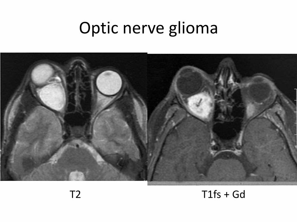
Optic nerve glioma
T2 T1fs + Gd
Diseases of the obit
• Globe
• Optic nerve & sheath
• Conal-intraconal area
• Extraconal area
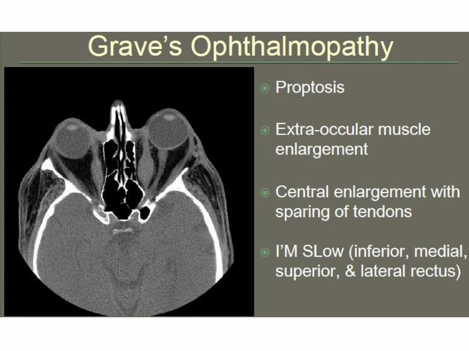
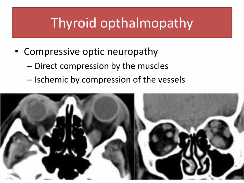
Thyroid opthalmopathy
• Compressive optic neuropathy
– Direct compression by the muscles
– Ischemic by compression of the vessels
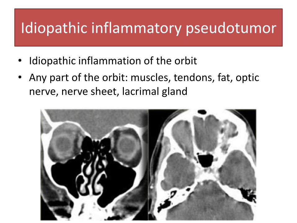
Idiopathic inflammatory pseudotumor
• Idiopathic inflammation of the orbit
• Any part of the orbit: muscles, tendons, fat, optic nerve, nerve sheet, lacrimal gland
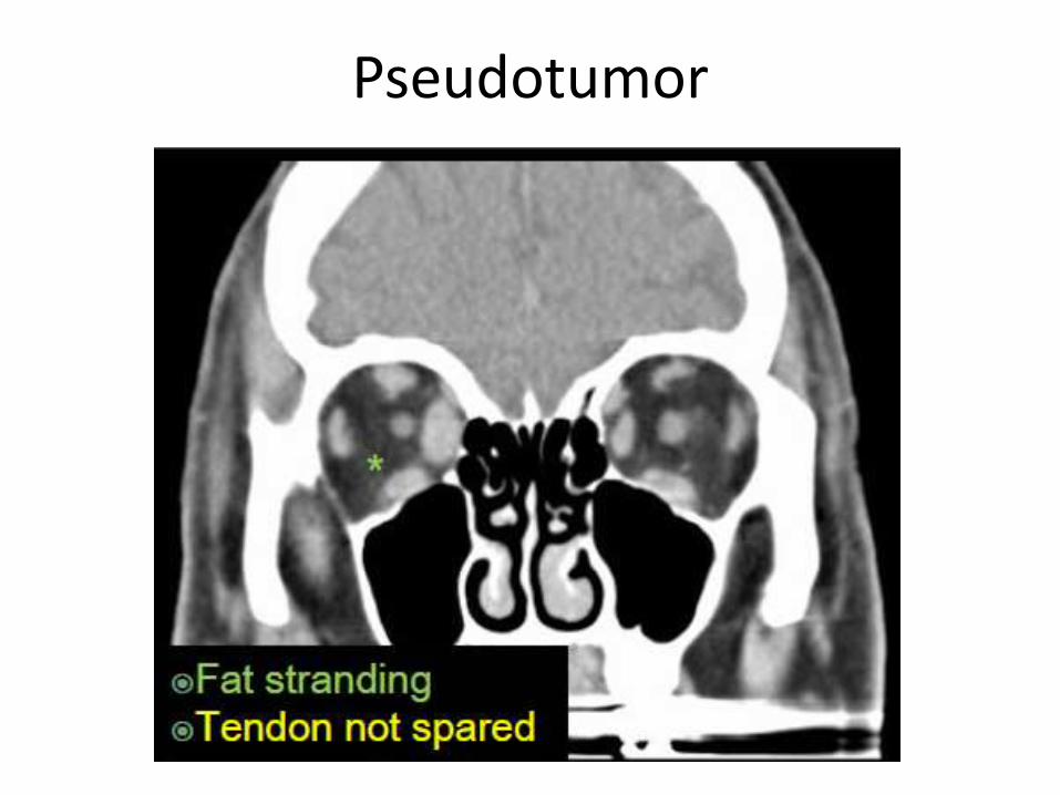
Pseudotumor
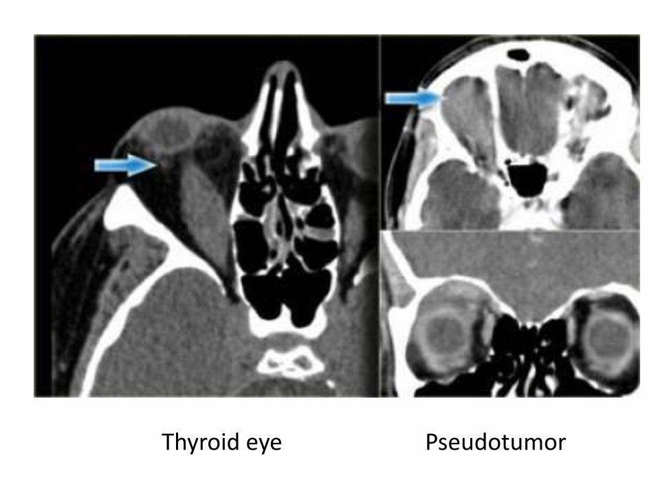
Thyroid eye Pseudotumor

Diseases of the obit
• Globe
• Optic nerve & sheath
• Conal-intraconal area
• Extraconal area
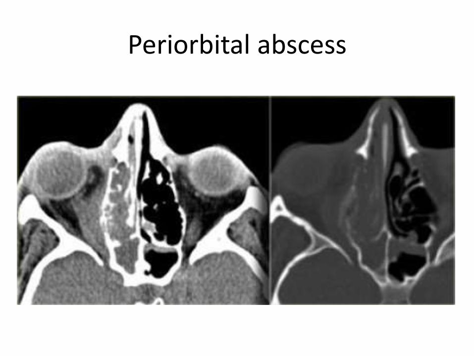
Periorbital abscess
Periorbital abscess
• Complications
– Venous thrombosis of the superior and inferior ophthalmic vein
– Cavernous sinus thrombosis and cavernous-carotid fistula in certain fungal sinusitis (e.g.aspergillosis)
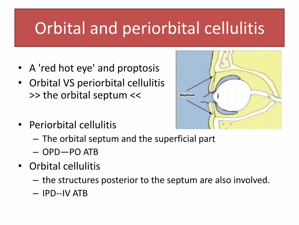
Orbital and periorbital cellulitis
• A 'red hot eye' and proptosis
• Orbital VS periorbital cellulitis -> >> the orbital septum <<
• Periorbital cellulitis – The orbital septum and the superficial part
– OPD—PO ATB
• Orbital cellulitis – the structures posterior to the septum are also involved.
– IPD--IV ATB
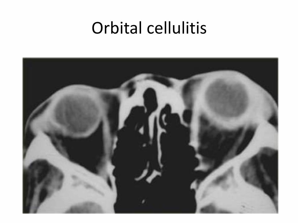
Orbital cellulitis
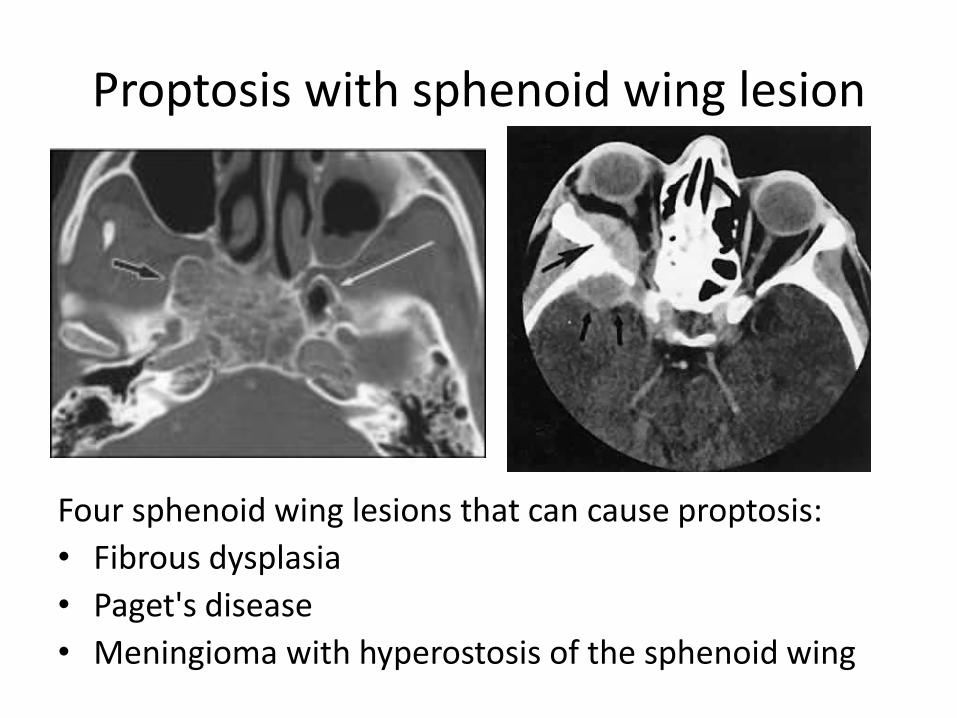
Proptosis with sphenoid wing lesion
Four sphenoid wing lesions that can cause proptosis:
• Fibrous dysplasia
• Paget's disease
• Meningioma with hyperostosis of the sphenoid wing
Orbital appendages
• Lacrimal gland
• lacrimal sac
• lacrimal duct
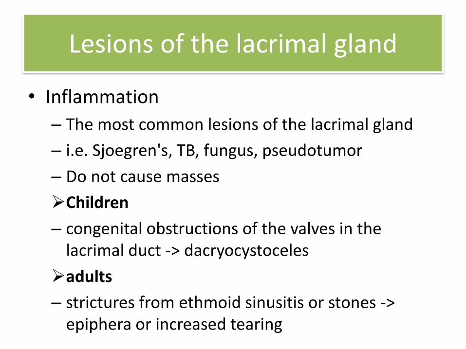
Lesions of the lacrimal gland
• Inflammation
– The most common lesions of the lacrimal gland
– i.e. Sjoegren's, TB, fungus, pseudotumor
– Do not cause masses
Children
– congenital obstructions of the valves in the lacrimal duct -> dacryocystoceles
adults
– strictures from ethmoid sinusitis or stones -> epiphera or increased tearing
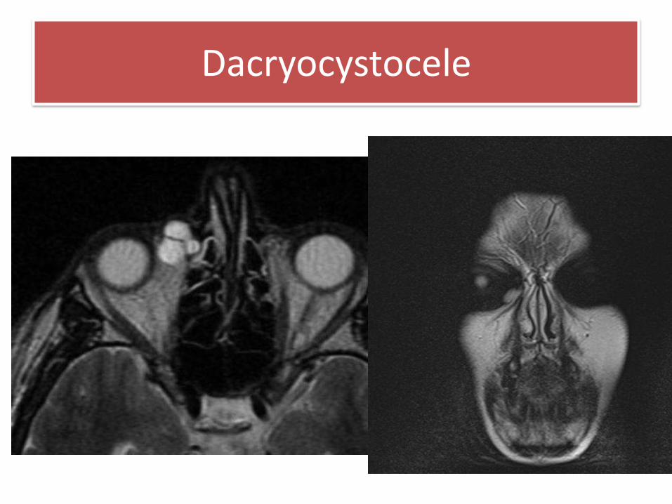
Dacryocystocele
• Lacrimal gland mass
– Lymphoma
– Pleomorphic adenoma
– Epithelial tumors (adenoid cystic tumor)
Lesions of the lacrimal gland
Tumors
• Epidermoid and dermoid tumors
– Developmental
– Usually arises anteriorly between the globe and orbital periosteum
– Well-circumscribed cystic masses containing debris
– Dermoid may contain fat, teeth, and hair
Vascular malformation
• Carotid cavernous sinus fistula
• Cavernous hemangioma
• Capillary hemangioma
• Lymphangioma
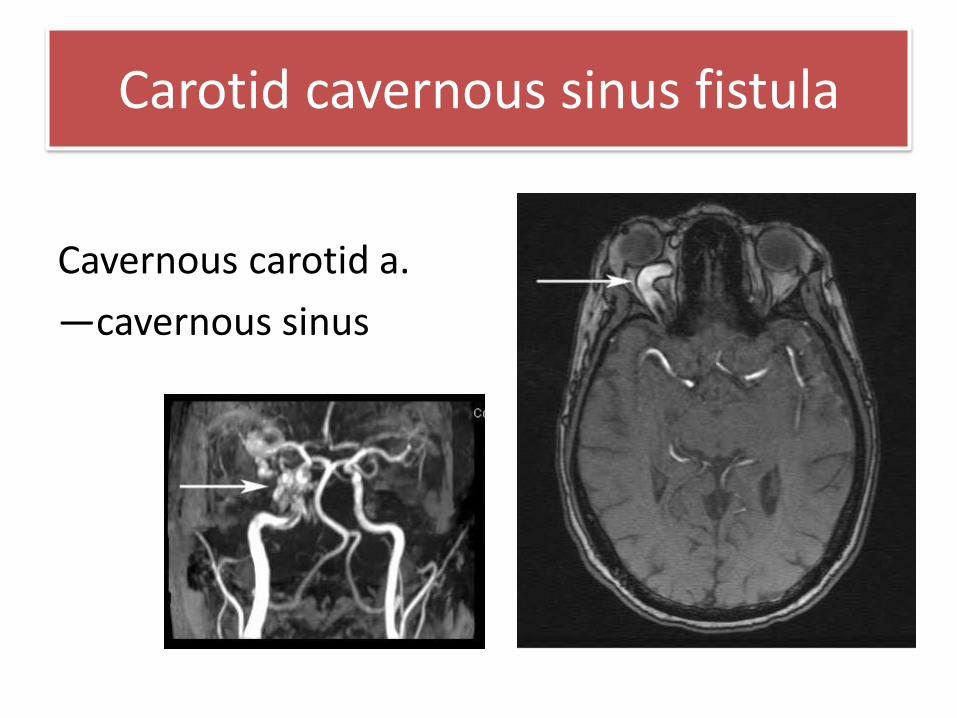
Carotid cavernous sinus fistula
Cavernous carotid a.
—cavernous sinus
Capillary hemangioma
• 6-12 months of age • Involutional at 5-7 years of age • Mostly skin, but also in the extraconal of the eye • PHACE-syndrome:
– Posterior fossa malformations, – Hemangiomas – Arterial anomalies – Cardiac malformation – Eye abnormalities such as coloboma, glaucoma
• CT—irregular margin
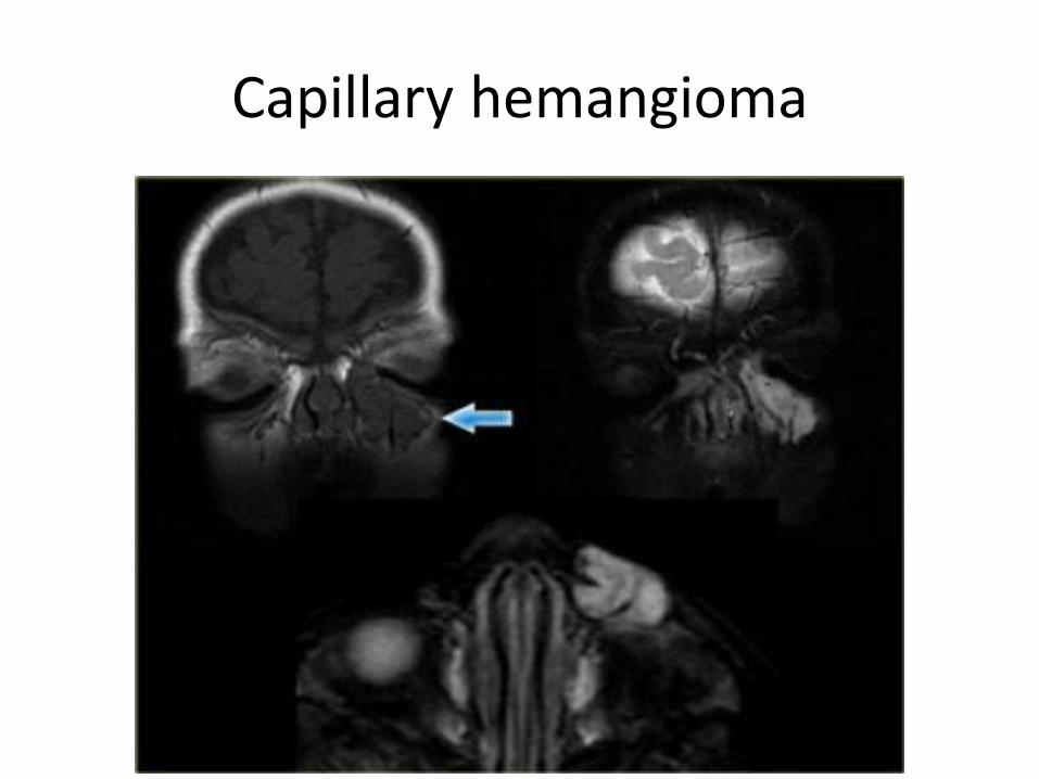
Capillary hemangioma
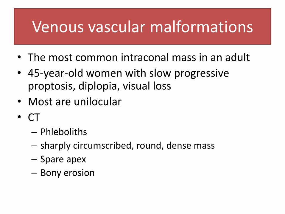
Venous vascular malformations
• The most common intraconal mass in an adult
• 45-year-old women with slow progressive proptosis, diplopia, visual loss
• Most are unilocular
• CT – Phleboliths
– sharply circumscribed, round, dense mass
– Spare apex
– Bony erosion
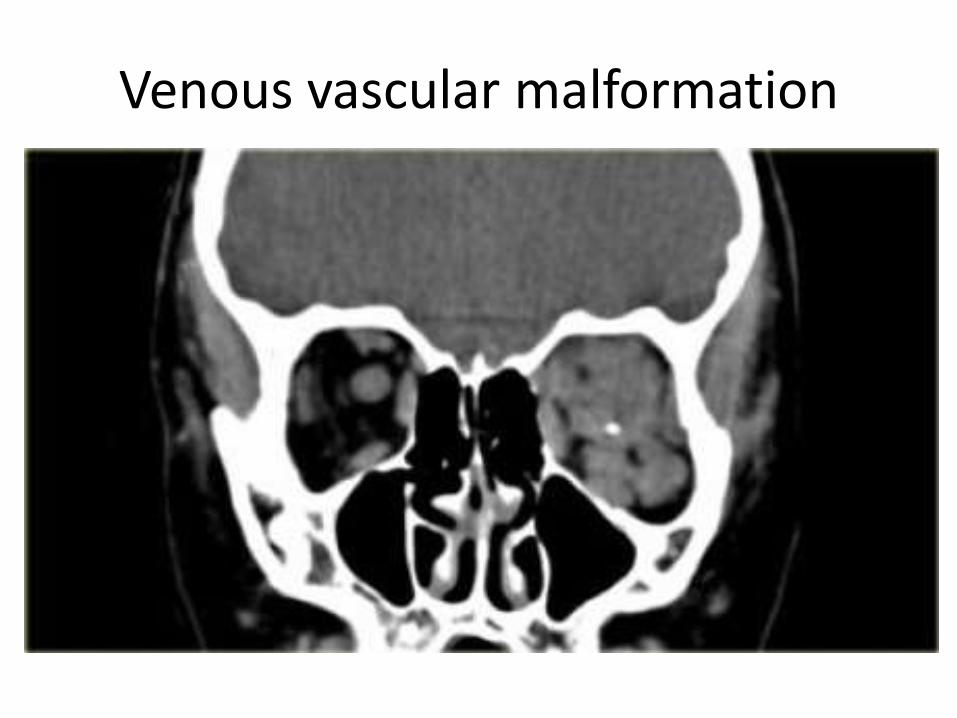
Venous vascular malformation
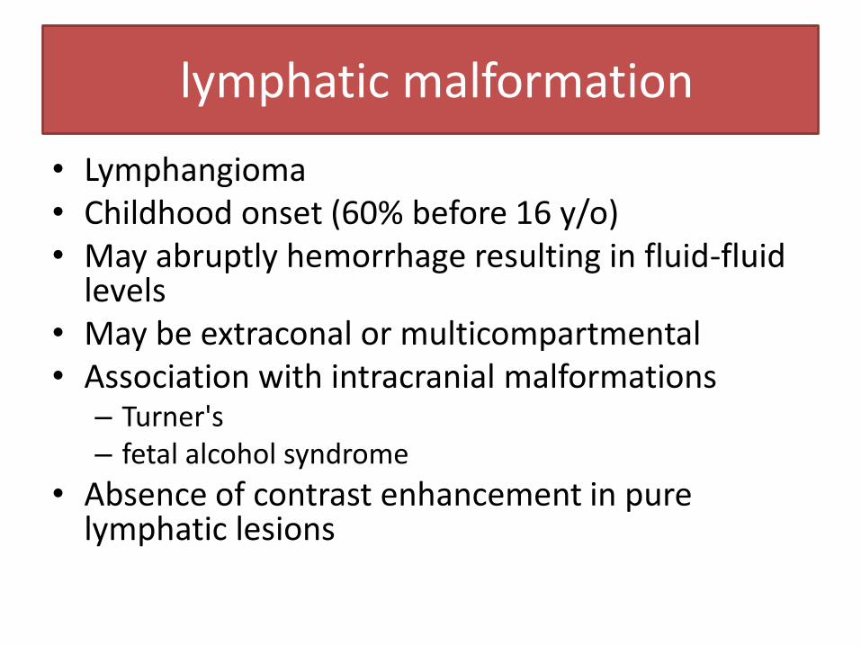
lymphatic malformation
• Lymphangioma • Childhood onset (60% before 16 y/o) • May abruptly hemorrhage resulting in fluid-fluid
levels • May be extraconal or multicompartmental • Association with intracranial malformations
– Turner's – fetal alcohol syndrome
• Absence of contrast enhancement in pure lymphatic lesions
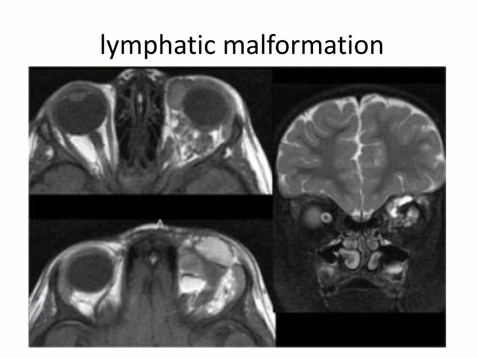
lymphatic malformation
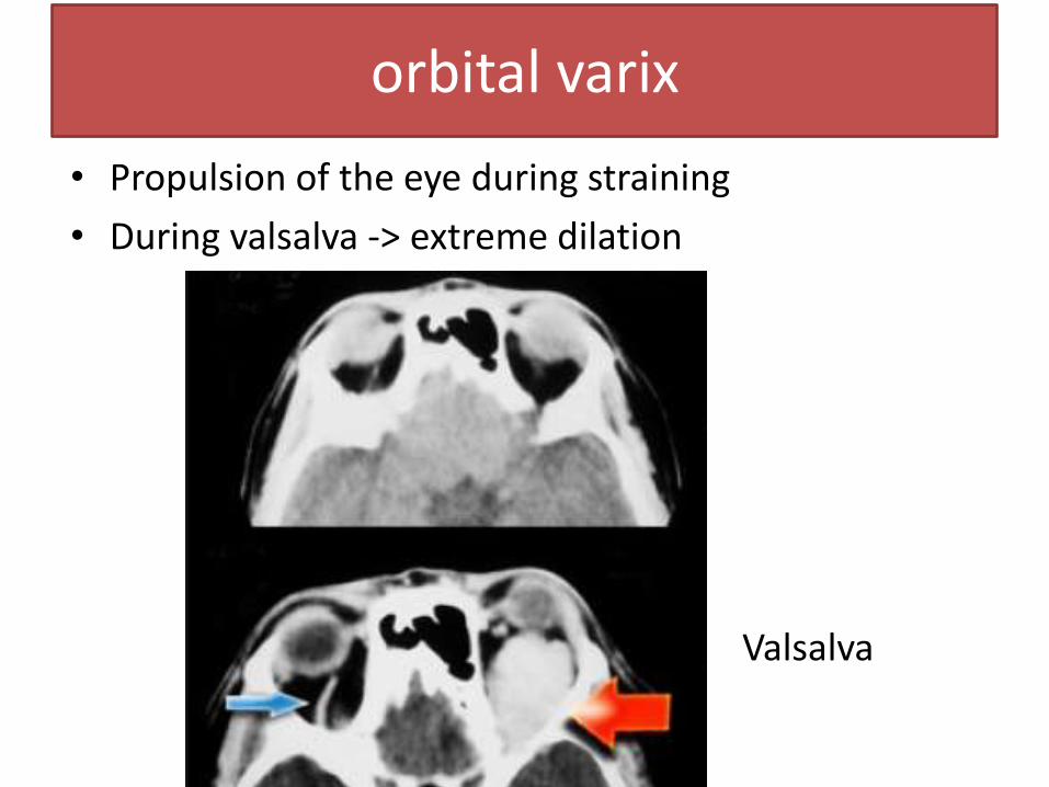
orbital varix
• Propulsion of the eye during straining
• During valsalva -> extreme dilation
Valsalva
Conclusion
• Orbital anatomy
• Disease approach
• Diseases of the orbit
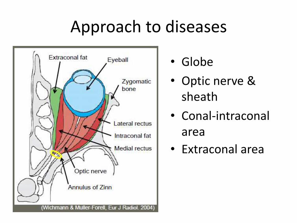
Approach to diseases
• Globe
• Optic nerve & sheath
• Conal-intraconal area
• Extraconal area
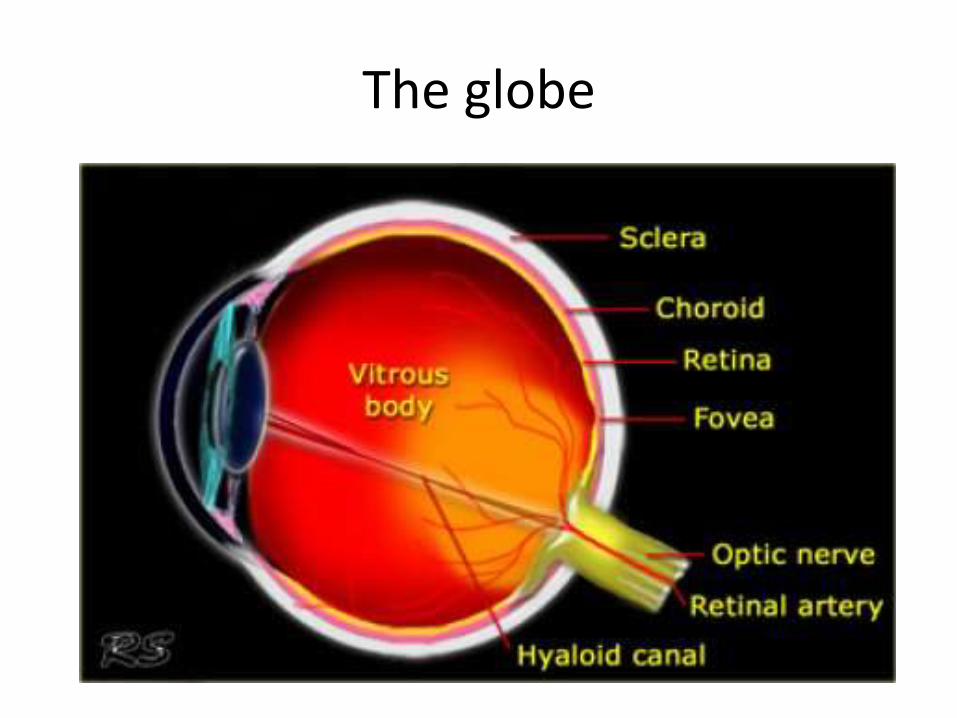
The globe
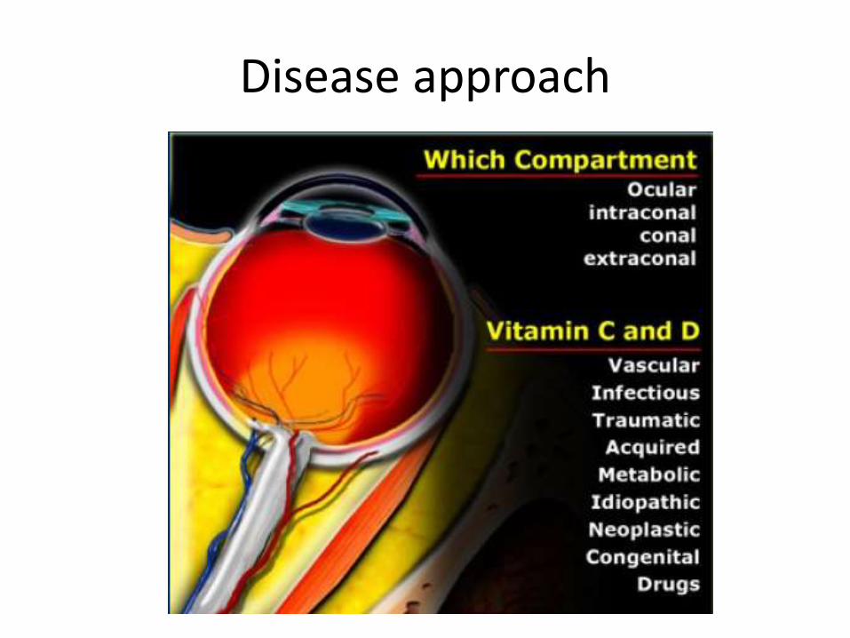
Disease approach

Q & A
References
• Harnsberger, H. Ric., and H. Ric. Harnsberger. Handbook of Head and Neck Imaging. St. Louis: Mosby, 1995. Print.
• Grossman, Robert I., and David M. Yousem. Neuroradiology. Philadelphia, PA: Mosby Elsevier, 2010. Print.
• "Orbita - Pathology." The Radiology Assistant :. N.p., n.d. Web. 20 May 2014

Thank You


![Orbit type: Sun Synchronous Orbit ] Orbit height: …...Orbit type: Sun Synchronous Orbit ] PSLV - C37 Orbit height: 505km Orbit inclination: 97.46 degree Orbit period: 94.72 min ISL](https://static.fdocuments.us/doc/165x107/5f781053e671b364921403bc/orbit-type-sun-synchronous-orbit-orbit-height-orbit-type-sun-synchronous.jpg)


