THE ICU SURVIVAL GUIDE - lthticu.files.wordpress.com · THE ICU SURVIVAL GUIDE Second Edition...
27
THE ICU SURVIVAL GUIDE Second Edition January 2016
Transcript of THE ICU SURVIVAL GUIDE - lthticu.files.wordpress.com · THE ICU SURVIVAL GUIDE Second Edition...

THE ICU SURVIVAL GUIDE Second Edition January 2016

1 | P a g e
Introduction
This guide is aimed at those of you starting your first attachment in Intensive Care Medicine. The Intensive Care Unit (ICU) can be a daunting environment when you first start and so we hope that this guide will give you some tips on the basics of ICU that you can build on during your attachment. In addition, the ICU is a truly multidisciplinary environment and you should always have someone to approach for support i.e. doctors, nurses, pharmacist, dietician, physiotherapist etc. It is important that you always ask someone if you are ever unsure of what to do.
During your time on the ICU you will be exposed to a number of new treatments and interventions that you may not have seen before. Whilst these are interesting and important to gain an understanding of, it is worthwhile noting that there are very few individual treatments that have made a significant impact on outcomes in critical illness. The main determinant of how patients do on ICU is often down to the more routine aspects of good clinical care e.g. hand-washing, safe prescribing, thromboprophylaxis, accurate handover, documentation & communication etc. It cannot be stressed enough how important it is to get this right for patients and you should aim at making sure you are organised and attentive to all of these aspects of care before tackling more complex ICU treatments and interventions.
Finally, many of the non-technical skills of ICU – e.g. decisions to admit or not; communication with relatives and other specialties; and major decisions around the escalation or withdrawal of treatment - are often complicated, sensitive, and challenging. For this reason, all admissions, referrals, major decisions, and communication with family should be discussed with the ICU consultant.
We hope that you will enjoy one of the most unique opportunities to see a wealth of pathology and learn some key skills in the management of critical illness.

2 | P a g e
Table of Contents Page
Section 1 : General duties
1.1 Infection control 4
1.2 Ward rounds 4
1.3 Night reviews 5
1.4 Routine blood tests 5
1.5 Referrals 6
1.5.1 Taking a referral
1.5.2 Assessing a referral
1.6 Admissions 7
1.7 Event logs 8
1.8 Ward 81 8
1.9 Handover 9
1.10 Death certification 10
1.11 Referral to the coroner 10
1.12 Referral for organ donation 11
1.13 Discharge Summary 12
1.14 Family communication 13
1.15 Summary of basic duties 14
Section 2: The basics of ICU support
2.1 Ventilation 15
2.1.1 Non-invasive ventilation 15 2.1.2 Invasive ventilation 16
2.2 Sedation and analgesia 17
2.3 Fluid balance 17
2.4 Vasopressors and inotropes 19
2.5 Renal replacement therapy 20

3 | P a g e
Section 3: Common problems on ICU
3.1 Acute hypoxia 22
3.2 Hypotension 22
3.3 Reduced urine output 22
3.4 Delirium 22
3.5 Pyrexia 23
3.6 Difficulty ventilating 23
Appendix 1: Drug infusions 24
Appendix 2: Coroner’s referral form 25
Appendix 3: Coroner’s referral information 26

4 | P a g e
Section 1: General Duties
1.1. Infection Control
- ICU patients are at particular risk of nosocomial infections and every effort should be made to comply with infection control practices [See LTHT intranet for more details]
- Always comply with the accepted ICU dress code (i.e. bare below the elbows, wedding bands only, plastic aprons & gloves etc.) and the World Health Organisation’s “Five moments for hand hygiene”
- Any new ICU admissions must have their microbiology reviewed prior to admission and those that require source isolation should be identified early and discussed with the nurse in charge so that timely and appropriate placement on the ICU can be arranged
- If an ICU patient develops an infection or symptoms of an infection that require source isolation, then they should be isolated at the earliest possible moment
- Take particular care when reviewing isolated patients and try to minimise the number of people entering the room and the frequency of visits
- Wherever possible, try to minimise the exposure of patients to multiple and extended courses of broad spectrum antibiotics. To facilitate this, ensure that antimicrobials are prescribed correctly (i.e. dose, indication, duration and review date) and review these on a daily basis
1.2 Ward rounds
- This is an opportunity to review each patient’s progress and establish the main objectives for the patient’s care
- They will be led by a consultant or advanced ICM trainee
- The main tasks on the ward round are to write the daily review sheet, examine the patient, review the blood results and imaging, review the drug chart, and identify jobs
- Make sure that you establish ‘who will do what’ at the beginning of the round so that the ward round runs smoothly
- If you are able to , try to complete jobs that can be done as you go a long i.e. prescribing drugs & fluids
- At the end of the ward round, do a quick review of what the main objectives for each patient are and prioritise the jobs
- Now go back and perform a more thorough review of each patient including a full examination and document your findings on the daily review sheet

5 | P a g e
- It’s important that the daily reviews are thorough, comprehensive, and well-documented i.e. make sure you complete ALL parts of the daily review sheet
- Microbiology ward rounds also frequently take place, and a member of the ICU team should take part and have the most up to date blood and culture results to hand
- An evening Consultant handover ward round takes place around 6pm. This is primarily to update the on-call Consultant, however trainees are encouraged to join this
- All ward rounds are an excellent learning opportunity if you take the initiative to get actively involved by presenting patients, discussing management options and asking questions. The more you put into them, the more you are likely to get out of them
1.3 Night Reviews
- This will be your main role at night
- Review the most urgent patients first - this can be determined from the handover and by asking the senior nurse on the ICU
- Start your review by checking the plan from the morning ward round, and any subsequent
changes or speciality reviews, in order to identify the main objectives for the patient’s care over night
- The night reviews should be more focussed and aim to ensure that the patients intended
plan is being followed and identify & resolve any acute issues - It is still however, important to have a systematic approach in order to ensure that things are
not missed - At the end of the review:
x Do a ‘FASTHUG’ – this is an acronym which reminds you to review some basic ICU
elements of patient care, i.e. Feeding, Analgesia, Sedation, Thromboprophylaxis, Head up 30˚, Ulcer prophylaxis, Glucose
x Document targets for the nurse to follow overnight (e.g. SpO2>95%, Hb>80, UO>0.5ml/kg/hr)
1.4 Routine blood tests
- At the end of your night review, let the nurse know which blood tests need to be sent in the morning for each patient and document this on the ICU chart
- Do not routinely send every test (e.g. daily clotting study) as this has no benefit for the
patient and is an unnecessary expense to the ICU

6 | P a g e
1.5 Referrals
- Referrals can initially be a source of anxiety, but they are an excellent way of learning about the presentation and management of critical illness
- It is a good idea to initially accompany one of the more senior doctors when they assess a referral in order to see how the process works
- Referrals can involve a range of issues i.e. IV access, drug dosing & administration, acutely unwell patient etc.
- At times there may be multiple referrals at once and these should be prioritised on the basis of patient need. Ask a senior ICU doctor for help with this.
1.5.1 Taking a referral:
x Record the Name, DOB, NHS number, Consultant team and location of the patient being referred
x Record the name, grade and contact details of the person making the referral x Identify what the main reason for the referral is i.e. airway support, hypotension
requiring vasopressors etc. x Ask the referrer what the likely diagnosis or differential diagnosis is? x Are there any urgent investigations or interventions that need to be performed before
admission to ICU e.g. CT head. If there is, make sure that the referrer has requested and arranged these.
x Does the patient have any limitations to their care in place? x Try to ascertain the urgency with which the patient needs to be reviewed, as the needs
of the ICU patients should be balanced with those of the patient being referred x Importantly, referrals must be seen by a trainee who has the appropriate level of skills &
competence, especially if actual or potential airway problems are involved. At the same time an airway trained doctor must be available to attend ICU if airway emergencies occur. During busy times, It may be necessary to call on the acute theatre anaesthetists for help.
1.5.2 Assessing a referral:
x Let the rest of the team know that you are leaving the ICU to see a referral x ‘Eye-ball’ the patient on arrival and have a look at the observation trends in order to get
a quick idea of how ill the patient is. x Identify anything that needs to be addressed immediately and instigate treatment as
soon as possible i.e. hypoxia, hypotension x Speak to the doctor or nurse responsible for the patient’s care and identify the main
concerns or issues x Examine & assess the patient thoroughly, using collateral information, to gain an insight
into the patient’s previous and current health status I.e. patient notes, old clinic letters, PPM, Ordercomms, the patient and relatives.

7 | P a g e
x Ask yourself:
9 Is the location of the patient suitable for the level of care that they are currently requiring?
9 If not, do they need level 2 (1:2 nurse : patient) or level 3 (1:1) care? 9 Does the patient need any additional investigations or procedures now? 9 Should any other specialities be involved in the care of the patient now? 9 Does the patient require any treatments/support that can only be provided on the
ICU? 9 Is the patient likely to deteriorate in the short term to a point that they will no
longer be suitable for ward level care?
- Throughout your assessment try to identify what the likely diagnosis and problems are, and importantly if there is any reversibility i.e. pneumonia, AKI as opposed to severe biventricular failure, advanced COPD on home NIV
- Discuss the case with a colleague on the ICU and involve the outreach team for advice and support
- ALL potential admissions must be discussed with the oncall ICU Consultant before admission and also inform the nurse in charge on ICU
- If the patient requires transfer to ICU then request the Oxylog® portable ventilator, portable monitor with end-tidal CO, emergency drug box and transfer bag from ICU
1.6 Admissions
- You should aim to review these patients as soon as possible after arrival on the ICU to prevent delays in treatment
- Use the structured admission documentation and ensure that this is completed as accurately
as possible - Make sure that you:
9 Provide a thorough overview of the sequence of events that occurred in the run up to admission and the reason for ICU admission.
9 Summarise any important investigation results and details of any surgical procedures or interventions and the dates on when these took place.
9 Provide a thorough examination of the patient and highlight important findings 9 Document an initial plan for the patient and communicate this to the nurse responsible
for looking after the patient

8 | P a g e
- Complete the VTE form
- Review the drug chart, omit or cancel appropriate drugs and remember to prescribe:
o VTE prophylaxis o MRSA prophylaxis o Ulcer prophylaxis (Omeprazole 20mg OD IV/NG) o Analgesia
- Use the ICU infusion chart to prescribe:
o Sedation o Vasopressors o Electrolytes o Fluids
1.7 Event Logs
- These are a record of the important events that have occurred during the patient’s ICU stay e.g. radiology results, surgical and non-surgical interventions & treatments, major deteriorations, going on/off haemofiltration
- These should be recorded on the documentation in the admission booklet and kept up-to-
date
- This helps to facilitate information transfer/handover between the different teams involved in the patient’s care during their ICU stay and prevents things being missed that may adversely affect patient care
- Use quieter times on the ICU to review and update this if it has not been updated, especially
patients who have been on the ICU for longer periods
1.8 Ward 81
- This is the main surgical HDU, however medical patients who require HDU care may also be admitted here if there is no capacity on the ICU
- This ward is located in Bexley wing, just on the other side of the link corridor
- These patient should remain under the care of their primary teams, however the ICU team provides support and advice when required
- Patients on this ward may be treated with inotropes, vasopressors, high flow nasal oxygen therapy and non-invasive ventilation

9 | P a g e
- You may be called to review a deteriorating patient who requires treatment advice and intervention, or transfer to the ICU for advanced airway and organ support
- You may also be called to insert central and arterial lines in patients when it is indicated
- During the night shift, the senior ICU registrar should perform a ‘trouble-shooting’ ward round on Ward 81 with the nurse in charge and identify any patients that need formal ICU involvement or management advice
- Any patient who has deteriorated or required intervention overnight, should be handed over to the ICU Consultant and Outreach teams at the morning ICU handover meeting
1.9 Handover
- Copies of the handover sheet should be printed out at the end of each shift and brought to the handover meeting
- When handing over, you should provide a succinct but thorough summary of each patient
that you have reviewed during your shift - This should be done in 2-3 minutes per patient and include:
9 The main diagnosis and reason for ICU admission or ongoing admission I.e. ventilation, inotropes, haemofiltration
9 A summary of the background medical/surgical problems and assessment of premorbid functional status
9 A general assessment of the patient’s progress i.e. there is no need for finite detail of ventilator settings etc.
9 A summary of important events , interventions, and investigation outcomes 9 The outstanding issues and management goals for the next 12-24 hours
- long term patient should also be handed over in this way to ensure that important information is always handed over and apparent to all members of the ICU team
- After handing over the unit, give details of any patients outside of the unit that require
review or admission and handover to the Outreach team
- It is important to update the handover sheet every day, otherwise it becomes easy for incorrect information to be repeatedly handed over, and eventually accepted as fact

10 | P a g e
1.10 Death certification
- The documentation for this is kept at the nursing station on the main unit - Discuss the cause of death with the ICU consultant and the consultant team responsible for
the care of the patient and establish whether it requires referral to the Coroner - Aim to complete the death certificate & cremation certificate as soon as possible after death
especially if you will be off the unit for the next day or two
1.11 Referral to the Coroner
- All referrals must now be made on a dedicated Coroner’s referral form which can be downloaded from the LTHT Intranet [See Appendix 2]
- This should be emailed to:
- The referral MUST be sent from a secure nhs.net email account and NOT from a personal account
- The referral will be received by the Trust bereavement team and automatically forwarded to
the Coroner’s office - All subsequent Coroners outcomes/decisions will be communicated to the referrer by the
Trust bereavement team - If you require any further guidance, then please refer to the “Coroner’s referral standard
operating procedure” on the LTHT Intranet [See Appendix 3] - You can also contact the Trust bereavement team on 0113 2064162 for further information
or assistance - Please note that deaths which occur outside of normal working hours and require reporting
to the Coroner’s office, must be reported by 10am on the first working day following the death. If you are finishing a shift then please make sure that there will be someone available to do this before going home.

11 | P a g e
1.12 Referral to a Specialist Nurse for Organ Donation (SNOD)
Criteria for referral
1. Any patient with ≥1 absent cranial nerve reflexes and a GCS ≤ 4 that is not explained by sedation
2. Any patient where a decision has been made to perform brainstem death testing 3. Any patient where there is an intention to withdraw life-sustaining treatment and the
patient’s condition is expected to result in circulatory death
Who are the SNOD’s in the LTHT?
x Geraldine Holmes (usually covers SJUH) x Cathy Jordan x Rachel Summers
How do I refer to a SNOD?
x Any of the SNODs can be contacted via switchboard x At SJUH, Geraldine shares an office with the unit matron near the relatives waiting area x Out of hours, or in hours if you can’t contact a LTHT SNOD, then call 07659 171979 x Leave your name, hospital and extension number and the on-call SNOD will phone you back
What might I be asked to do?
x Work with the SNOD in exploring the patient’s wishes regarding donation x Participate in brainstem death testing x Care for a brainstem dead patient whilst awaiting organ donation x Diagnose death on circulatory criteria before DCD can proceed x Accompany a donor to theatre and re-intubate the patient as part of DCD lung donation
More information about organ donation is available from:
x Organ donation intranet web pages x LTHT e learning package on organ donation x NICE guideline 135
12 | P a g e
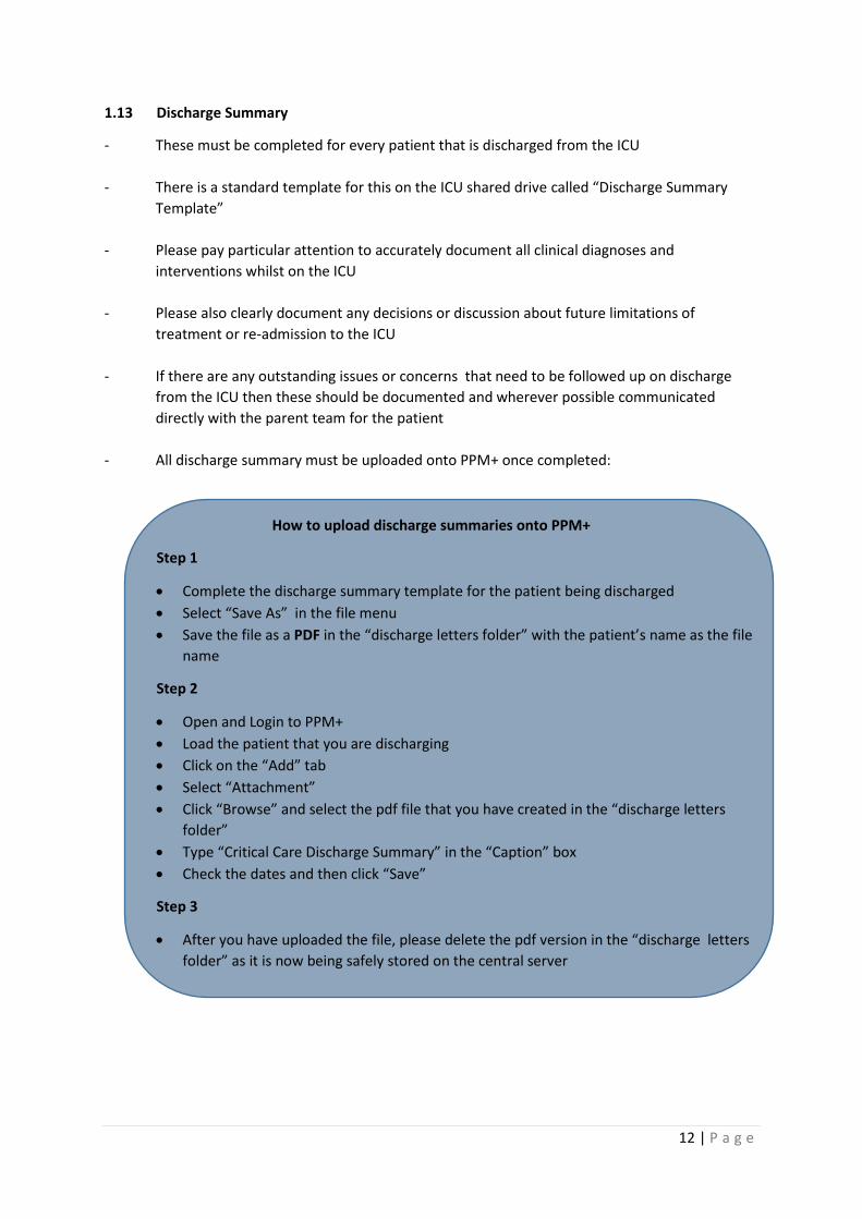
1.13 Discharge Summary
- These must be completed for every patient that is discharged from the ICU - There is a standard template for this on the ICU shared drive called “Discharge Summary
Template” - Please pay particular attention to accurately document all clinical diagnoses and
interventions whilst on the ICU - Please also clearly document any decisions or discussion about future limitations of
treatment or re-admission to the ICU - If there are any outstanding issues or concerns that need to be followed up on discharge
from the ICU then these should be documented and wherever possible communicated directly with the parent team for the patient
- All discharge summary must be uploaded onto PPM+ once completed:
How to upload discharge summaries onto PPM+
Step 1
x Complete the discharge summary template for the patient being discharged x Select “Save As” in the file menu x Save the file as a PDF in the “discharge letters folder” with the patient’s name as the file
name
Step 2
x Open and Login to PPM+ x Load the patient that you are discharging x Click on the “Add” tab x Select “Attachment” x Click “Browse” and select the pdf file that you have created in the “discharge letters
folder” x Type “Critical Care Discharge Summary” in the “Caption” box x Check the dates and then click “Save”
Step 3
x After you have uploaded the file, please delete the pdf version in the “discharge letters folder” as it is now being safely stored on the central server

13 | P a g e
1.14 Family Communication
- You may be asked to update or communicate with family members about a patient on the ICU
- If it is a discussion that you do not feel confident conducting (e.g. breaking bad news, talking
about tracheostomy) then you should always seek senior advice and support first - Take opportunities to accompany and observe more senior doctors when they communicate
with family in order to develop your communication skills - This is a basic plan for communicating information to family members:
9 Take time to review the notes and update yourself on the patient’s progress and previous family discussions first. It is important that the family receive consistent information as confusing them by not preparing first can cause a lot of unnecessary distress to both you and family members
9 Always take another qualified health professional with you, ideally the nurse looking after the patient and find an appropriate location to talk e.g. family room
9 Start with introductions and try to ascertain the family set-up and dynamics 9 Ask the family what they already know, in order to gauge their level of understanding,
and establish what the main issues are that they would like to focus on 9 Give a clear explanation to the family, speaking slowly and using minimal medical jargon,
emphasise important points and pay particular attention to your body language 9 Try not to overload them with information, but keep it simple initially and give them the
opportunity to request more detail if required 9 Check understanding and give opportunity for questions. If you do not know the
answers to these, do not feel pressured into giving an answer but reassure the family that you will discuss this with a more appropriate or senior person and report back to them
9 If you find yourself in a difficult position or with a difficult family and you feel unable to resolve the situation then try to bring the discussion to a close and offer the family an opportunity to discuss this with another more appropriate or senior person.
9 At the end, ask if the family have any more questions or concerns 9 Always document the discussion clearly in the notes

14 | P a g e
1.15 Summary of basic duties
x Take an active role in daily ward rounds and night reviews x Ensure complete and accurate documentation in line with GMC standards of good
medical practice x Request investigations and referrals in a timely fashion x Follow-up results of all investigations and tests x Identify which blood tests need to be sent each morning x Accompany visiting medical & surgical teams when they come to review patients in
order to provide information on the patient’s progress and acknowledge new management decisions
x Take part in practical procedures e.g. central lines, vascaths, arterial lines x Take part in communication with families x Take part in microbiology ward rounds and have up to date results of inflammatory
markers and culture results ready x Update event logs regularly x Complete discharge summaries accurately and in a timely fashion (during daytime
hours) to ensure safe and efficient discharge of patients x Print out copies of the handover sheet before the handover meeting. These are
located on the computer on the main unit x Re-stock and check the emergency drug box at the start of each shift x Give an accurate and succinct handover of the patients you have been looking after x Follow infection control practices - including basic hand hygiene, and the use of gloves
and gowns at each bed-space
x Identify and ensure that your mandatory training has been completed and that your educational requirements are being met

15 | P a g e
Section 2: The Basics of ICU Support
2.1 Ventilation
The aim of ventilation is to allow oxygenation and aid CO2 removal. Types of ventilation can be simply divided into non-invasive and invasive
2.1.1 Non-Invasive Ventilation (NIV)
x The patient breathes spontaneously and must be able to protect their own airway (GCS>8, no risk of aspiration)
x A patient interface is used to provide ventilation i.e. face mask, full face mask, hood x There are two main ways i.e. CPAP or BIPAP x Both provide a constant pressure at the end of expiration to prevent atelectasis and improve
oxygenation. x BIPAP also provides a pressure during inspiration to support the patient’s respiratory effort,
thereby increasing tidal volume and CO2 removal. It is therefore of benefit in patients with high PaCO2 e.g. COPD, depressed respiration (neuro-muscular or drugs), exhaustion
Indications for non-invasive ventilation
x Pulmonary oedema x Atelectasis or collapse x COPD x Pneumonia x Neuro-muscular weakness
NIV Trouble-Shooting
Discomfort
o Try a different patient interface o Make sure mask isn’t too tight o Apply padding to nasal bridge o Allow breaks for drinks etc. if not NBM
Agitation
o Screen for delirium and treat o Consider a small dose of anxiolytic
Dysynchrony
o Check for sputum retention or plugging o Screen for delirium
Failure to improve
o Check settings & mask positioning o Check for sputum retention or plugging o Is the patient tiring or GCS deteriorating o Is there another diagnosis i.e. pleural effusion or PE

16 | P a g e
2.1.2 Invasive Ventilation
x This may be via an endotracheal or tracheostomy tube x Newer ventilators have complex ventilator settings however there are 4 basic settings
that you should focus on:
Fraction of inspired Oxygen (FiO2)
x the percentage of oxygen delivered
Peak End Expiratory Pressure (PEEP)
x the pressure at the end of expiration that helps to prevent atelectasis
Tidal Volume (Vt)
x the volume of each inspiration which is either delivered by the ventilator as a set volume (Volume Control) or a set pressure difference (Pressure Control)
Respiratory Rate (ƒ)
x the number of breathes per minute
¾ Oxygenation is affected by FiO2 and PEEP
¾ Carbon dioxide removal is affected by Vt and ƒ
Ventilation may be:
(i) Mandatory - The patient makes no respiratory effort and the ventilator does all the work
(ii) Spontaneous - The patient makes a respiratory effort that triggers the ventilator to provide
a pressure to support this
(iii) Mixed (SIMV, BiLevel, Auto-mode) - The patient takes some breathes intermittently but the ventilator also
provides mandatory breathes when the patient doesn’t breathe to a set rate.

17 | P a g e
2.2 Sedation & Analgesia
- This is important for maintaining endotracheal tube tolerance for mechanical ventilation and facilitating interventions and treatments that may cause pain & distress to patients
- The main drug combination used on ICU is propofol and alfentanil - Over and under-sedation are both associated with adverse patient effects e.g. hypotension
or unplanned extubation, and therefore it is vital that the right balance is maintained - This can be assessed by using the Richmond Agitation-Sedation Scale (RASS) which can be
found on the ICU observation charts (Aim 0 to -2) - The aim is to have the patient on the minimum amount of sedation possible and this should
be assessed on a daily basis by a period of sedation reduction to ‘lighten’ the patient - Sometimes a patient may experience agitation when attempting to wean off sedation. If this
occurs, identify and treat other causes for this e.g. alcohol withdrawal, sepsis, fever, hypoglycaemia, before just re-sedating the patient unless they are at risk of causing harm to themselves.
- Occasionally it may be necessary to start an additional, less sedating agent (e.g. clonidine,
dexmedetomidine, trazadone) to facilitate weaning off sedation
2.3 Fluid Balance
- This can be difficult to assess in ICU patients who are often oedematous but intravascularly deplete
- When assessing fluid balance try to consider the following:
x What is the underlying diagnosis and stage in the disease process and how does this relate to their current fluid requirements i.e. Rhabdomyolysis vs ARDS?
x What was the fluid balance over the previous 24 hours and what are the likely fluid requirements over the next 24 hours?
x What other sources of fluid is the patient receiving other than IV fluids e.g. NG feed, TPN, drugs, infusions?
x What sources of fluid loss are not being quantified e.g. sweating, diarrhoea, burns, ascites
- Always make a thorough clinical assessment of the patient and use as many indirect markers of fluid balance as possible (e.g. CVP, central venous saturations, cardiac output monitoring, echo) rather than an isolated measurement e.g. CVP

18 | P a g e
- If in doubt, administer the patient a fluid bolus and monitor the effects of this on the patient i.e. change in heart rate, blood pressure, CVP, cardiac output and stroke volume (if cardiac output monitoring available)
A typical fluid bolus
x Hartmans 250 -500 ml over 15 minutes x Hartmans 100 – 200 ml, if concerned about fluid overload or myocardial impairment
In the early stages of fluid resuscitation aim for the following targets:
Heart Rate (HR) < 100
Mean Arterial Pressure (MAP) > 65mmHg
Urine Output (UO) > 0.5ml/kg/hr
Central venous saturations (SvO2) > 70%
x This can be determined from a venous blood gas taken from the central line x It is a global marker of the balance between oxygen supply and demand and therefore
adequacy of oxygen delivery
CVP > 10
Hb >7 or Haematocrit > 30%
Stroke Volume Variation (SVV) < 11%
x This can be determined from cardiac output monitoring x It is a marker of the variation of cardiac output as a result of the adequacy of filling
pressure or preload x If this value is >11% then the patient is likely to be fluid responsive

19 | P a g e
2.4 Vasopressors and inotropes
Put simply, vasopressors increase blood pressure by causing vasoconstriction and increasing systemic vascular resistance, whereas inotropes increase blood pressure by improving cardiac contractility and increasing cardiac output. You should keep this in mind when deciding which drugs are best suited to the particular clinical scenario. In some instances, e.g. Sepsis, it may be necessary to use a combination of vasoconstrictors and inotropes due to the underlying sequelae of the illness i.e. vasodilatation and septic cardiomyopathy.
Common Vasoconstrictors
Phenylephrine
This can be given through a peripheral cannula and is useful for the initial stages of resuscitation when the patient does not have central access. It can either be given in bolus doses of 50-100 micrograms or run as an infusion.
Noradrenaline
This is given centrally as an infusion and is more potent than phenylephrine.
Vasopressin
This is given centrally as an infusion and is a second line vasopressor used in severe sepsis where there is profound vasoplegia and hypotension. It is usually started when the noradrenaline requirement has reached 0.5mcg/kg/min
Adrenaline
This is given centrally as an infusion. Although it is a vasoconstrictor it also has inotropic effects and is therefore also useful when there is an element of myocardial impairment. It may adversely increase glucose and lactate levels.
Common Inotropes
Dobutamine
This is given centrally as an infusion. It also has a vasodilatory effect, especially when starting, and therefore usually requires the concomitant administration of a vasopressor e.g. noradrenaline. It should be used cautiously in patients with a tachycardia as it increases heart rate and may initiate tachyarrhythmia’s.
Calcium
This is an important component for muscular contraction and therefor myocardial contractility and cardiac output. Consider replacing when levels are low.

20 | P a g e
2.5 Renal Replacement Therapy (RRT)
This is a form of organ support that you may not have encountered before. RRT may either be intermittent or continuous. On ICU we usually use continuous RRT which allows us to use lower flow rates compared to those on the renal dialysis unit, which is more suitable for the haemodynamic instability of most ICU patients.
Before considering a patient for RRT it is important to establish the reason for why they require RRT and if there is reversibility i.e. AKI secondary to sepsis vs AKI secondary to end stage heart failure. It is also important to treat immediately reversible causes first i.e. hypovolaemia, hypotension and obstruction.
The main indications for starting RRT on ICU are:
x Hyperkalaemia resistant to medical therapy x Fluid overload x Symptomatic uraemia x Severe metabolic acidosis x Specific drug overdoses i.e. aspirin x Specific conditions i.e. rhabdomyolysis
There are 3 main mechanisms for RRT:
Continuous Veno-Veno Haemofiltration (CVVHF)
Blood flow over a filter membrane causes convection currents which draw fluid and small molecules (e.g. K+) out of the blood. The rate of removal is directly proportional to the speed of the blood flow over the filter membrane.
Continuos Veno-Veno Haemodialysis (CVVHD)
Blood and dialysate fluid flow in opposite directions (counter-current) on either side of a semi-permeable membrane and solutes diffuse by virtue of the concentration gradients between the two. The rate of removal is directly proportional to the concentration gradient of solute between blood and dialysate.
Continous Veno-Veno Haemodiafiltration (CVVHDF)
A combination of the two which aids fluid removal and improves the efficiency of solute removal.
Patients on RRT need to be anticoagulated to prevent clotting in the filter. On this unit we use citrate which acts as an anticoagulant by binding calcium (an important co-factor in coagulation). An infusion of calcium is then administered through the filter before returning blood to the patient in order to reverse the effects of citrate.

21 | P a g e
A simplified diagram of CVVHF circuit:
A simplified diagram of CVVHD circuit:
Blood from patient
Blood to patient
Replacement Fluid Anticoagulation
Effluent Waste
Blood from patient
Blood to patient
Ultra Filtrate
Dialysate
Anticoagulation

22 | P a g e
Section 3: Common problems on ICU
3.1 Acute Hypoxia
x Make sure this isn’t a measurement error i.e. sats probe off, hypotension, hypothermia x Put the patient on 100% oxygen and positon upright x Call for help x Check the position at which the tube is secured – has this changed? x Pass a suction catheter down the endotracheal or tracheostomy tube to exclude
obstruction and secretions x Examine the patient and initiate treatments based on findings i.e. pneumothorax,
bronchospasm, pulmonary oedema, pleural effusion etc. x Has the patient had a PE? x Request portable CXR x Consider referral to physiotherapy
3.2 Hypotension
x Make sure this isn’t a measurement error i.e. kinked arterial line x Check a non-invasive blood pressure to correlate with the invasive pressure x Assess fluid balance and consider giving a fluid bolus x Is the patient over sedated? x Is the patient vasodilated – increase or start vasopressors x Is the patient in heart failure – increase or start inotropes x Is there another possible diagnosis:
o bleeding – check Hb, NG aspirates etc. o pulmonary embolus o pneumothorax
3.3 Reduced urine output
x Check for misplaced, kinked or obstructed (blood, sediment) urinary catheters x Assess fluid balance and give a fluid bolus x Ensure the patient has an adequate MAP i.e. are they normally hypertensive x Use a bladder scan and if the bladder is full then change the catheter with antibiotic
cover (see trust guidelines on intranet)
3.4 Delirium
x This may be hypoactive or hyperactive x Use the CAM-ICU score (on the reverse of the observation chart) to assess and diagnose. x Treatment options include:

23 | P a g e
Non-pharmacological
x Stop causative agents if possible i.e. opiods and benzodiazepines x Orientate with verbal cues, clocks, family pictures x Improve chances of normal sleep pattern i.e. low lighting, low noise levels and limit
interventions at night. Encourage mobilisation and activity during the day x Prevent further injury i.e. secure drips and lines, apply mittens, use padded cot sides
Pharmacological
x Haloperidol 2.5-5mg IV QDS. If contraindicated (e.g. long QT) then consider Olanzepine 5mg OD
x Treat insomnia e.g. Zopiclone 3.75-7.5mg OD x Consider medications to reset diurnal rhythm e.g. melatonin
3.5 Pyrexia
x Is this a new temperature? x Examine the patient and don’t forget drains, lines, wounds, soft tissues , heart
murmurs x Send a septic screen x Consider additional investigations e.g. CXR, CT x Review antibiotic therapy and check culture results and sensitivities x Discuss with microbiology if complicated or advice required x Give Paracetamol and/or use a forced air cooling system
3.6 Difficulty ventilating
x Check that the patient is adequately sedated x Check that the ETT or tracheostomy is patent by passing a suction catheter down x If the patient has a tracheostomy and the catheter doesn’t pass, then remove the inner
cannula and attempt again x Examine the chest and treat obvious causes I.e. secretions, bronchospasm,
pneumothorax x Consider disconnecting the patient and hand bagging in order to exclude ventilator
problems and assess lung compliance x If air-trapping is a possibility (COPD or asthma) then disconnect from the ventilator and
manually decompress the chest

24 | P a g e
Appendix 1 – Drug Infusions
Drug Dose Dilution Dosage Guidance Acetylcysteine 10g 250ml – Glucose 5% Over 24 hours Adrenaline 10g 100ml – Glucose 5% 0-1 mcg/kg/min Alfentanil 25mg 50ml – NEAT 1-6ml/hr Aminophylline 1g 100-1000ml – NaCl 0.9% See protocol Amiodarone - loading 150-300mg 50ml – Glucose 5% Over 1 hour Amiodarone - maintenance 300-900mg 50ml – Glucose 5% 2ml/hr Atracurium 500mg 50ml – NEAT 0-5ml/hr Clonidine 600µg 60ml – NaCl 0.9% 0-6ml/hr Dobutamine 500mg 100ml – Glucose 5% 0-20mcg/kg/hr Furosemide 500mg 50ml – NEAT 0-10ml/hr Glyceryl Trinitrate 50mg 50ml – NEAT 0-10ml/hr Heparin 20,000 units 20ml – NEAT 200units/hr for liver Tx Insulin 50 units 50ml – NaCl 0.9% See protocol Midazolam 100mg 50ml – NEAT 0-5ml/hr Labetalol 200mg 40ml – NEAT 1-10ml/hr Magnesium Sulphate 20mmol 100ml – NaCl 0.9% Over 1-4 hours Noradrenaline 8mg 100ml – Glucose 5% 0-1mcg/kg/hr 16mg 100ml – Glucose 5% 0-1mcg/kg/hr Omeprazole 80mg 100ml – NaCl 0.9% 10ml/hr Phosphate Polyfuser 250-500ml Over 24 hours Potassium Chloride 50mmol 50ml – NEAT 0-20ml/hr (K+ 4-5mmol/l) Propofol 2g 50 ml – NEAT 0.5-3mg/kg/hr Vasopressin 20 units 50 ml – Glucose 5% 0-6ml/hr Vecuronium 100mg 50ml – NEAT 0-10mg/hr

25 | P a g e
Appendix 2 - Coroner’s Referral Form

26 | P a g e
Appendix 3 - Coroner’s Referral Information