THE HIDDEN HALF - Universität Vechta · Furthermore, our "in vivo tissue isolation" experiments...
13
THE HIDDEN HALF THIRD EDITION REVISED AND EXPANDED edlted by Yoav Waisel Amram Eshel Tel Aviv University Tel Aviv, Israel Uzi Kafkafi The Hebrew University of Jerusalem Rehovot, Israel MARCEL DEKKER, INC. NEW YORK' BASEL Copyright @ 2002 by Marcel Dekker, Inc. All Rights Reserved.
Transcript of THE HIDDEN HALF - Universität Vechta · Furthermore, our "in vivo tissue isolation" experiments...
THE HIDDEN HALF
THIRD EDITION REVISED AND EXPANDED
edlted by
Yoav Waisel Amram Eshel
Tel Aviv University
Tel Aviv, Israel
Uzi Kafkafi The Hebrew University of Jerusalem
Rehovot, Israel
MARCEL
DEKKER, INC.
NEW YORK' BASEL
Copyright @ 2002 by Marcel Dekker, Inc. All Rights Reserved.
MARCEL
MARCEL DEKKER, INc. DEKKER
Register now on dekker.com and receive a discount coupon towards your next Dekker purchase. Registered users enjoy these advantages:
� Preview content onIine for free � Quickly access your subscriptions and downloads � Sign up for email notifications about new products and special offers in YOUf subject of interest
Send vour book order to vour regular book supplier or directlv to vour nearest Marcel Oekker, Inc. office:
Please include quantity, ISBN,author, title, shipping andbilling addresses, telephone number/fax
number/e~mail address, method of shipment, and purehase order numbers or credit card information.
To purchase offprints of chapters that appear in any Marcel Dekker, Inc. book or reprints of articles that appear in any Marcel Dekker, Inc. journal: [email protected]
To inquire about special sales and bulk purchases of Marcel Dekker, Inc. books or journals: bulksale
@dekker.com
research + dekker.com -+ results
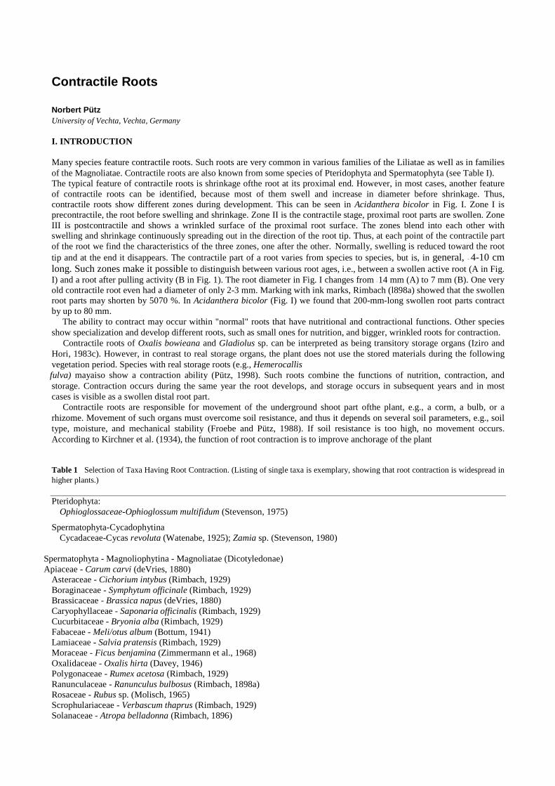
Contractile Roots Norbert Pütz University of Vechta, Vechta, Germany I. INTRODUCTION Many species feature contractile roots. Such roots are very common in various families of the Liliatae as weIl as in families of the Magnoliatae. Contractile roots are also known from some species of Pteridophyta and Spermatophyta (see Table I). The typical feature of contractile roots is shrinkage ofthe root at its proximal end. However, in most cases, another feature of contractile roots can be identified, because most of them swell and increase in diameter before shrinkage. Thus, contractile roots show different zones during development. This can be seen in Acidanthera bicolor in Fig. I. Zone I is precontractile, the root before swelling and shrinkage. Zone II is the contractile stage, proximal root parts are swollen. Zone III is postcontractile and shows a wrinkled surface of the proximal root surface. The zones blend into each other with swelling and shrinkage continuously spreading out in the direction of the root tip. Thus, at each point of the contractile part of the root we find the characteristics of the three zones, one after the other. " Normally, swelling is reduced toward the root tip and at the end it disappears. The contractile part of a root varies from species to species, but is, in general, ~ 4-10 cm long. Such zones make it possible to distinguish between various root ages, i.e., between a swollen active root (A in Fig. I) and a root after pulling activity (B in Fig. 1). The root diameter in Fig. I changes from ~ 14 mm (A) to 7 mm (B). One very old contractile root even had a diameter of only 2-3 mm. Marking with ink marks, Rimbach (l898a) showed that the swollen root parts may shorten by 5070 %. In Acidanthera bicolor (Fig. I) we found that 200-mm-long swollen root parts contract by up to 80 mm.
The ability to contract may occur within "normal" roots that have nutritional and contractional functions. Other species show specialization and develop different roots, such as small ones for nutrition, and bigger, wrinkled roots for contraction.
Contractile roots of Oxalis bowieana and Gladiolus sp. can be interpreted as being transitory storage organs (Iziro and Hori, 1983c). However, in contrast to real storage organs, the plant does not use the stored materials during the following vegetation period. Species with real storage roots (e.g., Hemerocallis fulva) mayaiso show a contraction ability (Pütz, 1998). Such roots combine the functions of nutrition, contraction, and storage. Contraction occurs during the same year the root develops, and storage occurs in subsequent years and in most cases is visible as a swollen distal root part.
Contractile roots are responsible for movement of the underground shoot part ofthe plant, e.g., a corm, a bulb, or a rhizome. Movement of such organs must overcome soil resistance, and thus it depends on several soil parameters, e.g., soil type, moisture, and mechanical stability (Froebe and Pütz, 1988). If soil resistance is too high, no movement occurs. According to Kirchner et al. (1934), the function of root contraction is to improve anchorage of the plant
Table 1 Selection of Taxa Having Root Contraction. (Listing of single taxa is exemplary, showing that root contraction is widespread in higher plants.) Pteridophyta: Ophioglossaceae-Ophioglossum multifidum (Stevenson, 1975)
Spermatophyta-Cycadophytina Cycadaceae-Cycas revoluta (Watenabe, 1925); Zamia sp. (Stevenson, 1980)
Spermatophyta - Magnoliophytina - Magnoliatae (Dicotyledonae) Apiaceae - Carum carvi (deVries, 1880)
Asteraceae - Cichorium intybus (Rimbach, 1929) Boraginaceae - Symphytum officinale (Rimbach, 1929) Brassicaceae - Brassica napus (deVries, 1880) Caryophyllaceae - Saponaria officinalis (Rimbach, 1929) Cucurbitaceae - Bryonia alba (Rimbach, 1929) Fabaceae - Meli/otus album (Bottum, 1941) Lamiaceae - Salvia pratensis (Rimbach, 1929) Moraceae - Ficus benjamina (Zimmermann et al., 1968) Oxalidaceae - Oxalis hirta (Davey, 1946) Polygonaceae - Rumex acetosa (Rimbach, 1929) Ranunculaceae - Ranunculus bulbosus (Rimbach, 1898a) Rosaceae - Rubus sp. (Molisch, 1965) Scrophulariaceae - Verbascum thaprus (Rimbach, 1929) Solanaceae - Atropa belladonna (Rimbach, 1896)
Spermatophyta - Magnoliophytina - Liliatae (Monokotyledonae)
Agavaceae - Agave americana (Rimbach, 1922) Alliaceae - Allium ursinum (Rimbach, 1897b) Amaryllidaceae - Narcissus pseudonarcissus (Draheim, 1922) Araceae - Philodendron bipinnatifidum (Rimbach, 1922) Arecaceae - Phoenix canariensis (Rimbach, 1922) Asparagaceae - Asparagus officinalis (Rimbach, 1927) Convallariaceae - Polygonatum multiflorum (Stroever, 1892) Hyacinthaceae - Muscari comosum (Kirchner et al., 1934) Iridaceae - Gladiolus segetum (Galil, 1969a) Lilaceae - Lilium martagon (Rimbach, 1898b) Musaceae - Musa ensete (Rimbach, 1922) Orchidaceae - Cattleya crispa (Stroever, 1892) Families according to Dah1gren et al., 1985.
(cf. Ennos, 1993; Ennos et al., 1993). It is interesting to note that even aerial roots of trees may be contractile (e.g., Coussapoa schottii [Nordhausen, 1913], Ficus ben jamina [Zimmermann et al., 1968]). Functioning like safety ropes, aerial root contraction seems to be useful in achieving better stability of the plant; however, detailed experimental proof of such function is lacking. II. ANATOMICAL MECHANISM OF ROOT CONTRACTION The primary question is, How does root contraction work? Most papers that deal with root contraction were based on structural aspects of the mechanism. Anatomical behavior of contractile roots differs among species and is unknown in many details. A short summary of the literature was given in Pütz and Froebe (1995) and Pütz (1996c). The common hypothesis of the anatomical mechanism goes back to the studies of deVries (1880), who found the cortex cells of the roots to be active to contract. The core of his study was that parenchyma cells expand radially and shorten longitudinally. DeVries (1880) distinguished between the immediate contraction of '" 1 % that in his experiments was achieved by water uptake, and the gradual contraction in normally growing roots. The latter is obtained by shortening of proximal root parts ofup to 70%. In may species, e.g., in Crinum capense, Gladiolus sp., Hyacinthus orientalis, Arum italicum, Narcissus pseudonarcissus, Allium polyanthum, and Chlorogalum pomeridianum, the radial expansion of root cortical cells is very obvious (Gravis, 1926; Pfeiffer, 1931; Wilson and Honey, 1966; Lamant and Heller, 1967; Chen, 1969; Deloire, 1980; Jernstedt, 1984). It is generally believed that the macroscopic features of contracti1e roots, wrinkling of the root surface and compression of the stele, are passive reactions to the shortening process within the root. Features of a passive compression can be seen from the turns of the spirals of metaxylem vessels being doser together or from the minute wavy folds of the longitudinal walls of individual cells (cf. Wilson and Honey, 1966; Chen, 1969; Zamski et al., 1983; Jernstedt, 1984).
Figure 1 Contractile roots of Acidanthera bicolor at different stages of root age. (A) Active root; (B) a root after contraction. Roman numbers indicate the different root zones. Sh, shrinkage; Sw, swelling. (From Pütz et a1., 1990.)
Apparently, the cells of the root cortex seem to be responsible for the active
shortening process. Thus, on the basis of deVries' experiments the anatomical mechanism is interpreted as reoriented cell growth: active radial expansion of cortical cells is linked to concurrent longitudinal shortening (Wilson and Honey, 1966; Chen, 1969; Wilson and Anderson, 1979; Deloire, 1980; Jernstedt, 1984). On the other hand, it was reported that in some species shortening of the radial expanding cells has not been detected (e.g., Arber, 1925, for Hypoxis; Ruzin, 1979, for Freesia). Therefore, Pütz and Froebe (1995) postulated a direct, active role of the inner cortical cells as being responsible for root contraction. To prove this we did some experiments (Pütz, 1999a) with Lapeirousia laxa (Iridaceae). In anatomical preparations we found that middle cortical cells of Lapeirousa laxa expand radially
without longitudinal change. On the other hand, the inner cortical cells shorten longitudinally but do not expand radially (see Fig. 2).

Furthermore, our "in vivo tissue isolation" experiments were made to show tissue tension during root contraction. The middle cortical cells in a swollen root shorten immediate1y after isolation by 76% (see Fig. 3) (Pütz, 1999a). The in vivo tissue isolation is a very simple methodical system, and is very similar to the experiments carried out by deVries (1880). The main difference between the experiments of deVries and of ours re1ates to the time of measurement. Active shortening of the middle cortical tissue is pronounced and occurs immediately after isolation. Thus, it is necessary to measure root portion length before tissue isolation
Figure 2 Schematic introduction to the change of dimension of inner and middle cortical cells during root contraction in Lapeirousia laxa. (From Pütz, 1999a.)
Figure 3 In vivo tissue isolation of one single contractile root of Lapeirousia taxa. (A) Position of tissue strips in the different root zones. (B) Change in dimension of the tissue strips of a single contractile root immediately after isolation. (From Pütz, 1999a.) to find the active shortening potential. Then it can be demonstrated that root contraction is due to the rapid shortening of expanded cortical cells. However, up to the present, even precise investigations into the position and orientation of fibrils and microtubuli in cortical cell walls (Lin and Jernstedt, 1988; Cyr et al., 1988; Smith-Huerta and Jernstedt, 1989, 1990) were unable to c1arify how "active shortening" during radial growth is regulated at the molecular level.
In a "constructional" context (cf. Kaplan, 1992; Pütz and Schmidt, 1999) it is possible to present a biomechanical model of the mechanism of root contraction, the "pneu model" (OUo, 1976). The pneu is a system in which a layer under tension envelopes a medium. The pneu model of root contraction comprises two phases. During the first phase,
longitudinal e1astic stress is built up within the root. During radial expansion of the middle cortical cells, the longitudinal walls become e1astically expanded (cf. Wilson and Anderson, 1979; De1oire, 1980) and thus store the energy for root contraction and pulling. However, there are no biomechanical data concerning cell wall elasticity of contracti1e roots.
In the second phase of root contraction, a (smalI) decrease of turgor pressure seems to be necessary. Again, it has to be said that there are currently no physiological data in relation to changes of turgor pressure of the active cells. However, a drop in pressure makes the longitudinally expanded cell walls reduce elastic tension by active shortening (Chen, 1969). One consequence is an overall shortening pressure exerted on the inner and outer root tissues to shorten passively and, indeed, develop a pulling force on the corm.
Recently, Cresswell et al. (1999) did some useful examinations in white c10ver (Trifolium repens) roots. Their anatomical findings resulted in an interesting hypothesis, based on Berkemeyer (1928). Cresswell et al. (1999) postulated that a continued pressure from inner root parts (the dividing and expanding outer phloem parenchyma cells) affected the longitudinally orientated fibers. These fibers form something like an open lauice. As with any laUice, if it is stretched in one
dimension it must shrink in another. The cylindrical laUice of fibers of a root increases in diameter following growth. Thus, it shortens in length and leads to root contraction (Cresswell et al., 1999).
There are no conc1usive data to prove which of the two models, the pneu model or the open-laUice model, applies to root contraction.
III. REACTION MODES OF THE PLANT FOR AN UNDERGROUND MOVEMENT A. Pulling Force of Contractile Roots There is a requirement for the contractile roots to build up a pulling force to overcome soil resistance for movement of underground plant organs ("pull roots"; Rimbach, 1898a; Duncan, 1925). This pulling force acts along the root axis toward the proximal as well as toward the distal part of the root. In normal cases, however, the distal part of a root is anchored, possibly by root hairs or by lateral roots. Thus pulling, created by root contraction, mainly affects the proximal plant part, i.e., bulbs or corms. Such a pulling force was quantified by us using two different methods, the direct lifting method (Pütz, 1992a) and the indirect experimental simulation (Pütz, 1992b). The lifting method was used with plants grown in a mist culture system. We fixed the storage organ (bulb, corm, rhizome) on a stand and connected a single contractile root to a given mass. Root contraction created a pulling force, which lifted the mass up. This can quantify the pulling effect of a single contractile root (Pütz, I 992a):
force or weight (N) = mass (kg) x acceleration (= 9.81 m/s2) (1)
work (I) = force (N) x distance of lifting
movement (m) (2)
power (W) = work (I) x pulling time (s) (3)
The second method measures the downward movement of a plant body in potted plants. Such a movement measures the total activity of all contractile roots of one plant. By dividing the total force by the number of contractile roots, a value for a single contractile root can also be obtained by this indirect method. Both methods have quantified the work done by a single contractile root, and results were of the same order of magnitude (Pütz, 1 992a). Some values for force, work, and power of contractile roots are presented in Table 2. These da ta become most interesting when compared to other biomechanical forces, e.g., these of an animal muscle. Such a comparison between the data for a contractile root of Sauromatum guttatum (Araceae; Pütz 1992a) and the data for a rat calf muscle (rat data from Flindt, 1988) was done (Pütz, 1999b). The force of a rat muscle is 1.42 N, the force of a Sauromatum root is 1.57 N. This similarity in force can even be seen in the work data (0.010 I for the rat muscle, 0.006 I for the root). However, when the exerted power is calculated, data are very different: 0.28 W (rat muscle) to 0.32 x 10⁻⁸ W (root). This difference occurs because a rat muscle contract in a few milliseconds (0.0036 s), the contraction of the root takes 22 days. Furthermore, a rat muscle can repeat its power effort. Contractile roots function only once; they are "botanical one-time muscles."
Table 2 Average Values for the Activity of Single Contractile Roots of Different Plant Species Fa Wb pe
Asphodelus aestivus (Asphodelaceae)d 0.9 0.01 2.4
Eucomis punctata (Hyacinthaceae)e 1.5 0.03 2.9 Sauromatum guttatum (Araceae)d, e 1.2 0.01 3.3 Tigridia pavonia (Iridaceae)e 2.1 0.05 5.8 Triteleia hyacinthina (Alliaceae)d 0.9 0.01 3.1 Oxalis pes-caprae (Oxalidaceae)f 0.9 0.043 1.8 Trifolium repens (Fabaceae)g 0.2 -# -#
aF: pulling force in [N]. bw: pulling work in [J]. cP: pulling power in [W x 10-9]. dPütz, 1992a. ePütz, 1992b. fPütz, 1994.

gCresswell et al., 1999.
B. Soil Channel Building by Contractile Roots (Pushing Force) As roots thicken, the soil around them is pushed aside. During root contraction, a soil cavity appears equal in size to the root diameter. Many species develop contractile roots at the base of the plant body; thus, it is through the soil cavity that the plant organ can be transported with only a small expenditure of energy. That way movement becomes greatly facilitated (Galil, 1969b, 1978, 1980). These cavities vary according to species, and Froebe and Pütz (1988) described this property of the contractile root as the "channel effect." Such an effect can be calculated in 10% steps (Pütz, 1992a). A root with a 100% channel effect forms a channel through which the shoot organs can move free of any soil resistance (e.g., Triteleia hyacinthina [Smith, 1930]; Oxalis pes-caprae [Galil, 1968a, Pütz, 1994]; Muscari parviflorum [Galil, 1983aD.
The soil channel thus formed enables only parts of the plant body to get through, and very often one can find a 10-20% channel, created by relatively sm all contractile roots (e.g., Hyacinthoides non-scripta or Allium ursinum). Larger contractile roots often create channels of ~ 40% and more (Tigridia pavonia and Strelitzia nicolai; Pütz, 1992a). Soil resistance acts only on those parts of the plant body which are free of contractile roots. Simulation experiments were carried out to clarify the role of a channel effect in terms of ist energetic efficiency (Pütz et al., 1995). We found that, in general, formation of a channel saves much of the work that would have been used for movement. Thus, it is c1ear why only very few species, e.g., Amorphophallus bulbifer, Sauromatum guttatum, and Zantedeschia alba-maculata, do not develop channels.
With respect to natural conditions of the soi!, it is c1ear that channel formation might be more efficient not only from an energetic point of view but also from a functional one. For instance, in stony soils a 100% channel formation may be the only possibility for a plant to move any of its organs or to overcome hindrances.
IV. ECOLOGICAL IMPORTANCE OF UNDERGROUND MOVEMENT Ecologically oriented investigations into the survival of plants c1arify the advantage of species that are able to locate their underground parts in the desired sites. Underground plant movement is necessary for fulfilling the following roles. A. Securing a Safe Position for Renewal Organs in the Soil Geophytes have to survive cold winters or dry summers and thus very often show movements of their underground organs, bulbs, rhizomes, or corms to regulate their depth. Depth optimization enables the individual plant to survive through unfavorable seasons, protected by an adequate amount of soil cover. In deeper soil positions, fluctuations in soil conditions such as temperature or water availability are smaller than at the soil surface, and conditions present some kind of an average. This me ans that plant organs buried deep in the soil are less likely to be subjected to drought or to freezing conditions during the dormancy period.
Earlier investigators have only assumed organ movement, on the basis of the position of the plant body (Rimbach, l898a; Arber, 1925; Troll, 19371943; Galil, 1962, 1969a, 1980). Time lapse photography that became available in the last decade allow direct observation of underground movement (Pütz, 1993, 1996a,b, 1998). An example is seen in Figure 4. In some species, e.g. Lapeirousia laxa, Gladiolus sp. or
Figure 4 Galtonia candicans. Underground movement as time lapse photography. Data of examination is given in the lower section of each photo. White controlline results from the control mark. Bars represent 10 mm. Bu, bulb; CR, contractile root. (From Pütz, 1996b.)

Crocus sp., pulling of the first contractile root on one side of the corm only results in an inversion of the old corm. Later, additional contractile roots appear, pulling the underground plant organ downward. The new corm grows out at the side of the old one, and normally has a sloping position.
Often, down ward movement of a corm is counteracted by the growing activity of the new one, which in extreme cases can work in the opposite direction. In the case of Sauromatum guttatum it is necessary to counteract the upward growth of vertical corms to secure an underground position. Direct observations of Sauromatum guttatum corms (Pütz, 1996a) showed that a defined position was maintained, or that small downward movement of adult corms occurred through the activity of contractile roots. Other species, e.g., Arum maculatum, orient their axial growth to the direction of the contractile root pulling. This results in synergistic effect of both components (Rimbach, 1897a; Pütz, 1996b).
The total amount of work involved in movement, in relation to both channel width and moving distance (Pütz et a1., 1995), characterizes, from an energetic point of view, an optimum system of movement. Many monocot species have contractile roots which produce small channels (1ü-40 %). Such species show only a slight underground movement (1 ü-40 mm) necessary, for example, to compensate for shoot growth of the vertical corm (Pütz, 1992b).
However, there are some indications that plants having their underground organs located ne ar the soil surface are able to increase their channel effect, partly by increasing root diameter (Phaedranassa chloracea; Rimbach, 1938) and partly by reducing the diameter of the bulb or corm as in the case of Crocus sativus (Negbi et a1., 1989). An increase in the channel effect results in the second moving system (Pütz et a1., 1995): for movements over greater distances (e.g., 50 mm and more) the 100% channel is superior. However, 100% channels are comparatively rare.
Shallow plan ted individual corms of Arisarum vulgare have a rather special moving system combining shoot growth and root contraction. Commonly, the pioneer roots achieve a 100% channel effect that enables the movement of the elongated corm (Galil, 1969b, 1978). The innovation buds of the corm can reach deeper positions in the soi1 of up to 80 mm (see time 1apse photography in Pütz, 1996b). It is interesting to note that the corm changes its shape thickens after reaching a "physio10gica1" depth (Galil, 1958). B. Vegetative Spreading of Daughter Bulbs or Corms In many cases, the mother plant creates lateral buds, which develop into daughter corms or bulbs and become separated. Over several years, large underground aggregates of bulbs or corms appear in such species. Natural cloning, i.e., building of ramets combined with underground movement, leads to another interesting aspect of underground plant behavior. The architecture of rhizomatous plants (e.g., Bell and Tomiinson, 1984; Bell, 1994) describes their growth process in time. However, together with some parameters like distance of ramets to the mother plant, integration, separation, movement, and productivity (cf. Pütz and Leskovsek, 1999), it is possible to distinguish several clonal strategies of vegetative dispers al (e.g., the "phalanx" strategy or the "guerilla" strategy (Stöcklin, 1992). However, it is obvious that many species develop contractile roots from lateral buds. Contractile root activity results in a movement of the lateral buds away from the mother plant. Very often such a vegetative spreading, e.g., in Nothoscordum inodorum (Pütz, 1993), extends only a few centimeters from the mother bulb.
Contractile roots with a 100% channel enable movement of a rather greater distance can be observed in several species. Triteleia hyacinthina (Smith, 1930; Pütz, 1992a) has big contractile roots but only on the small lateral corrns, which become separated in the horizontal direction by root activity over a distance of 4-10 cm. Oxalis pes-caprae has a very special mov-ing system (Pütz, 1994). This is produced by a combination of root contraction, and elongation of a few basal internodes of the shoot. Together they form a thin underground axis (see Fig. 5), which was named a "thread" (Galil, 1968a). Along this thread, several renewal bulbs appear over a length of ~ 20-30 cm, though some have reached the length of 47 cm (Galil, 1968a). Although in both species a 100% channel appears, the roots still create an important pulling force (see Table 2).
In most cases, the direction of contractile roots is downward, so that the plant renewal organ reaches a deeper soil position. Vegetative spreading also uses the horizontal root growth. Galil (1968a) pointed out that in shallow-planted individuals of Oxalis the direction of the root is downward. However, plants that reach their "physiological depth" develop roots in a horizontal direction, so that vegetative dispersal also occurs at the physiological depth. Such systems also function in Gynandriris sisyrinchium and Muscari
Figure 5 Oxa/is pes-caprae. Schematic presentation of the underground movement by root contraction and shoot elongation. The elongating internodes are colored gray and black. Cross lines on the contractile root indicate contraction. Ab, axillary bud; subsequently the new bulb; Sho, aboveground shoot; Bs, scales of the old bulb; Bb, bottom of the old bulb; In, internode; CR, contractile root. (From Pütz, 1994.)

parviflorum (Galil, 1981, 1983a) and aid the vegetative spreading. C. Establishment of Seedlings: Achievement of an Ecologically Useful Position
Seeds germinate beneath the soil surface. For survival of the perennial organs it is necessary for the young seedlings to reach a safe position in the soil. In many species this is made possible by elongation of the shoot, especially the hypocotyl. Sometimes seedling penetration becomes more specialized, e.g. in cases where a tube is made up from the primary root (e.g., in Oxalis rubella [Troll, 1937-43]; Colchicum steveni [Galil, 1968b]; Ixiolirion tataricum [Galil, 1983b); and Pancratium maritimum [pütz and Sukkau, 1995]).
Oownward movement of seedlings is also caused by contractile root activity. It is known that in many species (e.g., Lilium marthagon, Scilla festalis, Arum maculatum, Romulea bulbocodium, and Lapeirousia laxa), the primary root and the first adventitious roots are contractile (Arber, 1925; Kirchner et al., 1934; Bussen, 1951). Oownward movement of seedling is most obvious in many Apiaceae that build turnips in order to survive unfavorable seasons. Time lapse photography (Fig. 6) shows the development of a turnip of Foeniculum vulgare (Apiaceae) from a seedling (Pütz and Sukkau, 1995). Oownward movement and development of the perennating organ (here: the turnip) occur synchronously. After germination, the hypocotyl elongates upward and the two cotyledons become positioned '" 35 mm above the soil surface and function photosynthetically. Ouring later development, the cotyledons are pulled downward by shortening of the radicle and the hypocotyl and are located closer to the soil surface. The cotyledons now degenerate but owing to continuous contraction, their axillary buds reach the soil surface. Finally, this perennation zone is pulled into the soil by continuous root shortening. The total movement of the first innovation buds, in the axis of the cotyledons, in this example was 65 mm. However, seedling movements are in most cases rather large: Jernstedt (1984) has observed downward movement of Chlorogalum pomeridianum seedlings to some 64 mm over aperiod of 29 weeks. We have recorded the downward movement of Nothoscordum inodorum reaching a depth of 75 mm during a 35week period (Pütz, 1993) and in Sauromatum gut/atum reaching a depth of 100 mm within 20 weeks. Large movements are enabled by the pulling force of the contractile roots as well as by their pushing effect. The channel buildup in the case of such seedlings often reaches 100%. Thus, a small plant organ can be easily moved with a low soil resistance. From an ecological standpoint, seedling penetration into the soil is the most important underground movement, since it allows the seedlings to settle into their proper positions. Seedlings of geophytes reach a safe position during the first weeks after germination and can thus survive the first unfavorable season. v. INDUCTION OF CONTRACTILE ROOTS Shallow-planted plant propagules, even in their adult stage, often produce contractile roots and are able to show underground movement. However, deep-planted individuals of several species seem to register their position and, when satisfied, produce no more contractile roots (cf. Iziro and Hori, 1983b; Halevy, 1986). Galil (1958) was the first to explain the parameters responsible for depth detection by Leopoldia maritima and claimed that the main factor seems to be rapid fluctuations of soil temperature. Iziro and Hori (1983a) confirmed those temperature effects with Gladiolus and Oxalis bowieana. Another parameter
17.03.94 04.04.94 11.04.94 24.04.94 04.05.94 20.05.94

Fignre 6 Foeniculum vulgare. Underground movement of a seedling as time lapse photography. Date of examination is given in the lower section of each photo. The control marks are shown as a white dotted line. Bars represent 10 mm. Co, cotyledon; Hy, hypocotyl; Pz, perennation zone; Ra, radicle; Sh, shrinkage; Tu, turnip. (From Pütz and Sukkau, 1995.) influencing contractile root development in Gladiolus is the illumination of the sheath leaves (Jacoby and Halevy, 1970; Halevy, 1986).
We have carried out examinations with species that have contractile roots only-e.g., Sauromatum guttatum (Fig. 7) and Hemerocallis lulva (Fig. 8). To quantify the activity of contractile roots in such species, their pulling work was measured using the lifting method. We showed illumination of the basal leaf parts seems to be the main factor that induces contractile root activity in Nothoscordum inodorum (Alliaceae), in Narcissus tazetta (Amaryllidaceae) (Pütz, 1996b), and in Sauromatum guttatum (Pütz et al., 1997). Large day night temperature changes in the last growth period mayaiso induce some contractile root activity. However, in spite of the fact that light induction is more important and hierarchically superior, the temperature effect is only seen in plants whose sheath leaves are not illuminated.
The ornamental day lily, Hemerocallis lulva (Hemerocallidaceae), is different in several aspects (Pütz, 1998). This species possess contractile root tubers (Arber, 1925), and induction experiments make clear that the parameters (light and temperature fluctuations) that normally influence the contraction activity in other geophytes cannot regulate root contraction in Hemerocallis lulva. This means, contraction is a basic characteristic of Hemerocallis roots and always function to pull down the vertical shoot (Pütz, 1998). However, individuals of Hemerocallis are well adapted to secure the best soil position by having two mechanisms to regulate soil depth: the pulling effect of contractile roots, and, as an emergency response, the opposite effect of upward growth of a facultative shoot elongation. VI. OUTLOOK Movement of an underground plant body as effected by contractile roots seems to be generally understood. However, further examination of these topics would prove useful, especially when including more ecological aspects of underground movement, e.g., a comparison among different species at one habitat. This would provide us with a better knowledge of the contribution of contractile roots to the survival of plants in their respective environments.
However, anatomical features of root contraction still deserve attention. Less is known about the roles of changes in turgor pressure and the elasticity of the cell walls that occur during contraction of contractile
Figure 7 Sauromatum guttatum. Adult corm with roots developing at the top of the corm. The roots function for nutrition and contraction. Sh, shrinkage; Co, corm; lCo, lateral corm; Le, leaf (petiole). Bar represents 1 cm. (From Pütz, 1991.) roots. It is still unknown whieh tissue tensions, if any, are responsible for the eontraetion, and whieh theoretieal model, the pneu model or open lattiee, ean explain this meehanism. In this eontext, it seems neeessary to eonduet eomparative studies of root eontraetion of plants of various taxonomie groups to get an idea of the similar or different features of root eontraetion at the systematic level.
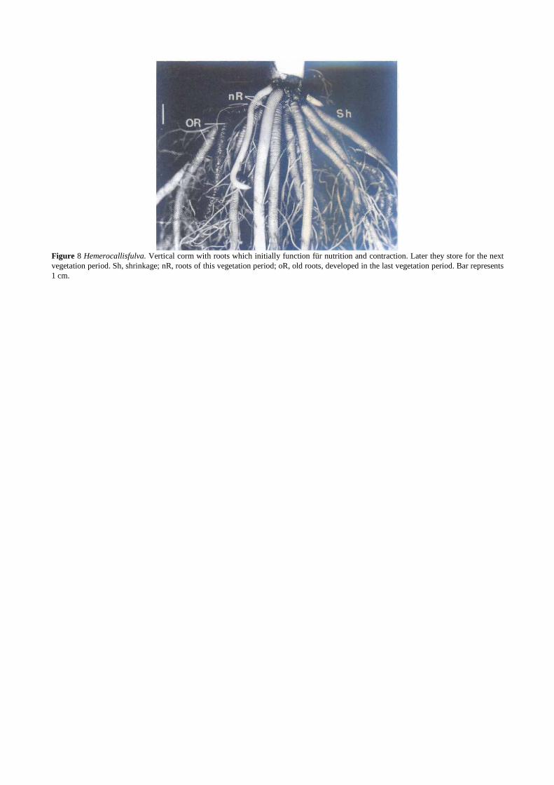
Figure 8 Hemerocallisfulva. Vertical corm with roots which initially function für nutrition and contraction. Later they store for the next vegetation period. Sh, shrinkage; nR, roots of this vegetation period; oR, old roots, developed in the last vegetation period. Bar represents 1 cm.

REFERENCES Arber A. 1925. Monocoty1edons-A Morpho10gica1 Study. Cambridge, U.K.: University Press. Bell AD. 1994. Illustrierte Morphologie der Blütenpflanzen. Heide1berg: Ulmer. Bell AD., Tomlinson PB. 1984. Adaptive architecture in rhi zomatous p1ants. Bot J Linn Soc 80:25-160. Berkemeyer W. 1928. Über kontraktile Umbelliferenwurzeln. Bot Archiv 24:273-318. Bottum FR. 1941. Histologica1 studies on the roots of Me/ilotus alba. Bot Gaz 103:132-145. Bussen M. 1951. Untersuchungen über die
Bewurzelung der Keimpflanzen im Verwandtschaftskreis der Monokotylen. Dissertation, Mainz, Germany. Chen S. 1969. The contractile roots of Narcissus. Ann Bot 33:421--426. Cresswell A, Hamilton NRS, Thomas H, Charnock RB, Cookson A, Thomas BJ. 1999. Evidence for root contraction in white clover
(Trifolium repens L.). Ann Bot 84:359-369. Cyr JR, Lin BL, Jernstedt JA. 1988. Root contraction in hyacinth. II. Changes in tubulin levels, microtubule number and orientation
associated with different cell expansion. Planta 174:446--452. Dahlgren RMT, Clifford HT, Yeo PF. 1985. The Families of the Monocotyledons. Structure, Evolution, and Taxonomy. New Y ork:
Springer-Verlag. Davey AJ. 1946. On the seed1ing of Oxalis hirta L. Ann Bot N Ser 39:237-256. Deloire A. 1980. Les racines tractrices de l'Allium polyanthum Roem. et Schult: une etude morphologique, anatomique et
histoenzymologique. Rev Cyto1 Biol Veg Bot 3:383-390. de Vries H. 1880. Ueber die Kontraktion der Wurzeln. Landwirthschaftl Jahrb 9:37-95. In: Hugo de Vries, Opera e Periodicis collata.
Vol. 11, Utrecht, Netherlands: A. Oosthoek, MCMXVIII. Draheim W. 1922. Beiträge zur Kenntnis des Wurzelwerks von Iridaceen, Amaryllidaceen und Liliaceen. Bot Archiv 23:385--440. Duncan JF. 1925. "Pull roots" of Oxalis esculenta. Trans Bot Soc Edinb 29:192-196. Ennos AR. 1993. The scaling of root anchorage. J Theor Biol 161:61-75. Ennos AR, Crook MJ, Grimshaw C. 1993. A comparative study of the anchorage systems of Himalayan balsam Impatiens glandulifera
and mature sunflower Helianthus annuus. J Exp Bot 44:133-146. Flindt R. 1988. Biologie in Zahlen. Berlin: Fischer. Froebe HA, Pütz N. 1988. Orientierende Versuche zur Verlagerung pflanzlicher Organe im Erdboden durch definierte Kräfte. Beitr Biol
Pflanzen 63:81-100. Galil J. 1958. Physiological studies on the development of contractile roots in geophytes. Bull Res Counc Isr 6:223-236. Galil J. 1962. Development cycle and ecology of Iris palaestina (Bak.). Boiss Bull Res Counc Isr 11:17-24. Galil J. 1968a. Vegetative
dispersal in Oxalis cernua. Am J Bot 55:68-73. Galil J. 1968b. Biological Studies on the Seedling of Colchicum steveni Kunth. Beitr Biol Pflanzen 45:243 256. Galil J. 1969a. Morpho-ecologica1 studies on Gladiolus segetum Gawl. Isr J Bot 18:43-54. Galil J. 1969b. On the laterally-contracting Root of Colchicum steveni. Beitr Bio1 Pflanzen 46:315-322. Galil J. 1978. Morpho-ecological studies on Arisarumvulgare Targ.-Tozz. Isr J Bot 27:77-89. Galil J. 1980. Kinetics of bu1bous plants. Endeavour 5:15 20. Galil J. 1981. Morpho-ecological studies on geophilic plants. Vegetative dispersal of Gynandiris sisyrinchium L. Isr J Bot 30:165-172. Galil J. 1983a. Vegetative dispers al of Muscari parviflorum Desf. Isr J Bot 32:221-230. Galil J. 1983b. Morpho-ecological studies of lowering in the seedling of the geophyte Ixiolirion tataricum (PalI.) Herb. New
PhytoI143:143-150. Gravis A. 1926. Contribution a I'etude anatomique du raccourcissement des racines. Acad R Belg Bull Clin Sci 12:48-69. Halevy AH. 1986. The induction of contracti1e roots in Gladiolus grandiflorus. P1anta 167:94--100. Iziro Y, Hori Y. 1983a. Effect of temperature on the growth of contractile root(s) of daughter corm or bulbs in Gladiolus and Oxalis
bowieana Lodd. J Jpn Soc Hort Sci 51 :459--465. Iziro Y, Hori Y. 1983b. Effect of p1anting depth on the growth of contractile root(s) of daughter corm or bulbs in Gladiolus and Oxalis
bowieana Lodd. J Jpn Soc Hort Sci 52:51-55. Iziro Y, Hori Y. 1983c. Retrans10cationof photoassimilates accumulated in contractile root(s) to daughter corm or bulbs in Gladiolus and
Oxalis bowieana Lodd. J Jpn Soc Hort Sci 52:56--64. Jacoby B, Halevy AH. 1970. Participation of light and temperature fluctuations in the inductionof contractile roots of Gladiolus. Bot Gaz
131:74--77. Jernstedt J. 1984. Seedling growth and root contraction in the soap plant, Chlorogalum pomeridianum (Liliaceae). Am J Bot 71:69-75. Kaplan DR. 1992. The relationship of cells to organisms in plants: problem and imp1ications of an organismal perspective. Int J Plant Sci
153:28-37. Kirchner 0, Loew E, Schröter C. 1934. Lebensgeschichte der Blütenpflanzen Mitteleuropas. Spezielle Ökologie der Blütenpflanzen Deutschlands, Österreichs und der Schweiz. Band 1.3 and 1.4. Berlin: Ulmer. Lamant A, Heller R. 1967. Sur 1a contraction des racines d'Arum italicum. Bull Soc Fr Physio1 Veg 13:179193.
Lin BL, Jernstedt J. 1988. Microtubu1e organization in root cortica1 cells of Hyacinthus orientalis. Protoplasma 141:13-23. Molisch H. 1965. Botanische Versuche und Beobachtungen ohne Apparate. Berlin: Fischer. Negbi M, Dagan B, Dror A, Basker D. 1989. Growth, flowering, vegetative reproduction, and dormancy in the saffron crocus (Croeus
sativus L.). Isr J Bot 38:95-113. Nordhausen M. 1913. Über kontraktile Luftwurzeln. Flora 105:102-126. Otto F. 1976. Das Konstruktionssystem pneu. In: Otto F, ed. Pneus in Natur und Technik. Bonn, Germany: Karl Krämer, pp 22-47. Pfeiffer NEA. 1931. Morpho10gica1 study of Gladiolus. Contrib Boyce-Thompson Inst 3:173-195. Pütz N. 1991. Die Zugbewegungstypen bei den Monokotylen. Bot Jahrb Syst 112:347-364. Pütz N 1992a. Measurement of the pulling force of a single contractile root. Can J Bot 70:1433-1439. Pütz N. 1992b. Das Verhältnis von Bewegung und Wurzelkraft bei Monokotylen. Beitr Biol Pflanzen 67:173-191. Pütz N. 1993. Underground plant movement. I. The bulb of Nothoseordum inodorum (Alliaceae). Bot Acta 106:338-343.

Pütz N. 1994. Underground plant movement. 11. Vegetative spreading of Oxalis pes-eaprae L. Plant Syst Evol 191:57-67. Pütz N. 1996a. Underground plant movement. 111. The corm of Sauromatum guttatum (Wall.) Schott (Araceae). Flora 191:275-282. Pütz N. 1996b. Underground plant movement. IV. Observance of the behaviour of some bulbs with special regard to the induction of root
contraction. Flora 191:313-319. Pütz N. 1996c. Development and function of contractile roots. In: Waisel Y, Eshel A, Kafkafi U, eds. Plant Roots. The Hidden Half. New
York: Marcel Dekker, pp 859-874. Pütz N. 1998. Underground plant movement. V. Contractile root tubers and their importance to the mobility of Hemeroeallis lulva L.
(Hemerocallidaceae). Int J Plant Sci 159:23-30. Pütz N. 1999a. In vivo tissue isolation in contractile roots of Lapeirousia laxa (Iridaceae). Flora 194:405-412. Pütz N. 1999b. Kontraktile
WurzeIn-ein unterirdischer Bewegungsmotor. PdN-Biol 48:20-29. Pütz N, Froebe HA. 1995. Are-evaluation of the mechanism ofroot contraction in monocotyledons using the exampie of Arisarum vulgare
Targ.-Tozz. (Araceae). Flora 190:285-297. Pütz N, Leskovsek C. 1999. Konstruktion geophiler Systeme. PdN-BioI48:1-12. Pütz N, Schmidt KHA. 1999. 'Underground plant mobility' and 'dispersal of diaspores'. Two exemplary case studies for useful
examinations of functional morphology (plant construction). Syst Geogr PI 68:39-50. Pütz N, Sukkau I. 1995. Comparative examination of the moving process in monocot and dicot seedlings using the examp1e Lapeirousia
laxa (Iridiaceae) and Foenieulum vulgare (Apiaceae). Feddes Repert 106:475-481. Pütz N, Froebe HA, Haese U. 1990. Quantitative Untersuchungen zum Mechanismus der Wurzelkontraktion bei Acidanthera bieolor
Hochst. (Iridaceae). Beitr Biol Pflanzen 65: 147-161. Pütz N, Hüning G, Froebe HA. 1995. Cost and advantage of soil channel formation by contractile roots in successful plant movement.
Ann Bot 75:633-639. Pütz N, Pieper J, Froebe HA. 1997. The induction of contractile root activity in Sauromatum guttatum (Araceae). Bot Acta 110:49-54. Rimbach A. 1896. Ueber die Tiefenlage unterirdisch ausdauernder Pflanzen. Ber Dtsch Bot Ges 14:164-168. Rimbach A. 1897a. Ueber die Lebensweise des Arum maeu latum. Ber Dtsch Bot Ges 15: 178-182. Rimbach A. 1897b. Lebensverhältnisse des Allium ursinum. Ber Dtsch Bot Ges 15:248-252. Rimbach A. 1898a. Die kontraktilen Wurzeln und ihre Thätigkeit. Beitr zur wissenschaft!. Botanik 2:1-26. Rimbach A. 1898b. Über Lilium marthagon. Ber Dtsch Bot Ges 16:104-110. Rimbach A. 1922. Die Wurzelverkürzung bei den großen Monokotylenformen. Ber Dtsch Bot Ges 40:196-202. Rimbach A. 1927. Die Geschwindigkeit und Dauer der Wurzelverkürzung. Ber Dtsch Bot Ges 45:127-130. Rimbach A. 1929. Die Verbreitung der Wurzelverkürzung im Pflanzenreich. Ber Dtsch Bot Ges 47:22-31. Rimbach A. 1938. Phaedranassa ehloraeea. Ber Deutsch Bot Ges 56:440-446. Ruzin S. 1979. Root contraction in Freesia (Iridaceae). Am J Bot 66:522-531. Smith FH. 1930. The corm and contractile roots of Brodiaea laetea. Am J Bot 17:916-927. Smith-Huerta NL, Jernstedt JA. 1989. Root contraction in hyaeinth. 111. Orientation of cortical microtubules visualized by
immunofluorescence microscopy. Protoplasma 151:1-10. Smith-Huerta NL, Jernstedt JA. 1990. Root contraction in hyacinth. IV. Orientation of cellulose microfibrils in radial, longitudinal and
transverse cell walls. Protoplasma 154:161-171. Stevens on DW. 1975. Taxonomic and morphological observations on Botryehium multifidum (Ophioglossaceae). Madrono 23:198-204. Stevenson DW. 1980. Observations on root and stern contraction in cycads (Cycadales) with special reference to Zamia pumila L. Bot J
Linn Soc 81:275-281. StöcklinJ. 1992. Umwelt, Morphologie und Wachstumsmuster klonaler Pflanzen - eine Übersicht. Bot Helv 102:3-21. Stroever V.1892. Ueberdie Verbreitung der Wurzelverkürzung. Dissertation, Jena, Germany. Troll W. 1937-43. Vergleichende Morphologie der höheren Pflanzen, 1. Vegetationsorgane. Berlin: Borntraeger. Watenabe K. 1925. Über die Kontraktion und daraus verursachte Anomalie in der Wurzel von Cycas revoluta. Jpn J Bot 2:293-297. Wilson K, Honey JN. 1966. Root contraction in Hyacinthus orienta/is. Ann Bot N Ser 30:47-61. Wilson K, Anderson GJH. 1979. Further observations on root contraction. Ann Bot N Ser 43:665-679. Zamski E, Ucko 0, Koller D. 1983. The mechanism of root contraction in Gymnarrhena_micranatha, a desert plant. New Phytol 95:29-
35. Zimmermann MH, Wardrop AB, Tomlinson PB. 1968. Tension wood in aerial roots of Ficus benjamina L. Wood Sci Technol 2:95-104.