The Helminthological Society of Washington - Peru State College
60
VOLUME 18 JULY, 195] NUMBER 2 PROCEEDINGS of TheHelminthologicalSociety ofWashington A semi-annual journal of research devoted to Helminthology andall branchesofParasitology Supportedinpartbythe BraytonH .RansomMemorialTrustFund EDITORIALCOMMITTEE EDWARDG.REINHARD,Editor TheCatholicUniversityofAmerica EMMETTW.PRICE WILLARDH .WRIGHT U .S .BureauofAnimalIndustry NationalInstitutesofHealth GILBERTF .OTTO GOTTHOLDSTEINER JohnsHopkinsUniversity U .S .BureauofPlantIndustry , Soils,andAgriculturalEngineering Subscription$1 .75 a Volume ;Foreign,$2 .00 Publishedby THEHELMINTHOLOGICALSOCIETYOFWASHINGTON
Transcript of The Helminthological Society of Washington - Peru State College

VOLUME 18
JULY, 195]
NUMBER 2
PROCEEDINGS
of
The Helminthological Society
of WashingtonA semi-annual journal of research devoted toHelminthology and all branches of Parasitology
Supported in part by theBrayton H. Ransom Memorial Trust Fund
EDITORIAL COMMITTEE
EDWARD G. REINHARD, EditorThe Catholic University of America
EMMETT W. PRICE
WILLARD H. WRIGHTU. S. Bureau of Animal Industry
National Institutes of Health
GILBERT F. OTTO
GOTTHOLD STEINERJohns Hopkins University
U. S. Bureau of Plant Industry ,Soils, and Agricultural Engineering
Subscription $1.75 a Volume; Foreign, $2 .00
Published byTHE HELMINTHOLOGICAL SOCIETY OF WASHINGTON
VOLUME 18
JULY, 1951
NUMBER 2
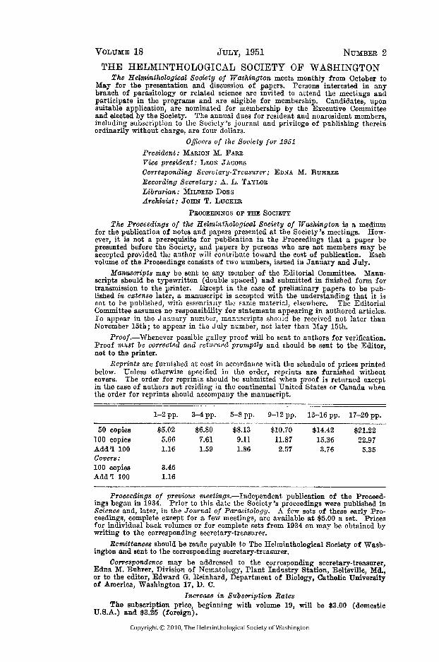
THE HELMINTHOLOGICAL SOCIETY OF WASHINGTONThe Helminthological Society of Washington meets monthly from October to
May for the presentation and discussion of papers . Persons interested in anybranch of parasitology or related science are invited to attend the meetings andparticipate in the programs and are eligible for membership. Candidates, uponsuitable application, are nominated for membership by the Executive Committeeand elected by the Society . The annual dues for resident and nonresident members,including subscription to the Society's journal and privilege of publishing' thereinordinarily without charge, are four dollars .
Officers o f the Society for 1951President : MARION M. FARRVice president :LEON JACOBSCorresponding Secretary-Treasurer : EDNA M.BUHRERRecording Secretary : A. L. TAYLORLibrarian : MILDRED DossArchivist : JOHN T. LUCKER
PROCEEDINGS OF THE SOCIETY
The Proceedings of the Helminthological Society of Washington is a mediumfor the publication of notes and papers presented at the Society's meetings. How-ever, it is not a prerequisite for publication in the Proceedings that a paper bepresented before the Society, and papers by persons who are not members may beaccepted provided the author will contribute toward the cost of publication . Eachvolume of the Proceedings consists of two numbers, issued in January and July.
Manuscripts may be sent to any member of the Editorial Committee . Manu-scripts should be typewritten (double spaced) and submitted in finished form fortransmission to the printer . Except in the case of preliminary papers to be pub-lished in extenso later, a manuscript is accepted with the understanding that it isnot to be published, with essentially the same material, elsewhere. The EditorialCommittee assumes no responsibility for statements appearing in authored articles .To appear in the January number, manuscripts should be received not later thanNovember 15th ; to appear in the July number, not later than May 15th.
Proof.-Whenever possible galley proof will be sent to authors for verification .Proof must be corrected and returned promptly and should be sent to the Editor,not to the printer.
Reprints are furnished at cost in accordance with the schedule of prices printedbelow. Unless otherwise specified in the order, reprints are furnished withoutcovers . The order for reprints should be submitted when proof is returned exceptin the case of authors not residing in the continental United States or Canada whenthe order for reprints should accompany the manuscript .
1-2 pp .
3-4 pp .
5-8 pp .
9-12 pp . 13-16 pp. 17-20 pp .
50 copies
$5.02
$6.80
$8.13
$10.70
$14.42
$21.22100 copies
5.66
7.61
9.11
11.87
15.36
22.97Add 11 100 1.16 1.59 1.86 2 .57 3 .76 5.35Covers :100 copies
3.46Add '1 100
1.16
Proceedings of previous meetings.-Independent publication of the Proceed-ings began in 1934 . Prior to this date the Society's proceedings were published inScience and, later, in the Journal of Parasitology. A few sets of these early Pro-ceedings, complete except for a few meetings, are available at $5 .00 a set. Pricesfor individual back volumes or for complete sets from 1934 on may be obtained bywriting to the corresponding secretary-treasurer.
Remittances should be made payable to The Helminthological Society of Wash-ington and sent to the corresponding secretary-treasurer .
Correspondence may be addressed to the corresponding secretary-treasurer,Edna M. Buhrer, Division of Nematology, Plant Industry Station, Beltsville, Md .,or to the editor, Edward G . Reinhard, Department of Biology, Catholic Universityof America, Washington 17, D . C .
Increase in Subscription RatesThe subscription price, beginning with volume 19, will be $3 .00 (domestic
U.S.A .) and $3 .25 (foreign) .

PROCEEDINGS OF THE HELMINTHOLOGICAL SOCIETYOF WASHINGTON
VOLUME 18
JULY, 1951
NUMBER 2
The Pathogenicity of the Common Sheep Tapeworm,Moniezia expansa
K. C. KATES and A. GOLDBERG1U. S. Bureau of Animal Industry, Beltsville, Md .
The tapeworm, Moniezia expansa, occurs in sheep and certain other ruminantsthroughout the world and, because of its large size, has probably been known toman since sheep became domesticated . Much uncertainty and difference ofopinion still exist among sheepmen, veterinarians, parasitologists, and others con-cerning the role of this parasite as a pathogen . There are several reasons for thisstate of affairs . Heavy infections have been observed in apparently healthy sheep,and, conversely, moderate to severe clinical symptoms, and even death, in somecases, have been ascribed to this parasite when no other cause was apparent .Until recently it was impossible to obtain experimental proof of pathogenicity orlack thereof, because the life cycle of the parasite was not known . Transmissionby oribatid mites was first reported by Stunkard (1937) . Some reports ofpathogenicity have appeared in connection with the successful use of anthelminticsfor expulsion of the worms from sheep and the coincident alleviation of variousclinical conditions . Reports of this kind, particularly in regard to use of leadarsenate as an anthelmintic, were recently summarized by Foster and Habermann(1948) . There have been only two reports, exclusive of the present one, of ex-periments designed to determine the possible pathogenicity of Moniezia by meansof experimentally induced infections, namely, those of Shorb (1939) and Hansenet al. (1950) . The purpose of this paper is to present a reasonably completesurvey of the literature on this subject and the results of experimental workwhich have been published previously only in abstract form by us (1949) .
REVIEW OF-LITERATURE
A summary of the widely scattered literature on this subject is not now avail-able, and an attempt is made here to bring these reports together . Opinions ex-pressed and observations reported in the past may be conveniently summarizedunder three categories as follows : (a) General statements in various publications,particularly in well-known books on veterinary medicine and helminthology ;(b) field observations and case reports many of which are concerned with the useof ruminant teniacides ; and (c) experimental work not primarily concernedwith anthelmintic medication. Many of the observations on the effect of M .expansa infections on ruminants were made before the life cycle of the parasitewas known, and before reasonably adequate information on the effects of otherparasites, such as gastrointestinal nematodes, coccidia, and other pathogens, wasavailable. Moreover, little or no information derived from experimentally inducedinfections was available to the earlier workers .
General statements .-The following writers, among others, have consideredM. expansa a serious pathogen of domestic ruminants under certain conditions
1 A considerable part of this work was done with the technical assistance ofMr. C. E . Runkel.
87

88
PROCEEDINGS OF THE
[VOL . 18
Curtice (1890), Neumann (1905), Daubney (1923), Skriabin and Schultz (1934),Neveu-Lemaire (1936), Monnig (1947), and Hutyra, Marek, Manninger, et al .-(1949) . The symptoms ascribed to infections by this parasite vary slightly indetail and wording in the different publications of these authors, but may besummarized for all of them as follows : Light infections, especially in olderanimals, usually do not cause serious symptoms, but because of the large size ofthe worms, even small numbers on rare occasions may cause various manifestationsof disease . Heavy infections in young animals cause serious digestive disturbances,diarrhea, and sometimes constipation ; stunting, emaciation, edema, anemia, andconsiderable loss of wool may occur. Death may ensue after a time as a resultof the cumulative effects of the tapeworms or suddenly after a period of con-vulsions or acute toxemia . The specific cause has been variously ascribed toreduction in available nourishment for the host as a result of the utilization offood by the rapidly growing worms, elimination of waste products and toxins bythe parasites, and mechanical blockage of the alimentary tract by masses of worms.Skriabin and Schultz (1934), for example, assert that in Russia enormous lossesare caused annually by moniezioses, and that in certain areas, in unfavorable years,a mortality of 80 percent of the young animals has occurred .
On the other hand, the opinion that M. expansa is seldom the cause of seriousdisease of sheep, with the reservation that reliable information on the subject islacking, has been expressed by Cameron (1934), Clunies Riss and Gordon (1936),Dikmans and Shorb (1942), and Morgan and Hawkins (1949) . Some of theseauthors believe that in supposed losses or serious effects thought to be due totapeworms, less obvious and more injurious parasites or other pathogens may havebeen present but were undetected . Furthermore, it has been expressed that untilmore proof is forthcoming that experimentally induced infections can causedefinite injury to the hosts, and that satisfactory evidence is brought forward ofimprovement in condition of sheep or alleviation of symptoms following removalof tapeworms only by anthelmintics under controlled conditions, it will not bepossible to reach any reliable conclusions concerning the pathogenicity of thistapeworm. In this connection, a recent statement by Gordon (1950) follows :"There is little doubt that heavy infestations (of tapeworms) in young sheepexposed to malnutrition are of some consequence ." To the writers' knowledge,no work to support this statement has been published .
Field observations and case reports.-Reports of original observations in thefield in which M. expansa and its close relative, M. benedeni, were thought to bethe primary cause of disease have appeared for almost a century, from 1855 to1950 . However, their total number is surprisingly small, considering the cosmo-politan distribution of the parasites . These reports are summarized in chrono-logical order.
Cox (1855) reported the loss in England of hundreds of lambs that wereunder his care. Before death they went off feed, became emaciated, and developeddiarrhea . On postmortem examination of the lambs, large quantities of tape-worms, which he considered the cause of the deaths, were recovered . No mentionwas made of other parasites present, if any, but reference was made to an un-usually severe winter preceding the losses and to the poor nutritional conditionof the lambs . Some years later-in 1877-Cross reported from England therelief of scouring in lambs when large quantities of tapeworms were removed byadministering a ruminant teniacide then in use. The same year there appearedan anonymous report of heavy losses in a large flock of lambs grazed in CentralPark, New York City . Many lambs in this flock became sick for two or threedays, developed convulsions, turned around in circles, and dropped dead . Onlyone lamb was examined postmortem and contained enough tapeworms to fill a

12-ounce measure. No other significant details were mentioned in this report .Neumann (1905) cited these reports as evidence of tapeworm pathogenicity . Inthe discussion of the general paper by Daubney (1923), J . F. Craig spoke of asimilar disease outbreak in lambs and ewes which he had observed ; diarrheaoccurred and death of some of the lambs was preceded by convulsions . One lambwas autopsied and "a large tract of the intestines was almost completely impactedwith tapeworms." A Mr. Martin, also participating in the discussion of Daub-ney's paper, said that "as regarded tapeworms, he did not think much of it ."
Since these early observations on alleged tapeworm disease under natural con-ditions, there has been, to the writers' knowledge, a dearth of reports of asimilar nature up to the last decade, when a revival of reports, somewhat reminis-cent of the earlier ones, occurred . McCulloch and McCoy (1941) reported treat-ing diarrheic, unthrifty lambs with lead arsenate, which removed "enormousnumbers" of tapeworms, and two months thereafter the owner reported the lambswere making better gains than in previous years . Baywater (1942) treated aflock of sheep, many of which showed clinical symptoms of parasitic gastritis,(gastroenteritis) accompanied by diarrhea, with a two percent solution of coppersulphate. In his own words : "The results were spectacular. Within an hour offbeing dosed the animals began to pass large numbers of tapeworms and there-after recovery was rapid, . . ." No mention was made of other parasites whichmay have been present and may have been affected by the medication . Likewise,Radeleff (1944) reported a mortality, resulting from monieziasis, as high as 50percent in herds of kids and calves and 25 percent in lambs, and prompt relief ofsymptoms and reduction of losses by use of lead arsenate as a ruminant teniacide .He made no mention of any other parasites or pathogens which may have beenpresent in the animals . Further tests of the value of lead arsenate in treatmentof lamb scours, and simultaneous removal of quantities of M. expansa from lambs,were reported by Habermann and Carlson (1946) . A few scouring lambs weretreated, tapeworms were effectively removed, scouring ceased shortly after treat-ment, and the condition of the animals improved . No other cause of the scouringother than tapeworms was observed . These workers did not note any signs ofconvulsions and sudden deaths, as reported by some other authors .
Two reports have appeared recently in which acute symptoms and suddendeaths in sheep were ascribed to tapeworms . Tableman (1946) observed thedeath of several lambs on a farm in Illinois, supposedly as a result of heavy tape-worm infections . He stated that "the outstanding characteristics of these caseswere : (1) the absence of stomach worms or intestinal roundworms, (2) the excel-lent condition of the lambs, and (3) the f act that all the lambs died in convulsions .. . . The cause of death in these cases was probably an intoxication due to absorp-tion of metabolic products of the worms-or was it?" Four dead lambs wereexamined on the farm, and, although it was stated that no parasites other thantapeworms were present, a very rare phenomenon for sheep, no mention was madeof a microscopic examination of the contents of the alimentary tract or feces orother diagnostic procedures . Lafenêtre (1948) reported heavy death losses insheep in numerous flocks pastured in southern France between Beziers and thePyrenees Mountains . Some of the affected flocks were locally owned, while somewintered in this area but came from summer pastures in the mountains of theRepublic of Andorra and the provinces of Pyrénées-Orientales and Ariège . Thedisease, affecting sheep of all ages, was characterized by sudden onset and rapidprogress. Some 250 to 300 deaths occurred among 3,000 to 3,500 animals . Theaffected animals isolated themselves from the flock, stood stiff-legged with headsdown, and mucoid saliva flowed from their lips . The mucous membranes werenormal or congested . Body temperatures were normal, but diarrhea was often

seen with abundant and fetid feces . Terminally, convulsions occurred, and theanimals became comatose and died. Recovery from the disease was exceptional .Animals killed during the acute phase of the disease showed no macroscopiclesions, tapeworms were present in all cases, some animals had a "few strongyles ",and no liver flukes were found . Bacteriological and other diagnostic proceduresgave negative results. Further observations were presented, supposedly to supportthe thesis that tapeworms were the primary cause of these losses . Some 85 animalsleft from an affected flock were sold for slaughter . These were examined fortapeworms, and "massive" (?) infestations of six to ten tapeworms were ob-served. Except for a few animals, the carcasses rated top grade . Also, medica-tion of several flocks with copper sulphate was tried, following the dosage recom-mended by Skriabin and Schultz (1934) . The author noted that treatment hadbeen instituted in these flocks when the number of death losses was declining, andthat losses had ceased in some flocks spontaneously. It was postulated that tape-worms may cause disease in sheep as a result of teniatoxins produced, or massiveinfections may favor infection by pathogenic organisms not yet known . In 1948,shortly after Lafenêtre's paper appeared, an anonymous editorial was published,which referred to Lafenêtre's report and certain other selected references, andthe suggestion was made that the current view of certain authorities that tape-worms of sheep are of little consequence should be restudied .
A recent report by Link et al . (1950) was summarized by the authors asfollows : "An extensive outbreak of Moniezia expansa infection in dairy calvesis reported. Out of a herd of 79, 19 had died, and 42 were emaciated, lackedvigor, and grew slowly . Treatment with lead arsenate removed the tapewormsand permitted the calves to make an uneventful recovery." The animals wereanemic, pot-bellied, and had profuse diarrhea . Pneumonia was present and wasconsidered a probable contributory cause of death. An autopsy was performedon one calf only, and areas of congestion were observed in the lungs and thesmall intestine . From the latter organ, which was markedly inflamed, 32 tape-worms were recovered. No other details were given regarding the affectedanimals, except that at the time of medication the yards and calf barn werecleaned and the calves moved to another pasture .
Experimental work.-As already stated, exclusive of the work reportedherein, there have been only two reports of work on experimentally induced, puretapeworm infections of lambs. Shorb (1939) fed four lambs 51 to 203 cysticer-coids of M. expansa each, over variable periods of time . Three lambs becameinfected, but no significant clinical symptoms developed during two to two andone half months. Weight gains of the infected lambs were slightly less thanthose of the controls, but these were not considered significant by the authorbecause of the small number of animals employed . The three infected lambscontained 4, 9, and 39 tapeworms, respectively, at autopsy. Hansen et al. (1950a)fed one lamb 5 cysticercoids, another lamb 60, and kept one uninfected lamb as acontrol. Later (1950b) in an abstract they stated that six lambs were infected .It was concluded on the basis of this work that infected lambs were retarded ingrowth, and it was reported that there was some depression in haemoglobin andhaematocrit values in comparison with the control lamb or lambs .
Three other reports of an experimental nature are of interest . Freeborn andBerry (1934) studied the weight gains of naturally infected lambs in comparisonwith the gains of exposed but uninfected lambs (i), and no significant differenceswere observed . When these lambs were assigned numerical grades at the time ofslaughter, the negative controls graded higher than lambs made negative for tape-worms by treatment and those that were infected . No mention was made of otherparasites which may have been, and probably were, present in many of the animals .

Shorb (1940) recorded the grades made by a group of .71 tapeworm infected,seven-month-old lambs . Most of these had Thysanosoma actinioides infections,a few had M . expansa only, and some had both species. No significant gradedifferences were noted between parasitized and non-parasitizd animals . Hawkins(1946) studied the effect of M. expansa infections in a flock of ewes and theirlambs over a four-year period . Heavy infections were acquired by the lambs inMay or June and were usually lost by August or September . No distinct symp-toms could be ascribed to tapeworms alone, and infected and uninfected lambsappeared much the same . It was concluded that for tapeworms alone treatmentwas not justified, because of the lack of marked symptoms or lesions and spon-taneous loss of worms.
MATERIALS AND METHODS
From the preliminary work of Shorb (1939) it was clear that the majorlimiting factor in making a study involving a larger number of experimentallyinfected animals than had been used by previous investigators, was the readyavailability of several thousand cysticercoids of M . expansa, which had to beobtained from oribatid mites . It was also necessary to have this source of infec-tive material available at the time young, parasite-free lambs were also availablefor the experiment . Details of the method used in this Bureau for securingadequate infective material have been described elsewhere by Kates and Runkel(1948) . Because over 3,000 cysticercoids were required for the work reportedherein, and the best yield of cysticercoids that could be expected from previousmite dissections was somewhat less than 100 from an equal number of mitescollected, it was necessary to collect over 4,000 mites, mainly of the speciesGalumna virginiensis, from a contaminated plot before dissecting the cysticercoidstherefrom and starting the experiment . Therefore, mites were collected for fiveto six weeks before the experiment was begun . This was done by means of abattery of modified Berlese funnels . The mites collected were placed in 50 ccweighing bottles with a small piece of moist filter paper on the bottom, and thebottle tops tightly inserted . Most of the mites remained alive in these bottles untildissected one to six weeks later, and were readil y , available as a source of cysticer-coids when needed.
When enough mites had been collected from the contaminated plot, andparasite-free lambs were available, dissection o€- mites for cysticercoids was' begun,as described by Kates and Runkel (1948) . The living cysticercoids were ac-cumulated in physiological .saline solution in small watch glasses until enough hadbeen obtained to infect one or more lambs, and then they were placed in smallgelatin capsules partially filled with moist, pulverized small animal feed . Thissmall capsule was then placed within a slightly larger one and given to the lambwith a balling gun. . Cysticercoids were thus fed lambs within a few hours of thetime they were removed from the mites . A total of 16 lambs were fed 121 to 411cysticercoids per lamb. The total dose was given to each lamb as a single feeding,except that two lambs were given two feedings separated by only two days in onecase and three days in another . Although it was not possible to feed the desirednumbers of cysticercoids to all the lambs on the same day, infection of all lambswas accomplished during the first week of the experiment after the initial weightsof the animals were taken and rations established .
The plan of the experiment was as follows : 16 lambs, two to three months ofage and previously weaned, were fed cysticercoids in numbers indicated in Tables1 and 2 . These lambs were divided into two groups of eight each . One groupwas killed and autopsied approximately one month after infection (Table 1), andthe other group approximately two months after infection (Table 2) . A thirdgroup of four lambs, of approximately similar age and weight, was not infected

TABLE 1 .Postmortem results on lambs approximately one month after being fed cysticercoids .
Cysticer-Lamb
coids Fed
ScolexCount
atAutopsy
PercentDevelopment
Volume ofMonieziain DiluteFormalin(quarts)
Volumeof MoistWorms by
Displacement(cc)
Variationin WormLength(cm)
Variationin Worm
Developmenta
WeightGains ofLambs(lbs .)
1
150 114 76 0 .50 70 37 to 270 Immature-Gravid 5.502
165 75 45 1.00 160 All worms do 14 .50
3
142 67 47 0.75 100fragmented67 to 420 . do 8 .50
4
216 53 24 1.00 140 50 to 360 do 11 .755
160 89b 55 0.66 80 4.3 to 180 do 6.006
166 56 53 0.50 57 22 to 360 do 6.007
121 9 7 0.50 55 175 to 420 All Gravid 8.00
a Eggs or proglottids had not appeared in the feces of the lambs before autopsy .b Nine of these tapeworms were less than 30 cm long.

TABLE 2 .Postmortem results on lambs approximately two months after being fed cysticercoids .
Feces PositiveaDay ofAutopsy
ScolexWorm Material Recovered in Feces Weight
Gainsof Lambs
(lbs .)
Cysticer-Lamb
coids Fed Count atAutopsy Total cc Estimated Percent
FirstDay
LastDay (centrifuged) Gravid
Non-Gravid
89
1011121314
411334357344256250264
30th31st32nd40th34th34th33rd
63rd51st61st68th. . . .. . . . .. . .57th56th
65th66th66th68th65th63rd63rd
0000
68b00
10440
1731172003483
29
7111
8939
6169
3149
5116
8414
86
1012.7510 .59 .5
13 .56 .59
a For non-gravid or gravid proglottids or pieces of strobilae ; non-gravid material usually passed in feces before gravid material or eggs .b Apparent volume of tapeworms in dilute formalin one pint ; volume of moist worms by displacement 80 cc. No terminal proglottids present, no eggs in
feces ; only one piece of strobila contained a few eggs, all other worms non-gravid . Intact strobilae with scoleces varied in length from 5 to 250 cm .

and used as controls . The reason for having both one-month and two-monthgroups concerned the fact that in the living animal it is difficult to determine thesize of infections established, not only because of the often rapid spontaneous lossof tapeworms from heavily infected animals, but also because of the poor correla-
tion between the production of eggs and proglottids, or both, and the degree ofinfection. From preliminary infections of several lambs fed variable numbers ofcysticercoids, it was observed that tapeworms were not usually lost spontaneouslyduring the first month of infection . Therefore, the one-month group was con-sidered primarily as an "infection control" group, which gave information onthe average degree of the infection established by the method employed and thenumber of cysticercoids fed . Also, this group afforded an opportunity to observeeffects of heavy infections during the major part of the prepatent period whenthe number of tapeworms and quantity of tapeworm material were known .
The infected lambs were observed from day to day for clinical effects of theinfections, and once eggs, or proglottids began passing in the feces, the animalswere bagged and all feces collected and examined daily for tapeworm materialuntil the experiment was terminated. The course of tapeworm infections in lambscannot be followed to any great advantage by fecal examinations for eggs or byproglottid counts . Therefore, the only practical method found for followingquantitatively the , course of infection in these lambs was to obtain all feceseliminated during patency and collect therefrom all tapeworm material, individualproglottids, and pieces of strobilae, expelled by the lambs .
The infected animals were autopsied with care to remove the tapewormsintact, if possible . This was usually successful, but in lamb 2 (Table 1) not oneintact tapeworm was obtained although 75 scoleces and a large quantity of brokenstrobilae were recovered . All intact tapeworms recovered postmortem were al-lowed to relax in water and were then measured . The relative quantities of mate-rial thus recovered from the lambs were determined in two ways, as follows :(1) All tapeworms recovered from each lamb were placed in quart fruit jars indilute formalin, allowed to settle, and the quantity estimated as a pint, quart, orfractions thereof . Results obtained by this rather unsatisfactory method arerecorded in the tables only because similar means were employed by certain otherworkers to measure quantities of tapeworms recovered postmortem or after treat-ment. (2) Volume of moist tapeworm material was also determined by displace-ment as follows : Tapeworms in dilute formalin were poured from the containeronto a coarse screen and the excess fluid drained off over a uniform period of fiveminutes, the tapeworms not being allowed to dry . Thereafter, they were trans-ferred to a graduated cylinder containing a known volume of water and the tape-worm volume determined by the water displaced . Volume determinations of theproglottids and fragments of strobilae recovered from dung of lambs of the two-month group were made by centrifugation at 1,000 rpm for two minutes ingraduated centrifuge tubes . This method was better than the water displacementmethod for this material because the material measured consisted of large numbersof individual proglottids and small pieces of strobilae, much of which was in poorcondition after being removed from the dung .
The lambs were fed measured quantities of a maintenance diet of hay andgrain, so that each group had available the same feed in equal quantities per lambper day. Any uneaten feed was removed daily and weighed before the next dailyallowance was given . Each group of lambs was kept in a concrete-floored pen,half of which had shelter and half did not . All debris was removed daily fromthe pens, which were then thoroughly washed with hot water . All lambs wereweighed weekly, and the total weight of feed eaten was recorded on a weekly basisand for the entire experiment.

RESULTS
Of the 16 lambs that were fed large numbers of cysticercoids, failure toestablish infection occurred in only two, one in the one-month group (Table 1)and one in the two-month group (Table 2) . The reason for these two failures isnot clear. A total of 14 lambs, therefore, were successfully infected experimentallyseven in the one-month group and seven in the two-month group . The data onthe two uninfected lambs are not included in the tables, as their weight gainswere intermediate between the maximum and minimum weight gains of the in-fected lambs, and the writers believe they contributed nothing to the results ofthe experiment .
One-Month Group.-Table 1 summarizes the results obtained from the sevenlambs killed approximately one month (30 to 34 days) after 121 to 216 cysticer-coids were fed per lamb. No tapeworms or eggs were observed in the feces of thelambs up to autopsy. The percentage of tapeworms recovered at autopsy, on thebasis of the cysticercoids fed, varied from 7 to 76 . The average percentage ofdevelopment for all lambs was 41 . The numbers of tapeworms developing hadno close relationship to the number of cysticercoids fed . The actual numbers oftapeworms recovered varied from 9 to 114, the average being 66, indicating thatheavy experimental infections were established by the method employed .
At autopsy it was immediately apparent that there was great variation indevelopment of the tapeworms in the six lambs having over 50 worms each, al-though cysticercoids were fed to each lamb at approximately the same time .Complete tapeworms, with terminal segments, recovered from lambs 1 to 6 variedin development from small, immature specimens to large, gravid ones . Thesmallest complete worm recovered from lamb 5 was only 4 .3 cm long, while thelargest worm was 180 cm long. The largest complete worm from any of the lambswas 420 cm long. The nine tapeworms recovered from lamb 7, which had thesmallest number at autopsy, were more uniform in size, and all were in the samestage of development, with gravid terminal proglottids . When the volume of moisttapeworms recovered from each lamb was determined by displacement, there waslittle correlation between the volume and the scolex count . The 9 worms fromlamb 7 had a volume of 55 cc, that of the 56 worms from lamb 6 was only 57 cc,while the 114 worms from lamb 1 had a volume of only 70 cc . Thus, in lambs1 to 7 the heaviest infection by volume was only about three times that of thelightest infection, namely, 160 cc against 55 cc ; by numbers, the heaviest infectionwas almost 13 times the lightest infection, namely, 114 against 9 . Therefore, thedata in Table 1 show that when large numbers of tapeworms are present in lambs,their growth is irregularly retarded, and when few worms are present they developmore or less uniformly. Therefore, these observations indicate that in patho-genicity studies volume of tapeworms appears to be a more reliable criterion ofdegree of an infection of M . expansa than number of individuals present . It isdifficult to think of the relatively small, unarmed scolex of this species as animportant factor in causing injury to the host.
Lambs of the one-month group did not show any effects of their relativelyheavy tapeworm infections during the experiment . At autopsy a considerableportion of the small intestines of these small lambs appeared to be filled withtapeworms, but there were no observable injurious effects on the intestines orother organs . There was considerable variation in weight gains of the lambs,but these did not seem to be related to the degree of infection . It is of interestto note, but probably not significant, that lambs 2 and 4, having the heaviestinfection by volume of worms, made the greatest gains . The average gains forboth the infected and control groups, during the same experimental period, were

identical, namely, 8 .6 pounds per lamb, and the feed consumption per lamb for thetwo groups was also the same . .
Two-Month Group.-Table 2 and Figure 1 summarize the data on the seveninfected lambs observed for about two months . These lambs were fed somewhatlarger numbers of cysticercoids than the one-month group, the total per lambvarying from 250 to 411 . Infections in the seven lambs were shown by thepresence in their feces of individual proglottids or pieces or strobilae, beginning
FIG . 1. Detailed summary of the tapeworm infections established in each ofthe lambs of the "two-month group" (Table 2 .), showing the daily output oftapeworm material by volume in the feces, and the extent of the patent period ofinfection and tapeworms recovered, if any, from lambs at autopsy .
on the 30th to 40th day after cysticercoids were fed. Six of the lambs ceasedpassing tapeworm material in their dung between the 51st and 68th days afterinfection . Lamb 12 was still passing some proglottids in the feces on the 65thday, when it was killed and examined . This lamb was the only one of the groupretaining some of its tapeworms to the time of autopsy, 68 tapeworms being re-

covered at that time . The other six lambs were tapeworm-free when autopsied63 to 68 days after cysticercoids were fed. Therefore, six of the lambs of thisgroup spontaneously expelled their tapeworms at some time during the secondmonth after infection between the 30th and 68th days .
An accurate check was obtained on the infections, even though at autopsysix of the lambs were tapeworm-free . All the tapeworm material passed in thefeces of the lambs was collected daily and measured volumetrically . Table 2gives the total volume of material obtained after centrifugation from each lambduring the patent period . This method of measuring the quantity of tapewormmaterial expelled in the feces is roughly comparable to the displacement methodemployed for the one-month group (Table 1), but because the material collectedfrom the feces is not in as good condition as similar material collected directlyfrom the small intestine at autopsy, the figures in table 2 are probably lower thanthey would be for fresh tapeworms . The seven lambs passed 40 cc to 200 cc oftapeworm material each during patent periods of 20 to 33 days. These totalsshow that heavy parasitism was established in all cases. In addition, as alreadystated, lamb 12 retained 68 tapeworms, amounting to 80 cc by displacement, whenautopsied 65 days after infection, making a total of 280 cc of tapeworm materialfrom this lamb .
Tapeworm material passed in the feces was examined to ascertain how muchconsisted of gravid segments and strobilae and how much was non-gravid ; con-siderable quantities of non-gravid strobilae is indicative that more or less completetapeworms were being expelled . Table 2 shows that in six of the lambs morethan 50 percent was non-gravid ; in one lamb only 31 percent was non-gravid.The data show that whole tapeworms were being spontaneously lost from time totime during the patent period, even though it was not possible to identify scolecesin 24-hour fecal collections .
Figure 1 shows the daily record of tapeworm material expelled from eachlamb . The first material expelled was almost entirely non-gravid, which showeda tendency early, in the infection toward expulsion of entire immature worms orparts thereof . In lambs 9, 13, and 14, the percentages of non-gravid materialpassed in the dung were respectively 89, 84, and 86, indicating that for theselambs most of the tapeworms were expelled before reaching the gravid stage .In lamb 9, over 60 percent of the total tapeworm material recovered during patencywas expelled on the 45th day after cysticercoids were fed . In lambs 13 and 14the tapeworms were expelled over a more or less extended period .
Figure 1 also shows that the greatest quantities of gravid segments andstrobilae were expelled from lambs 8, 10, 11, and 12 . Lamb 11 was the only oneof the group which expelled more gravid than non-gravid material, and had thelongest prepatent period (40 days) for its tapeworm infection . The tapewormsin this lamb probably became more firmly established than in the others, and alarger proportion of the worms were able to reach the gravid stage before beingexpelled. However, many worms were probably expelled on the 51st day and theremainder on the 67th day after infection, as indicated by the large quantities ofnon-gravid material expelled during these two days . In lambs 8, 9, 10, 13, and 14,all of which spontaneously expelled their entire infection before autopsy, the massof tapeworms was lost as follows : Lamb 8, on the 63rd day after infection ; lamb9, on the 45th day ; lamb 10, between the 58th and 61st days ; lamb 13, betweenthe 55th and 57th days, and lamb 14, during the 55th and 56th days .
These observations on relatively heavy infections show a strong tendency forspontaneous expulsion of all tapeworms from the host . However, there is evidencethat this is not necessarily true in relatively light infections, for in one infection,produced by feeding only a few cysticercoids to a lamb, elimination of gravid

proglottids in the feces began after the usual prepatent period and continued ata relatively steady pace for about ten and one half months, when . the animal . waskilled. At that time two large gravid specimens of M . expansa were recoveredfrom the small intestine.
There was no evidence of clinical effects of parasitism in the seven lambs ofthis group. during the entire experiment . As shown in Table- 2, there was con-siderable variation in weight gains of the lambs, but this was not related tovariations in degree of infection, as determined volumetrically . In fact, lamb 12,which expelled the greatest volume of tapeworm material, and still retained 68tapeworms having a volume of 80 cc, made the greatest gain . In contrast,lamb 13, which expelled the least tapeworm material and was tapeworm-free atautopsy, made the smallest gain . It is believed, however, that these data onindividual gains are of value only in showing that the heaviest infections did notdepress the gains of the lambs ; they are not interpreted to mean that heavyinfections have favorable effects on growth or on feed utilization by lambs . Theaverage gain of the infected lambs during the experiment was 10 .25 pounds,while the control lambs gained an average of 10 .75 pounds. The total feed con-sumption of the control lambs averaged one half pound higher than that of theinfected lambs . The slightly higher average gain by the controls than by theinfected lambs is not . significant .
DISCUSSION
Pathogenicity of Moniezia .-The literature on the pathogenicity of theMoniezia spp . t o ruminants, especially sheep, which has been summarized earlierin this paper, does not afford a strong case that tapeworms alone are the causeof clinically acute disease and death of the host. Most of the reports of tape-worm disease resulting from natural infections leave much to be desired, espe-cially in regard to detailed accounts of the disease conditions observed by thevarious authors and the possible causative organisms, which may have been presentin the animals . The nature of the disease conditions reported in the past forsheep and cattle and ascribed to tapeworms will probably never be determinedwith certainty. It is safe to conclude, however, that the acute symptoms anddeath losses reported as caused by tapeworms by Cox (1855), Cross (1877),Anonymous (1877), Lafenêtre (1948), Link et al. (1950), Radeleff (1944),Skriabin and Schultz (1934), Tableman (1946), and others are not confirmed bycritical observations, involving both experimental and naturally acquired infec-tions . . For instance, it is not possible to take seriously, on the basis of availableevidence today, the claims of Skriabin and Schultz (1934) that tapeworms causeda mortality as high as 80 percent of young animals in certain areas in Russia, andthat of Radeleff (1944), who claimed that moniezioses caused a mortality of 50percent in kids and calves and 25 percent in lambs in Texas . Neither of thesereports was documented with data to show that tapeworms were the sole cause ofthese losses. Many similar reports must also be discounted because of insufficientsupporting data.
The writers of this paper believe that a strong case can be made today, onthe basis of evidence now available, that Moniezia infections of sheep are rela-tively innocuous, even when the parasites are present in large numbers in younglambs. This belief is supported by the work of Shorb (1939), Hawkins (1946),that of the writers of this paper and to some extent by the work of Hansen et al .(1950) . Shorb made the first report on experimental infections, employing asmall number of lambs, and no significant evidence of tapeworm pathogenicitywas obtained. Hawkins carefully studied an experimental . flock of ewes and theirlambs during a four-year period under conditions very favorable to the develop-

ment of heavy natural tapeworm infections in the lambs, and reported that nodistinct symptoms ascribable to tapeworms alone were observed . The lambsusually developed heavy infections in May or June, which usually were spon-taneously lost by August or September . The writers of this paper producedinfections in 14 young lambs, most of them being the heaviest experimental tape-worm infections thus far reported . However, no injurious effects were observed .Hansen et al. also infected experimentally six lambs, using smaller numbers ofcysticercoids per lamb than those used by the present writers . They reportedonly slight retardation in growth and slight depression of haemoglobin andhaematocrit values in the infected animals, which they considered significant . Innone of this admittedly limited experimental work has any sign appeared thatheavy tapeworm infections in lambs can cause serious disease, with acute symp-toms and death. Furthermore, no proof has been presented by anyone that grossor histopathological changes, or both, in the small intestines or other organsaccompany, heavy infections of M. expansa alone. Certainly, no such changeswere observed by the present writers in 14 infected lambs, seven of which wereknown to retain from 53 to 114 tapeworms at autopsy .
This does not mean, however, that there is conclusive proof that Monieziainfections of lambs and older animals are at all times innocuous and can beignored . The contention of Gordon (1950) that heavy tapeworm infections ofyoung, malnourished sheep are of some consequence should be investigatedfurther . Also, the assertion by such an authority as Monnig (personal com-munication) that young lambs before weaning show the most serious effects ofheavy tapeworm infections should be thoroughly., investigated .
Variation in growth and development of M . expansa in heavy infections.--Measurements and observations of complete tapeworms obtained at autopsy of
lambs fed large numbers of cysticercoids approximately one month earlier, showedgreat variation in size and degree of development. As all the cysticercoids werefed at approximately the same time, in most cases as a single dose, it was ofinterest to note that complete worms obtained from these lambs varied in lengthfrom less than two inches to six or more feet, and in development from smallworms having a scolex and only a few immature proglottids to very large wormswith strobilae consisting of hundreds of proglottids, many of which were matureand gravid. If this great variation in growth of tapeworms in heavy infections is acommon phenomenon in sheep, as it seems to be, it is obvious that the actual scolexcount of tapeworms at autopsy does not give an accurate quantitative picture ofthe infection in individual cases. Therefore, in estimating the degree of infectiona more accurate method appears to be a measurement of volume of tapewormmaterial by some appropriate method, rather than by a count of the scoleces . Datapresented in Table 1 show that the scolex count in different lambs varied greatly,from 9 to 114, but the volume of tapeworm material varied only from 55 to 160cc. In addition, the lamb having the largest number of worms did not have thegreatest volume of tapeworm material .
The great variation in development of tapeworms in heavy infections hasbeen ascribed by some authors to the so-called "crowding effect", which has notbeen fully elucidated . The suggested explanations of this phenomenon of re-tarded growth in heavy infections were summarized by Reid (1942) as follows" (1) that a local immunity is developed in the host, (2) that insufficient food isavailable for all tapeworms, (3) that excretory products of the worms inhibitgrowth, (4) that an actual physical crowding takes place . There is some evi-dence against certain of these suggestions but no positive supporting evidenceis available . "
Recently, Read (1951) agreed with Reid (1942) in that previously suggested

causes of the "crowding effect" are unsupported by evidence, and stated " thatthe hypothetical limiting factor in the crowding effect is probably not a foodsubstance obtained from foodstuffs ingested by the hosts ." He suggested thatthe substance now known to be present in small, variable quantities in the smallintestine, which may fulfill the criteria for a growth limiting factor on tapeworms,is oxygen . This explanation is hypothetical and supported by a minimum ofevidence, but deserves as much consideration as the others .
Spontaneous loss of infections .-Hawkins ( 1946), Hansen et al. (1950), andothers have noted the tendency of spontaneous loss of tapeworms from sheep .This phenomenon has been demonstrated as a fairly consistent one in heavyinfections by the data presented in Table 2 and Figure 1 . Six of seven lambsspontaneously lost their tapeworms some time during the second month afteracquiring them ; one lamb did not, retaining 68 parasites on the 65th day afterinfection. These observations and those of others show that sheep tapeworms(M. expansa) in heavy infections apparently lead a precarious existence in thesmall intestine of lambs, and may be expelled before or soon after they reachtheir full development . Infections of a few worms, or rarely heavy infections,may be retained by sheep for long periods . This evidence of spontaneous lossof M. expansa, especially in heavy infections of lambs, should be taken intoconsideration in all critical work on tapeworm anthelmintics, and adds furthersupport to the contention that this parasite is of little consequence as a pathogen .
SUMMARY
1 . The important literature on the pathogenicity of Moniezia spp . t o ruminants,especially that of M. expansa to sheep, is briefly summarized .
2 . Heavy infections of M. expansa were induced experimentally in 14 lambsby feeding 121 to 411 cysticercoids per lamb .
3 . Seven of the parasitized lambs were autopsied approximately one monthafter being fed cysticercoids, and from 9 to 114 tapeworms were recovered fromeach lamb . Six of these lambs had over 50 tapeworms each . The volume of moisttapeworm material varied from 55 to 160 cc per lamb . The tapeworms obtainedfrom the lamb having only 9 parasites were all large and gravid, while those fromthe other six lambs varied greatly in size and in developmentfrom immature togravid specimens .
4. Seven of the infected lambs were observed for approximately two monthsbefore they were killed and examined for tapeworms . Six were tapeworm-free atautopsy, while one lamb still retained 68 tapeworms of variable sizes . All theselambs expelled rather large quantities of tapeworm material in the feces, much ofit being non-gravid pieces of strobilae, and some probably were complete wormsalthough not identifiable as such .
5. The infections did not result in any observable injurious effects to thelambs, or any significant retardation in growth in comparison with that of thecontrols .
LITERATURE CITEDANONYMOUS . 1877 . Enzootic amongst lambs . Amer. Vet. Rev . 1: 147-148 .
1948. Ovine and caprine monieziasis . Editorial, J . A. V. M. A .113: 270-271
BAYWATER, H, E. 1942 . Copper sulphate tolerance of sheep . Vet. Rec. 38 : 380 .CAMERON, T. W. M. 1934 . The internal parasites of domestic animals . London .CLUNIES Ross, I, and GORDON, H . McL . 1936. The internal parasites and para-
sitic diseases of sheep. Sydney .Cox, W . 1855 .
Taenia in lambs . Veterinarian, London 28 (332), 4 . s. 1 :471-472 .
CROSS, J. G . 1877 . Tapeworms in lambs, causing scour. Ibid . 50 : (595), 4, s .23 (271) : 471-472 ; notes by editors p . 472 ; (596), 4 . s. (272) : 552 .

CURTICE I C . 1890 . The animal parasites of sheep . U. S. Dept. Agric., Wash-ington .
DAUBNEY, R. 1923. The adult tapeworms of sheep, particularly those occurringin Great Britain . Vet. Rec . 3 : 679-686 .
DIKMANS, G. and SHORB, D . A. 1942. Internal parasites of sheep and goats .U. S. Dept. Agric. Yearbook, Keeping Livestock Healthy, Washington pp .859-903 .
FOSTER, A . 0., and HABERMANN, R. T. 1948. Lead arsenate for removal ofruminant tapeworms. J. A. V. M. A. 113 : 51-54 .
FREEBORN, S . B. and BERRY, L. J. 1934. Observations on the sheep tapeworm,Moniezia expansa, in California. Ibid . 85 : 611-616 .
GORDON, H. McL. 1950. Some aspects of parasitic gastro-enteritis of sheep .Austral. Vet. Jour . 26 : 14-28 .
HABERMANN, R. T. and CARLSON, F . N. 1946. Lead arsenate relieves scouring inlambs due to tapeworm infestation. . Vet. Med. 41 : 306-310 .
HANSEN, M . F., KELLEY, G . W., and TODD, A . C. 1950. (a) Observations on theeffects of a pure infection of Moniezia expansa on lambs. Trans. Amer .Microscop . Soc. 69 : 148-155 ; (b) Effects of a pure infection of the tapewormMoniezia expansa, on lambs. Jour. Parasitol. 36 (6, sec . 2) : 45 .
HAWKINS, P. A. 1946. Studies of sheep parasites . VII. Moniezia expansainfections . Ibid. 32 (6, see . 2) : 14.
HUTYRA, MAREK, MANNINGER, et al . 1949. Special pathology and therapeuticsof the diseases of domestic animals. Chicago, Vol . II, 5th ed . p . 284 .
KATES, K. C. and RUNKEL, C. E. 1948. Observations on oribatid nite vectorsof Moniezia expansa on pastures, with a report of several new vectors fromthe United States . Proc. Helminthol . Soc . Wash . 15 : 19-33 .
and GOLDBERG, A. 1949. Experimental tapeworm (Moniezia ex-pansa) infections in young lambs . Jour . Parasitol . 35 (6, see . 2) : 38 .LAFENÊTRE, H . 1948 . A propos d'une enzoitie de rnonieziose ovine. Bull. Acad .Vet. France 21 : 152-156 .
LINK, R . P., DEVINE, N. D., DANKS, A-G. and WOELFFER, E. A. 1950. Monieziainfection in a calf herd . J. A. V. M. A . 117 : 52-53 .
MCCULLOCH, E. C. and McCoy, J . E. 1941. Treatment of ovine teniasis withlead arsenate. Ibid . 99 : 496-497 .
MONNIG, H. O. 1947. Veterinary helminthology and entomology . Baltimore .MORGAN, B. B. and HAWKINS, P . A. 1949. Veterinary helminthology. Min-
neapolis .NEUMANN, L. G. 1905. Treatise on the parasites and parasitic diseases of
domesticated animals . London, 2nd ed. (English) .NEVEU-LEMAIRE, M. 1936. Traité D'Helminthologie Médicale et Vétérinaire .
Paris.RADELEFF, R. D. 1944. Lead arsenate an effective taeniacide for domestic
ruminants. Vet. Med. 39 : 453-454 .READ, C. P. 1951. The "crowding effect" in tapeworm infections. Jour .
Parasitol. 37 : 174-178 .REID, W. M. 1942. Certain nutritional requirements of the fowl cestode
Raillietina cesticillus (Molin) as demonstrated by short periods of starvationof the host . Ibid. 28 : 319-340 .
SHORB, D. A. 1939. Preliminary observations of the effect on sheep of pureinfestation with the tapeworm, Moniezia expansa . Proc. Helminthol . Soc .Wash. 6 : 77-79 .
. 1940. A note on the effect of tapeworm infestation on the con-dition of sheep. Vet. Med . 35 : 180-181 .
SKRIABIN, K . I. and SCHULTZ, R. E. 1934. La lutte contre les moniezioses .Invasions des moutons par les vers rubanes . Bull . Office Internat. Epizoot .8 : 355-378 .
STUNKARD, H. W. 1937 . The life cycle of Moniezia expansa. Science 86 : 312 .TABLEMAN, H. G. 1946. Taeniasis in lambs. Vet. Med. 41: 455-456 .

Tests with a Phenothiazine-Salt Mixture as aGrowth Stimulant for Sheep
REX W. ALLEN and PATRICIA M. KYLESZoological Division, Bureau of Animal Industry, Agricultural Research
Administration, U . S. Department of Agriculture
That the administration of a phenothiazine-salt mixture to sheep is often fol-lowed by an increased gain in weight of the treated animals has been demonstrated,by Britton and Miller (1944), Seghetti and Marsh (1945), and others . So far asthe writers have been able to ascertain, this effect has been observed only in con-junction with roundworm control, and the increased gains in weight have been at-tributed to the anthelmintic action of the phenothiazine . Apparently no attemptshave been made to determine whether phenothiazine possesses growth stimulatingproperties in the absence of nematode infections of consequence . The present re-port describes experiments which clarify this problem .
DATA
The observations were made for two consecutive years, starting in November,1949 . Lambs were made available by Professor P . E. Neale of the Animal Hus-bandry Department of New Mexico State College .
In each year 40 lambs, ranging in age from 8 to 10 months, were divided into4 groups of 10 each, and placed in dry lots . Their diet consisted of alfalfa cubes .Some of the lambs in each group came from the range and some came from drylots or small irrigated pastures . Two groups of the lambs had available to themthroughout the test period a mixture consisting of 1 part phenothiazine and 9parts common salt, while the other two groups received only salt.
During the first year one experimental group (Lot 6), with controls (Lot 5),was on test for 25 days, while the other experimental group (Lot 8), with controls(Lot 7), was on test for 46 days . Corresponding periods for the second year were33 days (Lots 6 and 5) and 47 days (Lots 8 and 7) .
Differential egg counts, based on the descriptions published by Kates andShorb (1943), were made on individual lambs at the beginning and end of eachtest. They indicated the presence of only small numbers of nematodes of thefollowing species : Haemonchus contortus, Trichostrongylus spp., Nematodirusspathiger, and Strongyloides papillosus .
All lambs were weighed individually at the beginning and end of each test .During the first year the average daily gains in weight in pounds were as follows :Lot 6, 0 .5 ; Lot 5, 0 .52 ; Lot 8, 0 .4 ; and Lot 7, 0 .49. During the second year thegains were : Lot 6, 0 .41 ; Lot 5, 0 .48 ; Lot 8, 0 .41 ; and Lot 7, 0 .56 . Thus, in eachinstance greater gains in weight were made by lambs receiving salt only than bylambs receiving the phenothiazine-salt mixture. Treatment of the data by analy-sis of variance showed that the differences during the first year were not signifi-cant. Those of the second year were found to be significant, but this was due tothe fact that 3 lambs in the groups receiving the phenothiazine-salt mixture forsome unknown reason made gains in weight far below the average .
CONCLUSIONS
It is concluded on the basis of these experiments that a mixture containing 1part phenothiazine and 9 parts common salt does not, in itself, possess propertiescapable of stimulating the growth of sheep . It seems probable, therefore, that theincreased growth rates which have been observed by other workers following theadministration of this mixture are due solely to the anthelmintic action of "the drug .

LITERATURE CITED
BRITTON, J. W . and MILLER, R. F. 1944. The practical application . of anthel-mintic medication of lambs. J . Am. Vet . Med. Assoc . 104 : 170-172 .
KATES, K. C . and SHORB, D. A . 1943. Identification of eggs of nematodes para-sitic in domestic sheep. Am. J . Vet. Res . 4 : 54-60 .
SEGHETTI, L . and MARSH, H. 1945. Control of intestinal parasitism in lambs bywinter treatment of ewes as compared with the use of phenothiazine in saltin summer. Am . J. Vet. Res. 6 : 159-164 .
Notes on the Trematode Genus Glypthelmins Stafford, 1905BERT B. BABERO
Arctic Health Research Center, U . S. Public Health Service, FSA,Anchorage, Alaska
Much of the controversy concerning the validity of species assigned to thetrematode genus Glypthelmins Stafford, 1905, may be considered to have resultedfrom : (1) the inadequate generic characterization ; (2) insufficient informationavailable to investigators who have reviewed or commented on the species of thegenus; and (3) the close relationship of other genera of the subfamily Plagi-orchiinae Pratt, 1902, to Glypthelmins . It is the purpose of this paper to presenta brief historical review of the genus and to propose suggestions which mightaid in clarifying the systematics of this group .
The gnus Glypthelmins was erected by Stafford (1905) with the speciesDistomum quietum Stafford, 1900 . As genotype, it may be considered that theinadequate description given by him laid the groundwork for much of the confu-sion concerning the validity of species later assigned to the genus . Recognizingthe controversy regarding the uncertainty of species assigned to Glypthelmins,Miller in 1930 redescribed the type species G. quieta (Stafford, 1900) . Althoughhe listed seven species [G. quieta ; linguatula (Rud., 1819) Travassos, 1924 ; parvaTravassos, 1924 ; repandum (Rud., 1819) Travassos, 1924 ; elegans Travassos, 1926 ;staffordi Tubanqui, 1928 ; and californiensis (Cort, 1919) Miller, 1930] he recom-mended that the genus be revised, since it was apparent that some of the speciesdid not fit the genus as originally described by Stafford . Olsen (1937), acceptingMiller's emendation to include species without, as well as with, pharyngeal glands,presented a key to the genus Glypthelmins to which he added two species, G. sub-tropica Harwood, 1931, and G. shastai Ingles, 1936. Caballero (1938), in his re-view of the genus and the presentation of his key, considered ten valid species-those listed by Miller and Olsen and one additional, G . rugocaudata (Yoshida,1916) Yahata, 1934 . (This . species had been returned to the genus EndiotremaLooss, 1900, by Syôgaki in 1937 .) G. palmipedis (Lutz, 1928) was omitted fromthe keys of Olsen and Caballero . Review of the literature revealed that this specieswas briefly described as Haplometra palmipedis Lutz, 1928 ; Travassos (1930),after having examined the original material of Lutz, transferred it to the genusGlypthelmins . In 1941, de Freitas described two new species, G . simulans andG. proximus . In this same work, de Freitas redescribed G . palmipedis and G . sub-tropica. The addition of these new species brought the total number assigned tothe genus to thirteen .
Following the suggestion of Miller (1930) that the genus be revised, Rankin(1944) made a systematic study of "hundreds of specimens from about seventy-five individual hosts." Employing the keys of Olsen and Caballero, Rankin stated,"Attempts to identify specimens found by the use of these keys failed ." Usingcharacters relating to the metraterm, uterine coils, and vitellaria, he concluded that

G. quieta, elegans, linguatula, and repandum were valid and that G. californiensis,parva, rugocaudata, shastai, staffordi, and subtropica were synonyms of the typespecies.
Cordero (1944) described as new G. festina and G. sera. Ruiz (1949) removedG. elegans to the genus Choledocystus Pereira and Cuocolo, 1941, and regardedC. eucharis Pereira and Cuocolo, 1941, and C. vesicalis Ruiz and Leão, 1942, assynonyms of Choledocystus elegans n. comb ., which, because of the law of priority,became the genotype . Byrd (1930) accepted Rankin's revision but removed G .repandum to the genus Microderma Mehra, 1931, retaining as valid G. quieta, lin-guatula, and elegans. Apparently, Byrd considered Glypthelmins a more suitablegenus for elegans than Choledocystus . Glypthelmins africana Dollfus, 1950, wasthe more recent species assigned to the genus .
The brief historical background of the species which have been assigned toGlypthelmins) indicates that the characters used in classification of the genusshould be more explicit . From the examination of available types and paratypesassigned to this genus, a study of literature, and discussions with several . investi-gators who have worked with the genus, two alternatives appear possible for cor-recting the systematics of the group : (1) create a new genus for the South Amer-ican species ; or (2) amend the genus so as to include these species.
It was pointed out by de Freitas (personal communication) that the speciesof Glypthelmins are divisable into two groups
a. species with peripharyngeal glands and without uterine coils developedin the pretesticular zone .
b . species without peripharyngeal glands and with uterine coils developedin the pretesticular zone .
On the basis of point b, a new genus could be created to include the species G.linguatula, repandum, palmipedis, simulans, and proximus . The species repandumdoes not belong in the genus Microderma, since that genus is characterized by theabsence of an esophagus and the possession of a coiled seminal vesicle. To thefirst group a belong G. quieta, californiensis, rugocaudata, shastai, staffordi, andsubtropica. The species G. parva might be considered intermediate, since it doesnot have peripharyngeal glands nor uterine coils in the pretesticular zone . Thisspecies appears to have been described from specimens which were not properlyfixed ; consequently, proper interpretation of its morphological details has beendifficult . The species elegans need not be considered since it has been removedfrom the genus .
Employing the characters used by Rankin in his determination of valid speciesof Glypthelmins, Caballero (personal communication) and the writer found thespecies G. simulans and G. sera to be synonyms of G . linguatula, the species G .proximus to be a synonym of G. repandum, and that the species G. festina could beplaced in the genus Choledocystus, since it possesses generic characters making itresemble very closely the type species, C . elegans. It must be pointed out that inmaking this study only the characters listed by Rankin were employed in derivingthe above conclusions .
Apparent generic relationships of the adult trematodes alone are often notsubstantiated when larval characters are considered . McMullen (1931), and Byrd,Parker, and Reiber (1940) have shown the advantage of studying the excretorysystem of a group in attempting to determine its systematic position. Baer (1924)pointed out, "If the . excretory systems were better known, it might perhaps beshown that the relationship between various families can be established by the
i Rankin (1944) and Ruiz (1949) offer extensive historical accounts of mostof the species assigned to this genus .

shape of the excretory vesicle .'' In this same work, Baer diagrammed what heconsidered the ontogeny of the excretory vesicle, showing that the remotely dif-ferent Y-shaped and I-shaped vesicles are derived from a common stem and thatsimilarity does exist during certain stages of its evolution. The genera, Choledo-cystus and Astiotrema Looss, 1900, appear to be phylogenetically related to Glyp-thelmins when a general morphological study of these groups is made . It hasprobably been due to this close relationship that specimens have been erroneouslyassigned to these groups . The excretory vesicle in Astiotrema is Y-shaped, inGlypthelmins it is I-shaped, and in Choledocystus there is a reduction in thebranches of the Y so as to make the appearance of the vesicles in the form of an I .Choledocystus, therefore, may be considered as being in a state of transition inter-mediate between Astiotrema and Glypthelmins . Ruiz (1949) made a similar anal-ogy, but apparently considered Choledocystus closer related to Plagiorchis Lühe,1899, than Astiotrema.
It has been shown that the genus Glypthelmins has been complicated by specieswhich do not fit the genus as originally described by Stafford . This, to a largeextent, has been due to the inadequate description of the characters of the genuswhich should readily serve to distinguish this genus from others of the subfamilyPlagiorchiinae. Two alternatives have been suggested to correct the systematicsof this group and to insure that differentiation of Glypthelmins from other generais sufficiently explicit . Until additional material becomes available so that a morecomprehensive morphological study may be continued, the writer feels no justifica-tion for making revisions at this time .
The writer extends his sincere gratitude to Drs . Eduardo Caballero y C .,Teixiera de Freitas, and Elon E . Byrd for their numerous comments in connectionwith this work .
LITERATURE CITED
BAER, J . G . 1924 . Description of a new genus of Lepodermatidae (Trematoda)with a systematic essay on the family. Parasit. 16 : 22-31 .
BYRD, E. E . 1950 . Allogyptus crenshawi, a new genus and species of digenetictrematode (Plagiorchiinae) from the chameleon . Trans. Amer. Mic. Soc .69 : 280-287 ., PARKER, M. V., and REIBER, R. J . 1940 . A new genus and two
new species of digenetic trematodes, with a discussion on the systematics ofthese and certain related forms . J. Parasit . 26 : 111-122 .
CABALLERO Y C., E. 1938 . Revision y clave de las especies del genero Glypthel-mins. An. Inst . Biol ., Mexico. 9 : 121-149 .
CORDERO, E. H . 1944 . Dos nuevas especies de tremátódos del genero Glypthelminsde los bactracios del Uruguay . An. Acad . Brasil . Cienc . 16 : 1-8 .
DE FREITAS, J . F . TEIXEIRA. 1941 . Sobre alguns tremátódeos parasitos de rãs .Rev . Brasil. Biol . 1 : 31-40 .
DOLLFUS, R. PH. 1950 . Trematodes recóltés au Congo Belge par le ProfesseurPaul Brien (mai-août 1937) . Ann. Mus. Congo Belge . C, Ser. V, 1 : 1-136 .
LUTZ, A. 1928 . Estudios sobre tremátódeos observados en Venezuela. In : Es-tudios de Zoologia y Parasitologia Venezolanas . pp. 101-125 .
MCMULLEN, D . B . 1937 . A discussion of the taxonomy of the family Plagior-chiidae Lühe, 1901, and related trematodes . J. Parasit . 23 : 244-258 .
MEHRA, H . R. 1931 c. On a new trematode Microderma elinquis, n.g ., n .s . Par-asit . 23 : 191-195 .
MILLER, E. L . 1930 . Studies on Glypthelmins quieta Stafford . J. Parasit . 16 :237-243 .
OLSEN, O. W . 1937 . A systematic study of the trematode subfamily Plagiorchi-inae Pratt, 1902 . Trans. Amer . Mic . Soc . 56 : 311-339 .
RANKIN, J. S . 1944. A review of the trematode genus Glypthelmins Stafford,1905, with an account of the life cycle of G . quieta (Stafford, 1900) Stafford,1905 . Trans. Amer. Mic. Soc. 63 : 30-43 .
Ruiz, J. M . 1949. Consideracões sobre o gênero Choledocystus Pereira and Cuo-colo, 1941 . (Trematoda, Plagiorchiidae .) Rev . Brasil . Biol . 9 : 167-174 .

STAFFORD, J . 1900. Some undescribed trematodes. Zool. Jahrb . Sys. 13 : 399-414 .. 1905. Trematodes from Canadian vertebrates . Zool. Anz . 28 :
681-694 .TRAVASSOS, L. 1930 . Pesquisas helmintologicas realizadas em Hamburgo . I .
Genero Haplometra Looss, 1899 (Trematoda : Plagiorchiidae) . Mem. Inst .Oswaldo Cruz . 23 : 163-168 .
Removing Nematodes from Soil
J. R. CHRISTIE' and V . G. PERRY2
The investigation of plant parasitic nematodes has always been hampered bylack of a simple and efficient technique for removing these organisms from thesoil. When working with species that feed on the surface of roots without pene-trating the tissues, some method of determining their abundance, distribution, andhabits in the soil is an absolute necessity . Our present ignorance of the enormousimportance of nematodes in this category is largely attributable to lack of a meansfor securing such pertinent information . The two methods most commonly usedfor removing nematodes from soil are sieving and the Baermann technique. Bothhave been described in various publications, the most recent description being thatof Goodey.3 What is needed, however, is a method whereby the nematodes can beobtained in clear water reasonably free from debris ; sieving does not accomplishthis nor does the Baermann technique, as this method is ordinarily employed .The writers have been using a combination of these two methods with very goodsuccess and have found it exceedingly useful . Such a combination seems an ob-vious and logical procedure but it does not appear to be in use, nor does Goodey(loc . cit .) mention such a possibility . In view of this, and believing that others willfind the method useful, the writers present the following description .
Equipment.-Two sieves are needed, a coarse one of about 20 to 24 meshes tothe inch that will allow the nematodes to pass through but will retain the coarsercomponents of the soil, and a fine one that will retain the nematodes . When it isnot necessary to retain the very small forms, such as larval stages, a sieve with150 meshes to the inch may be adequate . When it is desirable to retain as manyas possible of the small forms, a sieve with 200 meshes to the inch should be used .Even with such a sieve many of the very small nematodes will be lost, but with itthe writers have had no difficulty in removing larvae of the root knot nematodes(Meloidogyne spp .) and of the citrus nematode (Tylenchulus semi-penetrans Cobb)in large numbers. For special purposes, sieves of silk bolting cloth, as describedby Cobb4 and Goodey (loc. cit .) may be found useful . For the sieving operationa piece of rubber hose or tubing is attached to a faucet . The operation can beaccomplished somewhat more efficiently if the free end of the hose is attached to adevice that will throw a fine spray over a small area . Sprinklers used for moisten-ing clothes before ironing serve this purpose very well . The hose should be firmly
1 Senior Nematologist and 2 Agent, Nematologist ; Division of Nematology,Bureau of Plant Industry, Soils, and Agricultural Engineering, U . S. Departmentof Agriculture, stationed at Sanford, Florida, in cooperation with the FloridaAgricultural Experiment Station, University of Florida . Report of a study madeunder the Research and Marketing Act of 1946 .
3 Goodey, T. 1949 . Laboratory methods for work with plant and soil nema-todes . Tech. Bull . 2, Nematology Dept., Rothamstead Expt. Sta ., Min. Agr. andFisheries .
4 Cobb, N. A . 1918 . Estimating the nema population of the soil . Agr.Technology Cir . 1, U. S . Dept . Agr ., Bur. Plant Indust.

attached, to both the faucet and the sprinkling device, otherwise it will be blownoff when pressure is applied .
The Baermann apparatus consists of a funnel, the snout of which is insertedinto a short piece of thin-walled rubber tubing equipped with a clamp . Supportsare provided for as many funnels as are needed . The writers use glass funnels6 inches in diameter across the top . Cloth sacks (Fig . 1) are prepared from afairly heavy weight, closely woven muslin . These are fitted in the form of smallskullcaps and each is finished with a hem around the edge . Rings about 5-a' inches indiameter are made from number JO galvanized iron wire . The ends forming aring abut but are not soldered or otherwise fastened together . A ring is insertedinto the hem of a cloth sack and the sack placed in the top of a funnel . Suitablereceptacles such as buckets and beakers, paddles for roiling soil, and a block ofwood for rapping the side of a sieve, complete the necessary equipment .
FIG . 1 . A set of ten Baermann funnels used for removing nematodes fromsoil. Funnel on right shows sack in place and how cloth is sewn .
Procedure .-Thoroughly wet the fine sieves and place the coarse sieve on topof it . Place the sample of soil in a suitable container, add water equal to aboutthree or four times the volume of soil, and thoroughly roil . Allow the containerto stand a few seconds while the sand and heavier particles settle, then carefullypour off the water into the sieves . If this operation is repeated three times, mostof the nematodes will be removed from the sand . Wash the material caught in thecoarse sieve with a spray of water to carry through any nematodes that may havebeen caught on it, then discard . In the same manner, wash the material caughtin the fine sieve, which contains the nematodes, to carry through colloidal matterand fine particles and continue until the water from the sieve runs clear, then washthe material to the side of the sieve and into a beaker. When the sieve is tiltedfor removing this material it is sometimes helpful to direct a spray, without toomuch force, against the bottom from the outside . When the water does not runthrough the sieve, rap it sharply on the side with a block of wood .
Pour tepid water into the funnel until the cloth sack is partly filled but stillhas enough room to receive the contents of the beaker without overflowing . Roilthe contents of the beaker and pour it into this water, not onto the cloth . Afterabout five minutes, open the clamp and draw off a few milliliters of water . Thiswill remove most of the debris that may have been washed through the cloth sackduring the filling operation . Next, allow the material thus prepared in the funnelto stand where it will not be disturbed or jarred . The usual practice is to set upthe funnels during late afternoon and draw off the nematodes the following morn-

ing . However, if vigorous, living specimens are needed for experimental pur-poses, set up the funnels in the morning and draw off the nematodes every houror two .
Do not use cheesecloth in the funnels . Just as many nematodes will burrowthrough a more closely woven fabric and far less debris will go through . In coolweather fill the funnels with water heated to a temperature of about 90' F .
For sandy soils, one-pint samples are a convenient and satisfactory size . Claysoils are much more difficult to sieve but the difficulties are not insurmountable .When working with such soils it may be necessary to modify' the sieving pro-cedure somewhat and to use smaller samples of soil, or to divide a sample and sieveeach part separately .
Standard sieves, as fine as 200 meshes to the inch, can be purchased from com-panies that sell laboratory supplies . Very satisfactory sieves can be made bycutting the bottoms from tin pans and soldering on copper screening . The sidesof such sieves should be painted with asphalt varnish or some similar waterproofpaint . Square sieves made in this way are very convenient because the materialcaught in them can be poured from a corner . Fine sieves should always be thor-oughly washed and dried immediately after using .
Attempts to Transfer Plasmodium berghei Vincke andLips to Domesticated Animals'
CHARLES G. DURBINZoological Division, Bureau of Animal Industry, U . S. Department of Agriculture,
Beltsville, Maryland
INTRODUCTION
Plasmodium berghei was first described in 1948 by Vincke and Lips from thered blood cells of a wild tree rat, Thomnomys surdaster, in the vicinity of Elisa-bethville, Belgian Congo . Vincke and Lips were able to transfer the parasitesby blood inoculations to Rattus rattus, R . frugivorus, R. rattus alexandrinus,and white mice. They were unable to transfer the organism to guinea pigs andrabbits . Adler and associates (1950), working with P. berghei at the HebrewUniversity in Jerusalem, found this organism infective for the golden hamster,Mesocricetus auratus, and the field vole, Microtus guntheri .
According to Vincke and Lips, the insect vector is probably Anopheles dureniEdw., a mosquito native to the area in which the parasitized rats were captured .This mosquito is very difficult to raise in the laboratory, and so far as the presentwriter is aware passages through the mosquito to rats have not been successful .
Since P. berghei can easily be maintained in laboratory rats it is particularlyuseful for the testing of antimalarial drugs . For this reason the Division ofTropical Medicine, National Institutes of Health, requested the Bureau of AnimalIndustry, U . S. Department of Agriculture, for permission to bring this organisminto the United States. This Bureau is charged by law with supervision over theintroduction of disease organisms into the United States and their distributionwithin this country after introduction . Permission to do so was granted . Whenthis became known, other research organizations interested in the study of malarialorganisms and the chemotherapy of malaria, requested that P . berghei be placed attheir disposal. Before acceding to these requests, the Division of Tropical Diseases,
1 The writer wishes to express his thanks to Miss Nancy Allen, formerly of theDivision of Tropical Diseases, National Institutes of Health, for her assistance incarrying out the work reported in this paper .

National Institutes of Health, consulted the Bureau of Animal Industry . TheBureau, charged with protecting the livestock industry from diseases and disease-producing organisms not present in the United States, and mindful of the disas-trous results that on previous occasions have followed the introduction into thiscountry of foreign pests and their later dissemination, accidental or otherwise,hesitated to grant permission for the distribution of P. berghei to laboratoriesother than those of the National Institutes of Health . It was pointed out thatthere was no information available on the susceptibility of domestic animals toP. berghei, even though it was recognized that the various species of Plasmodiumare, generally speaking, rather definitely host specific . It was decided that suchinformation should be obtained concerning the possible transmission of P . bergheito domestic animals before permission for the distribution of the organism to re-search laboratories could be granted . In accordance with that decision the ex-periments described in this paper were carried out .
EXPERIMENTAL PROCEDURE AND RESULTS
The parasitized blood used in these experiments was obtained from white rats,Rattus rattus. Each rat was inoculated intravenously (i.v.) with about 5,000 para-sites . On the sixth day after inoculation the rats were bled out and destroyed .Blood smears were made and checked for parasites . This heavily parasitizedblood was used to inoculate experimental animals .
Two young lambs were each inoculated i .v . with 4 cc of blood from the afore-mentioned white rats . One young pig was inoculated i .v. with 5 cc of blood andanother was given 1 .5 cc of blood subcutaneously and 3 cc intraperitoneally . Tem-perature readings were taken on all inoculated animals beginning on the 4th dayafter inoculation and continuing through the 20th post-inoculation (P .I .) day.Thick and thin smears of peripheral blood were taken every other day, beginning onthe 4th day and continuing through the 20th day, and then every 4th day until the41st day . All blood smears were negative for parasites .
In order to determine whether any inapparent or subpatent infection waspresent in the lambs or pigs, blood was drawn on the 20th P .I. day and on the41st P.I . day and injected i.v . into four young white rats . Two rats were injectedwith blood from each animal, each rat receiving 0 .2 cc of blood . All the rat re-cipients were observed for 3 weeks following the subinoculations . Blood smearswere examined daily for parasites, and at no time did they show any evidence ofinfection with P. berghei.
A similar procedure was carried out using a calf and 2 young goats . Theamount of blood used for the initial inoculation varied somewhat from that usedin the first group of animals . The calf was inoculated i.v. with 6 cc of heavilyparasitized rat blood . The first goat received 6 .5 cc and the second received 9.5 ccof rat blood i .v. The first subinoculation into white rats from these test animalswas made on the 21st P .I . day . The second subinoculation was made on the 35thP.I. day . No evidence of infection was noted in the rats . Observations on thecalf and 2 goats were discontinued on the 35th P .I . day. At no time during theexperiment did any of the test animals show signs of infection with P. berghei,as determined by repeated examinations of blood smears .
A third group of animals, consisting of 2 puppies and 2 kittens, was inocu-lated with heavily infected rat blood . One puppy was inoculated i .v. with 2 cc ofheavily parasitized rat blood and the second puppy was inoculated i .v . with 4 ccof heavily parasitized rat blood . The same procedure of subinoculation was usedon all test animals, except that in this group the first subinoculation was madeinto white mice on the 6th P.I. day. The second subinoculation was made into

white rats on the 25th P.I. day . , The only exception was in the case of the secondpuppy. That animal showed symptoms of distemper soon after the inoculation ofrat blood ; therefore, the first subinoculation was made on the 3rd P .I . day . Thepuppy died of distemper on the 8th P .I . day . At no time did any of the animalsin this group show evidence of infection with P . berghei. All the subinoculatedrats and mice were negative for parasites. Observations on the first puppy werediscontinued on the 31st P .I. day and on the kittens on the 35th P .I . day.
The last group of animals used consisted of 5 ten-day-old chicks . Each birdwas inoculated i .v. with 0.1 cc of heavily parasitized rat blood. These animalswere observed for 32 P .I . days without evidence of P . berghei infection, as deter-mined by daily examination of blood smears .
SUMMARY AND CONCLUSIONS
A group of domestic animals, consisting of 2 lambs, 2 pigs, 1 calf, 2 puppies,2 kittens and 5 chicks, was inoculated i .v . with blood of white rats heavily para-sitized with P. berghei . The inoculated animals were observed for clinical symp-toms of parasitosis with negative results . Thick and thin blood smears were madeand found negative for parasites . Subinoculations were made to check for anyinapparent or subpatent infections ; all results were negative .
Since attempts to transfer Plasmodium berghei to the animals used in theseexperiments were unsuccessful, it is reasonable to conclude that the hosts listedwere not susceptible to infection with this parasite .
LITERATURE CITED
ADLER, S ., VOELI, M. AND ZUCKERMAN, A . 1950 . Behavior of Plasmodium bergheiin some Rodents . Nature 166 (4222) : 571 : Sept. 30.
VINCKE, I. H . et LIPS, M. S . 1948 . Un nouveau plasmodium d'un rongeur Sau-vage du Congo, Plasmodium berghei n.sp. Annales soc . Beige de Med . Trop .,28 (1) : 97-104 .
Observations on the Length of Dormancy in CertainPlant Infecting Nematodes
MAX J . FIELDINGU. S. Bureau of Plant Industry, Soils and Agricultural Engineering,
Salt Lake, Utah
Nematode-infected plant materials collected at various times have been keptat room temperatures at the Salt Lake City, Utah, station of the Division ofNematology to determine the length of dormancy of the nematodes . Corder [1933,J. Parasitol. 20 (2) 1 : 104] and McBeth [1937, Proc. Helminth . Soc . of Washing-ton 4 (2) : 53] published reports on this material, and the present examination,made in the fall of 1949, constitutes the third report . The reader is referred toCorder (loc . cit .) for samples discarded. Samples no. 19-32 are new .
A small portion of each lot of material was soaked in unchlorinated waterover night . The nematodes were then picked out and placed on a slide in a drop ofwater containing a few sand grains, and retained in a moist chamber for severaldays. The specimens were observed for movement and those showing no activitywere taken from the moist chamber and placed on a celluloid slide . When cutwith an eye knife the body contents gushed from live nematodes as if they wereunder pressure, while those of dead specimens oozed only slightly, if at all .Many specimens which did not become active in the moist chamber were found

TABLE 1.-Results of examination of dormancy samples, 1949
SampleNo .
Species Host Locality and Date No. Years No . SpecimensDormant
ExaminedPercentage alive by
Cutting Movement
3 .
Ditylenchus dipsaci4 .
"
"5.6 .
"
"7.
"
"10.17.
Tylenchus balsamophilus18.
Ditylenchus dipsaci19.
Ditylenchus phyllobius20 .
Anguina agrostis14.
Anguina tritici21 .22.23 .24.25 .26.27 .28.29 .30 .31 .
"
"32.
"
"
Dipsacus fullonumAllium sativumPlantago lanceolataDipsacus fullonumAvena sativaHypochoeris radicataBalsamorrhiza sagitattaMedicago sativaSolanum elaeagnifoliumFestuca rubra commutataTriticum aesticum
Secale cereale" "
Santa Clara Co . Cal ., 1926San Benito Co . Cal ., 1926Santa Cruz Co . Cal., 1927Molalla, Oregon, 1927Santa Cruz Co . Cal ., 1928Salem, Oregon, 1929Wellsville, Utah, 1925Midvale, Utah, 1933Verda Valley, Ariz ., 1930Corvallis, Oregon, 1945Madison, Wis., 1922Fulton County, Ga., 1940Ellicott City, Md ., 1929Arlington, Va ., 1928Pulaski, Va., 1939Pulaski, Va., 1938Pulaski, Va., 1938Leesburg, Va., 1923Arlington, Va., 1924Arlington, Va., 1928Wilkesboro, N . C., 1921Timberville, Va ., 1919Arlington, Va ., 1924
2323222221202416194
279
2021101111262521283025
60224560601824504
100125100180180150200200200200200200200200
1 .704
5853
100400
10090
1004117
10096
10000
100100
00
00000
100000
10085
10000
10084
10000
100100
00

alive when cut . Several discrepancies occurring between McBeth Is results (loc.cit .) and those reported herein are probably due to this cutting technique whichhe did not use . Results are given in Table 1 .
It is well known that certain species of nematodes can remain dormant for longperiods of time, and various investigators have reported on this dormancy, thelongest period recorded being that for Tylenchus polyhypnus Steiner & Albinrevived from rye after 39 years by Steiner and Albin [1946, Journal of the Wash-ington Academy of Sciences 36 (3) : 97-99] .
The 1949 examination showed the wheat nematode, Anguina tritici (Stein-buch), alive after 28 years . This corroborates the verbal statement to Steiner(loc . cit .) by Prof. Dr. Heinrich Simroth that the wheat nematode was revivedafter 28 years, and the published record by Needham [1775, in Observ. Phys.,Hist . nat . e. Arts 5 : 226-228] that Baker had revived the wheat nematode after 27years . However, Goodey (1923, Jour . Helminth . 1 : 47-52), and Byars (1920, U . S .Dept. of Agric . Bul. 842 : 1-40) found that A. tritici would not revive after 10years and considered this the longest period of dormancy for that nematode . Thedormancy period for A. tritici in wheat is variable (see Table 1) and undoubtedlydepends on the maturity of the specimens, the time of year collected, the locality,and the storage conditions .
The 1949 examination showed Ditylenchus dipsaci (Kahn) alive after 23 years,in fullers teasel, Dipsacus fullonum . This constitutes the longest period of dor-mancy reported for that genus .
All samples which contained living specimens were returned to storage forfuture examination .
A New Stage in the Life Cycle of the Golden NematodeHeterodera rostochiensis Wollenweber1
JOYCE W. HAGEMEYER2
Heterodera rostochiensis is one of the most important of the plant parasiticnematodes . In England and parts of Europe it is the most serious pest of pota-toes . H. rostochiensis has been studied by many workers but there is still muchto be learned concerning its life history . This paper will deal with only one phaseof the life cycle-the development of the first stage larva and its molt to thesecond and infective stage while inside the egg .
Strubell (1888) was the first to report a molt occurring inside the egg ofHeterodera schachtii Schmidt . His work was confirmed by Chatin in 1891 .Neither of these men considered it to be a separate stage . Raski (1950) estab-lished the molt in the egg as a separate stage. A molt has been reported in theegg of the root knot nematode, Meloidogyne sp . by Nagakura (1930) . Christieand Cobb (1941) also observed this molt . Jensen (1950) has found a molt in theegg of Pratylenchus vulnus. Chitwood and Buhrer (1945) reported that theycould find no molt in the egg of Heterodera rostochiensis .
Since the molt in the egg had been observed in several closely related speciesit seemed likely that it also occurred in H . rostochiensis. It was decided that the
1 This work was carried on at the Golden Nematode Research Laboratory,Hicksville, New York with funds supplied by the Dept . of Plant Pathology, CornellUniversity .
2 Graduate Student, Division of Entomology and Parasitology, University ofCalifornia, Berkeley.
The author wishes to thank Dr. Bert Lear, of the Dept . of Plant Pathology,Cornell University, for making the photomicrograph .

FIG. 1 . First stage larva of Heterodera rostochiensis ruptured from the eggwhile molting .
most favorable material to examine would be cysts containing eggs with bothembryonic and fully developed larvae present . It was found that such cysts wereusually yellow in color . Several yellow cysts were opened up and their contentsexamined under an oil immersion lens . Many eggs were found which demonstratedthe molt . By pressing lightly on the coverslip as suggested by Raski (1950) itwas possible to rupture the egg shell without damaging the larvae. Figure 1 is aphotomicrograph of a molting larva which has been ruptured from the egg .
The first stage larvae have very little resemblance to the succeeding stages .The head region is undifferentiated ; no spear or sclerotized structures are present .The esophagous is just beginning to take form (Fig . 2A) . As the larvae molt,
FIG . 2 . Development of first stage larva of Heterodera rostochiensis. A-First stage larva inside the egg. B-Beginning of first molt . C-First molt,advanced stage. X 600 .

the prorhabdions of the spear develop and annulations appear on the head . (Fig .2B) . As the molt progresses the remainder of the spear is formed, the head be-comes set off and its sclerotized framework appears (Fig . 2C) . The esophaguslikewise completes its differentiation . The first stage larval cuticle now has beencompletely cast off and the second stage larvae complete their development in theegg after which they are ready to hatch .
Due to the presence of this molt, descriptions of the life stages of the GoldenNematode should be revised . It is probable that the life cycle of Heteroderarostochiensis is similar to the life cycle of Heterodera schachtii as reported byRaski (1950) .
REFERENCES CITEDCHATIN. JOANNES. 1891. L'Anguillule de la betterave (Heterodera schlachtii)
Min. Agr. (France) Bull. Ann. 10 : 457-506 .CHITWOOD, B. G. and BUHRER, EDNA M.1946.The life history of the golden
nematode of potatoes, Heterodera rostochiensis Wollenweber under Long Is-land, New York conditions . Phytopath. 36 : 180-189 .
CHRISTIE, J. R. and COBB, GRACE S . 1941 . Notes on the life history .of the root-knot nematode, Heterodera marioni. Proc. Helm. Soc. Wash 8 : 23-26 .
JENSEN, H. J . 1950. The biology and morphology of the root lesion nematodeparasitic on walnuts in California . Ph.D. Thesis, Univ. of Calif.
NAGAKURA, KWAIICHIRO.1930 . Ueber den Bau und die Lebengeschichte derHeterodera radicicola (Greeff) Muller . Jap. Jour. Zool. 3: 95-160 .
RASKI, D . J . 1950. The life history and morphology of the sugar-beet nematode,Heterodera schachtii Schmidt. Phytopath. 2 : 135-152 .
STRUBELL, ADOLPH . 1888 . Untersuchungen fiber den Bau und die Entwecklungdes Rübennematoden, Heterodera schachtii Schmidt . Bibliotheca Zoologica1: 1-52 .
The Recovery of Encapsulated, Infective Larvae of Trichinellaspiralis Relatively Free of Muscle Tissue
C. H. HILLZoological Division, Bureau of Animal Industry, Agricultural Research
Administration, U . S. Department of Agriculture
Two techniques for the diagnosis of trichinosis and the recovery of trichinalarvae have been in use since their initial descriptions by Farre (1835) and byThornbury (189.7) . The first, described by Farre, is a simple press preparation inwhich small pieces of muscle tissue, suspected of containing trichinae, are com-pressed between two plates of glass and examined under a microscope for thecharacteristic cysts of the parasite . The second, described by Thornbury andelaborated by Ransom (1916), makes use of an artificial digestive solution to dis-solve muscle tissue surrounding the cysts and to weaken the cyst wall . This re-sults in freeing the contained larvae, which, in the liquid medium, are easily de-tectable. The artificial digestion method, while requiring more time than the presspreparation, gives more accurate results .
This comparative accuracy was shown by Schwartz (1938) in a quantitativedetermination of the efficiency of the sampling and subsequent direct microscopicexamination of pork muscle tissue. He found that when less than one larva pergram of muscle tissue was present only one out of 11 positive samples (foundpositive by the digestion technique), examined microscopically three times, showedtrichina larvae. When the infection was characterized by as many as three larvaeper gram of muscle tissue, only approximately 50 per cent of the known positivesshowed trichinae on direct microscopical examination.
For many years there has been a need for a more rapid method for a positive

diagnosis of trichinosis. This more rapid method should be at least as accurate asthe digestion technique. The procedure outlined herein' was found to be as re-liable and much more rapid than the digestion technique . The technique is brieflyas follows .
DESCRIPTION OF TECHNIQUE
Meat to be examined is ground through a food chopper or cut into small pieces .From 10 to 25 gms . are placed in a Waring blendor with enough cold water to coverthe meat . The blendor is run for about four minutes . When small amounts (10to 15 gms.) are used, the contents of the blendor are diluted with water and pouredthrough a 10-mesh screen (see Fig . 1) into a funnel inserted into a large separatory
FIG . 1 . Apparatus for the recovery of trichina larvae and cysts from blendedmuscle tissue.
funnel . The screen is washed with fine streams of water from an inverted Buchnerfunnel connected to a cold water tap, and the washings collected in the separatoryfunnel. The fibrous connective tissue remaining on the screen is discarded . Afterthe material has sedimented for 30 minutes, the stopcock of the separatory funnel
1 While this manuscript was in preparation the author's attention was directedto an abstract by Fallis (1943) in which is described briefly a technique of obtain-ing migrating young larval nematodes similar to the method outlined herein .

is opened and a specimen is collected in an examination dish . An N/10 solutionof sodium hydroxide is added to the content of the dish as a clearing agent .Search is now made under a stereoscopic, wide field microscope for parasites eitherfree or usually within their cysts (Fig. 2) .
When larger amounts of meat are to be examined or the blended material isespecially gelatinous or tends to flocculate, sedimentation in the separatory funnelis dispensed with and washing by screening is employed . The blended material isscreened through the 10-mesh screen to remove large pieces of connective tissue .A sedimentation jar large enough to fit under the 10-mesh screen is used to receivethe blended material and screen washings . After the screen has been washed, thecontents of the sedimentation jar are poured onto a standard screen of 200 mesh
FIG. 2 . Cysts of Trichinella spiralis recovered by the method described in thetext .
to the inch, a mesh sufficiently fine to prevent the passage of larvae and cysts .After washing and screening, the contents of the screen are washed into an exam-ination dish and the dish examined for parasites if such is desirable, or, if theobject is the recovery of large numbers of cysts, the contents are counted by thedilution technique .
The technique of the digestion method employed in these experiments is asfollows : The meat is ground through a food chopper and a weighed amount isplaced in the digestive fluid . The formula used for the digestive solution is givenby Schwartz (1939), who states that for digesting a one-half pound sample, thedigestive fluid was made up as follows : scale or spongy pepsin U.S .P ., 5 to 6 gms . ;hydrochloric acid, 10 cc . ; water, 600 cc. The water for the digestive fluid shouldbe at a temperature of 37* C . Digestion is carried out in an incubator at 37 .50 C .

with periodic stirring of the digest . After about 18 hours, the supernatant fluidis decanted and the material in the digesting jar poured through a 50-mesh screen,to remove any undigested particles, into a sedimentation vessel . Water is addedand after 30 minutes' sedimentation the supernatant is decanted and the sedimentpoured into a conical test glass . The concentrated sediment of the test glass isexamined after 30 minutes for possible larvae of Trichinella spiralis .
EXPERIMENTAL DATA
To test the reliability of the blendor method-the procedure just outlined, thefollowing experiment was performed. Thirteen young pigs were each fed 5 to 100cysts of Trichinella spiralis (Table 1) . The cysts in small pieces of tissue werecarefully dissected from infected hog meat, counted, and administered to the youngpigs by mouth. In 30 days after infection, the animals were killed and thediaphragm and its pillars were examined for trichinae . One pillar was examined
TABLE 1. Number larvae recovered from the diaphragm of pigs, 30 days afterinfection, by the digestion and the blendor methods .
by the peptic digestion method and the other by the blendor method . The remain-der of the diaphragm was digested and the number of larvae recovered recorded .As can be seen from Table 1, no larvae were recovered by either method from thediaphragms of three pigs fed 5, 10, and 20 cysts, respectively . Larvae were recov-ered from the remaining 10 pigs, fed 15 to 100 cysts of the parasite, using bothmethods for their discovery. No significant differences were noted in the effi-ciency of the two methods in detecting trichina larvae from very light infections .
In two other experiments designed to compare the number of larvae or cystsrecovered from infested flesh by the digestion and blendor methods, two lots ofmeat were used, one comparatively lightly infected and the other heavily infected .Ten 25-gm . samples were used for each method . For the digestion method, the 10samples, equally divided between the two lots of meat, were digested, screened anddiluted. Numbers of larvae in each sample were estimated by dilution count andrecorded (Table 2) . The second 10 samples, equally divided between the two lotsof meat, were blended for four minutes each and, because of the large amount ofblended material, were treated by passing first through a 10-mesh screen and then
PigNo .
Lar-vaeFed
Trichinae recovered from the pillars ofdiaphragm by : Trichinae recovered
from fibrous diaphragmby digestionBlendor method Digestion method
Weight(gms.)
No.recov-ered
Larvaepergram
Weight(gms.)
No.recov-ered
Larvaepergram
Weight(gms.)
No .recov-ered
Larvaepergram
2002 5 26 0 0 18 0 0 104 0 01971 10 28 0 0 20 0 0 96 0 01973 15 22 1 .04 18 1 .05 89 1 .012004 20 37 0 0 32 0 0 115 0 01981 25 28 3 .1 16 2 .12 102 15 .151972 30 16 13 .81 23 3 .13 83 10 .122001 35 19 4 .21 26 11 .42 83 9 .111985 40 42 11 .26 41 19 .46 125 48 .381984 45 26 10 .38 22 37 1 .68 112 57 .512003 50 14 47 3.35 17 51 3 .0 52 200 3.81979 55 41 29 .7 33 18 .54 127 145 1 .11983 60 7 9 1 .28 9 11 1 .22 81 105 1 .31974 100 21 0 0 25 2 .08 102 4 .04

through a 200-mesh screen as described above . The contents of the 200-meshscreen were washed into a dish and the cysts and larvae recovered were counted,using the dilution technique (Table 2) . Although the average number of larvaerecovered from each lot of meat is somewhat greater for the digestion method, theaverages for the two groups are within 10 per cent of each other .
TABLE 2 .Showing the number of larvae or cysts per gram of pork recovered bydigestion and blendor techniques from p5-gram samples of either
lightly or heavily infected meat
The question arose as to the viability and infectivity of the encapsulated larvaeobtained by use of the blendor . In order to answer this question, 20 rats wereselected and divided into two groups of 10 each . The first group was infectedwith decapsulated larvae obtained by digesting infected meat . The second group
TABLE 3.-Number of adult worms or larvae recovered from rats fed either 1,000decapsulated larvae or 1,000 encapsulated larvae of T . . spiralis each
was infected with encapsulated larvae obtained by blending infected meat. Eachrat of the two groups received 1,000 larvae (estimated by dilution technique) .Five days after infection, five rats from each group were killed and the small in-testine of each was opened and examined for adult trichinae . The worms foundwere counted by the dilution technique and recorded (Table 3) . Twenty-nine
Larvae recovered from :
Lightly infected meat by : Heavily infected meat by :
Blendortechnique
Digestiontechnique
Blendortechnique
Digestiontechnique
72 98 660 744104 130 660 82680 87 864 90693 73 660 77393 73 799 756
Total 442 461 3643 4005Average 88 92 729 801
Rats killed 5 days after infection Rats killed 29 days after infection
Adult worms recovered Larvae recovered
Rat No . Female
Male Total Rat No . No.
RATS INFECTED WITH DECAPSULATED LARVAE1 80 40 120 6 2,5002 120 60 180 7 4,0003 80 0 80 8 6,0004 40 0 40 9 5,0005 20 0 20 10 19,000
1 139RATS INFECTED WITH ENCAPSULATED LARVAE
23,00074 213 62 120 20 140 7 13,0003 120 0 120 8 23,0004 160 40 200 9 22,0005 440 20 460 10 24,000

days after infection the remaining 10 rats of the two groups were killed and thecarcass of each was ground in a food chopper and digested . Decapsulated larvaeresulting from this procedure were counted by the dilution technique and recorded(Table 3) . The experiment shows evidence that the encapsulatd larvae obtainedby use of the blendor were not only as vigorous as decapsulated larvae obtained bythe digestion technique, but were also superior in their infectivity.
DISCUSSION AND CONCLUSIONS
The recovery of larvae from muscle tissue suspected of harboring trichinae isof the utmost importance from the standpoint of diagnosis . Schwartz (1938) ina discussion of the diagnosis of trichinosis, states that "a positive diagnosis oftrichinosis in human beings can be made with certainty only when the parasitesare actually found in the patient, usually following a biopsy ." In the human orin the animal subject the tissue for examination is excised with a knife or with aharpoon . The harpooning of food animals for biopsy specimens was developed bythe Germans and has been described by Leuckart (1860) and by Althaus (1864) .Friedreich (1862) was apparently the first to use the harpoon to demonstratetrichinae in the living human muscle . The press preparation is extensively usedto examine biopsy specimens owing to the small pieces of biopsied flesh available .If larger amounts of muscle tissue can be secured, the peptic digestion methodwill give the more accurate and conclusive results . In experiments reported herein,the blendor method equals the digestion method in accuracy in detecting infectionsof small intensity, and approximates it in the recovery of larvae from larger in-fections. In addition, the larvae recovered from the blendor process seem to beof greater infectivity than those recovered from the peptic digestive procedure .Whether this is due to the greater vitality of the larvae from the blendor processor to their single exposure to gastric juice was not ascertained .
In favor of the new method is the comparatively short time required for itscompletion . Accurate examinations can be made in about five minutes with thescreen-washing technique and the 200-mesh screen and from 35 to 50 minutes withthe sedimentation technique . The digestion technique ordinarily requires 18 hours .
SUMMARY
The first and simplest method for the examination of muscle tissue for thepresence of trichinae-the press preparation-was described the year the trichinaworm was discovered and named. Sixty-two years later the digestion method wasdescribed . Nineteen years after its description, Ransom elaborated the method intothe accurate technique of modern procedure . A new method of quickly determiningthe presence of Trichinella spiralis in muscle tissue has been described . The recov-ery of large numbers of trichina cysts from heavily infected meat is possible withthis method . In employing it, a choice of sedimentation, which requires 35 to 50minutes and a small amount of blended material, and of screening, which requiresabout 5 minutes and the use of either gelatinous or voluminous blended material,is offered .
]Experimental evidence is given that the new technique is equal to the diges-tion technique in its ability to detect infections of small intensity and is approxi-mate to it in the numbers of larvae recovered from heavier infections . Larvaeso recovered by the new technique seem to possess greater infectivity than thoserecovered by the digestion method .
LITERATURE CITED
ALTHAUS, J . 1864 . On trichina disease, its prevention and cure . Med. Timesand Gaz., London, 1 (Apr. 2) : 362-364, (Apr . 9) : 390-392 .

FALLIS, A. M. 1943. Use of the Waring blendor to separate small parasites fromtissues . (Abstract) Canad. J. Pub. Health, 34 (1) : 44 .
FARRE, A . 1835. Observations on the Trichina spiralis. Lond. Med. Gaz ., 17(Dec . 12) : 382-387 .
FRIEDREICH, N . 1862. Ein Beitrag zur Pathologic der Trichinenkrankheit beimMenschen. Arch. fur Path. Anat., 25 (2F), 5 (3-4 : 399-413 .
LEUCKART, R . 1860. Untersuchungen uber Trichina spiralis . Leipzig. 57 pp .RANSOM, B. H. 1916. Effect of refrigeration upon larvae of Trichinella spiralis.
Jour. Agric . Research, 5 (18) : 819-854 .SCHWARTZ, B . 1938. Trichinosis in swine and its relation to public health . Jour.
Amer. Vet . Med. Assoc ., 92 (3) : 317-337 .1939. Freedom from viable trichinae of frankfurters prepared
under Federal meat inspection. Proc. Helminth . Soc . Wash. 6 (2) : 35-37.THORNBURY, F. L. 1897. The pathology of trichinosis . Original observations .
Univ. Med. Mag., 10 (2) : 64-79 .
Wellcomia evaginata (Smith, 1908) (Oxyuridae : Nematode) ofPorcupines -in Mule Deer, Odocoileus hemionus, in Colorado
O. WILFORD OLSEN1
Zoology Department, Colorado A & M College and Pathology Section, ColoradoExperiment Station, Fort Collins, Colorado
and
C. D. TOLMAN1Colorado Game and Fish Department, Denver, Colorado
Species of the nematode genus Wellcomia are parasites of rodents . Porcu-pines are both commonly and heavily parasitized by them . Approximately 30,500of these nematodes were collected from one pregnant and apparently healthy por-cupine, Erethrizon epixanthum, taken near Gunnison in western Colorado . Withsuch heavy infection of Wellcomia occurring in porcupines, it is apparent thatlarge numbers of eggs are produced and scattered over areas where these animalsforage. In regions where both porcupines and deer occupy the same range, thelatter are exposed to infection by these parasites .
During the course of a study of the parasites of the mule deer in Colorado,two individuals from the Gunnison area were found to be infected with nematodesof the same type as those in porcupines. There were approximately 20 and 70worms, respectively, in the deer . Both male and female nematodes were present ;the females were large but not gravid . Comparison of the specimens from thedeer and porcupine revealed similar morphological details .
Following a study of the species of Wellcomia Sambon, 1907, it was theopinion of Mao (1939 : Ann. Parasitol. Comp. et Hum. 17 : 336-354) that themembers of this genus found in American porcupines, Erethrizon dorsatum andE. epixanthum, is different from W. evoluta (Linstow, 1899) from the Malayanporcupine, Hystrix brachyura. His conclusion was based on two points, namely,(1) that Linstow's description is too vague to make a positive identification and,therefore, the name W. evoluta should be retained for the species from Hystrixbrachyura, and (2) that the great difference in geographic distribution of thehosts is justification for considering the nematodes as being different species .For the species of Wellcomia reported from Erethrizon by Smith (1908, Univ .Penn. Med. Bull . 20 : 266-267), Hall (1916, Proc . U. S. Nat. Mus. 50 : 70-74),and Jellison (1932, Trans . Am. Micro . Soc. 52 : 43-44), he assigned the name W .
'Appreciation is expressed by the authors to Mr . Gilbert Hunter, Game Man-ager, Colorado Game and Fish Department, for making this study possible .

FIGS . 1-2 . Wellcomia evaginata from deer . 1-En face view of large non-gravid female . 2-En face view of adult male.
evaginata (Smith, 1908) . The specimens reported in this paper from both deerand porcupines are designated as W. evaginata . This is the first record knownto the authors of W. evaginata occurring in deer .
Hall (loc . cit.) identified as Oxyurus evoluta Linstow, 1899 nematodes col-lected from Erethrizon epixanthum and E. dorsatum . He described and figuredthe mouth and lips. His description and figure, however, do not agree . He statedthat "there are three broad lips (Fig . 80) and between them three other inter-

mediate lip structures projecting nearer to the median longitudinal axis ." Hisfigure does not show the intermediate lips mentioned in the description. More-over, examination of our specimens from both deer and porcupines showed labialstructures very different from those figured by Hall . It is the opinion of thewriters that Hall's mounts of the head were not good and, therefore, did not showthe true nature of these structures.
Inasmuch as no accurate figures of the mouth and lips of W . evaginata exist,the character of these structures in our material is shown by drawings of a largenon-gravid female (Fig . 1) and an adult male (Fig . 2) from deer, and a photo-micrograph (Fig. 3) of a gravid female from the porcupine .
Studies on the Helminth Fauna of Alaska. VIII. Some CestodeParasites of the Pacific Kittiwake (Rissa tridactyla Ridgway)
with the Description of Haploparaxis rissae n. sp .EVERETT L. SCHILLER'
Thirty-seven adult Pacific kittiwakes (Rissa tridactyla Ridgway) were col-lected at St. Lawrence Island, Bering Sea, Alaska, during the latter part of August,1950 . Parasitological examinations revealed that 25 (70%) of these gulls con-tained cestodes representing three genera and four species . Tetrabothrium erostre(Lönnberg, 1889), previously recorded from this host, was found in two of thebirds. This species has been adequately described in the literature and thereforewill not be considered further here .
The majority of the gulls positive for cestodes harbored both Anomotaenialarina (Krabbe, 1869) and A. micracantha (Krabbe, 1869) . Although these spe-cies have also been previously recorded from the kittiwake, they are poorly knownand incompletely described . The present specific identifications were made pri-marily on the basis of rostellar hook number, size, and shape, together with cirrussac size . A redescription of each species, resulting from a study of this material,is presented.
Several cestodes of the genus Haploparaxis Clerc, 1902, were recovered fromthe small intestine of a single host . These worms constitute an hitherto unknownspecies which is herein described as new .
Anomotaenia micracantha (KRABBE, 1869)(Figs. 1-2)
Diagnosis.-Dilepididae. Length of strobila 100-120 mm . ; maximum width2 mm., attained at posterior end of strobila . Scolex 325 N, in diameter . Evagi-nated rostellum 288 g in length by 72 µ in diameter . Knob-like apex of rostellumbears 20-22 hooks measuring 25-32 Ft in length and disposed alternately in tworows. Suckers unarmed, very muscular, directed forward, 160 µ in diameter.Strobila 160 µ, wide immediately posterior to base of scolex . Genital pores irregu-larly alternate. Genital ducts pass between ventral and dorsal excretory canals.Cirrus sac about 216 it long by 24 µ in width . Vas deferens tightly coiled prior tojuncture with cirrus sac . Protrusible unarmed cirrus 8 µ in diameter. Testes16-20 in number contained within boundaries of excretory canals posterior to allfemale reproductive organs . Testes 72 µ in diameter in mature proglottids .Ovary located in anterior part of proglottid aporal to median line and composed
i Arctic Health Research Center, U . S . Public Health Service, FSA, Anchorage,Alaska.

of numerous short thick lobes . Vitelline gland lies ventral to ovary, prominent inmiddle of proglottid about 240X130µ. Piriform seminal receptacle lies justanterior to vitelline gland and measures 90X75µ in mature segments. Saccateuterus fills entire proglottid within boundaries of ventral excretory canals whengravid. Eggs about 40-45 1A in diameter ; hooks of onchosphere, 16 µ in length .
Host. Rissa tridactyla Ridgway.Locality.-St. Lawrence Island, Bering Sea, Alaska .Habitat.-Duodenum .One slide, No . 47088, containing an entire specimen has been deposited in the
Helminthological Collection of the U . S. National Museum.
Anomotaenia larina (KRABBE, 1869)(Figs . 3-4)
Diagnosis.-Dilepididae . Length of strobila about 80 mm. ; maximum width3 mm. attained at posterior end of strobila . Scolex 500 µ in diameter. Suckersunarmed, weakly muscled, about 216 µ in diameter. Evaginated rostellum 360 µ inlength and 144 µ in diameter . Rostellum provided with 20-22 hooks, 96-110 µ inin length and arranged alternately in two rows . Strobila about 390 j in diameterimmediately posterior to scolex . Genital pores irregularly alternate . Genitalducts pass between dorsal and ventral excretory canals . Cirrus sac 240-288 µ longby 72 p in greatest diameter. Cirrus unarmed, protrusible, about 10 µ in diameter .Testes about 30 to 40 in number measure 72 µ in diameter -in mature proglottids .Testes all situated posterior to ovary within boundaries of ventral excretory canals .Ovary occurs in anterior part of proglottid . Ovary deeply lobed with long finger-like projections which extend to excretory canals on either side . Vitelline gland,located directly posterior to ovary in middle of proglottid, measures about 1 .80 x 95 µin mature proglottids . Uterus sacculate and develops by lateral branching andenlargement, filling entire proglottid when gravid. Eggs about 32-36 µ in diam-eter . Ventral longitudinal excretory canals 12 µ in diameter ; dorsal canals 8 µin diameter .
Host .Rissa tridactyla Ridgway.Locality.-St . Lawrence Island, Bering Sea, Alaska .Habitat.-Duodenum .One slide, No . 47087, containing an entire specimen, has been deposited in the
Helminthological Collection of the U. S. National Museum .
Haploparaxis rissae n . sp .(Figs. 5-7)
Diagnosis.-Hymenolepididae . Length of strobila about 20 mm . ; maximumwidth 600 g . Scolex 170 µ in diameter. Suckers 75 It in diameter, unarmed,strongly muscled and directed forward . Evaginated rostellum 225 µ long and 72 µin diameter at apex . Rostellum provided with 10 hooks, 21 µ in length . Strobila130 µ wide immediately posterior to base of scolex . Genital pores unilateral anddextral . Cirrus spinose . Muscular cirrus sac 160 x 35 g . Saccate external sem-inal vesicle 50 µ long by 20 µ in greatest diameter. Single ovoid testis 95 x 80 µin mature proglottids and located in middle of segment . Ovary usually trilobed,230 µ long, contained within boundaries of excretory canals . Vitelline gland,75 x 30 µ, situated ventral and posterior to ovary, usually between posterior laterallobes . Vagina about 5 µ in diameter ; position variable, occurring posterior,directly ventral, or anterior to cirrus sac, and approaching ovary dorsally . Sem-inal receptacle not observed . Uterus sacculate . Completely gravid segmentsnot observed. Ventral longitudinal excretory canals measure 6 g in diameter.

FIGS . 1-7. Morphological details of some cestode parasites of the Pacifickittiwake . 1-Rostellar hook of Anomotaenia micracantha . 2-Mature prog-lottid of A. micracantha (ventral view) . 3-Rostellar hook of A . larina . 4-Ma-ture proglottid of A . larina (ventral view) . 5-Rostellar hook of Haploparaxisrissae n. sp . 6-Scolex of H. rissae. 7-Mature proglottid of H. rissae (ventralview) .
Host .Rissa tridactyla Ridgway.Locality.-St . Lawrence Island, Bering Sea, Alaska.Habitat.-Duodenum.

Type.-One slide, No. 47086, containing an entire specimen, has been depositedin the Helminthological Collection of the U . S. National Museum .
Discussion.-Six of the 25 known species of the genus Haploparaxis occurringin birds (24 as reported by Schiller, 1951, and one since described, H. galli Bausch,1951)2 possess a spinose cirrus . These are : H. fuliginosa Solowiow, 1911 ; H .veitchi Baylis, 1934 ; H. clerci Yamaguti, 1935 ; H. scolopacis Yamaguti, 1935 ;H. xemae Schiller, 1951 ; and H. galli Rausch, 1951. Of these, H. rissae n . sp .most closely resembles H. clerci and H. xemae. H. xemae is the only member ofthis group reported from lariforms . A critical- comparison of these species withH. rissae resulted in the following major differences :
H. clerci Yamaguti, 1935, (parasitic in Charadriiformes) has a strobila ofmuch greater length (130 mm.), and a considerably shorter rostellum (63-96 µ),which bears hooks of a characteristically different shape . This species differs, aswell, in having an elongate ovary which extends laterally beyond the excretorycanals when fully developed.
H. xemae Schiller, 1951, (parasitic in Lariformes) differs in the structure ofthe cirrus sac (extends past the median line of the proglottid), the shape of theovary (subspherical), and in the greater size of the rostellar hooks (25 µ) .
LITERATURE CITED
KRABBE, H . 1869. Bidrag til Kundskab om Fuglenes Baendelorme . Dansk.Vidensk. Selsk. Skr ., naturvid . math. Afd . 8 : 249-363 .
RAUSCH, R. 1951. Studien an der Helminthenfauna von Alaska. IV. Haplo-paraxis galli n . sp., ein Cestode aus dem Schneehuhn, Lagopus rupestris(Gmelin) . Zeitschr. Parasitenk . 15 : 1-3.
SCHILLER, E . L . 1951. Studies on the helminth fauna of Alaska . I. Two newcestodes from Sabines gull (Xema sabini) . J . Parasit . 37 : 266-292 .
2 The writer misplaced a data sheet during the preparation of an earlier manu-script (1951) relative to this group which later resulted in an erroneus report byBausch of 23 species of the genus Haploparaxis occurring in birds.
A Note on the Cell Inclusions of Syringolaimussmarigdus Cobb, 1928RICHARD W. TIMM
The Catholic University of America, Washington, D . C .
Numerous specimens of the' marine nematode, Syringolaimus smarigdus, werecollected from the common mud snail, Nassa obsoleta, at Woods Hole, Massachu-setts, in the summer of 1950 . The nemas occur in great abundance among thealgae on the shell of the snail, confirming Cobb's report of this association (J .Wash. Acad. Sci., 18 (9) : 249-253, 1928) . There is an encrusting orange alga(genus Ralfsia?) which adheres closely to the shell of the snail and a filamentousgreen alga which forms a thick felt over the orange alga. Cobb noted the brightgreen intestinal cell inclusions in S . smarigdus and designated them granulumviride intestinalis (sic) . He maintained that the nemas obviously feed upon theorange alga, since the intestinal content of the syringolaims has the same color asthe alga. However, for several reasons we feel that the cell inclusions are due topigment from the green alga . Firstly, there is an obvious difference in color be-tween the orange alga and the cell inclusions. Secondly, the orange pigment inthe intestine is uniform in density and color intensity from the esophagus to theanus. Thirdly, the majority of specimens examined had no orange -pigment or

only scattered patches. Fourthly, masses of green, algae were often observed inthe intestine. Finally, chemical tests showed that the cell inclusions were not de-rived from the orange alga. The standard tests for chlorophyll using concen-trated sulfuric and nitric acids and potassium iodide were run . No blue colorresulted, but when concentrated sulfuric and nitric acids were diffused slowly underthe cover slip as the nematodes were being observed under oil immersion a remark-able color change took place . The cell inclusions slowly passed from bright greento dark green, dark blue, red, orange, pink and eventually became colorless . Inthe same time the bright orange pigment of the intestinal lumen remained un-changed but the color gradually faded to a pale orange .
These tests are suggested for the demonstration of similar cell inclusions inother alga-eating nematodes . For example, Oncholaimium oxyure (Ditl., 1911)var . domesticum (Chitw., 1938) n . comb. (syn. 0. oxyuris var . domesticus Chitw .,1938), cultured for an entire year in an unaerated aquarium on filamentous greenalgae, showed no color changes of the olivaceous sphaeroids under similar tests .Perhaps the recent techniques of chromatography (vid. Ann. N. Y. Acad . Sci ., 18(9) : 141-326, 1948) could be effectively applied to the precise determination ofpigment cell inclusions in certain nematodes, since the limit of identification ofsome tests is often below one gamma .
The Morphology of Ascaris laevis Leidy 1856,and Notes on Ascarids in Rodents'
JACK D. TINER
During the summer of 1949 Dr . Asa Chandler, Rice Institute, Houston, Texas,informed the writer that he had obtained a bottle of ascarids from a woodchuckMarmota [Arctomys] monax in Pennsylvania .2 At about the same time Dr .Robert Rausch, Alaska Health and Sanitation Activities, U . S . Public Health Serv-ice, collected several Ascaris sp. from ground squirrels Citellus richardsoni on theArctic Slope of Alaska. Inasmuch as neither of these groups of roundwormscould be identified, Dr . Chandler generously loaned his specimens to the writer forcomparison . It is now evident that there is a distinct species of Ascaris naturallyparasitic in North American woodchucks and ground squirrels .
Leidy (1856) described an Ascaris laevis from the woodchuck . Inasmuch ashe stated "lips prominent" in his description, it seems probable that he was deal-ing with a member of the Ascaroidea . Leidy's measurements indicate that A .laevis is shorter and wider than Ascaris columnaris which he named and redescribedin the immediately preceding paragraph of the same paper . Consequently, itappears that he was dealing with a species which he regarded as distinct from, butpossibly similar to, A . columnaris . The writer has often observed that the wood-chuck and ground squirrel ascarid is proportionately of greater diameter thaneither the Ascaris spp. of North American carnivores or Ascaris lumbricoides .In female specimens of the North American members of the genus Ascaris themucronate tail which Leidy noted on A . laevis has been observed only on the species
1 A contribution from the Department of Zoology, University of Illinois,Urbana, Ill.
2 These specimens were collected by the Pennsylvania Mammal Survey, whichis a Pittman-Robertson Project, and is a cooperative feature between the Penn-sylvania Game Commission, the U . S. Fish and Wildlife Service, and the Carnegie Museum, Pittsburgh . The worms were made available for study by Dr . J .Kenneth Doutt, Curator of Mammals, Carnegie Museum .

from the woodchuck. The following original description of A . laevis quoted in itsentirety from Leidy (1856) was very probably based on a specimen of the wood-chuck Ascaris
11113 . ASCARIS LAEVIS Leidy. Body cylindrical, narrowing at the extremi-ties . Head naked ; lips prominent. Tail conical, mucronate. Length offemale 3J inches, breadth 1} lines . Male not seen.
"A single specimen was obtained by Mr. Packard, from the intestine ofArctomys monax ."
No type locality was specified and no mention was made of the disposal of thetype specimen of A . laevis. Neither Stiles and Hassall (1894) nor Walton (1927)made any reference to it in their listings of the Leidy collection specimens . Pre-sumably the original specimen and any accompanying labels or notes have beenlost . Dr. D . H . Wenrich of the Zoological Laboratory, University of Pennsylvania,(personal correspondence, 1950) stated that there are a considerable number ofLeidy's specimens at the University of Pennsylvania, but that he could not findany record of the specimen or the host of A . laevis. Dr. E'. W. Price, U . S .Bureau of Animal Industry has informed the writer that there is no type speci-men of A . laevis in the U . S . N. M. collection, and Dr. A . C. Walton, Knox College,writes that he did not encounter this species when he restudied the Leidy collection .
Owing to the apparent scarcity of adult Ascaris collected from rodents, Bauschand Tiner (1948) considered the possibility that the specimen which Leidy de-scribed as A . laevis might have been an accidental Ascaris lumbricoides or Ascariscolumnaris which happened to mature in the woodchuck's intestine. However, aredescription based entirely on the specimens from Marmota monax in Pennsyl-vania is possible, and is given below
Ascaris laevis LEIDY 1856Cervical alae absent. Oesophagus without ventriculus, and intestine without
projections or diverticula .Female.-Length 80-100 mm., width 2 .4-2.8 mm., in middle of body. Esoph-
agus, 4 .5-5 .1 mm. long, and .59-.71 mm. wide in posterior one fourth. Nerve ring.75-.90 mm., excretory pore 1.12-1.16 mm. (Fig. 6), and vulva 20-28 mm. fromanterior end. Anus .56-.83 mm. from posterior end of body (Figs . 5 and 7) .Eggs .067-.069 mm . long by .051-.054 mm. wide (Fig . 4) .
Male.-Length 30-48 mm., width 1.2-1.8 mm. in middle of body. Esophagus2.45-3.64 mm. long with a maximum width of .315-.500 mm . in its posterior onefourth. Nerve ring .44-.55 mm., and excretory pore .63-.73 mm. from anteriorend. Spicules equal or subequal, .36-.55 mm. long. Cloaca .375-.410 mm. fromposterior end (Fig . 1) . Five pairs post-cloacal papillae, of which most anteriortwo are doubled (Fig . 2) . About 50 pairs pre-cloacal papillae in irregular rowsbeginning beside cloaca on each side and running anteriorly. Roughened patchesof cuticle present just anterior to and just posterior to cloaca, extending intocloaca on its posterior wall (Fig . 1) . Cuticle on sides of worm in cloacal regionforms slight caudal alae (Fig. 2) .
Hosts.-Marmota monax in Pennsylvania, and Citellus parryi barrowensis inAlaska.
Localities .-Benson Swamp, 5 mi. East of Columbus, Warren Co ., Pennsyl-vania ; Anaktuvuk Pass (Tolugak Lake), Alaska, and Umiat, Alaska .
Location.-Small intestine.Specimens.-Neosyntypes, Carnegie Museum Number NR-CG 2108 ; Alaskan
specimens, U. S. National Museum Helminthological Collection No . 47359.When the ordinary morphological criteria are used, the nearest relatives of
A. laevis might be A . columnaris in North American skunks, badgers, and raccoons,


Ascaris schroederi McIntosh 1939 from an Asiatic panda in the New York Zoologi-cal Park, and Ascaris gulonis Gmelin 1790 of the wolverine Gulo luscus (L. 1766) .3The male of Ascaris laevis differs from males of these carnivore parasitizing as-carids by having smaller and less extensive roughenings in the vicinity of thecloaca. It also differs from the male A . lumbricoides which has no pericloacalcuticular roughenings. The spicule length of A . laevis ( .36- .55 mm.) can be usedto separate it from A . schroederi for which the original description gives thespicule length as .60 mm., and from A . gulonis in which the writer has measuredspicules .60-.63 mm . long . However, the spicule lengths of A . laevis would coin-cide with those of A . columnaris. Measurements of numerous specimens of A .columnaris from skunks and raccoons indicate that its spicules range from .45 to.55 mm. long. The " .8- .9 mm." recorded for the length of the spicules of A .columnaris by the writer in a previous note (Tiner and Chin, 1948) was evidentlymeasured through a 10 x instead of a 6 x ocular and should have been multipliedby a factor of .6 to give .48 to .54 mm., which falls in the correct range .
Probably the size of the host species to which an ascarid is adapted as anintestinal parasite can be correlated to some extent with the maximum size of theworm. Specimens of A . laevis, a parasite of rodents, tend to be shorter thanthose members of the genus parasitic in carnivores, or in man and pig (A . lumbri-coides), or in cattle (A. vitulorum) . Maximum lengths of A . laevis of either sexmeasured by the writer were less than maximum lengths of specimens of the cor-responding sex of other Ascaris. One of the female worms from the Umiat squir-rel measured 116 mm. long . McIntosh (1939) described the female of A . schroe-deri as being 125 mm . long, Goodey and Cameron (1923) reported a length of 225mm. for female A . columnaris. Ascaris gulonis females measured by the writerhave been 196-245 mm. long . Various textbooks have given the length of maturefemale A . lumbricoides as 200 mm. to 350 mm ., though in different units (cf .Chandler, 1949 ; Faust, 1949 ; and Belding, 1942) . Bhalerao (1935) gives 300mm. for the maximum length of females of A . vitulorum .
It was noted that Linsdale (1946) reported a length of 135 mm . for anAscaris in the California ground squirrel Citellus beecheyi which he tentativelycalled Ascaris columnaris, but which more probably is a variant of A . laevis.Through the courtesy of Dr. Linsdale it was possible to examine several of theseascarids from C . beecheyi collected on the Hastings Natural History Reservationof the University of California. Male specimens measured up to 72 mm . long,thus exceeding the lengths of the above described Pennsylvania male specimens .The spicules were similar in shape to other specimens identified as A . laevis, andranged from .410 to .540 mm. long . The left spicule was usually a little shorterthan the right. About 49 pre-cloacal papillae were present on each side. Thepericloacal roughenings of the male were inconspicuous like those of A . laevis .These roughenings were prominent on male ascarids from raccoons at the HastingsNatural History Reservation which were subsequently loaned by Dr . Linsdale .
3 The writer has examined ascarids collected from the wolverine by Dr . RobertRausch in Alaska and by Mr . H. F. Quick at Ft . Nelson, B . C. Thanks are ex-pressed to Mr. Merle Kuns for forwarding the British Columbia specimens tothe writer.
FIGS . 1-7. Morphology of Ascaris laevis . Scales for Figs . 1, 2, and 3 repre-sent 200 µ ; for Figs . 4 and 5, 30 µ ; for Figs . 6 and 7, 300 g ; for Fig. 8, 50 µ .1-Lateral view, posterior extremity of male . 2-Ventral view, posterior extremityof male . 3-En face view of female . 4-Egg. 5-Tip of tail, female (samespecimen as Fig . 7) . 6-Anterior end of female. 7-Tail of female . 8-Peri-anal region of male .

The identifications of A. lumbricoides from a fox squirrel reported by Bauschand Tiner (1948) and from a muskrat by Tiner and Chin (1948) are not affectedby proof of the existence of A. laevis. Both records are based on male wormswhich were more than twice the maximum known length of mature A . laevis males,and both possessed greater numbers of pre-cloacal papillae than the writer hasbeen able to count on A. laevis. Spicules of these worms were much longer thanthose known for either A . laevis or A. columnaris, and no pericloacal rougheningswere found .
Through the courtesy of Mr . Kuns, Department of Biology, Purdue Univer-sity, three additional vials of ascarids from fox squirrels were examined . Twoadditional records of A. lumbricoides in Sciurus niger rufiventer have resulted,and are listed in the following paragraphs :
One vial contained a male and fragments of a female worm, and was labelledAscarids x Fox sq . #216F Collected by C. G . Fredine during fall and winter of
1946 . Passed in feces ." The male specimen was 96 mm . long, had spicules1.4 mm. long, and in excess of 40 pre-cloacal papillae . An exact count of theselast mentioned was not possible because of infoldings of the cuticle of the curledposterior end of the worm. Pre- and post-cloacal roughenings were completelyabsent. Mr. Kuns (personal correspondence, 1949) supplied the following infor-mation : The squirrel was captured in Benton Co ., Indiana, September, 1946, andkept in the wildlife laboratory in a wire mesh cage until it died March 30, 1947 .Ascarids were found in the cage by Mr .. Fredine at different times between Oc-tober and December, 1946 ; although there was no apparent exposure to Ascariseggs during captivity .
A second vial from fox squirrel 245 F, Cunningham Wildlife Study Area,Tippecanoe Co., Indiana, April 14, 1947 contained a single male 90 mm. long inwhich the spicules were .86 (left) and .94 (right) mm., there were at least 45pre-cloacal papillae on either side, and pre- and post-cloacal roughenings wereabsent . The writer considers this specimen as also belonging to the species A .lumbricoides.
A third female ascarid, passed by a captive fox squirrel, Tippecanoe County,Indiana was 125 mm . long and 2.5 mm. wide . This specimen closely resembled theother ascarids from Indiana squirrels examined by the writer, and is most probablyA. lumbricoides.
Mr. Kuns writes that there are no records as to the area in Benton Co . inwhich squirrel 216 F was captured, but that squirrel 245 F was killed at the edgeof a 40-acre woodlot, and that pigs were present around farm buildings in theopposite corner of the woods about 300-400 yards away . In view of the distancesof several miles sometimes traveled by fox squirrels during their lifetime (Allen,1943), and the rapidity with which they moved into Allen's study area from othernearby woodlots to restore an experimentally destroyed population, it appearsprobable that, previous to capture, each of the above three squirrels had been ex-posed to A. lumbricoides eggs on soil frequented by infected pigs .
Thanks to Mr . James Gilford, collector, an additional record of Ascaris lum-bricoides in the muskrat Ondatra zibethica has also resulted . This- host specieswas first listed for A . lumbricoides by Tiner and Chin (1948) . A single muskratwas taken April 11, 1951 from the same area where two infected individuals werecollected among the 21 reported on in the previous paper, and a male worm 128 mm .long and 2.1 mm. wide with spicules 1 .14 mm. long was recovered . The exactlocality was at the origin of the Embarrass River, 11 miles south of the center ofthe City of Champaign, Ill. on U . S. Highway 45 . The University Pig farm ison the same watershed about j mile downstream and the stream runs within 300 ft.

of an area pastured by pigs at that point. It is possible, of course, that the in-fected muskrats had moved in from other farms further downstream.
To date we still lack evidence that A. lumbricoides which become accidentalparasites of rodents can be of epidemiological significance in spreading eggs (seeRausch and Tiner, 1948) . There is evidence that parasitic animal species dochange from one host to another (sometimes phylogenetically unrelated) speciesof host whenever two such hosts happen to occupy the same geographical area andwhen the food habits of one happen to be such that there is repeated exposure tothe parasites of the other. Some workers are of the opinion that the porcine va-riety of A. lumbricoides was acquired by pigs during their long association withman and constant exposure to the infective eggs . Evidently a similar process ofexposure is now taking place with native North American rodents selecting outascarids capable of maturing in their intestinal tracts . However, human and pigintestines do not differ markedly in size, and there is no reason to suppose that asize barrier existed to impede the change of a strain of A. lumbricoides from theone environment to the other . A process of selection toward a shorter and smallervariety would be expected to accompany permanent adaptation of any A. lumbri-coides progeny to the various wild rodent species which are now known to be occa-sional hosts .
LITERATURE CITED
ALLEN, D. L. 1943. Michigan fox squirrel management . Dept. Conservation,Lansing, Mich., Game Div. Publ. 100. 404 pp .
BELDING, D. L. 1942. Textbook of clinical parasitology. Appleton-Century-Crofts, New York .
BHALERAO, G. D. 1935. Helminth parasites of the domesticated animals inIndia. The Imperial Council for Agricultural Research . Scientific MonographNo. 6 . Delhi. 365 pp .
CHANDLER, A. C. 1949 . Introduction to parasitology . 8th ed. J. Wiley & Sons,New York.
FAUST, E. C. 1949. Human helminthology. 2nd ed. Lea & Febiger, Phila-delphia.
GooDEY, T . and CAMERON, T. W. M. 1923. Observations on the life history ofAscaris columnaris Leidy, a nematode parasite of the skunk. J. Helminthol.1 : 1-8 .
LEIDY, JOSEPH. 1856. A synopsis of entozoa and some of their ectocogenera ob-served by the author. Proc. Acad. Nat. Sci. Phila. 8 : 42-58 .
LINSDALE, J. M. 1946. The California ground squirrel. Univ. Calif . Press, Ber-keley and Los Angeles. 475 pp .
MCINTOSH, ALLEN . 1939. A new nematode Ascaris schroederi, from a giantpanda Ailuropoda melanoleuca . Zoologica 24 : 355-357 .
BAUSCH, R. and TINER, J. D. 1948. Studies of the parasitic helminths of theNorth Central States . I. Helminths of Sciuridae . Am. Midl. Nat . 39728-747 .
STILES, C. W. and HASSALL, A. 1894 . A preliminary catalogue of the parasitescontained in the collections of the U. S. Bureau of Animal Industry, U. S. ArmyMedical Museum, Biological Department of the University of Pennsylvania(Coll. Leidy) and in Coll. Stiles and Coll . Hassall. Vet. Mag. 1 : 245-253,331-354.
TINER, J . D. and CHIN, T . H. 1948. The occurrence of Ascaric lumbricoides L .1758 in the muskrat Ondatra zibethica L . J. Parasitol . 34 : 253 .
WALTON, A. C. 1927. A revision of the nematodes of the Leidy collection . Proc .Acad. Nat. Sci. Phila. 79: 49-163 .

Counting Nematodirus spathiger Eggs in Sheep Dung'JAMES H. TURNER2
Zoological Division, Bureau of Animal Industry, U. S . D . A .
During the course of certain experimental work, already reported in part inabstract by gates and Turner (1949) on Nematodirus spathiger, a trichostrongylidnematode parasite of sheep and other ruminants, accurate counts of the parasiteeggs in the host's feces were made in order to follow the normal course of infec-tion in the experimental animals . This presented an opportunity of testing theaccuracy of the egg count method employed, the modified direct centrifugal . flota-tion (D .C .F .) method of Stoll (1930) . Sheep feces, containing eggs of N. spathi-ger only, was used . The work of Stoll involving the D .C.F. method, as applied tosheep dung, was concerned only with eggs of Haemonchus contortus . The purposeof this paper is to present egg count data obtained from 42 different fecal speci-mens containing variable numbers of N. spathiger eggs .
MATERIALS AND METHODS
It has been reported by Kauzal (1933), gates (1947) and others that nema-todes of the genus Nematodirus produce relatively few eggs, and that relativelylow egg counts are encountered even in heavy infections . Consequently, a dilutionegg count method, as described by Stoll or a modification thereof, would not bepractical for counting Nematodirus eggs. As stated -by Stoll "high counts nat-urally favor the dilution method, low counts the D.C.F." Therefore, only D .C .F .counts were made on feces containing Nematodirus eggs only .
The fecal specimens were obtained rectally from five lambs experimentallyinfected with N. spathiger, and stored in an electric refrigerator in small stop-pered bottles until used. The D.C.F. egg count method employed was slightly, butnot essentially, modified from that of Stoll. It was preliminarily determined thatabout 99 to 100 percent of Nematodirus eggs were recovered when a total of fivesuccessive coverslips were examined from each D .C.F. preparation . In some cases,however, negative coverslips were obtained before the fifth one . Therefore, rou-tinely in the fecal egg counts reported herein, five successive coverslips from eachpreparation were examined for eggs, and the total number so obtained was consid-ered arbitrarily to be all, or 100 percent, of the eggs in the fecal sample examined.Duplicate preparations and counts were made from each fecal sample, so that atotal of ten coverslips were examined for each specimen . The numbers of eggscounted on the original and duplicate D .C.F. preparations of each specimen weretotaled for each of the five successive coverslips per tube, and their respective per-centages of the sum total of all eggs counted were calculated . The original andduplicate egg counts of the first coverslips and of the totals of all five coverslipswere plotted against a fitted correlation line to show the normal variation betweenoriginal and duplicate counts on the same fecal sample.
RESULTS AND DISCUSSION
In Fig . 1A, a scatter diagram of the original counts is plotted against theduplicate counts obtained on the first of five coverslips of the 42 separate samples.A correlation line has been fitted to the series . The original counts should equalthe duplicate counts in a perfect correlation ; therefore a simple linear equationwas applied . This was expressed by the formula Y = a + bX, and calculated to be
1 Report of a study made under the Research and Marketing Act of 1946 .2 The writer expresses his appreciation to Dr . A. 0 . Foster for helpful advice
in formulating the statistical data .

Y = 0 .238 + 1 .067X . The average variation between the number of eggs obtainedon the first original and the first duplicate coverslip was 9 percent, ranging froma low of 1 percent to a high of 43 percent, with the greater number below 15
FIG . 1 . Nematodirus spathiger eggs in . sheep dung. A-Comparison of orig-inal and duplicate egg counts on first coverslips only . B-Comparison of originaland duplicate egg counts, totaled on all coverslips .
percent. A scatter diagram showing the relationship between the total eggs re-covered on the five original and the five duplicate coverslips of each sample is alsorepresented in Fig . 1B. In this figure the derived equation was Y = 3 .34 + 0.94X .Total counts from the five original and five duplicate coverslips used in these data
FIG. 2 . Results of counting Nematodirus spathiger eggs in 42 sheep fecalsamples by means of the D .C .F. method. Graph shows the total of duplicates of 5successive coverslips from each sample .did not vary over 10 percent, except in one instance in which the duplicate countdiffered from the original count by 12 percent ; the majority varied less than5 percent.

All the egg counts are summarized in Fig. 2 . These data show that the aver-age percentage of eggs obtained on the first coverslips was 66, varying from 50 to81 percent on different fecal samples ; on the second coverslips the average was22 percent, varying from 9 to 36 percent on different specimens . The averagepercentage of eggs retained on the third coverslips was 8 .2, ranging from 1 to18 percent . Eleven samples showed an absence of eggs after the fourth coverslip,the average percentage of eggs on this coverslip being 2 .7 and varying from 0 to 10percent . Two samples were negative on the fourth coverslips and were not in-cluded in the calculation of percentage of eggs on the fifth coverslips . Twenty-nine of the 42 fecal samples counted were found to have eggs adhering to thefifth coverslips, the percentage averaging 1 .1 and varying from 0 to 4 on differentsamples .
The total number of eggs counted for each of the samples in duplicate variedfrom 44 to 524, the average number being 174. These figures represent the numberof eggs per one-fifth gram of feces . Thirty-one of the fecal samples containedover 100 eggs, or more than 500 eggs per gram .
Farr and Luttermoser (1941) using eggs of the chicken nematodes, Ascaridialineata and Heterakis gallinae, obtained from 78 to 90 percent on the first cover-slip, depending upon the type of flotation medium employed . However, physicaldifferences between poultry and sheep dung- and the different parasite eggs theystudied may have-been influential in their high reccovery. Kates (1947) statedthat with careful use of the D .C .F. technique over 90 percent of various helminthova could be obtained on the first coverslip. Although Nematodirus ova were in-cluded in his data, they constituted less than 7 percent of the total differentialegg counts on parasites of six genera . As eggs were counted on only one coverslipof duplicate samples by this worker, the data presented herein show that hiscounts of Nematodirus eggs probably represent only about 66 percent on theaverage of those actually present in the fecal samples . Stoll noted that in onegroup of tubes negative for Haemonchus eggs on the third coverslip, 90.1 percentof the eggs occurred on the first coverslip and 9 .9 percent on the second . Threeother groups containing 173 tubes had 1 percent or less on the third or fourth cover-slip or had none on four or more coverslips . When these three groups were com-bined, 87.3 percent of the Haemonchus eggs were on the first coverslip and 11percent on the second, the total for both being 98 .3 percent.
The percentage of Nematodirus eggs recovered on the first coverslip by thewriter was lower than that of Haemonchus, as reported by Stoll, and that of eggsof the poultry nematodes reported by Farr and Luttermoser . A reason for thismay be the large size and possibly greater specific gravity of the N. spathiger eggs.These particular eggs vary from 181 µ to 230 p. in length and 91 µ to 170 g inwidth . H. contortus measure 65 µ to 82 µ long and 39 µ to 46 µ wide (Kates andShorb 1943) ; Ascaridia lineata, 75 g to 80 µ long and 45 g to 50 g wide ; andHeterakis gallinae, 63 µ to 71 p. long and 38 p to 48 p. wide (Morgan and Hawkins1949) : Stoll was of the opinion that Nematodirus eggs would be "demonstrablewith greater facility" than those of Haemonchus because of the larger size of theformer. However, results reported here show the opposite to be true . It is quitepossible that the large Nematodirus eggs become crowded on the coverslip and inlifting the cover from the tube a number may be retained by the fluid . Also thereis a certain amount of human error regardless of the exactness with which a sampleis examined . Assuming the correct amount of salt solution has been added to thetube, the writer has found that the greatest source of error is in the removal ofthe coverslip from the tube . At times there will be encountered a slight adhesionbetween the ground lip of the tube and the coverslip, resulting in a none-too-smooth

pickup . As a result of this movement a small drop of liquid may fall from thecoverslip into the tube, causing a loss of some eggs on that particular sample .
Some of the duplicate counts on the first coverslips were not as close as de-sired. As shown in Fig. 1A there was greater variation between duplicate countsin samples containing the higher number of eggs . These data bear out Stoll'sstatement that counts by the D .C .F. method are less accurate in the samples con-taining the higher number of eggs than in those containing a comparatively lownumber . In Fig. 1B, however, the total counts of the original and duplicatecoverslips were more nearly equal than those of the first counts only, indicatingthat the eggs not picked up on the first coverslip were recovered on the succeedingones. This is in agreement with Stoll's findings that tubes with low yields on thefirst coverslip "uniformly compensate" on succeeding ones .
Counts were made to the fifth coverslip in the work reported herein, but thisis obviously too laborious for ordinary diagnostic work . A majority of N. spath-iger eggs are recoverable on the first count, but it is desirable to count the eggson at least three coverslips to approximate the total number per gram in the stoolof the sheep examined. As can be observed in Fig . 2, over 96 percent, on theaverage, of the total eggs obtained on five coverslips were on the first three .
SUMMARY
The efficiency of the direct centrifugal flotation method was studied in regardto counting Nematodirus spathiger eggs in sheep dung . Duplicate preparationsand counts were made from each of 42 fecal samples, utilizing five coverslips pertube .
It was found that an average of 66 percent of the total eggs counted on fivecoverslips were recovered on the first coverslip, 88 percent on first and second com-bined, 96.2 percent on three consecutive coverslips, 98 .9 percent on four, and theremainder on the fifth . The total eggs obtained from all five coverslips was con-sidered to be a 100 percent of the eggs in the fecal sample, as preliminary countsof a sixth coverslip were generally negative .
It is concluded, therefore, that if it is desired to count more than 90 percentof the N. spathiger eggs in a fecal sample by the D .C .F. method, the eggs on atleast three coverslips from each preparation should be counted in duplicate .
LITERATURE CITED
FARR; M. M. and LUTTERMOSER, G . W. 1941. Comparative efficiency of zinc sul-fate and sugar flotation for the simultaneous flotation of coccidial oocysts andhelminth eggs . Jour. Parasitol . 27 (5) : 417-424 .
KATES, K. C. 1947 . Diagnosis of gastro-intestinal nematode parasitism of sheepby differential egg counts . Proc. Helm. Soc. of Wash. 14 (1) : 44-53 .
and SHORB, D. A . 1943 . Identification of eggs of nematodes para-sitic in domestic sheep. Amer. Jour . Vet. Res . 4 (10) : 54-60 .
and TURNER, J . H. 1949 . Observations on the pathogenicity ofNematodirus spathiger in lambs. (Abstract) Jour. Parasitol . 35 (6), Sect.II : 13 .
KAUZAL, G. P. 1933 . Seasonal incidence of gastro-intestinal parasites of fatsheep in New South Wales . Australian Vet. Jour . 9 (5) : 179-186.
MORGAN, B. B. and HAWKINS, P. A. 1949. Veterinary Helminthology. BurgessPub. Co. 283-287 .
STOLL, N. R . 1930. On methods of counting nematode ova in sheep dung . Jour.Parasitol. 22 (1) : 116-136.

Some Alcohols and Hydrocarbons as NematocidesWILLIAM A. URICCHIO1
The Catholic University of America, Washington, D. C .
INTRODUCTION
A number of chemicals were tested with the intention of confirming the theoryof Chitwood that chemicals which do not dissolve or dissolve in (penetrate) bees-wax are probably of very little value as nematocides, while those which do dissolve(penetrate) beeswax readily may be of some value provided they have otherfavorable characteristics .
In discussing possible nematocides, the principles of soil fumigation shouldbe considered . Plant parasites may be in the roots, loose in the soil, or in theform of eggs enclosed in a mucoid mass . In any case the parasites are coveredby a film of moisture, and, therefore, a fumigant must : disperse through the soil,penetrate all barriers, kill, and leave no phytotoxic residue .Kühn (1881) introduced carbon disulfide as a nematocide, and it was used by
Bessey (1911) against root-knot nematodes . Bessey found that it could not beused with safety around living trees . At the same time, he tried potassium sulpho-carbonate, which was not only unsuccessful but also very expensive . No practicaldevelopment followed for many years . Mathews (1919) discovered nematocidalproperties in chloropicrin, and this chemical was developed by Johnson and God-frey (1932) as a soil fumigant. Taylor and McBeth (1940) found that methylbromide appeared to be • an effective soil nematocide for use against root-knot andfree-living nematodes . Carter (1943) discovered the very effective mixture of1-3 dichloropropylene, 1-2 dichloropropane . Meanwhile, various workers weretrying to find practical applications in the control of nematodes . Chitwood (1939)showed that the physical constants, including boiling point, vapor pressure, choles-terol solvency, and water solubility are correlated with efficacy and are apparentlylimiting factors . Soil texture was proven by Thorne (1939) to be an importantfactor . Temperature and moisture also seem to be factors determining the efficacyof soil treatment, according to McClellan, Christie, and Horn (1949) . Hoshinoand Godfrey (1932) found that the minimum period of time for killing the larvaeof plant nematodes in water at 40' C . is 2 hours and 7 .5 minutes, and for eggs itis 4.5 days ±0 .5 days . Christie (1945) found that Larvacide (chloropicrin) ismore effective when infected roots have undergone decay, but with D-D and Dow-fume G decay does not appear to be so important . Based on killing range as acriterion, the most pronounced nematocidal action was exhibited by mixtures con-taining dichloropropane (D-D and Dowfume N), by mixtures containing methylbromide (Dowfume G and Iscobrome No . 1), by carbon disulfide, by ethlene chloro-bromide and allyl bromide . Ellis et al (1949) found in field experiments thatroot-knot was effectively reduced and yields were markedly increased by soiltreatment with uramon . Although soil treatment with uramon is known to behighly effective in control of root-knot, its use often results in stunted plants andpoor yields.
PHYSIOLOGY
The egg is the most resistant stage since it is completely covered by a vitel-line membrane . Regulation of the environmental contact with the nema or nemic
1 A contribution from the Department of Biology, The Catholic University ofAmerica, Washington, D . C . This paper, prepared under the direction of Dr. Ben-jamin G . Chitwood, is based on Part I of the author's dissertation submitted inpartial fulfillment of the requirements for the degree of Master of Science . Sin-cere thanks are offered to Dr. B . G. Chitwood and Mr. W . F. Simpson.

embryo is apparently governed by a "lipoidal" membrane in the egg and a ther-molabile membrane in the larva or adult (Chitwood, 1938a) . The vitelline mem-brane of eggs of all the investigated ascarid species has been proven to dissolvein substances 'capable of dissolving lipoids. The vitelline membrane is, therefore,of lipoid nature, and only those substances which will dissolve the vitelline mem-brane of the eggs are capable of penetration (Zawadowsky, 1928) . Timm (1950)showed that the vitelline membrane in the living egg of Ascaris lumbricoides var .suis is similar to myricyl palmitate . He mixed equal amounts of the vitellinemembrane wax and refined beeswax (myricyl palmitate) and found it to melt at68* C. to 70* C . Chitwood (1951) observed crystallization of the vitelline mem-brane in the eggs . of Meloidogyne javanica . These crystals were very similar inform and optical behavior to those produced by refined beeswax. Crystals ofboth origins become optically extinct and amorphous on standing . Chitwood(1950) pointed out that the egg shell is commonly covered by a series of mucoidswhich are hygroscopic . This material tends to prevent drying and reduce thepenetration of many chemicals, such as fat solvents . In the presence of oxygen,this material commonly turns orange (possibly quinone tanning) on the surface,and is, therefore, less permeable to water .
EXPERIMENTAL PROCEDURE
The test organism used in this experiment was Rhabditis strongyloides rearedon agar cultures containing bacteria and a small piece of uncooked meat . Thechemicals which gave the best results against R. strongyloides were then testedagainst the root-knot nematode M. javanica. By this method of using R. strongy-loides as a screening organism, it was easy to eliminate the chemicals which wouldbe useless for M. javanica . In most cases, it was considered that if the chemicaldid not kill the free-living R. strongyloides, it would not bring death to the root-knot nematode .
A series of alcohols was tested for possible nematocidal activity, on a waterdilution basis at three different temperatures : 20', 30*, and 36* C. for a durationof two hours. A series of Wasserman tubes containing 1 cc . quantities of dilu-tions of the following percentages of various alcohols : 50, 5, 1, 0.5, 0.1, 0.05, 0 .01,0 .005, and 0.001, was set up for each of the three different temperatures . Abouttwenty-five specimens of R. strongyloides including adults and larvae of both sexeswere placed in each of the tubes . Two test tubes containing water were used ineach series as controls . The series of tubes was then placed in water baths at theparticular temperatures for a two hour period . At the end of this period, theseries of tubes was removed and examined for the effect of the chemicals . Thecontents of each tube was emptied into a 20 x 8 mm . watch glass and examinedunder a binocular dissecting scope for the number of dead and living of the adultsand larvae . A number of mature female worms, both living and dead, weremounted and examined under oil immersion lens to determine the effect on theembryonated eggs . The alcohols which gave the best results in the above methodwere tested against M . javanica in a similar manner . Results of this test areshown in Table 1 .
The hydrocarbons and the hydrocarbon derivatives were tested by a differentmethod because they were only slightly soluble in water . Other solvents or emul-sifiers could have been used, but they were considered unnecessary in this method oftesting. In testing the various hydrocarbons and derivatives, an effort was madeto simulate the problems of a nematocide as much as possible . One half cubiccentimeter of water was placed in a watch glass, which in turn was placed in a50 x 30 mm. stender-dish. To the watch glass were added : free nematode larvae,
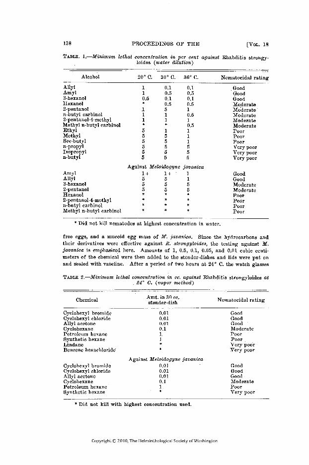
free eggs, and a mucoid egg mass of M . javanica. Since the hydrocarbons andtheir derivatives were effective against R. strongyloides, the testing against M .javanica is emphasized here . Amounts of 1, 0 .5, 0.1, 0.05, and 0 .01 cubic centi-meters of the chemical were then added to the stender-dishes and lids were put onand sealed with vaseline. After a period of two hours at 24 ° C. the watch glasses
TABLE 2.Minimum lethal concentration in cc. against Rhabditis strongyloides at24' C. (vapor method)
Chemical Amt. in 30 cc .stender-dish Nematocidal rating
Cyclohexyl bromideCyclohexyl chlorideAllyl acetoneCyclohexanePetroleum hexaneSynthetic hexaneLindaneBenzene hexachloride
Cyclohexyl bromideCyclohexyl chlorideAllyl acetone-CyclohexanePetroleum hexaneSynthetic hexane
0 .010 .010 .010 .11 .1**
Against Meloidogyne javanica0 .010 .010 .010 .11
GoodGoodGoodModeratePoorPoorVery poorVery poor
GoodGoodGoodModeratePoorVery poor
* Did not kill with highest concentration used .
TABLE . 1.-Minimum lethal concentration in per cent against Rhabditis strongy-loides (water dilution)
Alcohol 200 C.
300 C. 36° C . Nematocidal rating
AllylAmyl3-hexanolHexanol2-pentanoln-butyl carbinol2-pentanol-4-methylMethyl n-butyl carbinolEthylMethylSec-butyln-propylIsopropyln-butyl
AmylAllyl3-hexanol2-pentanolHexanol2-pentanol-4-methyln-butyl carbinolMethyl n-butyl carbinol
110.5*.111*555555
0 .10.50.10 .5511*155555
0 .1_0 .50 .10 .510 .510.5111555
GoodGoodGoodModerateModerateModerateModerateModeratePoorPoorPoorVery poorVery poorVery poor
GoodGoodModerateModeratePoorPoorPoorPoor
Against Meloidogyne javanica1 +555****
1 +555
***
1155
**
* Did not kill nematodes at highest concentration in water_

were removed and examined under a binocular dissecting scope and the number oflarvae, dead and living, was recorded . The eggs were then mounted and examinedunder oil immersion lens for the effect of the chemicals . Allyl acetone, a com-pound which is neither an alcohol nor a hydrocarbon, was also tested in the samemanner as the hydrocarbons . Results of this test are shown in Table 2 .
A beeswax solvency test was performed on each of the chemicals used in thisexperiment . One hundred milligrams of beeswax was placed in each test tube andone cubic centimeter of the chemical was added every 24 hours until the beeswaxwas either dissolved or a total of twelve cubic centimeters of the chemical hadbeen added . It was found that the chemicals which gave the best results as pos-sible nematocides either dissolved or emulsified beeswax at 24* C . in proportionsof 1 to 6 to 1 to 10 .
Of the alcohols tested, see-butyl, allyl, and amyl almost completely dissolvedthe beeswax ; hexanol, 3-hexanol, and 2-pentanol produced a slight change ; andethyl, methyl, isopropyl, n-propyl, n-butyl carbinol, 2-pentanol-4-methyl, and methyln-butyl carbinol gave no change . Twelve cubic centimeters of the above alcoholswas used in each test . All the hydrocarbons dissolved the beeswax and gave acloudy emulsion . Only six cubic centimeters of each of the chemicals was re-quired . Twelve cubic centimeters of allyl acetone produced only a slight changein the beeswax.2
SUMMARY
Among the alcohols tested as possible nematocides, amyl and allyl were themost toxic, followed by 3-hexanol and 2-pentanol. The following alcohols wereleast toxic : methyl, butyl, n-propyl, sec-propyl, isopropyl, hexanol, ethyl, 2-pentanol-4-methyl, normal butyl carbinol, and methyl n-butyl carbinol .
Among the hydrocarbons tested, the best results were obtained with cyclo-hexane, cyclohexyl chloride, and cyclohexyl bromide . The least toxic were : petro-leum, hexane, synthetic hexane, lindane, and benzene hexachloride . Allyl acetonetested as favorably as the best hydrocarbons and their derivatives .
From this experiment it can be seen that the chemicals which dissolved beeswaxalso tested favorably as nematocides, with the exception of allyl acetone which didnot dissolve beeswax .
LITERATURE CITED
BESSEY, E. A. 1911 . Root-knot and its control. U. S. Dept. Agric. Bur. PlantInd. Bull. No . 217 : 1-89 .
CARTER, W. 1943 . A promising new soil amendment and disinfectant. Science 97(2521) : 383-384 .
CHITWOOD, B. G . 1938a. Further studies on nemic skeletoids and their signifi-cance in the chemical control of nemic pests . Proc. Helm . Soc . Wash . 5 (2)68-75 .
1941 . Soil treatments with volatile liquids for control of nema-todes. Phytopath. 31 : 818-824 .
and CHITWOOD, M. B . 1939. Treatment of soil for the control of thebulb or stem nematode. Third Internat. Cong. Microbiol. N. Y . 169-170 .
and. 1950. An Introduction to Nematology, SectionI-Anatomy. Monumental Printing Co ., Baltimore, Maryland . 186-187 .
CHITWOOD, M. D . 1951. Notes on the physiology of Meloidogyne javanica (Nema-toda : Heteroderidae) . J. Parasitol . 37 (1) : 96-98 .
CHRISTIE, J. R. 1945 . Some preliminary tests to determine the efficacy of certainsubstances when used as soil fumigants against the root-knot nematode,Heterodera marioni (Cornu) Goodey . Proc. Helm . Soc . Wash. 12 (2) : 14-19 .2 Detailed tables of all the experiments may be found in the original thesis
in the library of The Catholic University of America, Washington, D . C .

ELLIS, D . E., CLAYTON, C . N. and OWENS, R. G . 1949 . Effects of soil treatmentswith uramon and certain fumigants upon plant growth and incidence of root-knot. Phytopath. 39 : 590-597 .
HOSHINO, H. M. and GODFREY, G. H . 1933. Thermal death point of Heteroderaradicicola in relation to time. Phytopath. 23 : 260-270 .
JOHNSON, M. O. and GODFREY, G. H . 1932 . Chloropicrin for nematode control.Indust. and Eng. Chem . 24 : 311-313 .
MATHEWS, D . J . 1919. Report on the work of the W. B. Randall research as-sistant . Expt. and Res . Sta., Chestnut, Herts, Ann. Rpt . 5 : 18-21 .
MCCLELLAN, W. D ., CHRISTIE, J . R. and HORN, N . L . 1949 . Efficacy of soil fumi-gants as affected by soil temperature and moisture . Phytopath . 39 (4)272-283 .
TAYLOR, A . L. and McBETH, C. W. 1940 . Preliminary tests of methyl bromideas a nematocide . Proc. Helm . Soc. Wash . 7 (2) : 94-96 .
THORNE, G . 1939 . Some factors governing the success of chemical treatment ofsoil for nematode control. Proc. Helm . Soc. Wash. 6 (2) : 60-62 .
TIMM, R. W. 1950 . The chemical composition of the .vitelline membrane of As-caris lumbricoides v. suis . Science 112 (2920) : 167-168 .
ZAWADOWSKY, M. M. 1928 . The nature of the egg shell of various species ofAscaris eggs. Trans. Lab. Expt. Biol. Zoopark Moscow 4 : 201-206.
MinutesTwo Hundred Ninety-third to Three Hundredth Meetings
The 293rd meeting was held at the Lee House, Washington, D . C. on October11, 1950, and was a dinner in celebration of the 40th Anniversary of the Society .Dr. F. G. Brooks, a corresponding member, was transferred to non-resident mem-bership . The following were elected to membership : Mr . B. B. Babero, Mr . V. G .Perry, Mr. E . L. Schiller, Mr . W. A. Uricchio, Lt . T . A. Jackowski, Jr ., Miss R. A .Buckner, Dr. S . Goto, Dr. P. V. Gustavson, and Dr. J . F. Kent. Dr. Foster actedas toastmaster for the evening, introducing the following speakers : Dr. PaulBartsch, Dr . W. W. Cort, Dr. E. B. Cram, Dr. G. Steiner and Dr . B. Schwartz .
The 294th meeting was held at the Zoological Division Laboratory, Bureau ofAnimal Industry, Beltsville, Maryland, on November 22, 1950 . The followingwere elected to membership : Dr. M. C. Meyer, Miss Ruth H . Bancroft, Dr .Benjamin F. Lownsbery, Mrs. Joyce W. Lownsbery, and 'Dr . C . A. Loos. Paperswere presented by Dr . R. Rausch, Dr. D. A. Shorb, Mr . J . T. Lucker, Mr . J. R.Elsea, Dr. G. Dikmans, and Dr. K. C. gates .
The 295th meeting was held at McMahon Hall, Catholic University of Amer-ica, on December 13, 1950 . The officers elected to serve during the year 1950were : Miss Marion M . Farr, President; Dr. Leon Jacobs, Vice President ; Mr .A. L. Taylor, Recording Secretary ; Miss Edna M. Buhrer, Cor .-Secretary andTreasurer . Papers were presented by Dr. C. G. Durbin, Dr. Thomas Burch, Dr .B. F. Lownsbery, Mr. A . C. Tarjan and Dr. E. G. Reinhard.
The 296th meeting was held at McMahon Hall, Catholic University of Amer-ica, Washington, D. C. on January 18, 1951. The following new members wereelected : Dr. George Dubois, Mr . Henry F. Mengoli, Mr. John V . Owens, Dr . W. D .Valleau and Dr. Richard A . Chapman. Dr. L. A. Spindler was elected to representthe Society as a vice-president of the Washington Academy of Science, succeedingDr. Price . The society voted to commemorate the deaths of the following mem-bers : Dr. H. E. Ewing, Dr. Banner Bill Morgan and Dr. A. B. Hardcastle .Papers were presented by Father R. W. Timm, Mr . William A. Uricchio, MissRita Buckner, Mr . J . V. Owens, and Mr. Donald Valentine .
. The 297th meeting was held at McMahon Hall, Catholic University of Amer-ica, Washington, D . C., on February 21, 1951 . Mr. James W. Ingalls, Jr., and

Dr. Dewey J . Raski were elected to membership . Papers were presented by Dr .F. D. Enzie, Dr . L . R . Sinclair, Mr. Ralph Barr, and Dr . C. G. Durbin. Shortnotes were presented by Dr . B. G. Chitwood and Dr . E . E. Wehr .
The 298th meeting was held at the Auditorium of the National Institutes ofHealth, Bethesda, Maryland, on March 21, 1951 . On the recommendation of theExecutive Committee, it was voted that : 1 . Annual dues are not be increased .2 . Subscription to future members of the proceedings, starting with volume 19 in1952 is to be $3.00 for subscribers residing in the United States and $3 .25 forsubscribers residing in foreign countries. 3. Prices for back numbers of theproceedings to non-members are to be $3 .00 domestic and $3 .50 foreign for volumes1, 2, 3 and 4, and $1 .75 domestic and $2.00 foreign for volumes 5 to 18, inclusive .Papers were presented by Dr. P. P. Weinstein, Dr. J. R . M. Innes, Drs . Josephson,Greenberg and Taylor, and Mr . Goldman.
The 299th meeting of the Helminthological Society of Washington was heldat the Johns Hopkins University School of Hygiene and Public Health, Baltimore,Maryland on April 20, 1951 . Dr. William F. Mai, Mr . Harry Herlich and Mr.David T. Clark were elected to membership . An invitation from Dr. Paul Bartschto hold a meeting at his home was accepted by the Society . Dr. W. W. Cortintroduced the following speakers : Mr. Eli Chernin, Mr . A. R. Barr, Mr. R. C .Wallis, Mr . R . E. Thorson, Mr. R. H. Foote and Dr. G . F. Otto .
The 300th meeting was a picnic held at "Lebanon", the home of Dr . andMrs. Paul Bartsch near Lorton, Va ., on June 16, 1951. Mr. A. S. Evans, Dr. BertLear, Mr. Julius Feldmesser and Dr . Alfred Edward A . Hudson were elected tomembership. Dr. N. R . Stoll, formerly a corresponding member, was transferredto nonresident membership .
A. L . TAYLORRecording Secretary
Report of the Brayton H. Ransom Memorial Trust FundFUNDS ON HAND, Jan . 1, 1950 $1696.17RECEIPTS : Interest rec'd in 1950 59.23DISBURSEMENTS : Expenses and grant to Helminthological Society of
Washington 31.00BALANCE ON HAND, Dec. 31, 1950 1724.40
ELOISEB.CRAM,Secretary-Treasurer
HENRY ELLSWORTH EWING1883-1951
Dr. Henry Ellsworth Ewing, retired entomologist and life member of the Hel-minthological Society of Washington, died January 5, 1951 .
Dr. Ewing was born in Arcola, Illinois, February 11, 1883 . He received hisA.B. degree from the University of Illinois in 1906 and his M .A. from the sameinstitution in 1908. The Ph .D. degree was conferred upon him in 1911 by CornellUniversity.
From 1911 to 1914, Dr . Ewing was assistant Entomologist for the OregonAgricultural Experiment Station ; from 1914 to 1919, he taught Entomology at theIowa State College ; and from 1919 until his retirement in 1945, he was a spe-cialist on arachnids and lice in what is now the Bureau of Entomology and PlantQuarantine of the U . S. Department of Agriculture .

Dr. Ewing was a member of many scientific societies, including the AmericanAssociation for the Advancement of Science ; Entomological Society of America ;Association of Economic Entomologists ; American Society of Mammalogists ; Amer-ican Society of Parasitologists (Pres . 1944) ; American Society of Ichthyologistsand Herpetologists ; Biological Society of Washington ; Washington Academy ofSciences ; Entomological Society of Washington ; and others. He was elected tomembership in the Helminthological Society of Washington, October 18, 1924,was its President in 1931, served as a member of the Editorial Committee of itsProceedings from 1934 to 1946, and was elected to life membership November 21,1945 . From the time of his election to membership until poor health prevented,Dr. Ewing rarely missed a meeting of the Society and took an active interest inits activities .
Dr. Ewing was a tireless worker and made many important contributions tothe systematics of lice and parasitic arachnids . His 'publications in the field ofparasitology consisted of one book and about 90 papers and notes .
In his death parasitology and entomology in general have lost a distinguishedscientist and his fellow workers a friend . The Helminthological Society extendsto his family its sincerest expression of sympathy .
E. W. PRICE

MEMBERS OF THE HELMINTHOLOGICAL SOCIETYOF WASHINGTON
The following membership list, arranged geographically, includes life, resideand nonresident members, as defined in Art . 3 of the Constitution (Vol. 13, Noof the Proceedings) .
Alabama
Kentucky
MontanaChamberlain, R . W.
Chapman, R. A.
Jellison, W. L.Porter, D. A .
Valleau, W . D .Nebraska
Alaska Louisiana Hanson, M. L.Babero, B. B . Mayhew, R. L. Manter, H . W .Bausch, R .
Maine
Prince, M. R.Meyer, M . C .
New JerseyCalifornia
Haenseler, C. M .Allen, M. W.
Maryland
Newman, D.Dougherty, E . C.
Bozicevich, J.McBeth, C. W .
von Brand, T.
New MexicoYoungson, C. R .
Buhrer, E. M.
Allen, R. W.Chitwood, M. B .Cobb, G. S.Colorado
OhioOlsen, . O . W.
Colglazier, M . L.
Harwood, P. D .Cort, W. W .Cram, E. B
OklahomaDelaware
Culbertson, J. T .
Marchant, E . H.Jaquette, D . S .
DeWitt, W. B .Lincicome, D. R .
Dikmans, G.
OregonSpaeth, F. W .
Doss, M. A.Durbin, C. G
Jensen,H . J ..District of Columbia
Enzie, F . D.
PennsylvaniaAbbott, R . T .
Farr, M. M .
Martin, H. M.Ballantyne, D . L .
Foster, A. O.
Martin, h N. A.Ballard, E. L .
Goldberg, A.Bauman, P . M .
Habermann, R . T .
TennesseeBuckner, R . A.
Hardcastle, F. B .
Riser, N. W.Chaffee, E . F .
Herman, C. M.Chitwood, B . G.
Hill, C. H .
TexasCole, B . A .
Howard, H. K.
Turk, R. D.Connolly, E . H.
Huff, C . G.Elsea, J. R .
Humphrey, J. M.Fife, E .H
Jacobs, L.
UtahFrick, L . P . Jones . M. F.
Fielding, M. J.Hodges, E . P .
Kates, K. C.
Thorne, G .Jahnes, W. G.
Lotze, J . C.Kent, J. F.
Lownsbery, B . F.
VirginiaMengoli, H. F.
Lownsbery, J . W.
Bartsch, P .Mollari, M .
Lucker, J. T.Reinhard, E . G .
Luttermoser, G. W.
WashingtonSchwartz, B .
McIntosh, A .
Courtney, W. D.Sonnenberg, B .
Morris, J . A.Timm, R. W.
Olivier, L. J.Traub, R .
Oteifa, B. A .Otto, G. F.
CeylonPhillips, B. P.
Loos, C. A.Florida
Price, E. W.Christie, J. R.
Reardon, L.
EgyptKincaid . R. R.
Rees, C. W .
Kuntz, R. E.Perry, V . G.
Roe. G. C.Swanson, L. E.
Sarles, M. P.
EnglandShorb, D. A.
Clapham, P. A.Georgia
Sinclair, L. R.Andrews, J. S .
Spindler, L. A.
IndiaDenton, J. F.
Steiner, G.
Basir, M. A.Machmer, J . H.
Stirewalt, M . A.
Chauhan, B. S .Illinois
Taylor, A. L.
JapanElishewitz, H .
Tobie, J. E .
Hunter, G. W.Harrington, R . F.
Turner, J. .Linford, M . B.
Weans E. E .Wenstein, P. P .
MexicoThomas, L. J.
Wright, W. H.
Caballero, E.Tiner, J . D .Van Cleave, H. J.
Michigan
SwitzerlandWalton, A. C.
DeGiusti, D.
Baer, J. G .
Iowa
Minnesota
VenezuelaBrooks, F. G .
Goto, S .
Vogelsang, E . G.

CONTENTSPAGE
ALLEN, REX W. AND PATRICIA' M. KYLES. Tests with a Phenothiazine-SaltMixture as a Growth Stimulant for Sheep 102
BABERO, BERT B . Notes on the Trematode Genus Glypthelmins Stafford, 1905 103
CHRIsTIE, J . R . AND V. G. PERRY. Removing Nematodes from Soil106
DURBIN, CHARLES G. Attempts to Transfer Plasmodium berghei Vincke andLips to Domesticated Animals:108
FIELDING, MAX J. Observations on the Length of Dormancy in Certain PlantInfecting Nematodes 110. .. . . . .. . .. .. . . . .. .. . .. ... .. .. . .. .. .. . .. . . . .. ... . . .. .. . .... . .. .. . .. .. . .. .. .. . .
HAGEMEYER, JOYCE W. A New Stage in the Life Cycle of the Golden Nema-tode Heterodera rostochiensis Wollenweber 112
HILL, C. H. The Recovery of Encapsulated, Infective Larvae of Trichinellaspiralis Relatively Free of Muscle Tissue 114
KATES, K. C. AND A. GOLDBERG. The Pathogenicity of the Common SheepTapeworm, Moniezia expansa 87
OLSEN, WILFORD, O. AND C. D. TOLMAN . WellcOmia evaginata (South, 1908)Oxyuridae: Nematode) of Porcupines in Mule Deer, Odocoileus hemionus,in Colorado 120
PRICE, E. W. Henry Ellsworth Ewing, 1883-1951 141
SCHILLER, EVERETT L. Studies on the Helminth Fauna of Alaska. VIII.Some Cestode Parasites of the Pacific Kittiwake (Rissa tridactyla Ridg-way) with the Description of Haploparaxis rissae n . sp122
TIMM, RICHARD W. A Note on the Cell Inclusions of Syringolaimus smarigdusCobb, 1928 125
TINER, JACK D. The Morphology of Ascaris laevis Leidy 1856, and Noteson Ascarids in Rodents 126
TURNER, JAMES H . Counting Nematodirus spathiger Eggs in Sheep Dung 132
URICCHIO, WILLIAM A . Some Alcohols and Hydrocarbons as Nematocides 136
Minutes 140. .. . . . .. .. ... .. .. . .. .. . .. .. . . . .. .