The great influenza pandemic of 1918 - … Vol24 No2 2 The influenza virus Influenza is caused by an...
8
Also in this issue (page 5): Microbes on building stone – for good or ill? Eric May Calcinogenic bacteria in laboratory culture, showing calcite crystals developing within colonies. Volume 24 No 2 September 2003 ISSN 0965-0989 Visit our website at http://www.oxoid.com ™ Introduction Severe acute respiratory syndrome (SARS) is currently causing alarm as speculation mounts that it could be the next global pandemic. Great pandemic diseases have swept the world ever since mankind ceased to live in small communities and began to breed domestic animals in close proximity to homes. Viruses in particular, crossing from one species to another, mutate to invade new hosts and can develop lethal characteristics. Man, pig and fowl form a dangerous domestic combination for the RNA virus of the orthomyxovirus group (Influenza virus). SARS is not of this group, it is possibly a coronavirus (Common cold virus) that has mutated and can now cause a 10- 15% death rate. Compared with influenza virus, SARS has low infectivity and low mortality. Influenza currently kills 4,000 people a year in the UK. In 1918 the mortality rate for influenza reached 60% in some communities and it caused more deaths than the whole of the 1914-18 war. SARS has demonstrated that rapid air travel can spread locally-constrained diseases across the world. To get the SARS outbreak into context it is necessary to re-examine the great influenza pandemic of 1918 when influenza swept across the globe and in less than a year it left some 40 million dead in its wake. The influenza virus was finally isolated and identified in 1930. The origin of pandemics Porter 1 considered that the era of human epidemics began as civilisation developed. Around 3000BC cities were arising in the Middle East with populations of scores of thousands. Such settlements required huge animal resources for food and animal byproducts eg., fur, leather. These large congregations of animals transferred lethal contagious pathogens (Zoonotic infections eg., smallpox, diphtheria, influenza, chickenpox, mumps and other devastating illnesses) which rapidly spread in the local population. Degrees of immunity developed in afflicted human populations but the increasing traffic of people between large settlements and the outreaches of explorers into new territories, continuously supplied new strains of infection and spread infections into new populations. Some epidemic diseases eventually became endemic, continuously infecting the young, culling the old and more susceptible individuals. Tropical endemic disease, which required intermediate hosts that could not survive in more temperate climates eg., malaria, yellow fever, remained in warmer latitudes. Historical descriptions, based on symptoms, indicate that influenza epidemics have probably been around human populations since before 5000BC. It was in the 20th century, however, when large population migrations both before and during the 1914–18 War, that helped create the most mobile lethal pandemic the world has ever witnessed, as it circumnavigated the globe in 1918–19. The country that suffered the greatest number of deaths was India where it is suggested that 16 million people died. Where did this pandemic originate? Although influenza epidemics have been recorded for centuries, the spread of this disease was never charted. The normally unremarkable death rate of influenza was a pale shadow of Plague (Yersinia pestis) where the Justinian outbreak in the 2nd century AD can be tracked in written records from Asia across Europe. Plague reached Europe in the 14th century, killing 25 million people. It remained for the next three centuries, waxing and waning in local epidemics across Europe. The pandemic may have arisen in troops at the Western Front and have been carried back to the USA in troopships arriving in 1918. The USA seems to have reported the first fatal cases from army barracks in Denver and South Carolina. It quickly spread into surrounding communities and a main wave of infection peaked in September-November 1918, killing over 10,000 people per week in US cities. Large proportions of the US population became ill (28%) with mortality rates >2.5% compared with <0.1% in previous influenza epidemics. An estimated total of 675,000 persons died in the USA from the 1918–19 pandemic. Another significant factor that differentiated this pandemic was that most deaths occurred among young adults. Influenza and pneumonia death rates for 15–34 year old persons were more than 20 times higher in 1918 than in previous years. The great influenza pandemic of 1918 Dr Eric Bridson MPhil, PhD, FIBMS, FIBiol
Transcript of The great influenza pandemic of 1918 - … Vol24 No2 2 The influenza virus Influenza is caused by an...

Also in this issue (page 5):
Microbes onbuilding stone –for good or ill?
Eric May
Calcinogenic bacteria in laboratory culture, showing calcitecrystals developing within colonies.
Volume 24 No 2 September 2003 ISSN 0965-0989
Visit our website at http://www.oxoid.com
™
IntroductionSevere acute respiratory syndrome (SARS) is currently causingalarm as speculation mounts that it could be the next globalpandemic. Great pandemic diseases have swept the world eversince mankind ceased to live in small communities and beganto breed domestic animals in close proximity to homes. Virusesin particular, crossing from one species to another, mutate toinvade new hosts and can develop lethal characteristics. Man,pig and fowl form a dangerous domestic combination for theRNA virus of the orthomyxovirus group (Influenza virus).
SARS is not of this group, it is possibly a coronavirus(Common cold virus) that has mutated and can now cause a 10-15% death rate. Compared with influenza virus, SARS has lowinfectivity and low mortality. Influenza currently kills 4,000people a year in the UK. In 1918 the mortality rate for influenzareached 60% in some communities and it caused more deathsthan the whole of the 1914-18 war. SARS has demonstratedthat rapid air travel can spread locally-constrained diseasesacross the world. To get the SARS outbreak into context it isnecessary to re-examine the great influenza pandemic of 1918when influenza swept across the globe and in less than a year itleft some 40 million dead in its wake. The influenza virus wasfinally isolated and identified in 1930.
The origin of pandemicsPorter1 considered that the era of human epidemics began ascivilisation developed. Around 3000BC cities were arising inthe Middle East with populations of scores of thousands. Suchsettlements required huge animal resources for food and animalbyproducts eg., fur, leather. These large congregations ofanimals transferred lethal contagious pathogens (Zoonoticinfections eg., smallpox, diphtheria, influenza, chickenpox,mumps and other devastating illnesses) which rapidly spread inthe local population. Degrees of immunity developed inafflicted human populations but the increasing traffic of peoplebetween large settlements and the outreaches of explorers intonew territories, continuously supplied new strains of infectionand spread infections into new populations. Some epidemicdiseases eventually became endemic, continuously infectingthe young, culling the old and more susceptible individuals.Tropical endemic disease, which required intermediate hoststhat could not survive in more temperate climates eg., malaria,yellow fever, remained in warmer latitudes.
Historical descriptions, based on symptoms, indicate thatinfluenza epidemics have probably been around human
populations since before 5000BC. It was in the 20th century,however, when large population migrations both before andduring the 1914–18 War, that helped create the most mobilelethal pandemic the world has ever witnessed, as itcircumnavigated the globe in 1918–19. The country thatsuffered the greatest number of deaths was India where it issuggested that 16 million people died.
Where did this pandemic originate?Although influenza epidemics have been recorded forcenturies, the spread of this disease was never charted. Thenormally unremarkable death rate of influenza was a paleshadow of Plague (Yersinia pestis) where the Justinian outbreakin the 2nd century AD can be tracked in written records fromAsia across Europe. Plague reached Europe in the 14th century,killing 25 million people. It remained for the next threecenturies, waxing and waning in local epidemics across Europe.
The pandemic may have arisen in troops at the WesternFront and have been carried back to the USA in troopshipsarriving in 1918. The USA seems to have reported the first fatalcases from army barracks in Denver and South Carolina. Itquickly spread into surrounding communities and a main waveof infection peaked in September-November 1918, killing over10,000 people per week in US cities. Large proportions of theUS population became ill (28%) with mortality rates >2.5%compared with <0.1% in previous influenza epidemics. Anestimated total of 675,000 persons died in the USA from the1918–19 pandemic. Another significant factor thatdifferentiated this pandemic was that most deaths occurredamong young adults. Influenza and pneumonia death rates for15–34 year old persons were more than 20 times higher in 1918than in previous years.
The great influenza pandemic of 1918
Dr Eric Bridson MPhil, PhD, FIBMS, FIBiol

Culture Vol24 No2
2
The influenza virusInfluenza is caused by an RNA virus of the orthomyxovirus group.Human influenza is transmitted from person to person through theair, primarily in droplets expelled during coughing and sneezing.The virus infects the mucous membranes of the upper respiratorytract and more occasionally invades the lungs. Recovery is usuallyspontaneous and rapid. The majority of the serious consequencesof influenza infection are not caused by the virus but by secondaryinvasion of pathogenic bacteria. Thus bacterial pneumonia isespecially prevalent in infants and the elderly.
Once a strain of influenza virus has passed through apopulation, the surviving majority are sufficiently immune tothat strain to give ‘herd’ immunity protection to the non-immune minority. It is then unlikely that a strain of virus ofsimilar antigenic type can cause another epidemic for 2–3years. Antigenic ‘drift’ is responsible for recurrent epidemics ofinfluenza to erupt in 2–3 year cycles.
Influenza virus was isolated from a human victim for thefirst time in 1933. The virus has two main types A and B, thereis a type C but it does not cause serious disease. Type B virusshows less variation and infects humans only. It causes regionalepidemics rather than pandemics. Type A is the more importantform, causing pandemics and infecting pigs, horses, seals,whales and birds, although not all variant forms infect allspecies (only four variants have been found in humans).
The simplified diagram of the influenza viron (Figure 1)shows the viral RNA is present in a number of separate pieces(segments). This segmentation is an important cause ofantigenic ‘shift’ because the host cell may be infected by twogenetically distinct strains and the RNA segments from bothstrains may re-associate to create a new strain. The new strainmay then be able to infect hosts that were immune to the twooriginal strains. Figure 1 also shows protein ‘spikes’ on theoutside of the viron envelope; haemagglutinin (H) andneuraminidase (N). The function of these proteins is to adhereto the host nucleated cell and then facilitate the transfer of theviron into the cell. Haemagglutination of erythrocytes is aconvenient measure of viral activity but the influenza virus haslittle interest in these non-nucleated cells. Immune antibodies,produced by infection or vaccination, are directed against theseproteins and neutralise the virus by preventing ingress into cellswith subsequent replication. It is considered to be mostprobable that these H and N proteins on the viron envelope areclosely associated with infectivity and virulence. Type A virushas 15 H and 9 N variants giving rise to many differentsubtypes eg., H1N1, H1N2, H2N2 etc. Analyses of antibodiescarried by survivors of the 1918 pandemic suggest that thestrain was a classic swine H1N1 subtype influenza A virus.
During the pandemic simultaneous outbreaks of influenza werereported in humans and pigs from around the world. Whetherthe virus passed from humans to pigs or vice versa has not beenresolved. The natural reservoir for influenza virus is thought tobe wild water fowl. Periodically, genetic material from avianstrains is detected in human infections. Pigs can be infectedwith both avian and human forms of influenza virus and the pigcould be an intermediate host.
Evidence so far suggests the H gene sequence of the 1918strain is associated with strains that infect pigs and humans butnot with strains that infect fowls. Little is known about thegenetic features of influenza virulence. Virulent strains show anability to infect various organs in the host, particularly spreadingthroughout the lungs to cause extensive, normally lethal damage.
A possible lead is that in some avian strains, a cleavage sitemutation in the H protein can cause systemic disease in birds,instead of the normally benign infection of the gastro-intestinaltracts of host birds. These mutant strains are associated withexceptionally high mortality among infected birds. So far, thereis no indication that the 1918 strain carried this mutation (oneamino-acid change in the H protein) which has only beenobserved in H5 or H7 subtype viruses associated with infectionin domestic poultry.
The search for evidence of the 1918 strainAs previously stated, in 1918 the influenza virus was unknownand virus study methodology was not available. Much later,opportunities were taken to get hold of tissue from victims ofthe pandemic buried in permafrost in the Arctic. Tissues couldbe expected to be preserved in these constant low temperaturesfor hundreds or possibly thousands of years. [A frozenmammoth body is currently being exhumed in Siberia that isalleged to be 10,000 years old.] Originally it was hoped that theinfluenza virus could be recovered but this proved impossible.
In 1951 a Swedish-born American pathologist, John Hultinfrom the University of Iowa, led a small party to exhume thebodies of victims who had died from the 1918 pandemic. Thesebodies had been buried in permafrost at Teller Mission, an Inuitfishing village on the Seward Peninsula of Alaska. This villagesuffered an extremely high mortality rate from influenza inNovember 1918. The disease spread through the village in fivedays and killed 72 people, about 85% of the adult population.No influenza virus could be recovered from the tissues.Molecular genetic analyses of the samples were impossible in1951, the structure of DNA (the beginning of moleculargenetics) was not determined until 1953.
In 1997, Hultin returned to Teller Mission and obtainedfrozen lung tissue from four more influenza victims. Thesewere placed in formaldehyde for histology and viral RNAisolation. One piece of lung tissue showed evidence of massivepulmonary haemorrhage, typical of virulent influenza and italso contained RNA fragments of the virus.
Meanwhile in 1995, Jeffrey Taubenberger, Head of a Divisionof Molecular Pathology, Armed Forces Institute of Pathology(AFIP) Washington DC, turned to archival formalin-fixed,paraffin-embedded autopsy tissues of 1918 influenza victimsstored in the AFIP. In total 78 autopsy cases from the pandemicwere examined. The majority of the victims had died fromsecondary bacterial pneumonia. Asmall subset of these cases diedwithin a week, with massive pulmonary oedema or haemorrhage.Death with these symptoms could occur in 48 hours.
Such rapid deaths also occurred in later epidemics of influenzabut they were a cardinal feature of the pandemic in 1918–19. Thefirst positive case was found in 1996 with lung sections showingacute focal bronchiolitis and alveolitis that are consistent with
Figure 1. A schematic influenza viron showing RNA segments and H and Nproteins on the envelope.
Neuraminidase
Haemagglutinin
Envelope
RNA genome (eight fragments)

primary influenzal viral pneumonia. Influenza RNA wasrecovered from the tissue blocks but it was fragmented into piecesnot longer than 150 bases in length. The eight RNA fragments inliving influenza virus vary from 900 to 2300 bases in length. Asecond case was identified in 1997 showing the same lungcharacteristics and RNA fragments no longer than 150 bases.
In 1998, John Oxford, professor of virology at the RoyalLondon Hospital led an expedition to Spitzbergen in Norway toexhume the frozen bodies of six young coal miners who haddied in the 1918 influenza pandemic. Tissues from variousorgans were removed, preserved and brought back to England.After months of testing at the National Institute of Medicalresearch, Mill Hill, fragmentary genetic traces of viral RNAwere found in lungs and other tissues.
This work continues in several centres but Taubenberger hastwo major goals for his investigation:
1. To determine the origin of the 1918 influenza virus ie., bird, pigor man and to discover how it can be transferred to humans.
2. To learn whether specific features of its nucleotide sequencecan explain the virulence of this or any other strains.
This second goal was repeated by Professor John Oxford in a“Times” newspaper article on November 17, 1999. He told thereporter that once ‘factor X’ was identified, it will be possibleto know what made this organism the most lethal virus everknown. It should be possible to detect the same gene sequencein recurring influenza epidemics and hopefully get an effectivevaccine to prevent influenza returning as the Grim Reaper.
Contrast John Oxford’s enthusiasm with Taubenberger’smore cautious opinion to a reporter in the ‘ASM News’ 65:7.1999. p.475. Taubenberger thought it unlikely that a ‘smokinggun’ specific genetic structure in a lethal virus will be found.Virulence may involve subtle changes in many genes. Hostimmune factors play a significant role in deciding the outcome
Culture Vol24 No2
3
of a serious infection. The unique massive movements of peoplein 1918–19 travelling or living in crowded conditions ontroopships, in barracks or in cattle trucks may have been acritical factor in the spread and virulence of the pandemic acrossthe world. An explanation is still required about the lethality ofthe 1918 strain for the 15–45 year age group (Figure 2).
Epidemiology of recent influenza epidemicsIt was not until the pandemic of Asian influenza in 1957 thatcareful analysis of its worldwide progress was carried out. Thisinfluenza outbreak first appeared in central China in lateFebruary 1957. By early April it had reached Hong Kong andAustralasia, spreading across to the USA. The USA epidemicreached a peak in October 1957. Meanwhile, it spread throughSouth America and across to Europe where it met another waveof this epidemic from the Middle east and Africa.
In 1968, a Hong Kong influenza pandemic occurred whichhad a similar transglobal spread. In 1979 an avian influenza Avirus entered the pig population in Northern Europe, forming astable viral lineage.
Until 1997, there was no evidence that wholly avianinfluenza virus could infect humans. This evidence wasprovided in Hong Kong in 1997 when a new strain of influenzakilled 4,500 chickens in Southern China. In Hong Kong 18people were infected with this avian virus and six rapidly died.Millions of domestic fowls were slaughtered by the Hong Kongauthorities to prevent the avian strain spreading, whilst the restof the world held its breath!
Humans have no pre-existing immunity to avian subtypesand the prompt, vigorous action in Hong Kong may havestopped a calamitous epidemic. This avian strain was latershown to be unique in that it leapt from birds to humans butfortunately it appeared not to spread human to human. No doubtgiven more time it would have overcome this problem.
Discussions are being held about whether to include aviansubtype strains in the human vaccine but this proposal is notwithout risk. Kolata2 described an unfortunate chain ofcircumstances that led to one of America’s worst public healthcatastrophes. In 1976, a USA soldier rapidly died from a swinetype influenza infection. The implications of this single deathheralding a major epidemic, grew from a remote possibilityexpressed in the army camps, up to projections of millions ofdeaths by the time it reached the White House. President GeraldFord was persuaded by his medical advisors that this could be arerun of the 1918 influenza pandemic. Ford ordered apopulation-wide vaccination programme with the slogan “Bettera vaccine without an epidemic than an epidemic without avaccine.” He could not have been more wrong. The epidemicnever materialised but the vaccine appeared to produce seriousand sometimes lethal side-effects. The resulting litigation costsand compensation claims amounted to 3.5 billion US$.
VaccinationWhilst new drug treatments for influenza are available or in thepipeline eg, ‘Relenza’ and ‘Tamiflu’, common sense suggeststhat prevention of the disease by vaccination is the mosteconomic and effective method of treatment.
Unfortunately, antigenic ‘drift’ by the virus means thatvaccination must be repeated every Autumn, using strains ofinfluenza that are considered by global reference laboratories tobe the greatest threat in the coming year. In 1999, the vaccinecontained the Beijing and Sydney strains which were a threat inBritain to unvaccinated people over Christmas. In 2000 a NewCaledonia strain of virus was included because it appeared tobe an antigenic ‘shift’ from previous strains. Selection of the
Figure 2. A. The rise in death rates of the 15-45 year age groups in the 1918-19influenza pandemic compared with previous influenza epidemics. B The sharpfall in US life expectancy in 1918-19 from the exceptional influenza death rate(Taubenberger 1999).
2500
2000
1500
1000
500
0<1 1-4 5-14 15-24 25-34 35-44 45-54 55-64 65-74 75-84 >85
Age divisions
Spec
ific
dea
th r
ate
US influenza and pneumonia deaths by age- - - 1911-1917 ––– 1918
A
70
62
54
46
38
3019001905 1910 1915 1920 1925 1939 1935 1949 1945 1950 19551960
Year
Lif
e ex
pect
ancy
(ye
ars)
US life expectancy 1900-1960B

correct strains is absolutely critical because it takes manymonths to prepare large stocks of vaccine and there is not timeto repeat the exercise.
The public health disaster in the USA in 1976 should not beused as an excuse for avoiding vaccination. What happened onthat occasion has not been fully explained. Attempts to rushproduction of adequate stocks of vaccine may have had a rolein the consequences. Equally, recipients of the vaccine with pre-existing egg-product hypersensitivity, may not have beenproperly screened out. It was a unique unfortunate calamitywhich current changes in vaccine production may prevent everhappening again.
Influenza vaccine productionInfluenza vaccines have been common in the USA since the1940s. Vaccines against any given influenza variant strain takeabout six months to produce, test for safety and distribute. Thisis no match for a fast moving pandemic. Antigenic ‘drift’ is thegradual revision of the amino-acid sequence in the surfaceproteins. This drift is a particular characteristic of Type Binfluenza which evolves gradually in the human host to avoidrecognition by the host immune system. Type A influenza canadditionally undergo a more drastic antigenic ‘shift’.
The change in the haemagglutinin or the neuraminidaseproteins may be so great that the virus can evade the antibodyrepertoire of all the people in the world and that causes apandemic. Such big changes do not occur through simple geneticmutation, it requires the mixing of two viral strains in one cell:
1918 pandemic H1N11957 pandemic H2N21968 pandemic H3N21997 nearly epidemic H5N1
The annual process of vaccine production begins each winter asinfluenza virus samples, collected by 110 surveillance sitesaround the world, are analysed. In February the WHO pinpointsthree strains – two Type A and one Type B that seem likely toaccount for most of the influenza that will occur in the comingseason (November to March in the Northern hemisphere).These three strains will comprise the new vaccine.
It would seem to be obvious that the next step must be togrow vast numbers of the selected strains, inactivate them sothat they cannot cause infection and then combine them into asingle vaccine. What seems to be obvious is not alwaysrealistic. The selected strains often grow slowly in thelaboratory. To overcome this problem, the immune-stimulatingproteins (haemagglutinin and neuraminidase – H and N) of theselected strains are inserted into an influenza strain that willgrow rapidly in laboratory conditions. After infecting chickembryos with both selected and fast growing strains, the fastgrowth strains with the selected H and N proteins are isolatedand delivered to the vaccine manufacturers. It is at this stagethat mass production of the vaccine strains commences.
Future developments of influenza vaccine includes:
1. Use vaccines composed solely of H and N proteins.
2. Use vaccines made from weakened live influenza viruses,which can stimulate T lymphocytes. T cells can cope with
Culture Vol24 No2
4
viral antigenic drift. Live viruses can be delivered as a nasalspray. WHO, however, has issued a warning about thefuture use of live-cell vaccines because of the increasingnumber of HIV/AIDS patients that will be put at risk.
3. Use vaccines composed of other viral hosts, bearingselected H and N proteins, which grow readily in culturedcells (influenza viruses grow poorly in tissue culture cells).The high yield of encoded influenza proteins can then bepurified for use in vaccines.
4. Use vaccines composed of DNA plasmids containing Hand N genes. When the plasmids are injectedsubcutaneously, nearby cells will take them up to produceH and N proteins. These ‘foreign’ proteins will be detectedby the host immune system which will then deployantibodies and T cells to neutralise free virus and eradicateinfected cells. Such ‘naked DNA’ influenza vaccines haveworked well in animals but have yet to be tested in humans.
ConclusionEighty years after the Influenza pandemic of 1918–19 we arestill no wiser about the cause of this most devastating killingdisease but we are better informed about the circumstances.
Influenza pandemics occur every 30 years or so and unlesssubstantial preventive measures are put into operation, therewill be more to come. Whilst correct antibiotic treatment willreduce deaths from secondary bacterial pneumonia, the bestprimary defence is vaccination.
More work needs to be done to determine how the influenzavirus cycles between the three major groups of hosts: birds,animals, humans and how often this occurs.
Influenza has been a well recognised disease for centuriesand if a previous outbreak of such lethal dimensions hadoccurred, it would surely be written in the medical history ofone or more countries. The English Sweate (1485 and fourmore visitations up to 1517) was well recorded, although thedeath rate was much smaller than the contemporary influenzaepidemics3.
There is a possibility that the 1918 influenza pandemic wasan entirely unique event that could never return. It would notbe wise, however, to take this possibility for granted.
References1. Porter R. “The Greatest Benefit to Mankind.” 1997; Harper Collins. London. pp.
22–25.2. Kolata G. “FLU: the story of the Great Influenza Pandemic of 1918 and the search
for the virus that caused it.” 1999; Macmillan. London.3. Bridson E. “The English ‘Sweate’ (Sudor Anglicus) and Hantavirus.” Med Sci
History. 1998; 14: 20–32.
Further readingTaubenberger JK. “Seeking the 1918 Spanish Influenza Virus.” 1999; ASM News.65: 473–478.Laver WG, Bischofberger N, Webster RG. “Disarming Flu Viruses.” 1999;Scientific American. january. pp.55-65.Collier R. “The Plague of the Spanish Lady.” 1974; Atheneum Press.Crosby AW. “America’s forgotten pandemic.” 1989; Cambridge University Press.Kilbourne ED. “Influenza.” 1987; Plenum Medical Book CompanyReid RH, Taubenberger JK. “The 1918 flu and other influenza epidemics ‘overthere’ and back again.” 1999; Lab Invest. 79: 95–101.
Correspondence: Dr Eric Bridson MPhil PhD FIBMS FIBiol, 3 Bellever Hill,Camberley, Surrey GU15 2HB UK; email: [email protected]
Culture provides an international publishing forum for papers on microbiology. Authoritative articles on microbiologicaltopics are welcomed. All submissions are peer reviewed by the Editorial Board. A synopsis of the proposed papershould be sent to the Managing Editor of Culture at Euromed Communications Ltd., The Old Surgery, Liphook Road,Haslemere, Surrey GU27 1NL, England. Tel: (0)1428 656665. Fax: (0)1428 656643. e-mail: [email protected]
5
Culture Vol24 No2
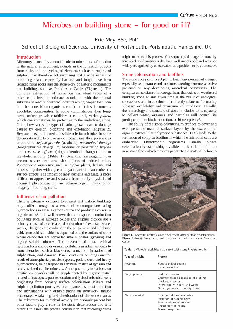
might make to this process. Consequently, damage to stone bymicrobial mechanisms is the least well understood and was notwidely recognised by conservators as a problem to be addressed3.
Stone colonisation and biofilmsThe stone ecosystem is subject to harsh environmental change,especially temperature and moisture, exerting extreme selectivepressure on any developing microbial community. Thecomplex consortium of micoorganisms that exists on weatheredbuilding stone at any given time is the result of ecologicalsuccessions and interactions that directly relate to fluctuatingsubstrate availability and environmental conditions. Initially,the mineralogy and structure of stone in relation to its capacityto collect water, organics and particles will control itspredisposition to biodeterioration, or bioreceptivity4.
The ability of the stone-colonizing microflora to cover andeven penetrate material surface layers by the excretion oforganic extracellular polymeric substances (EPS) leads to theformation of complex biofilms in which the microbial cells areembedded. Phototrophic organisms usually initiatecolonisation by establishing a visible, nutrient rich biofilm onnew stone from which they can penetrate the material below to
IntroductionMicroorganisms play a crucial role in mineral transformationin the natural environment, notably in the formation of soilsfrom rocks and the cycling of elements such as nitrogen andsulphur. It is therefore not surprising that a wide variety ofmicro-organisms, especially bacteria and fungi, have beenisolated from rocks and the stonework of historic monumentsand buildings such as Portchester Castle (Figure 1). Thecomplex interaction of numerous microbial types at amicroscopic level in intimate association with the mineralsubstrate is readily observed1 often reaching deeper than 3cminto the stone. Microorganisms can be on or inside stone, asendolithic communities. In some circumstances their long-term surface growth establishes a coloured, varied patina,which can sometimes be protective to the underlying stone.Often, however, some types of patina growth leads to damagecaused by erosion, biopitting and exfoliation (Figure 2).Research has highlighted a possible role for microbes in stonedeterioration due to one or more mechanisms: their presence asundesirable surface growths (aesthetic), mechanical damage(biogeophysical change) by biofilms or penetrating hyphaeand corrosive effects (biogeochemical change) due tometabolic activity (Table 1). Scientific investigation canpresent severe problems with objects of cultural value.Phototrophic organisms such as higher plants, lichens andmosses, together with algae and cyanobacteria, cause obvioussurface effects. The impact of most bacteria and fungi is moredifficult to appreciate and separate from purely physical andchemical phenomena that are acknowledged threats to theintegrity of building stone.
Influence of air pollution There is extensive evidence to suggest that historic buildingsmay suffer damage as a result of microorganisms usinghydrocarbons in air as a carbon source and producing corrosiveorganic acids2. It is well known that atmospheric combustionpollutants such as nitrogen oxides and sulphur dioxide are aprimary cause of accelerated deterioration of exposed stone-works, The gases are oxidized in the air to nitric and sulphuricacid, form acid rain which is deposited onto the surface of stonewhere carbonates are converted into sulphates (gypsum) andhighly soluble nitrates. The presence of dust, residualhydrocarbons and other organic pollutants in urban air leads tostone alterations such as black crust formation, nitratation, andsulphatation, and damage. Black crusts on buildings are theresult of atmospheric particles (spores, pollen, dust, and heavyhydrocarbons) being trapped in a mineral matrix of gypsum andre-crystallized calcite minerals. Atmospheric hydrocarbons onartistic stone-works will be supplemented by organic matterrelated to inadequate past restoration and lysis of microbial cellsoriginating from primary surface colonisation. Nitrate andsulphate pollution processes, accompanied by crust formationand incrustations with organic patina on stonework, induceaccelerated weakening and deterioration of the stone matrix.The substrates for microbial activity are certainly present butother factors play a role in the stone deterioration and it isdifficult to assess the precise contribution that microorganisms
Microbes on building stone – for good or ill?
Eric May BSc, PhDSchool of Biological Sciences, University of Portsmouth, Portsmouth, Hampshire, UK
Table 1. Microbial activities associated with stone biodeterioration
Type of activity Process
Aesthetic Surface colour changeSlime production
Biogeophysical Biofilm formationContraction and expansion of biofilmsBlockage of poresInteraction with salts and water Growth/movement through stone
Biogeochemical Excretion of inorganic acidsExcretion of organic acidsEnzyme attack of nutrientsChelation of mineralsMineral migration
Figure 1. Portchester Castle: a historic monument suffering stone biodeterioration.Figure 2 (inset). Stone decay and crusts on decorative arches at PortchesterCastle.

6
Culture Vol24 No2
seek protection from high light intensities or desiccation. StoneEPS trap aerosols, dust and nutrients, minerals, and organiccompound complexes and take up water from air and release itunder low RH conditions. Stone moisture and nutrients arethereby increased while porosity, water-uptake capacity andevaporation are reduced5.
Notably rich and homogeneous biofilms, composed mostlyof bacterial rods, are often observed on weathered stonesubstrates from sheltered areas (Figure 3). Microorganismsmay degrade stone mechanically, chemically and aestheticallythrough metabolic activities and biomineralisation processes inthese biofilms. The mechanical stress induced by shrinkingand swelling of the colloidal biogenic slimes inside stone poresmay damage stone and it may cause changes in the circulationof moisture to further enhance chemical dissolution andmineral loss from stone.
Interactions of microbes with stone saltsSalts acting on their own are very important decay agents andcan attack stones, mainly mechanically in pore spaces duringRH and temperature changes. Efflorescences present a nichefor halotolerant and halophilic bacterial populations which areosmotically well-adapted to an extreme existence, such asmembers of Archaea. Media containing high concentrations ofsodium chloride and magnesiumsulphate (up to 25%) may be appropriatefor studying efflorescences on stonemonuments6. It has also been shown thatmicroorganisms can enhance thephysical or chemical processes byinteracting with salts in stone7. Whenlimestone has been subjected to bothmicrobial and salt weathering, underdifferent temperature/wet/dry cyclingregimes, weight loss was higher withmicrobes alone (7.7%) than Na2SO4alone (4.9%) but the two agents togethermore than doubled the additive effectand caused extensive exfoliation andfissure formation (Figure 4). Thus, byinteracting with the effects of the salt,microbial biofilm growth can increasewater content and enhance physical, mechanical pressures onstone during wet/dry cycling.
Microorganisms associated with damageBiodeterioration of stone is rarely associated with one group ofmicroorganisms; weathering stone may support a balancedcommunity whose members co-evolve with time to enablerecycling of essential elements for activity and growth.Damage may thus be gradual through slow growth (biogenicdrift) or be sudden and harmful stimulated by a dramaticchange in environment, moisture or nutrients (biogenic shift).Microbial colonisation of building stones is characterised by abiological succession. Colonisation and conditioning of freshstone by predominantly phototrophic types (cyanobacteria,algae, lichens) will enrich the stone so that chemorganotrophicfungi, bacteria and actinomycetes can grow on accumulatedorganic matter, from dead cells and trapped debris.Chemolithotrophs (sulphur and nitrifying bacteria) willbecome significant wherever inorganic nitrogen or sulphurcompounds are available.
Algae are photosynthetic, developing on porous stoneprovided dampness, warmth and light are present. There aremany instances where algae have caused fouling of stone
surfaces or staining without surface changes (e.g. reddiscoloration of marble due to surface growth ofHaematococcus pluvialis). Algal communities on stone areoften embedded in surface slimy mats together withheterotrophic bacteria and these patinas undergo considerablevolume changes through repeated wetting and drying and thishas the effect of loosening the stone particles to promote decay.Although the main contributions to decay are to encouragewater retention and facilitate succession by more aggressivemicrobes, corrosive acids have been shown to be produced onmarble and limestone.
Cyanobacteria are oxygenic, phototrophic bacteria that cancolonise rocks and stone in buildings and produce aestheticchanges due to stains, coloured biofilms and incrustations.They are considered to be pioneers in the colonisation process,along with other autotrophic types, but they may assist thedamage process by supporting the growth of other more activedecay types. Their tolerance to desiccation, water stress andvarying light intensities help to explain their frequentoccurrence on stone surfaces.
Lichens are ‘microbial’ in the sense that they have algal andfungal cells in close association, forming a visible thallus.They can tolerate extreme dehydration and nutrient limitationin the absence of algae or mosses although they are sensitive
to air pollution. Growing slowly on(epilithic) and in (endolithic) stone, theyare undoubtedly the cause of damagethrough mechanical and/or chemicalmeans. Deterioration can be caused bythe mechanical effect of substratum-penetrating fungal hyphae (bleaching,blistering or sloughing), excretion ofoxalic acid and complexing and leachingof stone minerals by chelation.Fungi are associated with thedeterioration of stone and the mechanismof attack is thought to be bothmechanical, due to hyphal growth, andchemical, as a result of acid secretion.Fungal mycelia are found penetratingmany millimetres into porous stone. Onegroup of fungi isolated from stone are the
rock-inhabiting fungi consisting of black yeasts andmeristematic fungi, a heterogeneous group of black-pigmentedfungi that survive extreme conditions of humidity and sunlight.The latter group includes the Hyphomycetes andCoelomycetes that are more ubiquitous and widely distributedin soil and organic material.
Actinomycetes are filamentous bacteria that are oftenobserved on stone surfaces during in situ studies and a largerange of actinomycetes have been isolated from stone.Mechanical damage to stone by hyphal penetration ofactinomycetes occurs and SEM analysis reveals an extendedweb of hyphae. These hyphae penetrate the stone material,producing patches of biofilm on stone particles and around thestone pores often interacting with salt crystals. The mycelialnature of actinomycetes (and fungi) gives them a greatercapacity to penetrate the stone if it is friable. This may damagethe stone directly as well as indirectly by increasing the surfacearea of biofilm production, which further enhances the stonedamage. Laboratory investigations show that Streptomyces cangreatly enhance the deterioration caused by salts to limestone8.Nocardia restricta has also been to be prevalent on decayingsandstone, detected by molecular probes9.
Heterotrophic bacteria are readily isolated in large
Figure 3. Biofilms on weathered stone.Figure 4 (inset). Stone discs showing exfoliation aftertreatment with salts and mixed microbial populations.
10 µm

7
numbers from decaying stone (Figure 5) but their deteriogenicactivity was discounted because stone was thought to containlittle organic nutrient to support their growth. However allstonework probably possesses sufficient organic matter fromsoil, dust and dirt to sustain heterotrophic activity. Moreover,many stone bacteria have a preference for low concentrationsof organic nutrients and may even be oligotrophic. Populationactivity has been related to seasonal and climatic changes andisolated bacteria can produce acids that cause morphologicalalteration of the stone surface and elution of minerals.
Sulphur-oxidising bacteria are chemolithotrophs whichconvert inorganic sulphur compounds to sulphuric acid thatcan cause severe damage to mineral material. Bacteria such asThiobacillus thiooxidans, T. thiosporus and other thiobacillihave been isolated from decayed sandstone buildings andmarble monuments in urban and rural areas. Thiobacillusspecies have been implicated with concrete corrosion in theMelbourne and Hamburg sewer systems due to sulphuric acidformation. However, a role in stone decay is less certain sincesulphuric acid and calcium sulphate in stone can originate fromthe direct action of atmospheric pollution and acid rain.
Nitrifying bacteria are chemolithotrophs which oxidiseinorganic nitrogen compounds for energy and generate acidicend-products either nitrous acid or nitric acid. Ammonia maybe carried onto stone in dust as ammonium salts while nitritecan originate from the automobiles, soil or industry. Nitrifyingbacteria can be isolated from stone material but a role in stonedecay will be favoured in buildings with an obvious source ofammonia or nitrite. Nitrifiers often exist in a biofilm on thesurface and within the pores of the stone and Nitrosomonas,Nitrospira with Nitrosovibrio are commonly isolated10.
Investigating stone populationsAlthough microbial activity is not always correlated with thenumbers of microorganisms on stone, traditional counts ofmicrobial populations have tended to dominate the literature.The traditional approach using artificial growth media hassevere limitations due to inappropriate nutrient balance orquantity and inevitably neglects the important interactionsbetween different stone micro-organisms11. It is clear that the distortioninduced by the use of artificial mediagives an unrepresentative estimate of thein situ population. Direct microscopicobservation by SEM gives no indicationof metabolically-active cells. Lightmicroscopy, in combination with the useof fluorescent dyes or chemicals to detectdehydrogenase activity has been used todetect metabolically-active cells. Thisapproach reveals far higher numbers ofviable and active bacteria than platecounts and suggests substrate-accelerateddeath may be partially responsible for theapparent non-culturability of a highpercentage of colony-forming units foundon artificial media.
Culture-independent techniques based on molecularbiology have been used in the last ten years, initially forstudying communities on biodegraded wall paintings12 andextended to buildings and monuments by heritagemicrobiologists13. These methods of molecular ecology, basedon extraction of DNA, amplification by PCR and identificationby separation of marker sequences using DGGE, cancharacterise the entire microbial consortium on mineral
Culture Vol24 No2
materials, including the non-culturable majority and rareorganisms. Recently Fluorescent In Situ Hybridisation (FISH)techniques have been used to detect bacteria and Archaea onstone monuments14. Thus target bacteria can be identified andit is possible to detect catabolic genes involved inbiodeterioration such as those metabolic activities required forusing aromatic hydrocarbon pollutants in air15. Molecularmethods have been used successfully to assess biodiversity onstone and, as we suspected, our selective media are missingmuch microbial diversity. Heritage microbiologists arecertainly interested in what is there but we especially want toknow what they do. Much work is needed if molecularmethods can quantify microbial activities that lead to damage.Until this can be done, a polyphasic approach, combiningtraditional isolation and culture practices with thediscriminating power of molecular ecology, will provide thebasis for investigating stone damage. Above all, perhaps, theneed to understand what is there and how damage is causedmust lead to a consideration of how to control the problem.
Controlling microbial growthsIdeally, control of stone biodeterioration should start with theenvironment (moisture, temperature and nutrients) thatdetermines the growth of microbes. Direct interventionwithout such an understanding can sometimes lead to newproblems16. Conservation techniques for stone include manualcleaning to remove biological growths, stains and soluble salts,chemical biocide washes and the application of waterrepellants and resins.
Microorganisms are most often associated with a visualdisfigurement of buildings which can be physically removedby blasting with water or grit, or chemical cleaning.Unfortunately, it appears that such interventions remove onlysuperficial layers and may only reduce microbial numbers fora short time so eradication of established growths requirestoxic biocidal action.
Biocides have been widely used before and afterconservation treatments, to remove existing microbes(possibly with hydrophobic compounds) and prevent re-
growth of the restored surface. Therehave been concerns about safety in use,environmental effects and long-termeffectiveness. Toxic chemical washes,such as quaternary ammoniumcompounds, are used to eradicate orremove unsightly biological growthsfrom stone but they could be succeededby other microbes or mosses and higherplants with greater damage potential. InCambodia, treatment of Angkor Wat toremove a biopatina of algae and lichensled to extensive blackening of the treatedstone due to growth of melanin-producing fungi in the absence ofcompetition16.In recent years polymers and resins havebeen used in preservative treatments as
waterproofing, consolidant or protective coating. The maintypes are silicone-based chemicals, inorganics, syntheticorganic polymers and waxes/natural resins. Research hasshown that some preservative treatments may actually act as afood source and unintentionally stimulate biodeterioration17.
Bioremediation – microbes as restorers?While microorganisms have usually been associated with
Figure 5. Heterotrophic bacteria recovered from stone onselective media.

detrimental effects on stone, affectingmineral integrity or exacerbatingpowerful physical processes ofdeterioration, there had been growingevidence that some types can be used toreverse the deterioration processes onhistoric buildings and objects of art.Bacteria, such as Pseudomonas andDesulfovibrio, have shown potential toremove harmful salts such as nitrate andsulphate by denitrification and sulphatereduction18 and to mineralize organicresidues or pollutants like carbohydrates,waxes or hydrocarbons which commonlyoccur in crusts on stonework19.
Bacteria are also known to precipitatecalcium carbonate in their immediateenvironment (Figure 6) and encrust cellsin the process of carbonatogenesis (Figure7). This process of biomineral formationby calcinogenic bacteria occurs in thenatural environment but recently it hasbeen used on calcareous stones anddecorative reliefs (as in Figure 8). Bacilluscereus has been shown to protect exposedmineral surfaces by the formation ofsacrificial layers of calcite, vaterite oraragonite crystals, which may be dissolvedin a polluted environments but can berenewed when necessary20. Non-sporingbacteria such as Micrococcus xanthus mayalso produce calcite or vaterite crystalswhich strongly adhere to the original stoneand production can, be controlled bychanging the environmental conditions21.
Recently, the EU has funded projectsto develop bioremedation processes forconservation. One such project,BIOBRUSH (www.biobrush.org), aims to initially treatdamaging salt crusts with different bacteria that can removesulphate, nitrate and organics (as gases in sulphate reduction,denitrification and respiration) and then consolidate the stonewith calcinogenic bacteria using biocalcification. Researchwill aim to establish how the bacteria can be delivered to thestone surface and to identify the conditions favouringbiomineralising activity. Therefore our understanding of howmicrobes might damage stone provides us with a basis forputting some types to work for us to restore stoneworks andcontrol the damage to cultural heritage in European cities.
ConclusionsSince microorganisms transform minerals in nature, it is nosurprise to a microbiologist that many different groups ofmicrobes exist on building stone and may be linked to stonedeterioration. Alongside physical and chemical agents ofdecay, it is sometimes difficult to persuade conservators thatbiological mechanisms may be significant. Our understandingof the interaction between microorganisms and stone mineralshas advanced greatly in the last 10 years, mainly because ofdramatic improvements in methodology and research bymultidisciplinary groups. Not surprisingly, metabolic diversityand versatility, combined with remarkable tolerance to extremeenvironmental conditions, characterise microbial communities onstone. However, through a combination of biomineralisationprocesses, we may be able to tap this versatility and put microbes
to work to help us restore historicstonework.
AcknowledgementsMany thanks to my research collaborators DrAlison Webster, Dr Franz Möll, Dr Sally Tayler andSophia Papida for providing the photographs.
References1. Krumbein WE. Microbial interactions with mineral
materials. In: Biodeterioration 7. Houghton DR, Smith RN &Eggins HOW(eds), 1988; pp. 78–100. New York, US: Elsevier.2. Mitchell R and Gu Ji-D. Changes in biofilm microflora
of limestone caused by atmospheric pollutants.International Biodeterioration & Biodegradation, 2000;46: 299–303.3. Schnabel L. The treatment of biological growths on
stone: a conservator's viewpoint. InternationalBiodeterioration, 1991; 28: 125–131.4. Guillette O. Bioreceptivity: a new concept for building
ecology studies. The Science of the Total Environment,1995; 167: 215–220.5. Warscheid T, Becker TW, Braams J et al. Studies on
the temporal development of microbial infection ofdifferent types of sedimentary rocks and its effect on thealteration of the physico-chemical properties in buildingmaterials. In: Thiel M-J, (Ed.), Conservation of Stone andOther Materials, 1993; RILEM, vol. 1, pp. 303–310.6. Saiz-Jimenez C and Laiz L. Occurrence of halo-
tolerant/halophilic bacterial communities in deterioratedmonuments. Int. Biodeterioration & Biodegradation,2000; 46: 319–326.7. Papida S, Murphy Wand May E. Enhancement of physical
weathering of building stones by microbial populations. Int.Biodeterioration & Biodegradation 2000; 46(4): 305–317.8. May E, Papida S and Abdulla H. Consequences of the
microbe-biofilm-salt interaction for stone in monuments.In R Koestler (ed.), Art, Biology and Conservation:Biodeterioration of Works of Art, 2003, In Press,Metropolitan Museum of Art, New York.9. Palla E, Anello L, Pecorella S, Russo R and Damiani.
Characterisation of bacterial communities on stonemonuments by molecular biology tools. In: C. Saiz-Jimenez(ed.), Molecular Biology and Cultural Heritage, 2003, pp115–118.10. Bock E, Sand W, Meincke M et al. Biologically inducedcorrosion of natural stones – strong contamination ofmonuments with nitrifying organisms. In: Biodeterioration 7.Houghton DR, Smith RN, Eggins HOW (eds), 1988; pp.436–440. New York, US: Elsevier.11. Warscheid T, Petersen K and Krumbein WE.Physiological characterization of chemoorganotrophic
bacteria isolated from sandstones. In: VIth International Congress on Deteriorationand Conservation of Stone: Supplement, 1988; pp. 26-32. Torun, Poland: NicholasCopernicus University Press Department.
12. Röllecke S, Witte A, Wanner G and Lubitz W. Identification of bacteria in abiodegraded wall painting by denaturing gradient gel electrophoresis of PCR-amplified gene fragments coding for 16S rRNA. Applied & EnvironmentalMicrobiology, 1996; 62: 2059–2065.
13. Laiz L, Piñar G, Lubitz W and Saiz-Jimenez C. The colonisation of building materialsby microorganisms as revealed by culturing and molecular methods. In: C. Saiz-Jimenez (ed.), Molecular Biology and Cultural Heritage. 2003, pp 23–28.
14. Urzi C and Albertano P. Studying phototrophic and heterotrophic microbialcommunities on stone monuments. Methods in Enzymology, 2003; 336: 340–355.
15. Daffonchio D, Borin S, Zanardini E et al. Molecular tools applied to the study ofdeteriorated artworks. In: Of Microbes and Art: The role of microbial communities inthe degradation and protection of cultural heritage (ICMC 99, Florence), 2000; pp21–38, Kluwer/Plenum.
16. Warscheid T. Integrated concepts for the protection of cultural artefacts against bio-deterioration. In: Of Microbes and Art: The role of microbial communities in the degradationand protection of cultural heritage (ICMC 99, Florence), 2000; pp 185–201, Kluwer/Plenum.
17. Koestler RJ and Santoro ED. Assessment of the susceptibility to biodeterioration ofselected polymers and resins. GCI Scientific Program Report, 1988; pp. 56–66. NewYork, US: The Getty Conservation Institute.
18. Ranalli G, Chiavarini M, Guidetti V et al. The use of microorganisms for the removal ofnitrate and organic substances on artistic stoneworks. Proceedings of the VIIth InternationalCongress on Deterioration and Conservation of Stone, Berlin, 1996; pp1421–1427.
19. Saiz-Jimenez C. Biodeterioration vs Biodegradation: the Role of Microorganisms inthe Removal of Pollutants Deposited on to Historic Buildings. InternationalBiodeterioration & Biodegradation, 1997; 40: 225–232.
20. Castanier S, Le Metayer-Levrel G, Orial G, Loubiere JF and Perthuisot JP. Bacterialcarbonatogenesis and applications to preservation and restoration of historic property. In:Of Microbes and Art: The role of microbial communities in the degradation andprotection of cultural heritage (ICMC 99, Florence), 2000; pp 246–252, Kluwer/Plenum.
21. Rodriquez-Navarro C, Rodriguez-Gallego M, Chekroun and Gonzalez-Munoz MT.Conservation of ornamental stone by Myxococcus xanthus-induced carbonatebiomineralization. Applied & Environmental Microbiology, 2003; 69: 2182–2193.
Correspondence: Dr Eric May, Reader in Microbiology, School of BiologicalSciences, University of Portsmouth, Portsmouth PO1 2DY, UK. Tel: +44 (0)23 9284,email: [email protected]
8
Culture Vol24 No1
Figure 6. Calcinogenic bacteria in laboratory culture,showing calcite crystals developing within colonies.
Figure 7. Encrustation of bacteria cells (arrowed) as aresult of calcification during culture on stone.
Figure 8. Effect of conservation treatments usingcalcinogenic bacteria. (Courtesy of Dr Franz Möll, arsrestauro, Germany)
1 µm