The Graphic Recording of Reflexes, Clonl!s and Tremors
43
The Graphic Recording of Reflexes, Clonl!s and Tremors A Thesis Submitted to the Facidty of the Graduatl' SrhtJol of the University of Minnesotr1 DY R. M. D. IN PARTIAL F1.'LFILLl\IE., .T OF THE REQ!TIREME. FOR TIIE DEGREE OF DOC'TOR OF ::iC!E:-i(E 1917 [Reprinted from 1\11. :llEDICINE, July, 1919] THE H. W. KI'!'IGSTON COMPANY 527-533 Minnesot.A Street St. Paul, Minn.
Transcript of The Graphic Recording of Reflexes, Clonl!s and Tremors

The Graphic Recording of Reflexes, Clonl!s
and Tremors A Thesis Submitted to the Facidty of the Graduatl' SrhtJol
of the University of Minnesotr1
DY
R. EDWI~' ~!ORRIR, M. D.
IN PARTIAL F1.'LFILLl\IE., .T OF THE REQ!TIREME. ·T~ FOR TIIE
DEGREE OF DOC'TOR OF ::iC!E:-i(E
1917
[Reprinted from 1\11. ·.-E~OTA :llEDICINE, July, 1919]
THE H. W. KI'!'IGSTON COMPANY
527-533 Minnesot.A Street St. Paul, Minn.

[Reprinted from MINNESOTA MEDICINE, July' 1919)
THE GRAPHIC RECORDING OF REFLEXES, CLONUS AND TREMORS.
R. EDWIN MORRIS, l\I. D. Department of Medicine. University of Minnesota
Medical School.
Minneapolis, Minn.
In undertaking this work, two great problems were confronted-1st. The securing of c: onsistant constant graphic records of the reflexes, clonus and tremors; 2nd. Their interpretation and thf' underlying physiological basis.
So many were the difficulties and so great the time consumed in attempting to solve the first problem that all questions of interpretation will be deferred for later communication.
Prominent among the early workers making accurate studies and contributions to our knowledge of nervous phenomena, is Marshall Hall1, who c finding , dcm01.st rating the reflrx or excito-motor system of nerves, were published in 183 . Within the next decade he demon trated that the posterior columns of the cord were rn ory, while the anterior were motor in function.
Preceding and leading up to these important discoveries of Hall, were several investigations worthy of mention in this connection. J. H. Miller2 , of Baltimore, started hi work in 1809 and demonstrated important functions in relation to the great sympathetic plexus of nerves and included information relating to this work in his lecture delivered at the Washington 11edical College in 1827. Brachet3, carrying on researches in France, also made important contributions. Perhaps most important, however,

are the brilliant discoveries of Sir Chas. Bell of London4 demonstrating the distinct sensory and motor functions of the posterior and anterior roots of the spinal cord. His publication appeared in 1824. An earlier communication of Hall's presented to the Royal Medical Society of England in 1833 should also be mentioned.
Though the work of these men was conducted independently, they arrived at the same general conclusions and established definitely that the sympathetic plexus is connected with the posterior column of the cord and that the motor impulses pass from the anterior roots.
Three other important contributions should be mentioned before taking up the work of Westphal and Erb. George Morton5 , of Philadelphia, published a research in nerve physiology in 1839, entitled "Grania Americanae." The work of Magendie Floures6 on the relation of the brain and medulla oblongata to the functions of the cord, and that of H. H. Smith,7 are to be mentioned in connection with the development of our knowledge of the various reflexes.
Since Westphal and Erb8 recognized the values and introduced studies on the knee jerk, or patellar tendon reflex it has been a fruitful , .
field for scientific research and extensive literature has been gathered about it. The work of Fuerbringer9 on section of the cord in the upper dorsal region of rabbits showed that lively knee jerks could be elicited despite the claim of Rosenthal and Mendelsohn10 that, after transsection of the cord at different levels, the knee jerk is lost, and that the production of the reflexes required an intact arc in the region of the cervical enlargement.
In the work of Gad and Flatau11 with high trans-section of the cord in dogs, weak knee jerks were obtained or were sometimes tern-
2

porarily abolished, though with low trans-section of the cord the knee jerk continued.
A short time later Sherrington12 conducted comprehensive and extended studies on the knee jerk in monkeys, doing trans-section at various levels, and also observed carefully all the other reflexes affected. His report shows that knee jerks were lost for a time, though in rats and dogs that were transsected at the same time, the reflexe were not lost even on tran -section as high as the cervical region. The resulting shock produces a temporary di turbance, which in man is of longer duration.
Following this Margulies18 demonstrated that cutting the cord produced lesions resulting in flaccid palsy, and crushing the cord produced lesions resulting in loss of the knee jerks.
tewart P. Turner14 conclude that, though his findings were not constant on ection in other animals, in monkeys the higher the level of trans-section the greater the likelihood of loss of the knee jerk, while in man transver e injury above the lumbar enlargement u ually lead to los of the knee jerk, though in complete tran -verse injury it abolishes it only temporarily. Trans-section produces an impaired neurGmuscular tone. Voluntary movement is abolished while the true reflex is not impaired. A great autonomy exi t in the spinal egments in maintaining neuro-mu cular tone as we de-cend the vertebrate cale.
The work of Buchananu done with frogs, particularly in electric stimulation and the timing of r flexe , to my mind shows only that the action of a carefully excised frog' mu cle su pended in alt olution will differ greatly from that of the human mu cle in normal or abnormal conditions. W aller10 did important work along the same lines, publishing his re-
3

sults in the early eighties. Lombard 's work17
on the normal knee jerk is one of the most masterly studies made up to that time. In summing up his (.\Onclusions, he says-'' It is highly probable that the tendon phenomenon is a direct muscular contact and the integrity of the nervous arc is necessary for its production.''
Neuro-pathologists of the present day are following the teaching of Bastian that the cord is not tbe absolute center of reflexes: though there appears with most transverse lesions an increase in reflexes, the consensus of opinion is now that the tendon reflexes are independent of nervous action beyond the spinal cord.
Laborde (quoted by Jendrassik) reports instances showing that tendon reflexes persist after decapitation: hence brain action is not required for the production of all reflexes. Sahli18 believes that the cerebrum is essential. In contrast to the earlier ideas, Jendrassik19 has formulated a theory of reflexes which is based upon clinical evidence and is worth care 'ul consideration. "Reflexes are spinal or cerebral or a combination of the two.'' Sahli, discussing the subdivisions of J endrassik, says that "they do not consistently hold good" and he suggests that J endrassik 's group 3 be called corticonuclear, and those involving the spinal areas of innervation be called cerebrospinal reflexes, because in a transverse lesion of the cord the cerebellar reflex arc is also interrupted and one would expect a loss of cutaneous reflexes, but this does not occur since the lesion interrupts the sensory conduction through the cord and in a measure dams the sensory stimulation. ( J endrassik 's group 3). To this group belong reflexes which have complicated centers, within which the reflex occurs, not as a single movement but as a series of such, e. g., sneezing, vomiting,
4

swallowing, coughing, urinating, defecating, genital reflex (ejaculation). Therefore the peripheral impnlse must find a path in the region of the lower cord segment. It generally selects the customary path (the formed spinal reflex arc) of the coresponding cerebrospinal reflex and so the cutaneous reflexes become purely spinal. This explains how many of the preserved reflexes retain their complete and identical distribution. It also explains how other reflexes, by mean of the damming up of the exciting impulse at the lesion, attain both abnormal intensity and distribution by transmission of the impulse to the neighboring paths.
Sahli 's theory contrasted with J endrassik' simplifies the scheme of reflexes. There are two groups of reflexes: (1) spinal, or better, nuclear, because some of them occupy the region of the cranial nerves and would include the tendon, perio teal and joint reflexes: (2) the cerebro pinal or cerebro-nuclear reflexes, including both normal, cutaneous and mucou membrane reflexe and including al o J endra ik' group 3. In the latter group the brain and spinal cord act normally together: that i , the activity in the lower nuclear reflex arc is under physiologic conditions di charged by the cortex. In tran ver e le ions of the cord, reflexe of the econd group may originate exclusively by way of the cord and so be inereased or eYen deformed by reflex damming.
Lombard20 made careful studies in the variations of the knee jerk. Bowditch and W arren21
conducted interesting inve tigation as to the time element in the knee jerk and the effect of reinforcement. Waller22 , in a very instructive article, reviews the work of Lombard23 and of Bowditch and Warren and sum up in conclusion as follows: "It is highly probably, that the
5

tendon phenomenon is a direct muscular cantraction and the integrity of the nerve arc is necessary for its production.'' Devising some new apparatus, he secured excellent records of the knee jerk with time portrayal. On the completion of his experimental work with rabbits he concludes : ''The so-called .tendon reflex is a phenomenon of direct excitation, its lost time is practically identical with that of direct contraction, whereas the lost time of a reflex contraction is three times as long''.
Other experimenters have shown that the integrity of the spinal arc is a necessary condition. This time consideration as indicated by Waller, is, in my opinion, the most effective argument yet brought against the theory of reflex contraction.
The reflex may be obtained with the patient either in a sitting or recumbent position. If the patient is in bed, fl.ex the knee to an obtuse angle, the heel resting on the bed, support the knee with the left hand and tap the patellar tendon with the hammer and if the contraction i produced, the quadriceps extensor-if the reflex is aetive-may cause extension of the leg: if it is faint, the contraction may be just visible underneath the skin, though care should be taken that the jar of tapping the tendons does not produce a movement that resembles the reflex. Be sure that the muscles of the leg are not tense. This can be tested by suddenly removing the supporting hand and if the muscles are relaxed the leg will drop to the bed. This reflex may be reinforced according to the method of Jendrassik, pnllino" on the two hands hooked together.
If the reflex is elicited with the patient in sitting posture he is seated with legs crossed or with feet flat upon the floor and legs as nearly
6

vertical as possible. A better method is to have the patient seated on a table or edge of a bed, with legs hanging free. By placing the left hand on the thigh above the knee while tapping with the right hand, a contraction will be felt with the left hand. On tapping the left forefinger placed over the patellar tendon, increased extension of the tendon may be felt.
The method of Lanfanauer's reinforcement2 •
may be used. The patient is seated with both feet touching the floor, the examiner grasps the quadriceps with the left hand and the patient grasp the upper left arm of the examiner. As the patient squeezes the arm of the examiner the tendon i tapped with the hammer. In ~chonborn's method25 of reinforcement the patient queezes the left hand of the examiner while the latter tap the tendon with the hammer held in the right hand.
Knee jerks, a existing in apparently normal individuals, are usually described as absent, mild, strong and exaggerated. Very exten ive scales describin"' the degree of this phenomenon have been u ed, as many a ten degrees or variation being described in thus de ignating the strength of the reflex26•
A normal reflex i evidence that the spinal segment, by which it is connected with the extremitie of the arc Fig. 1. i in a tate of phy ical harmony. When there i a dimini hed reflex or an ab ence of reflex there i ome interruption of the r flex arc. Thi arc lead from the peripheral end organs through the sensory nerve leading to the po terior ensory ganglia on to the po terior horn through the egments, thence to the anterior motor roots in the anterior horn and on to the mu cle.
There are two great groups of reflexe , superficial and deep. There is a uperficial reflex
7

F1 ~ ttt
fl<trt.L • ·!l'ITtl:l>Wli:~
~"' ( t- ) 4 '~ (Ni;,Qo ~ ._,,,,,,,,.,
~ "A I;. "l'U UJ«!Ull~ tfh~
"·" ~·~.)· _...,.,._..._v _ _ , .;,;"'
Fig. 1.-1 '. Indirect (sensory) Tract, (Deep Reflex. ) 1 2 • Indirect (involuntary) Motor Tract. 1 3 • Route of the Normal Reflex with Results
of Lesions.
action coming from the end organs in the superficial tissue or at the surface of the skin: and there is a deep reflex or action coming from the muscle, bone or tendon. The superficial reflexes enter through the posterior root ganglia, at the arc directly through the cord and leave at the Rame level by way of their anterior motor roots. The deep reflexes enter in the same manner through the posterior root ganglia. From there they pass in part to the sensory root of the cord where they enter and follow the indirect sensory tract of the cortex.
8
(Fig. l1.) Others p<tS>' thronP-h the posterior horns to the anterior horn where the sensory impulse is translated to a motor response. (Fig. 13 .) Where the 1rnverning fibre>' from the cortex through the indirect motor tract join the reflex arc, the governing action either is inhibited or accompanied by the impulse from the cortical area. (Fig. 12 .) From the anterior horn cell the impulse passes along the motor nerve to the muscle.
Phelps21, in a study at the :Minnesota State Hospital for the Insane at Rochester, has carefully searched the literature of the knee jerk. He says, ''Rather surprisingly there is no scale by which to measure the degree of this phenomenon." In his work he finds that in some persons the knee jerk was quick and short: in others, longer and slower. In one hundred normal subjects (attendants) there was one case of absent reflex. This case was observed over a year and also two normal individuals showing the opposite extreme-an ankle clonus. He estimates that in about two per cent of healthy people the knee jerk is absent, though J endrassik 's method of reinforcement succeeds in eliciting it.
Recently a strong reaction against the prevalent conception of muscular tonus has set in28• It is maintained that there is no such thing as a general contraction of muscle when at rest, but that a so-called tonus is the coordinative performance that calls forth a certain attitude by means of finely graduated contractions.
Many of the so-called pathologic reflexes should be regarded as deformations of the normal reflexes depending upon the encroachment on the reflex impulse of the pathways which become accessible to the impulse only because of
9

an obstruction which is interposed in the ordinary reflex tract and in consequence of the reflex damming. The reflex, though modified, can still be recognized. In other cases reflexes occur as pathological phenomena, a peculiar exageration accompanied by reflex movements of a distinctly muscular type.
Sahli29, in closing his chapter on a discussion of the segmental localization of the spinal cord, says-'' The frequent contradictions which exist between the different writers show how unsettled the question remains; that accurate clinical and pathological examinations will not only supply numerous corrections but will extend our knowledge as well. These findings, especially where they concern the reflexes and their relation to the segments, must be critically examined for light on the genesis of reflexes.''
Engaged in electrocardiographic studies during the years of 1914 and 1915 and noting in these records the deflections (extra-cardiac or extraneous) produced by voluntary muscle movement, the writer began a more systematic study of extraneous voluntary and involuntary movements. A normal muscular contraction produced a definite curve or series of curves while muscular contractions associated with some pathological conditions, as tremors, produced a definite type of oscillations of the galvanometer string. These oscillations passed through stages according to the degree of involvement, to a definite series of rhythmic oscillations as observed in clonus. Noting the constant recurrence of these "records of muscular response, serious attempts were made to secure definite records of contraction of different groups of muscles. Se-
10

curing constant results from these, attempts were made to secure records of various reflexes.
The results were so promising that early in the spring of 1916, with the assistance of Dr. Henry W. Woltmann, a sy tematic study of various reflexes and tremors was begun and graphic records were made with the string galvanometer, using the same instrument as that employed for studying action currents in the heart.
Our instrument is the Cambridge Modification of the Einthoven String Galvanometer, or clectrocardiograh. (Fig. 2.)
Our earlier records were secured at the time of making the regular electrocardiograJ>hic runs, at the electrocardiographic station: with the recording apparatus in Millard Hall, University of Minnesota: the sending stations on the different floor of the University Hospital: the connection through under-ground cables some 1390 feet long. Communication with the operator was had by telephone wires pa sing through the cable.
In the fall of 1916 we returned to Millard Hall, taking records with the patients in the same room with the recording apparatus. The records obtained are identical in type \vith those from the ho pital. The writer has made several new appliances, among ther,n a device for recording the mechanical wing of the leg simultaneou ly and on the ame moving photographic paper with the record of the electric action of the string galvaro-neter. Electric contact is made by recording the exact time of the blow, giving the ignal on the margin of the paper.
Our attentions has been centered principally upon the study of the knee jerk and in recording this reflex. 1 he ' ppo.n tu::, as cw l m
11

Fig. 2.-Apparatus in use at Millard Hall, University of Minnesota.
ployed consists pf a chair (Fig. 3.) (1) with arms and back in order that the patient may sit in as comfortable a position as possible. The chair is elevated sufficiently so that the feet swing free from the floor. .Attached to the front edge of the chair are two light boards (F) that are fastened to the back of the patient's leg. The upper end of each board is attached by a hinged joint to a rod (T) that runs along the front edge of the chair, permitting a lateral movement in order to adjust the lei;t boards comfortably to the legs of the patient . .A vertical rod (E) passes through the centre of the front edge of the chair passing through a collar and is held in place by a set screw (V) so that it can be,. raised or lowered in adjusting the hammer position. The rod bends forward at right angles at the upper end and to this is attached a double set screw (S) which holds in position a rod (M) that passes at right· angles to the forward end of the hammer sup·
12

port and is parallel to the front edge of the chair. This rod supports the hammer (C) in position so that with proper adjustment it will strike the knee in the same spot each time and give blows of the same intensity as many times as desired. The ''electrical release'' was tried, but catching the hammer on the rebound with the hand and elevating it to the desired position, then relea ing, permits u ing a much simpler apparatus.
Several types of hammer have been used. Our earlier work was done with the ordinary percussion hammer, the blow being given with the hammer held in the band of the observer. Later this was modified in order to record electrically the moment of the blow. A little later the writer devised the swinging hammer held in position by the support de cribed above. A small metal-headed hammer was used, the metal head being placed in electric circuit, a small piece of metal gauze (D) being placed over the patellar tendon and being insulated so that no portion of it touched the body. Thi wa placed in the circuit with a signal magnet placed at the lower or right edge of the lit of the recording apparatus of th~ electrocardiograph. Thus at the moment the hammer touched the knee the circuit was clo ed and a record made of the moment of contact. The loss of time that occurs by the use of thi method amounts to approximately one fourtieth of a second. The hammer handle has five holes in it which permit it being hung upon the adjustable rod in such a manner as to allow adjustment for a blow of greater or le ser force.
The neces ity of exact control in the strength of the blow early became apparent. This was accomplished in two ways. 1st. by varying the length of the arc through which the hammer
13

Fig. 3.-The chair and its attachmen's, used in Ob:IJ:inReflexes. Also apparatus {below) in ob' ammg Clcnus Record.
fell, by suspending the hammer through different holes in the handle, 2nd, by allowing it to fall either 45 or 90 degrees. Figure 5a shows effect of dropping the hammer alternately through 45 and 90 degrees, and figure 5b, the ame with re-enforcement. The later model of hammer has a double con
tact head that does away with attachment of the metal gauze connections with the leg so
14
that, when the blow is struck, a cap over the head of the hammer, but not in contact with it,
forced into contact, closes the circuit and produces the swing of the signal magnet indicating the moment of the blow. The apparatus is arranged so that both legs are attached, double swing boards and electrodes being used. This permits the securing of repeated records of the same leg or of the opposite leg under the same conditions without disturbing the subject.
The electrocardiograph, such as we use in securing our cardiac records, consists of thre<" units-a lamp, recording apparatus, and an Einthoven string galvanometer.
In securing records of knee jerks, the subject is seated in the chair described above, attachments are made to the galvanometer by means of electroces (K and L) similar to those used in our electrocardiographic work, one electrode being placed upon the knee (L) and one placed on the chair (K) so that it comes in contact with the sacral region. The wires leading from either electrode pass directly to the tring galvanometer though it is possible to carry them to any distance.•
The attachment to the string galvanometer are connected to Lead I and Lead II of the switch board and if a third record i desired, with attachment not only from the knee to the sacrum but to the spine, there is attached the Lead ill which permits securing further records without disturbing the subject by simply throwing a switch as in cardiographing.
Various time markers have been used, giving records of one-tenth to one-hundreth of a econd. The one-tenth second marker is the one 'l.. ually employed by u and i of the Harv y type.
•At present we are taking records In the same room with the apparatus and also from the University Hospital. a distance of about 1,500 feet.
15

a
b
Fig. 5.-Shows effe~t of dropping the hammer alt.erilately through 45 and 90 degrees. ·
The apparatus for securing the mechanical swing consists of the swing board (F) in the inner edge of which are inserted three eyelets (0), to one of which is attached a hook which is secured to an inelastic cord which runs over an adjustable frictionless pulley (H) to a. hanging indicator suspended in front of the aperture in front of the lens of the . electro-
. c-ardiograph. The indicator produces a shadow parallel to that of the string galvanometer. Holding the indicator in position on the other side is a coiled ,.; Pel Pprine: (B in P Fig. 2)' the tension of which is adjustable. So long as the indicator remains in a vertical 'Position the tension on the string leadi~g to the swing board is constant. This apparatus has given very constant and satisfactory results and produces a record that is of the highest value.
The record, as it appears from a comparative point of view at the extre~e left or upper side of the paper as ordinarily held, indicates the movement of the time marker: next comes the record of the galvanometer string : and then the record of the mechanical indicator: and at the extreme right or lower side of the record is the record of the time indicator.
One great difficulty that presented itself in. securing records of reflexes was the adjustment
16

of the string of the galvanometer so that records of a constant value could be secured. To overcome this the following method was used. When ready to secure a record, the subject being properly attached and the photographic record paper started, one millivolt of current was thrown into the circuit from the switch board, the string of the galvanometer having been adjusted previously so that as nearly as po ible to secure a deflection of 10 mm. The introduction of this millivolt of current thus produces a deviation of the string so that the width of this primary deflection represents 1 millivolt (Fig. 4. )Thu we have a means of comparing the different record .
As a routine practice in recording knee jerks, we make three record of each limb, tarting with the left: fir t the knee jerk, then the knee jerk doubly reinforced, and then a voluntary swing representing as nearly as po ible the swing produced by the reflex. The greater care ltas to be used in securing the normal reflex to have the patient in as easy and comfortable a position as possible, with the eyes clo ed and the thoughts diverted from the reflex problem. Early in our work we found that the patient, being on the qui vive, and the operator of the galvanometer saying "Now" when ready, produced a reinforcement or increa e of the reflex. Since then the operator signal by raising the band when ready. In securing the reinforced knee jerk, a double reinforcement i u ed. The subject watches the hammer drop and the moment it is relea ed, pulls quickly. This reinforcement in most cases produces a marked incres se in the electrical reflex Fig. 6 though in i::ome subject it ha more of an inhibitory effect on the mechanical, the ubject holding the mu cle ten e.
17

There is a marked increase in sluggish reflexes wh~n reinforced by J endrassik 's method. In :Fig. 7 this is well shown. While on t he other hand reinforcement may have a t endency to decrease secondary and tertiary swings. This is well shown n Fig. ti, in which rec 1 and 3 without reinforcement should be compared with 2 and 4 with reinforcement. In our earlier work, the records taken were " f s", full swing, "s r" swing and return, that is the leg was stopped after completing one outward swing and return to stop. These records do not show the secondary and tertiary swings. As in Fig. ho11· h ' 1 're was o ten a jar o rebound that was shown. Later r ecords consisted of (a) the direct blow, (b ) the blow reinforced, ( c-) the voluntary swing, resembling nearly as possible the movement caused by the reflex swing <,>f the leg, with as complete relaxation as possiqle. The patient, having been instructed as to the voluntary swing, on signal kicks as nearly as possible in the same swing as produced by the reflex.
Three records are usually taken of each different phase for comparison. It is interesting to note in certain so-called normal cases that there may be a marked increase or diminution of the reflex evidenced with succeeding blows.
In securing a record of the knee jerk, the patient is seated comfortably in the chair with sufficient clothing removed to easily get at the parts desired. The electrodes are placed in position with the same care as in cardiographing, being covered with a warm felt pad saturated with salt solution and this in turn covered with a piece of tissue paper. With the finger as a hammer, the point of greatest reflex action is located and this marked with a pencil.
18

The leg swings are then secured to the leg and the cord to the mechanical indicator is attached and adjusted. The hammer is then placed in position and adjusted so that the point of greatest blow comes directly upon the spot previously marked. If a stronger blow is desired the hammer is moved to one of the holes higher in the handle. The indicator wire can be placed in position so that a blow of either ·90° or 45° can be determined. Thi means that a blow of half or full strength can be determined without rearranging the apparatus. Fig. 5.
The ~ifference of the knee jerk a ob erved by the eye and as recorded by our graphic methods i indeed marked. The knee jerk as ordinarily elicited may appear to be very imilar, but by the graphic methods of recording may be extremely different. Ca es, that by the ordinary method show no knee jerk, may, by the graphic method, show a marked electrical reaction with the ~tr n g Iv; nomet ' F ·~. 7 The initial reflex action i evidenced by the rapid o cillation of the tring re embling the harp pointed R-wave of the cardiogram. This
i u ually of the diphasic type a the R- wave, Fig. 63, though in occa. ional record , a. in neuritis, it may even appear as quadripha ic. This wave which we deem the true reflex action occurs in a very mall fraction of a econd after the blow i truck on the patellar tendon. Following this wave comes a wave or series of waves that are identical with the waves secured in the voluntary swing of the leg of equal length. Various factor , uch a fatigue, drugs and toxic conditions in general, eem to influence the width of excur ion of thi reflex action, but the individual type of record remains constant and its relation to the inception of the oscillation due to the mechanical swing of the leg is the same.
l!.l

1
2
3
4
Fig, 6.-Tracings :i and 3 show left and right K . K. Tracings 2 and 4 show sa!'.Ile reinforced.
Certain cases show a positive initial deflect.ion indicated by movement toward the upper right side of the records and is usually of opposite polarity to the reflex itself.
The reflex is of very short duration, about one-fortieth of a second. At present we have not the means at hand to accurately measure this, though later we hope to make a study of the various time elements. The mechanical swing of the leg usually begins one-tenth of a second after the reflex action and m~y consist of a single swing, or as many as five. The time element varies greatly ·in the swing in returning to the base line. If the subject holds the leg tense it is evidenced by a slower ascent and gradually receding descent of the marker to the ba e line, Fig. 4. This has been termed, by older writer , the "inhibitory effect."30
The knee jerk varies in different individuals under different normal conditions. Though they are constant under similar circumstances, they may be greater early in the morning than later in the day (fatigue) : increased after
20

meals: increased by mental activity: increased by cold, voluntary movements, strong sensory sensation, or emotion. They are lost in sleep31•
In the work of Lombard,82 Bowditch,83 Warren3• and Noyes3G elaborate examinations were made of this feature of the knee jerk and extensive records were made from a subject by use of a delicately constructed apparatu .
Diller36 examined the knee jerk of 103 student whom he considered normal. In no case was the phenomenon absent, though in one-fifth of the cases it was difficult to elicit, and repeated blows were neces ary to bring it out. His conclu ions were that the knee jerk varied greatly in length and rapidity of excursion. He says, "It would be desirable to measure definitely and record both of these elements".
Mitchell and Lewis37, in summing up an extended research of the knee jerk, say-"In some the ame weight of blow causes pretty constant effect when steadily repeated at like intervals, but in others the effect are inconstant and a series of slight motions are apt to be followed by an excessively exaggerated act.
uch per ons make bad subjects for experimentation. Thi explosivene is also apt to follow much excitation of the muscle. Even in the young and healthy the knee jerk varies markedly. It is more mark d in the morning, in relaxing tates of the weather, di ability, exhaustion, all tending to le sen the ea e of the reply to the tap.''
The fatigue of a reflex i ometimes responsible for mistakes in diagno is3 • The re ponse to the first tap should be ob erYed attentively because it may disappear after one or two repetitions. In testing periosteal and tendon reflexes it is especially important to use great care in eliciting reflexes, and to distract the at-
21

Fig. 7.-Reinforcement of Sluggish Reflexes. On. lheft are group with small action. On ng t, same under reinforcement.
tention of the patient from the part to be examined, for through voluntary tension the reflex may be inhibited.
Individuals show a constancy in type of reflex that has continued through the period of ot:r observations. The quickness with which the knee jerk responds and the muscular swing following are always of the same general type though they may be modified by various factors.
Knee jerks elicited early in the morning are much greater than those secured from the same individual later in the day or following extensive physical exercise. Fatigue exercises a remarkable influence on the strength and quickness of the reflex. I have taken records of runners under normal conditions and immediately following long cross-country runs. These records show decided lessening in all their forms. I have taken records, using the same individual, and secured records of repeated blows. Certain individuals show no marked change, though innumerable taps may be given, while others may show an increasing reflex and
22
others a diminishing reflex with ucceeding taps.
S. Weir Mitchell39 ays of Jendra sik's reinforcement that, "The discovery as to the power of the voluntary volitional muscular movement act to increa e the amount of the knee jerk". His conclu ions are-"The knee jerk i a true reflex caused by mechanical irritation of the nerves of the tendon. Duly repeated excitation of the knee jerk in some healthy per ons increa e the knee jerk. ome cases show gradual loss to oft repeated taps and the muscle ceases to contract. Such movements as frowning, moving the scalp or ear , moving the eye , all act as reinforcements. In piration and expiration act as a decided reinforcement. neezing, laughing, act of phonation, wallowing, and all of the ·coar er muscular acts increa e the knee jerk. Pain, inten e fear and cold also increa e it.''
Lombard40, studying the knee jerk variation , demonstrated that fatigue, hunger, enervating weather and sleep were condition which increa ed the activity of the whole nervou y -tern, and, in con equence, the activity of the knee jerks.
It is a constant observation that the reflex action of the right leg i more marked than the left, a is al o the accompanying mu cular reaction. This i still more increa ed by reinforcement. Inflammatory condition of mu cle and nerve bring out a marked reflex increase together with an increa ed mu cular respon e, though here the action i more irregular due to the irritation pre ent. Former injuries and ca e giving a hi tory of neuritis how marked decrease even to ab ence of the
knee jerk on the affected ide. t all time great care mu t be u ed in ecuring the knee
23

Fig. 4.-Three Knee Kicks, showing constancy of Record. The arrow indicates the effect of introduction of 1 millivolt.
Fig. 11.-TTacing or Knee Kick in Tabes Dorsalis. Lower band shows three points, at which time blow was struck on knee.
\
a
b
c
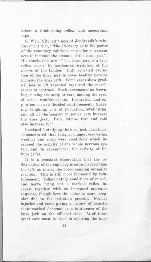
Fig. 8.-Knee Kick in Case of fultiple clerosis. a) Time marker. b) Electrical re ponse. c) Meche.nice.I response.
jerk, for, should the fir t respon e to the tap be smll.11, the examiner might con ider the knee jerk absent. He must ob erve each reflex quickly and accurately and make careful repeated examinations in order to determme definitely their presence or absence.
Sahli41 says-'' Any psychic excitement con.iderably increa es the tendon reflexes and this increa e may serve a an important sign of the tates of p ychic excitement.. The diminution
of reflex ometime observed in very acute, especially in traumatic, cord lesions i acceptable to the theory that thi diminution is due to inhibition or to injury to the lower cord segments from circulatory di turbances from the injury."
An injury to the cord produce a definite type of symptoms. The amount of trauma can only be determined by time, for a light injury may show an absence of knee jerk for a few days followed by complete re toration of this reflex. In the more severe lesion a trauma may show complete lo s of the knee jerk with no return, due to shock. The e produce di turbances of the circulations which clear up within a few <lay . Any inflammatory condition of the nerves of the leg will cau e an increa e of the
25

- --- -~----~~~- ~------------~----::=.--- -
Fig. 9.-Tracings 1 and 3 show sluggish reflexes, Zha?d 4 same, 45 minutes after gr. 1-10 stryc nia.
knee jerk. Sciatica, for example, produces a marked increase of the affected side that gradually becomes normal as the inflammatory conditions recede. .A hemiplegia, due to hemorrhage, produces a broken arc in the upper arc, thus cutting off the inhibition factor and causing a greater knee jerk on the opposite side from the lesion.
As has been mentioned before, a few apparently normal cases show absence of the knee jerk (Westphal 's sign) 42 by the ordinary clinical method of eliciting it, yet, with our method of graphically recording reflexes, a minute electric reflex action Fig. g1, followed by an electro-muscular action is recorded.
Pathological conditions such as fatigue43,
anesthesia narcosis, tabes44, Fig. 10. Entire absence of any leg swing is known in this condition. Thi failure is not mechanical. A large number of the tabetics have failed to show anY
26

effects whatever of electrical change when the attempt i" made to elicit the knee kick. Fig 10. 'l'hree tracings taken at three different time , over a period of two month . On the left the reflex record and on the right the voluntary swing. Diahctc ·, paraplegia, poliom:veliti ·, and in acute infection for a short time, al o the effect of certain drug a opiate , may cau e a diminution of the reflex action from a clinical standpoint, though, by our graphic method of recording, the e ca es will how a mall reflex with a small electro-mu cular and mechanical response. In students we made a careful study of the reflexes a effected by variou drug , among which were recorded, prior to and onehalf hour subsequent to the administration of caffein, grs. 5, and of strychnia sulphate, onetenth gr. An increa e following caffein and an increase following trychnia, and with increasing re pon e, each ~ucceeding blow. The caffein relieving apparent mu cular irritation, while the trychnia increases the irritability. In tran ver e le ion of the cord•5, the cuttinO' off of inhibition from the center above markedly increase the r fiexe . A a rule the knee jerk is permanently ab ent after total transverse le ion of the pinal cord above the level of the arc (Ba tian' Jaw), but the rea on is not known.
Certain pathological ca e pre ent an increase of both refiexe and mu cular action, uch as myelitis, spastic paraplegia, multiple clero i. 60 (Fig. ) and le ions that cau e a
degeneration of the cord it elf. Anv irritable condition of the mu cle, a in ome of the toxic tates whether from stimulant drugs as strych-
nia, (Fig. 9) or internal conditions, increa ·e the knee jerk.
27
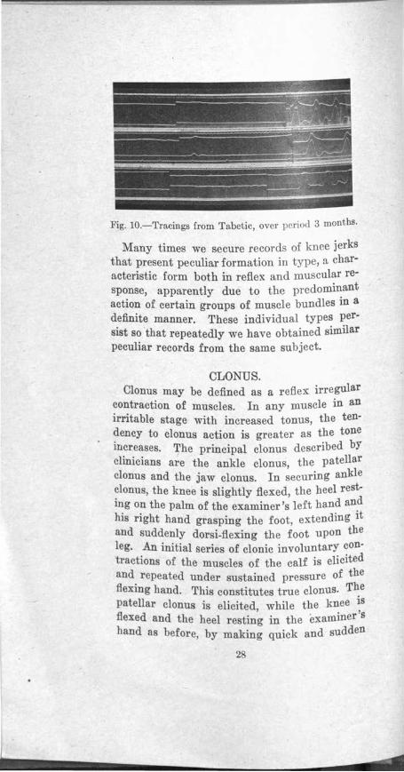
Fig. 10.-Tracings from Tabetic, over period 3 months.
Many times we secure records of knee jerks that present peculiar formation in type, a characteristic form both in reflex and muscular response, apparently due to the predominant action of certain groups of muscle bundles in a definite manner. These individual types persist so ·that repeatedly we have obtained similar peculiar records from the same subject.
CLO NUS. Clonus may be defined as a reflex irregular
contraction of muscles. In any muscle in an irritable stage with increased tonus, the tendency to clonus action is greater as the tone increases. The principal clonus described by clinicians are the ankle clonus, the patellar clonus and the jaw clonus. In securing ankle ~lonus, the knee is slightly flexed, the heel restmg on the palm of the examiner's left hand and his right hand grasping the foot, extending it and suddenly dorsi-flexing the foot upon the leg. An initial series of clonic involuntary contractions of the muscles of the calf is elicited and repeated under sustained pressure of the flexing hand. This constitutes true clonus. The patellar clonus is elicited while the knee is flexed and the heel restin1g in the examiner's hand as before, by making quick and sudden
28
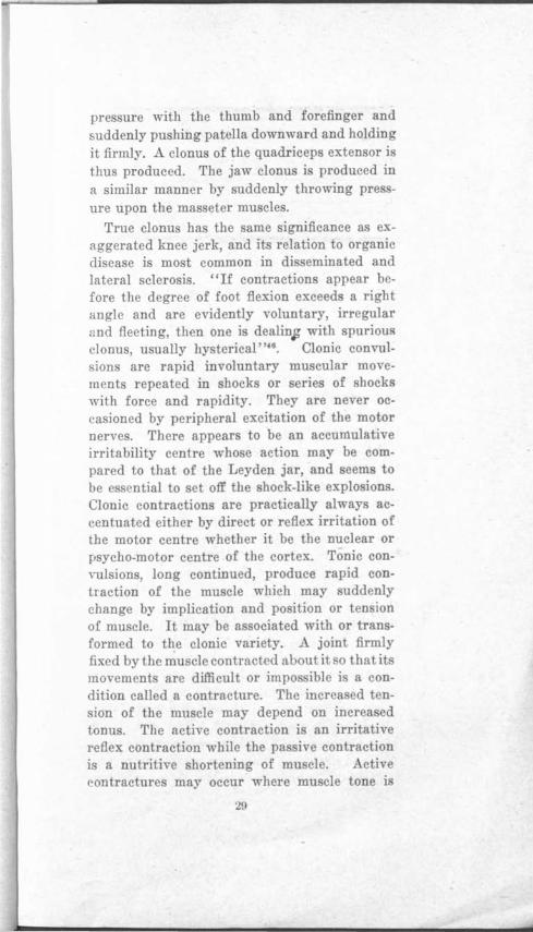
pressure with the thumb and forefinger and suddenly pushing patella downward and holding it firmly. A clonus of the quadriceps extensor is thus produced. The jaw clonus is produced in a similar manner by suddenly throwing pres -ure upon the masseter muscles.
True clonus has the same significance as exaggerated knee jerk, and its relation to organic disease is most common in disseminated and lateral sclerosis. "If contractions appear before the degree of foot flexion exceeds a right angle and are evidently voluntary, irregular and fleeting, then one is dealin$ with spurious clonus, usually hysterical "'6 • Clonic convulsions are rapid involuntary muscular movements repeated in shocks or series of shocks with force and rapidity. They are never occasioned by peripheral excitation of the motor nerves. There appears to be an accumulative irritability centre whose action may be compared to that of the Leyden jar, and seems to be essential to set off the shock-like explosions. Clonic contractions are practically always accentuated either by direct or reflex irritation of the motor centre whether it be the nuclear or psycho-motor centre of the cortex. Tonic convulsions, long continued, produce rapid contraction of the muscle which may suddenly change by implication and po ition or tension of muscle. It may be associated with or transformed to the clonic variety. A joint firmly fixed by the muscle contracted about it o that its movements are difficult or impo ible is a condition called a contracture. The increa ed tension of the muscle may depend on increased tonus. The active contraction is an irritative reflex contraction while the pa sive contraction is a nutritive shortening of mu cle. Active eontractures may occur where muscle tone is
2!)

~~v~~v-~-~~4~y\ ~JMMl\\~~~'r-~ ~ --.. . . Ef.f. £.t·f· 1 id J!j fff/i1r · - ~ ... _....-----.,...,..
·--~, , _______ .... ~\;"\' .. , ......... "~ ... ----\·~ ·---
1\. --~---... --
Fig. 12.-Records of Tremors, normal type.
increased since muscular tone is of reflex origin.
Bodwitch and Warren47 , in an article, say"There is good. evidence that clonus is a mechanical act the same as the knee jerk, hence should be similarly influenced by the peripheral nerve stimulation. Clonus may be reinforced or inhibited the same as a normal knee jerk of a healthy individual.''
Ankle clonus presents a definite record by the graphic method that will materially assist in a better understanding of the work. To the unaided eye all clonus appears alike, but records show that there are three distinct types50
due to fundamental genesis underlying as fu a case of chronic lenticular degeneration, hematomyelia and primary sclerosis. A constant record may be obtained not only showing the rate and frequency of the clonus but also Hs strength as recorded by the width of oscillations and also the duration, whether maintained for some time or quickly dropping off, and the question of its fatigue is recorded in a permanent and definite manner Fig. 15.
The apparatus used in securing records of clonus is shown in Fig. 3. The arm band (Ss.) and pump of a blood pressure apparatus is fastened about the limb from which the record is desired. Tubing leads to the Marey tambour
30

(R) which replaces the recording apparatus used in securing the mechanical swing of the knee jerk. Slight inflation is made with the pump. The electrodes are used as in securing the knee jerk and are attached to the string galvanometer.
TREMORS. Among the earlier medical writers was Clau
dius Galen48 who noted the fact that tremor existed and differed during voluntary movement and repose. Later Vieussens, in the latter part of the 14th century, was the first to point out the distinct and separate parts of the brain, and wrote extensively on the subject of tremors.
Haller49, in 1708, in researches on irritability, established the existence of irrital:iility as a property of living muscular tissue and that sensibility was due to the nerves alone.
Gilson50, the successor of Harvey, in the middle of the 17th century brought out many new discoveries regarding nerve ti sue. The doctrine of irritability, as taught by Haller, lead to a greater physiological study of nervou tissue. '
James Parkin onH, of London, in 1 17 pub-lished his es ay on shaking pal y and thi is his greatest and most. important contribution to Medicine. Parkin on 's definition is-" Involuntary tremations, motion with le ened muscular power in parts not in action even when supported.''
Tremors are rapid, minute mu cular contractions with a rhythmic tendency. Tremor may appear in healthy individuals as well as in pathological states, hence the need of careful study and record. It may appear in normal individuals following physical exercises, mental agitation, cold, etc. Tremors are of two classes:
31
•

Fig. 15.- Record of Ankle Clonus.
(a) intention, or that occurring during purposeful movement, and (b) passive, or that tremor persisting during rest. Putting the individual muscle group in action or under continued strain increases tremor. All tremors of the extremities are increased by extension and may be wholly absent when the patient is at rest. Most tremors are a spastic phenomenon and the contres are located above the reflex arc.
Sahli52 believed that tremors are essentially manifestations of spasm just as every spasm is explained by the damming up of stimuli which causes an explosive instead of a constant discharge of stimuli from the ganglion cells. It is analogous to the spark discharge of an induc-
• tion apparatus in contrast to the spray-like brush discharge following low resistance. To continue the analogy to explosive discharges further, we see that the succession of impulses underlying tremors may be based on the stronger stimulation of the motor ganglion cells on the one hand through the central neuron (paralysis agitans and nerve excitability), or through the reflex pathways (multiple sclerosis) on the other, because of the interruption o~ the motor current (peripheral palsies, fatigue, etc.) In all these cases there is the same disparity between the afferent and efferent nerve stimuli. This exposition is no mere hY-
32

pothesis. It rests upon the well-known general recognized property of the ganglion cells to accumulate impulses and di charge them explosively though we have as yet no further explanation. I t suggests a fundamental characteristic of nerve power which is a physiological observation. This brings us to a closer understanding of the different type of physiological tremor.
As has been stated previou ly, a a re ult of a careful study of the variou irregularities that appeared upon our cardiograph records and finding that many of the e finer movements were due to tremors, a more careful study of tremors was commenced. and oon the nece ity was apparent of an apparatu that would graphically record the variou movement of tremors. It was de ired that thi apparatus should be small and compact and capable of being u ed by the physician in gen ral practice. Fig. 13. As a rPsult of experiml'nL, the 'writ r presented the Tremograph,'0 Fig. 1 (L. .), and demon trated its u e at the )!innesota
tate Medical ociety in the fall of 1916. Thi apparatu is u ed in conjunction ' ith the recording apparatus of the modified i\fcKenzie Polygraph (B) 53, and giv a light, compact apparatu for making definite accurate record of tremor and clonus in a permanent form and with the time element portray d, Fig. 14
The Tremograph con i t of two tambours (E) set at right angle , o placed that one i vertical and the other horizontal to the axi of movement. The e tambour are connected with the tambours of the modified )foKenzie polygraph by rubber tubing. (G). The movement to be recorded is ·accentuated by the placing of spring vibrator of teel wire (H) in front of the tambour . Metal olive (F) with
33

Fig. 13.-Tremograph.
screw adjustment to permit their being placed in different positions along the spring vibrators, increase the oscillations and give a marked record of the vibration. Various handles are used which permit the securing of different types of records. These can be screwed on to the apparatus and are used for securing records of different parts of the body. (A)· Plain round handle (C) ordinarily used and held in either hand. (B). The t'onometer (D) with screw socket placed on one end which permits the apparatus being used for securing pressure tremors. (C). The slightly curved plate 2 "x3" with two straps and buckles th~t permit placing it immovably on any part. This is especially used for tremors and used principally on the lower ~xtremities.
In order to secure analogous records of patients under similar conditions the following
' procedure is adopted. The patient is made as comfortable as possible, whether seated erect in a chair or in bed. If in a chair, the feet should be flat on the floor and the hands on the knees. The handle of the Tremograph is then placed in one of the patient's hands and held between the thumb and extended forefingers, the horizontal arm to the front. The patient is then instructed as to the motion to be made with the instrument. The following movements have been used by us in securing records
34

as being representative of all voluntary movements.
1. "R. T." (Rand L) "Rest Tremor". This record is secured by having the patient raise the hand from the position on the knee, (a) front and upward to full exten ion of arm, (b) then holding arm at full exten ion, (c) then returning to knee position. These movement are made in a definite time- five cond to each move or position, the rate of movement being indicated to the patient by the operator's arm. Thi is of advantage, a the attention of the patient is focu ed and any unea ine s relieved.
2. "P. T." (R and L) . Pre ure Tremor, same a. Rest Tremor only tonometer i u ed a handle and held gently, arm raised (1) then at full exten ion, (2) the patient compre e handle to limit, then at (3) relea e to gentle grip and return to knee. R cord i made on lip of the grip pre ure.
3. "F .• r. T." (R and L) Finger o e Te t. The round handle (A) a u ed in (1) the movement con i t of four po. ition , each occupying five second -a in (1), (a) knee to full extension (b) then elbow i bent bringing the in trument, till vertical po ition, clo e to no , then (c) back to full ext n ion and (d) back to knee.
4. "R. T. T;eg." (R and L). Th metal pad ( ) u ed and attached to top of foot. Each foot i. brought up (a) to full ext n ion, (b) held, then (c) returned to floor.
5. "F. K. T. of Leg." (Rand L). Foot Knee 'rest. (C) attachment as in (4) movement (a) to full exten ion, (b) foot broul!h clo to oppoite knee (c) back to exten ion and (d) back
to floor. Thi produce a erie of tracing which
cover practically all the common movement .
35

Fig. 14.-Records of Tremors: a. Exophthalmic Goitre. b. Exophthalmic Goitre. c. Chronic Lenticular Degeneration. d. Multiple Sclerosis.
Tremors of the tongue may be tested by a little aluminum clip attached to one of the spring vibrators, and the tremograph attached to some solid object. The tongue is placed on the aluminum clip. For coarse tremors a smaller sized metal olive is used which produces a narrow oscillation of the writing lever.
In eliciting tremors by the older methods5•
"to distinguish between passive tremor and intention tremor direct the patient to
' d make some movement such as taking up an fastening a collar button buttoning a vest, or drinking a glass of water.' In this latter variet! the tremor is greatly increased by the co-ordinate movement involved and indeed maY be who~ly absent when the patient is at rest. ~y restmg the tips of the patient's fingers (Qu1Il· quad's phenomenon) upon the palm of the hand, a vibration otherwise imperceptible maY he readily detected." This is the usual form of noting tremors by clinical methods·. Though
36

1 his does not give a definite estimation of the rapidity or the strength of the tremor, still with a careful observation by this method the 1·linician was able to determine whether the tremor was fine or coarse.
:J[any type of apparatu have been u ed to estimate and record graphically the various forms of tremor but all of them were cumbersome and complicated for common u e.
"'Warner55, in the early eighties, brought out interesting but complicated apparatus con isting of an arrangement of rubber tubes, one for each finger, and each leading by the piece of tubing to an elaborate apparatus with a smoked drum, the frame supplied with recording tambour and electric signal . By means of thi :apparatu he brought out some intere ting figures on the movement of the hand and it Yarious parts.
Variou other device were used and much rnluable data has been gathered by11 Gra hey, , chafer, Peter on, Horsley, Wolfenden Ewald, Gower and Dana. Peter onH, in 1 94, published very intere ting re ult of exhau tive work on tremors. From the record . E. Hennely of the Edi on laboratory con truct d a very intere ting geometrical chart of the recorded wave . In hi summary Peter on ay -"Compared with the kymograph, the pbygmograph i coarse, crude and uncertain in the reproduction of variou tremor . fo t tremor can be placed in two categories-fine from 10 to 12 per second : and coarse, from 7 to per econd, corresponding to the normal innerva
tion rhythm a determined by Hor ley and chaefer58• slight tremor with normal in-
nervation wavelet which are fu ed in group of two gives the rate of 5 per econd."
37

In a study of tremors by Neustaedter59 in 1909, he brought out a new type of apparatus. By means of this apparatus a careful study of various types of tremors was made in some 600 cases of pathological type. His conclusions are as follows: '' 1. I want to say that the difference between different tremors are of kind, not of degree, and each form of tremor is distinctive of a form or group of diseases. 2. No definite relation exists between one form of tremor and any other. 3. The frequency of movements has no bearing upon the character of the tracing. 4. There is no material difference between the movements of the two sides of the
body."
CONCLUSIONS.
1. Graphic records of reflexes, clonus and tremors may be secured
a. By means of the string galvanometer, b. By means of apparatus recording move
ments of the regions involved, i. e., mechanical action. Such records may be designated broadly as reflexograms.
2. Graphic records of reflexes may be secured showing the form of electrical and mechanical response and also time elapsing between-
a. Stimulus and electrical response in muscle. b. Stimulus and mechanical response of
parts.
c. Electrical and mechanical responses. 3. In the normal reflexogram two elements
are found: a. An initial deflection of short duration
probably a definite reflex response. This appears to be present only in the records of electrical response.
38

b. Definite responses due to muscular action. These show both in the records of electrical and mechanical response. These responses may be reinforced, e. g., by J endrassik 's method, or inhibited, e. g., by 'Psychic factors, producing probably a state of tension or resistance in the muscles involved.
4. Tonus plays an important part in the reflex act. l\Iodifications may be due to an abnormal state in the afferent or efferent segments of the reflex arc and to conditions above the reflex arc.
5. In the reflexograms from abnormal individuals, the records may show modification of the reflex response (diminution, exaggeration or perversion) due to altered condition from fatigue, drugs and pathological condition in the reflex arc and in the upper neurons.
6. These records are so consistent, definite and permanent as to have a medico-legal value.
7. Irritability may be increased, in varying degrees and when this is sufficient, clonus may be produced. Clonus records show rate, amplitude, duration, response to increa e of pressure by the manipulator, and fatigue.
8. Records of tremors show rate, rhythm and amplitude. Tremor occurs in normal individuals and in them is increased by voluntary movement. Tremor is modified in individuals, otherwise normal, by such factors a toxic states, fatigue and drugs. In abnormal individuals alterations in rate, rhythm, amplitude and effects of voluntary movement are recorded.
3!l

BIBLIOGRAPHY.
1. Hail, Marshal. History of Med., Davis, '03. 2. Miiler, i. H. (1809). Ibid. 3. Brachet, Ibid. 4. Bell, Sir Chas. (London). Ibid. 5. Morton, George (Phil. '39). "Grania Americanae." 6. Magnedie Floures (Barker). 7. Smith. H. H., (Barker). 8. Westphal and Erb., Arch. f. Spychiartrie, Vol. V,
1875, p. 792. Berl. Klin. Wochen., '81, Vol. 18. Ibid., '78. ,
9. Fiirbringer, Centralblat. f. med. Wissenschr., 1875'. p. 929.
10. Rosenthal and Mendelsohn,-Monatschr. d. Roiilel Akadem. su. Berl. Feb. '73. Konigl. Preuss Akad. Berl., '82, '83, '85. Neur. Centralblat. '97, p. 878.
11. Gad and Flateau, Netirol. Centralblat, 1896, P· 147. 12. Sherrington, Phil. Trans. Roy. Soc. Lond., 1898,
p. 136.
13. Margulies, Wien. klin. Rundschau, 1899, pl. 9i5. 14. Turner, Stewart P.: J. N. and M. Dis., June, 1902,
Vol. 29, No. 6, p. 321. 15. Buchanan : Jour. of Physiol., Vol. xx, p. 95.190.
Ibid. Vol. vxi, p. 384, 1899. . 16~ Waller, A. D.: Lancet, 1881, Vol. 2, ·p. 83. Jo,ur. of
Physiol., Vol. vxi, 1890, p. 384. Brain, 1880, P· 179. 17. Lombard: Inter. J.M. Sc., 1887, p. 88. 18. Sahli: Treatise on Diagnostic Method of Examina
tion, 1914.
19. Jendrassik: · Beitrage zur lehre vor den Schneurefl.ex, A. F. K. M., 1883.
20. Lombard: Loe, Cit. 17. Also, Jour. M. and N. Disease\!, Feb. 1890.
21. Bodwitch and Warren: Bost. Med. and Surg. Jour., May 31st, 1888. Journ. Physiol., Vol. xi, 1890, p. 384.
22. Waller: Loe. Cit. No. 16. 23. Lombard : Loe. Cit. No. 17. Also, Jour. Med. Sc.,
Jan. 1887. 24· Lanfanauer's method (Sahli). 2°'· Schonborn's method (Sahli). 26· Phelps, R. M.; N. W. Lancet, St. Paul, Vol. xii,
p. 399.
40

27. Phelps, R. M., Loe. Cit. No. 26. 28. Noys: Amer. Jour. Psycho!., Aprll 1892. 29. Salhi: Loe. Cit. No. 18. 30. Lombard: Loe. Cit. No. 17.
Waller: Loe. Cit., No. 16. Piper, Kriel: Arch. f. ges. Physlol. Vol. cxlx, 1907,
p. 301. Zeitschr. f. Blolog. Vol. L. 1908, p. 393·504.
Buchanan, Florence: Quart. Jour. Exp. Pbyslol., 1908.
Bodwltch: Jour. Physiol., Vol. xi, 1890, p. 25.
31. Lombard: Amer. Jour. Physiol. 1887. 32. Waller: Loe. Cit. No. 16.
Erb: Loe. Cit. No. 8. Lombard: Loe. Cit. No. 31.
33. Waller: Loe. Cit. No. 16. 34. Waller: Loe. Cit. No. 16. 35. Waller: Loe. Cit. No. 16. 36. Diller: Penn. Med. Jour. 1899, Vol. 3, p. 44. 37. Mitchell, S. W. and Lewis, M. J., Med. News. 1886,
Feb. 13. t>. 20.
38. Sahli: Loe. Cit. No. 18. 39. Mitchell, S. W. Loe. Cit. No. 37. 40. Lombard: Loe. Cit. No. 31. 41. Sahli: Loe. Cit. No. 18. 42. Westphal: Loe. Cit. o. 8. Berl. Kiin. Woehen.
1878.
43. Lombard: Loe. Cit. No. 31. 44. Mitchell and Lewis: Loe. Cit. No. 37. 45. Piper: Loe. Cit. No. 30. Also, Arch. f. d. gee.
Physiol. Vol. exlx, p. 301, 1907. Zelt. f. Blolog., Vol. L, 393-504, 1908.
46. Med. Diagnosis, 1907. 47. Bodwiteh: Loe. Cit. o. 30. Also, Jour. Physiol.
Vol. xi, 1890, p. 25. 48. Rowntree: Johns Hop. Hosp. Bull., Vol xxill, o.
252, 1912. 49. Provost and Waller: Rev. Med. Sclesse-Romande,
June 15, 1881. 50. Provost and Woller: Loe. Cit. No. 49. 51. Rowntree: Loe. Cit. No. 48. 52. Sahli: Loe. Cit. o. 18. 53. Morris, R. E.; J. Amer. Med. Assoc., Vol. !xvi, 1916,
p, 1922.
41
54. Loe. Cit. No. 46. 55. Warner: Med. News, Philadel.1 1892. Also Jour.
Physiol., Aug. 3, 1884. 56. Grashey: Arch. f. Psych., 1885
Schafer and Horseley: Jour. Physlol., Vol. 5. Peterson: N. Y. Med. Jour. March 10, 1894. Wolfenden: British Med. Jour. May 19, 1888. Ewald: Berl. klin. Wochen., 1883, No. 52. G<>wers: Dis. of Nervous System, 1888, p. 1001. Dana: Medical News, Dec. 12, 1892.
57. Peterson: Jour. Nerv. and Ment. Dis., Feb. 1899. 58. Horsley and Schafer: Journ. Physlol. Vol. 5. 59. Neustaedter: Med. Record, N. Y., 1909, Vol. uvl,
p. 91. 60. Morris, R. E.: Journal Lancet, 1917: xxvii, p. 423.
42