The goal of physiology is to explain the physical and chemical factors that are responsible for the...
78
The goal of physiology is to explain the physical and chemical factors that are responsible for the origin,development, and progression of life Therefore, the vast field of physiology can be divided into viral physiology, bacterial physiology, cellular physiology, plant physiology,human physiology, and many more subdivisions . In Human Physiology we attempt to explain the specific characteristics and mechanisms of the human body that make it a living being
-
Upload
cory-powell -
Category
Documents
-
view
213 -
download
0
Transcript of The goal of physiology is to explain the physical and chemical factors that are responsible for the...

The goal of physiology is to explain the physical and chemical factors that are responsible for the origin,development, and progression of lifeTherefore, the vast field of physiology can be divided into viral physiology, bacterial physiology, cellular physiology, plant physiology,human physiology, and many more subdivisions.
In Human Physiology we attempt to explain the specific characteristics and mechanisms of the human body that make it a living being

Cells as the Living Units of the BodyThe basic living unit of the body is the cell. Each organ is an aggregate of many different cells held together by intercellular supporting structures. Each type of cell is specially adapted to perform one or a few particular functions.**.eg .RBCs.
-Although cell differ, but in all O2+ cho, prot, fat ----release energy
- -chemical processes for production of energy is the same.
--deliver waste nutrient to surrounding fluid.--reproduce for regerattion, & some degenerate .
Extracellular Fluid—The “Internal Environment”About 60 per cent of the adult human body is fluid, mainly a water solution of ions and other substances. Although most of this fluid is inside the cells and is called intracellular fluid, about one third is in the spaces outside the cells and is called extracellular fluid. This extracellular fluid is in constant motion throughout the body. It is transported rapidly in the circulating blood and then mixed between the blood and the tissue fluids by diffusion through the capillary walls.In the extracellular fluid are the ions and nutrients needed by the cells to maintain cell life. Thus, all cells live in essentially the same environment—the extracellularfluid. For this reason, the extracellular fluid is also
called the internal environment of the body .,

Differences Between Extracellular and Intracellular Fluids.The extracellular fluid contains large amounts ofsodium, chloride, and bicarbonate ions plus nutrients for the cells, such as oxygen, glucose, fatty acids, and amino acids. It also contains carbon dioxide that isbeing transported from the cells to the lungs to be excreted, plus other cellular waste products that are being transported to the kidneys for excretion. The intracellular fluid differs significantly from the extracellular fluid; specifically, it contains large amounts of potassium, magnesium, and phosphate ions
“Homeostatic” Mechanisms of he Major Functional SystemsHomeostasisThe term homeostasis is used by physiologists to meanmaintenance of nearly constant conditions in the internalenvironment. Essentially all organs and tissues of the body perform functions that help maintain these constant conditions. For instance, the lungs provide oxygen to the extracellular fluid to replenish the oxygen used by the cells, the kidneys maintain constant ion concentrations, and the
gastrointestinal system provides nutrientsم

Extracellular Fluid Transport andMixing System—The BloodCirculatory SystemExtracellular fluid is transported through all parts ofthe body in two stages. The first stage is movement ofblood through the body in the blood vessels, and thesecond is movement of fluid between the blood capillariesand the intercellular spaces.As blood passes through the blood capillaries,
continual exchange of extracellular fluid also occursbetween the plasma portion of the blood and the interstitial fluid that fills the intercellular spaces.

Therefore, large amounts of fluid and its dissolved constituents diffuse back and forth between the blood and the tissue spaces, s. This process of diffusion is caused bykinetic motion of the molecules in both the plasma andthe interstitial fluid. That is, the fluid and dissolvedmolecules are continually moving and bouncing in alldirections within the plasma and the fluid in the intercellularspaces, and also through the capillary pores.Few cells are located more than 50 micrometers froma capillary, which ensures diffusion of almost any substancefrom the capillary to the cell within a few seconds,

Thus, the extracellular fluid everywhere in thebody—both that of the plasma and that of the interstitialfluid—is continually being mixed, therebymaintaining almost complete homogeneity of the
extracellular fluid throughout the body.

Origin of Nutrients in theExtracellular FluidRespiratory System. The blood passes through the body, it also flows through the lungs. The blood picks up oxygen in the alveoli, thus acquiring the oxygen needed by the cells. The membrane between the alveoli and the lumen of thepulmonary capillaries oxygen diffuses by molecular motion through the pores of this membraneinto the blood in the same manner that water and ionsdiffuse through walls of the tissue capillaries.
Gastrointestinal Tract. A large portion of the blood the gastrointestinal tract. Here different dissolve
nutrients, including carbohydrates, fatty acids, an amino acids, are absorbed from the ingested food to the ECF .
.

Liver and Other Organs That Perform Primarily Metabolic Functions.Not all substances absorbed from the gastrointestinaltract can be used in their absorbed form by thecells. The liver changes the chemical compositions ofmany of these substances to more usable forms, andother tissues of the body—fat cells, gastrointestinalmucosa, kidneys, and endocrine glands—help modifythe absorbed substances or store them until they areneeded. pumped by the heart also passes through the wallsRemoval of Metabolic End ProductsRemoval of Carbon Dioxide by the Lungs.Removal of urea , uric acid,by kidneys.**.

Regulation of Body FunctionsNervous System. The nervous system is composed ofthree major parts: the sensory input portion, the centralnervous system (or integrative portion), and the motor output portion. Hormonal System of Regulation. Located in the body are eight major endocrine glands that secrete chemicalsubstances called hormones. Hormones are transportedin the extracellular fluid to all parts of the bodyto help regulate cellular function. For instance, , parathyroid, pancrease, thyroid

Control Systems of the BodyThe human body has thousands of control systems init. The most intricate of these are the genetic controlsystems that operate in all cells to help control Intracellular
function as well as extracellular function
examples of Control MechanismsRegulation of Oxygen and Carbon Dioxide Concentrations in the Extracellular Fluid ( oxygen-buffering function of hemoglobin)..

Carbon dioxide concentration in the extracellularfluid is regulated in a much different way. Carbondioxide is a major end product of the oxidative reactiona higher than normal carbon dioxide concentrationin the blood excites the respiratory center,causing a person to breathe rapidly and deeply. Thisincreases expiration of carbon dioxide and, therefore,removes excess carbon dioxide from the blood andtissue fluids. This process continues until the concentration returns to normal.

Regulation of Arterial Blood Pressure. Several systems contributeto the regulation of arterial blood pressure. One of these, the
baroreceptor system,.In the walls of the bifurcation region of the carotid arteries in the neck, and also in the arch of the aorta in the thorax, are many nerve receptors called baroreceptors, which are stimulated by stretch of thearterial wall.When the arterial pressure rises too high,the baroreceptors send barrages of nerve impulses to the medulla of the brain. Here these impulses inhibit the vasomotor center, which in turn decreases thenumber of impulses transmitted from the vasomotor center through the sympathetic nervous system to the heart and blood vessels. Lack of these impulses causes sdiminished pumping activity by the heart and also dilation of the peripheral blood vessels, allowingincreased blood flow through the vessels. Both of theseeffects decrease the arterial pressure back toward normal.

Conversely, a decrease in arterial pressure belownormal relaxes the stretch receptors, allowing thevasomotor center to become more active than usual,
thereby causing vasoconstriction and increased heartpumping, and raising arterial pressure back towardnormal.

Characteristics of Control SystemsNegative Feedback Nature of MostControl SystemsMost control systems of the body act by negative feedback,which can best be explained by reviewing someof the homeostatic control systems mentioned previously.In the regulation of carbon dioxide concentration,a high concentration of carbon dioxide in theextracellular fluid increases pulmonary ventilation.This, in turn, decreases the extracellular fluid carbondioxide concentration because the lungs expire greateramounts of carbon dioxide from the body. In otherwords, the high concentration of carbon dioxide initiatesevents that decrease the concentration towardnormal, which is negative to the initiating stimulus.
Conversely, if the carbon dioxide concentration falls too low, this causes feedback to increase the concentration.This response also is negative to the initiating stimulus.In the arterial pressure–regulating mechanisms, ahigh pressure causes a series of reactions that promotea lowered pressure, or a low pressure causes a seriesof reactions that promote an elevated pressure. In bothinstances, these effects are negative with respect to theinitiating stimulus.Therefore, in general, if some factor becomes excessiveor deficient, a control system initiates negativefeedback, which consists of a series of changes thatreturn the factor toward a certain mean value, thusmaintaining homeostasis.
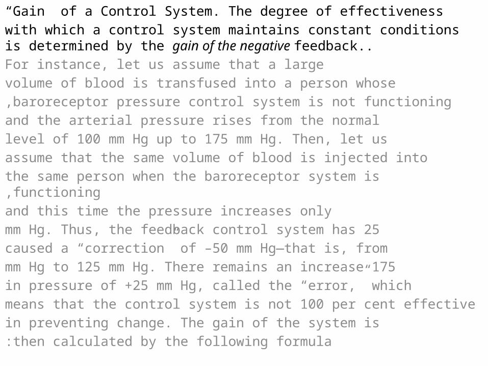
“Gain” of a Control System. The degree of effectivenesswith which a control system maintains constant conditions is determined by the gain of the negative feedback..For instance, let us assume that a largevolume of blood is transfused into a person whosebaroreceptor pressure control system is not functioning,and the arterial pressure rises from the normallevel of 100 mm Hg up to 175 mm Hg. Then, let usassume that the same volume of blood is injected intothe same person when the baroreceptor system is functioning,and this time the pressure increases only
25 mm Hg. Thus, the feedback control system hascaused a “correction” of –50 mm Hg—that is, from
175 mm Hg to 125 mm Hg. There remains an increasein pressure of +25 mm Hg, called the “error,” whichmeans that the control system is not 100 per cent effectivein preventing change. The gain of the system isthen calculated by the following formula:

Gain = correction / errorThus, in the baroreceptor system example, the correctionis –50 mm Hg and the error persisting is +25 mmHg. Therefore, the gain of the person’s baroreceptorsystem for control of arterial pressure is –50 dividedby +25, or –2. That is, a disturbance that increases ordecreases the arterial pressure does so only one thirdas much as would occur if this control system were notpresent.The gains of some other physiologic control systemsare much greater than that of the baroreceptorsystem. For instance, the gain of the system controllinginternal body temperature when a person is exposedto moderately cold weather is about –33. Therefore,one can see that the temperature control system ismuch more effective than the baroreceptor pressurecontrol system.
Positive Feedback Can Sometimes Cause Vicious Cycles and Death The heart of a healthy human being pumps about 5 litersof blood per minute. If the person is suddenly bled 2liters, the amount of blood in the body is decreased tosuch a low level that not enough blood is available forthe heart to pump effectively. As a result, the arterialpressure falls, and the flow of blood to the heart musclethrough the coronary vessels diminishes.This results inweakening of the heart, further diminished pumping,
a further decrease in coronary blood flow, and stillmore weakness of the heart; the cycle repeats itselfagain and again until death occurs. Note that eachcycle in the feedback results in further weakening ofthe heart. In other words, the initiating stimulus causesmore of the same, which is positive feedback

Positive feedback is better known as a “viciouscycle,” but a mild degree of positive feedback can beovercome by the negative feedback control
mechanisms of the body, and the vicious cycle fails to develop. For instance, if the person in the aforementioned example were bled only 1 liter instead of 2 iters, the normal negative feedback mechanisms for controlling cardiac output and arterial pressure would overbalance the positive feedback and the person would recover,
Positive Feedback Can Sometimes Be Useful.- Blood clott.- -uterine contraction

The Cell and Its Functions:al cell 2 major parts are the nucleus and the cytoplasm. . The nucleus is separated fromthe cytoplasm by a nuclear membrane, and the cytoplasm is separated from thesurrounding fluids by a cell membrane, also called the plasma membrane.
The different substances that make up the cell are collectively called
protoplasm.Protoplasm is composed mainly of five basic substances: water, electrolytes,proteins, lipids, and carbohydrates.

Cell membrane:Lipid Barrier of the Cell Membrane Impedes Water
Penetration.Its basic structure is a lipid bilayer, which is a thin,double-layered film of lipids—each layer only onemolecule thick—that is continuous over the entire
cell surface. Interspersed in this lipid film are large globular protein molecules. The basic lipid bilayer is composed of phospholipid molecules. One end of each phospholipid molecule is soluble in water; that is, it is hydrophilic. The other end
is soluble only in fats; that is, it is hydrophobic.

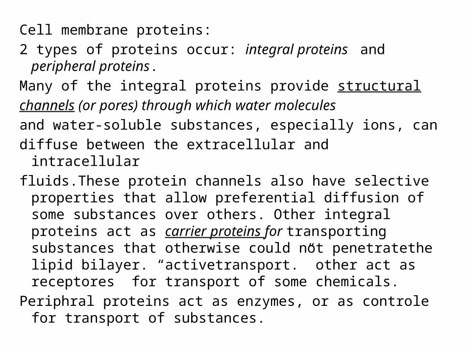
Cell membrane proteins: 2 types of proteins occur: integral proteins and peripheral
proteins.Many of the integral proteins provide structuralchannels (or pores) through which water moleculesand water-soluble substances, especially ions, candiffuse between the extracellular and intracellularfluids.These protein channels also have selective properties
that allow preferential diffusion of some substances over others. Other integral proteins act as carrier proteins for transporting substances that otherwise could not penetratethe lipid bilayer. “activetransport.” other act as receptores for transport of some chemicals.
Periphral proteins act as enzymes, or as controle for transport of substances.

Cytoplasm and Its Organelles:Dispersed in the cytoplasm are neutral fat globules,glycogen granules, ribosomes, secretory vesicles, andfive especially important organelles: the endoplasmicreticulum, the Golgi apparatus, mitochondria, lysosomes, and
peroxisomes.Endoplasmic reticulum is anetwork of vesicles & tubules. The
space in vesicles & tubules filled with fluid( E.R. matrix), the substances that frmed in some parts of cell transporetd through the space of E.R. to other parts.
On the outer surface of most E.R. are large numbers of minute granular particles called ribosomes. Where these are present, the reticulum is called the granular endoplasmic reticulum. The ribosomes are composed of a mixture of RNA and proteins,

Other type of ER were no granmles present called agranular or smooth ER. this way,
- Golgi apparatus: substances entrapped in the ER vesicles are transported from tendoplasmic reticulum to the Golgi apparatus. Thetransported substances are then processed in the Golgiapparatus to form lysosomes, secretory vesicles, andother cytoplasmic components Lysosomes:The lysosomes provide an intracellular digestivesystem that allows the cell to digest (1) damaged cellularstructures, (2) food particles that have beeningested by the cell, and (3) unwanted matter such asbacteria.

peroxisomes,they contain oxidases rather than hydrolases. Severalof the oxidases are capable of combining oxygen withhydrogen ions derived from different intracellularchemicals to form hydrogen peroxide (H2O2). Hydrogen peroxide is a highly oxidizing substance and is used in association with catalase, to oxidize many substances that might otherwise be
poisonous to the cell..

Secretory VesiclesOne of the important functions of many cells is
secretion of special chemical substances. Almost all such secretory substances are formed by the endoplasmic reticulum–Golgi apparatus system and are the released from the Golgi apparatus into the cytoplasm in the form of storage vesicles called secretory vesicle store protein proenzymes which are secreted later through the outer cell membrane into the pancreatic duct and hence into the duodenum, where they become activated and perform digestive functions
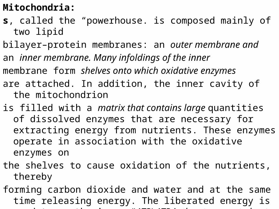
Mitochondria:s, called the “powerhouse. is composed mainly of two lipidbilayer–protein membranes: an outer membrane andan inner membrane. Many infoldings of the innermembrane form shelves onto which oxidative enzymesare attached. In addition, the inner cavity of the mitochondrionis filled with a matrix that contains large quantities of dissolved
enzymes that are necessary for extracting energy from nutrients. These enzymes operate in association with the oxidative enzymes on
the shelves to cause oxidation of the nutrients, therebyforming carbon dioxide and water and at the same time releasing
energy. The liberated energy is used to synthesize a “ATP).ATP is then transported out of the mitochondrion, and it diffuses throughout the cell to srelease its own energy wherever it is needed for performing
cellular functions.

NucleusThe nucleus is the control center of the cell. Briefly, thenucleus contains large quantities of DNA, which arethe genes. The genes determine the characteristics ofthe cell’s proteins, including the structural proteins, aswell as the intracellular enzymes that control cytoplasmicand nuclear activities.
The genes also control and promote reproduction ofthe cell itself. The genes first reproduce to give twoidentical sets of genes; then the cell splits by a specialprocess called mitosis to form two daughter cells, eachof which receives one of the two sets of DNA genes.

The nucleolus: it is simply an accumulationof large amounts of RNA and proteins of the types found in ribosomes.DNA in nucleuses form RNA , most of it remain in nuclease, burt some
transported to cytoplasm forming ribosome.s Functional Systems of the Cell
Ingestion by cells (endocytosis):. Most substances pass through the cell membrane by diffusion and
active transport.Diffusion involves simple movement through the membrane caused by
the random motion of the molecules of the substance; substances move either hrough cell membrane pores or, in the case of
lipid soluble substances, through the lipid matrix of the membrane.Active transport involves the actual carrying of a substance through the
membrane by a physical protein structure that penetrates all the way through the smembrane.

Very large particles enter the cell by a specializedfunction of the cell membrane called
endocytosis. The principal forms of endocytosis are pinocytosis and phagocytosis.
Pinocytosis means ingestion of minute particles that form vesicles of extracellular fluid and
particulate constituents inside the cell cytoplasm.Phagocytosis means ingestion of large particles,
such as bacteria, whole cells, or portions of degenerating tissue.
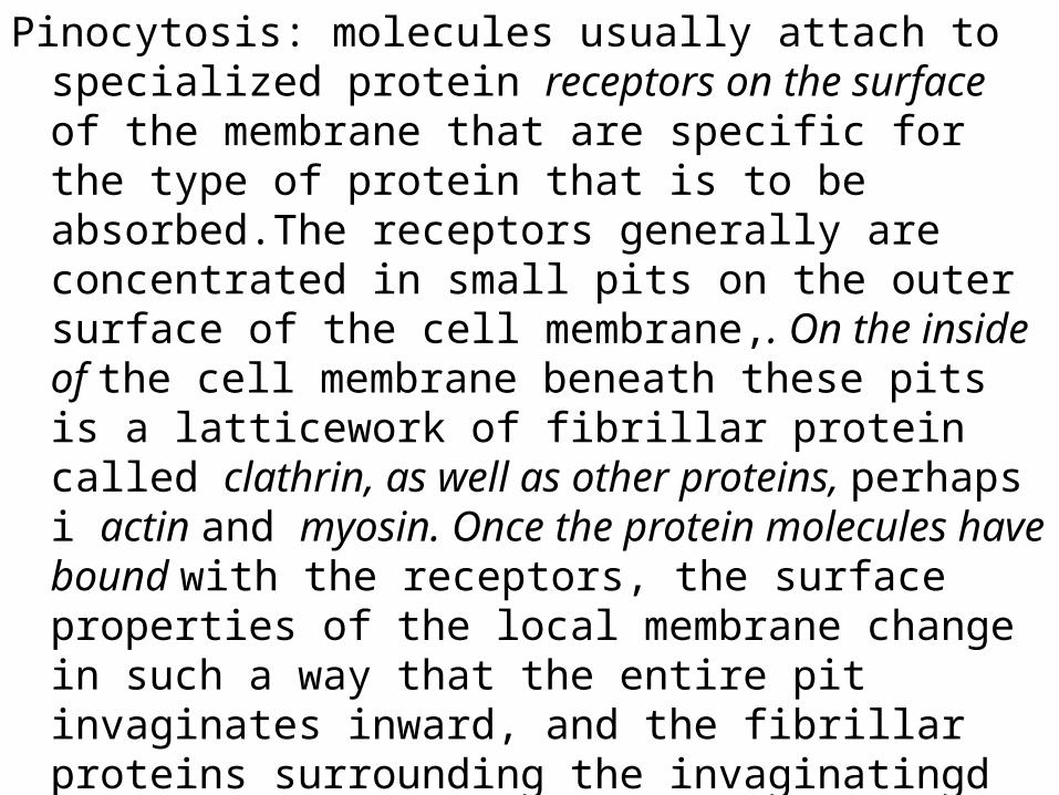
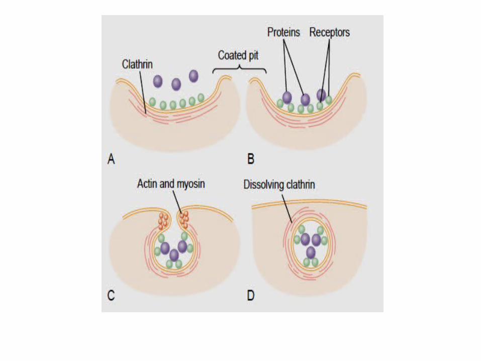
Pinocytosis: molecules usually attach to specialized protein receptors on the surface of the membrane that are specific for the type of protein that is to be absorbed.The receptors generally are concentrated in small pits on the outer surface of the cell membrane,. On the inside of the cell membrane beneath these pits is a latticework of fibrillar protein called clathrin, as well as other proteins, perhaps i actin and myosin. Once the protein molecules have bound with the receptors, the surface properties of the local membrane change in such a way that the entire pit invaginates inward, and the fibrillar proteins surrounding the invaginatingd pit cause its borders to close over the attached proteins as well as over a small amount of extracellular fluid. Immediately thereafter, the invaginated portion of the membrane breaks away from the surface of the cell, forming a pinocytotic vesicle.

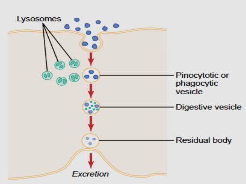
Phagocytosis. Phagocytosis occurs in much the same way as pinocytosis, except that it involves large particles. Phagocytic Foreign Substances Insidethe Cell—Function of the LysosomesAlmost immediately after a pinocytotic or phagocytic svesicle appears inside a cell, one or more lysosomes become attached to the vesicle and empty their acid hydrolases to the inside of the vesicle, Thus, a digestive vesicle is formed inside he cell cytoplasm in which the vesicular hydrolases The products of digestion are diffuse through the membrane of the vesicle into the cytoplasm. What is left of the digestive vesicle, called the residual body represent undigestible substances. In most instances this is finall excreted through the cell membrane by a process called exocytosis, which is essentially the opposite of endocytosis.

Extraction of Energy from Nutrients—Function of the Mitochondria—
Before intering the cells foodstuff converted to glucose, fatty acids, and amino acids—all entering the cell. Inside the cell, the foodstuffs react chemically with oxygen, under influence of enzymes that control th reactions and channel the energy released in the proper direction.
Briefly, almost all these oxidative reactions occurinside the mitochondria, and the energy that isreleased is used to form the high-energy compoundATP. Then, is used hroughout the cell to energize almost all the subsequent intracellular metabolic reactions.s

Uses of ATP for Cellular Function .Energy from ATP is used to promote three major categories of cellular functions:
( 1 )transport of substances through multiplemembranes in the cell, (2) synthesis of chemicalcompounds throughout the cell, and (3) mechanical
work. These are: (1 )to supply energy for the transport of sodium
through the cell membrane, (2) to promoteprotein synthesis by the ribosomes, and (3) to supplythe energy needed during muscle contraction.

Genetic Control of Protein Synthesis, Cell Function, and Cell Reproduction:
Each gene, which is a nucleic acid called deoxyribonucleic acid (DNA), automatically controls the formation of another nucleic acid, ribonucleic acid (RNA); this RNA then sspreads throughout the cell to control the synthesis of spcefic proteins, some of the cellular proteins are structural proteins, which, in association with various lipids and carbohydrates, form the structures of the various intracellular organelles However, by far the majority of the proteins are enzymes that catalyze the different chemical reactions in the cells. For instance, enzymes promote all the oxidative reactions that supply energy to the cell, and they promote synthesis of all the cell chemicals, such as lipids, glycogen, (ATP)..


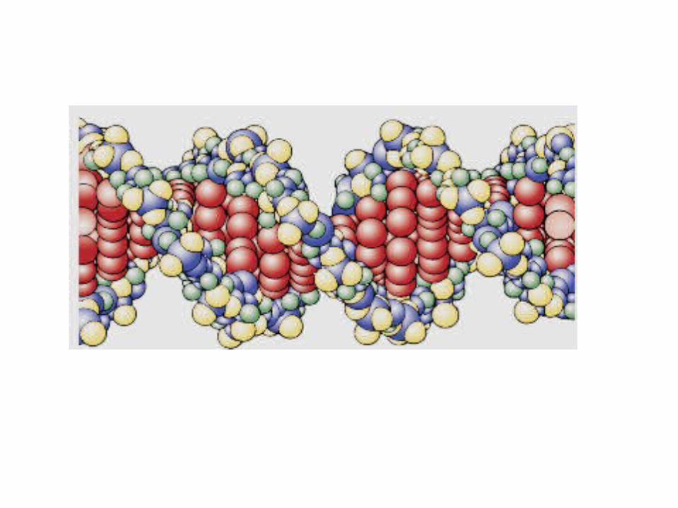
Genes in the Cell NucleusIn the cell nucleus, large numbers of genes are attached end
on end in extremely long double-stranded helical molecules of DNA
Basic Building Blocks of DNA. the basic chemical compounds involved in the formation of DNA. Are (1) phosphoric acid, (2) a sugar called deoxyribose, and (3) four nitrogenous bases (two purines, adenine and guanine, and two pyrimidines, thymine and cytosine). The phosphoric acid and deoxyribose form the two helical strands that are the backbone of the DNA molecule, and the
nitrogenous bases lie between the two strands and connectthem,. .

Nucleotides. The first stage in the formation of DNA is to combine one molecule of phosphoric acid, one molecule of deoxyribose, and one of the four bases to form an acidic nucleotide. Four separate nucleotides are thus formed, one for each of the 4 base. --Organization of multiple number of Nucleotides to Form Two Strands of DNA Loosely Bound to Each Other by lose cross linkage.
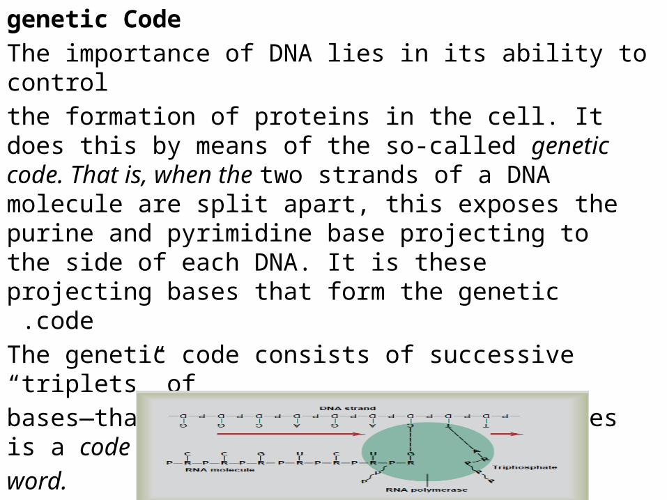
genetic CodeThe importance of DNA lies in its ability to controlthe formation of proteins in the cell. It does this by means of the so-called genetic code. That is, when the two strands of a DNA molecule are split apart, this exposes the purine and pyrimidine base projecting to the side of each DNA. It is these
projecting bases that form the genetic code .The genetic code consists of successive “triplets” ofbases—that is, each three successive bases is a codeword.
The successive triplets eventually control thesequence of amino acids in a protein molecule that isto be synthesized in the cell.The DNA Code in the Cell Nucleus Is Transferred to an
RNA Code in the Cell Cytoplasm—The Process of Transcription
Because the DNA is located in the nucleus of the cell, yet most of the functions of the cell are carried out in the cytoplasm, This is achieved through,RNA, the formation of which is controlled by the DNA this process is called Transcription, The RNA, in turn, diffuses from the nucleus through nuclear pores into the cytoplasmic compartment, where it controls protein synthesis.

Synthesis of RNADuring synthesis of RNA, the two strands of the DNA
molecule separate temporarily; one of these strands is used as a template for synthesis of an RNA molecule.
Basic Building Blocks of RNA. The basic building blocks of RNA are almost the same as those of DNA, except for two difference s.First, the sugar is ribose, Second, thymine is replaced by another pyrimidine, uracil.

Formation of RNA Nucleotides. The basic building blocks of RNA form RNA nucleotides,
types of RNA;1. Messenger RNA, which carries the genetic code to the
cytoplasm for controlling the type of protein formed.2. Transfer RNA, which transports activated aminoacids to the ribosomes to be used in assemblingthe protein molecule.3. Ribosomal RNA, which, along with about 75different proteins, forms ribosomes, the physicaland chemical structures on which proteinmolecules are actually assembled.4. MicroRNA(mi RNA), which regulate gene transcription
and gene translation

Messenger RNA—The CodonsMessenger RNA molecules are long, single RNAstrands that are suspended in the cytoplasm. These
molecules are composed of several hundred to several thousand RNA nucleotides in unpaired strands, and they contain codons that are exactly complementary to the code triplets of the DNA genes. Note that most of the amino acids are represented by more than one codon;
also, one codon represents the signal “start manufacturing the protein molecule,” and three codons represent “stop anufacturing the protein molecule

Transfer RNA—The AnticodonsIt transfers amino acid molecules to protein moleculesAs the protein is being synthesized. Each type of transferRNA combines specifically with 1 of the 20 aminoacids that are to be incorporated into proteins. Thetransfer RNA then acts as a carrier to transport itsspecific type of amino acid to the ribosomes, where protein
molecules are forming. Because the function of transfer RNA is to cause attachment of a specific amino acid to a forming protein chain, it is essential that each type of transfer RNA also have specificity for a particular codon in the messenger RNA. The specific code in the transferRNA that allows it to recognize a specific codon isagain a triplet of nucleotide bases and is called an anticodon.
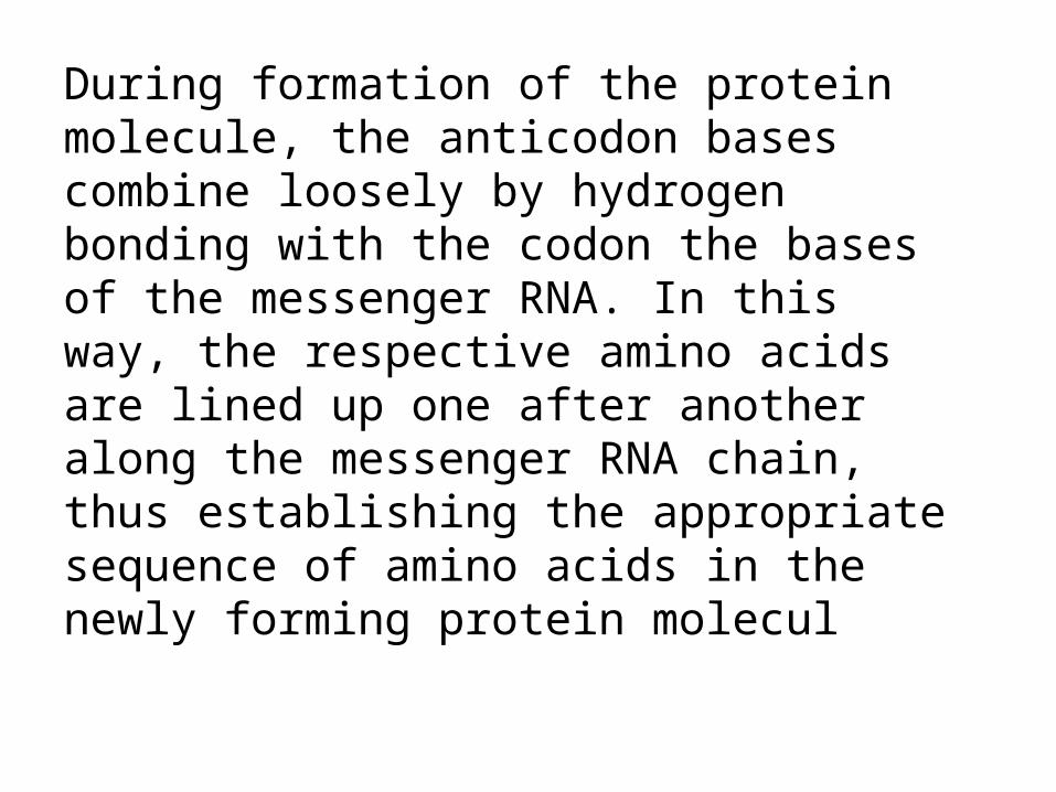
During formation of the protein molecule, the anticodon bases combine loosely by hydrogen bonding with the codon the bases of the messenger RNA. In this way, the respective amino acids are lined up one after another along the messenger RNA chain, thus establishing the appropriate sequence of amino acids in the newly forming protein molecul

Ribosomal RNAThe ribosome is the physical structure in the
cytoplasm on which protein molecules are actually synthesized. However, it always functions in association with the other two types of RNA as well: transfer RNA transports amino acids to the ribosome for incorporation into the developing protein molecule, whereas messenger RNA provides the information necessary for sequencing the amino acids in proper order for each specific type of protein to be manufactured.

Formation of Ribosomes in the Nucleolus. The DNA genes for formation of ribosomal RNA are located in five pairs of chromosomes in the nucleus. Ribosomal RNA is specially processed in the nucleolus, where it binds with “ribosomal proteins” to form granular condensation products that are primordial subunits of ribosomes. These subunits are then released from the nucleolus and transported through the large pores of the nuclear envelope to almost all parts of the cytoplasm. After the subunits enter the cytoplasm, they are assembled to form mature, functional ribosomes. Therefore, proteins are formed in the cytoplasm of the cell, but not in the cell nucleus, because the nucleus does not contain mature ribosomes.

Formation of Proteins on the Ribosomes—The Process of “Translation”
When a molecule of messenger RNA comes in contact with a ribosome, it travels through the ribosome, beginning at a predetermined end of the RNA molecule specified by an appropriate sequence of RNAbases called the “chain-initiating” codon. while the messenger RNA travels
through the ribosome, a protein molecule is formed—a process called translation. Thus, the ribosome readsthe codons of the messenger RNA in much the sameway that a tape is “read” as it passes through the playbackhead of a tape recorder. Then, when a “stop” (or“chain-terminating”) codon slips past the ribosome, the end
of a protein molecule is signaled and the protein molecule is freed into the cytoplasm..

Control of Gene Function and Biochemical Activity in Cells: the genes control both the physical and the chemical function of the cells. However, the degree of activation o f respective genes must be controlled as well; otherwise, some parts of the cell might overgrow or some chemical reactions might overact until they kill the cell.
Each cell has powerful internal feedback control mechanisms that keep the various functional operations of the cell in step with one another.For each gene there is at least one such feedback mechanismThere are basically two methods by which the biochemical activities in the cell are controlled. One of these is
genetic regulation and the other is enzyme regulation ,

Genetic regulation : Formation of all the enzymes needed for the synthetic
process often is controlled by a sequence of genes located one after the other on the same chromosomal DNA strand. This area of the DNA strand is called an operon, and the genes responsible for forming the respective senzymes are calledstructural genes.
Enzyme Regulation:Enzyme Inhibition. :Some chemical substances formed inthe cell have direct feedback effects in inhibiting thespecific enzyme systems that synthesize them. Almostalways the synthesized product acts on the firstenzyme in a sequence, rather than on the subsequentenzymes, usually binding directly with the enzyme andcausing an allosteric conformational change that inactivatesit.

Enzyme Activation. Enzymes that are normally inactive often can be activated when needed. An example of this occurs when most of the ATP has been depleted in a cell. In this case, cAMP begins to be formed as a breakdown product of the ATP; this cAMP, in turn, immediately activates the glycogen-splitting enzyme liberating glucose molecules that are rapidly metabolized and their energy used for replenishment of the ATP stores.
Other eg. Purin & pyrimidine.

The DNA-Genetic System Also Controls Cell Reproduction : The genes and their regulatory mechanisms determine the growth characteristics of the cells and also when or whether these cells will divide to form new cells. In this way, the all-important genetic systemn controls each stage in the development of the human being, from the single-cell fertilized ovum to the whole
functioning body. Cell reproduction begins in the nucleus itself.The firststep is replication (duplication) of all DNA in the
chromosomes. Only after this has occurred can mitosis take place.( the actual process by which the cell splits into two new cells is called mitosis) .

CancerCancer is caused in all or almost all instances by mutationor by some other abnormal activation of cellular genes that control
cell growth and cell mitosis. abnormal genes are called oncogenes. Also present in all cells are antioncogenes, which suppress the activation of specific oncogenes. Therefore, loss of or inactivation of antioncogenes can allow activation of oncogenes that lead to cancer. Only a minute fraction of the cells that mutate in the body ever lead to cancer.There are several reasons for this. First, most mutated cells have less survival capability than normal cells and simply die. Second, only a few of the mutated cells that do survive become cancerous, because even most mutated cells still have normal feedback controls that prevent excessive growth. hird, those cells that are potentially cancerous are
often, if not usually, destroyed by the body’s immunesystem before they grow into a cancer.