
The genome-wide binding profile of the Sulfolobus solfataricus transcription factor Ss-LrpB shows...
15
RESEARCH ARTICLE Open Access The genome-wide binding profile of the Sulfolobus solfataricus transcription factor Ss-LrpB shows binding events beyond direct transcription regulation Trong Nguyen-Duc 1,2,5 , Liesbeth van Oeffelen 3,4 , Ningning Song 3 , Gholamreza Hassanzadeh-Ghassabeh 1,2 , Serge Muyldermans 1,2 , Daniel Charlier 3 and Eveline Peeters 3* Abstract Background: Gene regulatory processes are largely resulting from binding of transcription factors to specific genomic targets. Leucine-responsive Regulatory Protein (Lrp) is a prevalent transcription factor family in prokaryotes, however, little information is available on biological functions of these proteins in archaea. Here, we study genome- wide binding of the Lrp-like transcription factor Ss-LrpB from Sulfolobus solfataricus. Results: Chromatin immunoprecipitation in combination with DNA microarray analysis (ChIP-chip) has revealed that Ss-LrpB interacts with 36 additional loci besides the four previously identified local targets. Only a subset of the newly identified binding targets, concentrated in a highly variable IS-dense genomic region, is also bound in vitro by pure Ss-LrpB. There is no clear relationship between the in vitro measured DNA-binding specificity of Ss-LrpB and the in vivo association suggesting a limited permissivity of the crenarchaeal chromatin for transcription factor binding. Of 37 identified binding regions, 29 are co-bound by LysM, another Lrp-like transcription factor in S. solfataricus. Comparative gene expression analysis in an Ss-lrpB mutant strain shows no significant Ss-LrpB-mediated regulation for most targeted genes, with exception of the CRISPR B cluster, which is activated by Ss-LrpB through binding to a specific motif in the leader region. Conclusions: The genome-wide binding profile presented here implies that Ss-LrpB is associated at additional genomic binding sites besides the local gene targets, but acts as a specific transcription regulator in the tested growth conditions. Moreover, we have provided evidence that two Lrp-like transcription factors in S. solfataricus, Ss-LrpB and LysM, interact in vivo. Keywords: Archaea, Sulfolobus, Leucine-responsive regulatory protein, CRISPR, ChIP-chip Background Transcription factors (TFs) belonging to the Leucine- responsive Regulatory Protein (Lrp) family (also known as AsnC or FFRP) are abundant in both bacteria and ar- chaea [1-4]. A sequence analysis of 52 archaeal genomes indicated that they are all predicted to contain at least one lrp-like gene, lrp-like genes constituting in total about 8% of all non-general TF genes in archaea [4]. Whereas bacterial Lrp-like TFs regulate amino acid biosynthesis in response to nutritional availability [5], archaeal Lrp members also regulate genes belonging to energy, central metabolism and transport pathways [6-9]. Furthermore, it has been observed and/or predicted by se- quence analyses that a subset of archaeal Lrp-like TFs do not interact with amino acids, in contrast to most other archaeal Lrp-like TFs [10-16] and to bacterial Lrp-like reg- ulators that invariably bind amino acids. Known archaeal Lrp-like TFs have regulon sizes ranging from one or a few targets to a large number of genes and operons. Examples of the former are LrpA from Pyrococcus [17], LrpA1 from * Correspondence: [email protected] 3 Research group of Microbiology, Vrije Universiteit Brussel, Pleinlaan 2, B-1050 Brussels, Belgium Full list of author information is available at the end of the article © 2013 Nguyen-Duc et al.; licensee BioMed Central Ltd. This is an open access article distributed under the terms of the Creative Commons Attribution License (http://creativecommons.org/licenses/by/2.0), which permits unrestricted use, distribution, and reproduction in any medium, provided the original work is properly cited. Nguyen-Duc et al. BMC Genomics 2013, 14:828 http://www.biomedcentral.com/1471-2164/14/828
Transcript of The genome-wide binding profile of the Sulfolobus solfataricus transcription factor Ss-LrpB shows...

RESEARCH ARTICLE Open Access
The genome-wide binding profile of the Sulfolobussolfataricus transcription factor Ss-LrpB showsbinding events beyond direct transcriptionregulationTrong Nguyen-Duc1,2,5, Liesbeth van Oeffelen3,4, Ningning Song3, Gholamreza Hassanzadeh-Ghassabeh1,2,Serge Muyldermans1,2, Daniel Charlier3 and Eveline Peeters3*
Abstract
Background: Gene regulatory processes are largely resulting from binding of transcription factors to specificgenomic targets. Leucine-responsive Regulatory Protein (Lrp) is a prevalent transcription factor family in prokaryotes,however, little information is available on biological functions of these proteins in archaea. Here, we study genome-wide binding of the Lrp-like transcription factor Ss-LrpB from Sulfolobus solfataricus.
Results: Chromatin immunoprecipitation in combination with DNA microarray analysis (ChIP-chip) has revealedthat Ss-LrpB interacts with 36 additional loci besides the four previously identified local targets. Only a subset of thenewly identified binding targets, concentrated in a highly variable IS-dense genomic region, is also bound in vitroby pure Ss-LrpB. There is no clear relationship between the in vitro measured DNA-binding specificity of Ss-LrpBand the in vivo association suggesting a limited permissivity of the crenarchaeal chromatin for transcription factorbinding. Of 37 identified binding regions, 29 are co-bound by LysM, another Lrp-like transcription factor in S. solfataricus.Comparative gene expression analysis in an Ss-lrpB mutant strain shows no significant Ss-LrpB-mediated regulationfor most targeted genes, with exception of the CRISPR B cluster, which is activated by Ss-LrpB through binding toa specific motif in the leader region.
Conclusions: The genome-wide binding profile presented here implies that Ss-LrpB is associated at additionalgenomic binding sites besides the local gene targets, but acts as a specific transcription regulator in the testedgrowth conditions. Moreover, we have provided evidence that two Lrp-like transcription factors in S. solfataricus,Ss-LrpB and LysM, interact in vivo.
Keywords: Archaea, Sulfolobus, Leucine-responsive regulatory protein, CRISPR, ChIP-chip
BackgroundTranscription factors (TFs) belonging to the Leucine-responsive Regulatory Protein (Lrp) family (also knownas AsnC or FFRP) are abundant in both bacteria and ar-chaea [1-4]. A sequence analysis of 52 archaeal genomesindicated that they are all predicted to contain at leastone lrp-like gene, lrp-like genes constituting in totalabout 8% of all non-general TF genes in archaea [4].
Whereas bacterial Lrp-like TFs regulate amino acidbiosynthesis in response to nutritional availability [5],archaeal Lrp members also regulate genes belonging toenergy, central metabolism and transport pathways [6-9].Furthermore, it has been observed and/or predicted by se-quence analyses that a subset of archaeal Lrp-like TFs donot interact with amino acids, in contrast to most otherarchaeal Lrp-like TFs [10-16] and to bacterial Lrp-like reg-ulators that invariably bind amino acids. Known archaealLrp-like TFs have regulon sizes ranging from one or a fewtargets to a large number of genes and operons. Examplesof the former are LrpA from Pyrococcus [17], LrpA1 from
* Correspondence: [email protected] group of Microbiology, Vrije Universiteit Brussel, Pleinlaan 2,B-1050 Brussels, BelgiumFull list of author information is available at the end of the article
© 2013 Nguyen-Duc et al.; licensee BioMed Central Ltd. This is an open access article distributed under the terms of theCreative Commons Attribution License (http://creativecommons.org/licenses/by/2.0), which permits unrestricted use,distribution, and reproduction in any medium, provided the original work is properly cited.
Nguyen-Duc et al. BMC Genomics 2013, 14:828http://www.biomedcentral.com/1471-2164/14/828

Halobacterium salinarum R1 [15], Ptr2 from Methanocal-dococcus jannaschii [6,7] and LysM from Sulfolobus solfa-taricus that has an intermediate number of target genes[16]. Examples of the latter are FL11 from PyrococcusOT3 [12], Lrp from H. salinarum R1 [15] and Sa-Lrp fromSulfolobus acidocaldarius [18].Lrp-like proteins generally have low sequence iden-
tities, but are structurally highly conserved [19]. Typic-ally, an Lrp monomer has a molecular mass of about15 kDa and consists of two domains: an amino-terminalDNA-binding domain with a helix-turn-helix (HTH)motif and a carboxy-terminal domain, named Regulationof Amino acid Metabolism (RAM) [20], which is respon-sible for protein multimerization and cofactor binding[8]. This RAM domain forms an αβ sandwich fold havingan antiparallel β sheet composed of four strands “sand-wiched” between two α helices. It has been observed that,in vitro, archaeal Lrp-like proteins associate into severalmultimeric forms via β strand exchange in the RAM do-main [10,17,21-28]. Oligomerization is a prerequisite forformation of the cofactor binding pocket. Furthermore,cofactor binding induces conformational changes that inturn could affect oligomerization [11,13].In S. solfataricus, a crenarchaeal model organism, three
Lrp-like TFs have been studied experimentally: LysM[10,16], Ss-Lrp [25] and Ss-LrpB [9,27,29], the latter beingone of the best characterized Lrp-like regulators in ar-chaea. Ss-LrpB performs both positive and negative auto-regulation in a concentration-dependent manner [30].Moreover, gene expression analysis in an Ss-lrpB deletionstrain demonstrates that Ss-LrpB acts as an activator onits neighbouring target operon/genes encoding a pyruvateferredoxin oxidoreductase (porDAB) and two putative per-meases (Sso2126, Sso2127) [9].At its target promoter regions, Ss-LrpB binds either a
single or multiple, regularly spaced, binding sites har-bouring a conserved motif [9,29]. Each binding site iscontacted by an Ss-LrpB dimer [27]. In the control re-gion of its own gene (Sso2131), three Ss-LrpB dimersbind cooperatively to juxtaposed sites [31]. Occupationof all three sites results in strong DNA deformationsand even DNA wrapping [27]. Based on the 15-bp palin-dromic consensus sequence 5'-TTGCAAAATTTGCAA-3', the sequence specificity of Ss-LrpB binding wasanalyzed by saturation mutagenesis [32].Despite extensive knowledge of the in vitro DNA-
binding properties of Ss-LrpB, nothing is known yet aboutits in vivo binding behaviour and furthermore, it is unclearwhether Ss-LrpB is a local or global acting TF. In thiswork, we investigate Ss-LrpB binding in an in vivo contextby performing chromatin immunoprecipitation combinedwith DNA microarray analysis (ChIP-chip). Besides merelyidentifying in vivo binding sites, we perform an extensivecomparative analysis of in vitro, in vivo and in silico
binding, exploiting the knowledge of the DNA-bindingspecificity model. By combining in vivo binding data withgene expression analysis, we provide new insights into thebiological functions of Ss-LrpB, which go beyond directtranscription regulation.
MethodsStrains and culture conditionsS. solfataricus P2 (DSM1617), PBL2025 [33] and Ss-lrpB::lacS [9] strains were grown aerobically at 80°C in Brockbasic medium supplemented with 0.1% tryptone as a car-bon and energy source [34]. Escherichia coli strain DH5αwas used for all cloning and plasmid propagation pur-poses. E. coli strain BL21(DE3) was used as a host for pro-tein overexpression.
Chromatin immunoprecipitationEach chromatin immunoprecipitation (ChIP) samplewas prepared from a 200 ml culture of S. solfataricus P2at mid-exponential growth phase. The entire ChIP pro-cedure, from collecting cells to obtaining amplifiedenriched and input DNA ready to use for microarrayhybridization was performed as described [35]. In con-trast to our previous work, in which a single ChIP sam-ple was analyzed [35], we prepared and analyzed threebiological replicate Ss-LrpB-specific ChIP samples. Priorto microarray hybridization analysis, samples were ana-lyzed for enrichment relative to input DNA, which istotal DNA extracted before immunoprecipitation, byquantitative PCR (qPCR) with primers specific for theSs-lrpB control region (Additional file 1: Dataset S1).Furthermore, after ChIP-chip, enrichment of newly dis-covered binding regions was quantified similarly by qPCR.All primers are listed in Additional file 2: Table S1. qPCRwas performed with a My-iQ Single Colour Real-TimePCR System (Bio-Rad) as described before [35], in tripli-cate and normalized to reference DNA, a non-related se-quence fragment amplified from E. coli gDNA and spikedat 30 ng/sample before sonication.
Microarray hybridization and data analysisMicroarray hybridizations were performed with custom-ized 385 K high-density tiling arrays by NimbleGen(Roche) as described previously [35]. ChIP input and out-put samples were labelled with Cy3 and Cy5, respectively.Each probe occured twice on each array, yielding technicalduplicate measurements for all samples. Microarray dataanalysis was performed using an extended version of theprogram described by Toedling and Huber [36], whichuses the Ringo package of R-Bioconductor. The sourcecode of the extended program is available via http://micr.vub.ac.be. It includes importing the data, data quality as-sessment, preprocessing of the data and identifying ChIP-enriched regions in a similar way as described in [16], with
Nguyen-Duc et al. BMC Genomics 2013, 14:828 Page 2 of 15http://www.biomedcentral.com/1471-2164/14/828

a threshold of 1 on the normalized log2 ratios. ChIP-enriched regions were selected as being co-associatedwhen a LysM binding region overlapped at least partiallywith an extended Ss-LrpB ChIP-enriched region.
Binding motif predictionsUsing a binding energy based position weight matrix ofSs-LrpB [32], binding motifs were predicted over (i) theentire S. solfataricus P2 genome sequence, (ii) the gen-omic regions comprising 200 bp upstream of the ORFstart, and (iii) the ChIP-enriched regions. The latter wasalso performed using the MEME suite [37]. Correspond-ing theoretical binding dissociation equilibrium con-stants (KDs) were calculated as well.
DNA manipulationsGenomic DNA (gDNA) was prepared from a 10 ml S.solfataricus P2 culture grown until late exponentialgrowth phase as described before [25]. Plasmid DNAwas extracted from E. coli DH5α strains using a mini-prep kit (Qiagen). For cloning of promoter regions,PCRs were performed with the FastStart High FidelityPCR System (Roche Applied Sciences), S. solfataricusgDNA as a template and oligonucleotides (Sigma-Al-drich) as listed in Additional file 2: Table S1. In case ofthe gpT-1/mtaP promoter region, the oligonucleotidescontained BamHI and PstI restriction sites, allowingsubsequent cloning into the ampicillin resistant vectorpUC18 [38]. In case of the Sso0049 promoter region, thefragment was cloned into the pCR2.1-TOPO vector byusing a TOPO TA cloning kit (Invitrogen). Individualbinding sites were cloned in pBend2 by annealing twocomplementary oligonucleotides and ligating them intoXbaI-restricted vector.
Electrophoretic mobility shift and footprinting assaysRecombinant non-tagged Ss-LrpB and LysM were over-expressed in E. coli BL21(DE3) and purified as describedpreviously [16,27]. Electrophoretic Mobility Shift Assays(EMSAs) were performed with gel-purified 5’-end 32P-la-belled PCR fragments generated by using ReadyMix TaqPCR Mix (Sigma-Aldrich). For validation of in vitrobinding to in vivo identified binding regions, S. solfataricusP2 gDNA was used as a template, whereas for study ofin vitro binding to the promoter regions and individualsites of mtaP and Sso0049, plasmid DNA was used as atemplate. The sequences of all oligonucleotides (Sigma-Al-drich) are provided in Table S1 (Additional file 2). The ex-periments were performed as described previously [22]using LrpB binding buffer [27]. The KD value for bindingto the CRISPR4 target was obtained using the Densitomet-ric Image Analysis Software, available at http://micr.vub.ac.be. DNase I and ‘in gel’ copper-phenantroline (Cu-OP)footprinting assays were executed as described [22,29].
Reference ladders were generated by chemical sequen-cing [39].
Quantitative reverse transcription PCR (qRT-PCR)For qRT-PCR analysis, 2 ml of an exponentially grown S.solfataricus PBL2025 or Ss-lrpB::lacS culture was mixedwith 4 ml RNAprotect Bacteria Reagent (Qiagen) andcentrifuged. Pelleted cells were subsequently lysed andRNA was extracted with the SV Total RNA IsolationSystem (Promega). To prevent gDNA contamination,RNA samples were treated with DNase I using theTURBO DNA-free kit (Invitrogen) according to manu-facturer’s instructions. First-strand cDNA was synthe-sized from 1 μg RNA with SuperScript III First-StrandSynthesis SuperMix kit (Invitrogen). Primers (Additionalfile 2: Table S1) were designed with Primer3 software[40] and purchased at Sigma-Aldrich. qPCR was per-formed in a Bio-Rad iCycler with each reaction mixturecontaining 12.5 μl iQ SYBR Green Supermix (Bio-Rad),forward and reverse primers and 1 μl 100-fold dilutedcDNA. The amplification protocol was as follows: initialdenaturation at 95°C during 3 minutes, 40 cycles of 95°Cduring 10 seconds and 55°C during 30 seconds and onecycle of 95°C during 1 minute and 55°C during 1 minute.The melt curve analysis demonstrated absence of primer-dimer formation. All qRT-PCR assays were carried out intechnical duplicate for at least four biological replicatesand with a no-template and a no-RT control. Quantifica-tion cycles (Cqs) were determined with Bio-Rad iQ5 soft-ware and mean relative gene expression ratios includingstandard deviations were calculated with the 2(−ΔΔCt)method [41]. Normalization was with respect to the ex-pression of tbp, which was comparable in both strains.Data were subjected to t-test analysis using the statisticalpackage Prism 6.0 (GraphPad).
ResultsGenome-wide binding profile of Ss-LrpBTo study genome-wide association of Ss-LrpB in vivo,we applied nanobody®-based ChIP-chip on exponentiallygrowing S. solfataricus cells. Previously, we have usedSs-LrpB-specific nanobodies® as a proof of principle forthe utilization of this class of antibodies in ChIP tech-nology [35]. In contrast to this first study, involving asingle immunoprecipitation, we now performed replicateexperiments with three independent biological samples.In addition, a nanobody® recognizing a target that is ab-sent in S. solfataricus cells was used in a mock ChIP ex-periment. Only regions exhibiting more than 2-foldenrichment in ChIP versus input DNA in all three Ss-LrpB-specific ChIP replicates, but not in the mock ChIPsample, were considered to be bound significantly to Ss-LrpB. In total 37 genomic regions, distributed over the en-tire genome, were identified (Figure 1A; Additional file 1:
Nguyen-Duc et al. BMC Genomics 2013, 14:828 Page 3 of 15http://www.biomedcentral.com/1471-2164/14/828

Dataset S1). Enrichment fold ratios ranged from 2.2 to10.8 and were further validated by qPCR experiments,which generally yielded higher enrichment fold ratios thanDNA microarray analysis, demonstrating a higher sensitiv-ity. Nevertheless, both datasets exhibit a positive correl-ation (Additional file 3: Figure S1).The genes that are overlapping or closest to the 37
ChIP peaks belong to various functional classes such asamino acid metabolism, energy metabolism, central me-tabolism and transport (Additional file 4: Figure S2). Wealso classified peaks according to their location with re-spect to open reading frames (ORFs) (Figure 1B and C;Additional file 1: Dataset S1). Although the majority ofidentified ChIP peaks are overlapping or located in anintergenic region (81%), many of these locations are un-usual for a typical TF and are distant from promoters, asalso demonstrated by the large distances between most
peak centers and the closest experimentally determinedtranscription start sites (TSSs) [42] (Additional file 1: Data-set S1). Several peaks, classified as “overlap” , (partially)cover two or more ORFs (Additional file 5: Figure S3).
In vitro analysis of Ss-LrpB binding to in vivo identifiedbinding regionsWe performed an EMSA screen for all 36 newly identifiedSs-LrpB-bound genomic regions to verify whether thesetarget regions also interact with purified protein in vitro(Figure 2A; Additional file 6: Figure S4). Fragment sizesranged from 200 to 700 bp and were designed to containthe best potential Ss-LrpB binding motif, predicted eitherusing the binding energy weighted position matrix [32] orwith the MEME suite (Additional file 1: Dataset S1).Twelve fragments displayed a concentration-dependent
Figure 1 Genome-wide distribution of Ss-LrpB binding regions. A. Ss-LrpB binding profile generated by ChIP-chip experiments across theentire S. solfataricus P2 genome. These experiments were performed using an Ss-LrpB specific nanobody® (three replicate experiments), and anirrelevant nanobody® (single mock ChIP-chip experiment), according to the colour code as indicated. S. solfataricus P2 genomic coordinates arementioned on the horizontal axis. Underneath plotted profiles, genomic locations of Clustered Regularly Interspaced Short Palindromic Repeats(CRISPRs) (gold bars) and IS elements (red bars) are shown, as extracted from the UCSC archaeal genome browser [56]. Targets that exhibit Ss-LrpB binding in vitro, are indicated. B. Schematic overview of location categories in which ChIP-enriched regions can be classified with respect togenomic organization. ChIP-enriched regions are indicated by black horizontal bars, whereas ORFs are depicted by horizontal arrows. C. Classificationof Ss-LrpB binding regions in location categories as mentioned in panel B. The central pie chart shows the portions of ChIP-enriched regionsoverlapping at least partially with an intergenic region (dark grey) and ChIP-enriched regions that are exclusively located in intragenic regions(light grey). Stacked column charts show further division into sub-categories of peaks overlapping intergenic and coding sequences, shown onthe left and right side of the pie chart, respectively.
Nguyen-Duc et al. BMC Genomics 2013, 14:828 Page 4 of 15http://www.biomedcentral.com/1471-2164/14/828
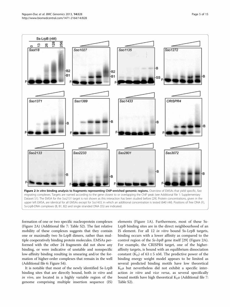
formation of one or two specific nucleoprotein complexes(Figure 2A) (Additional file 7: Table S2). The fast relativemobility of these complexes suggests that they containone or maximally two Ss-LrpB dimers, rather than mul-tiple cooperatively binding protein molecules. EMSAs per-formed with the other 24 fragments did not show anybinding, or were indicative of unstable and nonspecificlow-affinity binding resulting in smearing and/or the for-mation of higher-order complexes that remain in the well(Additional file 6: Figure S4).It is notable that most of the newly identified Ss-LrpB
binding sites that are directly bound, both in vitro andin vivo, are located in a highly variable region of thegenome comprising multiple insertion sequence (IS)
elements (Figure 1A). Furthermore, most of these Ss-LrpB binding sites are in the direct neighbourhood of anIS element. For all 12 in vitro bound Ss-LrpB targets,binding occurs with a lower affinity as compared to thecontrol region of the Ss-lrpB gene itself [29] (Figure 2A).For example, the CRISPR4 target, one of the higher-affinity targets, is bound with an equilibrium dissociationconstant (KD) of 63 ± 5 nM. The predictive power of thebinding energy weight model appears to be limited asseveral predicted binding motifs have low theoreticalKDs but nevertheless did not exhibit a specific inter-action in vitro and vice versa, as several specificallybound motifs have high theoretical KDs (Additional file 7:Table S2).
Figure 2 In vitro binding analysis to fragments representing ChIP-enriched genomic regions. Overview of EMSAs that yield specific, fastmigrating complexes. Targets are named according to the gene closest to or overlapping the ChIP peak (see Additional file 1: SupplementaryDataset S1). The EMSA for the Sso2131 target is not shown as this interaction has been studied before [29]. Protein concentrations, given in theupper left EMSA, are identical for all EMSAs except for Sso1433, in which an additional concentration is tested (640 nM). Positions of free DNA (F),Ss-LrpB-DNA complexes (B, B1, B2) and single stranded DNA (SS) are indicated.
Nguyen-Duc et al. BMC Genomics 2013, 14:828 Page 5 of 15http://www.biomedcentral.com/1471-2164/14/828

In vivo binding to Ss-lrpB, porDAB, Sso2126 and Sso2127operatorsUpon zooming into the profile at the Ss-lrpB genomicregion containing the regulatory target genes identifiedpreviously [9], we observed binding at all target promotersalthough signals in the Sso2126, Sso2127 and porDAB pro-moter regions did not reach the 2-fold enrichment thresh-old level in all replicates (Figure 3). Averaged peak heightsappear to correlate with both in vitro binding affinity andnumber of binding sites in the respective promoter/oper-ator (p/o) regions (binding affinities/number of bindingsites can be ranked as follows: p/oSs-lrpB > p/opor-DAB > p/oSso2127 > p/oSso2126 [9,29]). Furthermore,for the targets porDAB, Sso2126 and Sso2127 this cor-relation can be extended to the level of activation [9].An active copy of ISC1078 (containing a transposase
encoded by Sso2132) is located downstream of Ss-lrpBwith respect to genome sequence orientation [9,43].However, the steep decline in ChIP enrichment for theprobes representing the concerned IS sequence (Figure 3)and further PCR analysis (Additional file 8: Figure S5)demonstrated the absence of this element in a large sub-population of the cells that were subjected to ChIP.Interestingly, the EMSA screen also resulted in a further
unraveling of the Ss-lrpB operator for autoregulation: afragment comprising the sequence between the insertionsite of ISC1027 and the Sso2133 (glpK-2) promoter resultsin the formation of a single specific complex (Figure 2A,Sso2133 target). This observation implies sequence-specific recognition of another Ss-LrpB binding motif, lo-cated downstream (with respect to genome sequenceorientation) of Box3 with a center-to-center distanceof 174 bp (Additional file 9: Figure S6). Possibly, thissite, referred to as Box6, is an auxiliary operator sitethat supports binding to the main high-affinity oper-ator sites, besides the intragenic Box4 and Box5, which
were identified previously as secondary operator sites(Additional file 9: Figure S6) [35].
Ss-LrpB binds in CRISPR A and B leader regionsSs-LrpB is associated through direct high-affinity bindingwith two clusters of regularly interspaced short palin-dromic repeats (CRISPR) loci, which are essential ele-ments of an adaptive immunity system against viruses andother invading genetic elements. The concerned CRISPRloci, A and B, are paired family II CRISPRs sharing thesame repeat sequence and are bordered by quasi-identicalleader regions containing the elements to initiate and con-trol transcription of the long pre-crRNA [44]. Ss-LrpBbinding regions, previously annotated as “Sso1389” and“CRISPR4” , overlap the 502-bp long CRISPR A and Bleader regions, respectively (Figure 4A). ‘In gel’ Cu-OPfootprinting with a DNA probe representing the CRISPRB leader sequence clearly revealed Ss-LrpB-mediated pro-tection of a stretch of about 14 nucleotides (nt) corre-sponding to a relatively well conserved binding motif inthe promoter-proximal part of the leader (Figures 4Band C).Given the high sequence identity between the CRISPR
A and B leader regions, it is assumed that binding occursat the corresponding site with the same sequence in theCRISPR A leader (Figure 4D). The center of the identi-fied Ss-LrpB binding site is located 116/117 bp upstreamof the main pre-crRNA TSS [42] in the first CRISPR re-peat sequence, which is preceded by a strong promoter.This leads us to hypothesize that Ss-LrpB affects tran-scription of pre-crRNA through interaction with thebasal transcription machinery.
Permissivity of the genome for Ss-LrpB bindingUsing the binding energy position weight matrix, wesearched the entire S. solfataricus P2 genome for additional
Figure 3 Zoomed average ChIP-chip binding profile in the Ss-lrpB (Sso2131) genomic region. Below the profile, relative locations of genesand characterized Ss-LrpB binding sites [9,29] are shown in alignment with genomic positions. ORFs are indicated as horizontal arrows, whereasSs-LrpB binding sites are depicted as vertical lines.
Nguyen-Duc et al. BMC Genomics 2013, 14:828 Page 6 of 15http://www.biomedcentral.com/1471-2164/14/828

potential Ss-LrpB binding motifs. Setting the threshold forthe theoretical KD at 14 μM, a value still significantly lowerthan the average theoretical KD calculated for the novelidentified Ss-LrpB sites that are bound both in vivo andin vitro (290 μM), we detected 519 motifs of which 100 arelocated in regions 200 bp upstream of translational starts(Additional file 10: Table S3). Some of these motifs arecanonical Ss-LrpB binding motifs located at appropriatedistances from promoters to have the ability to exert
regulation. Nevertheless, binding is absent at these loca-tions in vivo.We selected two examples to illustrate this disagree-
ment between binding in vitro and in vivo (Figure 5;Additional file 11: Figure S7). The promoter region ofSso0049, encoding an unknown protein, contains a ca-nonical binding motif (5’-TTGTAATTTTTTCAA-3’)that is identical to the high-affinity Box 1 of the Ss-lrpBoperator 5’-TTGTAATTTTTACAA-3’ with the exception
Figure 4 Ss-LrpB binding to CRISPR A and CRISPR B leader regions. A. Zoomed average ChIP-chip binding profile of the CRISPR A/Bgenomic region. Below the profile, the genomic organization is schematically shown aligned to genomic positions. ORFs, mainly encodingCRISPR-associated and –related genes, are indicated as horizontal arrows, whereas the leader regions are depicted as grey boxes. Primarypre-crRNA TSSs are indicated by arrows. B. EMSA that was used for ‘in gel’ Cu-OP footprinting, using a 270-bp fragment representing the CRISPRB leader sequence. In this experiment, the bottom strand was 32P-labeled. Used protein concentrations are indicated. Populations of input DNA(I), free DNA (F) and bound DNA (B) are boxed in the same way as they were excised. C. Autoradiograph of the denaturing gel electrophoresis offootprinting samples. Direction of electrophoresis is indicated with an arrow, lanes contain either the I, B or F populations or the A + G and C + TMaxam-Gilbert sequencing ladders, as indicated. The protected region is indicated with a horizontal line and the corresponding sequence is given (5’ - > 3’).D. Alignment of the promoter-proximal CRISPR A and B leader sequences, with indication of the Ss-LrpB binding site, factor B recognition element(BRE), TATA box, first CRISPR repeat and primary TSS (arrow). Non-conserved residues are highlighted in grey; position numbering is with respect to theCRISPR B leader sequence.
Nguyen-Duc et al. BMC Genomics 2013, 14:828 Page 7 of 15http://www.biomedcentral.com/1471-2164/14/828

of one A → T substitution at a less critical position. AnEMSA probing binding of Ss-LrpB to a p/oSso0049 frag-ment revealed the formation of three distinct protein-DNA complexes (Figure 5B). DNase I footprinting showedthat it is indeed the predicted binding motif referred toas Box 1 that is bound at low protein concentration andis most likely protected in complex B1 in the EMSA(Figures 5B and C). Furthermore, an EMSA using afragment containing solely the Sso0049 Box 1 confirmed
a high-affinity interaction (KD ≈ 150 nM; Additional file 12:Figure S8). At higher Ss-LrpB concentrations, DNase Iprotection in the p/oSso0049 fragment was extended bothdownstream and upstream of Box 1. Upon manual inspec-tion of the sequence, we identified an additional bindingmotif (Box 2) with a center-to-center-distance of 20 bpupstream of Box 1 (Figure 5C). Despite the high-affinity,cooperative binding to multiple binding sites in vitro,no enrichment of this genomic region has been detected
Figure 5 Binding in the control region of Sso0049. A. Zoomed average ChIP-chip binding profile in the genomic region of Sso0049. Below theprofile, the genomic environment is schematically depicted by representing ORFs as grey arrows. The region corresponding to the fragmenttested in in vitro binding analyses, is boxed, whereas the identified Ss-LrpB binding sites are represented by vertical bars. B. EMSA of binding to a176 bp fragment encompassing the Sso0049 control region. DNA populations are indicated as follows: free DNA (F), Ss-LrpB-DNA complexes(B1, B2 and B3) and complexed DNA retained in the wells of the acrylamide gel (W). Protein concentrations are given (nM). C. DNase I footprinting withSs-LrpB binding to the Sso0049 control region fragment (having the top strand 32P labeled). A + G and C + T represent Maxam-Gilbert sequencingreactions whereas the other lanes contain DNase I-treated samples, with indication of the applied protein concentrations. Regions that areprotected against DNase I upon addition of a low Ss-LrpB concentration (< 500 nM) are indicated with a vertical line to the left-hand side of theautoradiograph, while regions that are protected at high concentration are indicated on the right-hand side. Similarly, hyperreactive sites areindicated with ball-and-stick symbols. D. Sequence of the Sso0048/Sso0049 intergenic region with indication of the regions that are protectedagainst DNase I in the experiment shown in subpanel C (dark grey = protected at all Ss-LrpB concentrations; light grey = only protected athigh Ss-LrpB concentrations) and in an experiment using DNA with the bottom strand labeled. Coding sequences are shown in uppercase(left side = Sso0048, right side = Sso0049). The TSS of Sso0049, indicated with an arrow, was experimentally determined with primer extensionanalysis (Additional file 13: Figure S9).
Nguyen-Duc et al. BMC Genomics 2013, 14:828 Page 8 of 15http://www.biomedcentral.com/1471-2164/14/828

in the ChIP-chip assay (Figure 5A). Notably, Ss-LrpB isassociated with the genome about 1 kb upstream of theSso0049 control region in the ORF of Sso0046. This obser-vation suggests that absence of Ss-LrpB at p/oSso0049 isnot caused by limited diffusion of the TF throughout thecell but rather to an inaccessibility of chromatin or theDNA sequence itself at this locus.A similar discrepancy between in vitro and in vivo
binding was shown to exist for the intergenic promoter re-gion shared between the divergently transcribed Sso2342,encoding a purine phosphoribosyltransferase (gpT-1), andSso2343, encoding a 5’-methylthioadenosine phosphoryl-ase (mtaP) (Additional file 11: Figure S7; Additional file12: Figure S8; Additional file 13: Figure S9). Altogether,these observations demonstrate that the target DNA siteswithin the S. solfataricus genome are not entirely permis-sive for Ss-LrpB binding.
Overlap between Ss-LrpB and LysM binding profilesS. solfataricus possesses additional Lrp-like TFs, such asthe lysine-responsive LysM. We compared the Ss-LrpBbinding profile to the locations of the LysM binding re-gions mapped previously [16] and observed a significantoverlap: 29 of the 37 Ss-LrpB binding regions were alsoassociated with LysM (Figure 6A; Table 1). Of note, nocross-reactivity of Ss-LrpB- and LysM-specific nanobodieswith other Lrp-type proteins has ever been observed[16,35]. Zoomed binding profiles for both TFs are highlysimilar, which is a striking observation given that bothprofiles were recorded in different growth conditions(sucrose-supplemented for LysM-specific ChIP versustryptone-supplemented medium for Ss-LrpB-specificChIP) (Figure 6B).The known regulatory targets of Ss-LrpB, Sso2126,
Sso2127 and porDAB, are not co-bound by LysM, incontrast to most of the newly discovered low-affinity Ss-LrpB binding targets. Conversely, the main local regulatorytarget of LysM, the lysWXJK operon for lysine biosynthesis,is also bound by Ss-LrpB. To distinguish between (i) amutually exclusive binding of either Ss-LrpB or LysM ata particular target in a subpopulation of cells, or (ii) asituation where the proteins bind as a co-complex tothis target, we compared in vitro and in silico bindingcharacteristics for these targets (Table 1). Shared bind-ing regions could be categorized in three classes: (i)binding regions that contain an Ss-LrpB binding motifand exhibit binding to Ss-LrpB but not LysM in vitro(class I); (ii) binding regions that contain a LysM bind-ing motif and exhibit (predicted) binding to LysM butnot Ss-LrpB in vitro (class II) and (iii) binding regionsthat do not contain an Ss-LrpB or LysM binding motifand do not interact with any of the two proteins in vitro(class III). There is a perfect inverse correlation patternbetween the presence of a bona fide Ss-LrpB or LysM
binding motif suggesting that the TFs are co-localizedthrough protein-protein interaction and that only oneof the protein partners directly interacts with the DNA.We further investigated the simultaneous interaction
of LysM and Ss-LrpB to one of the co-bound targets,Ssot28, in an in vitro assay (Figure 6C). Whereas Ss-LrpB does not form specific complexes with this target(Additional file 6: Figure S4), LysM forms a single com-plex by binding to a site located close to the promoter ofthe glutamate synthase (gltB) gene [16]. EMSA analysisdemonstrated that the addition of Ss-LrpB to reactionmixtures containing LysM and the DNA stimulatedslightly the complex formation (Figure 6C). Further-more, the addition of Ss-LrpB-specific antibodies re-sulted in a clear supershift of the complex. Theseobservations suggest that Ss-LrpB is present in the nu-cleoprotein complex, despite that it is unable to interactwith the DNA fragment by itself.
Gene expression analysis of transcripts associated withSs-LrpB binding regionsTo determine whether the identified binding eventscause transcription regulation of neighbouring genes, weinvestigated the effect of deleting Ss-lrpB on the ex-pression of a wide selection of potential target genesby quantitative reverse transcriptase PCR (qRT-PCR)(Figure 7). Some of these genes were associated with dir-ect Ss-LrpB binding (class I), others with indirect bind-ing (class II, III and IV), yet others with co-binding ofSs-LrpB and LysM (class I, II and III) (for a definition ofthe classes, see legend of Figure 7). For some of them,binding occurs relatively close to the promoter whereasfor other genes the binding is intragenic and far awayfrom the closest promoter region (e.g. Sso2233). Withthe exception of the CRISPR B pre-crRNA, deletion ofSs-lrpB did not significantly affect the expression of anyof these tested target genes. We have also confirmed thatthe TF does not significantly affect the expression ofSso0049, which contains high-affinity Ss-LrpB bindingsites in its promoter region that are however not associ-ated with Ss-LrpB in vivo.The expression of CRISPR B pre-crRNA is moderately
downregulated (2-fold) in the Ss-lrpB::lacS strain incomparison to the isogenic WT, indicating an Ss-LrpB-mediated activation. It is assumed that a similar regulationwill be exerted at the CRISPR A locus. In conclusion, Ss-LrpB is involved in CRISPR regulation whereas the otheridentified binding events appear to occur without appar-ent regulation under our conditions of growth.
DiscussionOur genome-wide localization study has revealed theassociation of Ss-LrpB with at least 37 loci in the S.solfataricus genome. A subset of these in vivo Ss-LrpB-
Nguyen-Duc et al. BMC Genomics 2013, 14:828 Page 9 of 15http://www.biomedcentral.com/1471-2164/14/828

targeted sites is clearly validated to be bound with thispure TF in vitro and to contain a bona fide sequencemotif. Obviously, the well-established, high sequencespecificity of Ss-LrpB [32] is responsible for the complexformation at these sites. However, computational ana-lysis with a binding energy based position weight matrixof the S. solfataricus genome demonstrated a vast
overrepresentation of appropriate sequence motifs ofwhich only a very small subset seems to be actuallybound in vivo. A high number of false negative signals inthe ChIP-chip analysis, e.g. due to a too stringent thresh-old, is a possible explanation for the observed lack ofin vivo binding at predicted motifs. However, a closer in-vestigation of the binding profiles at loci containing
Figure 6 Overlap between Ss-LrpB and LysM binding profiles. A. Venn diagram illustrating shared binding regions between Ss-LrpB andLysM. B. Zoomed binding profiles of Ss-LrpB- and LysM-specific ChIP assays for three selected targeted regions, belonging to one of three classes:class I: binding region that contains an Ss-LrpB binding motif and exhibits binding to Ss-LrpB but not LysM in vitro; class II: binding region thatcontains a LysM binding motif and exhibits binding to LysM but not Ss-LrpB in vitro; class III: binding region that does not contain an Ss-LrpB orLysM binding motif and does not interact with any of the two proteins in vitro. Aligned with the binding profiles, the genomic organization isdepicted by representing ORFs as arrows. C. EMSA demonstrating simultaneous binding of Ss-LrpB and LysM to a 348-bp probe encompassingthe Ssot28 target. The positions of free DNA (F) and bound DNA (B) are indicated. The following protein concentrations have been used: 40 nMLysM, 320 nM Ss-LrpB, 500 nM Ss-LrpB-specific Nb and 500 nM E. coli PepA, which was used as a negative control.
Nguyen-Duc et al. BMC Genomics 2013, 14:828 Page 10 of 15http://www.biomedcentral.com/1471-2164/14/828

multiple high-affinity binding motifs (promoter regionsof Sso0049 and mtaP) confirmed complete absence ofSs-LrpB-specific enrichment in these regions. Therefore,we conclude that the intrinsic DNA-binding sequencespecificity is a poor predictor of binding in vivo underour culture conditions of S. solfataricus and that add-itional factors are involved in determining site selectivityand occupancy in vivo.In vivo binding site selectivity could be influenced by
the structural landscape of the chromatin that imposesdifferential genome accessibility on a higher organizationallevel. Similarly to bacteria, Crenarchaeota have their nucleoid
structurally organized by small chromatin proteins [45],however it is unknown how this genome packaging affectsTF binding. Possibly, Ss-LrpB binding is restricted by theaction of nucleoid associated proteins resulting in dif-ferential accessible genomic regions. Apparently, acces-sibility is facilitated in a highly variable region of the S.solfataricus genome with multiple IS elements and lowabundance of essential genes. However, there are alternativeexplanations available for the lack of association at high-affinity binding motifs. For instance, it might be caused bythe presence of a co-repressor, ligand or post-translationalmodification that inhibits Ss-LrpB binding under the used
Table 1 Overview of the Ss-LrpB-specific ChIP-enriched regions that are also enriched in LysM-specific ChIP-chipanalysis
Targetname
Genomic coordinatepeak start
Genomic coordinatepeak stop
Relative position toLysM-specific peak
Presence of Ss-LrpBbinding motif
Presence of LysMbinding motif
Class
Sso0154 129616 130620 Inside - - III
Sso5317 131930 133008 Including - + II
Ssot18 290428 291131 Inside + - I
Ssot28 589572 590362 overlap (89%) - + II
Sso6904 835210 835839 Inside - + II
Sso1027 885479 886771 Inside + - I
Sso1114 960343 961574 Including - -* III
Sso1118 964702 966378 Including - -* III
Sso1135 974250 975551 overlap (99%) + -* I
Sso1272 1095398 1097098 Inside + -* I
Sso1371 1206614 1207226 overlap (97%) + -* I
Sso1389 1232759 1233642 overlap (99%) + -* I
CRISPR4 1260282 1261337 Including + -* I
Sso1433 1286420 1286928 Inside + -* I
Sso1463 1321798 1323084 overlap (98%) - -* III
Sso1890 1705187 1705830 Inside - -* III
Sso2043 1859092 1860291 overlap (75%) - + II
Sso2159 1985046 1985784 Inside - -* III
Sso2233 2050931 2052374 Including + -* I
Sso2289 2099312 2099902 Inside - -* III
Sso2309 2111358 2112389 Inside - -* III
Sso2310 2113136 2114378 overlap (93% ) - -* III
Sso2334 2132622 2134455 Including - + II
Sso2404 2187776 2188318 Inside - -* III
Sso2678 2437317 2437768 overlap (96%) - -* III
Sso2801 2561656 2562152 overlap (57%) - -* III
Sso2833 2594520 2595831 overlap (96%) - -* III
Sso3002 2742947 2743947 Including - -* III
Sso3072 2827204 2828448 overlap (94%) + -* I
The presence of an Ss-LrpB binding motif (measured by EMSA) or a LysM binding motif (either measured by EMSA or predicted in silico (indicated by an asterisk)[16]) is indicated. Classes are defined as follows: class I: binding region that contains an Ss-LrpB binding motif and exhibits binding to Ss-LrpB but not LysMin vitro; class II: binding region that contains a LysM binding motif and exhibits binding to LysM but not Ss-LrpB in vitro; class III: binding region that does notcontain an Ss-LrpB or LysM binding motif and does not interact with any of the two proteins in vitro. Ranking is according to genomic location.
Nguyen-Duc et al. BMC Genomics 2013, 14:828 Page 11 of 15http://www.biomedcentral.com/1471-2164/14/828

culture conditions or, on the contrary, by the absence of acritical ligand in vivo that activates binding and was presentin the in vitro binding reaction mixtures, possibly throughco-purification after heterologous expression of Ss-LrpB inE. coli. Technical details, such as unstable behavior of theTF-DNA complexes during formaldehyde fixation or sonic-ation, could also lead to certain binding events not beingdetected.There is a very low correlation between Ss-LrpB binding
and transcription regulation, which appears to be limitedto the Sso2126, Sso2127 and porDAB gene targets thatwere identified previously and to the CRISPR A and Bclusters. Furthermore, a significant fraction of binding re-gions is located at a significant distance from the nearestTSS and associated promoter and/or is even intragenic, anobservation made for bacterial [46-49] and for archaealTFs as well [16,50]. The newly discovered genomic targetsthat contain a binding motif are generally contacted by Ss-LrpB through non-cooperative low-affinity binding, incontrast to the local gene targets. Low-affinity bindingwithout apparent regulation appears to be universal as ithas been observed repeatedly for TFs of M. tuberculosis[51], yeast [52] and Drosophila [53]. However, the bio-logical function of these weak binding sites is still unclear.It has been hypothesized that they could display a subtleregulatory activity, which goes undetected and causes afine-tuning of gene regulation. In this way, these bindingsites alleviate selective pressure on specific loci, namelyclassical regulatory binding sites, and increase the abilityto evolve [53]. An alternative explanation for the biological
role of these low-affinity sites is that they might create abiological buffering system that serves as a reservoir to se-quester TF molecules, thereby thermodynamically regulat-ing the concentration of freely available protein [53,54].This could be a critical factor for correct functioning ofSs-LrpB, given the limited number of target genes and thecooperative nature of the interaction at these targets.The observed occupancy levels and regulatory effects of
the major regulatory targets (Sso2126, Sso2127, porDAB[9] and CRISPR B) are weak and were most probably re-corded in a growth condition yielding a non-regulated“ground state” . Possibly, a different, as yet unknown, cul-turing condition leads to higher occupation and corre-sponding activation levels of the regulatory target genes.Similarly, some of the newly identified binding sites mightmediate regulation of nearby genes under different growthconditions than those in which the binding profile wasmonitored. The exact functions and substrate specificitiesof the pyruvate ferredoxine oxidoreductase and the twopermeases are unclear although it is tentative to speculatethat these proteins function during lactate oxidation or arelated metabolic pathway [9]. Indeed, Sso2126 encodes apermease that exhibits homology with bacterial L-lactatepermeases and Sso2127 codes for a homolog of halophilicoxalate/formate antiporters. Protein sequence analysissuggests that if Ss-LrpB is bound by an effector molecule,it is a small molecule other than an amino acid [9].The growth condition that is more relevant for Ss-
LrpB regulation might be a more stressful condition forthe cells, given the Ss-LrpB-mediated CRISPR activation.
Figure 7 Expression analysis of genes of which the coding regions are located close to a selection of Ss-LrpB binding regions. Testedgenes are subdivided into the following classes: class I: gene associated to binding region bound by LysM and Ss-LrpB that contains an Ss-LrpBbinding motif and exhibits binding to Ss-LrpB but not LysM in vitro; class II: gene associated to binding region bound by LysM and Ss-LrpB thatcontains a LysM binding motif and exhibits binding to LysM but not Ss-LrpB in vitro; class III: gene associated to binding region bound by LysMand Ss-LrpB that does not contain an Ss-LrpB or LysM binding motif and does not interact with any of the two proteins in vitro; class IV: geneassociated to binding region solely bound by Ss-LrpB in vivo; class V: gene associated to region that is bound by Ss-LrpB in vitro but not in vivo.Relative gene expression ratios in an Ss-lrpB::lacS versus PBL2025 strain are normalized against tbp expression. Values are the average of biologicalquadruplicates and standard deviations represent the biological variation. An asterisk indicates a P-value of 0.085. All other tested genes renderP-values > 0.1, indicating no significant differences in expression. Absence of Ss-lrpB expression in the Ss-lrpB::lacS strain has been confirmed(fold ratio = 0.0031 ± 0.0020; P-value = 0.0001).
Nguyen-Duc et al. BMC Genomics 2013, 14:828 Page 12 of 15http://www.biomedcentral.com/1471-2164/14/828

It has been demonstrated that expression of CRISPRand CRISPR-associated (CAS) genes is inducible by abi-otic and/or biotic stress [55]. The high energetic cost ofexpressing the long pre-crRNA leads to hypothesize thatit is under a complex transcriptional regulation involvingmultiple regulators, of which we demonstrate that Ss-LrpB is the first promoter-interacting TF to be charac-terized in S. solfataricus. Co-regulation of porDAB,Sso2126 and Sso2127 on one hand and the CRISPR clus-ters on the other hand indicates that there is a possiblelink between metabolic regulation and immunity defensein S. solfataricus.Remarkably, the Ss-LrpB and LysM binding profiles dis-
play a significant overlap. The detection of the two pro-teins at the same genomic location in different ChIP-chipprofiles could be explained by (i) binding of a hetero-protein complex of LysM and Ss-LrpB to the DNA targetor (ii) a heterogeneous occupancy where Ss-LrpB is pre-sent on the DNA site in some cells, and LysM on the cor-responding DNA site in other cells or (iii) a combinationof both possibilities. In the case of these two Lrp-like TFs,the inverse correlation pattern between the presence of aLysM or Ss-LrpB binding motif suggests that they are sim-ultaneously associated as a complex with the target DNAsites in the S. solfataricus genome. Furthermore, this co-association occurs most likely via protein-protein interac-tions in which only one of the protein partners interactswith DNA, rather than by cooperative interactions be-tween distinct DNA-bound TF molecules. This proposalis supported by our in vitro experiments where Ss-LrpBwas shown to be present in the protein-DNA complex al-though it does not interact with the DNA by itself. For adistinct class (“class III”) of binding targets, direct DNArecognition by Ss-LrpB or LysM was clearly absent, sug-gesting the involvement of additional TFs. Remarkably, wedid not observe significant Ss-LrpB-mediated regulationof the major LysM targets to which co-association was ob-served. Possibly, the presence of Ss-LrpB results only insubtle regulatory effects, fine-tuning the regulatory actionof LysM, and the involvement of different partners is par-tially interchangeable. Our data demonstrate that archaealLrp-like TFs interact in vivo, supporting previous data thatPyrococcus Lrp-like TFs tend to form hetero-oligomericstructures in vitro [11,17].
ConclusionsIn conclusion, our genome-wide association study of Ss-LrpB yields novel insights into its in vivo interactions des-pite providing only limited additional information on itsphysiological role. Ss-LrpB interacts with multiple low-affinity sites throughout the genome without an apparentregulatory purpose and these sites are often associatedwith IS elements. Furthermore, the TF binds in theCRISPR A and B leader regions and activates expression
of pre-crRNA. Ss-LrpB also co-associates with anotherLrp-like TF, LysM. Hetero-oligomerization of archaeal Lrpproteins was previously observed in vitro and thus, weprovide the first indications of an interplay of two of thesefactors in vivo. Finally, the absence of Ss-LrpB in vivo onsites carrying a well-predicted binding motif suggests alimited permissivity of the S. solfataricus genome for asso-ciation with its cognate TF.
Availability of supporting dataAll the supporting data are included as additional files.
Additional files
Additional file 1: Dataset S1. Overview of all ChIP-enriched regionsidentified in the study, with indication of details of qPCR and EMSA ex-periments for validation.
Additional file 2: Table S1. List of oligonucleotide sequences used inthis work.
Additional file 3: Figure S1. Statistical analysis of Ss-LrpB ChIP enrichmentobtained by DNA microarray hybridization and qPCR analysis.
Additional file 4: Figure S2. Pie chart displaying the fractions offunctional classes to which the genes, closest to or overlapping theidentified Ss-LrpB binding regions, belong.
Additional file 5: Figure S3. Zoomed profiles of ChIP-enriched regions(partially) covering two open reading frames or more.
Additional file 6: Figure S4. In vitro binding analysis to fragmentsrepresenting ChIP-enriched genomic regions.
Additional file 7: Table S2. (Putative) binding motifs in the sequencesbound by Ss-LrpB in vivo and in vitro.
Additional file 8: Figure S5. PCR analysis of genomic DNA targetingthe genomic region that flanks Ss-lrpB and contains an ISC1078 elementbetween positions 1959047 and 1960125 in the published S. solfataricusP2 genome sequence.
Additional file 9: Figure S6. Schematic representation of the Ss-lrpBoperator structure in the case of absence of ISC1078.
Additional file 10: Table S3. In silico predicted Ss-LrpB binding sites(using the binding energy weight matrix) in regions upstream of S. solfataricusP2 ORFs (200 bp upstream of ORF start) with a theoretical KD lowerthan 5 μM.
Additional file 11: Figure S7. In vitro binding in the control region ofgpT-1 (Sso2342)/ mtaP (Sso2343).
Additional file 12: Figure S8. In vitro binding to the separate Ss-LrpBbinding sites identified in the Sso0049 and gpT-1/mtaP control regions.
Additional file 13: Figure S9. Primer extension analysis for TSSdetermination.
Competing interestsThe authors declare that they have no competing interests.
Authors' contributionsDesigned and coordinated the study: GH-G, SM, DC, EP. Performedexperimental work: TND, NS, DC, EP. Contributed to data analysis: TND,LvO, EP. Drafted the manuscript: TND, EP. All authors read and approvedthe final manuscript.
AcknowledgementsThe authors are grateful to Phu Nguyen Le Minh for the gift of purified PepAprotein. This work was supported by the Research Foundation Flanders(FWO-Vlaanderen) (a postdoctoral fellowship to EP and a pre-doctoral fellowshipto LvO), the Flemish Institute of Biotechnology (Vlaams Instituut voorBiotechnologie (VIB)), the Research Council of the Vrije Universiteit Brussel,
Nguyen-Duc et al. BMC Genomics 2013, 14:828 Page 13 of 15http://www.biomedcentral.com/1471-2164/14/828

the Vlaamse Gemeenschapscommissie, the China Scolarship Council (CSC) andthe European Union (EU) (project 241481 (AFFINOMICS) to SM).
Author details1Research group of Cellular and Molecular Immunology, Vrije UniversiteitBrussel, Pleinlaan 2, B-1050 Brussels, Belgium. 2Department of StructuralBiology, VIB, Pleinlaan 2, B-1050 Brussels, Belgium. 3Research group ofMicrobiology, Vrije Universiteit Brussel, Pleinlaan 2, B-1050 Brussels, Belgium.4IMEC, Kapeldreef 75, B-3001 Leuven, Belgium. 5Department of Microbiologyand Immunology, State University of New York at Buffalo, 321 Cary Hall, 3435Main Street, 14214Buffalo, NY, United States.
Received: 13 June 2013 Accepted: 15 November 2013Published: 25 November 2013
References1. Kyrpides NC, Ouzounis CA: Transcription in archaea. Proc Natl Acad Sci
USA 1999, 96:8545–8550.2. Pérez-Rueda E, Collado-Vides J, Segovia L: Phylogenetic distribution of
DNA-binding transcription factors in bacteria and archaea. Comput BiolChem 2004, 28:341–350.
3. Charoensawan V, Wilson D, Teichmann SA: Genomic repertoires of DNA-binding transcription factors across the tree of life. Nucleic Acids Res 2010,38:7364–7377.
4. Perez-Rueda E, Janga SC: Identification and genomic analysis oftranscription factors in archaeal genomes exemplifies their functionalarchitecture and evolutionary origin. Mol Biol Evol 2010, 27:1449–1459.
5. Brinkman AB, Ettema TJG, de Vos WM, van der Oost J: The Lrp family oftranscriptional regulators. Mol Microbiol 2003, 48:287–294.
6. Ouhammouch M, Dewhurst RE, Hausner W, Thomm M, Geiduschek EP:Activation of archaeal transcription by recruitment of the TATA-bindingprotein. Proc Natl Acad Sci USA 2003, 100:5097–5102.
7. Ouhammouch M, Langham GE, Hausner W, Simpson AJ, El-Sayed NMA,Geiduschek EP: Promoter architecture and response to a positiveregulator of archaeal transcription. Mol Microbiol 2005, 56:625–637.
8. Kawashima T, Aramaki H, Oyamada T, Makino K, Yamada M, Okamura H,Yokoyama K, Ishijima SA, Suzuki M: Transcription regulation by feast/famine regulatory proteins, FFRPs, in archaea and eubacteria. Biol PharmBull 2008, 31:173–186.
9. Peeters E, Albers SV, Vassart A, Driessen AJM, Charlier D: Ss-LrpB, atranscriptional regulator from Sulfolobus solfataricus, regulates a genecluster with a pyruvate ferredoxin oxidoreductase-encoding operon andpermease genes. Mol Microbiol 2009, 71:972–988.
10. Brinkman AB, Bell SD, Lebbink RJ, de Vos WM, van der Oost J: TheSulfolobus solfataricus Lrp-like protein LysM regulates lysine biosynthesisin response to lysine availability. J Biol Chem 2002, 277:29537–29549.
11. Okamura H, Yokoyama K, Koike H, Yamada M, Shimowasa A, Kabasawa M,Kawashima T, Suzuki M: A structural code for discriminating betweentranscription signals revealed by the feast/famine regulatory proteinDM1 in complex with ligands. Structure 2007, 15:1325–1338.
12. Yokoyama K, Ishijima SA, Koike H, Kurihara C, Shimowasa A, Kabasawa M,Kawashima T, Suzuki M: Feast/famine regulation by transcription factorFL11 for the survival of the hyperthermophilic archaeon Pyrococcus OT3.Structure 2007, 15:1542–1554.
13. Kumarevel T, Nakano N, Ponnuraj K, Gopinath SCB, Sakamoto K, Shinkai A,Kumar PKR, Yokoyama S: Crystal structure of glutamine receptor proteinfrom Sulfolobus tokodaii strain 7 in complex with its effector L-glutamine:implications of effector binding in molecular association and DNA binding.Nucleic Acids Res 2008, 36:4808–4820.
14. Miyazono KI, Tsujimura M, Kawarabayasi Y, Tanokura M: Crystal structure ofSTS042, a stand-alone RAM module protein, from hyperthermophilicarchaeon Sulfolobus tokodaii strain 7. Proteins 2008, 71:1557–1562.
15. Schwaiger R, Schwarz C, Furtwangler K, Tarasov V, Wende A, Oesterhelt D:Transcriptional control by two leucine-responsive regulatory proteins inHalobacterium salinarum R1. BMC Mol Biol 2010, 11:40.
16. Song N, Nguyen Duc T, van Oeffelen L, Muyldermans S, Peeters E, CharlierD: Expanded target and cofactor repertoire for the transcriptionalactivator LysM from Sulfolobus. Nucleic Acids Res 2013, 41:2932–2949.
17. Yokoyama K, Ishijima SA, Clowney L, Koike H, Aramaki H, Tanaka C, MakinoK, Suzuki M: Feast/famine regulatory proteins (FFRPs): Escherichia coli Lrp,
AsnC and related archaeal transcription factors. FEMS Microbiol Rev 2006,30:89–108.
18. Vassart A, van Wolferen M, Orell A, Hong Y, Peeters E, Albers SV, Charlier D: Sa-Lrp from Sulfolobus acidocaldarius is a versatile, glutamine-responsive, andarchitectural transcriptional regulator. Microbiology Open 2013, 2:75–93.
19. Peeters E, Charlier D: The Lrp family of transcription regulators in archaea.Archaea 2010, 2010:750457.
20. Ettema TJG, Brinkman AB, Tani TH, Rafferty JB, van der Oost J: A novelligand-binding domain involved in regulation of amino acid metabolismin prokaryotes. J Biol Chem 2002, 277:37464–37468.
21. Brinkman AB, Dahlke I, Tuininga JE, Lammers T, Dumay V, de Heus E,Lebbink JH, Thomm M, de Vos WM, van der Oost J: An Lrp-like transcriptionalregulator from the archaeon Pyrococcus furiosus is negativelyautoregulated. J Biol Chem 2000, 275:38160–38169.
22. Enoru-Eta J, Gigot D, Thia-Toong TL, Glansdorff N, Charlier D: Purification andcharacterization of Sa-Lrp, a DNA-binding protein from the extreme ther-moacidophilic archaeon Sulfolobus acidocaldarius homologous to the bac-terial global transcriptional regulator Lrp. J Bacteriol 2000, 182:3661–3672.
23. Leonard PM, Smits SH, Sedelnikova SE, Brinkman AB, de Vos WM, van derOost J, Rice DW, Rafferty JB: Crystal structure of the Lrp-like transcriptionalregulator from the archaeon Pyrococcus furiosus. EMBO J 2001,20:990–997.
24. Ouhammouch M, Geiduschek EP: A thermostable platform for transcriptionalregulation: the DNA-binding properties of two Lrp homologs from thehyperthermophilic archaeon Methanococcus jannaschii. EMBO J 2001,20:146–156.
25. Enoru-Eta J, Gigot D, Glansdorff N, Charlier D: High resolution contact probingof the Lrp-like DNA-binding protein Ss-Lrp from the hyperthermoacidophiliccrenarchaeote Sulfolobus solfataricus P2. Mol Microbiol 2002,45:1541–1555.
26. Koike H, Ishijima SA, Clowney L, Suzuki M: The archaeal feast/famineregulatory protein: potential roles of its assembly forms for regulatingtranscription. Proc Natl Acad Sci USA 2004, 101:2840–2845.
27. Peeters E, Willaert R, Maes D, Charlier D: Ss-LrpB from Sulfolobussolfataricus condenses about 100 base pairs of its own operator DNAinto globular nucleoprotein complexes. J Biol Chem 2006,281:11721–11728.
28. Pritchett MA, Wilkinson SP, Geiduschek EP, Ouhammouch M: Hybrid Ptr2-likeactivators of archaeal transcription. Mol Microbiol 2009, 74:582–593.
29. Peeters E, Thia-Toong TL, Gigot D, Maes D, Charlier D: Ss-LrpB, a novel Lrp-likeregulator of Sulfolobus solfataricus P2, binds cooperatively to threeconserved targets in its own control region. Mol Microbiol 2004,54:321–336.
30. Peeters E, Peixeiro N, Sezonov G: Cis-regulatory logic in archaealtranscription. Biochem Soc Trans 2013, 41:326–331.
31. Peeters E, van Oeffelen L, Nadal M, Forterre P, Charlier D: A thermodynamicmodel of the cooperative interaction between the archaeal transcriptionfactor Ss-LrpB and its tripartite operator DNA. Gene 2013, 524:330–340.
32. Peeters E, Wartel C, Maes D, Charlier D: Analysis of the DNA-bindingsequence specificity of the archaeal transcriptional regulator Ss-LrpBfrom Sulfolobus solfataricus by systematic mutagenesis and highresolution contact probing. Nucleic Acids Res 2007, 35:623–633.
33. Schelert J, Dixit V, Hoang V, Simbahan J, Drozda M, Blum P: Occurrenceand characterization of mercury resistance in the hyperthermophilicarchaeon Sulfolobus solfataricus by use of gene disruption. J Bacteriol2004, 186:427–437.
34. Brock TD, Brock KM, Belly RT, Weiss RL: Sulfolobus: a new genus of sulfur-oxidizingbacteria living at low pH and high temperature. Arch Mikrobiol 1972,84:54–68.
35. Nguyen Duc T, Peeters E, Muyldermans S, Charlier D, Hassanzadeh-Ghassabeh G:Nanobody(R)-based chromatin immunoprecipitation/micro-array analysisfor genome-wide identification of transcription factor DNA binding sites.Nucleic Acids Res 2012, 41:e59.
36. Toedling J, Huber W: Analyzing ChIP-chip data using bioconductor. PLoSComput Biol 2008, 4:e1000227.
37. Bailey TL, Boden M, Buske FA, Frith M, Grant CE, Clementi L, Ren J, Li WW,Noble WS: MEME SUITE: tools for motif discovery and searching. NucleicAcids Res 2009, 37:W202–W208.
38. Vieira J, Messing J: The pUC plasmids, an M13mp7-derived system forinsertion mutagenesis and sequencing with synthetic universal primers.Gene 1982, 19:259–268.
Nguyen-Duc et al. BMC Genomics 2013, 14:828 Page 14 of 15http://www.biomedcentral.com/1471-2164/14/828

39. Maxam AM, Gilbert W: Sequencing end-labeled DNA with base-specificchemical cleavages. Meth Enzymol 1980, 65:499–560.
40. Rozen S, Skaletsky H: Primer3 on the WWW for general users and forbiologist programmers. Methods Mol Biol 2000, 132:365–386.
41. Livak KJ, Schmittgen TD: Analysis of relative gene expression data usingreal-time quantitative PCR and the 2(−Delta Delta C(T)) Method.Methods 2001, 25:402–408.
42. Wurtzel O, Sapra R, Chen F, Zhu Y, Simmons BA, Sorek R: A single-baseresolution map of an archaeal transcriptome. Genome Res 2010,20:133–141.
43. She Q, Singh RK, Confalonieri F, Zivanovic Y, Allard G, Awayez MJ, Chan-WeiherCC, Clausen IG, Curtis BA, De Moors A, et al: The complete genome of thecrenarchaeon Sulfolobus solfataricus P2. Proc Natl Acad Sci USA 2001,98:7835–7840.
44. Lillestøl RK, Shah SA, Brügger K, Redder P, Phan H, Christiansen J, Garrett RA:CRISPR families of the crenarchaeal genus Sulfolobus: bidirectionaltranscription and dynamic properties. Mol Microbiol 2009, 72:259–272.
45. Driessen RPC, Dame RT: Structure and dynamics of the crenarchaealnucleoid. Biochem Soc Trans 2013, 41:321–325.
46. Smollett KL, Smith KM, Kahramanoglou C, Arnvig KB, Buxton RS, Davis EO:Global analysis of the regulon of the transcriptional repressor LexA, akey component of SOS response in Mycobacterium tuberculosis. J BiolChem 2012, 287:22004–22014.
47. Grainger DC, Aiba H, Hurd D, Browning DF, Busby SJW: Transcription factordistribution in Escherichia coli: studies with FNR protein. Nucleic Acids Res2007, 35:269–278.
48. Shimada T, Ishihama A, Busby SJW, Grainger DC: The Escherichia coli RutRtranscription factor binds at targets within genes as well as intergenicregions. Nucleic Acids Res 2008, 36:3950–3955.
49. Cho BK, Federowicz S, Park YS, Zengler K, Palsson BØ: Deciphering thetranscriptional regulatory logic of amino acid metabolism. Nat Chem Biol2011, 8:65–71.
50. Schmid AK, Reiss DJ, Pan M, Koide T, Baliga NS: A single transcriptionfactor regulates evolutionarily diverse but functionally linked metabolicpathways in response to nutrient availability. Mol Syst Biol 2009, 5:282.
51. Galagan J, Lyubetskaya A, Gomes A: ChIP-Seq and the complexity ofbacterial transcriptional regulation. Curr Top Microbiol Immunol 2013,363:43–68.
52. Tanay A: Extensive low-affinity transcriptional interactions in the yeastgenome. Genome Res 2006, 16:962–972.
53. Li XY, MacArthur S, Bourgon R, Nix D, Pollard DA, Iyer VN, Hechmer A,Simirenko L, Stapleton M, Luengo Hendriks CL, et al: Transcription factorsbind thousands of active and inactive regions in the Drosophilablastoderm. PLoS Biol 2008, 6:e27.
54. Macquarrie KL, Fong AP, Morse RH, Tapscott SJ: Genome-widetranscription factor binding: beyond direct target regulation.Trends Genet 2011, 27:141–148.
55. Manica A, Schleper C: CRISPR-mediated defense mechanisms in thehyperthermophilic archaeal genus Sulfolobus. RNA biology 2013,10:671–678.
56. Chan PP, Holmes AD, Smith AM, Tran D, Lowe TM: The UCSC ArchaealGenome Browser: 2012 update. Nucleic Acids Res 2012, 40:D646–D652.
doi:10.1186/1471-2164-14-828Cite this article as: Nguyen-Duc et al.: The genome-wide binding profileof the Sulfolobus solfataricus transcription factor Ss-LrpB shows bindingevents beyond direct transcription regulation. BMC Genomics 2013 14:828.
Submit your next manuscript to BioMed Centraland take full advantage of:
• Convenient online submission
• Thorough peer review
• No space constraints or color figure charges
• Immediate publication on acceptance
• Inclusion in PubMed, CAS, Scopus and Google Scholar
• Research which is freely available for redistribution
Submit your manuscript at www.biomedcentral.com/submit
Nguyen-Duc et al. BMC Genomics 2013, 14:828 Page 15 of 15http://www.biomedcentral.com/1471-2164/14/828


