The geko™ a neuromuscular electrostimulation (NMES) device ... · The geko™ a neuromuscular...
1
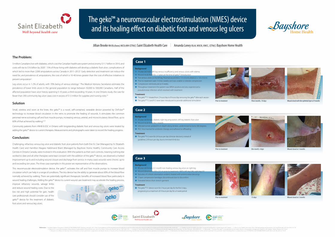
The geko™ a neuromuscular electrostimulation (NMES) device and its healing effect on diabetic foot and venous leg ulcers Jillian Brooke RN BSc(hons) MClScWH CETN(C) Saint Elizabeth Health Care | Amanda Loney BScN, WOCN, IIWCC, CETN(C) Bayshore Home Health The Problem: 3 million Canadians live with diabetes, which cost the Canadian healthcare system and economy $11.7 billion in 2010, and costs will rise to $16 billion by 2020 1 . 15% of those living with diabetes will develop a diabetic foot ulcer, complications of which led to more than 2,000 amputations across Canada in 2011–2012 2 . Early detection and treatment can reduce the need for, and prevalence of, amputations, the cost of which is 10-40 times greater than the cost of effective initiatives to prevent amputation 1 . Leg ulcers occur in 1-2% of adults, with 70% being of venous etiology 3 . The Medical Advisory Secretariat estimates the prevalence of lower limb ulcers in the general population to range between 50,000 to 500,000 Canadians. Half of the affected population have ulcer history spanning 5-10 years; a third exceeding 10 years. In one Ontario study, the care for fewer than 200 community leg ulcer cases cost in excess of $1.5 million for supplies and nursing visits. 4,5 Solution Small, wireless and worn at the knee, the geko™ is a novel, self-contained, wearable device powered by OnPulse™ technology to increase blood circulation in the veins to promote the healing of wounds. It stimulates the common peroneal nerve activating calf and foot muscle pumps, increasing venous, arterial, and microcirculatory blood flow, up to 60% of that achieved by walking. 6,7,8 Community patients from HNHB & ESC in Ontario with longstanding diabetic foot and venous leg ulcers were treated by adding the geko™ device to current therapies. Measurements and photographs were taken to record the healing progress. Conclusion: Challenging, refractory venous leg ulcer and diabetic foot ulcer patients from both Erie St. Clair (Managed by St. Elizabeth Health Care) and Hamilton Niagara Haldimand Brant (Managed by Bayshore Home Health), Community Care Access Centres in Ontario Canada, were involved in this evaluation. With the patients as their own controls, meaning nothing else worked to date and all other therapies were kept constant with the addition of the geko™ device, we observed a marked improvement up to and including wound closure and discharge from service. In many cases wounds were chronic up to and exceeding ten years. The three case examples in this poster are representative of the observations. This neuromuscular electrostimulation device, the geko™, activates the calf and foot muscle pumps to increase blood circulation which can help in a range of conditions. The tiny device has the ability to generate about 60% of the blood-flow normally achieved by walking. There are potentially significant therapeutic benefits of increased blood flow particularly in wound healing challenges. Adding the geko™ device to current wound care treatment may accelerate the healing process, improve refractory wounds, salvage limbs and reduce wound healing costs. Due to the low risk and high potential for gain, health care professionals should consider use of the geko™ device for the treatment of diabetic foot ulcers and venous leg ulcers. References: 1. Canadian Diabetes Association. CANADA AT THE TIPPING POINT: Charting a new path. 2011. https://www.diabetes.ca/CDA/media/documents/publications-and-newsletters/advocacy-reports/canada-at-the-tipping-point-policy-backgrounder-english.pdf | 2. Canadian Institute for Health Information, Compromised Wounds in Canada August 2013 Update | 3. Alavi A, et al, Diabetic foot ulcers: Part I. Pathophysiology and prevention J Am Acad Dermatol. 2014 Jan;70(1):1.e1-18 4. Shannon RJ. A cost utility evaluation of best practice implementation of leg and foot ulcer care in the Ontario Community. Wound Care Can. 2007;5:@53-S56 | 5. Graham ID, Harrison MB, Shafey M, Keast D. Knowledge and attitudes regarding care of leg ulcers. Can Fam Physician. 2003;49:896-902 | 6. Wainwright, T. The role of the geko™, a portable electrical stimulation device, in the podiatric management of patients with diabetes. Podiatry Review, Vol 70, No 6 7. Zhang Q. et al, Effects of electrical nerve stimulation on force generation, oxygenation and blood volume in muscles of the immobilized human leg. Scandinavian Journal of Clinical & Laboratory Investigation, 2014 | 8. Warwick DJ, et al, Neuromuscular electrostimulation via the common peroneal nerve promotes lower limb blood flow in a below-knee cast. A POTENTIAL FOR THROMBOPROPHYLAXIS Bone Joint Res 2013;2:179–85 Case 1 Background n 74 year old male, left leg venous insufficiency and venous ulcers with edema n Wound recalcitrant for >7 years at the time of geko™ introduction n Two venous ulcers on the left leg. Proximal wound (4.6 x 1.9 x 0.5cm), distal (2.5 x 1.8 x 0.3cm) n Prior to treatment seen 3 times weekly and was unable to tolerate compression n Compression reintroduced at 4 weeks n Throughout treatment the patient was MRSA positive and also experienced a pseudomonas infection which resolved with treatment Treatment n The geko™ T-1 titrated from 2 hours/day to 8 hours/day removing the geko™ after each session n The geko™ T-2 and R-2 were later introduced to provide additional stimulation Prior to treatment Three months, 10 days Wound closed with thin epithelial layer at 9 months Case 2 Background n 50 year old female, diabetic right leg amputation, left leg diabetic foot ulcer n Wound recalcitrant for 1 month n Infected tunnel, probed 8 cm depth along tendon, undermined to bone 360 degrees n PICC line inserted for antibiotic therapy and adhered to offloading Treatment n The geko™ R-2 worn for 24 hours per day (clinician decision), instead of guidelines 2-8 hours per day, due to imminent limb loss Prior to treatment One month, 4 days Wound closed at 3 months Case 3 Background n 80 year old female, 6.5 month non-healing venous leg ulcer on right leg n Wound dimensions 4.8 x 2.0 cm prior to treatment. ABPI’s left leg 0.86, right leg 0.9 n Episodes of critical colonization evident, treated with antimicrobial dressings n 2 layer compression bandages discontinued due to discomfort n Tolerated Velcro short stretch garment Treatment n The geko™ T-1 device worn for 2 hours per day for the first 3 days, progressing to a maximum of 4 hours per day for a 4 week period Prior to treatment 15 days Wound closed at 2 months
-
Upload
truongkhue -
Category
Documents
-
view
226 -
download
1
Transcript of The geko™ a neuromuscular electrostimulation (NMES) device ... · The geko™ a neuromuscular...
The geko™ a neuromuscular electrostimulation (NMES) deviceand its healing effect on diabetic foot and venous leg ulcers
Jillian Brooke RN BSc(hons) MClScWH CETN(C) Saint Elizabeth Health Care | Amanda Loney BScN, WOCN, IIWCC, CETN(C) Bayshore Home Health
The Problem:
3 million Canadians live with diabetes, which cost the Canadian healthcare system and economy $11.7 billion in 2010, and
costs will rise to $16 billion by 20201. 15% of those living with diabetes will develop a diabetic foot ulcer, complications of
which led to more than 2,000 amputations across Canada in 2011–20122. Early detection and treatment can reduce the
need for, and prevalence of, amputations, the cost of which is 10-40 times greater than the cost of effective initiatives to
prevent amputation1.
Leg ulcers occur in 1-2% of adults, with 70% being of venous etiology3. The Medical Advisory Secretariat estimates the
prevalence of lower limb ulcers in the general population to range between 50,000 to 500,000 Canadians. Half of the
affected population have ulcer history spanning 5-10 years; a third exceeding 10 years. In one Ontario study, the care for
fewer than 200 community leg ulcer cases cost in excess of $1.5 million for supplies and nursing visits.4,5
Solution
Small, wireless and worn at the knee, the geko™ is a novel, self-contained, wearable device powered by OnPulse™
technology to increase blood circulation in the veins to promote the healing of wounds. It stimulates the common
peroneal nerve activating calf and foot muscle pumps, increasing venous, arterial, and microcirculatory blood flow, up to
60% of that achieved by walking.6,7,8
Community patients from HNHB & ESC in Ontario with longstanding diabetic foot and venous leg ulcers were treated by
adding the geko™ device to current therapies. Measurements and photographs were taken to record the healing progress.
Conclusion:
Challenging, refractory venous leg ulcer and diabetic foot ulcer patients from both Erie St. Clair (Managed by St. Elizabeth
Health Care) and Hamilton Niagara Haldimand Brant (Managed by Bayshore Home Health), Community Care Access
Centres in Ontario Canada, were involved in this evaluation. With the patients as their own controls, meaning nothing else
worked to date and all other therapies were kept constant with the addition of the geko™ device, we observed a marked
improvement up to and including wound closure and discharge from service. In many cases wounds were chronic up to
and exceeding ten years. The three case examples in this poster are representative of the observations.
This neuromuscular electrostimulation device, the geko™, activates the calf and foot muscle pumps to increase blood
circulation which can help in a range of conditions. The tiny device has the ability to generate about 60% of the blood-flow
normally achieved by walking. There are potentially significant therapeutic benefits of increased blood flow particularly in
wound healing challenges. Adding the geko™ device to current wound care treatment may accelerate the healing process,
improve refractory wounds, salvage limbs
and reduce wound healing costs. Due to the
low risk and high potential for gain, health
care professionals should consider use of the
geko™ device for the treatment of diabetic
foot ulcers and venous leg ulcers.
References: 1. Canadian Diabetes Association. CANADA AT THE TIPPING POINT: Charting a new path. 2011. https://www.diabetes.ca/CDA/media/documents/publications-and-newsletters/advocacy-reports/canada-at-the-tipping-point-policy-backgrounder-english.pdf | 2. Canadian Institute for Health Information, Compromised Wounds in Canada August 2013 Update | 3. Alavi A, et al, Diabetic foot ulcers: Part I. Pathophysiology and prevention J Am Acad Dermatol. 2014 Jan;70(1):1.e1-184. Shannon RJ. A cost utility evaluation of best practice implementation of leg and foot ulcer care in the Ontario Community. Wound Care Can. 2007;5:@53-S56 | 5. Graham ID, Harrison MB, Shafey M, Keast D. Knowledge and attitudes regarding care of leg ulcers. Can Fam Physician. 2003;49:896-902 | 6. Wainwright, T. The role of the geko™, a portable electrical stimulation device, in the podiatric management of patients with diabetes. Podiatry Review, Vol 70, No 6
7. Zhang Q. et al, Effects of electrical nerve stimulation on force generation, oxygenation and blood volume in muscles of the immobilized human leg. Scandinavian Journal of Clinical & Laboratory Investigation, 2014 | 8. Warwick DJ, et al, Neuromuscular electrostimulation via the common peroneal nerve promotes lower limb blood flow in a below-knee cast. A POTENTIAL FOR THROMBOPROPHYLAXIS Bone Joint Res 2013;2:179–85
Case 1
Backgroundn 74 year old male, left leg venous insufficiency and venous ulcers with edeman Wound recalcitrant for >7 years at the time of geko™ introductionn Two venous ulcers on the left leg. Proximal wound (4.6 x 1.9 x 0.5cm), distal (2.5 x 1.8 x 0.3cm)n Prior to treatment seen 3 times weekly and was unable to tolerate compressionn Compression reintroduced at 4 weeksn Throughout treatment the patient was MRSA positive and also experienced a
pseudomonas infection which resolved with treatment
Treatmentn The geko™ T-1 titrated from 2 hours/day to 8 hours/day removing the geko™ after each sessionn The geko™ T-2 and R-2 were later introduced to provide additional stimulation Prior to treatment Three months, 10 days Wound closed with thin epithelial layer at 9 months
Case 2
Backgroundn 50 year old female, diabetic right leg amputation, left leg diabetic foot ulcern Wound recalcitrant for 1 monthn Infected tunnel, probed 8 cm depth along tendon, undermined to bone 360 degreesn PICC line inserted for antibiotic therapy and adhered to offloading
Treatmentn The geko™ R-2 worn for 24 hours per day (clinician decision), instead of
guidelines 2-8 hours per day, due to imminent limb loss
Prior to treatment One month, 4 days Wound closed at 3 months
Case 3
Backgroundn 80 year old female, 6.5 month non-healing venous leg ulcer on right legn Wound dimensions 4.8 x 2.0 cm prior to treatment. ABPI’s left leg 0.86, right leg 0.9n Episodes of critical colonization evident, treated with antimicrobial dressingsn 2 layer compression bandages discontinued due to discomfortn Tolerated Velcro short stretch garment
Treatmentn The geko™ T-1 device worn for 2 hours per day for the first 3 days,
progressing to a maximum of 4 hours per day for a 4 week period
Prior to treatment 15 days Wound closed at 2 months


![A Highly Adaptable Glaing Robot · 2018. 8. 13. · Geko 425 [425kg] The Geko 425 stand on glazing robot boasts a lifting power of 425kg and can precisely lift and install glass,](https://static.fdocuments.us/doc/165x107/60109b9a0b57185d822ebd01/a-highly-adaptable-glaing-robot-2018-8-13-geko-425-425kg-the-geko-425-stand.jpg)


![109 A Must-Have For Glazing Contractors · Geko 350 PV [350kg] The Geko 350 PV is the big brother of our popular Geko 250 PV robot and boasts 350kg of lifting power for effortless](https://static.fdocuments.us/doc/165x107/60109a04eb6c0d7da25b33fc/109-a-must-have-for-glazing-contractors-geko-350-pv-350kg-the-geko-350-pv-is-the.jpg)