The gametophytes ofNeptunia oleracea Lour
13
THE GAMETOPHYTES OF N.i 3PT UNIA OLEIM CEA LOUR.' By B H_kDUR SINGH, M.SC. AND T. N. SHIVAPURI, M.SC., Botany Department, Agra College, Agra. Received January 21, 1035. (Communicated by Dr. P. Maheshwari, n,sc.) Introduction. ALTHOUGH a fair amount of work has been done on the family Leguminosc^ in general, the sub-family Mimosacece has so far received little attention. Schnarf (1931) has recently reviewed the literature in this connection and it is unnecessary for us to repeat it. Two important papers have appeared since then, one on Albizzia lebbek (Maheshwari, 1931) and the other on Acacia Baileyana (Newman, 1933). In Albizzia lebbek (Maheshwari, 1931) the archesporial cells in the anther are rather late in differentiation. The mother cells, four in each lobe, undergo the reduction divisions in the usual way, but the microspores do not separate and the entire product of the sporangium is liberated as a sixteen-celled pollinium. The nucellus has one, sometimes more archespo- rial cells. The megaspore mother cell, on reduction, gives rise to a linear or T-shaped tetrad of megaspores, of which the chalazal produces an eight- nucleate gametophyte of the normal type. The polar nuclei do not fuse till the time of fertilisation, and the antipodals are persistent. Degenera- tions are very common in both anthers and ovules, and the majority of flowers fall off without producing fruits. In Acacia Baileyana (Newman, 1933) are found varying degrees of sterility within the flower heads. The pollen grains form a pollinium of 16 cells as in Albizzia. The integuments are very poorly developed. They arise about the time of megaspore formation, and even at fertilisation they are only minute ridges round the chalazal so that the ovule is virtually naked. Three or four megaspores may be formed, and either the distal or the chalazal may function. 2 In one degenerated ovule two embryo sacs were 1 This paper was originally submitted by the junior author in part fulfilment of the requirements of the M.Sc. degree of the Agra University, in the year 1933. Since then the work was continued singly by the senior author, who is mainly responsible for the paper as it appears at present in its final form.—P. Maheshwari. 2 A tendency towards occasional growth of the distal megaspore was also noted by Maheshwari (1931; fig. 21) in 4lbizzia. 423
-
Upload
bahadur-singh -
Category
Documents
-
view
214 -
download
0
Transcript of The gametophytes ofNeptunia oleracea Lour
THE GAMETOPHYTES OF N.i3PT UNIA OLEIM CEA LOUR.'
By B H_kDUR SINGH, M.SC.
AND
T. N. SHIVAPURI, M.SC.,
Botany Department, Agra College, Agra.
Received January 21, 1035.
(Communicated by Dr. P. Maheshwari, n,sc.)
Introduction.
ALTHOUGH a fair amount of work has been done on the family Leguminosc^in general, the sub-family Mimosacece has so far received little attention.Schnarf (1931) has recently reviewed the literature in this connection andit is unnecessary for us to repeat it. Two important papers have appearedsince then, one on Albizzia lebbek (Maheshwari, 1931) and the other onAcacia Baileyana (Newman, 1933).
In Albizzia lebbek (Maheshwari, 1931) the archesporial cells in theanther are rather late in differentiation. The mother cells, four in eachlobe, undergo the reduction divisions in the usual way, but the microsporesdo not separate and the entire product of the sporangium is liberated as asixteen-celled pollinium. The nucellus has one, sometimes more archespo-rial cells. The megaspore mother cell, on reduction, gives rise to a linearor T-shaped tetrad of megaspores, of which the chalazal produces an eight-nucleate gametophyte of the normal type. The polar nuclei do not fusetill the time of fertilisation, and the antipodals are persistent. Degenera-tions are very common in both anthers and ovules, and the majority offlowers fall off without producing fruits.
In Acacia Baileyana (Newman, 1933) are found varying degrees ofsterility within the flower heads. The pollen grains form a pollinium of 16cells as in Albizzia. The integuments are very poorly developed. Theyarise about the time of megaspore formation, and even at fertilisation theyare only minute ridges round the chalazal so that the ovule is virtuallynaked. Three or four megaspores may be formed, and either the distal or thechalazal may function. 2 In one degenerated ovule two embryo sacs were
1 This paper was originally submitted by the junior author in part fulfilment of therequirements of the M.Sc. degree of the Agra University, in the year 1933. Since thenthe work was continued singly by the senior author, who is mainly responsible for thepaper as it appears at present in its final form.—P. Maheshwari.
2 A tendency towards occasional growth of the distal megaspore was also noted byMaheshwari (1931; fig. 21) in 4lbizzia.
423
424 Bahadur Singh and T. N. Shivapuri
seen, and they are interpreted as being the result of the functioning of twospores. This is supported by another case where two megaspores of thesane tetrad had begun to enlarge. One case of proliferation was foundwhere a small flower had developed inside another flower.
In a later communication the same author (Newman, 1934) reports thepresence of polysperniy in connection with endosperm formation. Thepresence of more than three nucleoli and a very much higher numberof chromosomes are cited in support. It is believed that the extra spermnuclei resulted from more than one pollen tube getting into the embryo sac.
Material and Methods.
Neptunia oleracea, the floating sensitive plant, occurs in many places inIndia on the surface of water in pools or swamps and has stout floatingstems with clusters of roots at the nodes and a remarkable development ofspongy white aerenchyma 3 on the internodes. The leaflets are small andsensitive and up to 15 pairs may be present in each pinna, of which theremay be 4-6 in a leaf. The plants have a wonderful capacity to withstandunfavourable conditions; for even in the months of March and April whenthe pools have dried up, some can still be seen creeping on the dry land.
To start with Dr. P. Maheshwari very kindly handed over to us someprepared slides and some material fixed in formalin-acetic-alcohol. InOctober 1933 and again in October 1934 further material of different ageswas fixed from Parkham, a place about 25 miles from Agra.
The following fixatives were used: Formalin-acetic-alcohol, Chromo-acetic acid, Licent's fluid and Nawaschin's fluid. Most of the slides werestained in Iron-alum Haematoxylin, but Crystal violet and Erythrosin werealso used in some cases.
The Flower.
The inflorescence is a globose head borne on a long peduncle in the axilof a bipinnate leaf. All the flowers (usually ranging between 50 and 60)in an inflorescence are not alike. There is seen here a good illustration ofdivision of labour, as pointed out by Geitler (1927). The upper flowers arebisporangiate and typically Mimosean in character. The lower are sterileWith petaloid staminodes having no anthers at their tips. These flowers aremeant merely to attract insects for pollination. Between these two typesare a few flowers showing an intermediate structure. In these the petaloidstaminodes bear rudimentary anthers at their tips and the upper among
3 The development of aerenchyma in this plant has recently been studied byMetcalfe (1931) and our observations are in complete agreement with his.

The Garnetothytes of Neptunia oleracea Lour 425
them also possess pistillodes. On an average, the total number of fruitsappearing in an inflorescence never exceeds half-a-dozen. In Albizzia(Maheshwari, 1931) and Acacia (Newman, 1933) sterility is so pronouncedthat whole inflorescences drop off entirely and of those that remain,usually no more than one or two flowers form fruits.
Microssorogenesis and Male Gametoolayte.
Wall layers.—A transverse 'section through a staminode shows a flattenedstructure with two air spaces one on each side (Fig. 1). The filament of a fertile
FIG s. 1-5.
Fia. 1.—T. S. filament of a staminode showing two large air spaces and a singlevascular bundle. x 175.
FIG. 2.—L. S. ovary showing orientation of ovules. X 30.
FIG, 3.—I,. S. ovary showing one abnormal orthotropous ovule at the upper end. X 30.
FIG. 4.—L. S. abnormal ovule, shown in Fig. 3, slightly enlarged. X 110.
FIc. 5.—Abnormal ovule showing two nucelli with one embryo sac in each (reconstruct-ed from eight serial sections). X 110.
stamen is circular in transverse section with many small air spaces. Thereis a single vascular bundle in each stamen or staminode. The anther is atfirst composed of a mass of homogeneous cells, which becomes four-lobed.As reported in Albizzia lebbek (Maheshwari, 1931) the archesporial cells are

426 Bahadur Singh and T. N. Shivapuri
rather late in differentiation and can only be distinguished after one or twolayers of wall cells have been formed.
The parietal tissue is composed of a tapetal layer, two middle layersand the endothecium, lying just beneath the epidermis. The endotheciumdevelops the characteristic fibrous thickenings during the maturation of thepollen grains. The tapetum remains uni-nucleate (Fig. 6) and attains itsmaximum development at the time of reduction of microspore mother cells.The uni-nucleate condition of the tapetum seems to be of wide occurrencein the family Legum nose. It has been reported in Albizzia lebbek (Mahesh-wari, 1931), Phaseolus vulgaris (Weinstein, 1926), Lathyrus odoratus (Latter,1926), Medicago sip. (Reeves, 1930; Cooper, 1933), Melilotus alba (Castetter,1927), but in Cassia did niobotrya (Sethi, 1930) the cells become progressivelybi-nucleate, tri-nucleate and even tetra-nucleate. 4 At the time of microsporeformation the nuclei of the tapetum take a denser stain and the cells givean indication of separating from one another. There are clear signs of theirdegeneration at this stage, and by the time the pollen grains are maturethere is no trace of them. Thus the wall of the loculus is finally composedof only the endothecium and the epidermis, the middle layers having dis-appeared earlier.
Formation of microspores.—A transverse section of an anther showsonly one or two mother cells but a longitudinal section shows arow of eight to ten cells. They are polygonal in shape with veryconspicuous and densely staining cytoplasm (Fig. 6). They increaseconsiderably in size before the heterotypic division sets in, and at thesynizesis stage they begin to separate from one another, though they still re-tain their angular form. Later they round up and secrete a thick wall whichis only faintly visible in sections stained with iron-alum haematoxylin, butappears homogeneous when sections are stained with fast green. Theappearance of such gelatinous matrix has also been reported in Phaseolusvulgaris (Weinstein, 1926) and Melilotus alba (Castetter, 1925). No wall isformed at the close of the first division, and the spindle fibres entirelydisappear after the telophase 5 . The two spindles in the homotypic divisionare nearly at right angles to each other and the resulting microspores are,therefore, tetrahedrally arranged (Fig. 7) .
4 An especially interesting condition of the tapetum has been noted by Gates andRees (1921) in Lactuca. sxtiva. In this case a variety of transitional stages occur betweentapetal cells and microspore mother cells, some of the former even showing the occasionaloccurrence of synizesis. The cells become bi- or even tetra-nucleate and at a later stagea tapetal plasmodium is formed.
5 In Ulex europceus a transitory cell plate is formed after the heterotypic divison(Yamaha, 1926; Fig. 38).
The Gamelophytes of Neptunia oleracea Lour 427
Cytokinesis.—Here, as in Melilotaus alba (Castetter, 1925), the spindlefibres are completely reabsorbed at the time of the homotypic mitosis. Thequadripartition of the tetra-nucleate mother cell is preceded by the appear-ance of hyaline areas between the four nuclei. This is due to the generalreceding of cytoplasm from this region. Small vacuoles appear in thisregion and as they gradually increase in size by fusion with one another,furrows originate at the surface by the pinching-in of the peripheralcytoplasm between the nuclei, but it could not he determined whether thefirst furrow develops between the daughter nuclei after the heterotypicdivision or between those formed after homotypic division. In Cassiadidvmobotrya (Sethi, 1930) the first furrow is said to develop between thedaughter nuclei formed after the heterotypic division. The outer wall ofthe mother cell projects into the groove as a wedge-shaped structure, and asthe grooves advance the wedges keep pace with them. The inwardly grow-ing furrows meet the vacuoles and complete the cleavage of the cytoplasm.The wedges also meet in the centre separating the four microspores (Fig. 8).The vacuoles in this case are never so large as those figured by Castetter(1925) for Melilotus alba, but they are quite clearly seen inside the hyalineareas. The appearance of similar hyaline areas, though not vacuoles, wasalso observed by Reeves (1930) in Medicago sativa.
The young microspores lie inside the wall of the mother cell, but withthe gradual thickening of their exine and intine, the mother cell wall dis-appears setting the microspores free. The whole process of quadripartitiontakes place very quickly, and it is not always easy to find many such cases,which show the vacuoles in an advanced stage. Moreover, wall formationin the tetrads does not take place simultaneously even in all the locules ofthe same anther. It is often seen that while some of the tetrads show onlythe hyaline areas, others have already completed the division and formedthe microspores.
Pollen grains.—After the quadripartition of the mother cell, the uni-nucleate pollen grains separate from one another in contrast to thecondition found in Albizzia (Maheshwari, 1931), Acacia (Newman, 1933),Dicrostachys cinerea 6, Calliandra, Parkia (Engler and Prantl, 1926, p. 32) andsome other genera where the grains are held together in the form of apollinium. The young microspores have a very vacuolated cytoplasm andshrink easily during fixation, although at later stages they are not soreadily affected by the killing fluids. The microspore nucleus moves from its
6 Some material of this plant was collected by my friend Mr. V. Puri from B'iaratpurand I intend to investigate the development of its anthers at an early date.—B. Sin;/h.
428 Bahadur Singh and T. N. Shivapuri
central position towards the wall and divides here to give rise to a largertube nucleus and a smaller generative nucleus. The cytoplasm round thegenerative nucleus is separated from that of the vegetative nucleus by anarrow space so that the pollen grain is two-celled (Fig. 9). There are threefurrows on the grain, inside each of which is found a rounded germpore.The exine is very thin at this point but the intine is thick, and when thepollen is in a turgid condition it protrudes out through the pores in the formof small projections.
Megasporogenesis and .Female Gametophyte.
Ovary .—Six to ten ovules are borne on the ventral sutures of the ovary.They first appear as blunt processes consisting of a group of homogeneouscells. After having come close to the opposite wall of the ovarian cavity,they begin to curve upward towards the stylar end (Fig. 2) . The same condi-tion is found in Albizzia (Maheshwari,° 1931), Phaseolus (Weinstein, 1926),Arachis (Reed, 1924), Pisum sativum, Pachyrizus angulatus, Cajanus indicus,Dolichos lablab and Lathyrus sativus (Roy, 1933). In Medicago sativa (Reeves,1930), Melilotus alba (Cooper, 1933) and Melilotus offacinalis (Coe andMartin, 1920), on the contrary, all the ovules curve downward, but theuppermost might sometimes curve upwards. The primordia of theinteguments appear at the time when the ovules begin to curve upward. Theinner appears first and the outer follows a little later. The outer integu-ment, particularly on the side away from the placenta, grows faster thanthe inner and envelopes it. In Melilotus alba, Medicago offzcinalis (Coe andMartin, 1920), Medicago sativa (Reeves, 1930), Phaseolus vulgaris (Weins-tein, 1926) and Dolichos lablab (Roy, 1933) also, the inner integumentappears first but shows a tardy growth so that the outer ultimately outgrowsand envelopes it. In Paclsyrhizus angulatus and Pisum sativum (Roy, 1933)the two integuments appear simultaneously but the inner is again slower ingrowth and becomes enveloped by the outer. In Cajanus indicus andLathyrus sativus (Roy, 1933) the outer appears first and the inner appears alittle later. In Acacia Baileyana (Newman, 1933), as mentioned before, theinteguments are so poorly developed that the ovule is virtually naked.
The mature ovule of Neptunia oleracea is half-anatropous with themicropyle formed by the outer integument (Fig. 2) . The inner integument.is mostly composed of two layers of cells, while the outer is 7-12 layersthick and in later stages its cells are filled with light brown contents.
The nucellus.—This is a massive structure. The epidermal cells of thenucellus at the micropylar end (but not those situated at the sides) dividerepeatedly and push in the sporogenous tissue. This increase of the parietal
The Gametop/iytes of Neptunia oleracea Lour 429
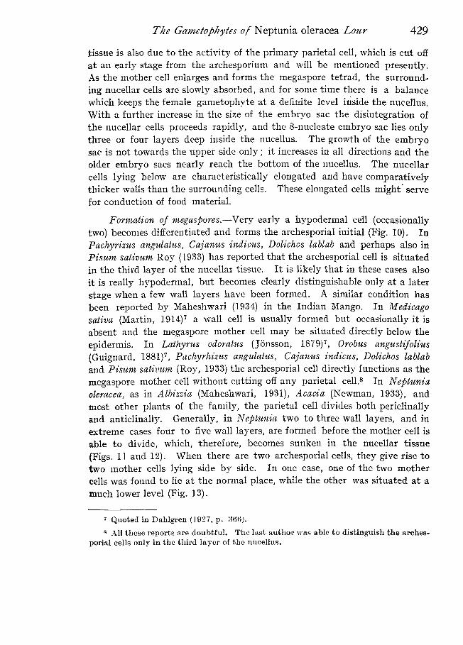
tissue is also due to the activity of the primary parietal cell, which is cut offat an early stage from the archesporium and will he mentioned presently.As the mother cell enlarges and forms the megaspore tetrad, the surround-ing nucellar cells are slowly absorbed, and for some time there is a balancewhich keeps the female gametophyte at a definite level inside the nucellus.With a further increase in the size of the embryo sac the disintegration ofthe nucellar cells proceeds rapidly, and the 8-nucleate embryo sac lies onlythree or four layers deep inside the nucellus. The growth of the embryosac is not towards the upper side only; it increases in all directions and theolder embryo sacs nearly reach the bottom of the nucellus. The nucellarcells lying below are characteristically elongated and have comparativelythicker walls than the surrounding cells. These elongated cells might' servefor conduction of food material.
Formation of megaspores.—Very early a hypodermal cell (occasionallytwo) becomes differentiated and forms the archesporial initial (Fig. 10). InPachyrizus angulatus, Cajanus indicus, Dolichos lablab and perhaps also inPisum sativum Roy (1933) has reported that the archesporial cell is situatedin the third layer of the nucellar tissue. It is likely that in these cases alsoit is really hypodermal, but becomes clearly distinguishable only at a laterstage when a few wall layers have been formed. A similar condition hasbeen reported by Maheshwari (1934) in the Indian Mango. In Medicagosaliva (Martin, 1914) 7 a wall cell is usually formed but occasionally it isabsent and the megaspore mother cell may be situated directly below the
epidermis. In Lathyrus odoratus (Jonsson, 1879) 7 , Orobus angustifolius(Guignard, 1881) 7 , Pachyrhizus angulatus, Cajanus indicus, Dolichos lablaband Pisum sativum (Roy, 1933) the archesporial cell directly functions as themegaspore mother cell without cutting off any parietal cell. 8 In Neptuniaoleracea, as in Albizzia (Maheshwari, 1931), Acacia (Newman, 1933), andmost other plants of the family, the parietal cell divides both periclinallyand anticlinally. Generally, in Neptunia two to three wall layers, and inextreme cases four to five wall layers, are formed before the mother cell isable to divide, which, therefore, becomes sunken in the nucellar tissue(Figs. 1]. and 12). When there are two archesporial cells, they give rise totwo mother cells lying side by side. In one case, one of the two mothercells was found to lie at the normal place, while the other was situated at amuch lower level (Fig. 13).
7 Quoted in Dahlgren (1927, P. 366).
8 All these reports are doubtful. The last author was able to distinguish the arches-porial cells only in the third layer of the nucellus.
430 Bahadur Singh and T. N. Shivapuri
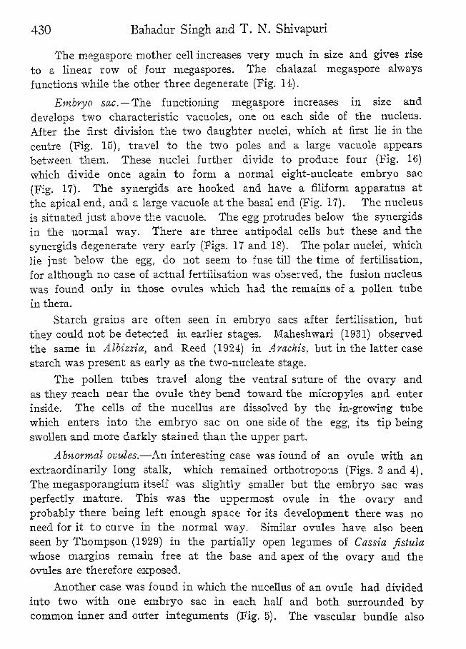
The rnegaspore mother cell increases very much in size and gives riseto a linear row of four megaspores. The chalazal megaspore alwaysfunctions while the other three degenerate (Fig. 14) .
Embryo sac. —The functioning megaspore increases in size anddevelops two characteristic vacuoles, one on each side of the nucleus.After the first division the two daughter nuclei, which at first lie in thecentre (Fig. 15), travel to the two poles and a large vacuole appearsbetween them. These nuclei further divide to produce four (Fig. 16)which divide once again to form a normal eight-nucleate embryo sac(Fig. 17). The synergids are hooked and have a filiform apparatus atthe apical end, and a large vacuole at the basal end (Fig. 17). The nucleusis situated just above the vacuole. The egg protrudes below the synergidsin the normal way. There are three antipodal cells but these and thesynergids degenerate very early (Pigs. 17 and 18). The polar nuclei, whichlie just below the egg, do not seem to fuse till the time of fertilisation,for although no case of actual fertilisation was observed, the fusion nucleuswas found only in those ovules which had the remains of a pollen tubein them.
Starch grains are often seen in embryo sacs after fertilisation, butthey could not be detected in earlier stages. Maheshwari (1931) observedthe same in Albizzia, and Reed (1924) in Arachis, but in the latter casestarch was present as early as the two-nucleate stage.
The pollen tubes travel along the ventral suture of the ovary andas they reach near the ovule they bend toward the micropyles and enterinside. The cells of the nucellus are dissolved by the in-growing tubewhich enters into the embryo sac on one side of the egg, its tip beingswollen and more darkly stained than the upper part.
_Abnormal ovules.—An interesting case was found of an ovule with anextraordinarily long stalk, which remained orthotropous (Figs. 3 and 4).The megasporangium itself was slightly smaller but the embryo sac wasperfectly mature. This was the uppermost ovule in the ovary andprobably there being left enough space for its development there was noneed for it to curve in the normal way. Similar ovules have also beenseen by Thompson (1929) in the partially open legumes of Cassia fistulawhose margins remain free at the base and apex of the ovary and theovules are therefore exposed.
Another case was found in which the nucellus of an ovule had dividedinto two with one embryo sac in each half and both surrounded bycommon inner and outer integuments (Fig. 5) . The vascular bundle also
The GczmeEophytes of Neptunia oleracea Lour 431
forked into two in this case. One further case was found of an embryosac which had four well-organised antipodal cells instead of the usualthree (Fig. 19) .
A general discussion of the results will be taken up in a subsequentpaper which will deal with some other plants of the MimosaceT.
Summary.1. The inflorescence is divisible into three regions, the lower of
completely sterile flowers, the upper of fertile and bisexual flowers anda small middle region with flowers showing an intermediate structure.
2. The archesporial cells in the anther are rather late in differentiation.The parietal tissue is composed of an endothecium, two middle layers anda uni-nucleate tapetum.
3. The divisions of the microspore mother cells are simultaneous.Cytokinesis occurs by vacuolation and furrowing.
4, The pollen grains are shed at the hi-celled stage.5. In the young ovule there is usually a single hypodermal
archesporial cell, but occasionally two may appear.6. The primary parietal cell and also the epidermal cells undergo
divisions, so that the mother cell comes to lie 3 to 5 layers deep in thetissue of the nucellus.
7. A linear tetrad of four megaspores is produced, of which thechalazal functions.
8. The mature embryo sac is of the usual 8-nucleate type. Thesynergids and antipodals are ephemeral. The fusion of the polar nucleiseems to be delayed till the time of fertilisation.
In conclusion, we wish to express our gratitude to Dr. P. Maheshwariunder whose directions this investigation was carried out and to whomwe are greatly indebted for criticism and kind advice. We are alsothankful to Messrs. Baba Lal Gupta and B. M. Johri for the helprendered by them.
PostScript.After this paper had already gone to the press two other papers on
Acacia Baileyana have just been published by Newman (1.934, a and b).It is reported that in the anther there are eight separate unicellular
archesporia, each of which gives rise to four mother cells. The microsporesare held together in the form of a pollinium. The thickened exine, whichis sculptured with reticulate grooves, is deposited only towards the outsideof the pollinium. The pollen grains are binucleate at the time of shedding.
432 Bahadur Singh and T. N. Shivapuri
The development of the megaspores is quite normal, but either the proximalor the distal may function. The megaspore is smaller in size than themicrospore. The embryo sac is of the normal eight-nucleate type. Thesynergids are hooked and possess a filiform apparatus. The antipodals arepersistent and degenerate only after the formation of the primary endospermnucleus. The integuments are very tardy in their growth and it is onlyafter fertilisation that they develop beyond their primordia. There is asingle vascular bundle in the outer integument. A cusion-like aril is deve-loped on the funiculus. Starch appears in the early stages of germinationof the megaspore.
LITER 1TURE CITED.
Castetter, E. F. .. .. " Studies on the comparative cytology of the annualand biennial varieties of Meliilotus alba," AmericanJour. Bot., 1925, 12, 270.
Coe, H. S., and Martin, J. N. .. " Sweet-clover seed," Bull. U. S. Dept. Agr., 1920,844, 1..
Cook, M. T. .. .. " Development of the seed of C'rotalar-ia sagittalis,"Bet. Gas., 1924, 77, 440.
Cooper, D. C. .. .. " Microsporogenesis and embryology of Melilotusalba," Bot. Gas., 1933, 95, 143.
Do. "Nuclear divisions in the tapetal cells of certainAngiosperms," American Jour. Bot., 1933, 20, 348.
Dahlgren, K. V. 0. .. .. " Die Morphologie des Nuzellus mit besondererBerrficl:sichtigung der deckzellosan Typen," Jahrb.Wise. Bot., 1927, 67, 317,
Engler, A., and Prantl, K. .. "Die Naturlichen Pflanzenfamilien," Zweite Auflage,1926.
Gates, R. R., and Rees, E. M. .. "A cytological study of pollen development inLactuca," Ann. Bot., 1921, 35, 365.
Geitler, L. .. .. .. " Zur MorphoIogie der Infloreszenzen and Bluten vonNeptunia oleracea," Ber. Deutsch. Bet. Ges., 1927,45, 36.
Latter, J. .. .. .. "The p'ollen development of Lathyrus odoratus,"Ann. Bot., 1026, 40, 2 77.
Maheshwari, P. .. .. "Contribution to the morphology of Albizzia lebbek,"Jour. Indian Dot. Soc., 1931, 10, 241.
Do. .. .. "The Indian Mango," Curr. Sci., 1934, 3, 97.
Metcalfe, C. R.. .. .. " The ' Aerenchyma' of Sesbania and Neptunia,"Bull. Mis. Information, Kew, 1931, 3, 151.
Newman, I. V. .. .. "The life-history of Acacia Bailcyana (F. V. M.),"Jour. Linn.. Soc., 1933, 49, 145.
Do. .. .. " Polyspermy and the endosperm," Nature, 1934,133, 650.
T'he Garnetophytes of Neptunia oleracea Lou, 433
Newman, I. V. .. .. "Studies in the Australian Acacias III. Supplemen-tary observations on the habit, carpel, sporeproduction and chromosome of Acacia BaileyanaF. V. M." Proc. Linn. Soc. New South Wales, 1934,59, 237.
Do. •. .. "Studies in the Australian Acacias IV. The lifehistory of Acacia Baiteyana F.V.. ." Part 2. Game-tophytes, Fertilisation, Sped Production and Ger-mination, and General Conclusion. Proc. Linn.Soc. New South Wales, 1934, 59, 297.
Reed, E. L. .. .. "Anatomy, embryology and ecology of Arach.ishypogea," Bot. Gaz., 1924, 78, 289.
Reeves, R. G. .. .. " Nuclear and cytoplasmic division in microsporo-genesis of Alfalfa," American Jour. Bot., 1930,17, 29.
Do. .. .. "Development of the ovule and embryo sac ofAlfalfa," American Jour. Rot., 1,430, 17, 239.
Roy, Basudev .. .. "Studies in the development of the female gameto-phyte in some Leguminous crop plants of India,"Indian Jour. Agr. Sci., 1933, 3, 1098.
Schnarf, K. .. .. .\T ergleich ende Embryologic der Angiospermen,"
Berlin, 1931.
Sethi, M. L. .. .. " Microsporogenesis in Cassirz didymobotrya," Jour.Indian Bot. Soc., 1930, 9, 126.
Thompson, J. M. .. .. " Studies in advancing sterility. Part IV. TheLegume," Hartley Bot. Lab. Publ., 1929, No. 6, 29.
Weinstein, Arthur I. .. .. "Cytological studies on Phassolus vulgaris," AmericanJour. Bot., 1926, 13, 248.
Wodehouse, R. P. .. .. "Morphology of Pollen grains in relation to plantclassification," Jour. New York Bot. Garden, 1926,27, 145.
Yamaha, G. .. .. Uber die Zytokinese bei der Pollentetradenbildung,zugleich weiter Beitrage zur Kenntnis fiber dieZytokinese im Pfianzenreichen," Japanese Jour.Bot., 1926, 3, 130.
EXPLANATION OF FIGURES.
FIGS. 6-19. are all magnified. x 720.
Flo. 6.—L. S. through part of an anther showing a row of microspore mother cells, theuni-nucleate tapetum, two middle layers, endothecium and the epidermis.
FIG. 7.—Microspore nuclei showing vacuoles appearing in the hyaline areas, and theorigin of furrows at the periphery.
FIG. S.—Mi.crospore tetrad still enclosed inside the mother cell wall.
Fio. 9.—Cross-section of mature pollen grain with three germ pores.
FIG. 10.—Nucellus with a hypodermal archesporial cell.
FIG. 11.—Nucellus showing young megaspore mother cell separated from the epidermisby two wall cells.

434 Bahadur Singh and T. N. Shivapuri
FIG. 12.—Ovule showing the primordia of the integuments and a megaspore mother cellin the prophase of the first reduction division.
FIG..13. —Same, with two megaspore mother cells in synizesis.
FIG. 14. —Older ovule with a linear tetrad of megaspores.
FIG. 15.—Young two-nucleate embryo sac.
FIG. 16.—Four-nucleate embryo sac.
FIG. 17.—Mature embryo sac with hooked synergids having a filiform apparatus at thetop, three degenerating antipodals (reconstructed from two sections).
FIG. 18.—Mature embryo sac showing early degeneration of synergids (reconstructedfrom two sections).
FIG. 19.—Lower part of an embryo sac showing four antipodal cells.

Bahadur Sin 'h & Proc. Ind. Acad. Sci., B, vol. I, Pl. 1V LIV.T. 1V. Shivap uri
FIGS. 6-19.