The Female Reproductive System
40
FEMALE REPRODUCTION Unlike males, who are able to produce sperm cells throughout their reproductive lives, females produce a finite number of egg cells. During early fetal development germ cells migrate into the ovaries and differentiate into oogonia
description
the female reproductive system anatomy cervix ovarium uterus
Transcript of The Female Reproductive System
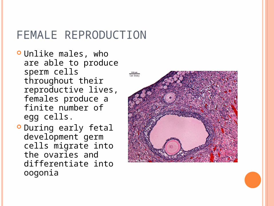
FEMALE REPRODUCTION Unlike males, who
are able to produce sperm cells throughout their reproductive lives, females produce a finite number of egg cells.
During early fetal development germ cells migrate into the ovaries and differentiate into oogonia

OOGONIA
The oogonia divide by mitosis for the next few months and some differentiate into primary oocytes.
By fifth month there are about 7 million primary oocytes, but most will degenerate during the next 2 months
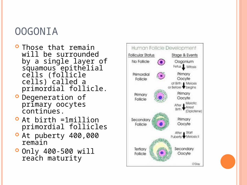
OOGONIA Those that remain will
be surrounded by a single layer of squamous epithelial cells (follicle cells) called a primordial follicle.
Degeneration of primary oocytes continues.
At birth =1million primordial follicles
At puberty 400,000 remain
Only 400-500 will reach maturity

OVARIAN CYCLE Monthly changes that
occur in the ovary during a woman’s reproductive life.
Each month FSH stimulates primordial follicles to grow and mature (follicular phase)
Ovulation- release of the egg (LH)
Luteal phase the corpus luteum produces progesterone that maintains uterine walls
If fertilization does not occur, the corpus luteum degenerates, within 2 weeks into a mass of scar tissue called the corpus albicans
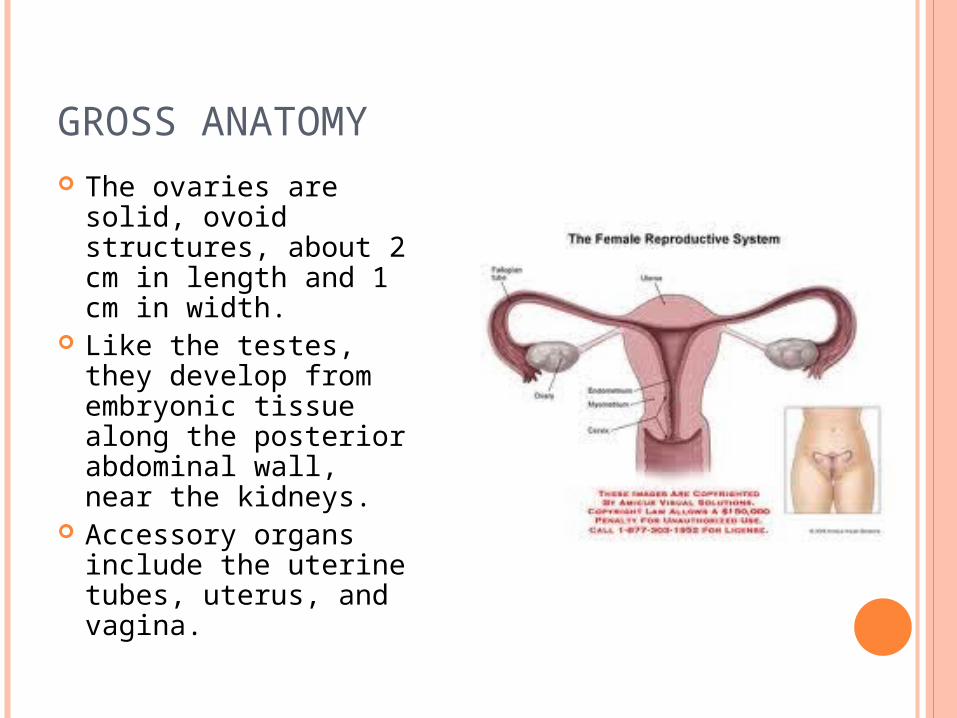
GROSS ANATOMY The ovaries are solid,
ovoid structures, about 2 cm in length and 1 cm in width.
Like the testes, they develop from embryonic tissue along the posterior abdominal wall, near the kidneys.
Accessory organs include the uterine tubes, uterus, and vagina.
6
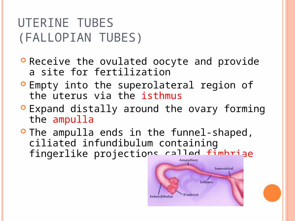
UTERINE TUBES (FALLOPIAN TUBES)
Receive the ovulated oocyte and provide a site for fertilization
Empty into the superolateral region of the uterus via the isthmus
Expand distally around the ovary forming the ampulla
The ampulla ends in the funnel-shaped, ciliated infundibulum containing fingerlike projections called fimbriae

7
UTERINE TUBES (FALLOPIAN TUBES)
Function: events occurring in the uterine tube
Fimbriae sweep oocyte into tube, cilia & peristalsis move it along, sperm reaches oocyte in ampulla, fertilization occurs within 24 hours after ovulation & zygote reaches uterus about 7 days after ovulation

8
FALLOPIAN TUBE HISTOLOGY
Cilia sweep egg/zygote toward the uterus

9
UTERUS
Hollow, thick-walled organ located in the pelvis anterior to the rectum and posterosuperior to the bladder
Body: Major portion of the uterus Fundus: Rounded region superior to the
entrance of the uterine tubes Isthmus: Narrowed region between the body
and cervix

10
UTERUS

11
UTERINE HISTOLOGY
Endometrium Simple columnar epithelium Stroma of connective tissue and endometrial glands
Stratum functionalis: Shed during menstruation
Stratum basalis: Replaces stratum functionalis each month
Myometrium 3 layers of smooth muscle
Perimetrium Visceral peritoneum

12
UTERINE HISTOLOGY

13
ENDOMETRIUM
Simple columnar epithelium
Endometrial glands
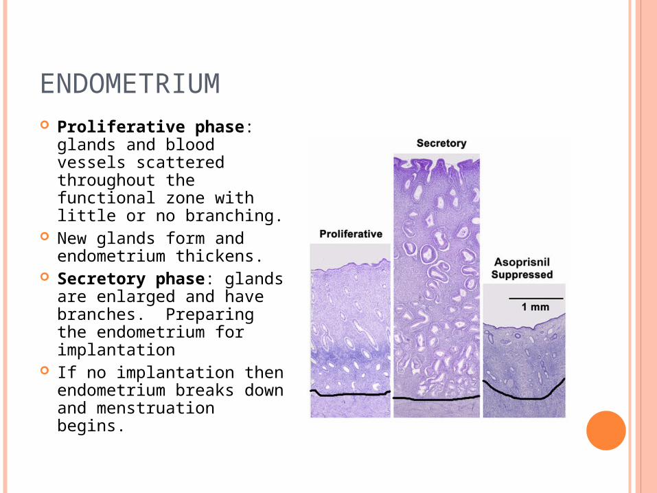
ENDOMETRIUM Proliferative phase:
glands and blood vessels scattered throughout the functional zone with little or no branching.
New glands form and endometrium thickens.
Secretory phase: glands are enlarged and have branches. Preparing the endometrium for implantation
If no implantation then endometrium breaks down and menstruation begins.
15
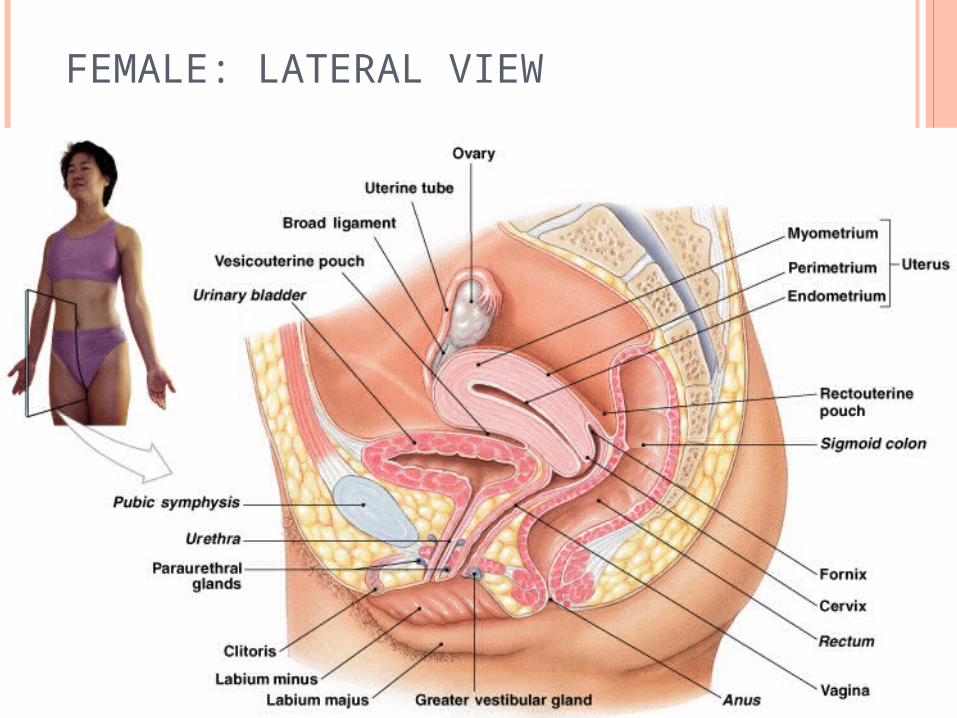
FEMALE: LATERAL VIEW
16
CERVIX
Narrow lower neck of the uterus which projects into the vagina inferiorly
Cervical canal – cavity of the cervix that communicates with: The vagina via the external os The uterine body via the internal os
Cervical glands secrete mucus that covers the external os and blocks sperm entry except during midcycle

17
Fornix
Endocervical canal
18
VAGINA
Thin-walled tube lying between the bladder and the rectum, extending from the cervix to the exterior of the body
Wall consists of three coats: fibroelastic adventitia, smooth muscle muscularis, and a stratified squamous mucosa
Mucosa near the vaginal orifice forms an incomplete partition called the hymen
Vaginal fornix: upper end of the vagina surrounding the cervix
19
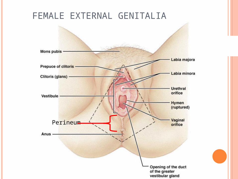
FEMALE EXTERNAL GENITALIA
Mons pubis: fatty pad over the pubic symphysis Labia majora & minora: folds of skin encircling
vestibule where find urethral and vaginal openings
Clitoris: small mass of erectile tissue Bulb of vestibule: masses of erectile tissue just
deep to the labia on either side of the vaginal orifice
Perineum: Area between the vagina and anus
20
FEMALE EXTERNAL GENITALIA
Perineum

21
BARTHOLIN’S GLANDS
(AKA: VESTIBULAR GLANDS) The Bartholin's glands are located on each side
of the vaginal opening. They secrete fluid that
helps lubricate the vagina. Sometimes the ducts of
these glands become obstructed. Fluid backs up into the gland
and causes swelling (Bartholin's cyst)

22
MAMMARY GLANDS
Modified sweat glands that produce milk (lactation) Amount of adipose determines size of breast Milk-secreting glands open by lactiferous ducts at
the nipple Areola is pigmented area around nipple Suspensory ligaments suspend breast from deep
fascia of pectoral muscles (aging & Cooper’s droop) Mammary line is a thickened ridge of embryonic
tiwwue that extends from the axilla to the groin.

23
BREAST

24
BREAST
Prolactin from the pituitary gland stimulates the synthesis of milk
Oxytocin from the posterior pituitary gland stimulates milk ejection
25
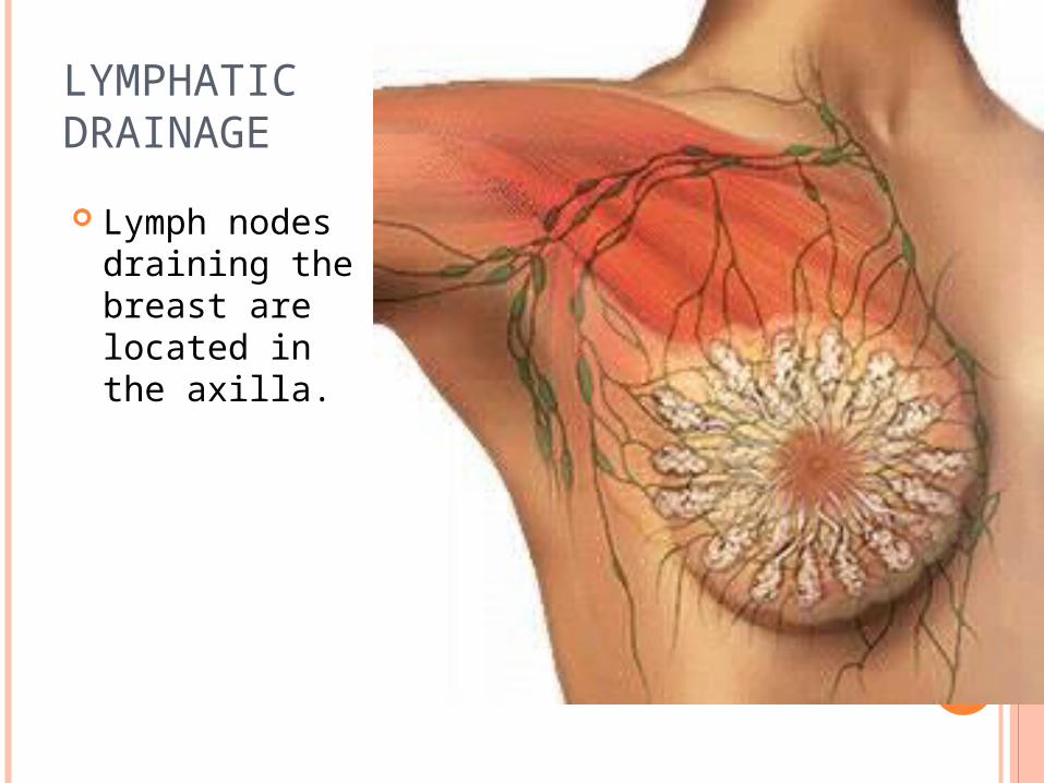
LYMPHATIC DRAINAGE
Lymph nodes draining the breast are located in the axilla.
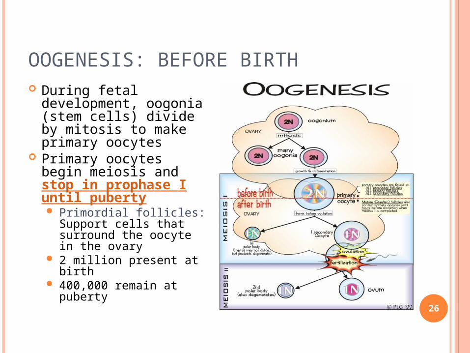
OOGENESIS: BEFORE BIRTH
26
During fetal development, oogonia (stem cells) divide by mitosis to make primary oocytes
Primary oocytes begin meiosis and stop in prophase I until puberty Primordial follicles:
Support cells that surround the oocyte in the ovary
2 million present at birth
400,000 remain at puberty

27
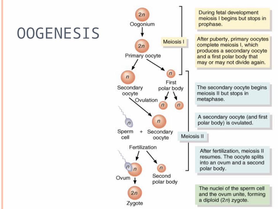
OOGENESIS: AFTER PUBERTY
Each month, hormones cause several follicles to develop, which triggers the primary oocyte to resume meiosis I
Polar bodies: When the cell divides, all the cytoplasm and organelles stay with one of the new cells, the other cell is just DNA, and is called a polar body and is discarded
Secondary oocyte: The stage at which ovulation occurs.

28
OOGENESIS: AFTER PUBERTY
The secondary oocyte begins meiosis II, but stops in metaphase II
The secondary oocyte is ovulated Meiosis II is completed only if it is fertilized.
29
OOGENESIS
30
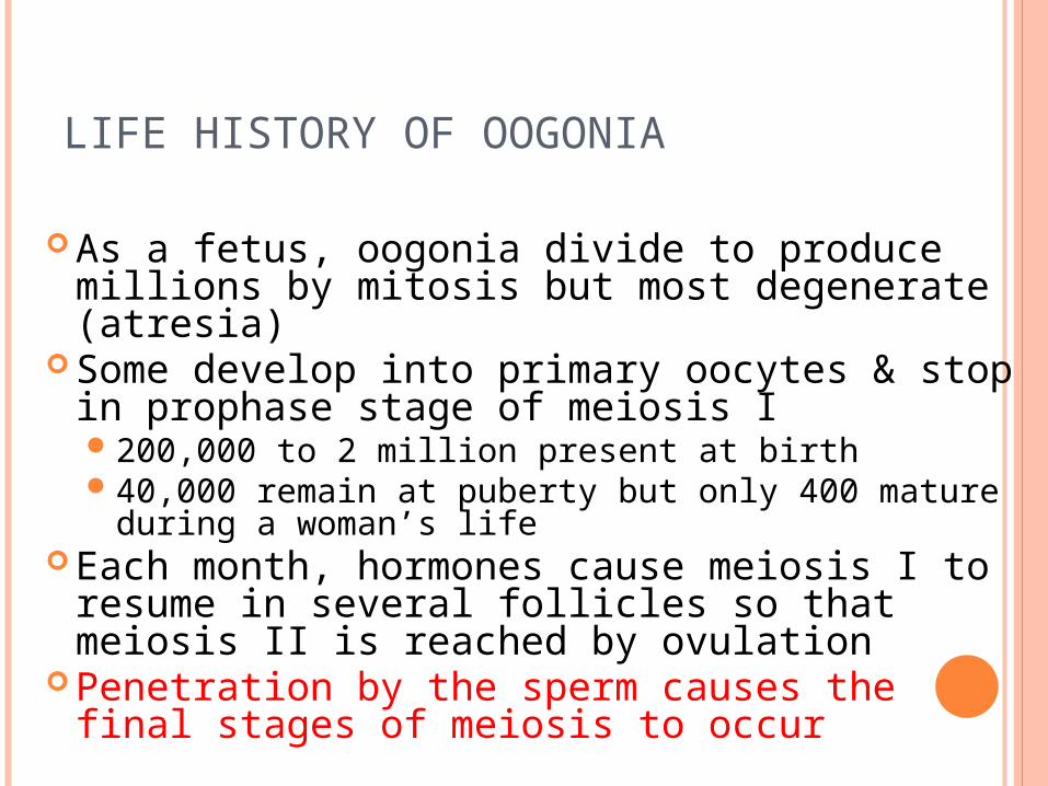
LIFE HISTORY OF OOGONIA
As a fetus, oogonia divide to produce millions by mitosis but most degenerate (atresia)
Some develop into primary oocytes & stop in prophase stage of meiosis I200,000 to 2 million present at birth40,000 remain at puberty but only 400 mature
during a woman’s life Each month, hormones cause meiosis I to
resume in several follicles so that meiosis II is reached by ovulation
Penetration by the sperm causes the final stages of meiosis to occur

31
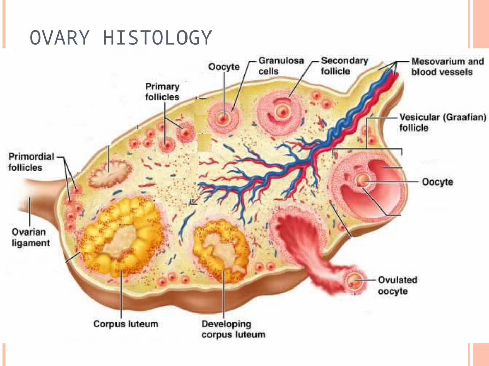
OVARIES
Each follicle consists of an immature egg called an oocyte
Cells around the oocyte are called: Follicle cells (one cell layer thick)
Stimulated to mature by FSH from the pituitary gland Granulosa cells (when more than one layer is
present) Thecal cells: Cells in the ovarian stroma
Thecal & granulosa cells work together to produce estrogen
A protective layer of glycoprotein forms around the egg called the zona pellucida
32
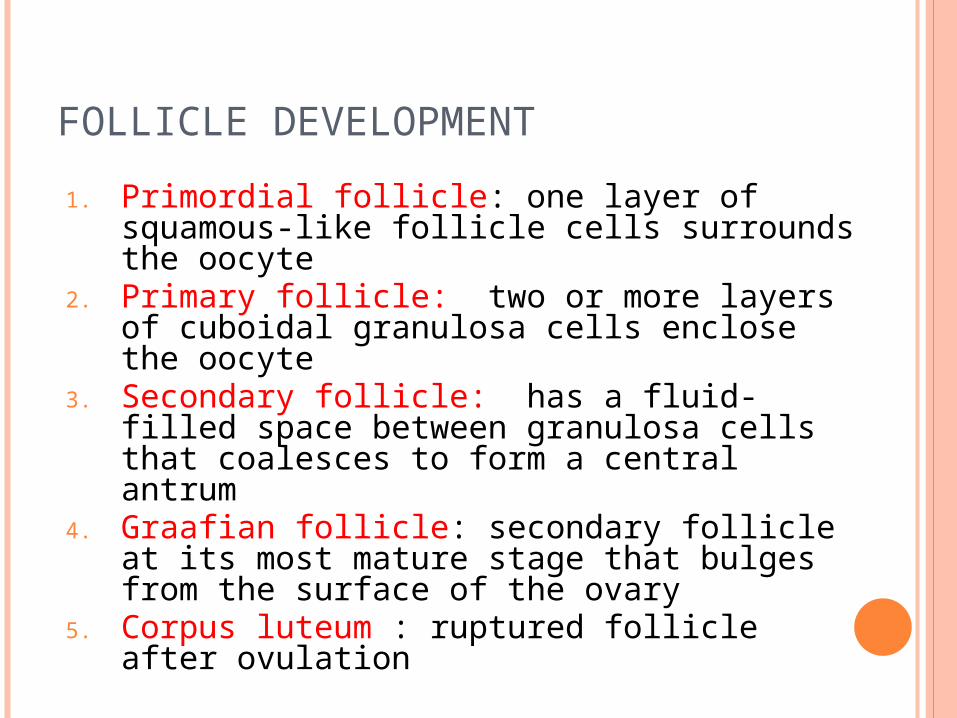
FOLLICLE DEVELOPMENT
1. Primordial follicle: one layer of squamous-like follicle cells surrounds the oocyte
2. Primary follicle: two or more layers of cuboidal granulosa cells enclose the oocyte
3. Secondary follicle: has a fluid-filled space between granulosa cells that coalesces to form a central antrum
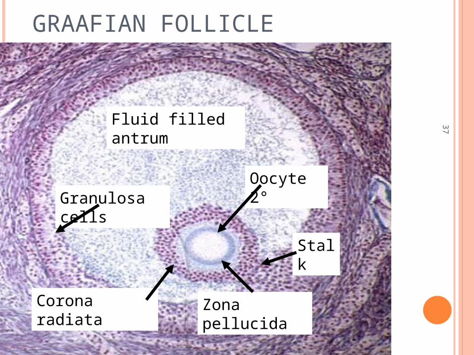
4. Graafian follicle: secondary follicle at its most mature stage that bulges from the surface of the ovary
5. Corpus luteum : ruptured follicle after ovulation
33
OVARY HISTOLOGY
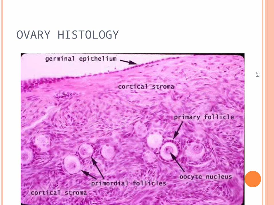
34
OVARY HISTOLOGY
35
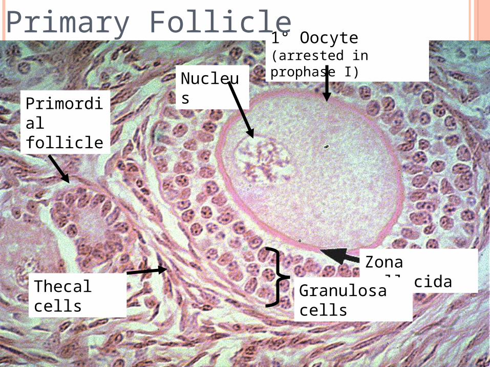
Zona pellucida
1° Oocyte(arrested in prophase I)
Granulosa cells
Thecal cells
Nucleus
Primordial follicle
Primary Follicle

36
SECONDARY FOLLICLEFluid-filled antrum
37
GRAAFIAN FOLLICLE
Fluid filled antrum
Granulosa cells
Oocyte 2°
Corona radiata
Stalk
Zona pellucida
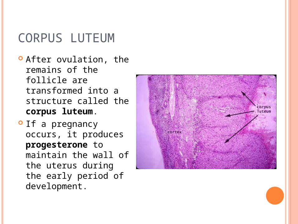
CORPUS LUTEUM
After ovulation, the remains of the follicle are transformed into a structure called the corpus luteum.
If a pregnancy occurs, it produces progesterone to maintain the wall of the uterus during the early period of development.

CORPUS ALBICANS
If fertilization does not occur, the corpus luteum will begin to break down about 2 weeks after ovulation.
Degeneration occurs when fibroblasts enter the corpus luteum and a clump of scar tissue forms called the corpus albicans.
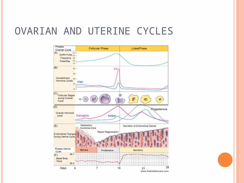
OVARIAN AND UTERINE CYCLES