The feasibility and efficacy of implementing a focused cardiac ultrasound …€¦ · ·...
9
RESEARCH ARTICLE Open Access The feasibility and efficacy of implementing a focused cardiac ultrasound course into a medical school curriculum Sergio L. Kobal 1* , Yotam Lior 2 , Alon Ben-Sasson 2 , Noah Liel-Cohen 1 , Ori Galante 3 and Lior Fuchs 3 Abstract Background: Teaching cardiac ultrasound to medical students in a brief course is a challenge. We aimed to evaluate the feasibility of teaching large groups of medical students the acquisition and interpretation of cardiac ultrasound images using a pocket ultrasound device (PUD) in a short, specially designed course. Methods: Thirty-one medical students in their first clinical year participated in the study. All were novices in the use of cardiac ultrasound. The training consisted of 4 hours of frontal lectures and 4 hours of hands-on training. Students were encouraged to use PUD for individual practice. Finally, the students’ proficiency in the acquisition of ultrasound images and their ability to recognize normal and pathological states were evaluated. Results: Sixteen of 27 (59%) students were able to demonstrate all main ultrasound views (parasternal, apical, and subcostal views) in a six-minute test. The most obtainable view was the parasternal long-axis view (89%) and the least obtainable was the subcostal view (58%). Ninety-seven percent of students correctly differentiated normal from severely reduced left ventricular function, 100% correctly differentiated a normal right ventricle from a severely hypokinetic one, 100% correctly differentiated a normal mitral valve from a rheumatic one, and 88% correctly differentiated a normal aortic valve from a calcified one, while 95% of them correctly identified the presence of pericardial effusion. Conclusions: Training of medical students in cardiac ultrasound during the first clinical year using a short, focused course is feasible and enables students with modest ability to acquire the main transthoracic ultrasound views and gain proficiency in the diagnosis of a limited number of cardiac pathologies. Background The diagnosis of cardiac disease is based on medical his- tory, physical examination, and complementary studies. The cardiovascular physical exam, based mainly on car- diac auscultation, can be performed during the first patient-doctor encounter. However, its diagnostic accur- acy is suboptimal [1–8]. Some cardiovascular pathologies are difficult to identify by means of physical examination due to imperceptible or barely detectable clinical mani- festations [4, 6, 9–12], with no connection to physicians’ skills. In order to improve their diagnostic capability, physicians make use of complementary diagnostic im- aging techniques. Ultrasound offers precise anatomical and functional information on the cardiovascular system and is the most commonly used technique for diagnos- ing cardiovascular diseases [2, 6, 10, 13, 14]. Decara et al. showed that the utilization of hand-held ultrasound devices significantly augmented the accuracy of medical students’ cardiac diagnoses at the bedside compared to traditional physical examination [3]. How- ever, the practicability of echocardiography is limited due to the cost of the device, its cumbersome size, and the lack of availability of expert personnel to perform and interpret studies. New, small, user-friendly ultrasound equipment has been in use for the last decade. Recently, the pocket ultra- sound device (PUD), with high imaging resolution, has be- come commercially available [14]. The PUD has the same characteristics as the traditional stethoscope routinely used by physicians in their physical examinations for the last 150 years: it is portable, free of adverse effects, and * Correspondence: [email protected] 1 Cardiology Department, Soroka University Medical Center, Beer-Sheva, Israel Full list of author information is available at the end of the article © The Author(s). 2017 Open Access This article is distributed under the terms of the Creative Commons Attribution 4.0 International License (http://creativecommons.org/licenses/by/4.0/), which permits unrestricted use, distribution, and reproduction in any medium, provided you give appropriate credit to the original author(s) and the source, provide a link to the Creative Commons license, and indicate if changes were made. The Creative Commons Public Domain Dedication waiver (http://creativecommons.org/publicdomain/zero/1.0/) applies to the data made available in this article, unless otherwise stated. Kobal et al. BMC Medical Education (2017) 17:94 DOI 10.1186/s12909-017-0928-x
Transcript of The feasibility and efficacy of implementing a focused cardiac ultrasound …€¦ · ·...

RESEARCH ARTICLE Open Access
The feasibility and efficacy of implementinga focused cardiac ultrasound course into amedical school curriculumSergio L. Kobal1*, Yotam Lior2, Alon Ben-Sasson2, Noah Liel-Cohen1, Ori Galante3 and Lior Fuchs3
Abstract
Background: Teaching cardiac ultrasound to medical students in a brief course is a challenge. We aimed toevaluate the feasibility of teaching large groups of medical students the acquisition and interpretation of cardiacultrasound images using a pocket ultrasound device (PUD) in a short, specially designed course.
Methods: Thirty-one medical students in their first clinical year participated in the study. All were novices in theuse of cardiac ultrasound. The training consisted of 4 hours of frontal lectures and 4 hours of hands-on training.Students were encouraged to use PUD for individual practice. Finally, the students’ proficiency in the acquisition ofultrasound images and their ability to recognize normal and pathological states were evaluated.
Results: Sixteen of 27 (59%) students were able to demonstrate all main ultrasound views (parasternal, apical, andsubcostal views) in a six-minute test. The most obtainable view was the parasternal long-axis view (89%) and theleast obtainable was the subcostal view (58%). Ninety-seven percent of students correctly differentiated normalfrom severely reduced left ventricular function, 100% correctly differentiated a normal right ventricle from a severelyhypokinetic one, 100% correctly differentiated a normal mitral valve from a rheumatic one, and 88% correctlydifferentiated a normal aortic valve from a calcified one, while 95% of them correctly identified the presence ofpericardial effusion.
Conclusions: Training of medical students in cardiac ultrasound during the first clinical year using a short, focusedcourse is feasible and enables students with modest ability to acquire the main transthoracic ultrasound views andgain proficiency in the diagnosis of a limited number of cardiac pathologies.
BackgroundThe diagnosis of cardiac disease is based on medical his-tory, physical examination, and complementary studies.The cardiovascular physical exam, based mainly on car-diac auscultation, can be performed during the firstpatient-doctor encounter. However, its diagnostic accur-acy is suboptimal [1–8]. Some cardiovascular pathologiesare difficult to identify by means of physical examinationdue to imperceptible or barely detectable clinical mani-festations [4, 6, 9–12], with no connection to physicians’skills. In order to improve their diagnostic capability,physicians make use of complementary diagnostic im-aging techniques. Ultrasound offers precise anatomicaland functional information on the cardiovascular system
and is the most commonly used technique for diagnos-ing cardiovascular diseases [2, 6, 10, 13, 14].Decara et al. showed that the utilization of hand-held
ultrasound devices significantly augmented the accuracyof medical students’ cardiac diagnoses at the bedsidecompared to traditional physical examination [3]. How-ever, the practicability of echocardiography is limiteddue to the cost of the device, its cumbersome size, andthe lack of availability of expert personnel to performand interpret studies.New, small, user-friendly ultrasound equipment has
been in use for the last decade. Recently, the pocket ultra-sound device (PUD), with high imaging resolution, has be-come commercially available [14]. The PUD has the samecharacteristics as the traditional stethoscope routinelyused by physicians in their physical examinations for thelast 150 years: it is portable, free of adverse effects, and
* Correspondence: [email protected] Department, Soroka University Medical Center, Beer-Sheva, IsraelFull list of author information is available at the end of the article
© The Author(s). 2017 Open Access This article is distributed under the terms of the Creative Commons Attribution 4.0International License (http://creativecommons.org/licenses/by/4.0/), which permits unrestricted use, distribution, andreproduction in any medium, provided you give appropriate credit to the original author(s) and the source, provide a link tothe Creative Commons license, and indicate if changes were made. The Creative Commons Public Domain Dedication waiver(http://creativecommons.org/publicdomain/zero/1.0/) applies to the data made available in this article, unless otherwise stated.
Kobal et al. BMC Medical Education (2017) 17:94 DOI 10.1186/s12909-017-0928-x

has no contraindications. The PUD allows for a quick andaccurate observation of a patient’s internal structures aswell as their function, and thus enhances the diagnosticpotential of the “auscultation-assisted” physical examin-ation [1–5, 8, 11, 14–18].Acquiring ultrasound skills in medical school is vitally
important, but teaching these skills presents challenges.Recently, a consensus of 34 experts in the field of ultra-sound in medical education established recommendedultrasound training milestones that all medical studentsshould reach before they graduate [19].The main limitations of ultrasound-assisted physical
examination are related to time of training until profi-ciency is achieved in obtaining echo views and in theability to identify cardiac pathologies. Teaching the ac-quisition of cardiac ultrasound imaging is based mainlyon hands-on practice in a one-on-one fashion, under thedirect supervision of highly qualified instructors. Thismakes the teaching of large numbers of medical personnelunfeasible, primarily due to the limited number of instruc-tors and teaching costs.
MethodsDevising a time-efficient and effective method of teach-ing cardiac ultrasound to larger groups is challenging,but could facilitate the incorporation of this importantdiagnostic modality into medical school curriculums.Our study’s objectives were to assess the feasibility and
efficacy of having novice medical students perform andread basic cardiac ultrasound studies during their firstclinical rotation after participating in a newly designed,brief (eight-hour), focused cardiac ultrasound courseusing a PUD. Specifically, we aimed to validate thisteaching method by testing whether students couldachieve standard echocardiographic views and analyzebasic transthoracic cardiac ultrasound images and basiccardiac pathologies.
Study populationA total of 31 medical student volunteers in their firstclinical year (fourth year of a six-year medical schoolprogram) were enrolled in the study. The course tookplace during the first 2 weeks of an eight-week internalmedicine rotation. The importance of the course wasconveyed to the class and the schedule was presented amonth before the initiation of the rotation. Next, thestudents were notified simultaneously about the courseand all the students agreed to participate in it. The stu-dents all signed an informed consent form. None of thestudents had any prior experience with the use of ultra-sound, so pretesting did not appear to be necessary. Theresults of students’ performance in this study were notavailable to medical school faculty and therefore did notinfluence their evaluations.
The interventionThe course consisted of frontal lectures and hands-ontraining sessions. The course objectives were to teachstudents how to obtain standard cardiac ultrasound im-ages from standard transthoracic views and how torecognize normal cardiac ultrasound anatomy as well aspathological states of the left ventricle, the right ven-tricle, the mitral and aortic valves, the inferior vena cava,and the pericardium.Students’ training consisted of 4 hours of frontal lec-
tures and an additional 4 hours of hands-on practiceunder the direct supervision of cardiologists and echo-cardiographic sonographers. The eight-hour course wasincorporated into the eight-week internal medicine rota-tion in place of lectures that students could view inde-pendently in a pool of lectures that is available to themas e-Learning material. During their eight-week rotation,following the course, students were encouraged to usePUD autonomously (without supervision), practicingimage acquisition on healthy volunteers and hospitalizedpatients. Students were asked to record acquired imagesfor later revision with a tutor for feedback and skill im-provement. This post-course self-training was notobligatory.
LecturesThe 4 hours of lectures were divided into two weeklysessions of 2 hours each. The subjects covered in thelectures were the following: basic ultrasound physics,principles of two-dimensional ultrasound imaging, theDoppler effect, and cardiac ultrasound anatomy fromparasternal, apical, and subcostal views. Previous clinicalechocardiographic cases were used to teach studentspattern recognition of normal and abnormal LV and RVfunction, identification of aortic valve structure (normal,bicuspid, and degenerative calcified), normal mitral valveversus mitral valve with rheumatic damage, presence ofpericardial effusion, and dilated versus normal IVC.Color Doppler imaging was used to teach students toidentify aortic and mitral regurgitation of any degree.
Hands-on practiceDuring the same week the lectures were given, studentshad a total of 4 hours of guided hands-on training (di-vided into two sections of 2 hours each). Students prac-ticed image acquisition utilizing standard ultrasounddevices (Vivid 6 and Vivid I, General Electric). These de-vices optimized image acquisition for teaching purposesby providing a larger screen and better image qualitythan the pocket devices. Training sessions were led bycardiologists or echocardiographic sonographers withteaching experience on healthy student volunteers whoserved as models. Training was performed in small
Kobal et al. BMC Medical Education (2017) 17:94 Page 2 of 9

groups (up to four students) and focused on the acquisi-tion of standard cardiac ultrasound images.The practiced views included the parasternal long-axis
view, the parasternal short-axis view (three levels: aortic,mitral, and midpapillary), the apical four-, two-, andthree-chamber views (4-ch, 2-ch, 3-ch respectively), andthe subcostal view. Students were taught to move thetransducer with gentle maneuvers and to perform threemain motions: alignment, rotation, and tilt, in order toproduce the optimal image in each of the ultrasoundwindows. Students were also instructed to use general2D gain and image depth controls for better ultrasoundimages. Figures 1, 2 and 3 show details of the anatomicalpoints that the students had to obtain in each of theultrasound views.
Individual practice using the pocket ultrasound deviceStudents were encouraged to practice image acquisitionduring the eight-week clinical rotation. This PUD prac-tice was completely voluntary and no number of re-quired practice hours was defined. Every five studentswere provided with a PUD (Vscan from General Electric)for individual practice. The easy-to-use device is shapedlike a small flip-phone and has an attached 2.5 MHz car-diac transducer. It has a 3.5-in. display screen andweighs 390 g. With two separate control settings (depthand gain), the Vscan produces a black-and-white real-time two-dimensional ultrasound image as well as acolor-coded blood flow image. Images can be frozen andsimple linear measurements can be performed. All im-ages produced with the device can be saved on the sys-tem’s memory card for later viewing and feedback.
Practice was neither predefined nor mandatory. Stu-dents performed cardiac ultrasound studies on one an-other as well as on their patients as part of the physicalexamination. We therefore advised our students to in-form the patients they examined that the ultrasoundstudy they performed did not in any way replace a com-prehensive echocardiographic study, and that no diagno-sis or clinical decision would be made based on it.
a
c
b
a
b
Fig. 1 a Parasternal long axis view and main anatomic points. bParasternal short axis view at three levels. a Aortic level. b Mitrallevel. c Mild papillary level
a
b
c
Fig. 2 Apical 4, 3 and 2 chamber views. a Aplical 4 chamber view. bAplical 3 chamber view. c Aplical 2 chamber view
Kobal et al. BMC Medical Education (2017) 17:94 Page 3 of 9

Post-intervention assessmentAt the end of the eight-week clinical rotation, enrolledstudents underwent two exams. The first was designedto assess hands-on cardiac ultrasound image acquisitionof the main transthoracic views (the six-minute exam,see below), while the second aimed to validate theirabilities to recognize different cardiac ultrasound views,particularly their ability to differentiate normal frompathologic ventricular, valvular, IVC, and pericardial find-ings (ultrasound image interpretation exam).
The six-minute examThe goal of this exam was to assess students’ ability to pro-duce transthoracic cardiac ultrasound images. Each studenthad 6 minutes to obtain the main echocardiographic viewsin a predetermined order using a PUD: PLAV (Fig. 1a),PSAV (at the aortic, mitral, and mid-papillary levels; Fig. 1b),apical views (4-ch, 2-ch, 3-ch; Fig. 2) and subcostal views(standard and focused on IVC; Fig. 3).
During the exam, the supervising physician used achecklist of cardiac anatomical points that each studentwas required to identify in each one of the main transtho-racic ultrasound views and a final score was obtained ac-cording to the student’s performance (Additional file 1:Appendix A). The views were obtained on healthy modelswho were pre-screened and had good ultrasound windows.By the end of the six-minute period, the exam ended,
regardless of the number of windows acquired and thestudents were asked to report their personal self-trainingtime during the rotation in hours.
Ultrasound image interpretation examThe exam consisted of 23 questions, each with one echoclip. The students were evaluated for their ability toidentify the main cardiac ultrasound views and recognizenormal and abnormal LV and RV function, the anatomyof the aortic and mitral valves, IVC size and respiratoryvariation, valvular dysfunction, and the presence of peri-cardial effusion.
Statistical analysisThe data collected was documented using summary ta-bles. Continuous variables with normal distribution werepresented as mean and standard deviation with 95%confidence interval when appropriate. Ordinary variablesor continuous variables with non-normal distributionwere presented as median with an interquartile range(IQR). Categorical variables were presented as countsand percentages of the total.Continuous variables were analyzed using student t-test.
Non-parametric procedures (Mann–Whitney) were usedif parametric assumptions could not be satisfied even afterdata transformation attempts. Parametric model assump-tions were assessed using Normal-plot or the Shapiro-Wilks test for verification of normality and Levene’s testfor verification of homogeneity of variances.Categorical variables were tested using Pearson’s χ2
test for contingency tables or Fisher Exact test when ap-propriate. Correlations between variables were testedusing Pearson or Spearman tests, depending on variabledistribution.All statistical tests and/or confidence intervals, as ap-
propriate, were performed at α = 0.05 (two-sided) andpresented with their 95% confidence interval whenappropriate.
ResultsAll 31 students completed the eight-hour course, as re-quired by the study protocol. All students but one couldvisualize the parasternal long-axis view (PLAV), all visu-alized at least one level (aortic, mitral, mid-papillary) ofthe parasternal short-axis view (PSAV), all visualized atleast one of the three apical views (four-, three- and
b
a
Fig. 3 Subcostal views. a Subcostal long axis view. b Subcostal IVC view
Kobal et al. BMC Medical Education (2017) 17:94 Page 4 of 9

two-chamber views) and 89% visualized at least one ana-tomical view of the subcostal view (Table 1).The average grade on the cardiac ultrasound view rec-
ognition test was 73%. Thirty students (97%) were able tocorrectly identify normal LV function and LV dysfunction.
All 31 students differentiated normal mitral valve fromrheumatic mitral valve injury and 95% of the students di-agnosed the presence of pericardial effusion.The average PUD individual practice time (which was
not obligatory) was 3.3 ± 2.7 h per student. The maximumindividual practice time was 10 h (reported by one student).Four students reported no individual practice at all.
The six-minute examTwenty-seven of 31 (87%) students performed the exam.All participants were able to obtain at least one anatomicalview of the eight transthoracic views within the 6 minutesallowed. The most obtainable view (the view that the lar-gest number of students could achieve) was the PLAV,with a median rate of 89% (SD 23%), while the least ob-tainable view was the subcostal view, with a median rateof 58% (SD 34%).
Parasternal viewsTwenty-six of 27 students (96%) obtained at least oneimage with one of the four anatomical points requiredfor the PLAV view (correct alignment, total endocardialdemarcation of LV, and visualization of mitral leaflets oraortic valve cusps). Although seven students (26%) couldnot achieve the correct alignment for PLAV anatomicalpoints, endocardial demarcation and mitral and aorticvalves were visualized by most students (89%, 96%, and95% respectively; Table 1).All students were able to obtain at least one image of the
eight anatomical points defined for the PSAV view at theaortic level (aortic, tricuspid, and pulmonic valves, intera-trial septum), mitral level (complete LV endocardial demar-cation, mitral valve), and midpapillary level (complete LVendocardial demarcation, papillary muscles). Students per-formed better in scanning the midpapillary than the mitralor the aortic level (average grades of 89%, 85%, and 78% re-spectively; Table 1).
Apical viewsAll of the students were able to obtain at least one of thethree apical views, namely 4-ch, 2-ch, and 3-ch. Eighteenstudents (67%) obtained all apical views. Six (22%) stu-dents obtained two out of the three apical views, and three(11%) students were able to obtain only one of the threeapical views. The most obtainable apical view was 4-ch(89%) and the least obtainable was 3-ch (77%). Table 1shows that students had less success in imaging entireventricles (up to 56% for the LV and up to 48% for theRV) than for imaging the valves (up to 89% for the mitraland 70% for the aortic valve) from the apex.
Subcostal viewThe subcostal view comprised the last part of the six-minute exam and the students were required to show
Table 1 The six-minute exam of ability to obtain correct ultrasoundimages
Ultrasound views and defined anatomical points Score
Parasternal Long-Axis View 88.89 ± 23.34
Correct alignment 20/27 (74.1%)
Endocardial demarcation 24/27 (88.9%)
Mitral valve visualization 26/27 (96.3%)
Aortic valve visualization 26/27 (96.3%)
Parasternal Short-Axis View 78.24 ± 24.41
Aortic level demonstration 69.4 ± 37.6
Aorta visualization 22/27 (81.5%)
Tricuspid valve visualization 21/27 (77.8%)
Pulmonic valve visualization 18/27 (66.7%)
Interatrial septum visualization 14/27 (51.9%)
Mitral level demonstration 85.2 ± 30.43
Complete left ventricle visualization 21/27 (77.8%)
Mitral valve visualization 25/27 (92.6%)
Midpapillary level demonstration 88.9 ± 32
Complete left ventricle visualization 24/27 (88.9%)
Papillary muscle visualization 24/27 (88.9%)
Apical View 62.96 ± 24.29
Four-Chamber View 59.3 ± 34.4
Left ventricle visualization 11/27 (40.7%)
Right ventricle visualization 13/27 (48.1%)
Mitral valve visualization 24/27 (88.9%)
Tricuspid valve visualization 22/27 (81.5%)
Atrium visualization 10/27 (37%)
Two-Chamber View 61.7 ± 38.9
Left ventricle visualization 14/27 (51.9%)
Mitral valve visualization 23/27 (85.2%)
Left atrium visualization 13/27 (48.1%)
Three-Chamber View 67.9 ± 41.8
Left ventricle visualization 15/27 (55.6%)
Mitral valve visualization 21/27 (77.8%)
Aortic valve visualization 19/27 (70.4%)
Subcostal View 57.78 ± 34.34
Right ventricle visualization 18/27 (66.7%)
Pericardial visualization 20/27 (74.1%)
Interatrial septum visualization 11/27 (40.7%)
Inferior vena cava visualization 16/27 (59.3%)
Inferior vena cava respiratory variation visualization 13/27 (48.1%)
Kobal et al. BMC Medical Education (2017) 17:94 Page 5 of 9
two views: the long-axis four-chamber standard viewand the tilted view of the IVC. Twenty-four of 27 stu-dents (89%) were able to produce at least one anatomicalview of five predetermined anatomical points for thisview (Appendage A). Imaging of the IVC was achievedby 16 of 27 students (59%). The rest of the results forthis view are summarized in Table 1.
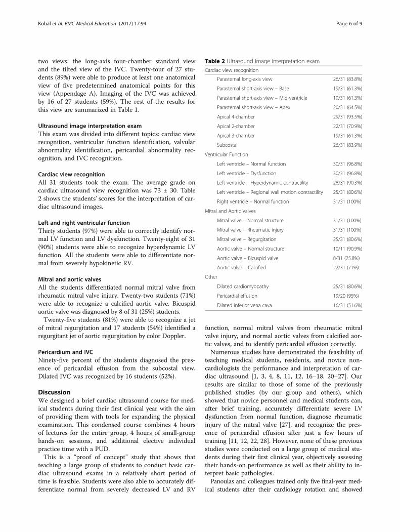
Ultrasound image interpretation examThis exam was divided into different topics: cardiac viewrecognition, ventricular function identification, valvularabnormality identification, pericardial abnormality rec-ognition, and IVC recognition.
Cardiac view recognitionAll 31 students took the exam. The average grade oncardiac ultrasound view recognition was 73 ± 30. Table2 shows the students’ scores for the interpretation of car-diac ultrasound images.
Left and right ventricular functionThirty students (97%) were able to correctly identify nor-mal LV function and LV dysfunction. Twenty-eight of 31(90%) students were able to recognize hyperdynamic LVfunction. All the students were able to differentiate nor-mal from severely hypokinetic RV.
Mitral and aortic valvesAll the students differentiated normal mitral valve fromrheumatic mitral valve injury. Twenty-two students (71%)were able to recognize a calcified aortic valve. Bicuspidaortic valve was diagnosed by 8 of 31 (25%) students.Twenty-five students (81%) were able to recognize a jet
of mitral regurgitation and 17 students (54%) identified aregurgitant jet of aortic regurgitation by color Doppler.
Pericardium and IVCNinety-five percent of the students diagnosed the pres-ence of pericardial effusion from the subcostal view.Dilated IVC was recognized by 16 students (52%).
DiscussionWe designed a brief cardiac ultrasound course for med-ical students during their first clinical year with the aimof providing them with tools for expanding the physicalexamination. This condensed course combines 4 hoursof lectures for the entire group, 4 hours of small-grouphands-on sessions, and additional elective individualpractice time with a PUD.This is a “proof of concept” study that shows that
teaching a large group of students to conduct basic car-diac ultrasound exams in a relatively short period oftime is feasible. Students were also able to accurately dif-ferentiate normal from severely decreased LV and RV
function, normal mitral valves from rheumatic mitralvalve injury, and normal aortic valves from calcified aor-tic valves, and to identify pericardial effusion correctly.Numerous studies have demonstrated the feasibility of
teaching medical students, residents, and novice non-cardiologists the performance and interpretation of car-diac ultrasound [1, 3, 4, 8, 11, 12, 16–18, 20–27]. Ourresults are similar to those of some of the previouslypublished studies (by our group and others), whichshowed that novice personnel and medical students can,after brief training, accurately differentiate severe LVdysfunction from normal function, diagnose rheumaticinjury of the mitral valve [27], and recognize the pres-ence of pericardial effusion after just a few hours oftraining [11, 12, 22, 28]. However, none of these previousstudies were conducted on a large group of medical stu-dents during their first clinical year, objectively assessingtheir hands-on performance as well as their ability to in-terpret basic pathologies.Panoulas and colleagues trained only five final-year med-
ical students after their cardiology rotation and showed
Table 2 Ultrasound image interpretation exam
Cardiac view recognition
Parasternal long-axis view 26/31 (83.8%)
Parasternal short-axis view – Base 19/31 (61.3%)
Parasternal short-axis view – Mid-ventricle 19/31 (61.3%)
Parasternal short-axis view – Apex 20/31 (64.5%)
Apical 4-chamber 29/31 (93.5%)
Apical 2-chamber 22/31 (70.9%)
Apical 3-chamber 19/31 (61.3%)
Subcostal 26/31 (83.9%)
Ventricular Function
Left ventricle – Normal function 30/31 (96.8%)
Left ventricle – Dysfunction 30/31 (96.8%)
Left ventricle – Hyperdynamic contractility 28/31 (90.3%)
Left ventricle – Regional wall motion contractility 25/31 (80.6%)
Right ventricle – Normal function 31/31 (100%)
Mitral and Aortic Valves
Mitral valve – Normal structure 31/31 (100%)
Mitral valve – Rheumatic injury 31/31 (100%)
Mitral valve – Regurgitation 25/31 (80.6%)
Aortic valve – Normal structure 10/11 (90.9%)
Aortic valve – Bicuspid valve 8/31 (25.8%)
Aortic valve – Calcified 22/31 (71%)
Other
Dilated cardiomyopathy 25/31 (80.6%)
Pericardial effusion 19/20 (95%)
Dilated inferior vena cava 16/31 (51.6%)
Kobal et al. BMC Medical Education (2017) 17:94 Page 6 of 9
improved clinical diagnosis [21]. Andersen and colleaguestrained 30 fifth-year medical students, but in this studythe intensity (teacher-student ratio) of hands-on teachingwas not specified and the study suffered from selectionbias, as the assessment of scanning quality was dependenton students’ log books, where students chose their bestclips and were not evaluated in an objective test as theywere in our study [11]. Stokke and colleagues brieflytrained (in a four-hour course) 21 medical students usingPUD, but compared their findings to auscultation and didnot present objective scanning quality assessment [29].We have objectively measured students’ performance
and showed that a large class can become proficient inconducting and assessing basic cardiac ultrasound clipsby participating in relatively short hands-on training ingroups of four. Our students did only fairly well in termsof their ability to acquire all cardiac images. Sixteen of27 (59%) students were able to demonstrate all the views(parasternal, apical, and subcostal) within 6 minutes. Butmost students obtained at least one transthoracic echo-cardiographic view, while the most obtainable view wasthe parasternal long-axis view (89%) and the least ob-tainable was the subcostal view (58%). It is possible thatthe six-minute test was too short to allow novice opera-tors to obtain all main cardiac views. This notion is sup-ported by the fact that the last view to be obtained inthe exam (the subcostal view) was also the least obt-ainable. A longer exam would probably have improvedstudents’ scores.Another novel intervention was the incorporation of
non-obligatory individual PUD practice time during theeight-week clinical rotation. This teaching modality mayhave contributed to the students’ performance as mea-sured by the six-minute exam. However, we could notassess the correlation between the number of individualpractice hours and final exam scores due to inadequateself-reporting by the students.Strzecka and colleagues recently showed a learning
curve effect among novice operators after a short courseon PUD. Medical students detected major abnormalitieswith an acceptable diagnostic value that increased withthe number of examinations performed [12]. Ruddoxand colleagues found poor scanning performance whenmedical residents received only a two-hour training pro-gram (a one-hour practical demonstration followed by aone-hour hands-on training session) [22]. Students dem-onstrated poor ability to recognize impaired cardiacfunction, pericardial effusion, and valvular heart diseaseafter this very brief training. Anderson and colleaguestrained 30 fifth-year volunteer medical students who re-ceived a total of 9 hours (three sessions) of combinedpractical and theoretical training in the use and inter-pretation of ultrasound images. Students were encour-aged to perform at least 75 PUD examinations prior to
being tested [11]. Seventy-four percent of the studentsrecorded acceptable cardiovascular organ imaging. Inagreement with our results, students were better atinterpreting cardiac images than acquiring them. Theirdiagnostic accuracy in correctly identifying reduced LVfunction, pericardial effusion, pleural effusion, lungcomets, IVC diameter, and respiratory variation was 94%(CI 89.0–96.5). At present, the recommendations regard-ing a minimum number of studies (performance of at least75 echocardiographic studies and interpretation of 150complete studies under supervision) are indicated forsonographers and physicians who are learning compre-hensive echocardiographic studies with premium echocar-diographic devices. Due to inconsistent data that exists todate, there is no consensus on the minimum number ofstudies or hours of training needed to teach cardiac ultra-sound as a diagnostic tool in medical schools.A prolonged teaching program would be impractical
when teaching novice medical personnel on a large scale.On the other hand, very short training could be insuffi-cient to achieve the true benefit of ultrasound-assistedphysical examination and comprise a source of mistakesand misdiagnoses. It seems that at least 8 hours of PUDtraining (according to our results and previously pub-lished data) may be sufficient for medical students tolearn to accurately assess basic cardiac parameters byechocardiography, but insufficient to generate independ-ent ultrasound cardiac imaging using pocket devices.The acquisition of the ability to use PUDs independentlymay be enhanced by more structured guidance for indi-vidual use of PUD during clinical rotations. We do believethat the “consolidation process” of bedside ultrasoundstudies is directly related to the availability of PUDs forstudent use. In our hospital, each internal medicine wardhas one PUD device. In addition, in our course we usedfour PUDs provided by the cardiology department of thehospital affiliated with the medical school to the studentsfor their free practice during the eight-week internal medi-cine rotation. The PUDs were available for each studentevery 4 days. In our estimation, this number of PUDsallowed our students to practice ultrasound image acquisi-tion at an acceptable frequency.Practicing by utilizing PUD in relevant medical school
clinical rotations will improve students’ ability to per-form focused cardiac ultrasound examinations. We be-lieve that incorporating the six-minute hands-on examand the pathological clip exam into the final rotation’sformal student assessment as well as requiring studentsin clinical rotations to submit logbooks of PUD imageswill further improve their bedside point-of-care ultra-sound performance.Our study has some limitations. Students did not ac-
curately report self-training time, and thus provided onlyestimated data that limited our ability to reliably test the
Kobal et al. BMC Medical Education (2017) 17:94 Page 7 of 9

correlations between the amount of self-training and thefinal exam scores. Perhaps obligatory guided self-trainingsessions during the clinical rotation would have improvedhands-on performance on the six-minute exam. Inaddition, we used young, healthy models, known to havegood acoustic windows, for the six-minute hands-on test,and not actual medical patients.The ultrasound image recognition exam made use of
pathological pre-recorded clips, which were presented tothe students on a screen instead of real patients with path-ologies in real medical settings. No pretest was needed asnone of the students had any prior experience with theuse of ultrasound.Teaching cardiac ultrasound to large groups of medical
students is challenging. Incorporating ultrasound intoanatomy and physiology curriculums in the preclinicalyears could improve students’ skills during the clinicalyears. Replacing highly qualified sonographers and cardi-ologists with medical students who have received formalinstruction could provide an option for coping with thereality of a limited number of instructors being availablefor hands-on practice [25, 26]. More support of training inthe wards may be possible once more physicians beginusing this tool as part of regular practice during clinicalrounds. The use of web-based learning modules as well assimulators (currently available for learning basic as well asadvanced ultrasound) to complement practice sessions areoptions that we are assessing both to reduce costs and tomake this type of course more efficient.
ConclusionsLarge-scale instruction of medical students in cardiacultrasound during the first clinical years is feasible. Brieftraining in cardiac ultrasound that includes lectures,hands-on sessions, and individual practice allows medicalstudents to gain proficiency in the diagnosis of a limitednumber of cardiac states and provides them with a modestability to acquire transthoracic ultrasound views. There-fore, in order to modify traditional methods of physicalexamination and diagnosis by including ultrasound assess-ment as a complement of the physical examination, itseems appropriate to incorporate basic courses similar tothe one described here into medical school curriculums.
Additional file
Additional file 1: Appendix A. (DOCX 92 kb)
AbbreviationsAL: Anterolateral; AML: Anterior mitral leaflet; AoV: Aortic valve; DesAO: Descending Aorta; IAS: Interatrial septum; IVC: Inferior vena cava;IVS: Interventricular septum; LA: Left atrium.; LC: Left coronary cusp; LV: Leftventricle; NC: Noncoronary cusp; PM: Posteromedial; PML: Posterior mitralleaflet; PV: Pulmonic valve; RA: Right atrium; RC: Right coronary cusp;RV: Right ventricle; RVOT: Right ventricle outflow tract; TV: Tricuspid valve
AcknowledgementsNone.
FundingThis study was not funded.
Availability of data and materialsThe datasets used and analysed during the current study are available fromthe corresponding author on reasonable request.An English language copy of the six-minute exam used is provided as anadditional file.
Authors’ contributionsSLK and LF conceptualized and designed the study. All authors wereinvolved in data collection, analyses, and interpretation. SLK, AB, and LFdrafted the manuscript. NLS, OG, and YL critically revised the manuscript forimportant intellectual content. All authors approved of the version to bepublished and agreed to be accountable for all aspects of the work inensuring that questions related to the accuracy or integrity of any part ofthe work are appropriately investigated and resolved.
Competing interestsThe authors declare that they have no competing interests.
Consent for publicationNot applicable.
Ethics approval and consent to participateEthical approval for the data collection was obtained from the EthicsCommittee of Soroka Medical Center. All the students who participated weregiven assurance of confidentiality that the information gathered would beused exclusively for research purposes. We also indicated that participationin the study was anonymous and voluntary and filling in the written exammeant that the students were willing to participate in the study.
Publisher’s NoteSpringer Nature remains neutral with regard to jurisdictional claims inpublished maps and institutional affiliations.
Author details1Cardiology Department, Soroka University Medical Center, Beer-Sheva, Israel.2Clinical Research Center, Soroka University Medical Center, Beer-Sheva, Israel.3Medical Intensive Care Unit, all at Soroka University Medical Center and TheFaculty of Health Sciences, Ben-Gurion University of the Negev, Beer-Sheva,Israel.
Received: 10 September 2016 Accepted: 11 May 2017
References1. Mjolstad OC, Dalen H, Graven T, Kleinau JO, Salvesen O, Haugen BO.
Routinely adding ultrasound examinations by pocket-sized ultrasounddevices improves inpatient diagnostics in a medical department. Eur JIntern Med. 2012;23(2):185–91.
2. Steinberger J, Moller JH, Berry JM, Sinaiko AR. Echocardiographic diagnosis ofheart disease in apparently healthy adolescents. Pediatrics. 2000;105(4 Pt 1):815–8.
3. Decara JM, Kirkpatrick JN, Spencer KT, Ward RP, Kasza K, Furlong K, et al. Use ofhand-carried ultrasound devices to augment the accuracy of medical studentbedside cardiac diagnoses. J Am Soc Echocardiogr. 2005;18(3):257–63.
4. Kobal SL, Atar S, Siegel RJ. Hand-carried ultrasound improves the bedsidecardiovascular examination. Chest. 2004;126(3):693–701.
5. Spencer KT, Anderson AS, Bhargava A, Bales AC, Sorrentino M, Furlong K,et al. Physician-performed point-of-care echocardiography using a laptopplatform compared with physical examination in the cardiovascular patient.J Am Coll Cardiol. 2001;37(8):2013–8.
6. Attenhofer Jost CH, Turina J, Mayer K, Seifert B, Amann FW, Buechi M, et al.Echocardiography in the evaluation of systolic murmurs of unknown cause.Am J Med. 2000;108(8):614–20.
7. Mangione S. Cardiac auscultatory skills of physicians-in-training: acomparison of three English-speaking countries. Am J Med.2001;110(3):210–6.
Kobal et al. BMC Medical Education (2017) 17:94 Page 8 of 9
8. Kobal SL, Trento L, Baharami S, Tolstrup K, Naqvi TZ, Cercek B, et al.Comparison of effectiveness of hand-carried ultrasound to bedsidecardiovascular physical examination. Am J Cardiol. 2005;96(7):1002–6.
9. Sonderegger-Iseli K, Burger S, Muntwyler J, Salomon F. Diagnostic errors inthree medical eras: a necropsy study. Lancet. 2000;355(9220):2027–31.
10. Jaffe WM, Roche AH, Coverdale HA, McAlister HF, Ormiston JA, Greene ER.Clinical evaluation versus Doppler echocardiography in the quantitativeassessment of valvular heart disease. Circulation. 1988;78(2):267–75.
11. Andersen GN, Viset A, Mjolstad OC, Salvesen O, Dalen H, Haugen BO.Feasibility and accuracy of point-of-care pocket-size ultrasonographyperformed by medical students. BMC Med Educ. 2014;14:156.
12. Filipiak-Strzecka D, John B, Kasprzak JD, Michalski B, Lipiec P. Pocket-sizeechocardiograph–a valuable tool for nonexperts or just a portable devicefor echocardiographers? Adv Med Sci. 2013;58(1):67–72.
13. Aguirre FV, Pearson AC, Lewen MK, McCluskey M, Labovitz AJ. Usefulness ofDoppler echocardiography in the diagnosis of congestive heart failure. AmJ Cardiol. 1989;63(15):1098–102.
14. Rugolotto M, Hu BS, Liang DH, Schnittger I. Rapid assessment of cardiac anatomyand function with a new hand-carried ultrasound device (OptiGo): a comparisonwith standard echocardiography. Eur J Echocardiogr. 2001;2(4):262–9.
15. Colli A, Prati D, Fraquelli M, Segato S, Vescovi PP, Colombo F, et al. The useof a pocket-sized ultrasound device improves physical examination: resultsof an in- and outpatient cohort study. PLoS One. 2015;10(3):e0122181.
16. Andersen GN, Graven T, Skjetne K, Mjolstad OC, Kleinau JO, Olsen O, et al.Diagnostic influence of routine point-of-care pocket-size ultrasoundexaminations performed by medical residents. J Ultrasound Med. 2015;34(4):627–36.
17. Kimura BJ, Amundson SA, Willis CL, Gilpin EA, DeMaria AN. Usefulness of ahand-held ultrasound device for bedside examination of left ventricularfunction. Am J Cardiol. 2002;90(9):1038–9.
18. Duvall WL, Croft LB, Goldman ME. Can hand-carried ultrasound devices beextended for use by the noncardiology medical community?Echocardiography. 2003;20(5):471–6.
19. Dinh VA, Lakoff D, Hess J, Bahner DP, Hoppmann R, Blaivas M, et al. Medicalstudent Core clinical ultrasound milestones: a consensus among directors inthe United States. J Ultrasound Med. 2016;35(2):421–34.
20. Bruce CJ, Montgomery SC, Bailey KR, Tajik J, Seward JB. Utility of hand-carried ultrasound devices used by cardiologists with and withoutsignificant echocardiographic experience in the cardiology inpatient andoutpatient settings. Am J Cardiol. 2002;90(11):1273–5.
21. Panoulas VF, Daigeler AL, Malaweera AS, Lota AS, Baskaran D, Rahman S,et al. Pocket-size hand-held cardiac ultrasound as an adjunct to clinicalexamination in the hands of medical students and junior doctors. Eur HeartJ Cardiovasc Imaging. 2013;14(4):323–30.
22. Ruddox V, Stokke TM, Edvardsen T, Hjelmesaeth J, Aune E, Baekkevar M,et al. The diagnostic accuracy of pocket-size cardiac ultrasound performedby unselected residents with minimal training. Int J Cardiovasc Imaging.2013;29(8):1749–57.
23. Prinz C, Dohrmann J, van Buuren F, Bitter T, Bogunovic N, Horstkotte D,et al. The importance of training in echocardiography: a validation studyusing pocket echocardiography. J Cardiovasc Med (Hagerstown). 2012;13(11):700–7.
24. Rugolotto M, Chang CP, Hu B, Schnittger I, Liang DH. Clinical use of cardiacultrasound performed with a hand-carried device in patients admitted foracute cardiac care. Am J Cardiol. 2002;90(9):1040–2.
25. Celebi N, Zwirner K, Lischner U, Bauder M, Ditthard K, Schurger S, et al.Student tutors are able to teach basic sonographic anatomy effectively - aprospective randomized controlled trial. Ultraschall Med. 2012;33(2):141–5.
26. Kuhl M, Wagner R, Bauder M, Fenik Y, Riessen R, Lammerding-Koppel M, et al.Student tutors for hands-on training in focused emergencyechocardiography–a randomized controlled trial. BMC Med Educ. 2012;12:101.
27. Shmueli H, Burstein Y, Sagy I, Perry ZH, Ilia R, Henkin Y, et al. Briefly trainedmedical students can effectively identify rheumatic mitral valve injury usinga hand-carried ultrasound. Echocardiography. 2013;30(6):621–6.
28. DeCara JM, Lang RM, Koch R, Bala R, Penzotti J, Spencer KT. The use ofsmall personal ultrasound devices by internists without formal training inechocardiography. Eur J Echocardiogr. 2003;4(2):141–7.
29. Stokke TM, Ruddox V, Sarvari SI, Otterstad JE, Aune E, Edvardsen T. Briefgroup training of medical students in focused cardiac ultrasound mayimprove diagnostic accuracy of physical examination. J Am SocEchocardiogr. 2014;27(11):1238–46.
• We accept pre-submission inquiries
• Our selector tool helps you to find the most relevant journal
• We provide round the clock customer support
• Convenient online submission
• Thorough peer review
• Inclusion in PubMed and all major indexing services
• Maximum visibility for your research
Submit your manuscript atwww.biomedcentral.com/submit
Submit your next manuscript to BioMed Central and we will help you at every step:
Kobal et al. BMC Medical Education (2017) 17:94 Page 9 of 9