The expression of EGFR, cerbB2, p16, and p53 and their ...
6
411 http://journals.tubitak.gov.tr/medical/ Turkish Journal of Medical Sciences Turk J Med Sci (2014) 44: 411-416 © TÜBİTAK doi:10.3906/sag-1305-9 e expression of EGFR, cerbB2, p16, and p53 and their relationship with conventional parameters in squamous cell carcinoma of the larynx* Hülya ŞİMŞEK 1,2 , Ünsal HAN 1 , Binnur ÖNAL 1 , Gülçin ŞİMŞEK 2, ** 1 Department of Pathology, Ministry of Health Ankara Keçiören Training & Research Hospital, Ankara, Turkey 2 Department of Pathology, Ministry of Health Ankara Dışkapı Training & Research Hospital, Ankara, Turkey 1. Introduction Many complex biological events in laryngeal carcinomas remain poorly understood despite extensive study of the disease. Epithelial carcinogenesis is divided into 3 phases (1–3): 1) Cellular loss of control over division. 2) Acquiring features of infinite division. 3) Acquiring characteristics of malignancy and aggressiveness. Understanding the molecular pathways associated with these events is an important step in obtaining new information for the diagnosis, treatment, and prevention of the disease. To gain a greater understanding of these pathways, studies have been carried out on tumor suppressor genes and oncogenes that play a part in a variety of pathophysiological events. In the present study, the expression of the oncogenes epidermal growth factor receptor (EGFR) and cerbB2, and of the tumor suppressor genes p16 and p53, was analyzed in patients with laryngeal SCC by immunohistochemistry (IHC). Additionally, the relationship of the expression of these genes with conventional parameters was investigated. A spectrum of epithelial alterations is seen in the larynx. ese are termed, from one end to the other, hyperplasia, atypical hyperplasia, dysplasia, carcinoma in situ (CIS), and invasive carcinoma. A correlation exists between the grade of dysplasia/CIS and the incidence of aneuploidy, IHC reactivity for EGFR, cell proliferation, and expression of the p53 product. Obviously, the most important question in this group of patients is the estimation of the risk for the development of invasive SCC (1,3). e staining patterns and intensities obtained by using the previously mentioned antibodies have been reported to vary from mild dysplasia to SCC (4,5) . e aim of this study was to determine the staining characteristics and differences in laryngeal SCC and to evaluate the relationship between the identified expression characteristics and conventional parameters to generate data with prognostic value. 2. Materials and methods 2.1. Demographic characteristics of the patients and study protocol Total laryngectomy and functional neck dissection material from 92 patients diagnosed with SCC was evaluated retrospectively. Hematoxylin and eosin-stained Background/aim: To investigate the expression of epidermal growth factor receptor¬-HER1 (EGFR), cerbB2 (HER2), p16, and p53, as well as the relationship of the expression of these genes with conventional parameters in squamous cell carcinoma (SCC) of the larynx. Materials and methods: Samples from 92 cases of diagnosed laryngeal SCC between 2001 and 2011 from the Pathology Department of Ministry of Health Ankara Dışkapı Yıldırım Beyazıt Teaching & Research Hospital were studied by immunohistochemistry using EGFR, cerbB2, p16, and p53 antibodies. Results: An increase in the TNM stage and pathological tumor size status correlated with an increase in EGFR and cerbB2 expression. In the cases with lymphovascular invasion, the expression was detected at a higher ratio. Cases in which high levels of p16 and p53 expression were observed did not show any lymphovascular invasions. Conclusion: Expressions of p53 and p16 were considered to be most effective in early carcinogenesis stages of laryngeal SCC. In comparison with p53 and p16 expression levels, EGFR and cerbB2 expression levels were observed to be associated with poor prognostic parameters and were higher at later stages of laryngeal carcinogenesis development. Key words: Laryngeal carcinoma, carcinogenesis, prognosis, EGFR, cerbB2, p16, p53, immunohistochemistry Received: 02.05.2013 Accepted: 05.08.2013 Published Online: 31.03.2014 Printed: 30.04.2014 Research Article * is study was partially presented as a poster at the 21st National Congress of Pathology, 16–20 November 2011, İzmir, Turkey. ** Correspondence: [email protected]
Transcript of The expression of EGFR, cerbB2, p16, and p53 and their ...
411
http://journals.tubitak.gov.tr/medical/
Turkish Journal of Medical Sciences Turk J Med Sci(2014) 44: 411-416© TÜBİTAKdoi:10.3906/sag-1305-9
The expression of EGFR, cerbB2, p16, and p53 and their relationship with conventional parameters in squamous cell carcinoma of the larynx*
Hülya ŞİMŞEK1,2, Ünsal HAN1, Binnur ÖNAL1, Gülçin ŞİMŞEK2,**1Department of Pathology, Ministry of Health Ankara Keçiören Training & Research Hospital, Ankara, Turkey2Department of Pathology, Ministry of Health Ankara Dışkapı Training & Research Hospital, Ankara, Turkey
1. Introduction Many complex biological events in laryngeal carcinomas remain poorly understood despite extensive study of the disease. Epithelial carcinogenesis is divided into 3 phases (1–3):
1) Cellular loss of control over division. 2) Acquiring features of infinite division. 3) Acquiring characteristics of malignancy and
aggressiveness. Understanding the molecular pathways associated with
these events is an important step in obtaining new information for the diagnosis, treatment, and prevention of the disease. To gain a greater understanding of these pathways, studies have been carried out on tumor suppressor genes and oncogenes that play a part in a variety of pathophysiological events. In the present study, the expression of the oncogenes epidermal growth factor receptor (EGFR) and cerbB2, and of the tumor suppressor genes p16 and p53, was analyzed in patients with laryngeal SCC by immunohistochemistry (IHC). Additionally, the relationship of the expression of these genes with conventional parameters was investigated.
A spectrum of epithelial alterations is seen in the larynx. These are termed, from one end to the other, hyperplasia,
atypical hyperplasia, dysplasia, carcinoma in situ (CIS), and invasive carcinoma. A correlation exists between the grade of dysplasia/CIS and the incidence of aneuploidy, IHC reactivity for EGFR, cell proliferation, and expression of the p53 product. Obviously, the most important question in this group of patients is the estimation of the risk for the development of invasive SCC (1,3).
The staining patterns and intensities obtained by using the previously mentioned antibodies have been reported to vary from mild dysplasia to SCC (4,5) . The aim of this study was to determine the staining characteristics and differences in laryngeal SCC and to evaluate the relationship between the identified expression characteristics and conventional parameters to generate data with prognostic value.
2. Materials and methods 2.1. Demographic characteristics of the patients and study protocolTotal laryngectomy and functional neck dissection material from 92 patients diagnosed with SCC was evaluated retrospectively. Hematoxylin and eosin-stained
Background/aim: To investigate the expression of epidermal growth factor receptor¬-HER1 (EGFR), cerbB2 (HER2), p16, and p53, as well as the relationship of the expression of these genes with conventional parameters in squamous cell carcinoma (SCC) of the larynx.Materials and methods: Samples from 92 cases of diagnosed laryngeal SCC between 2001 and 2011 from the Pathology Department of Ministry of Health Ankara Dışkapı Yıldırım Beyazıt Teaching & Research Hospital were studied by immunohistochemistry using EGFR, cerbB2, p16, and p53 antibodies.Results: An increase in the TNM stage and pathological tumor size status correlated with an increase in EGFR and cerbB2 expression. In the cases with lymphovascular invasion, the expression was detected at a higher ratio. Cases in which high levels of p16 and p53 expression were observed did not show any lymphovascular invasions.Conclusion: Expressions of p53 and p16 were considered to be most effective in early carcinogenesis stages of laryngeal SCC. In comparison with p53 and p16 expression levels, EGFR and cerbB2 expression levels were observed to be associated with poor prognostic parameters and were higher at later stages of laryngeal carcinogenesis development.
Key words: Laryngeal carcinoma, carcinogenesis, prognosis, EGFR, cerbB2, p16, p53, immunohistochemistry
Received: 02.05.2013 Accepted: 05.08.2013 Published Online: 31.03.2014 Printed: 30.04.2014
Research Article
* This study was partially presented as a poster at the 21st National Congress of Pathology, 16–20 November 2011, İzmir, Turkey. ** Correspondence: [email protected]
412
ŞİMŞEK et al. / Turk J Med Sci
sections of tumor blocks from all cases were reviewed. Subtyping and grading was performed. Information on other parameters was obtained from pathology reports and clinical files. Clinicopathological parameters were evaluated in the cases and are shown in Table 1. The classification of tumors was based on the World Health Organization 2005 classification. 2.2. ImmunohistochemistryTumors were fixed with 10% formalin and embedded in paraffin blocks. After routine procedures, sections at a thickness of 2.5–4 µm were prepared on slides. EGFR (Santa Cruz, sc-L0308, 1/100), cerbB2 (Biocare Medical, sc-021610-2, 1/100), p16 (Santa Cruz, sc-G0908, 1/50), and p53 (Biocare Medical, sc-100909-R6, 1/100) primary antibodies were applied to the sections by an indirect peroxidase method. For EGFR, cerbB2, and p53, breast cancer tissue was used as a positive control, and for p16, nonneoplastic tonsil tissue was used as a positive control. Antigen retrieval was achieved using citrate for EGFR and using EDTA for cerbB2, p16, and p53.2.3. Scoring of the preparations The stained slides were evaluated by 2 pathologists, the first an experienced pathologist and the second a medical resident, under an Olympus BX50 light microscope. Areas with the most intensive staining were taken into account. The presence of staining was evaluated as present (positive) or absent (negative) and as a subjective percentage in stained cells.
For EGFR, the staining pattern was cytoplasmic and membranous; the degree of staining was evaluated as 1: <25%, 2: 25%–50%, and 3: >50%.
For cerbB2, the staining pattern was membranous; the degree of staining was evaluated as 1: <50%, 2: 50%–75%, and 3: >75%.
For p16 and p53, nuclear positivity was considered to be a relevant staining pattern and the degree of staining was evaluated as 1: ≤10%, 2: 10%–50%, 3: 50%–80%, 4: ≥80%, and 5: 100%.2.4. Statistical analysis Data analysis was conducted using SPSS 15.0. In the evaluation of the data, frequency distributions, means, standard deviations, percentage values, and cross-tables were used. In categorical comparisons, Pearson’s chi-squared and Fisher’s exact tests were used. A P-value of lower than α = 0.05 was considered significant.
3. Results The distribution of the cases based on clinicopathological parameters and epidemiological data are shown in Table 1. Most of the cases of patients over 50 years old were TNM stage IV and pT3 or pT4, while patients under 50 years old were stage II and pT2. An increase in the number of supraglottic and subglottic cases was correlated with an
advanced stage. Among the glottic cases, lymphovascular invasion and lymph node involvement were not detected.
Results of IHC staining of tumor sections are shown in Table 2. Positive staining for EGFR, cerbB2, p16, and p53 was compared with clinicopathological data, and it was established that none of the stage I cases expressed EGFR. There was a statistically significant correlation between EGFR and cerbB2 (Figures 1 and 2) staining intensity and both the pT status and TNM stage of the cases (P < 0.05). However, there was no significant correlation between p16 and p53 (Figure 3 and 4) staining intensity and grade, pT, pN, extracapsular spread, or TNM stage (P > 0.05).
A statistically significant inverse correlation was found between the increased expression of p16 and p53, as affected by each antibody, and lymphovascular invasion in all cases (P < 0.05). The results of the statistical comparison of each antibody with the clinicopathological data are shown in Table 3.
4. Discussion More than 80% of the laryngeal SCC cases occur between the ages of 50 and 70. When sex distribution is evaluated, 96% of the cases are male (6,7). The mean age of the patients in this study was 56.61 ± 9.16 years and 94.6% were male and 5.4% were female, which is consistent with the literature. Similar to our previous study (8), when patients were divided into 2 groups, an “older than 50” group and a “younger than 50” group, there was no significant difference between the 2 groups in terms of the degree of lymphovascular invasion, extracapsular spread, or pN status. Conversely, a significant difference was observed in the TNM stage and pT status between the 2 groups. In the literature, it has been reported that 60% of laryngeal cancers are located in the glottic region, 40% are located in the supraglottic region, and 1% or less are located in the subglottic region (4,5,9–11). In this study, 38% of the laryngeal tumors were located in the glottis, 42.4% were in supraglottic regions, and 19% were in subglottic regions.
Recently, many studies have been carried out to investigate the carcinogenesis pathways involved in the pathogenesis of laryngeal cancers (10). The most important of these pathways involve the activation of oncogenes (EGFR and cerbB2) and/or inactivation of tumor suppressor genes (p16 and p53) (12,13). Koynova et al. found that EGFR and cerbB2 amplification had a relatively low correlation with laryngeal carcinogenesis and had no significant correlation with tumor progression. Additionally, no significant correlation was found between EGFR and cerbB2 amplification and age, tumor size, degree, stage, clinical course, or lymph node involvement (14).
Micozkadioğlu et al. investigated the association between cerbB2 oncoprotein expression and age,

413
ŞİMŞEK et al. / Turk J Med Sci
sex, pT status, lymph node status, localization, and histopathological degree and did not find a significant correlation. They concluded that cerbB2 should not be considered an important prognostic factor (15). However, Brunner et al. investigated the prognostic impact of HER2 on disease-specific survival and found HER2 expression to be an independent negative prognostic factor in head and neck SCC (16).
Wei et al. reported EGFR overexpression in 87.5% of laryngeal primary tumors, of which 82.5% had lymph node metastases (17). CerbB2 overexpression occurred in only 10.5% of the cases while lymph node metastasis occurred in only 1% of the cases. EGFR levels were related to aggressive behavior and poor clinical outcome, and these cases were reported to be more resistant to radiotherapy. In a similar study of 219 laryngeal SCC cases, EGFR
Table 1. Evaluated clinicopathological parameters and distribution of cases.
Age group n %
50 and below 26 28.3Over 50 66 71.7
Sex
Female 5 5.4Male 87 94.6
Tumor localization
Glottic 35 38.0Subglottic 18 19.6Supraglottic 39 42.4
Histological grade
Well differentiated 28 30.4Moderately differentiated 31 33.7Poorly differentiated 33 35.9
pT
T1 20 21.7T2 33 35.9T3 23 25.0T4 16 17.4
pN
N0 67 72.8N1 12 13.0N2 13 14.1
TNM stage
I 19 20.7II 29 31.5III 18 19.6IV 26 28.3
Extracapsular spread Present 17 18.5Absent 6 6.5
Lymphovascular invasion
Present 28 30.4Absent 64 69.6
414
ŞİMŞEK et al. / Turk J Med Sci
expression was reported in 49% of the cases and cerbB2 expression in 6.6% of the cases, and it was stated that in neck and head SCC cases, cerbB2 overexpression was an independent prognostic factor (17).
Zhang et al. determined that EGFR is an important indicator for the progression of laryngeal cancer (18).
In the carcinogenesis of laryngeal SCC, the most common genetic alteration is a loss in the chromosomal region of 9p21 and the subsequent inactivation of the p16 gene (4). One of the most frequent mutations is a p53 (located in 17p13) gene mutation. Mutation in p53 has been identified in 50% of head and neck cancers (1,4) .
Mills et al. found p53 mutations in laryngeal cancers but reported that they did not correlate with prognosis (7).
Hellquist et al. and Alvarez-Marcos et al. reported that, in laryngeal cancers, p53 overexpression was associated with a favorable prognosis while EGFR overexpression was associated with a poor prognosis (19,20).
Swellam et al. studied p16 and p15 gene deletions and observed their relationships with advanced stage, high grade, high proliferation index, and DNA aneuploidy (21).
Cardesa and Slootweg established that p16 expression was present in 89% of cases with the presence of a laryngeal
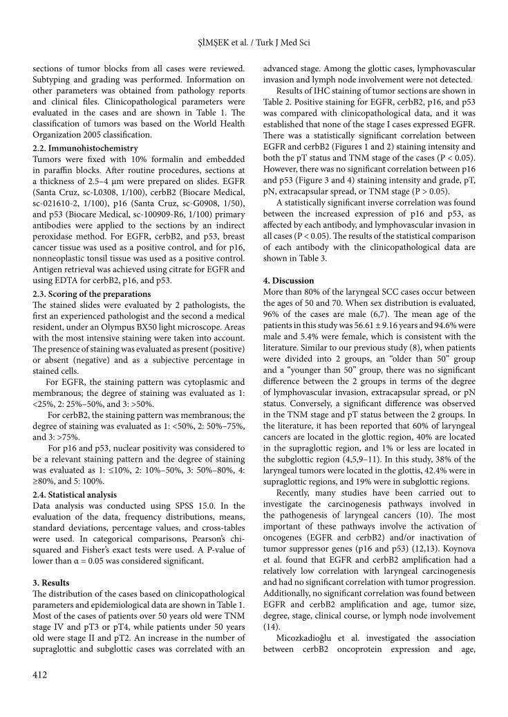
Table 2. Staining status of immunohistochemical antibodies.
EGFR n %
Negative (–) 42 45.7Positive (+) 50 54.3Overall 92 100.0
cerbB2
Negative (–) 61 66.3Positive (+) 31 33.7Overall 92 100.0
p16
Negative (–) 25 27.2Positive (+) 67 72.8Overall 92 100.0
p53
Negative (–) 47 51.1Positive (+) 45 48.9Overall 92 100.0
Figure 1. EGFR positivity in tumor tissue.
Figure 2. cerbB2 positivity in tumor tissue.
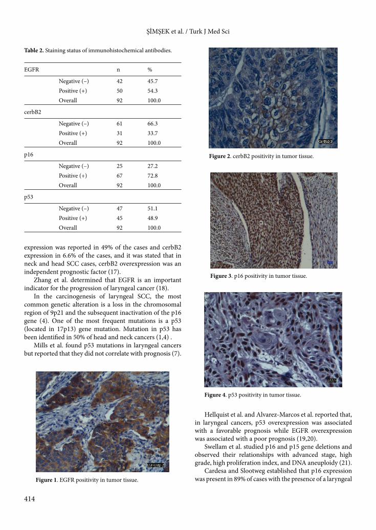
Figure 3. p16 positivity in tumor tissue.
Figure 4. p53 positivity in tumor tissue.

415
ŞİMŞEK et al. / Turk J Med Sci
tumor and reported that loss of p16 was important in the early tumorigenic phase (4).
In conclusion, consistent with the literature, p16 expression was present in 72.8% of the cases in this study, and p53 expression was present in 49.9% of the cases in this study. An increase in p16 and p53 expression levels in cases without lymphovascular invasion suggests that tumor suppressor genes (p16 and p53) have an effective role in laryngeal SCCs, mostly at the early stages of carcinogenesis and without an association with poor prognosis.
EGFR expression was present in 54% of the cases in this study, and cerbB2 expression was present in 33.7% of the cases of the cases in this study. Relative to previously reported studies, the expression levels of both proteins were higher. An increase in the expression levels of EGFR and cerbB2 oncogenes in cases with lymphovascular invasion agrees with the literature and suggests that EGFR and cerbB2 are associated with an advanced stage and poor prognosis in the development of laryngeal carcinoma.
Table 3. The comparison of immunohistochemical staining with clinical parameters.
Activation of protooncogenes Inactivation of tumor suppressor genes
EGFR (+) cerbB2 (+) EGFR (+)cerbB2 (+) p16 (+) p53 (+) p16 (+)
p53 (+)
Histological grade (P = 0.923, χ2 = 1.967)* (P = 0.711, χ2 = 0.682)**
Good 13 (14.1) 6 (6.5) 6 (6.5) 19 (20.7) 11 (12.0) 8 (8.7)Moderate 17 (18.5) 14 (15.2) 12 (13.0) 22 (23.9) 15 (16.3) 10 (10.9)Poor 20 (21.7) 11 (12.0) 11 (12.0) 26 (28.3) 19 (20.7) 14 (15.2)
Overall n (%) 50 (54.3) 31 (33.7) 29 (31.5) 67 (72.8) 45 (48.9) 32 (34.8)
pT stage (P = 0.000, χ2 = 31.948)* (P = 0.002, χ2 =14.374)**
T1 1 (1.1) 1 (1.1) 0 (0.0) 14 (15.2) 12 (13.0) 8 (8.7)T2 10 (10.9) 3 (3.3) 2 (2.2) 23 (25.0) 12 (13.0) 8 (8.7)T3 23 (25.0) 14 (15.2) 14 (15.2) 16 (17.4) 14 (15.2) 9 (9.8)T4 16 (17.4) 13 (14.1) 13 (14.1) 14 (15.2) 7 (7.6) 7 (7.6)
Overall n (%) 50 (54.3) 31 (33.7) 29 (31.5) 67 (72.8) 45 (48.9) 32 (34.8)
pN stage (P = 0.100, χ2 = 10.641)* (P = 0.070, χ2 = 5.317)**
N0 25 (27.2) 13 (14.1) 11 (12.0) 47 (51.1) 30 (32.6) 21 (22.8)N1 12 (13.0) 7 (7.6) 7 (7.6) 10 (10.9) 7 (7.6) 6 (6.5)N2 13 (14.1) 11 (12.0) 11 (12.0) 10 (10.9) 8 (8.7) 5 (5.4)
Overall n (%) 50 (54.3) 31 (33.7) 29 (31.5) 67 (72.8) 45 (48.9) 32 (34.8)
TNM stage (P = 0.000, χ2 = 36.848)* (P = 0.004, χ2 = 13.475)**
I 0 (0.0) 1 (1.1) 0 (0.0) 13 (14.1) 11 (12.0) 7 (7.6)II 6 (6.5) 2 (2.2) 1 (1.1) 20 (21.7) 9 (9.8) 6 (6.5)III 18 (19.6) 6 (6.5) 6 (6.5) 13 (14.1) 10 (10.9) 7 (7.6)IV 26 (28.3) 22 (23.9) 22 (23.9) 21 (22.8) 15 (16.3) 12 (13.0)
Overall n (%) 50 (54.3) 31 (33.7) 29 (31.5) 67 (72.8) 45 (48.9) 32 (34.8)
Extracapsular spread (P = 0.928, χ2 = 0.458)* (P = 0.895, χ2 = 0.018)**
(+) 17 (18.5) 12 (13.0) 12 (13.0) 13 (14.1) 11 (12.0) 8 (8.7)(–) 6 (6.5) 4 (4.3) 4 (4.3) 6 (6.5) 3 (3.3) 3 (3.3)
Overall n (%) 23 (25.0) 16 (17.4) 16 (17.4) 19 (20.7) 14 (15.2) 11 (12.0)
Lymphovascular invasion (P = 0.010, χ2 = 11.367)* (P = 0.028, χ2 = 4.844)**
(+) 27 (29.3) 20 (21.7) 20 (21.7) 22 (23.9) 17 (18.5) 12 (13.0)(–) 23 (25.0) 11 (12.0) 9 (9.8) 45 (48.9) 28 (30.4) 20 (21.7)
Overall n (%) 50 (54.3) 31 (33.7) 29 (31.5) 67 (72.8) 45 (48.9) 32 (34.8)
*: EGFR(+), cerbB2(+), p16(+), p53(+), **: EGFR and cerbB2 (+), p16 and p53 (+).

416
ŞİMŞEK et al. / Turk J Med Sci
References
1. Robbins SL, Kumar V, Cotran RS. Robbins and Cotran Pathologic Basis of Disease. 8th ed. Philadelphia, PA, USA: Saunders/Elsevier; 2010.
2. Thompson L. World Health Organization classification of tumours: pathology and genetics of head and neck tumours. Ear Nose Throat J 2006 ; 85: 74.
3. Rosai J, Ackerman LV. Rosai and Ackerman’s Surgical Pathology. 9th ed. Edinburgh, UK: Mosby; 2004.
4. Cardesa A, Slootweg PJ. Pathology of the Head and Neck. Berlin, Germany: Springer; 2006.
5. Pilch BZ. Head and Neck Surgical Pathology. Philadelphia, PA, USA: Lippincott Williams & Wilkins; 2001.
6. Sternberg SS, Mills SE, Carter D. Sternberg’s Diagnostic Surgical Pathology. 4th ed. Philadelphia, PA, USA: Lippincott Williams & Wilkins; 2004.
7. Sternberg SS. Histology for Pathologists. 2nd ed. Philadelphia, PA, USA: Lippincott-Raven; 1997.
8. Kocaturk S, Yilmazer D, Onal B, Erkam U, Urunal B. Do micrometastases detected with cytokeratin immunoperoxidase reactivity affect the treatment approach to neck in supraglottic cancers? Otolaryngol Head Neck Surg 2003; 128: 407–411.
9. Moore KL. Clinically Oriented Anatomy. 3rd ed. Baltimore, MD, USA: Williams & Wilkins; 1992.
10. Şimşek G, Han Ü, Önal B, Koybaşıoğlu F, Akın İ, Dağlı M. Expression of cyclin D1, p27, p21, BCL-2 and p53 in laryngeal squamous cell carcinoma and investigation of correlation with conventional prognostic factors. Turk J Med Sci 2013; 43: 27–32.
11. Arıncı K, Elhan A. Anatomi. Ankara, Turkey: Güneş Tıp Kitabevi; 2006.
12. Chomchai JS, Du W, Sarkar FH, Li YW, Jacobs JR, Ensley JF, Sakr W, Yoo GH. Prognostic significance of p53 gene mutations in laryngeal cancer. Laryngoscope 1999; 109: 455–459.
13. Sun J, Xiong J, Zhen Y, Chen ZL, Zhang H. P53 and PCNA is positively correlated with HPV infection in laryngeal epitheliopapillomatous lesions in patients with different ethnic backgrounds in Xinjiang. Asian Pac J Cancer Prev 2012; 13: 5439–5444.
14. Koynova DK, Tsenova VS, Jankova RS, Gurov PB, Toncheva DI. Tissue microarray analysis of EGFR and HER2 oncogene copy number alterations in squamous cell carcinoma of the larynx. J Cancer Res Clin Oncol 2005; 131: 199–203.
15. Micozkadioğlu D, Unal M, Pata YS, Baştürk M, Cinel L. Prognostic value of expression of p53, proliferating cell nuclear antigen, and c-erbB-2 in laryngeal carcinoma. Med Sci Monit 2008; 14: CR299–304.
16. Brunner K, Fischer CA, Driemel O, Hartmann A, Brockhoff G, Schwarz S. EGFR (HER) family protein expression and cytogenetics in 219 squamous cell carcinomas of the upper respiratory tract: ERBB2 overexpression independent prediction of poor prognosis. Anal Quant Cytol Histol 2010; 32: 78–89.
17. Wei Q, Sheng L, Shui Y, Hu Q, Nordgren H, Carlsson J. EGFR, HER2, and HER3 expression in laryngeal primary tumors and corresponding metastases. Ann Surg Oncol 2008; 15: 1193–1201.
18. Zhang S, He X, Li X, Wang Y. Expression of EGFR about different region tissues and its relationship with histological differentiation in laryngeal carcinoma. Lin Chung Er Bi Yan Hou Tou Jing Wai Ke Za Zhi 2012; 26: 123–124, 128 (article in Chinese with English abstract).
19. Hellquist H, Cardesa A, Gale N, Kambic V, Michaels L. Criteria for grading in the Ljubljana classification of epithelial hyperplastic laryngeal lesions. A study by members of the Working Group on Epithelial Hyperplastic Laryngeal Lesions of the European Society of Pathology. Histopathology 1999; 34: 226–233.
20. Alvarez-Marcos C, Lopez F, Alonso-Guervos M, Dominguez F, Suarez C, Hermsen MA, Liorente JL. Genetic and protein markers related to laryngeal epithelial precursor lesions and their neoplastic progression. Acta Otolaryngol 2013; 133: 281–290.
21. Swellam M, El-Arab LR, Adly A. Prognostic value of cell-cycle regulators and cellular biomarkers in laryngeal squamous cell carcinoma. Clin Biochem 2008; 41: 1059–1066.

![Theranostics · Web viewThus, several molecular markers, such as TP53 mutations [9, 10], P53/P16 immunohistochemistry [11], HPV genotyping [12, 13], gene expression [14], and loss](https://static.fdocuments.us/doc/165x107/60e49b4976d2144a4809da27/theranostics-web-view-thus-several-molecular-markers-such-as-tp53-mutations-9.jpg)