The evolution of the classification of nephrotic syndrome · ˝The evolution of the classification...
43
“ “ The evolution of the classification of The evolution of the classification of nephrotic syndrome nephrotic syndrome ” ” Laura Barisoni, MD Department of Pathology and Medicine, Division of Nephrology New York University
Transcript of The evolution of the classification of nephrotic syndrome · ˝The evolution of the classification...

““The evolution of the classification of The evolution of the classification of nephrotic syndromenephrotic syndrome””
Laura Barisoni, MD
Department of Pathology andMedicine, Division of Nephrology
New York University

Old classification schemes:
Proteinuria and nephrotic syndrome
Good prognosis andResponse to steroidTherapy
Poor prognosis andPoor Response to Steroid therapy
MCD FSGS

Nephrotic syndrome - the 80’s and 90’s• While the definition of minimal change disease did not change over the years, in the mid
80’s other patterns of glomerular damage have became part of the FSGS spectrum.
• Collapsing glomerulopathy:- first description in 1978 as “ malignant FSGS” (Brown Clin Nephrol 1978)- 1980’s frequent diagnosis during HIV pandemic (HIV-AN)- first described in non-HIV pts in 1986 (Weiss et al AJKD 1986) – “collapsing
glomerulopathy” – new clinical-pathologic entity.- in mid 90’s became “idiopathic collapsing FSGS”
(Detwiler et al KI 1994 & Valeri et al JASN 1996)
• Cellular lesion:- Term used first by Schwarz and colleagues to indicate a group of lesions with
endocapillary and/or extracapillary increased cellularity.- Other authors used the term cellular to indicate intracapillary cellularity only.
• Tip lesion:- Howie et al described tip lesion as a well-defined and specific pathological entity
with clinical similarity to MCD. (J Pathol 1984)- Tip lesions are also seen in associations with other glomerular diseases such as
diabetic nephropathy or membranous glomerulopathy.

Relatively recent classification schemes:Columbia classification - FSGS variants
Perihilar NOS Tip
Cellular Collapsing

Limitations of the morphologic classification
• Various histopathologic lesions are listed under “focal segmental glomerulosclerosis” regardless the presence or absence of segmental sclerosis.
• Lack of correlation with pathogenetic mechanisms and etiology.
• Lack of correlation with treatment

Proteinuria and nephrotic syndrome in the 21st century
• The attention of scientists, nephrologists and pathologists has been recently focused on the role of podocytes as cause of proteinuria
• In the last 10 years lot of progress has been made in the understanding the biology of podocytes, how they function and how they are injured.
“Taxonomy of the podocytopathies”where morphologic diagnosis are integrated with etiology
(Barisoni, Schnaper, Kopp, CJASN 2007)

TaxonomyTaxonomyταξισ + νοµια
arrangement or division law or method
A taxonomy is organized into multiple levels, each of which represents a taxon with one or more elements (taxa), which are mutually exclusive, unambiguous, and all-encompassing categories.
Taxonomies provide classification and conceptual framework for analysis, discussion, and hypothesis generation.
ETHIOLOGYidiopathic genetic reactive
HISTOPATHOLOGY- pattern of glom injury- podocyte number
Ταξον
Ταξα
Taxonomy of Podocytopathies

PodocytopathiesPodocytopathiesDEFINITION: Proteinuric diseases in which pathologic processes arise from intrinsic or extrinsic “primary”podocyte injury and where the podocyte genotype/phenotype is altered.

Podocytopathies: Podocytopathies: 4 morphologic patterns of glomerular injury4 morphologic patterns of glomerular injury
Normal Histology
MCN
Mesangial Sclerosis
DMS
Segmental Sclerosis
FSGS
Collapse ofthe GBM
CG

Common denominator of podocytopathies: Common denominator of podocytopathies: Podocyte injury = foot process effacementPodocyte injury = foot process effacement

Causes of foot process effacementCauses of foot process effacement1. Impaired formation of the slit diaphragm complex2. Abnormalities of the adhesive interaction between podocytes and GBM3. Alterations of transcription factors4. Abnormalities of the actin-based cytoskeleton5. Alterations of the apical domain of podocytes6. Mitochondria abnormalities7. Abnormalities of cell metabolism8. Mechanical stress9. Viral infection10. Acute ischemic injury11. Toxic / metabolic effect12. Immunologic stimuli
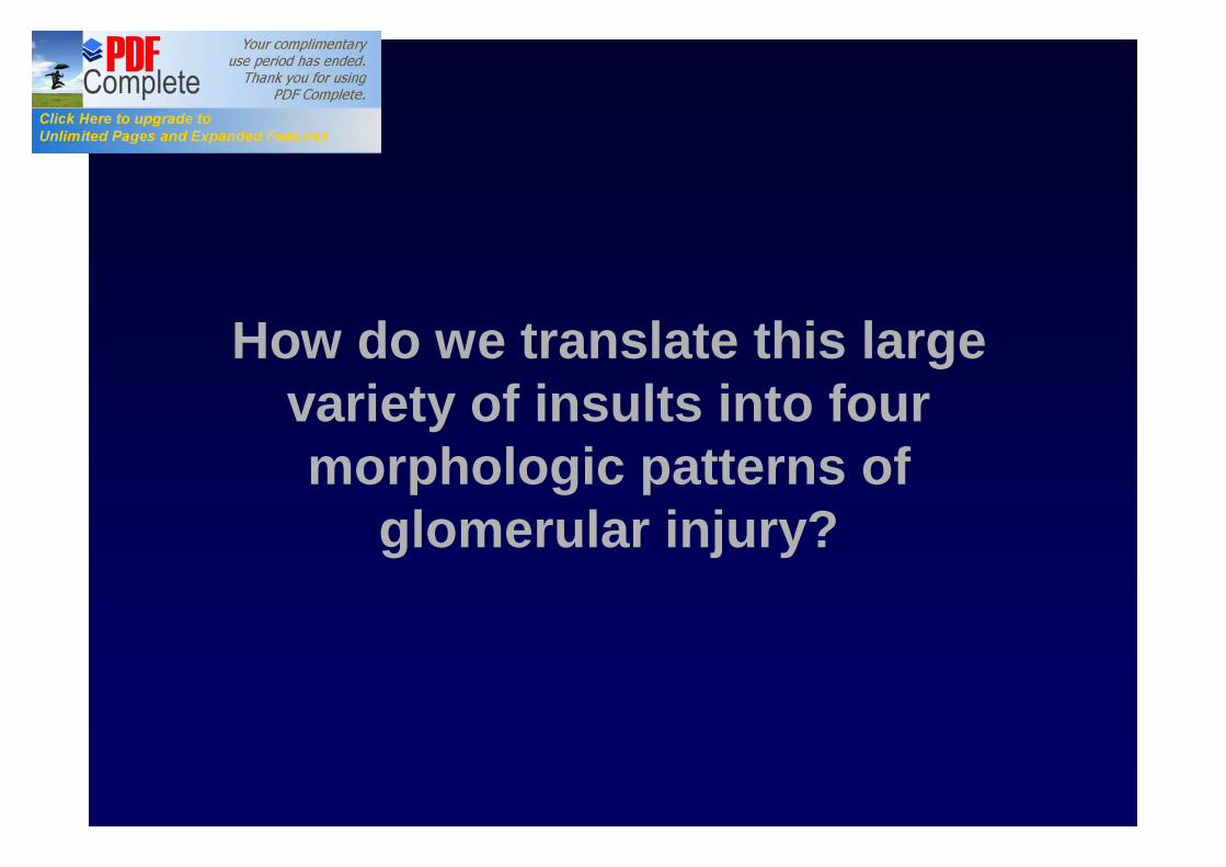
How do we translate this large variety of insults into four morphologic patterns of
glomerular injury?

Hypothesis #1:Hypothesis #1:Injured podocytes can take different pathwaysInjured podocytes can take different pathways
Podocyte injury
Collapse
Proliferation(high)
Engagement of apoptotic pathways
Cell death
No change
De-differentiation
FSGS CG
No change in podocyte number
MCN
Mesangial sclerosis
DMS
Segmentalsclerosis
Altered phenotype
Proliferation(low)
Developmental arrest

Copyright ©2009 American Society of Nephrology
Ronconi, E. et al. J Am Soc Nephrol 2009;20:322-332
Hypothesis #2: The role of the renopoietic system
Hierarchical distribution of CD133+CD24+PDX- and CD133+CD24+PDX+ cells within human glomeruli

Hypothesis #2Hypothesis #2The role of CD24+CD133+ renal progenitors in FSGS & CG.
Podocyte injury
pseudocrescents
Exuberant CD24+CD133+
Activity
Podocytedeath
InsufficientCD24+CD133+Repair activity
No change
FSGS CG
No change in podocyte number
MCN
Mesangial sclerosis
DMS
Segmentalsclerosis
Altered phenotype
Proliferation(low)
Developmental arrest
Podocyte death

MINIMAL CHANGE MINIMAL CHANGE NEPHROPATHYNEPHROPATHY
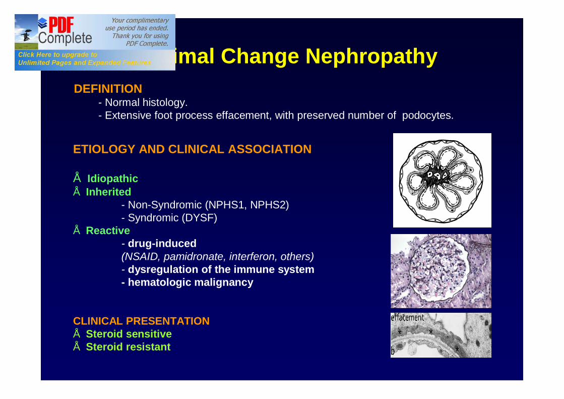
Minimal Change NephropathyMinimal Change NephropathyDEFINITION
- Normal histology.- Extensive foot process effacement, with preserved number of podocytes.
ETIOLOGY AND CLINICAL ASSOCIATION
• Idiopathic• Inherited
- Non-Syndromic (NPHS1, NPHS2)- Syndromic (DYSF)
• Reactive- drug-induced (NSAID, pamidronate, interferon, others)- dysregulation of the immune system- hematologic malignancy
CLINICAL PRESENTATION• Steroid sensitive• Steroid resistant

FOCAL SEGMENTAL FOCAL SEGMENTAL GLOMERULOSCLEROSISGLOMERULOSCLEROSIS

FSGSFSGSDEFINITION
Segmental solidification of the tuft accompanied by sinechiae. Hyalinosis and foam cells can also be present. Low number of podocytes (podocytopenia).
ETIOLOGY AND CLINICAL ASSOCIATION
• Idiopathic• Inherited
- syndromic- non-syndromic
• Reactive- hyperfiltration-mediated
normal renal massreduced renal mass
- medication-induced- permeability factor (?)

Idiopathic FSGSIdiopathic FSGS
APOL1 is a major-effect risk gene for FSGS.(Genovese et al Science 2010)
APOL1 risk alleles are more frequent in AA. Odd ratios of 10.5 in FSGS and 7.3 in HTN-ESRD
Is idiopathic really idiopathic?

Genetic forms of FSGSGenetic forms of FSGS
• Associated with other organ abnormalities (syndromic):– Freiser Syndrome (WT-1).– Nail-patella syndrome (LMX1B)– Renal-coloboma syndrome with oligomeganephronia (PAX2)– Alport’s disease (COL4A3, A4, A5)– Metabolic disorders (GLA – Fabry’s)– Mitochondriopathies (mtDNA tRNALeu and tRNATyr,CoQ2 NP, CoQ6 NP)
• Limited to the kidney (non-syndromic):- NPHS1 – nephrin – autosomal recessive- NPHS2 – podocin – autosomal recessive- NPHS3 – phospholipase Cε1 – autosomal recessive- ACTN4 – α-actinin-4 - autosomal dominant - INF2 – autosomal dominant- TRPC6 – Transient Receptor Potential channel 6 - autosomal dominant - WT1 – sporadic/isolated FSGS- CD2AP – susceptibility to FSGS - MYH 9 – susceptibility to FSGS - APOL1 – susceptibility to FSGS
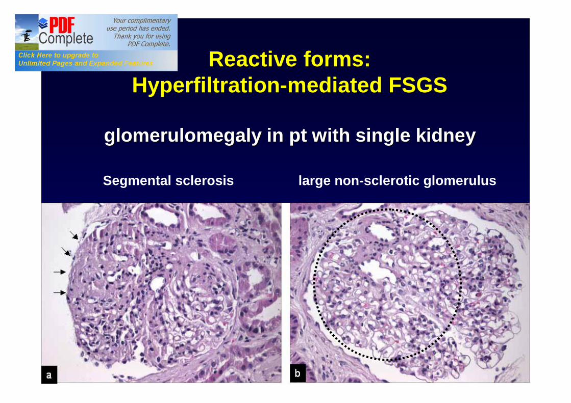
Segmental sclerosis large non-sclerotic glomerulus
Reactive forms: Reactive forms: HyperfiltrationHyperfiltration--mediated FSGSmediated FSGS
glomerulomegaly in pt with single kidneyglomerulomegaly in pt with single kidney

DIFFUSE MESANGIAL DIFFUSE MESANGIAL SCLEROSISSCLEROSIS
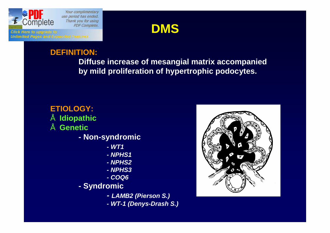
DMS
DEFINITION:Diffuse increase of mesangial matrix accompanied by mild proliferation of hypertrophic podocytes.
ETIOLOGY:• Idiopathic• Genetic
- Non-syndromic- WT1 - NPHS1 - NPHS2- NPHS3- COQ6
- Syndromic- LAMB2 (Pierson S.)- WT-1 (Denys-Drash S.)

COLLAPSING COLLAPSING GLOMERULOPATHYGLOMERULOPATHY

CG Definition: GBM collapse and CG Definition: GBM collapse and pseudocrescent formationpseudocrescent formation
HIV
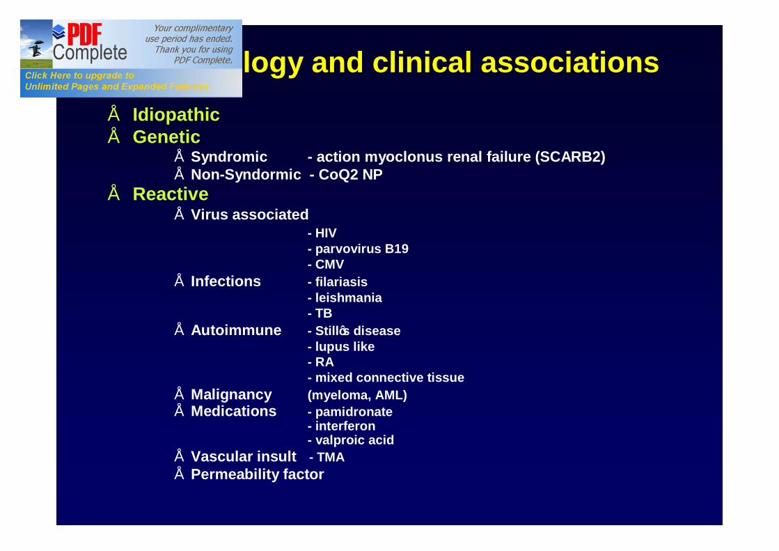
CG: etiology and clinical associationsCG: etiology and clinical associations• Idiopathic• Genetic
• Syndromic - action myoclonus renal failure (SCARB2)• Non-Syndormic - CoQ2 NP
• Reactive• Virus associated
- HIV- parvovirus B19- CMV
• Infections - filariasis- leishmania- TB
• Autoimmune - Still’s disease- lupus like- RA - mixed connective tissue
• Malignancy (myeloma, AML)• Medications - pamidronate
- interferon- valproic acid
• Vascular insult - TMA• Permeability factor

Differently from other podocytopathies, in HIV-CG podocytes do not express maturity
markers
Barisoni et al. JASN 1999

In CG de-differentiated podocytes re-enter the cell cycle and proliferate
p57 Ki-67 Ki-67
Barisoni et al. KI 2000

In idiopathic and HIV-associated CG dedifferentiated podocytes have a
dysregulated phenotype

In inherited CG (COQ2In inherited CG (COQ2--NP)NP)podocyte phenotype is dedifferentiated podocyte phenotype is dedifferentiated
but not dysregulatedbut not dysregulated
WT1Synpo Ki67

Which is the underlying mechanism of pseudocrescent formation and
collapse of the basement membranes?

Pathogenesis of CG:from podocyte injury to pseudocrescent formation
The dysregulated podocyte
Podocytes are injured
They dedifferentiate and dysregulate their phenotype
Dedifferentiated podocytes can re-enter the cell cycle and
proliferate
Pseudocrescent formation
The exuberant renopoietic system
Podocytes are injured
They undergo apoptosis/death
CD24+CD133+ cells migrate from the Bowman’s capsule
Exuberant proliferation
Pseudocrescent formation

Collapsing glomerulopathy is a proliferative disease
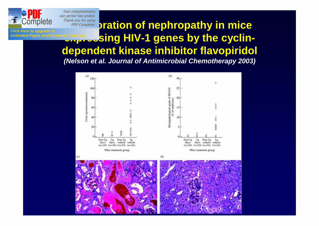
Amelioration of nephropathy in mice expressing HIV-1 genes by the cyclin-dependent kinase inhibitor flavopiridol(Nelson et al. Journal of Antimicrobial Chemotherapy 2003)

TAXONOMY OF PODOCYTOPATHIESidiopathic genetic reactive
MCN Idiopathic•Steroid-sensitive•Steroid-resistant
Non-syndromic•NPHS1•NPHS2
Syndromic•DYSF
Clinical association(immunologic stimuli, Tumors)
Medications(NSAID,gold, penicillamine, lithium, IF, pamidronate)
FSGS Idiopathic•Steroid-sensitive•Steroid-resistant
Non-syndromicITGB4, NPSH2, NPHS3, NPHS1 + NPHS2, COQ2, MHY9, ACTN4, CD2AP, TRCP6, WT-1, SYNPO, INF2
SyndromicMtDNA, WT1, PAX2, COQ6, COL4,GLA, LMBX1
Post-adaptive•nephron mass•Initially normal nephron mass
Medications (tacrolimus, lithium, IF, pamidronate)
DMS Idiopathic Non-syndromicWT1, NPHS1, NPSH2, NPHS3, LAMB2,
SyndromicWT1, LAMB2, COQ6,
CG Idiopathic Non-syndromicCOQ2MHY9
SyndromicSCARB
Infections (viruses, TB, others)
Clinical association•Autoimmune, TMA, tumors
Medications (IF, pamidronate, valproic acid)

MCN, FSGS, DMS and CG are patterns of glomerular damage where the common denominator is podocyte injury .
Morphologic classifications alone are insufficient to capture the complexity and heterogeneity of diseases presenting with NS.- multiple specific disease processes can present with indistinguishable histopatholology- a specific monogenetic disorder can present with more than one form of histopathologic pattern of glomerular damage.
Final diagnosis of the podocytopathies should occur in 3 steps: a. clinical evaluation b. morphologic evaluation c. additional clinical tests, such as genetic or serology for evidence of
infections, or others, when indicated.
Proteinuria and nephrotic syndrome: the present

Nephrotic Syndrome: the future
NEPTUNE : The Nephrotic Syndrome Study NetworkInternational effort with the following major goals:
• Determination of rates and predictors of clinical remission or progression in NS
• Identification of gene expression profiles• Identification of patient specific molecular signatures• Clinically useful classification based on morphologic & molecular
phenotype

NEPTUNE: evolution of the renal biopsy procedure

Evolution of morphologic analysis methodology

NEPTUNE Pathology Scoring Sheet