The evolution of paralogous enzymes MAT and ... - UBC Botany
12
RESEARCH ARTICLE Open Access The evolution of paralogous enzymes MAT and MATX within the Euglenida and beyond Jana Szabová 1,2* , Naoji Yubuki 3 , Brian S Leander 3 , Richard E Triemer 4 and Vladimír Hampl 1,2* Abstract Background: Methionine adenosyltransferase (MAT) is a ubiquitous essential enzyme that, in eukaryotes, occurs in two relatively divergent paralogues: MAT and MATX. MATX has a punctate distribution across the tree of eukaryotes and, except for a few cases, is mutually exclusive with MAT. This phylogenetic pattern could have arisen by either differential loss of old paralogues or the spread of one of these paralogues by horizontal gene transfer. Our aim was to map the distribution of MAT/MATX genes within the Euglenida in order to more comprehensively characterize the evolutionary history of MATX. Results: We generated 26 new sequences from 23 different lineages of euglenids and one prasinophyte alga Pyramimonas parkeae. MATX was present only in photoautotrophic euglenids. The mixotroph Rapaza viridis and the prasinophyte alga Pyramimonas parkeae, which harbors chloroplasts that are most closely related to the chloroplasts in photoautotrophic euglenids, both possessed only the MAT paralogue. We found both the MAT and MATX paralogues in two photoautotrophic species (Phacus orbicularis and Monomorphina pyrum). The significant conflict between eukaryotic phylogenies inferred from MATX and SSU rDNA data represents strong evidence that MATX paralogues have undergone horizontal gene transfer across the tree of eukaryotes. Conclusions: Our results suggest that MATX entered the euglenid lineage in a single horizontal gene transfer event that took place after the secondary endosymbiotic origin of the euglenid chloroplast. The origin of the MATX paralogue is unclear, and it cannot be excluded that it arose by a gene duplication event before the most recent common ancestor of eukaryotes. Keywords: Methionine adenosyltransferase, Horizontal gene transfer, Deep paralogy, Gene evolution, Euglenozoa Background Methionine adenosyltransferase (MAT) is a cytosolic ubi- quitous enzyme that synthesizes S-adenosyl-L-methionine (SAM), a molecule that is one of the most important me- tabolites in living cells. SAM serves as the major methyl donor to phospholipids, DNA, RNA and other small mol- ecules and is the second most widely used enzyme sub- strate after ATP [1,2]. MAT is a well-conserved enzyme that is encoded in the genomes of most eukaryotes, eubac- teria, and archaebacteria (which have a highly divergent version of the gene) and has been well studied at the primary, secondary, and tertiary structural levels [3-5]. Except for the mammalian MAT II, which is a hetero- oligomer [6], members of the MAT family are homo- oligomers that usually form tetramers consisting of four identical subunits; the two active sites are located between the subunits in each dimer [3]. Mammalian MAT III and archaeal MATs form dimers [7]. Multiple sequence alignments of MAT genes from a wide diversity of eukaryotes demonstrated a paralogue of MAT, named MATX, with distinctive features that are absent in all other eukaryotic MATs. These features include four specific insertions and a large number of unique substitutions [8]. The recombinant MATX from Euglena gracilis has been found to function as a homo- dimer with activities comparable to MATs from other eukaryotes [9]. Molecular phylogenetic analyses clearly showed that MATX is related to other eukaryotic MATs, but it forms a long branch in the eukaryotic subtree [8]. The majority of MATX paralogues occur in four distantly * Correspondence: [email protected]; [email protected] 1 Department of Parasitology, Charles University in Prague, Faculty of Science, Vinicna 7, Prague 2 128 44, Czech Republic 2 Biotechnology and Biomedicine Center of the Academy of Sciences and Charles University in Vestec, Prague, Czech Republic Full list of author information is available at the end of the article © 2014 Szabová et al.; licensee BioMed Central Ltd. This is an Open Access article distributed under the terms of the Creative Commons Attribution License (http://creativecommons.org/licenses/by/2.0), which permits unrestricted use, distribution, and reproduction in any medium, provided the original work is properly credited. Szabová et al. BMC Evolutionary Biology 2014, 14:25 http://www.biomedcentral.com/1471-2148/14/25
Transcript of The evolution of paralogous enzymes MAT and ... - UBC Botany
Szabová et al. BMC Evolutionary Biology 2014, 14:25http://www.biomedcentral.com/1471-2148/14/25
RESEARCH ARTICLE Open Access
The evolution of paralogous enzymes MAT andMATX within the Euglenida and beyondJana Szabová1,2*, Naoji Yubuki3, Brian S Leander3, Richard E Triemer4 and Vladimír Hampl1,2*
Abstract
Background: Methionine adenosyltransferase (MAT) is a ubiquitous essential enzyme that, in eukaryotes, occurs intwo relatively divergent paralogues: MAT and MATX. MATX has a punctate distribution across the tree of eukaryotesand, except for a few cases, is mutually exclusive with MAT. This phylogenetic pattern could have arisen by eitherdifferential loss of old paralogues or the spread of one of these paralogues by horizontal gene transfer. Our aimwas to map the distribution of MAT/MATX genes within the Euglenida in order to more comprehensivelycharacterize the evolutionary history of MATX.
Results: We generated 26 new sequences from 23 different lineages of euglenids and one prasinophyte algaPyramimonas parkeae. MATX was present only in photoautotrophic euglenids. The mixotroph Rapaza viridis and theprasinophyte alga Pyramimonas parkeae, which harbors chloroplasts that are most closely related to the chloroplastsin photoautotrophic euglenids, both possessed only the MAT paralogue. We found both the MAT and MATXparalogues in two photoautotrophic species (Phacus orbicularis and Monomorphina pyrum). The significant conflictbetween eukaryotic phylogenies inferred from MATX and SSU rDNA data represents strong evidence that MATXparalogues have undergone horizontal gene transfer across the tree of eukaryotes.
Conclusions: Our results suggest that MATX entered the euglenid lineage in a single horizontal gene transfer eventthat took place after the secondary endosymbiotic origin of the euglenid chloroplast. The origin of the MATXparalogue is unclear, and it cannot be excluded that it arose by a gene duplication event before the most recentcommon ancestor of eukaryotes.
Keywords: Methionine adenosyltransferase, Horizontal gene transfer, Deep paralogy, Gene evolution, Euglenozoa
BackgroundMethionine adenosyltransferase (MAT) is a cytosolic ubi-quitous enzyme that synthesizes S-adenosyl-L-methionine(SAM), a molecule that is one of the most important me-tabolites in living cells. SAM serves as the major methyldonor to phospholipids, DNA, RNA and other small mol-ecules and is the second most widely used enzyme sub-strate after ATP [1,2]. MAT is a well-conserved enzymethat is encoded in the genomes of most eukaryotes, eubac-teria, and archaebacteria (which have a highly divergentversion of the gene) and has been well studied at theprimary, secondary, and tertiary structural levels [3-5].
* Correspondence: [email protected]; [email protected] of Parasitology, Charles University in Prague, Faculty of Science,Vinicna 7, Prague 2 128 44, Czech Republic2Biotechnology and Biomedicine Center of the Academy of Sciences andCharles University in Vestec, Prague, Czech RepublicFull list of author information is available at the end of the article
© 2014 Szabová et al.; licensee BioMed CentraCommons Attribution License (http://creativecreproduction in any medium, provided the or
Except for the mammalian MAT II, which is a hetero-oligomer [6], members of the MAT family are homo-oligomers that usually form tetramers consisting of fouridentical subunits; the two active sites are located betweenthe subunits in each dimer [3]. Mammalian MAT III andarchaeal MATs form dimers [7].Multiple sequence alignments of MAT genes from a
wide diversity of eukaryotes demonstrated a paralogue ofMAT, named MATX, with distinctive features that areabsent in all other eukaryotic MATs. These featuresinclude four specific insertions and a large number ofunique substitutions [8]. The recombinant MATX fromEuglena gracilis has been found to function as a homo-dimer with activities comparable to MATs from othereukaryotes [9]. Molecular phylogenetic analyses clearlyshowed that MATX is related to other eukaryotic MATs,but it forms a long branch in the eukaryotic subtree [8].The majority of MATX paralogues occur in four distantly
l Ltd. This is an Open Access article distributed under the terms of the Creativeommons.org/licenses/by/2.0), which permits unrestricted use, distribution, andiginal work is properly credited.
Szabová et al. BMC Evolutionary Biology 2014, 14:25 Page 2 of 12http://www.biomedcentral.com/1471-2148/14/25
related groups of photosynthetic eukaryotes: haptophytes,photosynthetic euglenids, diatoms, and dinoflagellates.MATX was also detected in a pelagophyte alga Aureococ-cus anophagefferens [10]. All organisms possess either theMAT or the MATX form of the gene, with the exceptionof five diatom species that have both paralogues and A.anophagefferens that harbors two different homologues ofMAT in addition to MATX [8,10].A similar punctate distribution of two paralogues with
the same function was reported for “elongation factor 1-alpha” (EF-1α) and its paralogue “elongation factor like”(EFL), which are highly conserved members of a GTPasesuperfamily involved in translation. Like MAT/MATX,the EF-1α/EFL paralogues have a patchy distributionacross the tree of eukaryotes and rarely occur togetherin the same organism. EFL has been localized so far ineight groups of unrelated organisms: dinoflagellates,haptophytes, cercozoans, green algae, choanoflagellates,fungi, diatoms, and radiolarians [11-17].The punctate distributions of MAT/MATX and EF-1α/
EFL across the tree of eukaryotes can be explained by twoscenarios: (1) a deep paralogy, whereby both paralogueswere present in an ancient common ancestor followed bydifferential loss of one or the other paralogue in descend-ant lineages; and (2) a horizontal (syn., lateral) gene trans-fer (HGT), whereby a more recent origin of one paralogue(most likely the less frequent one, such as MATX) inone lineage of eukaryotes is followed by the spread ofthis paralogue to other distantly related lineages viahorizontal transfer.These scenarios differ in their assumptions. The first sce-
nario hypothesizes coexistence and probably co-expressionof both paralogues in one cell for a long time withoutnegative effects on the organism. This scenario explainsthe distribution purely by vertical transmission. In thiscase, MATX must have originated by gene duplicationfrom the MAT already present in the common ancestor ofall MATX containing taxa. This organism was very ancientand not very distantly related, maybe identical, to the mostrecent common ancestor of eukaryotes. Since that time,MAT and MATX must have been propagated side by sidein the genomes of the descendants to much more recentnodes of eukaryotic evolution and in some cases (diatoms)even to extant organisms.The second scenario assumes that one (MATX) can be
horizontally transferred and is capable of functional re-placement of the MAT form soon after the transfer. Ourprevious work on the model systems of Euglena gracilisand Trypanosoma brucei indicates that MATX fulfillsthe assumptions for both of these scenarios, because thisparalogue can be co-expressed with MAT and can imme-diately take over its function [18]. By contrast, EFL wascapable of long-term co-expression, but was not able tofunctionally replace EF1-α. Based on these results, neither
of the two evolutionary scenarios can be refuted forMAT/MATX. However, in the case of EF1-α/EFL, HGTis apparently more difficult and likely played a less im-portant role in the evolutionary history of this paraloguecouple [18].There are several questions associated with the putative
HGT explanation for the origin and distribution of theMATX paralogue that remain unanswered. For instance,under what circumstances would the highly divergentMATX evolve within one recent group of eukaryotes andin which lineage could it happen? One hypothesis positsthat MATX evolved during a secondary endosymbiotic ori-gin of plastids from the endosymbiont copy of the MATgene, which was released from purifying selection andunderwent accelerated sequence evolution [8]. Therefore,an analysis of the distribution of MAT/MATX in euglenidsprovides an opportunity to evaluate this possibility.The Euglenida is a large group of marine and freshwater
eukaryotic flagellates with diverse modes of nutrition, in-cluding phagotrophy, osmotrophy, photoautotrophy, anda recently discovered example of mixotrophy (a euglenidcapable of both phagotrophy and photosynthesis) [19,20].Photosynthetic and secondarily osmotrophic euglenids(i.e., colorless euglenids that have lost photosynthesis)form a monophyletic group that is the sister lineage to themixotrophic Rapaza viridis and is nested within a para-phyletic assemblage of phagotrophic euglenids. It is in-ferred that the secondary chloroplast was gained throughsecondary endosymbiosis in the most recent common an-cestor of all photosynthetic euglenids, including R. viridis[19-22]. The marine flagellate Pyramimonas (Pyramimo-nadales, Prasinophyta) is inferred to be the closest knownrelative of the euglenid chloroplasts (Turmel et al. 2009).In this study, we investigated the distribution of MAT andMATX in euglenids and Pyramimonas in order to evalu-ate whether the origin of MATX occurred simultaneouslywith the secondary endosymbiotic origin of the euglenidchloroplast. These data were also expected to provide in-sights into whether euglenids were the first group of eu-karyotes to evolve the MATX paralogue.
ResultsMAT and MATX phylogeny and distribution of MATX ineuglenidsWe generated six new sequences of MAT and 20 newsequences of MATX. The MAT sequences were obtainedfrom heterotrophic euglenids (Petalomonas cantuscygniand Distigma sp.), the mixotroph Rapaza viridis, twophotoautotrophic euglenids (Phacus orbicularis andMonomorphina pyrum) and the prasinophyte alga Pyra-mimonas parkeae. The MATX sequences were obtainedfrom all investigated photoautotrophic euglenids, exceptRapaza viridis (Table 1). The sequences retrieved fromtranscriptome projects were complete; sequences amplified

Table 1 Sources of sequences applied in this study
Taxon Protein MAT/MATX SSU
Euglena clara † supplement AJ532423.1*
Euglena stellata † supplement AF150936.1*
Euglena gracilis † supplement AY029409.1*
Euglena hiemalis † supplement DQ140157.1*
Euglena proxima † supplement EU624027.1*
Euglena viridis † supplement AJ532415.1*
Euglenaria anabaena † supplement AF242548.1*
Eutreptiella braarudii † supplement AJ532397.1*
Eutreptiella gymnastica ▲ KF383289 ▲ KF559331
Distigma sp. ▲ KF383287
Eutreptia viridis † supplement AF157312.1*
Lepocinclis tripteris † supplement AF286210.1*
Lepocinclis playfairiana † supplement KF267871*
Monomorphina aenigmatica ▲ KF383291 AF283313.1*
Monomorphina parapyrum † supplement AF112874
Monomorphina pyrum ▲ KF383286 MAT▲ KF383290 MATX
▲ KF559330
Phacus inflexus † supplement FJ719629.1*
Phacus orbicularis † supplement AF283315.1*
Pyramimonas parkeae ▲ KF383285
Rapaza viridis ▲ KF383288 AB679269.1*
Trachelomonas ellipsoidalis † supplement DQ140135.1*
Trachelomonas sp. ▲ KF383292 AJ532447.1*
Trachelomonas volvocina † supplement AF096995.1*
Strombomonas accuminata † supplement EU624029.1*
The sequences downloaded from GenBank are marked by *; sequencesobtained by Sanger sequencing method in this study are marked by ▲,sequences obtained from transcriptome projects sequenced by Roche 454sequencing were marked by † and are available in supplement.
Szabová et al. BMC Evolutionary Biology 2014, 14:25 Page 3 of 12http://www.biomedcentral.com/1471-2148/14/25
from cDNA (Pyramimonas parkeae, Trachelomonas sp.,Distigma sp., Monomorphina aenigmatica and Monomor-phina pyrum) were partial (approximately 430 aminoacids). We found additional so far unnoticed partial MATXhomologues in GenBank from the haptophyte Prymne-sium, the plant Lactuca serriola and the beetle Dendrocto-nus frontalis. Further database searches revealed thatLactuca and Dendroctonus also contain the MAT paralo-gue. The presence of the MATX paralogue in the singlespecies of plant and metazoa is highly suspicious, and wetreat this data with caution because we cannot exclude thepossibility of contamination by foreign RNA in the Lactucaand Dendroctonus transcriptome data sets. The MATsequences of Rhodomonas sp., Rhodomonas salina, Tha-lassionema sp. and Peranema trichophorum and theMATX sequence of Karenia brevis retrieved from Gen-Bank were also incomplete. Despite their incomplete-ness, all MAT and MATX sequences were suitable fordetermining the paralogue type and for phylogenetic
analyses; therefore, all sequences were added to thealignment with published MAT/MATX sequences forphylogenetic analysis (Figure 1).In the phylogenetic tree (Figure 1), MATX paralogues
formed a well-supported clade that was separated fromthe MAT paralogues by a long stem. The tree wasrooted by five bacterial outgroups within the MAT para-logues, with Trichomonas vaginalis MAT being themost basal branch. However, the backbone topology ofthe MAT tree was weakly supported, and the MATXbranch was situated only one node apart from prokary-otes. We used Kishino Hasegawa (KH), weighted KH(WKH), Shimodaria Hasegawa (SH) and weighted SH(WSH) tests to evaluate whether the root position be-tween MAT and MATX paralogues is significantly worsethan the suggested root on the T. vaginalis branch. Thetests showed that this root position cannot be ex-cluded (p = 0.076 for KH and WKH, p = 1.00 for SH andp = 0.945 for WSH).The MATX sequences from photoautotrophic eugle-
nids formed a well-supported subclade (bootstrap 77%)within the more inclusive MATX clade and branched asthe sister group to a clade consisting of Lactuca, dinofla-gellates and Dendroctonus. The MAT sequences fromthe heterotrophic euglenids clustered together with kine-toplastids; the MAT sequence from P. parkeae branchedtogether with other green algae; and the MAT sequencesfrom M. pyrum and P. orbicularis clustered with ciliatesand Aureococcus, respectively.We also performed an independent analysis of MATX
sequences that enabled us to use more alignment posi-tions to reconstruct the phylogenetic relationships withinthe MATX clade (Figure 2). The tree was rooted with thebranch of diatoms, haptophytes and Aureococcus accord-ing to Figure 1.
Comparison of MATX and SSU rRNA gene phylogenyWe investigated whether or not the phylogeny of theMATX paralogues differs significantly from the speciesphylogeny. Significant differences would indicate thatMATX has not evolved vertically but instead experiencedHGTs between the MATX containing taxa. As “speciestrees”, we have used topologies inferred from small sub-unit (SSU) rRNA gene sequences and also manuallyconstructed topologies reflecting current view of speciesrelationships. The SSU rRNA gene tree and manual spe-cies topologies differed in minor details and they are re-ported in Additional file 1 and in Additional file 2: FigureS1 and Additional file 3: Figure S3. We used the KH andSH tests to compare the species topologies with the bestMATX topology and the set of 500 bootstrap topologiescalculated from MATX alignment (Table 2). The testsshowed that the “species topologies” are strongly rejected(p value = < 7*10-6). To be sure that the conflict with the

Figure 1 Maximum likelihood phylogeny of MAT and MATX. The tree was constructed by maximum likelihood method in RAxML from the347 amino acid positions. The values at nodes represent maximum likelihood bootstraps/Bayesian posterior probabilities; only values above 50%and 0.5, respectively, are shown. Euglenid taxa are marked in red.
Szabová et al. BMC Evolutionary Biology 2014, 14:25 Page 4 of 12http://www.biomedcentral.com/1471-2148/14/25
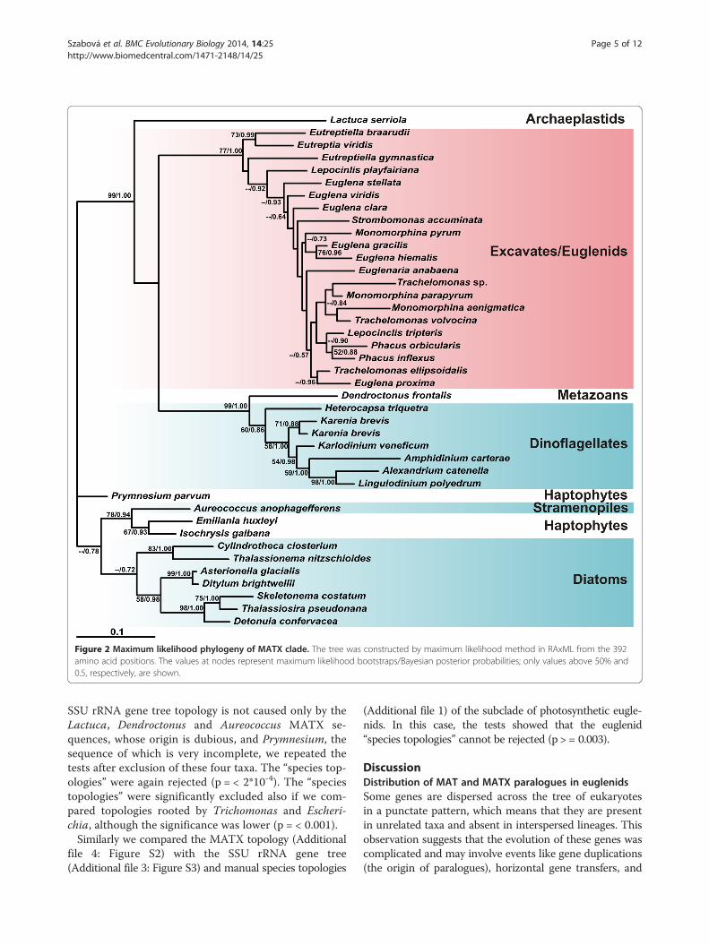
Figure 2 Maximum likelihood phylogeny of MATX clade. The tree was constructed by maximum likelihood method in RAxML from the 392amino acid positions. The values at nodes represent maximum likelihood bootstraps/Bayesian posterior probabilities; only values above 50% and0.5, respectively, are shown.
Szabová et al. BMC Evolutionary Biology 2014, 14:25 Page 5 of 12http://www.biomedcentral.com/1471-2148/14/25
SSU rRNA gene tree topology is not caused only by theLactuca, Dendroctonus and Aureococcus MATX se-quences, whose origin is dubious, and Prymnesium, thesequence of which is very incomplete, we repeated thetests after exclusion of these four taxa. The “species top-ologies” were again rejected (p = < 2*10-4). The “speciestopologies” were significantly excluded also if we com-pared topologies rooted by Trichomonas and Escheri-chia, although the significance was lower (p = < 0.001).Similarly we compared the MATX topology (Additional
file 4: Figure S2) with the SSU rRNA gene tree(Additional file 3: Figure S3) and manual species topologies
(Additional file 1) of the subclade of photosynthetic eugle-nids. In this case, the tests showed that the euglenid“species topologies” cannot be rejected (p > = 0.003).
DiscussionDistribution of MAT and MATX paralogues in euglenidsSome genes are dispersed across the tree of eukaryotesin a punctate pattern, which means that they are presentin unrelated taxa and absent in interspersed lineages. Thisobservation suggests that the evolution of these genes wascomplicated and may involve events like gene duplications(the origin of paralogues), horizontal gene transfers, and
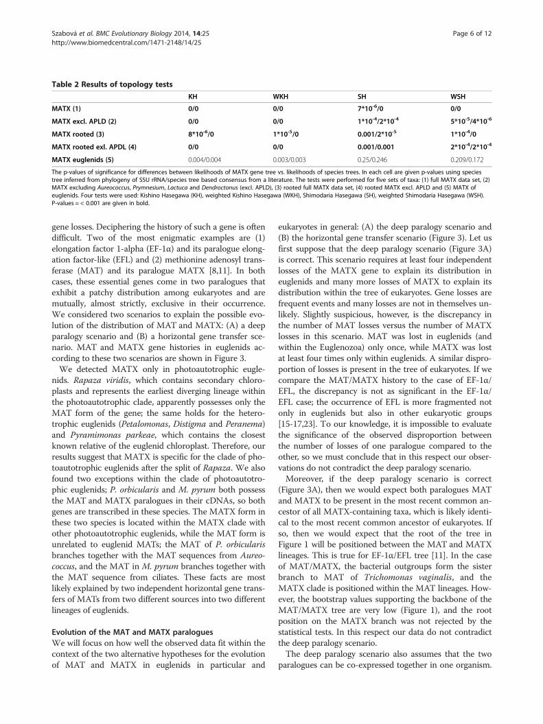
Table 2 Results of topology tests
KH WKH SH WSH
MATX (1) 0/0 0/0 7*10-6/0 0/0
MATX excl. APLD (2) 0/0 0/0 1*10-4/2*10-4 5*10-5/4*10-6
MATX rooted (3) 8*10-6/0 1*10-5/0 0.001/2*10-5 1*10-4/0
MATX rooted exl. APDL (4) 0/0 0/0 0.001/0.001 2*10-4/2*10-4
MATX euglenids (5) 0.004/0.004 0.003/0.003 0.25/0.246 0.209/0.172
The p-values of significance for differences between likelihoods of MATX gene tree vs. likelihoods of species trees. In each cell are given p-values using speciestree inferred from phylogeny of SSU rRNA/species tree based consensus from a literature. The tests were performed for five sets of taxa: (1) full MATX data set, (2)MATX excluding Aureococcus, Prymnesium, Lactuca and Dendroctonus (excl. APLD), (3) rooted full MATX data set, (4) rooted MATX excl. APLD and (5) MATX ofeuglenids. Four tests were used: Kishino Hasegawa (KH), weighted Kishino Hasegawa (WKH), Shimodaria Hasegawa (SH), weighted Shimodaria Hasegawa (WSH).P-values = < 0.001 are given in bold.
Szabová et al. BMC Evolutionary Biology 2014, 14:25 Page 6 of 12http://www.biomedcentral.com/1471-2148/14/25
gene losses. Deciphering the history of such a gene is oftendifficult. Two of the most enigmatic examples are (1)elongation factor 1-alpha (EF-1α) and its paralogue elong-ation factor-like (EFL) and (2) methionine adenosyl trans-ferase (MAT) and its paralogue MATX [8,11]. In bothcases, these essential genes come in two paralogues thatexhibit a patchy distribution among eukaryotes and aremutually, almost strictly, exclusive in their occurrence.We considered two scenarios to explain the possible evo-lution of the distribution of MAT and MATX: (A) a deepparalogy scenario and (B) a horizontal gene transfer sce-nario. MAT and MATX gene histories in euglenids ac-cording to these two scenarios are shown in Figure 3.We detected MATX only in photoautotrophic eugle-
nids. Rapaza viridis, which contains secondary chloro-plasts and represents the earliest diverging lineage withinthe photoautotrophic clade, apparently possesses only theMAT form of the gene; the same holds for the hetero-trophic euglenids (Petalomonas, Distigma and Peranema)and Pyramimonas parkeae, which contains the closestknown relative of the euglenid chloroplast. Therefore, ourresults suggest that MATX is specific for the clade of pho-toautotrophic euglenids after the split of Rapaza. We alsofound two exceptions within the clade of photoautotro-phic euglenids; P. orbicularis and M. pyrum both possessthe MAT and MATX paralogues in their cDNAs, so bothgenes are transcribed in these species. The MATX form inthese two species is located within the MATX clade withother photoautotrophic euglenids, while the MAT form isunrelated to euglenid MATs; the MAT of P. orbicularisbranches together with the MAT sequences from Aureo-coccus, and the MAT in M. pyrum branches together withthe MAT sequence from ciliates. These facts are mostlikely explained by two independent horizontal gene trans-fers of MATs from two different sources into two differentlineages of euglenids.
Evolution of the MAT and MATX paraloguesWe will focus on how well the observed data fit within thecontext of the two alternative hypotheses for the evolutionof MAT and MATX in euglenids in particular and
eukaryotes in general: (A) the deep paralogy scenario and(B) the horizontal gene transfer scenario (Figure 3). Let usfirst suppose that the deep paralogy scenario (Figure 3A)is correct. This scenario requires at least four independentlosses of the MATX gene to explain its distribution ineuglenids and many more losses of MATX to explain itsdistribution within the tree of eukaryotes. Gene losses arefrequent events and many losses are not in themselves un-likely. Slightly suspicious, however, is the discrepancy inthe number of MAT losses versus the number of MATXlosses in this scenario. MAT was lost in euglenids (andwithin the Euglenozoa) only once, while MATX was lostat least four times only within euglenids. A similar dispro-portion of losses is present in the tree of eukaryotes. If wecompare the MAT/MATX history to the case of EF-1α/EFL, the discrepancy is not as significant in the EF-1α/EFL case; the occurrence of EFL is more fragmented notonly in euglenids but also in other eukaryotic groups[15-17,23]. To our knowledge, it is impossible to evaluatethe significance of the observed disproportion betweenthe number of losses of one paralogue compared to theother, so we must conclude that in this respect our obser-vations do not contradict the deep paralogy scenario.Moreover, if the deep paralogy scenario is correct
(Figure 3A), then we would expect both paralogues MATand MATX to be present in the most recent common an-cestor of all MATX-containing taxa, which is likely identi-cal to the most recent common ancestor of eukaryotes. Ifso, then we would expect that the root of the tree inFigure 1 will be positioned between the MAT and MATXlineages. This is true for EF-1α/EFL tree [11]. In the caseof MAT/MATX, the bacterial outgroups form the sisterbranch to MAT of Trichomonas vaginalis, and theMATX clade is positioned within the MAT lineages. How-ever, the bootstrap values supporting the backbone of theMAT/MATX tree are very low (Figure 1), and the rootposition on the MATX branch was not rejected by thestatistical tests. In this respect our data do not contradictthe deep paralogy scenario.The deep paralogy scenario also assumes that the two
paralogues can be co-expressed together in one organism.
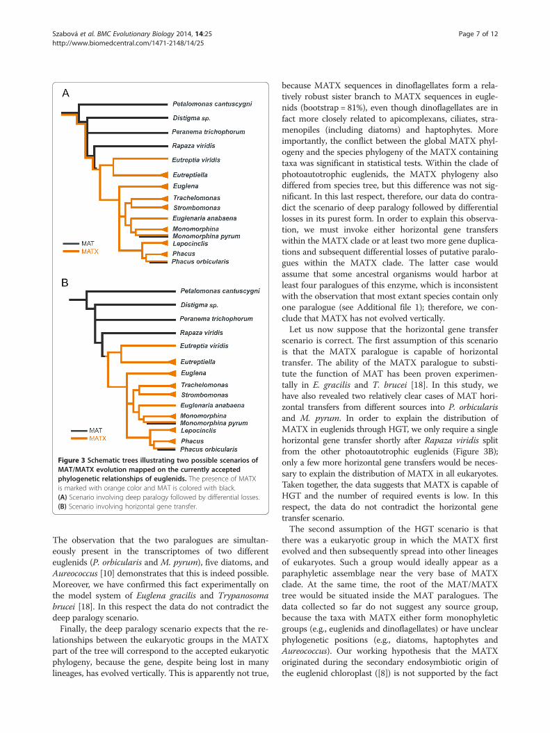
Figure 3 Schematic trees illustrating two possible scenarios ofMAT/MATX evolution mapped on the currently acceptedphylogenetic relationships of euglenids. The presence of MATXis marked with orange color and MAT is colored with black.(A) Scenario involving deep paralogy followed by differential losses.(B) Scenario involving horizontal gene transfer.
Szabová et al. BMC Evolutionary Biology 2014, 14:25 Page 7 of 12http://www.biomedcentral.com/1471-2148/14/25
The observation that the two paralogues are simultan-eously present in the transcriptomes of two differenteuglenids (P. orbicularis and M. pyrum), five diatoms, andAureococcus [10] demonstrates that this is indeed possible.Moreover, we have confirmed this fact experimentally onthe model system of Euglena gracilis and Trypanosomabrucei [18]. In this respect the data do not contradict thedeep paralogy scenario.Finally, the deep paralogy scenario expects that the re-
lationships between the eukaryotic groups in the MATXpart of the tree will correspond to the accepted eukaryoticphylogeny, because the gene, despite being lost in manylineages, has evolved vertically. This is apparently not true,
because MATX sequences in dinoflagellates form a rela-tively robust sister branch to MATX sequences in eugle-nids (bootstrap = 81%), even though dinoflagellates are infact more closely related to apicomplexans, ciliates, stra-menopiles (including diatoms) and haptophytes. Moreimportantly, the conflict between the global MATX phyl-ogeny and the species phylogeny of the MATX containingtaxa was significant in statistical tests. Within the clade ofphotoautotrophic euglenids, the MATX phylogeny alsodiffered from species tree, but this difference was not sig-nificant. In this last respect, therefore, our data do contra-dict the scenario of deep paralogy followed by differentiallosses in its purest form. In order to explain this observa-tion, we must invoke either horizontal gene transferswithin the MATX clade or at least two more gene duplica-tions and subsequent differential losses of putative paralo-gues within the MATX clade. The latter case wouldassume that some ancestral organisms would harbor atleast four paralogues of this enzyme, which is inconsistentwith the observation that most extant species contain onlyone paralogue (see Additional file 1); therefore, we con-clude that MATX has not evolved vertically.Let us now suppose that the horizontal gene transfer
scenario is correct. The first assumption of this scenariois that the MATX paralogue is capable of horizontaltransfer. The ability of the MATX paralogue to substi-tute the function of MAT has been proven experimen-tally in E. gracilis and T. brucei [18]. In this study, wehave also revealed two relatively clear cases of MAT hori-zontal transfers from different sources into P. orbicularisand M. pyrum. In order to explain the distribution ofMATX in euglenids through HGT, we only require a singlehorizontal gene transfer shortly after Rapaza viridis splitfrom the other photoautotrophic euglenids (Figure 3B);only a few more horizontal gene transfers would be neces-sary to explain the distribution of MATX in all eukaryotes.Taken together, the data suggests that MATX is capable ofHGT and the number of required events is low. In thisrespect, the data do not contradict the horizontal genetransfer scenario.The second assumption of the HGT scenario is that
there was a eukaryotic group in which the MATX firstevolved and then subsequently spread into other lineagesof eukaryotes. Such a group would ideally appear as aparaphyletic assemblage near the very base of MATXclade. At the same time, the root of the MAT/MATXtree would be situated inside the MAT paralogues. Thedata collected so far do not suggest any source group,because the taxa with MATX either form monophyleticgroups (e.g., euglenids and dinoflagellates) or have unclearphylogenetic positions (e.g., diatoms, haptophytes andAureococcus). Our working hypothesis that the MATXoriginated during the secondary endosymbiotic origin ofthe euglenid chloroplast ([8]) is not supported by the fact

Szabová et al. BMC Evolutionary Biology 2014, 14:25 Page 8 of 12http://www.biomedcentral.com/1471-2148/14/25
that the MATX paralogue is absent in both Rapaza viridisand the closest relative of the euglenid chloroplast, Pyra-mimonas. Moreover, the MATX paralogues in euglenidsdo not form a paraphyletic group, but instead form a ro-bust clade within the more inclusive MATX clade. Theposition of the root between MAT and MATX lineagescannot be rejected, and both paralogues might have beenpresent in the common ancestor of all eukaryotes. Thecurrent data are in this respect not in direct conflict but,at the same time, they are also not supportive of the hori-zontal gene transfer scenario.
ConclusionsOur data are not entirely consistent with either of thetwo scenarios for MAT/MATX evolution in their purestforms. The hypothesis of deep paralogy followed by dif-ferential losses is rejected by the fact that MATX did notevolve purely by vertical transmission. The hypothesis ofa more recent origin of MATX followed by spread viahorizontal gene transfers is complicated by the absenceof a source of the first MATX paralogue and the factthat both paralogues could be present in the most recentcommon ancestor of all eukaryotes. Therefore, we inferthat the MATX paralogue spread among eukaryotes viaHGT; however, the original source of MATX is not yetknown and it could originate by gene duplication fromMAT in the last eukaryotic common ancestor.We also infer that euglenids were not the group in
which the MATX paralogue evolved. Instead, a foreignMATX paralogue substituted the ancestral euglenid MATparalogue in a single horizontal gene transfer event thatoccurred after the secondary endosymbiotic origin of theeuglenid chloroplast (Figure 3B). Although the donor ofthe euglenid MATX paralogue is not known, the MATXparalogue, once established, may have evolved verticallywithin the clade of photoautotrophic euglenids. Two pho-toautotrophic euglenids (P. orbicularis and M. pyrum)regained a new version of the MAT paralogue by recenthorizontal gene transfers from two different eukaryoticlineages and now contain both paralogues. Overall, thecase study of MAT/MATX illustrates the complex evolu-tionary histories of some eukaryotic genes and highlightsthe prevalence of gene duplications, differential losses ofparalogues, and horizontal gene transfer events during thecourse of eukaryotic evolution.
MethodsEuglenid strains and culture conditionsAll cultures used in this study are listed in Table 1. Strainsof Eutreptiella gymnastica (SCCAP K-0333), Trachelomo-nas sp. (SCCAP K-1380) and Pyramimonas parkeae(SCCAP K-0007) were obtained from the ScandinavianCulture Collection of Algae and Protozoa (SCCAP).Strains of Monomorphina pyrum (CCAP 1261/4B) and
Monomorphina aenigmatica (CCAP 1261/9) were ob-tained from the Culture Collection of Algae and Protozoa(CCAP). Distigma sp. was isolated from samples collectedfrom freshwater sediment from Czech Republic (50°27’N,13°20’E). This culture was not monoeukaryotic andcontained various other protists, therefore, we used amethod of single cell cloning by serial dilution to obtaina monoclonal Distigma sp. culture. Rapaza viridis wasisolated and cultured from marine sediment samplesfrom Canada (48° 47.551’ N, 125° 06.974’ W) [20]. Eu-glena clara (SAG 25.98), Euglena gracilis (SAG 1224-5/25), Euglena proxima (SAG 1224-11a), Eutreptia viridis(SAG 1226-1c), were obtained from the Culture Collec-tion of Algae at Goettingen, Germany. Euglena stellata(UTEX 372), Trachelomonas volvocina (UTEX 1327),Monomorphina parapyrum (UTEX 2354) and Eugle-naria anabaena (UTEX 373) were obtained from theCulture Collection of Algae at the University of Texas,Austin Texas, USA. Euglena viridis (ATCC PRA110) wasfrom the American Type Culture Collection, Manassas,Virginia, USA and Eutreptiella braarudii (CCMP 1594)was obtained from the National Center for Marine Algaeand Protozoa, East Boothbay, Maine, USA. Phacus inflexus(ACOI 1336) and Phacus orbicularis (ACOI 996) were ob-tained from the Coimbra Collection of Algae, Coimbra,Portugal. Culture of Petalomonas cantuscygni (CCAP1259/1) was provided by Dr. Mark Farmer at theUniversity of Georgia, Athens, Georgia, USA and it wasoriginally obtained from the Culture Collection of Algaeand Protozoa. Strombomonas accuminata NJ, S 716 andTrachelomonas ellipsoidalis NJ, ST1 are cultures main-tained in the Triemer lab which were originally isolatedfrom pond samples from New Jersey, USA; Lepocinclistripteris MI 101 and Lepocinclis playfairiana MI 102 arecultures isolated from ponds near Michigan State Uni-versity, East Lansing, MI, USA.
DNA, RNA isolation and preparation of cDNAGenomic DNA from Eutreptiella gymnastica, Trachelo-monas sp., Pyramimonas parkeae, Monomorphina pyrum,Monomorphina aenigmatica, and Distigma sp. was ex-tracted from strains using the Qiagen Blood and Tissuekit and total RNA was isolated from 150 ml of well-growncultures (approx. 25*106 cells) using TRIzol Reagent(Invitrogen). Total RNA from Rapaza viridis was isolatedusing Ambion® RNAqueous-Micro Kit (Life technologies).mRNA was purified from total RNA with the use ofDynabeads mRNA Purification Kit (Invitrogen). cDNAwas then prepared using Smarter PCR cDNA SynthesisKit (Clontech) according to the manufacturer’s protocolwith 15 to 27 cycles of cDNA amplification (depending onthe amount of mRNA used in the first-strand synthesis).In case of E. gracilis, M. parapyrum, S. accuminata and
L. playfairiana the total RNA was extracted by grinding
Szabová et al. BMC Evolutionary Biology 2014, 14:25 Page 9 of 12http://www.biomedcentral.com/1471-2148/14/25
wet biomass in liquid nitrogen followed by purificationusing RNA/DNA Maxi Kit (Qiagen); mRNA, wheneverused for cDNA synthesis, was purified from total RNAusing Qiagen Oligotex mRNA Maxi Kit. cDNA was pre-pared using Smart (later Smarter) cDNA synthesis Kit(Clontech) or by similar technology provided by MINTcDNA synthesis Kit (Evrogen). cDNA libraries werenormalized using Trimmer cDNA normalization Kit(Evrogen). The resulting normalized cDNA was adaptedfor Roche 454 sequencing by performing a multiple lastamplification step, pooling the PCR products in order toachieve the overall amount of cDNA acceptable forsequencing.For the remaining euglenid strains, total RNA was iso-
lated using RNAzol RT RNA Isolation Reagent (Molecu-lar Research Center, Inc.). High level purification of totalRNA was achieved using MEGAclear Kit (Ambion).Next, mRNA was isolated using MIcroPoly(A)Purist Kit(Ambion). Preparation of cDNA suitable for the nextgeneration sequencing was according to cDNA RapidLibrary Preparation Manual (Roche, GS FLX TitaniumSeries, later GS FLX + Series - XL+).
Amplification, sequencing and assemblyIn case of Pyramimonas parkeae, Eutreptiella gymnastica,Trachelomonas sp., Distigma sp., Monomorphina aenig-matica and Monomorphina pyrum we have amplified theMAT or MATX genes from cDNA template using slightlymodified primers of Kamikawa et al. [10]: Forward primerMATA3-F (5’-GAGYMMGTSAVYGARGGYCAYCCXGACAA-3‘) directed at the consensus amino acid (aa) se-quence GHPDK and the reverse primer MATB3-R (5’-CCRTGNGCNCCCCADCCDCCRTAXGT-3’) directed atthe eukaryotic consensus aa sequence TYGGWGAH in-side a conserved block. Amplification was carried out in25-μl reactions with 1.5 μl of the diluted cDNA as a tem-plate using EmeraldAmp MAX PCR Master Mix (TaKaRaBio Inc.) and the following program: a hot start at 95°Cfor 4 min, followed by 35 cycles of denaturation at 95°Cfor 30 s, annealing at 55°C for 60 s and extension at 72°Cfor 90 s, finishing with an extension at 72°C for 15 min.The PCR products were excised from the gel, cloned intopGEM-T Easy Vector System (Promega) and sequenced.The new sequences were deposited in GenBank under theaccession numbers listed in Table 1.Small subunit (SSU) ribosomal RNA gene from E. gym-
nastica was amplified from genomic DNA with “universal”eukaryote SSU primer pairs Medlin A (5’-CTGGTTGATCCTGCCAG-3‘), Medlin B (5’-TGATCCTTCTGCAGGTTCACCTAC-3’) described by Medlin et al. [24]. Amp-lification was carried out using the following program: ahot start at 95°C for 4 min, followed by 35 cycles of de-naturation at 95°C for 30 s, annealing at 55°C for 60 s andextension at 72°C for 90 s, finishing with an extension at
72°C for 15 min. Medlin A, Medlin B, EPA-23 (5’- GTCATATGCTTYKTTCAAGGRCTAAGCC -3’), EPA-2286(5’- TCACCTACARCWACCTTGTTACGAC -3’) accord-ing to Müllner et al. [25] and our primers SSU 633-F (5’-GGCAGCAGGCRCGCAAATTGC -3’) and SSU 2031-R(5’- TCAACCAGACAAATCACTYCACCAA -3’) wereused for sequencing of PCR products.Small subunit (SSU) ribosomal RNA gene from L. play-
fairiana and M. parapyrum was amplified from genomicDNA with nuclear SSU primers 18S_1A (AAYCTGGTTGATCCTGCCAGT) and 18S_1520B (TGATCCTTCTGCAGGTTCACCTAC). Amplifications were carried outusing 5 min of denaturation at 94°C and 30 cycles of thefollowing: 94°C for 30 s, 45°C – 50°C for 1 min, 72°C for2 min, a final extension at 72°C for 11 min. For sequencingof PCR products were used primers 18S_1A, 18S_1520B,18S_300F (WGGGTTYGATTCCGGAG), 18S_528F (CGGTAATTCCAGCTCC), 18S_516R (ACCAGACTTGCYCTCC), 18S_960F (TTTGACTCAACRCGGG) and 18S_1055R (CGGCCATGCACCACC).For the 454 sequences obtained from cDNAs, the raw
reads (SFF File format) from 454 were filtered to removereads shorter than 50 bp and all reads which had morethan 30% of the bases with a Phred quality score less than30 using NGS QC TK [26] were excluded. The resultinghigh quality reads were assembled using Roche's propri-etary "Newbler" software version 2.6 with "cDNA" option.Assembled contigs shorter than 200 bp were excluded.The full length of euglenid MATX genes were 1290 bp.
Some of the sequences were incomplete: P. orbicularis(length 1257 bp), M. pyrum (length 906 bp), M. aenigma-tica (length 843 bp), Trachelomonas sp. (length 909 bp)and E. anabaena (length 1266 bp). The length of theMAT genes were 1167 bp for P. cantuscygni, 1137 for P.orbicularis, 795 bp for R. viridis, 774 bp for M. pyrum,765 bp for Distigma sp. and 720 bp for P. parkeae.
Phylogenetic analysesThe MAT and MATX protein sequences were aligned inClustalX [27], the SSU rRNA gene sequences were alignedin MAFFT (http://www.genome.jp/tools/mafft/) using G-INS-I option [28]. The alignments were manually refinedin BioEdit 7.0.5.3. [29]. The regions, which could not beunambiguously aligned, were excluded from the analyses.A phylogeny of eukaryotic MAT and MATX was in-
ferred from 123 sequences using 347 aligned amino acidpositions; the phylogenetic relationships within the MATXclade were inferred from 41 sequences and 405 positions;the phylogenetic relationships within the euglenid sub-group of the MATX clade were inferred from 21 sequencesand 399 alignment positions. Maximum likelihood treeswere estimated by RAxML_HPC version 2.3.3 [30] usingthe best fitting models as determined by Prottest (http://darwin.uvigo.es/software/prottest2_server.html) [31] and

Szabová et al. BMC Evolutionary Biology 2014, 14:25 Page 10 of 12http://www.biomedcentral.com/1471-2148/14/25
10 replicates of starting tree construction. The modelswere PROTGAMMALG for MAT+MATX and MATX ofeuglenids and PROTGAMMAWAG for analysis ofeukaryotic MATX clade. Bootstrap supports (BS) were cal-culated from 500 replicates. Bayesian trees were estimatedby MrBayes version 3.1.2 (Ronquist and Huelsenbeck2003) using the WAG+GAMMA+ Invariants + covarionmodel of substitution. In case of MAT+MATX analysis(Figure 1), two MCMC were run for 5 860 000 generations,trees from the first 1000 000 generations were discarded asburn-in. In case of MATX analysis (Figure 2), two MCMCwere run for 17 775 000 generations, trees from the first 2818 500 generations were discarded as burn-in.For the purposes of topology testing, pruned and rooted
data sets of MATX clade were analyzed – 40 sequences(only one Karenia brevis sequence was used), 36 se-quences (without Aureococcus, Prymnesium, Dendrocto-nus and Lactuca) and both previous data sets rooted byTrichomonas and Escherichia (i.e. 42 and 38 sequences).All alignments contained 405 amino acid positions andwere analysed as described above. Phylogenetic trees ofSSU rDNA were inferred by maximum likelihood methodfrom the corresponding set of taxa – 40 and 36 sequencesin unrooted, 42 and 38 sequences in rooted analyses ofMATX clade and 21 sequences of MATX containingeuglenids. Unrooted and rooted SSU alignments con-tained 1525 and 1282 positions respectively. A maximumlikelihood trees were estimated by RAxML_HPC version2.3.3 [30] using the GTRGAMMA model of nucleotidesubstitution, 10 replicates of starting tree constructionand BS were calculated from 500 replicates.All data sets and trees generated in this study have
been deposited in TreeBASE (study accession number is15062).
Topology testingThe Kishino Hasegawa (KH) [32] and ShimodariaHasegawa tests [33] implemented in Consel 0.1j [34]were used for topology testing. We have decided not toreport the results of approximately unbiased test [35]because we have realized that the test behaves very un-stably for our data sets; re-testing of the same data setsproduced very different p-values that sometimes dif-fered in significance. Regarding the significance or non-significance at the p = 0.001 level, the results of the AUtests were in agreement with the results of KH and SHtests in most cases; however due to their instability, wehave decided to report only the results of KH and SHtests.A set of 503 topologies was created in order to test
whether the relationships between MATX paralogues arein conflict with the relationship of MATX containing taxaas inferred from SSU rDNA sequences. This set of topolo-gies contained the best topology inferred from an analysis
of the MATX protein alignment by RAxML, 500 topolo-gies from bootstrap permutations of the MATX alignmentgenerated by RAxML, the best tree inferred by RAxMLfrom the SSU rRNA alignment of the same set of taxa,and the manually constructed topology reflecting thecurrent view of species relationships. The latter two top-ologies representing species trees are given in Additionalfile 1 and in Additional file 2: Figure S1 and Additional file3: Figure S3. Site likelihoods for topologies 1–501 were in-ferred by Treepuzzle 5.2. [36] using MATX gene align-ment, WAG+ I + Γ model of amino acid substitution andparameter values inferred from the topology nr. 1. Sitelikelihoods for topologies 502 and 503 were inferred byTreepuzzle using MATX gene alignment, WAG+ I + Γmodel of amino acid substitution and parameter valuesinferred from these topologies. The sets of site likeli-hoods were then compared by the KH, weighted KH(WKH), SH and SH (WSH) test in Consel 0.1j [34]. Thetests were performed for (1) the full set of MATX paralo-gues from 40 taxa, (2) a set of MATX paralogues,excluding MATX from Aureococcus, Prymnesium, Den-droctonus and Lactuca, (3) data set 1 rooted by Tri-chomonas and Escherichia, (4) data set 2 rooted byTrichomonas and Escherichia, and (5) a set of MATXparalogues from euglenids.The same tests were used to evaluate whether or not
the root position between MAT and MATX paraloguescan be rejected. For these tests, we used topology shownin Figure 1, 500 bootstrap topologies calculated from thesame alignment, and a topology that differed from Figure 1only in the position of prokaryotic outgroups that weremoved on the branch separating MAT and MATX paralo-gues. The tests were performed as described above.
Availability of supporting dataAll the supporting data are included as additional files.
Additional files
Additional file 1: Reconciliation of MATX gene tree with speciestree. We have used the software Jane (http://www.cs.hmc.edu/~hadas/jane/) to reconcile the MATX gene tree with the species tree. For thisanalysis we have excluded taxa with very incomplete sequence(Prymnesium) or taxa, whose MATX sequences could be result ofcontamination (Lactuca and Dencroctonus). If we set the cost of gene lossto 0, which could be a realistic value in case of loss of one of twoparalogues, then the discrepancy between MATX gene tree and speciestree can be explained by the same number of events if we considerduplications and differential losses (A) or horizontal gene transfers (B).
Additional file 2: Figure S1. Maximum likelihood phylogeny of MATXcontaining taxa based on SSU rRNA gene. The tree was constructed bymaximum likelihood method in RAxML from the 1525 nucleotidepositions. The values at nodes represent maximum likelihood bootstraps,only values above 50% are shown.
Additional file 3: Figure S3. Maximum likelihood phylogeny of MATXcontaining euglenid taxa based on SSU rRNA gene. The tree wasconstructed by maximum likelihood method in RAxML from the 1525
Szabová et al. BMC Evolutionary Biology 2014, 14:25 Page 11 of 12http://www.biomedcentral.com/1471-2148/14/25
nucleotide positions. The values at nodes represent maximum likelihoodbootstraps, only values above 50% are shown.
Additional file 4: Figure S2. Maximum likelihood phylogeny ofeuglenid MATX. The tree was constructed by maximum likelihoodmethod in RAxML from the 399 amino acid positions. The values atnodes represent maximum likelihood bootstraps, only values above 50%are shown.
Competing interestsThe authors declare that they have no competing interests.
Authors’ contributionsJS participated on cDNA preparation (for Distigma sp., P. parkeae, E.gymnastica, Trachelomonas sp., M. pyrum, M. aenigmatica and R. viridis), dataanalysis, in the sequence alignments and drafted the manuscript. NYprovided Rapaza viridis RNA and revised the manuscript. BSL revised themanuscript. RET provided the transcriptome data for the rest of euglenidspecies and revised the manuscript. VH supervised the study, performed thephylogenetic analyses and helped to draft the manuscript. All authors readand approved the final manuscript.
AcknowledgementsThe work on the project was supported by the project "BIOCEV –Biotechnology and Biomedicine Centre of the Academy of Sciences andCharles University" (CZ.1.05/1.1.00/02.0109), from the European RegionalDevelopment Fund and by the Czech Science Foundation (P506/11/1320)awarded to VH and by grants to BSL from the Tula Foundation (Centre forMicrobial Diversity and Evolution at the University of British Columbia) andthe Canadian Institute for Advanced Research, Program in IntegratedMicrobial Biodiversity. RET was supported by an Assembling the Tree of Lifegrant from the National Science Foundation (DEB-0830056). Many of theEuglenozoan MATX sequences were generated as part of this larger project.RET would like to thank his collaborators at Virginia CommonwealthUniversity, Dr. Gregory A. Buck (PI on the grant), Dr. Vishal N. Kopardé andDr. Andrey V. Matveyev for their roles in generating these sequences.
Author details1Department of Parasitology, Charles University in Prague, Faculty of Science,Vinicna 7, Prague 2 128 44, Czech Republic. 2Biotechnology and BiomedicineCenter of the Academy of Sciences and Charles University in Vestec, Prague,Czech Republic. 3Departments of Botany and Zoology, Canadian Institute forAdvanced Research, Program in Integrated Microbial Biodiversity, Universityof British Columbia, Vancouver, British Columbia V6T 1Z4, Canada.4Department of Plant Biology, Michigan State University, East Lansing,Michigan 48824, USA.
Received: 20 August 2013 Accepted: 30 December 2013Published: 11 February 2014
References1. Cantoni GL: Biological methylation: selected aspects. Annu Rev Biochem
1975, 44:435–451.2. Chiang PK, Gordon RK, Tal J, Zeng GC, Doctor BP, Pardhasaradhi K, McCann
PP: S-adenosylmethionine and methylation. FASEB J 1996, 10:471–480.3. Takusagawa F, Kamitori S, Misaki S, Markham GD: Crystal structure of
S-adenosylmethionine synthetase. J Biol Chem 1996, 271:136–147.4. Graham DE, Bock CL, Schalk-Hihi C, Lu ZJ, Markham GD: Identification of a
highly diverged class of S-adenosylmethionine synthetases in the ar-chaea. J Biol Chem 2000, 275:4055–4059.
5. Gonzalez B, Pajares MA, Hermoso JA, Alvarez L, Garrido F, Sufrin JR,Sanz- Aparicio J: The crystal structure of tetrameric methionine adenosyl-transferase from rat liver reveals the methionine-binding site. J Mol Biol2000, 300:363–375.
6. Kotb M, Kredich NM: S-Adenosylmethionine synthetase from humanlymphocytes purification and characterization. J Biol Chem 1985,260:3923–3930.
7. Markham GD, Pajares MA: Structure – function relationships inmethionine Adenosyltransferases. Cell Mol Life Sci 2009, 66:636–648.
8. Sanchez-Perez GF, Hampl V, Simpson AGB, Roger AJ: A new divergent typeof eukaryotic methionine adenosyltransferase is present in multiple
distantly related secondary algal lineages. J Eukaryot Microbiol 2008,55:374–381.
9. Garrido F, Estrela S, Alves C, Sánchez-Pérez GF, Sillero A, Pajares MA:Refolding and characterization of methionine adenosyltransferase fromEuglena gracilis. Protein Expr Purif 2011, 79:128–136.
10. Kamikawa R, Sanchez-Perez GF, Sako Y, Roger AJ, Inagaki Y: Expandedphylogenies of canonical and non-canonical types of methionineadenosyltransferase reveal a complex history of these gene families ineukaryotes. Mol Phylogenet Evol 2009, 53:565–570.
11. Keeling PJ, Inagaki Y: A class of eukaryotic GTPase with a punctatedistribution suggesting multiple functional replacements of translationelongation factor 1α. Proc Natl Acad Sci U S A 2004, 101:15380–15385.
12. Noble GP, Rogers MB, Keeling PJ: Complex distribution of EFL and EF-1αproteins in the green algal lineage. BMC Evol Biol 2007, 7:82.
13. Kamikawa R, Inagaki Y, Sako Y: Direct phylogenetic evidence for lateraltransfer of elongation factor-like gene. Proc Natl Acad Sci U S A 2008,105:6965–6969.
14. Keeling PJ, Palmer JD: Horizontal gene transfer in eukaryotic evolution.Nat Rev Genet 2008, 9:605–618.
15. Gile GH, Faktorova D, Castlejohn CA, Burger G, Lang BF, Farmer MA, Lukes J,Keeling PJ: Distribution and phylogeny of EFL and EF-1α in Euglenozoasuggest ancestral co-occurrence followed by differential loss. PLoS One2009, 4:e5162.
16. Kamikawa R, Yabuki A, Nakayama T, Ishida K, Hashimota T, Inagaki Y:Cercozoa comprises both EF-1α-containing and EFL-containingmembers. Eur J Protistol 2011, 47:24–28.
17. Ishitani Y, Kamikawa R, Yabuki A, Tsuchiya M, Inagaki Y, Takishita K:Evolution of elongation factor-like (EFL) protein in Rhizaria is revised byradiolarian EFL gene sequences. J Eukaryot Microbiol 2012, 59:367–373.
18. Szabova J, Ruzicka P, Verner Z, Hampl V, Lukes J: Experimental examinationof EFL and MATX eukaryotic horizontal gene transfers: coexistence ofmutually exclusive transcripts predates functional rescue. Mol Biol Evol2011, 28:2371–2378.
19. Leander BS, Esson HJ, Breglia SA: Macroevolution of complex cytoskeletalsystems in euglenids. Bioessays 2007, 29:987–1000.
20. Yamaguchi A, Yubuki N, Leander BS: Morphostasis in a novel eukaryoteilluminates the evolutionary transition from phagotrophy tophototrophy: description of Rapaza viridis n. gen. et sp. (Euglenozoa,Euglenida). BMC Evol Biol 2012, 12:29.
21. Leander BS: Did trypanosomatid parasites have photosyntheticancestors? Trends Microbiol 2004, 12:251–258.
22. Takahashi F, Okabe Y, Nakada T: Origins of the secondary plastids ofeuglenophyta and chlorarachniophyta as revealed by an analysis of theplastid-targeting, nuclear-encoded gene psbO. J Phycol 2007,43:1302–1309.
23. Gile GH, Novis PM, Cragg DS, Zuccarello GC, Keeling PJ: The distribution ofElongation Factor-1 Alpha (EF-1alpha), Elongation Factor-Like (EFL), anda non-canonical genetic code in the ulvophyceae: discrete geneticcharacters support a consistent phylogenetic framework. J EukaryotMicrobiol 2009, 56:367–72.
24. Medlin L, Elwood HJ, Stickel S, Sogin ML: The characterization ofenzymatically amplified eukaryotes 16S like ribosomal RNA codingregions. Gene 1988, 71:491–499.
25. Müllner AN, Angeler DG, Samuel R, Linton EW, Triemer RE: Phylogeneticanalysis of phagotrophic, photomorphic and osmotrophic euglenoids byusing the nuclear 18S rDNA sequence. Int J Syst Evol Microbiol 2001,51:783–791.
26. Patel RK, Jain M: NGS QC toolkit: a toolkit for quality control of nextgeneration sequencing data. PLoS ONE 2012, 7:e30619.
27. Thompson JD, Gibson TJ, Plewniak F, Jeanmougin F, Higgins DG: The clustalxwindows interface: flexible strategies for multiple sequence alignmentaided by quality analysis tools. Nucl Acids Res 1997, 24:4876–4882.
28. Katoh K, Asimenos G, Toh H: Multiple alignment of DNA sequences withMAFFT. Methods Mol Biol 2009, 537:39–64.
29. Hall TA: BioEdit: a user-friendly biological sequence alignment editor andanalysis program for Windows 95/98/NT. Nucl Acids Symp Ser 1999, 41:95–98.
30. Stamatakis A: RAxML-VI-HPC: maximum likelihood-based phylogeneticanalyses with thousands of taxa and mixed models. Bioinformatics 2006,22:2688–2690.
31. Abascal F, Zardoya R, Posada D: ProtTest: selection of best-fit models ofprotein evolution. Bioinformatics 2005, 21:2104–2105.
Szabová et al. BMC Evolutionary Biology 2014, 14:25 Page 12 of 12http://www.biomedcentral.com/1471-2148/14/25
32. Kishino H, Hasegawa M: Evaluation of the maximum likelihood estimateof the evolutionary tree topologies from DNA sequence data, and thebranching order in Hominoidea. J Mol Evol 1989, 29:170–179.
33. Shimodaira H, Hasegawa M: Multiple comparisons of log-likelihoods withapplications to phylogenetic inference. Mol Biol Evol 1999, 16:1114–1116.
34. Shimodaira H, Hasegawa M: CONSEL: for assessing the confidence ofphylogenetic tree selection. Bioinformatics 2001, 17:1246–1247.
35. Shimodaira H: An approximately unbiased test of phylogenetic treeselection. Syst Biol 2002, 51:492–508.
36. Schmidt HA, Strimmer K, Vingron M, Von Haeseler A: TREE-PUZZLE:maximum likelihood phylogenetic analysis using quartets and parallelcomputing. Bioinformatics 2002, 18:502–504.
doi:10.1186/1471-2148-14-25Cite this article as: Szabová et al.: The evolution of paralogous enzymesMAT and MATX within the Euglenida and beyond. BMC EvolutionaryBiology 2014 14:25.
Submit your next manuscript to BioMed Centraland take full advantage of:
• Convenient online submission
• Thorough peer review
• No space constraints or color figure charges
• Immediate publication on acceptance
• Inclusion in PubMed, CAS, Scopus and Google Scholar
• Research which is freely available for redistribution
Submit your manuscript at www.biomedcentral.com/submit