
The evolution of amphibian metamorphosis: insights...
15
J. Anat. (2007) 210, pp379– 393 doi: 10.1111/j.1469-7580.2007.00710.x © 2007 The Authors Journal compilation © 2007 Anatomical Society of Great Britain and Ireland Blackwell Publishing Ltd The evolution of amphibian metamorphosis: insights based on the transformation of the aortic arches of Pelobates fuscus (Anura) Hana Kolesová 1 , Alois Lametschwandtner 2 and Zbyn´k RoCek 3 1 Institute of Anatomy, First Faculty of Medicine, Charles University, Prague, Czech Republic 2 Department of Organismic Biology, Vascular and Muscle Research Unit, University of Salzburg, Austria 3 Laboratory of Palaeobiology, Geological Institute, Academy of Sciences, Prague, Czech Republic, and Department of Zoology, Charles University, Prague, Czech Republic Abstract In order to gain insights into how the aortic arches changed during the transition of vertebrates to land, trans- formations of the aortic arches during the metamorphosis of Pelobates fuscus were investigated and compared with data from the early development of a recent ganoid fish Amia calva and a primitive caudate amphibian Salamandrella keyserlingi. Although in larval Pelobates, as in other non-pipid anurans, the gill arches serve partly as a filter-feeding device, their aortic arches maintain the original piscine-like arrangement, except for the mandibular and hyoid aortic arches which were lost. As important pre-adaptations for breathing of atmospheric oxygen occur in larval Pelobates (which have well-developed, though non-respiratory lungs and pulmonary artery), transformation of aortic arches during metamorphosis is fast. The transformation involves disappearance of the ductus Botalli, which results in a complete shunting of blood into the lungs and skin, disappearance of the ductus caroticus, which results in shunting of blood into the head through the arteria carotis interna, and disappearance of arch V, which results in shunting blood to the body through arch IV (systemic arch). It is supposed that the branching pattern of the aortic arches of permanently water-dwelling piscine ancestors, of intermediate forms which occasionally left the water and of primitive tetrapods capable of spending longer periods of time on land had been the same as in the prematamorphic anuran larvae or in some metamorphosed caudates in which the ductus caroticus and ductus Botalli were not interrupted, and arch V was still complete. Key words Anura; circulatory system; development; evolution; metamorphosis. Introduction The transition of vertebrates from an aquatic environ- ment to dry land in the Devonian, some 360 million years ago, was associated with profound changes in their physiology and anatomy. In piscine ancestors of tetrapods, oxygen was taken from the water and brought to the blood and tissues through the gills located on the surface of gill arches. In water-dwelling gnath- ostomes, each of these arches includes skeletal components and the branchial muscles and nerves. In addition, each arch is traversed by the arterial blood vessels collectively termed the aortic arch, and is supported by endoskeletal elements called the branchials. The ensemble of branchials that form the skeleton of each gill segment is called the skeletal gill arch. In early gnathostomes it typically consisted of two pharyngobranchials that connected the entire skeletal arch to the neurocranium, and (dorsally to ventrally) the epibranchial, ceratobranchial and hypobranchial elements of each gill arch; skeletal arches of the left and right sides were interconnected by median ventral structures, the basibranchials (Bjerring, 1977). As early as in the early Devonian gnathostomes, however, some branchials either fused with surrounding structures, became reduced or were entirely lost (Moy-Thomas & Miles, 1971; Jarvik, 1980). Correspondence Hana Kolesová, Institute of Anatomy, First Faculty of Medicine, Charles University, U Nemocnice 3, CZ-128 00 Prague 2. E: [email protected] Accepted for publication 18 January 2007
Transcript of The evolution of amphibian metamorphosis: insights...

J. Anat.
(2007)
210
, pp379–393 doi: 10.1111/j.1469-7580.2007.00710.x
© 2007 The Authors Journal compilation © 2007 Anatomical Society of Great Britain and Ireland
Blackwell Publishing Ltd
The evolution of amphibian metamorphosis: insights based on the transformation of the aortic arches of
Pelobates fuscus
(Anura)
Hana Kolesová
1
, Alois Lametschwandtner
2
and Zbyn
´
k Ro
C
ek
3
1
Institute of Anatomy, First Faculty of Medicine, Charles University, Prague, Czech Republic
2
Department of Organismic Biology, Vascular and Muscle Research Unit, University of Salzburg, Austria
3
Laboratory of Palaeobiology, Geological Institute, Academy of Sciences, Prague, Czech Republic, and Department of Zoology, Charles University, Prague, Czech Republic
Abstract
In order to gain insights into how the aortic arches changed during the transition of vertebrates to land, trans-
formations of the aortic arches during the metamorphosis of
Pelobates fuscus
were investigated and compared with
data from the early development of a recent ganoid fish
Amia calva
and a primitive caudate amphibian
Salamandrella
keyserlingi
. Although in larval
Pelobates
, as in other non-pipid anurans, the gill arches serve partly as a filter-feeding
device, their aortic arches maintain the original piscine-like arrangement, except for the mandibular and hyoid
aortic arches which were lost. As important pre-adaptations for breathing of atmospheric oxygen occur in larval
Pelobates
(which have well-developed, though non-respiratory lungs and pulmonary artery), transformation of
aortic arches during metamorphosis is fast. The transformation involves disappearance of the ductus Botalli, which
results in a complete shunting of blood into the lungs and skin, disappearance of the ductus caroticus, which results
in shunting of blood into the head through the arteria carotis interna, and disappearance of arch V, which results
in shunting blood to the body through arch IV (systemic arch). It is supposed that the branching pattern of the
aortic arches of permanently water-dwelling piscine ancestors, of intermediate forms which occasionally left
the water and of primitive tetrapods capable of spending longer periods of time on land had been the same as in
the prematamorphic anuran larvae or in some metamorphosed caudates in which the ductus caroticus and ductus
Botalli were not interrupted, and arch V was still complete.
Key words
Anura; circulatory system; development; evolution; metamorphosis.
Introduction
The transition of vertebrates from an aquatic environ-
ment to dry land in the Devonian, some 360 million
years ago, was associated with profound changes in
their physiology and anatomy. In piscine ancestors of
tetrapods, oxygen was taken from the water and
brought to the blood and tissues through the gills located
on the surface of gill arches. In water-dwelling gnath-
ostomes, each of these arches includes skeletal components
and the branchial muscles and nerves. In addition, each
arch is traversed by the arterial blood vessels collectively
termed the aortic arch, and is supported by endoskeletal
elements called the branchials. The ensemble of branchials
that form the skeleton of each gill segment is called the
skeletal gill arch. In early gnathostomes it typically
consisted of two pharyngobranchials that connected
the entire skeletal arch to the neurocranium, and
(dorsally to ventrally) the epibranchial, ceratobranchial
and hypobranchial elements of each gill arch; skeletal
arches of the left and right sides were interconnected
by median ventral structures, the basibranchials
(Bjerring, 1977). As early as in the early Devonian
gnathostomes, however, some branchials either fused
with surrounding structures, became reduced or were
entirely lost (Moy-Thomas & Miles, 1971; Jarvik, 1980).
Correspondence
Hana Kolesová, Institute of Anatomy, First Faculty of Medicine, Charles University, U Nemocnice 3, CZ-128 00 Prague 2. E: [email protected]
Accepted for publication
18 January 2007

Changes in the aortic arches of the Anura, H. Kolesová et al.
© 2007 The AuthorsJournal compilation © 2007 Anatomical Society of Great Britain and Ireland
380
Tetrapods typically are air-breathing vertebrates in
which gills were replaced by lungs (originally the swim
bladder of their Devonian crossopterygian ancestors).
Most gill slits became obliterated (except for the
spiraculum, a vestigial slit between the mandibular and
hyoid arches) and, consequently, the skeletal arches
lost their original function and became reduced. They
persist as the hyoid, which is a skeletal support of the
muscles in the bottom of the mouth cavity, and the
columella auris. The transformation of the skeletal gill
arches into the hyoid was associated with reduction of
their dorsal elements so that the hyoid lost its firm
connection with the neurocranium. Although the
transition of the vertebrates to land was associated
with arrested ossification that also affected the gill
arches (so the arches remain cartilaginous to various
extent), transformations of at least some parts (e.g.
columella) may be inferred from fossils (Clack, 1994,
1998; Clack et al. 2003; Brazeau & Ahlberg, 2006). How-
ever, the transformation of soft parts, including the aortic
arches, may be inferred only from extant amphibians.
Some of the principal anatomical changes that
occurred during the transition of vertebrates to land
are reflected in the metamorphosis of contemporary
amphibians with bi-phasic ontogeny. Their water-dwelling
larvae take oxygen from the water, whereas terrestrial
adults use lungs for gas exchanges. Metamorphosis involves
not only the transformation of the gill-supporting
skeleton, but also changes in the associated aortic
arches. These processes are reflected in caudates better
than in anurans because their skeletal arches, both in
larvae and in metamorphosed adults, retain to a larger
extent the original segmentation. The same holds true
also for caecilians (see Wake, 2003). However, although
caecilians have long been considered significant to
understanding relationships among extinct and extant
amphibians, very little is known about development of
their branchial and aortic arches (Ramaswami, 1944;
for reviews of the literature, see also Hafferl, 1933;
Stadtmüller, 1936; Exbrayat, 2000; Wake, 2003). In
anurans, on the other hand, the hyoid lost its original
segmentation, supposedly because of modification of
the larval branchial skeleton into a filter-feeding
device in which the epibranchials disappeared whereas
all remaining branchials fused together (for a review of
the literature, see Ro
C
ek, 2003). It is therefore inter-
esting to explore several questions: (1) how the aortic
arches of anuran larvae are transformed into the
adult pattern; (2) to what degree adults retain larval
arrangement; and (3) whether anuran metamorphosis
may provide evidence to infer the evolutionary
transformations of aortic arches during the transition
of vertebrates from water to land.
Information on the aortic arches in adult anurans is
based mainly on
Xenopus laevis
(e.g. Grobbelaar,
1924a,b; Nikitin, 1925; Millard, 1941, 1942, 1945;
Paterson, 1942; Graaf, 1957) although this differs
remarkably from the anatomy of non-pipid anurans,
for instance
Leiopelma hochstetteri
(Szarski, 1951),
L. archei
(Stephenson, 1951),
Rana esculenta
(Gaupp,
1899),
Pelobates fuscus, Bombina bombina
and
Bufo
bufo
(Szarski, 1937, 1948).
In addition, development of the aortic arches is best
known in
Xenopus laevis
(Nikitin, 1925; Paterson, 1942;
Millard, 1945; Weisz, 1945; Nieuwkoop & Faber, 1967;
Viertel & Richter, 1999), whereas only a small number
of papers deal with aortic arch development or larval
anatomy in non-pipid anurans (e.g. Schulze, 1889, 1892
in
Pelobates fuscus
; Schmalhausen, 1953a in
Bombina
bombina
; Strawinski, 1956 in
Rana esculenta
; Schmal-
hausen, 1953a and Lanot, 1962 in
Rana temporaria
; De
Saint-Aubain, 1985 in
Rana catesbeiana
and
Bufo bufo
;
Magnin, 1959 in
Alytes obstetricans
; Witschi, 1956 in
Rana
sp.).
Materials and methods
We chose
Pelobates fuscus
because of the comparatively
primitive phylogenetic position of the genus
Pelobates
.
The large size of
P. fuscus
tadpoles makes them suita-
ble for vascular corrosion casting and scanning electron
microscopical analyses. The 34 individuals examined in
this study include specimens from the advanced larval
period to the end of metamorphosis (stages 50–66;
staging after Nieuwkoop & Faber, 1967). Tadpoles
were raised in the laboratory from a clutch collected
near the town of P
r
íbram, Czech Republic. The total
length of tadpoles ranged from 6.2 to 9.2 cm (stage
58), and their snout–vent length at the end of meta-
morphosis (stage 66) ranged from 2.4 to 3.2 cm.
The vascular system was prepared by injecting a
polymerizing resin, Mercox-Cl-2B (Ladd Research Inc.,
Burlington, VT, USA), into the blood vessels (Aichhorn
& Lametschwandtner, 1996; Bartel & Lametschwandtner,
2000). First, tadpoles were narcotized in an aqueous
solution of tricaine methansulfonate (MS 222, Sandoz,
Basle, Switzerland). Then the heart and the conus
arteriosus were exposed, a glass cannula was introduced

Changes in the aortic arches of the Anura, H. Kolesová et al.
© 2007 The Authors Journal compilation © 2007 Anatomical Society of Great Britain and Ireland
381
via the ventricle into the conus arteriosus, ligatured in
place, and the vascular system was rinsed with Ringer
solution. Injection of Mercox followed immediately
after clear reflux of rinsing solution escaped the
opened atria. Subsequent corrosion of the soft tissues
was achieved in 7.5% KOH (24 h, 40
°
C). Specimens
were analysed with Cambridge 250 and Sigma 300
scanning electron microscopes at accelerating voltages
of 5–10 kV. For further details on vascular casting,
specimen processing and scanning electron microscopy
analyses see Minnich et al. (2002).
In agreement with Taylor & Kollros (1946), we divided
the examined developmental period into (1) the larval
period, (2) prometamorphosis and (3) metamorphosis
(= metamorphic climax). Numbering of the aortic
arches was I–VI, which reflects also earlier larval
stages in which all aortic arches, including I and II, were
developed. The nomenclature of vessels is based on
Gaupp (1899).
Results
Larval period (stages 50, 52, 53)
Blood leaves the ventricle by way of the conus arteriosus,
the interior of which is divided longitudinally by a
spiral valve. The conus continues as a short unpaired
truncus arteriosus (aorta ventralis), which gives rise to
three aortic arches, called (anterior to posterior) the
carotid (III), systemic (IV) and pulmo-cutanean (com-
mon stem for V and VI) arches. Arches I and II are
absent (Fig. 1A,B).
Arch III gives off the a. carotis externa, which turns
backwards first and then immediately anteriorly
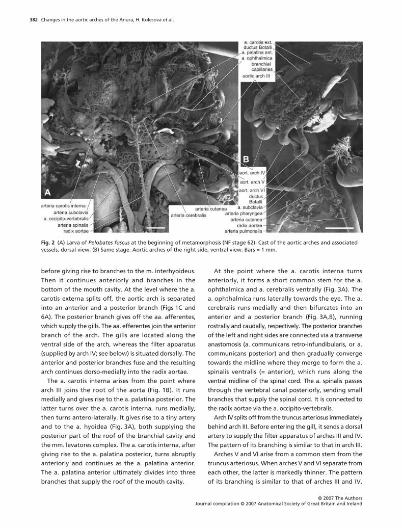
Fig. 1 (A) Larva of Pelobates fuscus at NF stage 50. Cast of the aortic arches and associated vessels, dorsal view. (B) Larva of Pelobates fuscus at NF stage 56. Dorsal view of the area corresponding to that marked by square in A. (C) Larva of Pelobates fuscus at NF stage 56. Dorsal view of the right arch III. Bars = 1 mm.
Changes in the aortic arches of the Anura, H. Kolesová et al.
© 2007 The AuthorsJournal compilation © 2007 Anatomical Society of Great Britain and Ireland
382
before giving rise to branches to the m. interhyoideus.
Then it continues anteriorly and branches in the
bottom of the mouth cavity. At the level where the a.
carotis externa splits off, the aortic arch is separated
into an anterior and a posterior branch (Figs 1C and
6A). The posterior branch gives off the aa. afferentes,
which supply the gills. The aa. efferentes join the anterior
branch of the arch. The gills are located along the
ventral side of the arch, whereas the filter apparatus
(supplied by arch IV; see below) is situated dorsally. The
anterior and posterior branches fuse and the resulting
arch continues dorso-medially into the radix aortae.
The a. carotis interna arises from the point where
arch III joins the root of the aorta (Fig. 1B). It runs
medially and gives rise to the a. palatina posterior. The
latter turns over the a. carotis interna, runs medially,
then turns antero-laterally. It gives rise to a tiny artery
and to the a. hyoidea (Fig. 3A), both supplying the
posterior part of the roof of the branchial cavity and
the mm. levatores complex. The a. carotis interna, after
giving rise to the a. palatina posterior, turns abruptly
anteriorly and continues as the a. palatina anterior.
The a. palatina anterior ultimately divides into three
branches that supply the roof of the mouth cavity.
At the point where the a. carotis interna turns
anteriorly, it forms a short common stem for the a.
ophthalmica and a. cerebralis ventrally (Fig. 3A). The
a. ophthalmica runs laterally towards the eye. The a.
cerebralis runs medially and then bifurcates into an
anterior and a posterior branch (Fig. 3A,B), running
rostrally and caudally, respectively. The posterior branches
of the left and right sides are connected via a transverse
anastomosis (a. communicans retro-infundibularis, or a.
communicans posterior) and then gradually converge
towards the midline where they merge to form the a.
spinalis ventralis (= anterior), which runs along the
ventral midline of the spinal cord. The a. spinalis passes
through the vertebral canal posteriorly, sending small
branches that supply the spinal cord. It is connected to
the radix aortae via the a. occipito-vertebralis.
Arch IV splits off from the truncus arteriosus immediately
behind arch III. Before entering the gill, it sends a dorsal
artery to supply the filter apparatus of arches III and IV.
The pattern of its branching is similar to that in arch III.
Arches V and VI arise from a common stem from the
truncus arteriosus. When arches V and VI separate from
each other, the latter is markedly thinner. The pattern
of its branching is similar to that of arches III and IV.
Fig. 2 (A) Larva of Pelobates fuscus at the beginning of metamorphosis (NF stage 62). Cast of the aortic arches and associated vessels, dorsal view. (B) Same stage. Aortic arches of the right side, ventral view. Bars = 1 mm.

Changes in the aortic arches of the Anura, H. Kolesová et al.
© 2007 The Authors Journal compilation © 2007 Anatomical Society of Great Britain and Ireland
383
Arch V, however, is much shorter than arches III and IV,
and re-joins arch VI just before the latter joins the radix
aortae. The posterior branch of arch V sends an artery
which supplies the filter plates of arches V and VI.
Arch VI is, in contrast to the other arches, very thin
and runs posteriorly after its separation from the
common stem with arch V. It does not divide into an
anterior and a posterior branch. It produces only a small
number of arteries that enter the gills; blood from the
gills flows back into the arch. Therefore, its gill supply
system is different from that of other arches. Before
arch VI enters the radix aortae, it re-joins arch V. The a.
pulmonalis arises from the common stem of re-united
arches V and VI. Consequently, the ductus Botalli,
which is the vessel connecting the origin of the a.
pulmonalis and the point where the common stem of
arches V and VI joins the radix aortae, is very short
(Fig. 4B). The a. cutanea, which runs laterally to supply
the skin, also originates in the ductus Botalli. In two
individuals the a. pulmonalis (and a. cutanea) originated
from the radix aortae, which may suggest that their
ductus Botalli was incorporated into the radix aortae.
The a. pulmonalis gives rise to the a. pharyngea,
which turns medially and supplies the dorsal pharyngeal
Fig. 3 (A) Larva of Pelobates fuscus at NF stage 56. Details of branching of the left arteria carotis interna, dorsal view. (B) Metamorphosing larva at NF stage 63. Details of branching of the left arteria carotis interna, dorsal view. Note triangular arrangement of the roots of the a. cerebralis. (C) Same stage as in B. Dorsal view of the aortic arches of the left side. Bars = 0.5 mm.

Changes in the aortic arches of the Anura, H. Kolesová et al.
© 2007 The AuthorsJournal compilation © 2007 Anatomical Society of Great Britain and Ireland
384
wall (Fig. 1A). Another branch (not previously mentioned
by other authors) originating from the a. pulmonalis
passes along the ventral side of the branchial basket,
then turns laterally to circumvent the branchial cavity,
and terminates on its dorsal side to supply the dorsal
velum. Branches of the a. pulmonalis enter the anterior
ends of the lungs, but the arteries continue along the
lateral sides of the lungs all the way up to their posterior
ends, producing small branches that break up into the
capillary nets around the alveoli.
The radix aortae, after accommodating the fused
arches V and VI, runs postero-medially, joins its
counterpart and gives rise to the aorta dorsalis. Each
root sends the a. palato-nasalis which passes on the
dorsal side of the head anteriorly up to the external
naris. Another branch originating from the radix
aortae is the a. subclavia (Fig. 1B), which is present even
if the anterior limb is still rudimentary and not yet
protruding from the body wall.
Prometamorphosis (stages 56, 57, 58)
At stage 56, the general pattern of the aortic arches is
the same as in the premetamorphic larvae; we found
only some individual variation in the branching of the
a. palatina posterior and the a. carotis interna.
Later (stage 57), the posterior branch of arch III
separates from the main stem of the arch and remains
connected to the anterior branch only by means of the
gill capillaries. In addition, the posterior branch of arch
IV separates from the main stem; ventrally, it gives off
two branches that pass anteriorly to supply the m.
interhyoideus. The artery arising from the posterior
branch and supplying the filter plates also sends branches
to the roof of the branchial cavity (Fig. 6C). Arches V
and VI arise by a common stem whereby arch V seems
to be the direct prolongation of the stem, whereas arch
VI seems to be only its side branch. The pattern of arch
VI remains the same as in the premetamorphic larvae;
however, in one individual we found that it was divided
into an anterior and a posterior branch (Fig. 6B), as
were the anterior arches. In addition, in some indi-
viduals, arch V only joins the ductus Botalli, so arches V
and VI re-unite after the origin of the a. pulmonalis
(Fig. 4A). Given that arch VI is much thinner than the
a. pulmonalis, the main supply for the lungs is most
probably via the ductus Botalli, not via arch VI.
The a. pharyngea retains its larval branching pattern,
but sends some branches towards the dorsal surface of
the lungs. The lungs continue to grow posteriorly so as
to reach the posterior end of the trunk at stage 58. The
length of the ductus Botalli varies considerably among
individuals. The a. cutanea has become thicker and
splits into two branches that supply the skin of the
ventral side of the body.
At stage 58, the a. ophthalmica begins to wind in its
course, which seems to be associated with the tadpole’s
increasing ability to move the eyeball.
The radix aortae and the a. palato-nasalis display the
same branching pattern as in the earlier stages. However,
the palato-nasal artery sends one additional branch,
which ends in the orbit (Fig. 5). In addition, the radix
aortae produces an additional artery, the a. oesophagea,
which supplies the dorsal surface of the digestive tube.
The a. subclavia gives rise to several small branches,
then passes beneath the a. cutanea and enters the
developing anterior limb (Fig. 6B).
Metamorphosis (metamorphic climax; stages 59–66)
Stage 59
. Afferent and efferent gill arteries begin to
regress. The a. palatina anterior passes in the roof of
the buccal cavity towards the anterior part of the orbit
where it sends an artery which runs below the orbit
into the roof of the branchial cavity. The a. palatina
posterior passes laterally and then anteriorly along the
surface of the head, and produces branches that supply
the temporal region. The a. cutanea is now divided into
the anterior and posterior branches, supplying the skin
of the corresponding parts of the body (Fig. 6D).
Stage 61
. Close to the area where the external carotid
splits from the main stem of arch III and where the arch
splits into anterior and posterior branches, the stem
begins to thicken and produces a swelling. This swelling
is an early rudiment of the glomus caroticum (Fig. 2B;
see also Kusakabe, 1992). Arch IV remains unchanged,
but its artery that supplied the filter plates has dis-
appeared. Arch V is thinner than arch IV, which means
that it has begun to regress; its basic pattern, however,
remains unchanged. In contrast, arch VI increases in
size and attains approximately the same diameter as
arch V (Fig. 6B).
Stage 62
. Gills become shorter than in previous stages
(stage 62 is a fully aquatic metamorphosing froglet
with the tail still longer than body). The basic branching
pattern of the a. carotis interna is preserved; there is
only a larger number of tiny branches splitting from

Changes in the aortic arches of the Anura, H. Kolesová et al.
© 2007 The Authors Journal compilation © 2007 Anatomical Society of Great Britain and Ireland
385
the main stems (Figs 2A, 3B and 6F). From this stage on,
arch IV is the thickest arch (Fig. 2B). The pulmonary
alveoli are already filled with air, indicating that
pulmonary respiration has begun. Accordingly, the
ductus Botalli is noticeably thinner than in the previous
stage. It should be noted that inflation of lungs occurs
as early as shortly after hatching in
Xenopus
(Wasser-
sug, in litt.) and
Discoglossus pictus
(ZR, pers. obs.).
Lung ventilation is a crucial phenomenon associated
with anuran metamorphosis (Wassersug & Yamashita,
2000).
Stage 63
. Gills on arch III disappeared completely; part
of the arch that supplied them is noticeably shorter
and without afferent and efferent arteries. This is an
important observation because in the previous stage
the gills were still well developed, though shorter. Only
the anterior branch of arch III is preserved, whereas the
posterior one disappears. Moreover, arch III loses its
connection with the posterior section of the radix
aortae because the ductus caroticus, which is the
section of the radix aortae interconnecting the dorsal
parts of arches III and IV, disappeared (Figs 4E,F and
6G). Consequently, the a. carotis interna is the only
continuation of aortic arch III. The a. carotis interna
passes beneath the parasphenoid (one of the earliest
ossifications) and the a. palatina posterior branches
from it. This artery turns posteriorly and anastomoses
with the a. cutanea. The a. palatina posterior then
turns anteriorly and breaks into the capillary plexus of
the orbit. The further course and the branching pattern
of the a. carotis interna remains the same as in previous
stages. Aortic arch IV is now the only arch which still
retains its connection with the radix aortae. Conse-
quently, it is now the main arterial vessel that supplies
the trunk, i.e. the systemic arch (Fig. 3C). Arch V lost its
Fig. 4 (A) Larva of Pelobates fuscus at NF stage 61, lateral view of the right side (anterior is to the right). Connection of aortic arches V and VI to the radix aortae. Arrow marks position of the arteria cutanea (broken off on the cast). (B) Larva of Pelobates fuscus at NF stage 62, ventral view of the right side. Connection of the aortic arches V and VI to the radix aortae. (C) Metamorphosing larva of Pelobates fuscus at NF stage 63, dorsal view of the left side. Reduction of the arch V. Arteria cutanea is broken off. White arrows show scars indicating former fusion of the arches V and VI to the radix aortae. Black arrow marks reduced section of the arch V adjacent to the m. petrohyoideus. (D) Metamorphosing larva of Pelobates fuscus at NF stage 65, ventral view of the right side of the body. Note reduction of the arch V shortly before the end of metamorphosis. (E) Larva of Pelobates fuscus at NF stage 61, dorsal view of the right side. Ductus caroticus still present. (F) Larva of Pelobates fuscus at NF stage 63, same view as in E. Ductus caroticus already absent (scar is marked by an arrow). Note also that the a. cutanea takes its origin from the arch V, because of the larger extent of the m. petrohyoideus posterior. In all pictures except for A anterior is orientated to the top. Bars = 0.5 mm.

Changes in the aortic arches of the Anura, H. Kolesová et al.
© 2007 The AuthorsJournal compilation © 2007 Anatomical Society of Great Britain and Ireland
386
connection with the root of the aorta and gradually
regresses from that side. This regression correlates with
the increase in size of the m. petrohyoideus posterior.
In arch VI, the ductus Botalli has disappeared and,
consequently, the arch has lost its connection with the
radix aortae. Now, the arch is straight and laterally
directed, not bent dorsally as in previous stages; this is
associated with expansion of the lungs. Arch VI gives
off the a. cutanea anteriorly and the a. pulmonalis
posteriorly. The a. cutanea gives off a branch that
supplies the dorsal skin. In its most anterior portion, the
a. cutanea anastomoses with the a. palatina posterior.
Then it turns posteriorly and gives off branches that
supply the skin of the back and flanks. In one individual
at this stage (of four investigated) the a. cutanea
became, during regression of the ductus Botalli, a
branch of aortic arch V. Points where arches III and V
connected with the radix aortae in earlier stages are
indicated by scars (Fig. 4C).
Stage 65
. The glomus caroticum on arch III is surrounded
by a capillary plexus. The a. carotis externa sends tiny
branches to the m. interhyoideus and the ventral
velum, which is considerably reduced. Arch IV remains
the principal artery supplying the body, whereas arch V
is preserved as a tiny vestigial artery that terminates
after a short distance, seemingly branching from arch
VI (Fig. 4D). The a. pulmonalis is well developed and
much thicker than the a. cutanea; the lungs have
become fully functional.
The a. spinalis passes beneath the brain and spinal
cord. Below the brain, it splits into two branches that
turn around each other and then fuse again in a single
vessel (Fig. 6H).
Fig. 5 (A) Larva of Pelobates fuscus at NF stage 59, cast of the vascular system of the head in dorsal view. The specimen was not decalcified, which is the reason why rudiments of the frontoparietals are preserved. (B) Same specimen after further preparation that revealed course of the arteria palatonasalis. Arrows mark branches to eyeballs. Bars = 1 mm.

Changes in the aortic arches of the Anura, H. Kolesová et al.
© 2007 The Authors Journal compilation © 2007 Anatomical Society of Great Britain and Ireland
387
Before entering the forelimb, the a. subclavia gives
off the a. axillaris, which passes posteriorly along the
flanks.
Stage 66
. Metamorphosis of the aortic arch system is
terminated by complete disappearance of aortic arch V.
Other arteries remain the same as in the previous stage.
Discussion
Four aortic arches occur in premetamorphic larvae;
they are homologues to the posterior four aortic arches
in early gnathostomes. We did not observe arches I and
II (mandibular and hyoid) in our stages of
Pelobates
fuscus
. They would probably be early and transitory,
Fig. 6 Main developmental stages of the aortic arches of Pelobates fuscus in dorsal view (only right half is illustrated, semi-schematic). (A) Premetamorphic larva, stages 50–53. (B) Prometamorphosis, stage 56. (C) Prometamorphosis, stages 57–58. (D) Beginning of metamorphosis, stage 58. (E) Metamorphosis, stage 61. (F) Metamorphosis, stage 62. (G) Metamorphosis, stage 63. (H) Metamorphosis, stage 65. (I) End of metamorphosis, stage 66. Drawn after original specimens, not to scale.

Changes in the aortic arches of the Anura, H. Kolesová et al.
© 2007 The AuthorsJournal compilation © 2007 Anatomical Society of Great Britain and Ireland
388
and therefore not observed in our material. The
incomplete arches I and II were described only in early
embryonic stages of
Xenopus laevis
(Millard, 1945;
Nieuwkoop & Faber, 1967; Delsol & Flatin, 1972), and
Rana esculenta
and
R. temporaria
(Schmalhausen,
1953a). Weisz (1945) noted that the anterior two
arches in
Xenopus laevis
were present up to hatching
and several hours thereafter as antero-ventral lacunar
spaces which soon disappear. However, because he
referred to arch III as a small arch, which is never
associated with gills, and which disappears in early
embryonic development, most probably he referred to
vestigial arch II. Nevertheless, it is obvious that both
anterior arches disappeared early in development.
There are some speculations as to whether the aa.
palatinae may be their persisting vestiges but this
cannot be supported by any substantial evidence.
In premetamorphic larvae the ventral aorta, through
which blood is directed anteriorly from the heart, is
extremely shortened. Blood leaves the heart by way of
the conus arteriosus and the truncus arteriosus. Both
are very short and the latter divides immediately into
three trunks leading to the arches III and IV, and a
common stem for arches V and VI. Therefore, blood
is distributed to the aortic arches immediately in the
periphery of the heart, and the truncus arteriosus may
be considered to be fused ventral parts of the afferent
branchial arteries.
Aortic arch III is the tract connecting the glomus
caroticum and the radix aortae. It is, like other larval
aortic arches, divided into an anterior and a posterior
branch. Gills are always supplied by blood coming from
the posterior branch through the arteriae afferentes,
and blood is carried from the gills by the arteriae
efferentes to the anterior branch. In accordance with
this direction of blood flow, the proximal section of the
posterior branch is thicker than the anterior, whereas
the situation is reverse in its distal section. After both
branches re-unite, the oxygenated blood is carried to
the radix aortae. In
Pelobates fuscus
, there is no branch
from arch III to the filter apparatus but such connection
may occur in other anurans (e.g. in
Litoria
; McIndoe &
Smith, 1984). According to Millard (1945), arch III in
Xenopus laevis
is, from stage 46 until metamorphosis,
the main arch supplying the body with oxygenated
blood (‘larval aorta’). However, Weisz (1945) considered
the third aortic arches to be very small, joined to the
radices aortae, and not associated with external gills;
he believed that arches III disappear during the second
day. Undoubtedly, he misidentified the vestigial second
arch (see above). This is also supported by the fact that
he considered the ductus caroticus as persisting.
Arches V and VI take a common origin from the truncus
arteriosus, and both re-unite before joining the radix
aortae. Arch V is fully developed and functional in
Pelobates fuscus
(in
Rana temporaria
, it consists of a
single tube which lacks any gill-supplying capillaries
and thus is not functional; Lanot, 1962). Arch VI is
reduced, and although it has functional gills it does not
divide into an anterior and a posterior branch. Besides,
arch VI gives off the a. pulmonalis, which most probably
vascularizes the lung tissue for growth and maintenance
before the lung is functional (their pouches are still not
yet fully inflated in premetamorphic larvae). Moreover,
the proximal part of the a. pulmonalis is important for
cutaneous respiration as evidenced by the well-developed
a. cutanea. We note that in
Pelobates fuscus,
the a.
pulmonalis develops as early as in the larval period
whereas in
Rana
sp. it appears only before the end of
metamorphosis (Delsol & Flatin, 1972).
The common trunk of arches V and VI between the
origin of the pulmonary artery and the fusion to the
radix aortae (i.e. the ductus Botalli) is thick in the larvae
and the main blood flow to the a. pulmonalis is thus
most likely via the ductus Botalli. The unstable functional
arrangement in the area of the ductus Botalli is
expressed by the fact that in some premetamorphic
larvae the a. cutanea, usually originating from the a.
pulmonalis, may branch from the ductus Botalli, or that
the a. pulmonalis splits from the radix aortae. In meta-
morphotic stages (stage 63) we also found one individual
in which the a. cutanea was a branch of arch V. This
finding suggests that the ductus Botalli (and thus arch
VI) does not connect to the radix aortae but rather joins
the upper part of arch V.
Originally, the a. carotis interna was the anterior
prolongation of the radix aortae. It begins at the point
where arch III joins the radix. As in other anurans, it
sends the a. palatina posterior which supplies the
posterior part of the roof of the branchial cavity, the a.
palatina anterior which supplies the roof of the buccal
cavity, and the common stem of the a. ophthalmica
which supplies the eyeball and the oculomotor muscles
of the orbit, and the a. cerebralis which, together with
its counterpart from the opposite side, gives rise to the
a. spinalis ventralis (Millard, 1941). This pattern is basically
the same in larvae and in adults, although there may be
some variation in branching patterns and in anastomoses.

Changes in the aortic arches of the Anura, H. Kolesová et al.
© 2007 The Authors Journal compilation © 2007 Anatomical Society of Great Britain and Ireland
389
The climax of metamorphosis begins at stage 59 with
the regression of the terminal branchial capillaries. In
stage 61, arch V becomes thinner than the other arches
and further regresses. The m. petrohyoideus posterior,
the main breathing muscle of the adult frog, begins to
develop between arches V and VI. It takes its origin on
the otic capsule (more precisely, on the lower surface of
the crista parotica) and inserts on the hyoid, therefore
filling the space between the two arches (Lanot, 1962).
The muscle is vascularized from the radix aortae via
branches of the a. occipito-vertebralis. It is probably
the increase in size of the muscle that causes arch V to
regress. In stage 62, the gills are shorter, the branching
pattern of the afferent and efferent arteries is markedly
simplified, and their number is significantly reduced.
The posterior branch lost its terminal connection to the
arch; hence, the anterior branch connects directly to
the radix aortae. This is undoubtedly a consequence of
the shortening of the arch. Lungs are filled with air,
which implies that they begin to take part in respiration.
Simultaneously, the ductus Botalli becomes thinner.
The main metamorphic changes occurred in stage
63. Gills were completely reduced and aortic arches
became short, continuous and sigmoid-shaped vessels.
It may be inferred from their course that each arch is
now represented by its persisting anterior branch. The
ductus caroticus, which is still unchanged at stage 62,
has entirely disappeared, leaving only small elevated
scars at the former points of attachment. Consequently,
the internal carotid artery and all its branches becomes
the direct and sole continuation of arch III. Arch V lost
its connection to the radix aortae and starts to regress
from its distal end. Also the ductus Botalli is completely
reduced, so arch VI only carries blood to the lungs and
skin via the a. pulmonalis and a. cutanea. It is obvious
that only arch IV is preserved as the main channel for
the blood flowing from the heart to the body (‘systemic
arch’). It should be noted that in
Pipa pipa
the a.
cutanea splits off from arch IV (Klinckowström, 1894)
and not from arch VI or the pulmonary artery, as is the
case in
Xenopus laevis
(Grobbelaar, 1924b; Millard,
1941),
Pelobates fuscus
and other frogs.
The final phases of metamorphosis complete the
processes that began in earlier stages, namely that (1)
arch V entirely disappears in stage 66 and (2) the radix
aortae becomes the direct and sole continuation of
arch IV, or the systemic arch. The roots of the a. carotis
externa and interna approach each other but remain
separated at the end of metamorphosis. In
Rana
esculenta
, they fuse to form a common carotid artery
(Gaupp, 1899).
The main changes associated with the transition of
the metamorphosed froglets to dry land are (1) com-
plete regression of the gills, (2) disappearance of the
ductus Botalli, and (3) the disappearance of the carotid
duct. Disappearance of the ductus Botalli, and a slightly
delayed disappearance of arch V, result in a complete
shunting of the deoxygenated blood into the lungs
and skin, i.e. into the main respiratory organs of adult
anurans. Disappearance of the carotid duct, which
leads to an interruption of the radix aortae between
the points where arches III and IV join the radix aortae,
results in shunting the oxygenated blood into the head
through the internal carotid artery and in shunting the
oxygenated blood to the body through the systemic
arch and radix aortae.
The loss of the ductus Botalli, arch V and the carotid
duct (rarely, however, arch V and the ductus caroticus
of both sides may persist in adult frogs too; Eales, 1949)
are obviously primary adaptations to air breathing in
adult anurans (and all amniotes). It may be argued that
their disappearance had not occurred in the earliest
tetrapods, such as
Ichthyostega
and
Acanthostega
. The
pattern of their aortic arches could have been the same
as in recent adult caudates, in which the piscine pattern
persists (see below). This means that even if their blood
was oxygenated in the lungs (and no doubt also in the
skin), the blood supplying the head and trunk was only
partly oxygenated because it was mixed with venous
blood.
If we want to infer the situation in the Devonian
piscine ancestors of the earliest tetrapods, we may
examine larval stages of the extant ganoid fish
Amia
calva
(Bjerring, 1967, 1973, 1977; Jarvik, 1980), which is
one of the closest contemporary relatives. In larval
Amia calva
(Fig. 7A), five pairs of afferent arteries split
off the truncus arteriosus, which is one that is vestigial
to the hyoid skeletal arch and one to each of the four
branchial units. The efferent branchial arteries of the
branchial units empty into the radix aortae or directly
into the dorsal aorta. Anterior to them, there are two
more arteries. The posterior one is the a. hyoidea efferens,
the anterior one is the a. mandibularis efferens (pseu-
dobranchial artery
sensu
Jarvik, 1980). Although the hyoid
aortic arch is, compared with those of the posterior
branchial units, developed to a lesser degree and is
apparently vestigial, it is still divided into two branches,
an afferent dorsal branch which supplies the hyoid

Changes in the aortic arches of the Anura, H. Kolesová et al.
© 2007 The AuthorsJournal compilation © 2007 Anatomical Society of Great Britain and Ireland
390
hemibranch, and an efferent ventral branch joining the
radix aortae. However, as in elasmobranchs, there
is another efferent artery which is an anastomosis
between the hyoid and mandibular aortic arches,
supplying the spiracular pseudobranch with arterial
blood. Anteriorly, close to the efferent mandibular
artery, the radix aortae (the internal carotid in later
stages) sends the a. ophthalmica magna which enters
the eye ball.
In adult
Amia calva
, the a. mandibularis efferens
together with the a. ophthalmica are separated from
the a. carotis interna, and connect with the latter only
by a thin anastomosis. This separation is due to the
occurrence of a new artery called the orbital artery
(also termed the ‘external carotid’), which supplies the
eye muscles by its supraorbital and infraorbital
branches.
In the Devonian fish
Eusthenopteron foordi
(Jarvik,
1980), the ventral aorta apparently produced afferent
arteries to the branchial units as did
Amia calva
, and
possibly there was an afferent hyoid artery too. The
efferent arteries presumably started with two branches
which soon merged into a single vessel. It is not clear
whether the situation in
Eusthenopteron foordi
agreed with that in adult
Amia calva
or if there was an
efferent mandibular artery as in larval
Amia calva
.
The hyoid hemibranchs have disappeared in all
recent Amphibia (except, possibly, for some vestiges in
the Gymnophiona; Brauer, 1897); however, their
afferent and efferent arteries are preserved. In adult
caudates, the most primitive condition was found in
Pleurodeles waltl
(Schmalhausen, 1953b) in which the
internal carotid produces the hyoid artery and its
branch, the orbital artery. In larval caudates (e.g.
Fig. 7 (A) Amia calva, recent ganoid fish, embryo 8 mm. Dorsal part of the aortic arches in ventral view. (B1) Salamandrella keyserlingi, recent primitive caudate amphibian, embryo 8.5 mm. Arterial arches in ventral view. (B2) Salamandrella keyserlingi, 15 mm. Dorsal part of the aortic arches in ventral view. A, after Bjerring (1977), from Jarvik (1980); B, after Schmalhausen (1953a, 1968).

Changes in the aortic arches of the Anura, H. Kolesová et al.
© 2007 The Authors Journal compilation © 2007 Anatomical Society of Great Britain and Ireland
391
Ranodon sibiricus, Salamandrella keyserlingi; Fig. 7B1,
B2; see also Schmalhausen, 1954), the complete hyoid
arch develops, although it disappears before metamor-
phosis (Schmalhausen, 1955). In anurans, only vestiges
of the afferent hyoid arteries are preserved before
metamorphosis (Schmalhausen, 1953b).
The piscine arrangement of the aortic arches is partly
retained in anuran tadpoles, disregarding the anterior
two that persist as mere vestiges in early development.
The main difference from fish is that the ventral aorta
was reduced and, consequently, the ventral sections of
arches III–VI fused with each other to various degrees,
thus giving rise to a short and partly bifurcated vessel
called the truncus arteriosus. In adult Pelobates fuscus,
its paired section is longer than in tadpoles, and its
lumen is divided into parallel trunks (Szarski, 1948). As
evidenced by the cast of a stage 50 tadpole, it is not yet
divided in premetamorphic tadpoles. The a. carotis
externa is generally believed to be a vestigial anterior
end of the ventral aorta (Romer & Parsons, 1977; Balinsky,
1981). Noteworthy is the fact that it occurs only in
tetrapods and lungfishes (Dipnoi). In contrast, a. carotis
externa is absent in the early gnathostomes and teleost
fishes. According to Goodrich (1958), in adult anurans
the external carotids are represented by the lingual
arteries. These arteries, together with branches of the
a. carotis communis, supply the mucous membrane
of the mouth cavity and probably also take part in
gaseous exchange (Szarski, 1948). It should be noted
that what is called the a. carotis externa in lungfishes
develops from the a. carotis interna (Robertson, 1914)
and consequently is not homologous with the like-named
vessel in amphibians and amniotes.
In caudates, the piscine pattern of the aortic arches,
disregarding external gills which are supplied by accessory
capillary loops, persists into adulthood when the ductus
Botalli and ductus caroticus may persist in reduced form
in at least some taxa (e.g. Triturus cristatus, Boas, 1882;
Hynobius dunni, Kato & Kurihara, 1989). Although it
would seem logical to consider disappearance of the ductus
Botalli to be associated with the beginning of pulmonary
respiration, the persistent ductus Botalli in adult caudates,
which use pulmonary respiration, seems to contradict
this strict correlation (Baker, 1949). Nevertheless, the
primitive pattern in caudates is structurally associated
with their hyobranchial apparatus which largely main-
tains its segmentation. In anuran tadpoles, on the other
hand, the hyobranchial skeleton is transformed into
the branchial basket with filter-feeding function, and in
adult anurans it is transformed into the hyoid in which
it is difficult to recognize the original segmentation.
We may speculate that in the Devonian crossopterygian
fishes Eusthenopteron, Panderichthys and Tiktaalik
(Jarvik, 1980; Vorobyeva, 1995; Daeschler et al. 2006),
which represent successive evolutionary stages of
piscine ancestors of tetrapods and which were per-
manently or predominantly aquatic, a limited degree
of air breathing existed in water but because it was
rather inefficient, pulmonary respiration was used
only in cases of extreme necessity. In addition, dermal
respiration could not have had great significance in
fishes living in water because its effectiveness would
not compare with the normal function of the special-
ized organs of branchial respiration (Schmalhausen,
1968). The general pattern of their aortic arches agrees
with that in other aquatic gnathostomes (Jarvik, 1980).
However, Romer (1937) reconstructed part of the
arterial circulation in the Carboniferous crossopterygian
fish Megalichthys and found some peculiarities that are
normally not present in aquatic gnathostomes, e.g.
the presence of the palatine artery, and that anticipate
the condition typical for amphibians. We therefore
suggest that these fishes could survive in moist environ-
ments using their dermal and pulmonary respiration
for which some prerequisites in the circulatory system
already existed, but they could also return to water and
to branchial respiration. This intermediary condition is
evidenced by vestigial preopercular and subopercular
skeletal elements, which covered the branchial skeletal
arches in the early tetrapods Ichthyostega and Acan-
thostega (Jarvik, 1952, 1995; Clack, 2000). In the larvae
of early tetrapods the aortic arches probably remained
unaffected.
Furthermore, we hypothesize that the basic branching
pattern of the aortic arches of permanently water-dwelling
piscine ancestors, of intermediate forms occasionally
leaving water, and of primitive tetrapods capable of
spending longer periods of time on land was the same
as in the premetamorphic anuran larvae or in some
metamorphosed caudates in which the ductus caroticus
and ductus Botalli were not yet interrupted, and arch
V was still complete. The process of a complete trans-
formation of the aortic arches from piscine condition
to that of a permanent land-dweller could last several
million years (Carroll, 1995). The changes characteriz-
ing anuran metamorphosis may thus reflect later
evolutionary transformations that occurred in more
advanced amphibians.

Changes in the aortic arches of the Anura, H. Kolesová et al.
© 2007 The AuthorsJournal compilation © 2007 Anatomical Society of Great Britain and Ireland
392
Acknowledgements
We are indebted to David Fischer who provided us with
a mating pair of Pelobates fuscus, and to Jan Kacvinsky
who prepared drawings for Fig. 6. Václav Seichert,
Milo7 Grim, and two anonymous reviewers provided
useful comments. This research was made possible by
grants GAUK 54/203 209 and MSM 0021620806 to H.K.,
and supported from grant AVOZ30130516 to the
Geological Institute, Academy of Sciences of the Czech
Republic.
References
Aichhorn H, Lametschwandtner A (1996) Vascular regressionduring the amphibian metamorphosis – a scanning electronmicroscope study of vascular corrosion cast of the ventralvelum in tadpoles of Xenopus laevis Daudin. Scanning 18,447–455.
Baker CL (1949) The comparative anatomy of the aortic archesof the urodeles and their relation to respiration and degreeof metamorphosis. J Tennessee Acad Sci 24, 12–40.
Balinsky BI (1981) An Introduction to Embryology, 5th edn.Philadelphia: Saunders College Publishing.
Bartel H, Lametschwandtner A (2000) Intussusceptive micro-vasculant growth in the lung of larval Xenopus laevis. A lightmicroscope, transmission electron microscope and SEM studyof microvascular corrosion casts. Anat Embryol 202, 55–66.
Bjerring HC (1967) Does a homology exist between thebasicranial muscle and the polar cartilage? Colloques intCent Natn Rech Scient 163, 223–267.
Bjerring HC (1973) Relationships of coelacanthiforms. InInterrelationships of Fishes (eds Greenwood PH, Miles RS,Patterson C), pp. 179–205. London: Academic Press.
Bjerring HC (1977) A contribution to structural analysis of thehead of craniate animals. Zool Scr 6, 127–183.
Boas JEV (1882) Über den Conus arteriosus und die Arterien-bogen der Amphibien. Morph Jahrb 7, 488–568.
Brauer A (1897) Beitrag zur Kenntniss der Entwicklungsges-chichte und der Anatomie der Gymnophionen. Zool JahrbAbt Anat Ontog Tiere 10, 389–472.
Brazeau MD, Ahlberg PE (2006) Tetrapod-like middle eararchitecture in a Devonian fish. Nature 439, 318–321.
Carroll R (1995) Between fish and amphibian. Nature 373,389–390.
Clack JA (1994) The earliest known tetrapod braincase and theevolution of the stapes and fenestra ovalis. Nature 369,392–394.
Clack JA (1998) The neurocranium of Acanthostega gunnariand the evolution of the otic region in tetrapods. Zool J LinnSoc 122, 61–97.
Clack JA (2000) The origin of tetrapods. In Amphibian Biology,Vol. 4: Palaeontology (eds Heatwole H, Carroll RL), pp. 979–1029. Chipping Norton: Surrey Beatty & Sons.
Clack JA, Ahlberg PE, Finney SM, Dominguez Alonso P, Robin-son J, Ketcham RA (2003) A uniquely specialized ear in avery early tetrapod. Nature 425, 65–69.
Daeschler EB, Shubin NH, Jenkins FA Jr (2006) A Devoniantetrapod-like fish and the evolution of the tetrapod bodyplan. Nature 440, 757–763.
De Saint-Aubain ML (1985) Blood flow patterns of therespiratory system in larval and adult amphibian: functionalmorphology and phylogenetic significance. Z Zool Syst Evol23, 229–240.
Delsol M, Flatin J (1972) Anatomie du système vasculaire destêtards de batraciens. Paris: Librairie de la Faculté des Sciences.
Eales N (1949) Persistent fifth arterial arch in the frog. Nature165, 648.
Exbrayat J-M (2000) Les Gymnophiones. Ces Curieux Amphibiens.Paris: Societe nouvelle des editions Boubee.
Gaupp E (1899) Anatomie des Frosches. Abt. 2. Lehre VomNerven- und Gefäßsystem. Braunschweig: Vieweg u. Sohn.
Goodrich ES (1958) Studies on the Structure and Developmentof Vertebrates, Vol. 2. New York: Dover Publications Inc.
Graaf AR (1957) Investigations into the distribution of bloodin the heart and aortic arches of Xenopus laevis (Daud.). JExp Biol 34, 143–172.
Grobbelaar CS (1924a) On the venous and arterial system ofthe ‘Platanna’ (Xenopus laevis, Daud). Z Anat Entwicklungs72, 392–398.
Grobbelaar CS (1924b) Beiträge zu einer anatomischenMonographie von Xenopus laevis (Daud.). Z Anat Entwick-lungs 72, 131–168.
Hafferl A (1933) Das Arteriensystem. In Handbuch der Ver-gleichenden Anatomie der Wirbeltiere, 6: Urogenital undGefäßsystem (eds Bolk L, Göppert E, Kallius E, Lubosch W),pp. 563–684. Berlin: Urban & Schwarzenberg.
Jarvik E (1952) On the fish-like tail in the ichthyostegid stego-cephalians with descriptions of a new stegocephalian and anew crossopterygian from the Upper Devonian of EastGreenland. Meddr Grønland 114, 1–90.
Jarvik E (1980) Basic Structure and Evolution of Vertebrates.London: Academic Press.
Jarvik E (1995) The Devonian tetrapod Ichthyostega. FossilsStrata 40, 1–213.
Kato S, Kurihara K (1989) The blood vascular architecture ofthe salamander external gill: a scanning microscopic studyof corrosion cast. Okajimas Folia Anat Japon 66, 171–194.
Klinckowström AV (1894) Zur Anatomie der Pipa Americana.3. Gefässsystem und subcutane Lymphsäcke. Zool Jahrb AbtAnat Ontog Tiere 7, 647–666.
Kusakabe T (1992) Ultrastructural characteristic of glomus cellsin the external carotid artery during the larval developmentand metamorphosis in bullfrogs, Rana catesbeiana. AnatRec 233, 461–466.
Lanot R (1962) Evolution des arcs artériels postérieurs au coursde la métamorphose chèz la grenouille rousse (Rana tempo-raria). B Biol Fr Belg 96, 703–722.
Magnin E (1959) Anatomie du têtard d’Alytes obstetricansLaur. Acta Soc Linn Bordeaux 98, 1–60.
McIndoe R, Smith DG (1984) Functional anatomy of theinternal gills of the tadpole Litoria ewigni (Anura: Hylidae).Zoomorphology 104, 280–291.
Millard N (1941) The vascular anatomy of Xenopus laevis(Daudin). Trans R Soc S Afr 28, 387–439.
Millard N (1942) Abnormalities and variations in the vascularsystem of Xenopus laevis (Daudin). Trans R Soc S Afr 29, 9–28.

Changes in the aortic arches of the Anura, H. Kolesová et al.
© 2007 The Authors Journal compilation © 2007 Anatomical Society of Great Britain and Ireland
393
Millard N (1945) The development of the arterial system ofXenopus laevis, including experiments on the destruction ofthe larval aortic arches. Trans R Soc S Afr 30, 217–234.
Minnich B, Bartel H, Lametschwandtner A (2002) How a highlycomplex threedimensional network of blood vessels regresses:the gill blood vascular system of tadpoles of Xenopus duringmetamorphosis. A SEM study on microvascular corrosioncasts. Microvasc Res 64, 425–437.
Moy-Thomas JA, Miles RS (1971) Palaeozoic Fishes. London:Chapman & Hall.
Nieuwkoop PD, Faber J (1967) Normal Table of Xenopus laevis(Daudin). Amsterdam: North-Holland Publishing Co.
Nikitin B (1925) Some particularities in the development of thevascular system of Xenopus laevis. B Soc Nat Moscou 34,286–308.
Paterson NF (1942) The anterior blood-vessels of Xenopuslaevis. S Afr J Sci 38, 279–291.
Ramaswami LS (1944) An account of the heart and associatedvessels in some genera of Apoda (Amphibia). Proc Zool Soc114, 117–139.
Robertson JI (1914) The development of the heart and vascularsystem of Lepidosiren paradoxa. Quart J Micr Sci 59, 53–132.
Ro!ek Z (2003) Larval development and evolutionary origin ofthe anuran skull. In Amphibian Biology, Vol. 5: Osteology(eds Heatwole H, Davies M), pp. 1877–1995. Chipping Norton:Surrey Beatty & Sons.
Romer AS (1937) The braincase of the Carboniferous crossop-terygian Megalichthys nitidus. B Mus Comp Zool 82, 1–73.
Romer AS, Parsons TS (1977) The Vertebrate Body. Philadelphia:W.B. Saunders Company.
Schmalhausen II (1953a) The first arterial arches and thedevelopment of the carotid artery system in Amphibia. ZoolZh 32, 937–954. [In Russian].
Schmalhausen II (1953b) Development of the arterial systemin the head of tailed amphibians. Zool Zh 32, 642–661. [InRussian].
Schmalhausen II (1954) Development of gills, their bloodvessels, and musculature in Amphibia. Zool Zh 33, 848–868.[In Russian].
Schmalhausen II (1955) Development of the visceral muscula-ture in tailed amphibians. Zool Zh 34, 162–174. [In Russian]
Schmalhausen II (1968) The Origin of Terrestrial Vertebrates.New York: Academic Press.
Schulze FE (1889) Über die inneren Kiemen der Batrachierlarven
1. Epithel der Lippen, der Mund-, Rachen- und Kiemenhöhleerwachsener Larven von Pelobates fuscus. Abh König AkadWiss 1889, 1–59.
Schulze FE (1892) Über die inneren Kiemen der Batrachierlarven2. Skelet, Musculatur, Blutgefäβe, Filterapparat, respiratorischeAnhänge und Athmungsbewegungen erwachsener Larvenvon Pelobates fuscus. Abh König Akad Wiss 1892, 1–66.
Stadtmüller F (1936) Kranium und Visceralskelett der Stego-cephalen und Amphibien. In Handbuch der VergleichendenAnatomie der Wirbeltiere, 4: Skelettsystem (eds Bolk L, GöppertE, Kallius E, Lubosch, W), pp. 501–698. Berlin: Urban &Schwarzenberg.
Stephenson EM (1951) The anatomy of the head of the NewZealand Frog, Leiopelma. Trans Zool Soc Lond 27, 255–305.
Strawinski S (1956) Vascularization of respiratory surfaces inontogeny of the edible frog, Rana esculanta. Zool Pol 7,327–365.
Szarski H (1937) The blood vessels of the thymus gland in someof the Salientia. B Acad Pol Sci Lett B 1937, 139–149.
Szarski H (1948) On the blood-vascular system of the Salientia.B Acad Pol Sci Lett B 1948 (1947), 145–211.
Szarski H (1951) Remarks on the blood-vascular system of thefrog Leiopelma hochstetteri Fitzinger. Trans Roy Soc NZ 79,140–147.
Taylor C, Kollros JJ (1946) Stages of the normal developmentof Rana pipiens larvae. Anat Rec 94, 7–24.
Viertel B, Richter S (1999) Anatomy. Viscera and endocrines. InTadpoles. The Biology of the Anuran Larvae (eds McDiarmid W,Altig R), pp. 92–148. Chicago: The University of Chicago Press.
Vorobyeva E (1995) The shoulder girdle of Panderichthysrhombolepis (Gross) (Crossopterygii), Upper Devonian, Latvia.Geobios 19, 285–288.
Wake M (2003) The osteology of caecilians. In AmphibianBiology, Vol. 5: Osteology (eds Heatwole H, Davies M),pp. 1809–1875. Chipping Norton: Surrey Beatty & Sons.
Wassersug RJ, Yamashita M (2000) The mechanics of air-breathing in anuran larvae: implications to the developmentof amphibians in microgravity. Adv Space Res 25, 2007–2013.
Weisz PB (1945) The development and morphology of thelarva of the South African clawed toad, Xenopus laevis.J Morph 77, 163–217.
Witschi E (1956) Integration of larval organs. In Developmentof Vertebrates (ed. Witschi E), pp. 115–137. Philadelphia:Saunders.


![Data Evaluation Record (DER) for Amphibian … 21-day assay of [test chemical] on amphibian metamorphosis of [common name and scientific name] was studied under [flow-through/static-renewal]](https://static.fdocuments.us/doc/165x107/5ce0d84988c99388178c477f/data-evaluation-record-der-for-amphibian-21-day-assay-of-test-chemical-on-amphibian.jpg)

