
THE EVALUATION OF UTERINE ELECTRICAL ACTIVITY BY ...
10
Analele Științifice ale Universității „Alexandru Ioan Cuza”, Secțiunea Genetică și Biologie Moleculară TOM XVIII, Fascicula 2, 2017 THE EVALUATION OF UTERINE ELECTRICAL ACTIVITY BY ANTEPARTUM ELECTROMYOGRAPHY ANDA GHEORGHIȚĂ 1 , EDUARD CRAUCIUC 2 , DRAGOȘ NEMESCU 3 , ELENA MIHALCEANU 3 , OVIDIU TOMA 4 , DRAGOȘ CRAUCIUC 5 , MIRCEA ONOFRIESCU 3 Received: 27 March 2017 / Revised: 10 April 2017 / Accepted: 5 May 2017 / Published: 6 July 2017 Keywords: electromyography, cervical length, uterine activity Abstract. It is known that the cervical length is a predictive factor for pregnancy evolution to term, but there have not been conducted any trials yet to correlate it with myometrial electrical activity on long term. Nowadays, we don’t dispose of a simple, widely available technique to objectively monitor uterine contractility during pregnancy, in order to assess the risk of preterm birth. Our aim was to evaluate the relation between clinical elements, the cervical length and the uterine contractility as recorded by antepartum electromyography. In this study were included 113 women with singleton pregnancies, with gestational age 20-29 weeks divided in two groups: 44 women with short cervical length and 69 with cervical length >25mm. The cervical length is indirectly correlated to the mean contraction frequency, and the contraction percentage was significantly lower in the normal cervix group when compared to the short cervix group. The mean number of contractions was slight increase in the short cervix group (11.27±9.59 vs 9.80±7.05 contractions/hour). 58% of the short cervix patients present a significantly high number of contractions/hour, of low amplitude, and more than 55% present great amplitude and duration of contractions. About 67% of the short cervix patients present high contraction areas. Maternal activity between 10 am-11pm was more pronounced for both study groups. Electromiography has proven its efficacy and utility in obstetrical practice for identify a short cervix, which is a risk factor for premature birth, and recommend the proper treatment. INTRODUCTION It is widely accepted that cervical length is correlated to premature birth risk. Many studies have reported its use in the second trimester (Iams, J.D., et al., 1996; Hassan, S.S., et al., 2000) and the inverse correlation between premature birth risk and cervical length, for the symptomatic patient as well as for the general population (Grimes-Dennis, J. & Berghella, V., 2007). The evolution of a short cervix in asymptomatic patients is unclear (McIntosh, J., et al., 2016). The most frequent diagnosis that leads to hospitalisation during pregnancy is threatened preterm birth because the patient’s individual perception of uterine contractions varies too much. That’s why, about half of patients admitted for threatened preterm labour are not in true labour and will eventually deliver at term (McPheeters, M.L., et al., 2005). Almost 20% of symptomatic patients who are diagnosed as not being risky for preterm labour, however, will deliver prematurely (Iams, J.D., et al., 2001). These symptoms are not helpful for predicting the risk of prematurity and lead to unnecessary treatments, on the one hand, or missed opportunities to improve neonatal outcome, on the other hand (Luckovnik, M., et al., 2011). Whether the pregnancy is low or high risk, one of the risk factors strongly associated with spontaneous preterm birth before 35 weeks is cervical length less than 25 mm, measured by transvaginal ultrasound in the mild trimester, (Iams, J.D., et al., 1996). The pathophysiology of cervical shortening in asymptomatic women is unclear. Is the shortened cervix the first event, meaning a weak or insufficient cervix which will then lead to premature birth in the absence of uterine contractions, or do these patients actually have asymptomatic contractions that shorten the cervix (Lewis, D., et al., 2005). Our difficult task is to differentiate uterine contractions that will lead to cervical change, from physiological uterine activity that will not lead to preterm birth (Iams, J.D., 2003; McNamara, H.M., 2003). Some authors suggested a positive correlation between the increased electrical activity of the uterus and the shortening of the cervical canal (Grgic, O. & Matijevic, R., 2008). It is well known that contractile activity is the result of electrical activity propagation through the myocites and electromyography is a technique that monitors quantitatively this uterine activity by measuring the variation of the membrane potential in myometrial cells (Vinken, M.P.G.C., et al., 2009). PURPOSE AND OBJECTIVES The aim was to find a pattern of contractility belong normal pregnancy, without risk, comparing with high risk preterm birth depending of cervical length. Measuring uterine activity in the mild trimester could become an additional tool to estimate the incidence of prematurity in general population. 87
Transcript of THE EVALUATION OF UTERINE ELECTRICAL ACTIVITY BY ...

Analele Științifice ale Universității „Alexandru Ioan Cuza”, Secțiunea Genetică și Biologie Moleculară TOM XVIII, Fascicula 2, 2017
THE EVALUATION OF UTERINE ELECTRICAL ACTIVITY BY ANTEPARTUM ELECTROMYOGRAPHY
ANDA GHEORGHIȚĂ1, EDUARD CRAUCIUC2, DRAGOȘ NEMESCU3, ELENA
MIHALCEANU3, OVIDIU TOMA4, DRAGOȘ CRAUCIUC5, MIRCEA ONOFRIESCU3
Received: 27 March 2017 / Revised: 10 April 2017 / Accepted: 5 May 2017 / Published: 6 July 2017 Keywords: electromyography, cervical length, uterine activity Abstract. It is known that the cervical length is a predictive factor for pregnancy evolution to term, but there have not been conducted any trials yet to correlate it with myometrial electrical activity on long term. Nowadays, we don’t dispose of a simple, widely available technique to objectively monitor uterine contractility during pregnancy, in order to assess the risk of preterm birth. Our aim was to evaluate the relation between clinical elements, the cervical length and the uterine contractility as recorded by antepartum electromyography. In this study were included 113 women with singleton pregnancies, with gestational age 20-29 weeks divided in two groups: 44 women with short cervical length and 69 with cervical length >25mm. The cervical length is indirectly correlated to the mean contraction frequency, and the contraction percentage was significantly lower in the normal cervix group when compared to the short cervix group. The mean number of contractions was slight increase in the short cervix group (11.27±9.59 vs 9.80±7.05 contractions/hour). 58% of the short cervix patients present a significantly high number of contractions/hour, of low amplitude, and more than 55% present great amplitude and duration of contractions. About 67% of the short cervix patients present high contraction areas. Maternal activity between 10 am-11pm was more pronounced for both study groups. Electromiography has proven its efficacy and utility in obstetrical practice for identify a short cervix, which is a risk factor for premature birth, and recommend the proper treatment.
INTRODUCTION
It is widely accepted that cervical length is correlated to premature birth risk. Many studies have reported its use in the second trimester (Iams, J.D., et al., 1996; Hassan, S.S., et al., 2000) and the inverse correlation between premature birth risk and cervical length, for the symptomatic patient as well as for the general population (Grimes-Dennis, J. & Berghella, V., 2007). The evolution of a short cervix in asymptomatic patients is unclear (McIntosh, J., et al., 2016). The most frequent diagnosis that leads to hospitalisation during pregnancy is threatened preterm birth because the patient’s individual perception of uterine contractions varies too much. That’s why, about half of patients admitted for threatened preterm labour are not in true labour and will eventually deliver at term (McPheeters, M.L., et al., 2005). Almost 20% of symptomatic patients who are diagnosed as not being risky for preterm labour, however, will deliver prematurely (Iams, J.D., et al., 2001). These symptoms are not helpful for predicting the risk of prematurity and lead to unnecessary treatments, on the one hand, or missed opportunities to improve neonatal outcome, on the other hand (Luckovnik, M., et al., 2011). Whether the pregnancy is low or high risk, one of the risk factors strongly associated with spontaneous preterm birth before 35 weeks is cervical length less than 25 mm, measured by transvaginal ultrasound in the mild trimester, (Iams, J.D., et al., 1996). The pathophysiology of cervical shortening in asymptomatic women is unclear. Is the shortened cervix the first event, meaning a weak or insufficient cervix which will then lead to premature birth in the absence of uterine contractions, or do these patients actually have asymptomatic contractions that shorten the cervix (Lewis, D., et al., 2005). Our difficult task is to differentiate uterine contractions that will lead to cervical change, from physiological uterine activity that will not lead to preterm birth (Iams, J.D., 2003; McNamara, H.M., 2003). Some authors suggested a positive correlation between the increased electrical activity of the uterus and the shortening of the cervical canal (Grgic, O. & Matijevic, R., 2008). It is well known that contractile activity is the result of electrical activity propagation through the myocites and electromyography is a technique that monitors quantitatively this uterine activity by measuring the variation of the membrane potential in myometrial cells (Vinken, M.P.G.C., et al., 2009).
PURPOSE AND OBJECTIVES
The aim was to find a pattern of contractility belong normal pregnancy, without risk, comparing with high risk preterm birth depending of cervical length. Measuring uterine activity in the mild trimester could become an additional tool to estimate the incidence of prematurity in general population.
87

Anda Gheorghiță et al – The evaluation of uterine electrical activity by antepartum electromyography
MATERIAL AND METHODS
This was a prospective observational study conducted at the „Cuza-Vodă” Obstetrics and Gynecology Clinical Hospital in Iași, România. We included 113 women with singleton pregnancies, with gestational age between 20 and 29 weeks. After measuring the cervical length while respecting all the requirements of the Society for Maternal-Fetal Medicine (SMFM), two groups were created: the short cervix group - 44 women with cervical length ≤ 25 mm and the control group - 69 women with cervical length >25mm. Steps for proper cervical length measurement (SMFM, 2016): (1)Ensure patient has emptied her bladder. (2)Prepare the cleaned probe using a probe cover. (3)Gently insert the probe into the patient’s vagina. (4)Guide the probe into the anterior fornix. (5)Obtain a sagittal, long-axis image of the entire cervix. (6)Remove the probe until the image blurs and then reinsert gently until the image clears (this ensures you are not using excessive pressure). (7)Enlarge the image so that the cervix occupies two thirds of the screen. (8)Ensure both the internal and external os are seen clearly. (9)Measure the cervical length along the endocervical canal between the internal and external os. (10)Repeat this process twice to obtain 3 sets of images/measurements. (11)Use the shortest best measurement All patients gave written informed consent to participate and the measurement of uterine activity was performed using non-invasive electromyography (EMG), recorded at the maternal abdominal surface. At the beginning of the study we performed short EMG recordings, up to 2 hours long. Afterwards, the EMG recordings were conducted for as long as the battery charge would last, that is up to 20-23 hours. Because of that, we analyzed the uterine contractility for each hour of the day and for the entire recording. Uterine activity was monitored using a portable EMG recorder Monica AN24 (Monica Healthcare Ltd, Nottingham, United Kingdom), which has 5 disposable electrodes that must be positioned on the maternal abdomen. Prior to electrode placement, it is necessary prepare the skin, in order to ensure that skin impedance was <5 kΩ in all recordings. The electrodes were positioned as follows: 2 electrodes vertically along the midline, one below the umbilicus (at the uterine fundus) and the second above the pubic symphysis; 2 electrodes horizontally, approximately symmetrical, 3-5 cm from midline, at the edge of the uterus; a ground electrode on the right flank (fig. 1).
Fig. 1. Placing electrodes on the abdomen (personal collection) The recordings show five elements: maternal heart rate, fetal heart rate, uterine electrical activity, maternal movements, fetal movements. The monitored parameters were: maximal amplitude, duration and integral time-amplitude (area of contraction) for each contraction, on one hand, and the characterization of all contractions for each recording, on the other hand: the total number of contractions, the ratio between the number of contractions and the recording length, the maximum, mean and median amplitude, duration, and integral time-amplitude of all contractions, the rate of contractions from recording length, the
88
Analele Științifice ale Universității „Alexandru Ioan Cuza”, Secțiunea Genetică și Biologie Moleculară TOM XVIII, Fascicula 2, 2017
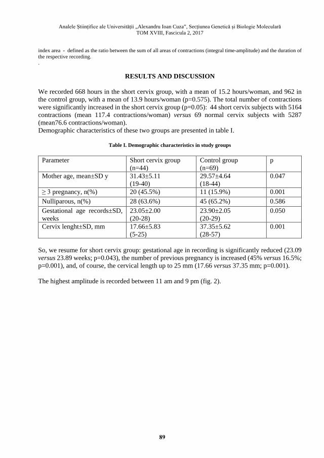
index area - defined as the ratio between the sum of all areas of contractions (integral time-amplitude) and the duration of the respective recording. .
RESULTS AND DISCUSSION
We recorded 668 hours in the short cervix group, with a mean of 15.2 hours/woman, and 962 in the control group, with a mean of 13.9 hours/woman (p=0.575). The total number of contractions were significantly increased in the short cervix group (p=0.05): 44 short cervix subjects with 5164 contractions (mean 117.4 contractions/woman) versus 69 normal cervix subjects with 5287 (mean76.6 contractions/woman). Demographic characteristics of these two groups are presented in table I.
Table I. Demographic characteristics in study groups
Parameter Short cervix group (n=44)
Control group (n=69)
p
Mother age, mean±SD y 31.43±5.11 (19-40)
29.57±4.64 (18-44)
0.047
≥ 3 pregnancy, n(%) 20 (45.5%) 11 (15.9%) 0.001 Nulliparous, n(%) 28 (63.6%) 45 (65.2%) 0.586 Gestational age records±SD, weeks
23.05±2.00 (20-28)
23.90±2.05 (20-29)
0.050
Cervix lenght±SD, mm 17.66±5.83 (5-25)
37.35±5.62 (28-57)
0.001
So, we resume for short cervix group: gestational age in recording is significantly reduced (23.09 versus 23.89 weeks; p=0.043), the number of previous pregnancy is increased (45% versus 16.5%; p=0.001), and, of course, the cervical length up to 25 mm (17.66 versus 37.35 mm; p=0.001). The highest amplitude is recorded between 11 am and 9 pm (fig. 2).
89

Anda Gheorghiță et al – The evaluation of uterine electrical activity by antepartum electromyography
Fig. 2. The diurnal distribution of contractions depending on their amplitude in the control group
We analyzed the signals by each hour of the day, trying to find correlations between maternal movements and uterine contractility in both groups. We found variability in median maternal movements, probably the circardian physiological rhythms, and we decided to divide the day in two intervals, between 10 am -11 pm=activity period and between 12 pm - 9 am=resting period (fig. 3).
Fig. 3. Mean maternal movements
4 8 12 16 20
0102030405060708090
5102030405060708090
100
110
120
130
140
150
160
170
180
190
200
210Hour of Day
Cont
ract
ions
/ho
ur
Amplitude0-10 10-20 20-30 30-40 40-50 50-60 60-70 70-80 80-90
90

Analele Științifice ale Universității „Alexandru Ioan Cuza”, Secțiunea Genetică și Biologie Moleculară TOM XVIII, Fascicula 2, 2017
We tried to characterize all particularities of the recorded uterine activity according to those two different time periods. There are significant statistical differences of mean contraction amplitude; the patients from the short cervix group had significantly increased contractions during the active period (83.02 vs 47.58; p<0.05), as well as during the resting period (82.68 vs 47.52; p<0.05) (fig. 4).
Fig. 4. Mean values of contraction amplitude according to
active versus resting period in study groups The mean duration of contraction was a little bit increased in the short cervix group, during both periods, activity (31.77 vs 25.48; p>0.05), versus resting (31.44 vs 24.35; p>0.05) (fig. 5).
Fig. 5. Mean values of duration of contraction according to
active versus resting period in study groups The short cervix group had mean of contraction area significantly increased, during active status (6400 vs 3752; p<0.05) and during resting status (6238 vs 3515; p<0.05) (fig. 6).
0
20
40
60
80
100
120
active period resting period active period resting period
Short cervix group Control group
0
10
20
30
40
active period resting period active period resting period
Short cervix group Control group
91

Anda Gheorghiță et al – The evaluation of uterine electrical activity by antepartum electromyography
Fig. 6. Mean values of contraction area according to
active versus resting period in study groups Maternal activity associated to uterine contractions is significantly increased between 10 am-11 pm, compared to the 12 pm-9 am period, both in the short cervix group (1.66 vs 1.25; p=0.005) as well as in the control group (1.67 vs 1.25; p=0,005) (fig. 7).
Fig. 7. Mean values of maternal activity according to
active versus resting period in study groups The mean number of contractions per hour was slightly increased in the short cervix group, for both active (24.62vs 23.50; p>0.05) and resting time (9.09 vs 7.38; p>0.05) (fig. 8).
0
3000
6000
9000
active period resting period active period resting period
Short cervix group Control group
0
0,5
1
1,5
2
active period resting period active period resting period
Short cervix group Control group
92

Analele Științifice ale Universității „Alexandru Ioan Cuza”, Secțiunea Genetică și Biologie Moleculară TOM XVIII, Fascicula 2, 2017
Fig. 8. Mean values of number of contractions per hour according to
active versus resting period in study groups Correlation of the parameters of uterine activity to time periods, for both groups (tab. II).
Table II. Statistic differences between utero activity according to
active versus resting period in study groups
Parameter Short cervix group
(n=44) Control group
(n=69) activity resting p activity resting p
contraction amplitude 83.02± 38.60
82.68± 31.54
0.976 47.58a) ± 9.04
47.52 a) ± 11.57
0.998
duration of contraction 31.77± 4.56
31.44± 4.57
0.882 25.48 ± 2.21
24.35 ± 2.81
0.852
contraction area 6400± 3567
6238± 3235
0.820 3752 a) ± 1224
3515 a) ± 1425
0.751
maternal activity 1.66± 0.42 1.25± 0.44
0.005 1.67 ± 0.31
1.25 ± 0.49
0.005
contractions per hour 24.62 ± 11.94
9.09 ± 4.6
0.001 23.50± 10.29
7.38± 6.91 0.001
% duration of contraction from record
17.81 ± 10.0
6.18 ± 3.44
0.001 15.77± 11.12
5.75± 3.33 0.001
area index 29.11± 22.30
10.10± 9.55
0.001 28.24 ± 22.98
9.36 ± 8.87
0.001
a) p<0.05 short cervix group vs control groups It seems the uterus responds to maternal activity. This is a newly demonstrated phenomenon and it must be confirmed by further studies, because the electrical activity of the abdominal muscles could be mistaken for uterine contractility. As for the timeline of the contractions, we found that between 10 am-11 pm there is more intense uterine activity for both control and short cervix group (tab. II) and this seems to be a physiological event.
0
10
20
30
40
active period resting period active period resting period
Short cervix group Control group
93

Anda Gheorghiță et al – The evaluation of uterine electrical activity by antepartum electromyography
There were previous studies to attempt to understand the uterine activity and to quantify the contraction frequency in asymptomatic women. Moore et al in 1994 provided contraction data in low risk pregnancies after 20 weeks of gestation and showed that there is a diurnal variation in uterine contractions but the majority of them occurred during the night (Moore, T.R., et al., 1994). We observed the opposite situations. This could be due to the fact that in our study women were monitored by electrohisterography and, in Moore’s study, the contractility was recorded by tocography (the tocodynamometer is a strain gauge positioned over the uterine fundus, witch responds to changes in uterine tension transmitted to the abdominal wall; that’s way, the device identifies the frequency of contractions, but not their intensity. It suffers misalignment following maternal movements). The antepartum uterine activity in normal pregnancy is not known to date. Our study tried to identify, characterize and compare the electromyographic parameters in normal pregnancies and in short cervix pregnancies, with high risk of preterm birth. As far as we know, it is the first long term study (as the duration of each recording) using electromyography. 48.7% of the recordings lasted more than 20 hours. This way, by examining all the recordings, we can evaluate the electrical activity and the maternal activity for the entire 24 hours/day. The uterine electrical activity was intense between 10 am – 11 pm and correlated with maternal activity; it was manifested through increased frequency and duration percentage of contractions reported to the total recording length, but without modifications in amplitude, duration and area of contractions. During the active period (10 am- 11 pm), we can see that the uterus contracts more intensely than during the resting period (12 pm – 9 am), in all study groups. In the short cervix group, we can see that the total number of contractions, frequency, contraction percentage as reported to the total recording length are much more important. The cervical length is indirectly correlated to the mean contraction frequency, and the contraction percentage was significantly lower in the normal cervix group when compared to the short cervix group. The mean number of contractions was 7, but we can observe a slight increase in the short cervix group (11.27±9.59 vs 9.80±7.05 contractions/hour). Even though in normal pregnancies there are 2 peaks of contractions frequency, these are at about half of the maximum frequency of the short cervix patients. The cervical length indirectly correlated with the mean frequency of contractions in the short cervix group. The mean duration of contractions is around 31.5 for both groups. There are no significant differences between the studied groups, as well as for the two time periods. The contraction percentage was significantly lower in the normal cervix group than in the short cervix group. The amplitude of contractions is quite similar between the 2 groups, but more than 58% of the short cervix patients present a significantly high number of contractions/hour, of low amplitude, and more than 55% present great amplitude and duration of contractions. In both groups, the correlation between the duration and mean contraction area was direct, of medium intensity, statistically significant. About 67% of the short cervix patients present high contraction areas. Comparing the 2 time periods (active versus resting state), we can see there are significant differences in amplitude and contraction area; these are particularly high in the short cervix group, in the resting phase included.
94

Analele Științifice ale Universității „Alexandru Ioan Cuza”, Secțiunea Genetică și Biologie Moleculară TOM XVIII, Fascicula 2, 2017
Maternal activity between 10 am-11pm was more pronounced for both study groups, but without significant differences. The number of contractions/hour, the contraction duration reported to the total recording length and the area index are significantly higher in both groups in active state when compared to resting state. Electromiography has proven its efficacy and utility in obstetrical practice. But there are no standardized criteria which can be quantified and reproduced in the general population (Maner, W.L. & Garfield, R.E. 2007, Lucovnik, M., et al., 2012).
CONCLUSIONS
It seems the uterus is responsive to maternal activity. This is a newly highlighted phenomenon and it must be confirmed by other means, because it could be wrongly mistaken for the electrical activity of the abdominal muscles. We must not overlook the influence of the abdominal muscles, especially related to maternal physical activity, not the existence of a permanent uterine tonus, relatively regular, which could be physiological. Therefore, it is very important to measure the cervical length during second trimester fetal morphology evaluation. We can identify a short cervix, which is a risk factor for premature birth, and recommend the proper treatment. Most of subjects were recruited during fetal second trimester screening. The Monica Holter Fetal AN24 device has several advantages; it allows long recordings and storing data for further analysis, the signal quality is not influenced by the maternal BMI, fetal position or movements. It is well tolerated because it does not limit the patients daily activities, it does not need strapping for fixation, nor bed rest. Premature birth screening remains an open discussion, in order to better identify risk factors (uterine contractility, cervical modification, infection), through clinical, biochemical and imagistic methods. Further studies should focus on implementing already known methods, like electrohysterography, and on understanding and interpreting recorded data so that these can be used better and more frequently in our daily practice.
REFERENCES
Iams, J.D., Goldenberg, R.L., Meis, P.J., et al. (1996). The length of the cervix and the risk of spontaneous premature delivery. N Engl J Med, 334: 567–572. Hassan, S.S., Romero, R., Berry, S.M., et al. (2000). Patients with an ultrasonographic cervical length\or =15 mm have nearly a 50% risk of early spontaneous preterm delivery. Am J Obstet Gynecol, 182: 1458–1467. Grimes-Dennis, J., Berghella, V. (2007). Cervical length and prediction of preterm delivery. Curr Opin Obstet Gynecol 19: 191–195. McIntosh, J., Feltovich, H., Berghella, V., et al. (2016). Role of routine cervical length screening for preterm birth prevention. Am J Obstet Gynecol, B2-B6. McPheeters, M.L., Miller, W.C., Hartmann, K.E., et al. (2005). The epidemiology of threatened preterm labor: a prospective cohort study. Am J Onstet Gynecol, 192: 1325-1329. Iams, J.D., Newman, R.B., Thom, E.A., et al. (2001). Frequency of uterine contractions and the risk of spontaneous preterm delivery. N Engl J Med, 346: 250-255. Lucovnik, M., Kuon, R.J., et al. (2011). Use of Uterine Electromyography to diagnose Term and Preterm labor. Acta Obstet Gynecol Scand, 90: 150-157. Lewis, D., Pelham, J.J., Done, E., Sawhney, H., Talucci, M., Berghella, V. (2005). Uterine contractions in asymptomatic pregnant women with a short cervix on ultrasound. The Journal Of Maternal-Fetal & Neonatal Medicine 18 (5): 325-328.
95

Anda Gheorghiță et al – The evaluation of uterine electrical activity by antepartum electromyography
Iams, J.D. (2003). Prediction and early detection of preterm labor. Obstet Gynecol, 101: 402-412. McNamara, H.M. (2003). Problems and challenges in the management of preterm labour. BJOG, 110(suppl 20): 79–85. Grgic, O., Matijevic, R. (2008). Uterine electrical activity and cervical shortening in the midtrimester of pregnancy. Int J Gynaecol Obstet, 102(3): 246–248. Vinken, M.P.G.C., Rabotti, C., et al. (2009). Accuracy of frequency related parameters of the electrohysterogram for predicting preterm delivery. A review of the literature. Obstet Gynecol Surv, 64: 529-541. SMFM. McIntosh, J., Feltovich, H., Berghella, V., Manuck, T. (2016). Role of routine cervical length screening for preterm birth prevention. Am J Obstet Gynecol, 215(3): B2-7. doi: 10.1016/j.ajog.2016.04.027. Moore, T.R., Iams, J.D., Creazy, R.K., Burau, K.D., Davidson, A.L. (1994). Diurnal and gestational paterns of uterine activity in normal human pregnancy. Obstet Gynecol, 83: 517-523. Maner, W.L., Garfield, R.E. (2007). Identification of human term and preterm labor using artificial neural networks on uterine electromyography data. Ann Biomed Eng, 35: 465-473. Lucovnik, M., Novak-Antolic, Z., Garfield, R.E. (2012). Use of noninvasive uterine electromyography in the diagnosis of preterm labour. Facts, Views & Vision in Obgyn, 4: 66–72. 1 PhD student „Gr.T.Popa” University of Medicine and Pharmacy, Iași, România; „Cuza Vodă” Clinical Hospital of Obstetrics and Gynaecology Iași 2 „Gr.T.Popa” University of Medicine and Pharmacy, Iași, România; „Elena Doamna” Clinical Hospital of Obstetrics and Gynaecology Iaşi 3 „Gr.T.Popa” University of Medicine and Pharmacy, Iași, România; „Cuza Vodă” Clinical Hospital of Obstetrics and Gynaecology Iași 4 ”Alexandru Ioan Cuza” University, Iași, România 5 IML, Iași, România [email protected]
96