The evaluation of the diagnostic utility and sensitivity ...
69
The evaluation of the diagnostic utility and sensitivity of the Xpert® MTB/RIF in the detection of Mycobacterium tuberculosis and rifampicin resistance on bone marrow aspirate samples NADHIYA SUBRAMONY STUDENT NUMBER: 0500378R MMED HAEMATOLOGY A research report submitted to the Faculty of Health Sciences, University of the Witwatersrand, Johannesburg, in partial fulfilment of the requirements for the degree of Master of Medicine in the Branch of Pathology (Haematology).
Transcript of The evaluation of the diagnostic utility and sensitivity ...

The evaluation of the diagnostic utility and
sensitivity of the Xpert® MTB/RIF in the
detection of Mycobacterium tuberculosis and
rifampicin resistance on bone marrow aspirate
samples
NADHIYA SUBRAMONY
STUDENT NUMBER: 0500378R
MMED HAEMATOLOGY
A research report submitted to the Faculty of Health Sciences, University of the
Witwatersrand, Johannesburg, in partial fulfilment of the requirements for the degree of
Master of Medicine in the Branch of Pathology (Haematology).

ii
DECLARATION
I, Nishanti Nadhiya Subramony declare that this thesis is my own work. It is being submitted
for the degree of Master of Medicine in the University of the Witwatersrand, Johannesburg. It
has not been submitted before for any degree or examination at this or any other University.
20 November 2017

iii
DEDICATION
To the Almighty God.
Dedicated to my parents, Sam and Tholsie and to my husband, Nash.

iv
ABSTRACT
In South Africa, the World Health Organisation estimated 454 000 new cases of
Mycobacterium Tuberculosis (M.tb) infection (MTB) in 2015. Disseminated tuberculosis
arises from haematogenous spread of the bacilli and seeding of the bacilli in extrapulmonary
sites. The current gold standard for the detection of MTB of the bone marrow is TB culture
which has an average turnaround time of 6 weeks. Although shorter, histological
examinations of trephine biopsy cores to diagnose MTB also have a time delay owing mainly
to the 5-7 day processing period prior to microscopic examination. Adding to the diagnostic
delay is the non-specific nature of granulomatous inflammation which is the hallmark of
MTB involvement of the bone marrow. A Ziehl-Neelson stain (which highlights acid-fast
bacilli) is therefore mandatory to confirm the diagnosis but can take up to 3 days for
processing and evaluation. Owing to this delay in diagnosis, many patients are lost to follow
up or remain untreated for up to six weeks while the results are awaited, thus encouraging the
spread of undiagnosed TB.
The Xpert MTB/RIF (Cepheid, Sunnyvale, CA) is the molecular test used in the South
African national TB program as the initial diagnostic test for pulmonary TB in adults and
children. In 2013 the Xpert MTB/RIF was applied to diagnose extrapulmonary TB and
despite being available in 207 testing sites nationwide, it was never investigated for its
potential application in diagnosing TB in bone marrow. Therefore, this study investigates the
optimisation and performance of Xpert MTB/RIF on bone marrow aspirate specimens.
BMA specimens received for routine immunophenotypic analysis as part of the investigation
into disseminated MTB or in the evaluation of cytopenias in immunocompromised patients
were used in this study. Processing BMA on the Xpert® MTB/RIF was optimised to ensure
bone marrow in EDTA and heparin did not inhibit the PCR reaction. Inactivated M.tb from an
Xpert MTB/RIF external quality assessment program was spiked into the clinical bone
marrow specimen and distilled water (as a control). A volume of 500µl and an incubation time
of 15 minutes with sample reagent were investigated as the processing protocol.
A total of 135 BMA specimens had sufficient residual volume for Xpert® MTB/RIF testing
however 22 specimens (16.3%) were not included in the final statistical analysis as an adequate
trephine biopsy and/or TB culture was not available. Xpert MTB/RIF testing was possible in

v
the presence of heparin or EDTA, but the overall detection of MTB in BMA was low compared
to histology and culture.
The sensitivity of the Xpert® MTB/RIF when compared to both histological and culture
findings was 8.7% with a 95% confidence interval (CI) of 1.07-28.04%. The sensitivity of the
Xpert® MTB/RIF compared to histological findings only was 11.1% with a 95% CI of 1.38-
34.7%. The specificity of the Xpert® MTB/RIF was 98.9% (95% CI: 93.9-99.7%).
Although the Xpert® MTB/RIF has a shorter turnaround time than histology and TB culture
and is less expensive than culture and drug susceptibility testing, the low sensitivity of the
Xpert® MTB/RIF in this study limits its current use for the diagnosis of MTB in bone marrow
aspirate specimens until the diagnostic algorithm or the assay is further defined.
vi
ACKNOWLEDGEMENTS
To the Almighty God without who none of this would be possible.
To my parents for their unwavering love and continuous support through my undergraduate
and postgraduate training.
To my husband for his encouragement and belief in me.
To Prof Scott, Dr Vaughan and Dr Black (microbiology) for their assistance, guidance and
support.
To the staff in the flow cytometry laboratory and to Natasha Gouws and Anura David, thank
you for your assistance and patience.
To my colleagues and friends for their encouragement.

vii
Table of Contents
DECLARATION ......................................................................................................................................... ii
DEDICATION ........................................................................................................................................... iii
ABSTRACT ............................................................................................................................................... iv
ACKNOWLEDGEMENTS .......................................................................................................................... vi
LIST OF FIGURES ..................................................................................................................................... ix
LIST OF TABLES ........................................................................................................................................ x
LIST OF ABBREVIATIONS ........................................................................................................................ xi
CHAPTER 1: INTRODUCTION ................................................................................................................... 1
1.1 History and characteristics of Mycobacteria .............................................................................. 1
1.2 Pathophysiology .......................................................................................................................... 1
1.2.1 M.tb infection (MTB) in the HIV positive population .................................................................. 2
1.3 Epidemiology ............................................................................................................................... 3
1.3.1 Global perspective of MTB .......................................................................................................... 3
1.3.2 MTB and HIV in South Africa ...................................................................................................... 5
1.4 Extrapulmonary MTB .................................................................................................................. 6
1.5 Disseminated MTB involving the bone marrow ......................................................................... 7
1.5.1 Current diagnostic practices for disseminated TB involving the bone marrow ......................... 8
1.6 Other diagnostic methods for TB .............................................................................................. 12
1.6.1 WHO endorsed technologies ................................................................................................... 12
1.6.1.1 Xpert® MTB/RIF ......................................................................................................................... 12
1.6.1.2 Line probe assays ...................................................................................................................... 21
1.6.1.3 Loop-mediated isothermal amplification assay ........................................................................ 22
1.6.1.4 Lipoaribinomannan assay ......................................................................................................... 23
1.6.2 Tests under evaluation ............................................................................................................. 27
1.6.2.1 RealTime MTB and RealTime MTB-INH/RIF assays ................................................................... 27
1.7 Objectives of this MMED research report ................................................................................ 28
CHAPTER 2: MATERIALS AND METHODS .............................................................................................. 29
2.1 Specimen selection ........................................................................................................................ 29
2.2 Optimisation of the Xpert® MTB/RIF for bone marrow aspirate specimens .................................. 29
2.2.1 Assessment for PCR inhibitors in BMA and to assess the appropriate specimen volume .......... 29
2.2.2 Assessment of the effect of anticoagulants on the Xpert® MTB/RIF PCR reaction ..................... 30
2.2.3 Distilled water as a control: ......................................................................................................... 30
2.3 Sample processing .......................................................................................................................... 30

viii
2.4 Evaluation of the Xpert ® MTB/RIF for the detection of M.tb in patient BMA specimens ............. 31
2.5 Data collection ............................................................................................................................... 31
2.6 Exclusion criteria ............................................................................................................................. 31
2.7 Statistics .......................................................................................................................................... 32
CHAPTER 3: RESULTS ............................................................................................................................. 33
3.1 Specimens processed ...................................................................................................................... 33
3.2 Optimisation of the Xpert® MTB/RIF for BMA specimens .............................................................. 33
3.2.1 Assessment for PCR inhibitors in BMA and to assess the appropriate specimen volume .......... 33
3.2.2 Assessment of the effect of anticoagulants on the Xpert® MTB/RIF PCR reaction ..................... 35
3.2.3. Distilled water as a control ......................................................................................................... 36
3.3 Evaluating the Xpert® MTB/RIF on BMA specimens ....................................................................... 38
3.3 Inconclusive results ......................................................................................................................... 39
3.4 Calculation of sensitivity and specificity ......................................................................................... 43
CHAPTER 4: DISCUSSION ....................................................................................................................... 45
4.1 Conclusion ....................................................................................................................................... 47
REFERENCES .......................................................................................................................................... 48
APPENDIX .............................................................................................................................................. 53

ix
LIST OF FIGURES
Figure Title
Page number
1 Global incidence of TB for 2015
4
2 Estimated HIV prevalence of new and relapsed TB cases in 2015
5
3 MTB incidence in South Africa from 2000-2015
6
4 Outline of the components involved in the diagnosis of MTB in
bone marrow as followed by the NHLS
8
5 Schematic representation of the processing of bone marrow
Bactec Myco-F Lytic culture bottles in the TB reference
laboratory of the NHLS
11
6 The 81bp RRDR of the rpoB gene showing the overlapping
molecular probes
12
7 Components of the Xpert® MTB/RIF assay
14
8 Interpretation of results as reported by the Xpert® MTB/RIF test
15
9 A radar plot of the sensitivity of the Xpert® MTB/RIF compared
to liquid culture for various specimen types and including
performance among the HIV+ population
19
10 Flow diagram demonstrating the processing of specimens using
LPAs
21
11 Test strip and reference card of the Determine™ TB LAM antigen
kit
24
12 Flow diagram of results obtained in this study
40
13 100% stacked bar graph depicting MTB results for each
diagnostic modality used in this study
42

x
LIST OF TABLES
Table Title
Page
number
1 Extrapulmonary specimens received for TB culture
7
2 The reported MTB result based on the CT range
16
3 A meta-analysis of sensitivity and specificity of the Xpert®
MTB/RIF in diagnosing EPTB of the commonest sites involved
18
4 Studies utilising bone marrow aspirate specimens on the Xpert®
MTB/RIF
20
5 Summary of the WHO endorsed technologies
26
6 Results using 1ml heparinised BMA specimens with 25µL of
inactivated M.tb in a single sample run in quintuplicate.
33
7 Results using 500µL heparinised BMA specimens with 25µL of
inactivated M.tb in a single sample run in quintuplicate.
34
8 Results using 500µL BMA specimens obtained from ETDA tubes
with 25µL of inactivated M.tb added in a single sample run in
quintuplicate
35
9 Results of 500µL BMA specimens obtained from the same patient
and from EDTA and heparinised tubes run in triplicate for each
anticoagulant. 25µL of inactivated M.tb added.
35
10 Results using 500µl heparinised BMA specimens with serial
dilutions of inactivated M.tb (ranging from 5µl to 25µl).
36
11 Results using 500µL distilled water with 25µL of inactivated M.tb in
a single sample run in quintuplicate.
36
12 Results using 500µL distilled water and serial dilutions of inactivated
M.tb
37
13 Positive Xpert® MTB/RIF results in this study
41
14 Summary of statistical analysis of the Xpert® MTB/RIF assay on
bone marrow aspirate specimens
44

xi
LIST OF ABBREVIATIONS
AFB: acid fast bacilli
ART: anti-retroviral treatment
BCG: Bacillus Calmette-Guérin
BM: bone marrow
BMA: bone marrow aspirate
CMJAH: Charlotte Maxeke Johannesburg Academic Hospital
Ct: cycle threshold
EDTA: Ethylenediaminetetraacetic acid
ELISA: enzyme linked immunosorbent assay
EPTB: extra-pulmonary TB
HIV: Human Immunodeficiency Virus
IFN-γ: interferon gamma
IGRA: interferon gamma release assays
INH: isoniazid
LAM: lipoaribinomannan
LAMP: loop-mediated isothermal amplification assay
LF-LAM: lateral flow lipoaribinomannan assay
LIS: laboratory information system
LMTBI: latent MTB infection
LPA: line probe assays
MDR-TB: Multi drug resistant-TB
MGIT: Mycobacteria Growth Indicator Tube
MOTT: Mycobacterium Other Than TB
M.tb: Mycobacterium tuberculosis
MTB: M.tb infection
NAAT: nucleic acid amplification test
NHLS: National Health Laboratory Services
PCR: polymerase chain reaction
RIF: rifampicin
RRDR: Rifampicin Resistance Determining Region
SOP: standard operating procedure
SPC: sample processing control

xii
SR: solvent-reagent
TAT: turnaround time
TB: Tuberculosis
TNF: Tumour Necrosis Factor
TST: tuberculin skin test
WHO: World Health Organisation
ZN: Ziehl-Neelson

CHAPTER 1: INTRODUCTION
1.1 HISTORY AND CHARACTERISTICS OF MYCOBACTERIA
The genus Mycobacterium is hypothesised to have emerged more than 150 million years ago
however Hermann Heinrich Robert Koch was the first to identify the tubercle bacillus
(Mycobacterium tuberculosis(M.tb)) and postulate its infectious nature in humans in 1882 (1,
2). The M.tb bacilli are rod-shaped, non-spore forming, aerobic organisms spread by airborne
droplets (3). They are classified as acid-fast bacilli owing to their ability to retain the colour of
arylmethane dyes when exposed to dilute mineral acid (4). The lipid cell wall of the bacillus is
comprised of mycolic acid attached to a polysaccharide arabinogalactan and has many integral
roles which include providing resistance to antibiotics and influencing the growth rate of the
organism (5). As the organism replicates, it acquires mutations within its genome as a result of
random DNA transcriptional errors. This allows for phenotypic variation between infections
such as sensitivity to drugs, interaction with the host immune cells and the ability to
disseminate to other tissues such as bone marrow (4).
The term MOTT (Mycobacterium Other Than Tuberculosis (TB)) was used when subsequent
studies identified the organism in other species. This includes M. avium (avian) and M. bovis
(bovine) (4).
1.2 PATHOPHYSIOLOGY
Once inhaled, most of the bacilli are trapped in the upper airways in close proximity to the
goblet cells. These cells serve as the first line of defence owing to their mucus producing
properties and are sufficient for the prevention of full-blown infection in healthy individuals
(6, 7). Those bacilli that are capable of evading the goblet cells reach the alveoli and are
phagocytosed by macrophages. This is enhanced by the binding of complement protein C3 to
the bacterial cell wall (opsonisation). Despite being engulfed by alveolar macrophages, the
bacilli continue multiplying at a rate of one cell division every 25-32 hours (6). The cytokines
released from the macrophages recruit T lymphocytes thus shifting the immune response from
an initial innate mediated to a cell mediated response. This process can take 2-12 weeks (8).

2
In an immunocompetent host, granulomatous inflammation occurs around the bacillus as the
next step in containing the infection. Granulomas are predominantly comprised of T
lymphocytes and macrophages and are characterised by low pH levels, low oxygen content and
limited nutrients. These areas then undergo fibrosis and calcification resulting in a latent
bacillus contained within a healed lesion (9). In immunocompromised patients, the inability of
the immune system to contain the primary infection leads to symptomatic pulmonary disease.
In addition, the formation of a granuloma is usually unsuccessful with early breakdown and
extradition of the material into a blood vessel. This allows for haematogenous dissemination
of the bacilli to extrapulmonary sites (9).
1.2.1 M.tb INFECTION (MTB) IN THE HIV POSITIVE POPULATION
Human Immunodeficiency Virus (HIV) is the biggest risk factor for acquiring M.tb infection
(MTB) which in turn is the leading cause of death in people with HIV (10). As mentioned
above, cell mediated immunity plays a vital role in the initial containment of MTB. This is
driven by CD4+ T cells and is therefore compromised in HIV where the hallmark of disease is
the steady depletion of CD4+ T cells. Other mechanisms that contribute to increased
susceptibility to MTB (both primary and reactivation/post primary disease) include impaired
tumour necrosis factor (TNF)-mediated macrophage apoptotic response to the bacillus (11),
up-regulation of receptors on macrophages facilitating MTB entry (12) and impaired
chemotaxis (13).
The TB-HIV relationship is a mutually beneficial one as M.tb increases the expression of CCR5
and CXCR4 receptors on CD4+ T cells which are known entry points for the HIV virus (14).
Replication of the HIV virus is increased and has been demonstrated at sites of MTB infection
in the lung and within macrophages and lymphocytes of the pleural space (15, 16). This is
reflected in vivo by the rising viral loads noted in patients with both infections. Increased viral
replication occurs as a bystander effect when TNF is secreted by T lymphocytes with the
primary aim to inhibit M.tb growth (17). M.tb can directly stimulate TNF production through
a component of its cell wall, lipoarabinomannan. This occurs via the NF-κB pathway which
leads to the transcriptional activation of the long terminal repeat promotor thus enhancing HIV
replication (18).

3
The diagnosis of MTB in a HIV positive person is challenging given the frequency of smear
negative cases, the unusual/atypical presentations and the lack of granuloma formation on
histological specimens. A high index of suspicion is required as well as novel diagnostic
methods to shift the focus away from acid-fast bacilli detection in the diagnosis of active TB.
Latent MTB infection (LMTBI) is described as an entity in which there is no manifestation of
disease and M.tb cannot be cultured however clinical suspicion of infection exists (19).
Identifying LMTBI is beneficial as it allows for an early diagnosis in individuals at high risk
for transformation to active MTB (such as the immunocompromised). This can be done via
two methods - the tuberculin skin test and interferon gamma release assays. Whilst much
information is available regarding these tests, LMTBI is not the focus of this study.
1.3 EPIDEMIOLOGY
1.3.1 GLOBAL PERSPECTIVE OF MTB
In 2015, there were approximately 10.4 million new cases of MTB globally with a male to
female ratio of 1.5:1. South Africa was one of six countries that accounted for 60% of these
new cases (Figure 1). Despite this seemingly large number, the World Health Organisation
(WHO) reports a decline in MTB incidence at a rate of 1.5% from 2014 to 2015. Deaths owing
to MTB were estimated at 1.4 million in 2015 with MTB retaining its position as one of the top
ten causes of death worldwide (20).
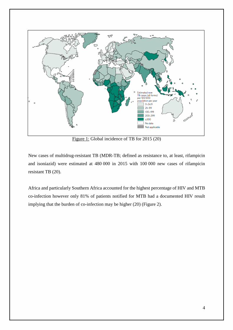
4
Figure 1: Global incidence of TB for 2015 (20)
New cases of multidrug-resistant TB (MDR-TB; defined as resistance to, at least, rifampicin
and isoniazid) were estimated at 480 000 in 2015 with 100 000 new cases of rifampicin
resistant TB (20).
Africa and particularly Southern Africa accounted for the highest percentage of HIV and MTB
co-infection however only 81% of patients notified for MTB had a documented HIV result
implying that the burden of co-infection may be higher (20) (Figure 2).

5
Figure 2: Estimated HIV prevalence of new and relapsed TB cases in 2015 (20)
1.3.2 MTB AND HIV IN SOUTH AFRICA
MTB in South Africa is rife in poorer socio-economic environments making MTB
identification, control and reporting a challenge (21).
The WHO Global TB Report 2016 reported 454 000 new cases of MTB in South Africa of
which 56% were co-infected with HIV(20). Data regarding incidence (reported as rate per
100 000 population) from 2000 – 2015 is represented graphically in Figure 3 and shows an
initial upward growth but a decline after 2005. From 2014 however, the total incidence appears
to have stabilised.

6
Figure 3: MTB incidence in South Africa from 2000 – 2015 (20)
Overall mortality from MTB amounted to 98 000 cases with a fatality ratio of 0.22 (0.1-0.42).
The majority (~74%) occurred in the HIV positive population with a mortality rate of 133 per
100 000. These statistics are similar to the previous year (22).
In 2015, MTB accounted for 0.4 million deaths in the HIV positive population (20). Of the new
and relapsed cases of MTB in 2015 in South Africa, 57% were positive for HIV. Of these, 85%
were on anti-retroviral treatment (ART). Whilst this appears impressive, it is not in line with
the WHO recommendation that all HIV positive people with MTB infection should receive
ART (23). This recommendation by the WHO is supported by a study performed in South
Africa which showed that by increasing the ART roll-out, the incidence of confirmed
pulmonary MTB declined over a period of four years (2008-2012) (24).
1.4 EXTRAPULMONARY MTB
Extrapulmonary MTB infection (EPTB) is defined as MTB outside of the pulmonary system
and occurs as a result of seeding of the M.tb bacilli via haematogenous, lymphoid or mucosal
routes. If two or more non-contiguous sites are involved, the term disseminated TB (synonym:
miliary TB) is used. EPTB is more common in those with advanced HIV as the associated
immunosuppression allows for rapid progression of the disease following
exposure/reactivation of the organism (4).

7
In South Africa, as per the WHO Global TB report 2016, EPTB cases accounted for 10%
(20). Further specification regarding the site involved was not included in this report however
a study performed locally (at the National Health Laboratory Services (NHLS)
Mycobacteriology Referral Laboratory in Johannesburg) revealed the most common
extrapulmonary specimens received for TB culture. These are cerebrospinal fluid, fine needle
aspirates (predominantly from lymph nodes) and serous effusions (pleural or ascitic) (Table
1). In this study, the prevalence of EPTB was 23% (277 of 1,175 specimens tested) (25).
Table 1: Extrapulmonary specimens received for TB culture (in
order of frequency). Table adapted from (25).
1.5 DISSEMINATED MTB INVOLVING THE BONE MARROW
Bone marrow involvement by the M.tb bacilli occurs as a result of haematogenous spread and
can present with specific haematology findings (such as cytopenias), pyrexia of unknown
origin and/or non-specific constitutional symptoms (fever, night sweats and loss of weight)
(26). In an 18 month period at Chris Hani Baragwanath Academic Hospital, south of
Johannesburg, 15% of the bone marrow aspirates and trephines received were for suspected
disseminated TB. Over half of these cases had unexplained cytopenias (27). A study conducted
in 2010 in India concluded that bone marrow examinations are highly useful in HIV positive

8
patients presenting with pyrexia of unknown origin (PUO) as MTB was the commonest cause
of PUO, accounting for 60% of all cases in this study (28).
Disseminated TB with bone marrow involvement requires ~9 months of treatment (comparable
to other extra pulmonary sites) with the same drugs used for pulmonary TB. There is however
a higher mortality rate among patients with bone marrow TB involvement when compared to
other sites (29). For these reasons, if clinical suspicion of disseminated TB involving the bone
marrow exists (e.g. the presence of cytopenias), a bone marrow aspirate and trephine biopsy
with material for TB culture is indicated.
1.5.1 CURRENT DIAGNOSTIC PRACTICES FOR DISSEMINATED TB
INVOLVING THE BONE MARROW
When a bone marrow procedure is performed as part of the investigation of disseminated
mycobacterial infection, three components are essential. These are outlined in Figure 4 and
explained in detail in the text that follows. Of note, three pathology disciplines are involved in
the processing and diagnosis of MTB. This includes haematology (analyses bone marrow
aspirate smears and trephine biopsies), microbiology (processes TB cultures) and anatomical
pathology (processes the trephine biopsies and performs the Ziehl-Neelson (ZN) stain).
Figure 4: Outline of the components involved in the diagnosis of MTB in bone marrow as
followed by the NHLS
Bone marrow trephine biopsy Bone marrow aspirate
Smears TB culture Assessed for granulomatous inflammation
ZN stain ordered by attending pathologist

9
1) Bone marrow (BM) aspirate slides are fixed in 10% methanol and stained twice with
Giemsa’s stain. Examination usually reveals reactive features which are non-diagnostic.
The slides can be stained with auramine O for the identification of acid fast bacilli using
fluorescent microscopy however this is not commonly done as a study performed locally
revealed an overall low yield of positive cases using this method (7 of 123 cases) (27).
2) Bone marrow trephine biopsies undergo 24 hours of formalin fixation followed by a
minimum of 48 hours decalcification in ethylenediaminetetraacetic acid (EDTA) disodium
salt. The specimen is then embedded into moulds, cut and stained with haematoxylin and
phloxine.
As mentioned previously, the hallmark of MTB is granulomatous inflammation. This is
however non-specific and seen with several other infections (including cryptococcus),
sarcoidosis, Hodgkin and Non-Hodgkin Lymphoma (30). As a result, a ZN stain is
mandatory for confirmation of MTB. In our setting, this stain is performed only at the
request of the attending pathologist. Due to the large amounts of lipid (mycolic acid) in the
cell wall of the mycobacterium, traditional staining methods such as the Gram stain are
ineffective. The ZN stain utilises heated carbolfuchsin to break through this lipid capsule.
Initially, all viable cells stain with carbolfuchsin however after a process of acid and alcohol
decolourisation only cells with the protective lipid layer retain the carbolfuchsin dye.
Mycobacteria are thus acid and alcohol fast. The specimen is then stained with methylene
blue which is taken up by non-acid fast organisms. Acid-fast bacilli (AFB) retain the pink
colour of carbolfuchsin (31).
From the description above, it is easy to see that this method of diagnosing MTB in the bone
marrow can be protracted, taking on average 4-7 days to reach the pathologist. In addition, the
diagnostic yield of bone marrow trephine examinations is influenced by poor quality specimens
(owing to clinician inexperience, degree of bone marrow infiltration or patient factors. The
latter may include obesity, pelvic bed sores or fractures that interfere with obtaining a good
quality, diagnostic specimen), differential sampling and paucibacillary specimens (that may
appear negative for AFB on a ZN stain).
3) TB culture and drug susceptibility analysis is the gold standard for detection of
disseminated MTB. Myco-F lytic culture vials are required with a minimum of 5ml of bone
marrow sample. This is incubated in the Bactec 9240 automated system with weekly

10
inspections for six weeks. If growth is detected, a smear is prepared from the TB culture
bottle containing the bone marrow aspirate specimen and a ZN stain performed. One of the
following scenarios can then occur (outlined in Figure 5 below):
a. Only contaminating bacteria are identified and the ZN stain is negative for AFB.
No further testing is performed in this scenario.
b. Both AFB and contaminating bacteria are not identified. This implies a false
positive flag by the instrument and the sample is re-incubated.
c. If the ZN stain is positive for AFB regardless of the presence of contaminating
bacteria, a rapid antigen test (the TB Ag MPT64 Rapid) is performed. This test
identifies M.tb therefore if negative, polymerase chain reaction (PCR; specifically
the Hain Genotype Mycobacterium CM, Hain Lifescience, Germany) is performed
to identify the presence of MOTT. If the rapid antigen test is positive and drug
susceptibility has been requested by the clinician, a subculture into a Mycobacteria
Growth Indicator Tube (MGIT) is performed which is followed by a line probe
assay (the MTBDRplus, Hain Lifescience, Germany) to identify susceptibility to
the first line anti-TB agents, rifampicin and isoniazid.
The protracted nature of this TB culture leads to delayed diagnosis of the disease with the
knock on effect of losing patients to follow up and hindering TB infection control.

11
Figure 5: Schematic representation of the processing of bone marrow Bactec Myco-F Lytic
culture in the TB reference laboratory of NHLS
Incubate Myco-F Lytic culture bottle in BD Bactec 9240 instrument for 42
days
Growth detected
ZN stain performed
No growth detected
Negative result
ZN positive for AFBs
with/without
contaminating bacteria
Rapid antigen test
(TB Ag MPT64 Raid)
MTBDRplus (a first line line probe assay) performed for the
confirmation of MTB and identification of RIF and/or INH
resistance
MTB confirmed
No AFBs and no
contaminating
bacteria
PCR (Hain Genotype Mycobacterium
CM) performed for the identification
of MOTT
BD Bactec Myco-F Lytic liquid culture
bottle received in laboratory
Re-incubate
No AFBs,
contaminating
bacteria only
No further testing
Positive Negative
Subculture into a MGIT tube which
is placed into a MGIT 960
instrument
If drug susceptibility requested, a
subculture into a MGIT tube is
performed which is placed into a
MGIT 960 instrument

12
1.6 OTHER DIAGNOSTIC METHODS FOR TB
1.6.1 WHO ENDORSED TECHNOLOGIES
1.6.1.1 XPERT® MTB/RIF
The Xpert® MTB/RIF assay from Cepheid (Sunnyvale, California) is a cartridge based assay
using hemi-nested real time PCR to amplify the rpoB gene which is specific to M.tb. In
addition, the Xpert® MTB/RIF assay is capable of simultaneously detecting resistance to the
most commonly used first line anti-TB drug, rifampicin (RIF).
The assay utilises five probes (A-E; also known as molecular beacons) which are
complementary to overlapping regions of the rpoB gene (Figure 6) and the 81 base pair RIF
Resistance Determining Region (RRDR) which is the site of mutation for more than 95% of
all RIF resistant strains (32).
Figure 6: The 81bp RRDR of the rpoB gene showing the overlapping molecular probes (33)
Each probe is allocated a specific fluorescent marker. When unhybridised, the probes do not
fluoresce as the quencher and the fluorescent dye are in close proximity. This is enabled by the
curved conformation of the probes. Once hybridised to the RRDR region, the probe flattens
out and the quencher no longer suppresses the fluorescent dye. An increase in fluorescence
above the background is reported by the software as a positive result. A positive result for the
presence of M.tb is deemed if two or more probes emit a signal within two cycles of each other

13
(34). If there is resistance to RIF, hybridisation of the probe will not occur as there is a change
in sequence in the RRDR region of the rpoB gene owing to the mutation/s present (34). As a
result, a fluorescent signal will not be released. Resistance to RIF is therefore identified by the
lack of a detectable cycle threshold for a specific probe or if the difference in cycle threshold
between the first and last rpoB signals is >4.1. If <4.1, a result of rifampicin sensitive is
reported.
Included in the cartridge is a Sample Processing Control (SPC) which is a separate PCR
reaction (but occurs simultaneously to the sample PCR reaction) and assesses for the presence
of Bacillus globigii spores (34). This serves as an internal control and allows for the verification
of the integrity and effectiveness of processing.
The Xpert® MTB/RIF is fully automated but does require the addition of a reagent buffer to
liquefy and inactivate any M.tb bacilli present in the specimen. The limit of detection for the
Xpert® MTB/RIF is 150 cfu/mL (34) however with the advent of the Xpert® MTB/RIF Ultra
(Cepheid), this may be improved upon (35). This latest assay utilises a new cartridge that allows
for double the amount of sample DNA (as compared to the Xpert® MTB/RIF) to be used in
the PCR reaction. The five original probes described above are replaced with four probes which
detect mutation in the rpoB gene as well as real-time probes that target the IS6110 and
IS1081genes of M.tb. Whilst being as easy to use, this new assay has a 10x lower limit of
detection compared to the original Xpert® MTB/RIF and can detect a wider variety of rpoB
gene mutations (36). As of March 2017, the WHO recommends the use of Xpert® MTB/RIF
Ultra as a replacement assay for the current Xpert® MTB/RIF (37).
The steps involved in the Xpert® MTB/RIF assay are shown in Figure 7 (38).

14
Figure 7: Components of Xpert® MTB/RIF assay. Adapted from (38).

15
The results are interpreted using the algorithm shown in Figure 8 (33):
Figure 8: Interpretation of results as reported by the Xpert® MTB/RIF test (33)
Check probe
Fail Pass Result: Error
Two or more rpoB gene probes hybridise
No Yes
All 5 probes
hybridise
1/more probes do not
hybridise or show
delayed hybridisation
with a difference in
cycle threshold of
>4.1
Result:
M.tb detected
RIF resistance
not detected
Result:
M.tb detected
RIF resistance
detected
Internal control
positive
Internal control
negative
Result:
Invalid
Result:
M.tb not
detected

16
In the instances where M.tb is detected, the instrument reports levels of M.tb DNA in the sample
by analysis of the cycle thresholds (Ct). Based on an average of the Ct values of all five probes,
a lower Ct denotes a higher DNA concentration and a higher Ct denotes a lower DNA
concentration (Table 2) (21, 33).
Table 2: The reported MTB result based on the Ct range
The 2013 WHO policy update for the use of the Xpert® MTB/RIF assay on pulmonary
specimens was based on 27 studies with more than 9000 participants. A meta-analysis of these
studies revealed the following (39):
As an initial diagnostic test used instead of smear microscopy, the Xpert® MTB/RIF
assay had a pooled sensitivity of 88% (95% CI: 84–92%) and a pooled specificity of
99% (95% CI: 98– 99%).
If performed after a negative smear-microscopy result, the Xpert® MTB/RIF had a
pooled sensitivity of 68% (95% CI: 61–74%) and a pooled specificity of 99% (95% CI:
98–99%).
For smear-positive culture-positive MTB, the pooled sensitivity of the Xpert®
MTB/RIF was 98% (95% CI: 97–99%).
In the HIV positive population, the pooled sensitivity of Xpert® MTB/RIF was 79%
(95% CI: 70–86%).
In the HIV negative population, the pooled sensitivity was 86% (95% CI: 76–92%).
When used to identify RIF resistance, Xpert® MTB/RIF resulted in a pooled sensitivity
of 95% (95% CI: 90–97%) and a pooled specificity of 98% (95% CI: 97–99%).
Based on the above findings, the WHO strongly recommends the use of Xpert® MTB/RIF in
place of conventional testing (microscopy, culture and drug susceptibility testing) as the initial
diagnostic test in both adults and children suspected of having MDR-TB or HIV-associated
Result
Ct range
High <16
Medium 16-22
Low 22-28
Very low >28

17
TB. A conditional recommendation (acknowledging resource constraints) is also included
whereby the Xpert® MTB/RIF can be used instead of the conventional testing mentioned
above as the initial diagnostic test in children and adults suspected of having pulmonary TB. It
can also be used as a follow-up test to microscopy (especially if smear negative) in adults
suspected of having pulmonary TB if they are not at risk of MDR-TB or HIV-associated TB.
The use of Xpert® MTB/RIF assay on pulmonary specimens in South Africa commenced in
March 2011 following WHO recommendations and is used in the National TB programme as
the initial diagnostic tool for diagnosing MTB. In 2017, evidence 5 years post implementation
showed a decline in the TB notification rate in both the HIV positive (19% decrease) and HIV
negative (12% decrease) populations since the advent of the rollout. A positive Xpert®
MTB/RIF result also surpassed a positive microscopy result as the number one reason for
commencing TB treatment. This shows how well clinicians have taken to this relatively new
assay in our setting (40).
The results of the meta-analysis performed by the WHO in determining the sensitivity and
specificity of the Xpert® MTB/RIF in diagnosing EPTB is summarised in Table 3.

18
Table 3: A meta-analysis of the sensitivity and specificity of the Xpert® MTB/RIF in
diagnosing EPTB at the commonest sites involved (39).
Sample type Pooled sensitivity (95%
confidence interval)
Pooled specificity (95%
confidence interval)
Lymph node 84.9% (72-92%)
92.5% (80-97%)
Cerebro-spinal fluid 79.5% (62-90%) when
compared against culture
55.5% (51-81%) when
compared against a
composite reference
standard
98.6% (96-100%)
98.8% (95-100%)
Pleural fluid 43.7% (25-65%) when
compared against culture
17% (8-34%) when
compared to a composite
reference standard
98.1% (95-99%)
99.9% (94-100%)
Overall, pleural fluid is a poor specimen for the diagnosis of MTB however if Xpert®
MTB/RIF is positive, this should be considered evidence of disease and treated as such (39).
A study performed locally demonstrated the sensitivity of the Xpert® MTB/RIF in comparison
to liquid culture for various specimen types (including pulmonary and extrapulmonary
specimens) (41). This is shown in Figure 9.

19
Figure 9: A radar plot of the sensitivity of the Xpert® MTB/RIF compared to liquid culture
for various specimen types and including performance among the HIV+ population. Adapted
from (41).
It is evident that the sensitivity of the Xpert® MTB/RIF is higher with pulmonary samples (and
especially so with smear positive specimens) compared to the extrapulmonary samples
assessed. Lymph node tissue/aspirate in the HIV positive population is the exception noted in
the local study.
Owing to limited availability, data on the use of the assay on bone marrow aspirate (BMA)
specimens was omitted from the WHO systematic review and meta-analysis. Often, these
specimens are included with other extra-pulmonary specimens and an overall assessment of
the performance of the Xpert® MTB/RIF assay is reported. The findings of some of the studies
that included BMA samples are shown in Table 4.

20
Table 4: Studies utilising bone marrow aspirate specimens on the Xpert® MTB/RIF
Authors: No. of BMA
specimens/total specimens
(%)
Sensitivity: Specificity:
Armand et al (42)
6/37 (16.2%)
53%
-
Malbruny et al (43)
2/89 (2.2%)
85.7%
97.3%
Clemente et al (44)
1/72 (1.4%)
73.6%
99.9%
Kim et al (45)
11/1540 (0.7%)
67.7%
98.1%
Moure et al (46)
1/149 (0.7%)
58.3%
100%
The 2013 WHO policy recommendations regarding the use of the Xpert® MTB/RIF assay for
extra pulmonary specimens (with the exception of blood, urine and stool) is summarised below
(39).
Strong recommendations:
o If TB meningitis is suspected, CSF specimens should be processed first on the
Xpert® MTB/RIF instead of conventional testing (microscopy, culture and drug
sensitivity testing).
Conditional recommendations (acknowledging resource constraints):
o For certain non-respiratory specimens, the Xpert® MTB/RIF can be used instead
of conventional practices for the diagnosis of EPTB. This includes lymph node
specimens.

21
Although the Xpert® MTB/RIF has a broad global footprint and has been highly successful
(especially in the diagnosis of pulmonary TB), there are other technologies endorsed by the
WHO which can be used together with or instead of the Xpert® MTB/RIF. The main
characteristics of these tests are highlighted in Table 5.
1.6.1.2 LINE PROBE ASSAYS
Line probe assays (LPAs) are strip-based tests that utilise PCR and reverse hybridisation
methods to amplify DNA. In addition to identifying the presence of M.tb, these assays also
detect genotypic susceptibility to RIF and isoniazid (INH) by identifying mutations in three
genes - rpoB, katG and inhA genes (47).
As noted above, the 81 base pair region of the rpoB gene is the commonest site for mutations
that confer resistance to rifampicin. Similarly, 80-90% of all INH resistant strains can be
localised to mutations within the katG and inhA genes (47).
Figure 10 shows an outline of the steps involved in this assay (48):
DNA extraction from the specimen
The target gene is amplified using PCR and biotinylated primers
The PCR products are hybridised to specific oligonucleotide probes located on a strip
Hybridisation is visually observed by the presence of coloured bands at the site of probe
binding. This signals the presence of M.tb as well as wild-type rpoB, katG and inhA genes
Figure 10: Flow diagram demonstrating the processing of specimens using LPAs
If a genetic mutation is present in one or more of the target genes, the PCR products will not
hybridise with the wild type probes but will bind to the specific probes for the commonly
occurring mutations.
Advantages of this type of technology include:
Able to be performed directly on sputum or other specimens
Detects INH resistance
Rapid turnaround time (TAT) when compared to culture

22
Disadvantages include:
Longer time to result (when compared to Xpert® MTB/RIF)
Personnel trained in PCR are required
Regional/centralised laboratory set-up required
Based on the two first-generation assays (INNO-LiPARif.TB assay (Innogenetics, Ghent,
Belgium) and Genotype MTBDR assay (HainLifescience GmbH, Nehren, Germany), the
WHO approved the use of line probe assays for the diagnosis of MTB and identification of RIF
resistance in smear positive cases in 2008 (48). The use of these first generation LPAs for smear
negative specimens is not recommended by the WHO, a statement supported by the findings
of a recent meta-analysis which showed a 50% difference in sensitivity between smear positive
cases (89.4-99.4%) and smear negative cases (20.2-71.7%) (49).
Since 2008, the pioneering LPAs are no longer in use however newer versions have been
developed. A meta-analysis commissioned by the WHO evaluated 74 studies which looked at
three of these new technologies (Hain Genotype MTBDRplusV1, MTBDRplusV2 and Nipro
NTM+MDRTB). This revealed a pooled sensitivity of 96.7% (95% CI: 95.6-97.5%) and
pooled specificity of 98.8% (95% CI: 98.2-99.2%) with regards to the detection of RIF
resistance. INH resistance had a pooled sensitivity of 90.2% (95% CI: 88.2–91.9%) and a
pooled specificity of 99.2% (95% CI: 98.7–99.5%) (49).
LPAs for the detection of resistance to the commonly used second line anti-MTB therapy
(termed second-line line probe assays or SL-LPA) utilises probes within the gyrA, rrs and gyrB
genes as well as the eis promoter to identify resistance to fluroquinolones or the injectable
agents. These assays are recommended by the WHO for patients with confirmed rifampicin-
resistant or multidrug-resistant MTB as the initial test for the identification of resistance to
fluroquinolones (50).
1.6.1.3 LOOP-MEDIATED ISOTHERMAL AMPLIFICATION ASSAY
Loop-mediated isothermal amplification (LAMP) was described by Notomi et al as a DNA
amplification method using a DNA polymerase with strand displacement activity, four primers
which recognise six sites on the target gene and a constant temperature (~65°C) (51).

23
Subsequently, the technique has been modified to increase the effectiveness and reduce the
turnaround time of the test (52, 53).
A meta-analysis performed in 2016 revealed a pooled sensitivity of 93% (95% CI: 92-95%)
and a pooled specificity of 94% (95% CI: 92-95%)(54).
The advantages of this assay include (55):
Temperature independent amplification of DNA
Rapid TAT (requires less than an hour to perform)
Simple to perform and requires minimal laboratory equipment – therefore can be used
at peripheral healthcare centres
Safe – similar biosafety requirements as those for sputum smear microscopy
Easy to read as the endpoint depends on degree of turbidity which is analysed visually
under ultraviolet light
Currently, the WHO recommends (on condition) the use of the TB-LAMP assay in adults as a
replacement or add-on test for smear microscopy for the diagnosis of pulmonary TB if the signs
and symptoms are consistent with TB (56).
As this assay is incapable of detecting rifampicin resistance, the Xpert® MTB/RIF remains the
first choice. However, if the Xpert® MTB/RIF cannot be implemented (for example in places
with poor electrical supply, poor temperature control, excessive humidity and/or excessive
dust), the TB-LAMP assay can be considered as an alternative (56).
1.6.1.4 LIPOARIBINOMANNAN ASSAY
Lipoaribinomannan (LAM) is a 17-19kDa lipopolysaccharide found in the cell wall of M.tb
microbes and account for 15-18% of the total weight of the bacteria. It is secreted into urine
during active MTB infection and is therefore an accessible antigen to assay. Previously,
enzyme-linked immunosorbent assays (ELISA) formed the basis of LAM diagnostic testing
however these are not feasible in a resource limited setting. Hence, a lateral flow version of the
assay (LF-LAM) has been developed as a qualitative point of care test (57).

24
The commercially available kit is the Determine™ TB LAM Ag (shown in Figure 11).
Figure 11: Test strip and reference card of the Determine™ TB LAM Ag kit (57)
A small amount of urine (60µL) is placed onto the sample pad of the test strip and incubated
at room temperature for 25-35 minutes. Provided the control band is present, the patient’s result
can be accepted. This is facilitated by the provision of a reference card to which the band
intensity can be compared.
A study performed in South Africa by Peter et al evaluated the Determine™ TB LAM among
hospital inpatients. Various methods were used to assess the sensitivity which varied between
45% and 71% (58). A meta-analysis performed by the WHO revealed a pooled sensitivity of
44% (95% CI: 31-60%) and a pooled specificity of 92% (95% CI: 83-96%).
As of 2015, the WHO recommends the LF-LAM assay in HIV positive hospitalised patients
with signs and symptoms of pulmonary TB or EPTB and a CD4 count less than or equal to 100

25
cells/µL. This assay is also recommended in patients who are unable to produce sputum or in
seriously ill HIV positive people regardless of CD4 count (definition of seriously ill:
tachypnoea of >30 breaths/minute, temperature >39ºC, tachycardia >120 beats/minute and/or
inability to walk unaided). There is a strong recommendation that this assay should not be used
as a screening tool (59).

26
Table 5: Summary of the WHO endorsed technologies

27
1.6.2 TESTS UNDER EVALUATION
1.6.2.1 REALTIME MTB AND REALTIME MTB-INH/RIF ASSAYS
The RealTime MTB assay from Abbott Molecular (Des Plaines, Illinois) is a novel nucleic acid
amplification test (NAAT) utilising real time PCR to qualitatively detect M.tb in pulmonary
specimens (60). It is fully automated and has a higher throughput compared to the original
Xpert® MTB/RIF (capable of processing 94 specimens per batch). This test amplifies two
genes of M.tb namely protein antigen B (paB) and the multicopy insertion element IS6110 for
the detection of MTB infection. In addition, the Abbott RealTime MTB-INH/RIF resistance
assay allows for the detection of drug resistance in positive specimens. Whilst this does not
occur simultaneously like in the Xpert® MTB/RIF assay, it does allow for the detection of
resistance against INH which the Xpert® MTB/RIF assay does not. Similar to the line probe
assays, the RealTime MTB INH/RIF resistance assay identifies mutations in the katG and inhA
genes for INH resistance and in the rpoB gene for resistance to rifampicin. A study conducted
by Hofmann-Thiel et al. revealed an overall sensitivity of the assay in pulmonary samples of
92.1% (95% CI: 87.9-95.1%) with a lower sensitivity noted in smear negative samples
compared to smear-positive cases (76.2% vs 100% respectively). This sensitivity was
comparable in extrapulmonary samples. The specificity of this assay was shown to be 99.6%
(95% CI: 98.3-99.9%) and with regards to identification of resistance, there was a 99.5%
agreement between the RealTime MTB-INH/RIF resistance assay and the genotypic and
phenotypic manifestations. A local study included HIV positive patients and found a sensitivity
of 82.5% (CI: 67.2-92.7%) and specificity of 93.1% (CI: 86.2-97.2) on raw sputum when
compared to liquid culture. If the sputum was concentrated prior to processing, the sensitivity
declined by 5% whilst the specificity improved by 2%.
Studies are ongoing using this platform in the diagnosis of MTB but so far demonstrate positive
findings and certain definite advantages over the Xpert® MTB/RIF assay (60, 61). As it is
widely used for HIV viral load testing, it offers the opportunity to integrate multiple tests on a
single platform.

28
1.7 OBJECTIVES OF THIS MMED RESEARCH REPORT
In the current South African landscape, the Xpert® MTB/RIF is improving patient care
through the rapid diagnosis of MTB. However, an area where data is lacking is the diagnostic
utility of the Xpert® MTB/RIF in bone marrow aspirate specimens. As shown previously, as
part of the investigation into disseminated TB involving the bone marrow, three modalities
are performed – bone marrow aspirate smear, trephine biopsy and TB culture. The last two
tests are associated with a substantial delay in diagnosis. If the Xpert® MTB/RIF is
performed at the same time as these other modalities, a reportable result could be obtained
within 24 hours allowing timeous initiation of treatment. As all three modalities require
processing by different pathology departments, the Xpert® MTB/RIF could assist in
integrating these disciplines.
The primary objectives of this study were therefore:
a) To assess the ability of the Xpert® MTB/RIF assay to identify MTB in anticoagulated
bone marrow aspirate specimens
b) To evaluate the sensitivity and specificity of the Xpert® MTB/RIF compared to the gold
standard histological testing, liquid culture and drug susceptibility assays in BMA
specimens.
Secondary objective:
a) To investigate the utility of the Xpert® MTB/RIF using BMA specimens as part of the
diagnostic algorithm of MTB in a high prevalence TB and HIV setting.

29
CHAPTER 2: MATERIALS AND METHODS
2.1 SPECIMEN SELECTION
BMA specimens collected as part of the investigation for disseminated TB or for the
investigation of cytopenias in immunocompromised patients and sent for routine
immunophenotypic analysis at the NHLS flow cytometry laboratory in Charlotte Maxeke
Johannesburg Academic Hospital (CMJAH) between July 2015 and October 2015 were used
in this study. BMA specimens for immunophenotypic analysis are routinely collected in EDTA
and heparinised tubes. An aliquot of each sample (0.5-1ml) was collected prior to
immunophenotypic analysis to prevent possible interference from immunophenotypic
processing (e.g. isolating cells by the Ficol method or the addition of phosphate buffer
solution). A maximum of 1ml of specimen was utilised as this was found to be the most
common volume available for testing that would still allow for adequate cell numbers for
immunophenotypic analysis. The longest time delay between collection and processing of the
specimens in this study was 3 days and is attributed to the weekend when the flow cytometry
laboratory is closed. The same specimen selection criteria were used for the Xpert® MTB/RIF
test optimisation and the performance evaluation arms of the study.
Ethical approval was obtained from the University of the Witwatersrand Human Research
Ethics Committee (M09-06-88; Appendix 3).
2.2 OPTIMISATION OF THE XPERT® MTB/RIF FOR BONE MARROW
ASPIRATE SPECIMENS
2.2.1 ASSESSMENT FOR PCR INHIBITORS IN BMA AND TO ASSESS THE
APPROPRIATE SPECIMEN VOLUME
To establish if BMA contains inhibitors to the Xpert® MTB/RIF PCR reaction and to assess
test performance using varying volumes of bone marrow, the following were processed (and
repeated five times on the same sample):

30
1) 1ml heparinised BMA specimens with 25µL of inactivated M.tb.
2) 500µl heparinised BMA specimens with 25µL of inactivated M.tb
3) 500µl heparinised BMA specimens with serial dilutions of inactivated M.tb (ranging
from 5µl to 25µl).
2.2.2 ASSESSMENT OF THE EFFECT OF ANTICOAGULANTS ON THE XPERT®
MTB/RIF PCR REACTION
The following were performed and repeated five times on the same sample:
1) 500µL BMA specimens from EDTA tubes with 25µL of inactivated M.tb.
2) From the same patient, 500µL BMA specimens from EDTA tubes and 500µL BMA
specimens from heparinised tubes with 25µL of inactivated M.tb.
2.2.3 DISTILLED WATER AS A CONTROL:
1) 500µL distilled water with 25µL of inactivated M.tb.
2) 500µL distilled water with serial dilutions of inactivated M.tb (ranging from 5µl to
25µl).
2.3 SAMPLE PROCESSING
Sample-reagent (SR) buffer was added to the specimens to obtain a 2ml volume of
sample/buffer mixture (minimum required for Xpert® MTB/RIF processing (38)). These
mixtures were spiked with inactivated rifampicin sensitive M.tb that is used as dried culture
spots as part of the Xpert® MTB/RIF external quality assurance programme (62) to ensure a
known quantity of detectable M.tb in the clinical specimen. The mixtures were incubated for
a minimum of 15 minutes at room temperature as per the manufacturer’s guidelines. The
sample/SR buffer mixture was then transferred into the GeneXpert cartridges and inserted into
the instrument.

31
In addition, distilled water was processed in the same way to assess for differences in results
that could be attributed to the inhibition of the Xpert® MTB/RIF PCR reaction by BMA and/or
the anticoagulant.
The Ct of the sample processing control was documented to ensure validity of the results. In
addition, the CT of each probe was documented to assess for possible inhibition to the reaction.
2.4 EVALUATION OF THE XPERT ® MTB/RIF FOR THE DETECTION
OF M.tb IN PATIENT BMA SPECIMENS
On the basis of the results of the optimization study, the evaluation arm of the study
commenced using 500µL of patient specimens from both EDTA and heparinised tubes. This
was added to SR buffer in a 1:3 ratio and incubated for 15 minutes at room temperature prior
to transfer into the cartridge. The cartridges were immediately inserted into the GeneXpert
instrument. The ratio of specimen to buffer differs from the manufacturer’s guidelines (Figure
7) and was based on obtaining the total minimum sample/buffer mixture (2ml) required for
processing.
2.5 DATA COLLECTION
The Laboratory Information System (LIS) was used to obtain minimal information on each
specimen (Appendix 1). This included the HIV status of the patient from which the specimen
was obtained as well as the results of the TB culture and trephine biopsy to which the Xpert®
MTB/RIF result was compared. The Ct of each probe was documented as this correlates to the
amount of M.tb DNA present in the specimen and the result reported by the instrument (see
Table 2).
2.6 EXCLUSION CRITERIA
Specimens with no corresponding trephine biopsy or TB culture result were not
included in the final statistical analysis.
Insufficient specimens at the time of receipt (<1ml) were excluded to allow for
adequate cell numbers for immunophenotyping.
32
2.7 STATISTICS
Statistical analysis was performed using the MedCalc Statistical Software programme (Ostend,
Belgium) (25). Sensitivity and specificity as well as the positive and negative predictive values
were calculated using the number of true positive, true negative, false positive and false
negative results. The likelihood ratios were calculated using the sensitivity and specificity:
Positive likelihood ratio = sensitivity/(100-specificity)
Negative likelihood ratio = (100-sensitivity)/specificity

33
CHAPTER 3: RESULTS
3.1 SPECIMENS PROCESSED BMA specimens obtained for this study:
Optimisation phase:
o Five BMA specimens, each of which was analysed five times
o EDTA and heparinised BMA specimen from the same patient (each analysed in
triplicate)
Evaluation phase:
o 135 BMA specimens were analysed. Of these:
11 specimens were specifically submitted by the requesting clinican for
the exclusion of TB involving the bone marrow
The remainder (124 specimens) were chosen as the request form
indicated the presence of both HIV and cytopenias in the patient.
3.2 OPTIMISATION OF THE XPERT® MTB/RIF FOR BMA
SPECIMENS
3.2.1 ASSESSMENT FOR PCR INHIBITORS IN BMA AND TO ASSESS THE
APPROPRIATE SPECIMEN VOLUME
Table 6: Results using 1ml heparinised BMA specimens with 25µL of inactivated M.tb in
a single sample run in quintuplicate.
*MTB, Mycobacterium tuberculosis; RIF, rifampicin; QC, quality control; Ct, cycle
threshold; SPC, sample processing control
Test MTB status RIF status Internal QC
status
Ct of probes SPC Ct
I Detected – low Susceptible Passed 24.5-26.2 26.8
II Detected – low Susceptible Passed 25.0-26.9 30.2
III Detected – medium
Susceptible Passed 18.4-20.4 26.7
IV Detected – medium
Susceptible Passed 18.3-20.3 29.9
V Detected – high Susceptible Passed 15.0-17.0 25.6

34
Table 7: Results using 500µL heparinised BMA specimens with 25µL of inactivated M.tb
in a single sample run in quintuplicate.
These results demonstrate that a minimum of 500µL BMA specimen is sufficient to generate a
result using the Xpert® MTB/RIF assay. The results are overall comparable to a higher volume
of specimen (1ml) as low and medium results are obtained and the specific Ct values appear to
overlap. There is however a single “very low” result seen with the fourth repeat of 500µL of
specimen and a “high” result is seen with the fifth repeat of 1ml of specimen, which suggests
imperfect reproducibility in Xpert® MTB/RIF results. As the performance between the 2 tested
sample volumes was similar, a sample volume of 500µL was opted for the evaluation arm of
this study as it allowed for a higher residual volume for immunophenotypic processing. Going
forward, using a lower specimen volume is also helpful in the cases of difficult BM aspirations
when the amount of specimen obtained can be limited.
Test MTB status RIF status Internal QC
status
Ct of probes SPC Ct
I Detected – medium
Susceptible Passed 18.2-20.2 27.0
II Detected – low Susceptible Passed 22.2-23.9 27.7
III Detected – medium
Susceptible Passed 19.1-20.8 25.7
IV Detected – very low
Susceptible Passed 33.0-35.3 28.2
V Detected – medium
Susceptible Passed 20.7-22.5 28.0

35
3.2.2 ASSESSMENT OF THE EFFECT OF ANTICOAGULANTS ON THE XPERT®
MTB/RIF PCR REACTION
Table 8: Results using 500µL BMA specimens obtained from ETDA tubes with 25µL of
inactivated M.tb added in a single sample run in quintuplicate.
Table 9: Results of 500µL BMA specimens obtained from the same patient and from
EDTA and heparinised tubes run in triplicate for each anticoagulant. 25µL of inactivated
M.tb added.
Test MTB status RIF status Internal
QC status
Ct of probes SPC Ct
I Detected – high
Susceptible Passed 13.7-15.6 25.2
II Detected – medium
Susceptible Passed 16.3-17.9 25.5
III Detected – high
Susceptible Passed 15.9-17.6 26.0
IV Detected – medium
Susceptible Passed 21.9-23.8 29.0
V Detected – medium
Susceptible Passed 17.2-19.2 26.5
Test MTB status RIF status Internal
QC status
Ct of
probes
SPC Ct
EDTA Detected – medium
Susceptible Passed 21.5-23.3 24.9
EDTA Detected – medium
Susceptible Passed 21.9-23.7 24.4
EDTA Detected – low
Susceptible Passed 24.2-25.8 26.2
HEPARIN Detected – medium
Susceptible Passed 19.2-20.8 25.6
HEPARIN Detected – medium
Susceptible Passed 21.5-23.2 24.1
HEPARIN Detected – medium
Susceptible Passed 21.2-23.0 27.0

36
The analysis performed with the same BMA specimen showed comparable results for both of
the common anticoagulant tubes (EDTA and heparin). This is helpful as these tubes are widely
available and should the Xpert® MTB/RIF test be implemented for BMA specimens, it would
be easy for the clinicians to submit an extra tube for processing.
3.2.3. DISTILLED WATER AS A CONTROL:
Table 10: Results using 500µl heparinised BMA specimens with serial dilutions of
inactivated M.tb (ranging from 5µl to 25µl).
Table 11: Results using 500µL distilled water with 25µL of inactivated M.tb in a single
sample run in quintuplicate.
Test MTB status RIF status Internal
QC status
Ct of probes SPC Ct
25µL Detected – low
Susceptible Passed 26.3-28.1 25.2
20µL Detected – low
Susceptible Passed 25.6-27.6 27.0
15µL Detected- medium
Susceptible Passed 20.4-22.2 25.2
10µL Detected – medium
Susceptible Passed 21.5-23.5 27.1
5µL Detected – low
Susceptible Passed 25.2-27.1 25.2
Test MTB status RIF status Internal QC
status
Ct of probes SPC Ct
I Detected – high
Susceptible Passed 15.7-17.7 26.8
II Detected – high
Susceptible Passed 14.3-16.2 28.1
III Detected – high
Susceptible Passed 14.5-16.5 27.0
IV Detected – high
Susceptible Passed 13.9-16.2 26.5
V Detected – high Susceptible Passed 14.5-16.6 25.7

37
Table 12: Results using 500µL distilled water and serial dilutions of inactivated M.tb.
As expected, the spiked specimens generated a positive result for the detection of M.tb however
when the Ct values were analysed to provide a semi-quantitative assessment of the M.tb
concentration in the specimen, the “high” MTB status was seen predominantly with distilled
water. The BMA specimens ranged from low to high with a prominence of low and medium
results. This implies that BMA or heparin partially inhibits the PCR reaction but not enough to
prevent the generation of a result. When serial dilutions of inactivated M.tb were utilised,
distilled water demonstrated the expected response with a decline in MTB status with the lower
concentrations. Interestingly, when BMA specimens were used under the same conditions, a
“medium” MTB status was reported with 10µL and 15µL of inactivated M.tb yet a “low” result
was seen with higher concentrations. The reason for this is unclear but it would be of value to
assess if the viscosity of the specimens influenced the result. As the presence of M.tb was not
excluded in the specimen prior to processing, it is possible that the “medium” MTB status noted
with the lower concentrations could be attributed to the M.tb bacilli already present in the
specimen.
In summary, the results demonstrated the following:
500µL of BMA material (clinical specimen) incubated with SR buffer in a 1:3 ratio for
15 minutes does not meaningfully inhibit the PCR reaction of the Xpert® MTB/RIF
assay.
Results are comparable between heparin and EDTA anticoagulated specimens however
the higher Ct value seen with the anticoagulated specimens (compared to distilled
water) may indicate partial PCR interference.
Test MTB status RIF status Internal
QC status
Ct of probes SPC Ct
25µL Detected – high
Sensitive Passed 14.4-16.4 25.7
20µL Detected – high
Sensitive Passed 15.6-17.3 27.2
15µL Detected – high
Sensitive Passed 15.5-17.3 25.7
10µL Detected – medium
Sensitive Passed 16.6-18 24.6
5µL Detected – medium
Sensitive Passed 19.3-20.8 27.2

38
3.3 EVALUATING THE XPERT® MTB/RIF ON BMA SPECIMENS
Figures 12 and 13 outline the results obtained in this study. During the study period at the
CMJAH NHLS flow cytometry laboratory, 135 bone marrow aspirate specimens had sufficient
residual volume for Xpert® MTB/RIF testing. Of these, 85.2% (115/135) had a corresponding
adequate trephine biopsy and/or had a TB culture submitted. Two (1.5%) had an inadequate
trephine biopsy and although a TB culture was submitted, this was reported as contaminated.
Therefore, in total, 16.3% (22/135) of the specimens were not included in the final statistical
analysis. The prevalence of HIV seropositivity in this study was 85.9% (116/135).
Granulomatous inflammation was seen on the trephine biopsy in 15.9% (18/113) of the
specimens, half of which had a positive Ziehl-Neelson (ZN) stain. Five of these 18 specimens
had a confirmatory positive result on TB culture (four of which already had a positive ZN stain)
and six were negative on TB culture. Seven of the 18 positive results could not be confirmed
as the TB culture was either not done or contaminated.
A positive culture result was shown in 8.8% (10/113) with M.tb identified in 8 (species
identification was not possible in two specimens). All 10 of these positive cases were negative
on the Xpert® MTB/RIF and only half showed concordance with histology. Time to positivity
of these cases ranged from 1-27 days with a median of 18 days.
Xpert® MTB/RIF positivity was detected in four specimens, one of which had an inadequate
trephine biopsy and a contaminated TB culture therefore this specimen was excluded from the
final statistical analysis. Positivity on the Xpert® MTB/RIF was therefore shown in 2.7%
(3/113) of the specimens. Two of these positive results concurred with the histological finding
of AFB positive granulomas however in both instances the TB culture results were negative.
One positive result failed to correlate with either the TB culture or the trephine biopsy. Two of
the four cases resulted in a “very low M.tb detection” whilst the other two had a “low M.tb
detection” result (Table 13). None of the four positive cases on the Xpert® MTB/RIF showed
RIF resistance.

39
3.3 INCONCLUSIVE RESULTS
The Xpert® MTB/RIF error rate was 1.5% (2/135) and these specimens were not repeated. In
both instances, a cartridge error (error code 5007) was reported as the probe check value was
below the minimum. This can be linked to sample viscosity, incorrect volume, improperly
filled reaction tube or if the probe integrity has been compromised (see Appendix 2).
There was a 12.4% (14/113) contamination rate on culture and all of these specimens produced
a result on the Xpert® MTB/RIF (one positive result and 13 negative results). Twelve of the
14 specimens had a corresponding adequate trephine biopsy, two did not.
Figures 12 and 13 summarises the results obtained in this study. Of note, histology and TB
culture shared five positive results however the total positive cases of each modality is
included.

40
Figure 12: Flow diagram of the results obtained
Bone marrow aspirate specimens with sufficient residual volume for
Xpert® MTB/RIF testing
n = 135
Specimens with adequate trephine
biopsies and/or TB Bactec was
submitted
n=113
Specimens with inadequate trephine
biopsies and/or TB Bactec was not
submitted/contaminated
n=22
Positive
on culture
n=10
Negative on
culture and
histology
n=90
Xpert®
MTB/RIF
positive
n=1
Xpert®
MTB/RIF
negative
n=21
Xpert®
MTB/RIF
positive
n=2
Xpert®
MTB/RIF
negative
n=16
Xpert®
MTB/RIF
negative
n=87
Xpert®
MTB/RIF
positive
n=1
Xpert®
MTB/RIF
error result
n=2
Xpert®
MTB/RIF
positive
n = 0
Positive
on
histology
n=18
Xpert®
MTB/RIF
negative
n=10

41
Table 13: Positive Xpert® MTB/RIF results in this study
Case
Correlating with
histology/culture
MTB
Result
RIF Result
Ct* of probes
Ct of SPC*
1
Yes (histology)
Low
Not
detected
Probe A – 24.7
Probe B – 25.5
Probe C – 25.2
Probe D – 25.7
Probe E – 26.5
25.7
2
Yes (histology)
Very low
Not
detected
Probe A – 29.2
Probe B – 29.3
Probe C – 28.8
Probe D – 29.7
Probe E – 30.9
25.3
3
No
Low
Not
detected
Probe A – 26.9
Probe B – 27.6
Probe C – 27.0
Probe D – 28.1
Probe E – 28.8
24.6
4
Unknown
Very low
Not
detected
Probe A – 31.7
Probe B – 30.9
Probe C – 31.6
Probe D – 31.7
Probe E – 33.6
25.1
*Ct, cycle threshold; SPC, sample processing control.

42
Figure 13 describes the separate performance of histology, TB culture and the Xpert®
MTB/RIF in this study. The latter confers the shortest time to reportable result but had the
fewest positive results. Overall, histology reported the most number of positive cases. TB
culture has the highest number of “no results” as it was either not performed by the clinician
or was contaminated.
*No result owing to inadequate/contaminated specimens, test not done or error code reported
by Xpert® MTB/RIF instrument
Figure 13: 100% stacked bar graph depicting MTB results for each diagnostic modality used
in this study
1810
4
95
75
129
22
50
2
0%
10%
20%
30%
40%
50%
60%
70%
80%
90%
100%
Histology TB Culture Xpert®MTB/RIF
Positive Negative No result*

43
3.4 CALCULATION OF SENSITIVITY AND SPECIFICITY
The overall prevalence of MTB in this study based on the positive TB culture and histology
results was 20.4% (23/113). Table 14 summaries the findings that follow. The sensitivity of
the Xpert® MTB/RIF when compared to both histological and culture findings was 8.7% with
a 95% confidence interval (CI) of 1.07-28.04%. The sensitivity improves when the Xpert®
MTB/RIF is compared to histological findings only (11.1% with a 95% CI of 1.38-34.7%). The
specificity of the Xpert® MTB/RIF was 98.9% (95% CI: 93.9-99.7%). The positive predictive
value of the Xpert® MTB/RIF assay in bone marrow aspirate specimens was found to be 66.7%
(95% CI: 15.93-95.48) with a positive likelihood ratio of 7.7. The negative predictive value of
the Xpert® MTB/RIF assay for bone marrow aspirate specimens was 80.7% (95% CI: 78.66-
82.65%) with a negative likelihood ratio of 0.92 (25).

44
Table 14: Summary of statistical analysis of Xpert® MTB/RIF assay on bone marrow
aspirate specimens
Xpert® MTB/RIF assay
(%)
95% Confidence Interval
Sensitivity
Histology and culture
Histology only
8.7%
11.1%
1.07-28.04%
1.38-34.7%
Specificity
98.9% 93.9-99.7%
Positive predictive value
66.7% 15.93-95.48%
Positive likelihood ratio
7.7%
Negative predictive value
80.7% 78.66-82.65%
Negative likelihood ratio
0.92

45
CHAPTER 4: DISCUSSION
This study evaluated the performance of the Xpert® MTB/RIF in bone marrow specimens
submitted to the CMJAH immunophenotyping laboratory in Johannesburg, South Africa as
part of the investigation of disseminated TB or to assess the cause/s of cytopenias in
immunocompromised patients. The initial optimisation stage of the study showed that:
500µL of BMA material (clinical specimen) incubated with SR buffer in a 1:3 ratio for
15 minutes does not meaningfully inhibit the PCR reaction of the Xpert® MTB/RIF
assay.
Results are comparable between heparin and EDTA anticoagulated specimens however
the higher Ct value seen with the anticoagulated specimens (compared to distilled
water) may indicate partial PCR interference.
These findings support the feasibility of Xpert® MTB/RIF testing on bone marrow aspirate
specimens.
The second arm of this study assessed the sensitivity and specificity of the Xpert® MTB/RIF
assay in comparison to the current diagnostic practices (i.e. TB culture (employing Myco-F
Lytic culture vials) and histological assessment of the trephine biopsy) in 113 bone marrow
specimens. This showed the sensitivity of the Xpert® MTB/RIF assay was 8.7% (95% CI:
1.07-28.04%). The two specimens in which the TB culture failed to identify a species may have
been non-tuberculous mycobacteria which the Xpert® MTB/RIF would not have detected. The
sensitivity improved when the Xpert® MTB/RIF was compared to histological findings only
(11.1% with a 95% CI of 1.38-34.7%). When compared to other studies utilising
extrapulmonary specimens, the sensitivity obtained in this study is lower. Possible reasons for
this include paucibacillary specimens (this is supported by the high Ct values demonstrated in
the positive specimens) and may be compounded by the low volume of specimen used. In
addition, bone marrow as a specimen type and/or the presence of anticoagulants may interfere
with the Xpert® MTB/RIF PCR reaction and result in a higher Ct. This may hamper detection
of the bacilli particularly in instances where the bacterial load is already low. Studies performed
previously to evaluate the sensitivity and specificity of the Xpert® MTB/RIF assay on
extrapulmonary specimens did not often include bone marrow aspirate specimens. When they
did, an overall assessment of the sensitivity and specificity was reported with no information

46
pertaining to bone marrow aspirate specimens specifically (42, 43, 45). In these cases, the
sensitivity of the Xpert® MTB/RIF assay may be skewed especially if certain specimens (such
as CSF and lymph node) are included in the study as the assay has already been shown to have
a higher sensitivity with these specimens (39). A meaningful comparison between studies
regarding the sensitivity of the Xpert® MTB/RIF assay on bone marrow aspirate specimens is
therefore not possible owing to the limited data available.
The specificity of the Xpert® MTB/RIF assay in this study was shown to be 98.9% (95% CI:
93.9-99.7%). This correlates well with other studies performed on extrapulmonary specimens.
The last objective of this study was to develop a protocol to include the Xpert® MTB/RIF
assay in the processing of bone marrow aspirate specimens to diagnose or exclude MTB
infection. As mentioned above, the specimens were collected at the flow cytometry laboratory
of CMJAH. Whilst this is a quaternary hospital, the Xpert® MTB/RIF instrument is compact
and does not require a Biosafety Level 3 laboratory. It can therefore be used at the peripheral
hospital performing the bone marrow investigation. This will reduce the need for specimen
transportation, reduce the number of lost specimens and will allow same-day results. When
performing a bone marrow investigation, material is aspirated for immunophenotypic analysis.
Similarly, a small amount of material can be aspirated and placed into a separate heparin or
EDTA tube for Xpert® MTB/RIF testing. The specimen will then be processed as per the
manufacturer’s guidelines and as determined in this study.
This study shows that a separate bone marrow aspirate specimen could be submitted for
processing on the Xpert® MTB/RIF as it will generate a faster result than histology and TB
culture. The assay is less expensive than culture and drug sensitivity testing (63) however the
low sensitivity of the Xpert® MTB/RIF in this study warrants alternative methods (see
recommendations in section 4.2) to improve the sensitivity before introducing the assay into
the diagnostic algorithm for the identification of MTB in bone marrow. Negative Xpert®
MTB/RIF results do not necessarily exclude disease but may be related to a low bacterial
burden in the specimen. This observation can be compared to the lower sensitivity of the
Xpert® MTB/RIF assay noted in smear negative pulmonary cases.

47
Limitations to this study are:
Only a limited number of specimens were tested with the final number included in the
analysis further reduced as an adequate corresponding trephine biopsy and/or TB
culture was not available.
Owing to the small volume of specimen used, paucibacillary specimens may have
resulted in false negative results.
It would be of benefit to expand on this study to assess if a higher volume of BMA specimen
may have improved the outcome and resulted in a better detection of MTB using the Xpert®
MTB/RIF.
This study used 15 minutes as the incubation time for the sample/SR buffer mixture as per the
manufacturer’s guidelines. It would however be of interest to assess if an increased exposure
to the SR buffer would improve the sensitivity of the assay. It is postulated that this longer time
prior to processing may allow macrophages to release the ingested bacilli.
With the recent recommendation by the WHO (37), assessment of the sensitivity of the Xpert®
MTB/RIF Ultra assay to diagnose MTB in bone marrow would be a valuable study considering
the improved sensitivity of this assay particularly in paucibacillary and extrapulmonary
specimens.
4.1 CONCLUSION
Although the Xpert® MTB/RIF has a shorter turnaround time than histology and TB culture
and is less expensive than culture and drug susceptibility testing, the low sensitivity of the
Xpert® MTB/RIF in this study limits its current use for the diagnosis of MTB in bone marrow
aspirate specimens until the diagnostic algorithm or the assay is further defined.
Alternative methods (such as the Xpert® MTB/RIF Ultra assay, longer incubation times and/or
higher volume of BM specimens) are warranted to further optimise the assay. Negative Xpert®
MTB/RIF results do not necessarily exclude disease but may be related to a low bacterial
burden in the specimen. More sensitive TB diagnostic tools such as the Xpert® MTB/RIF Ultra
assay warrant re-evaluation of the clinical and diagnostic algorithms for patient care. Although
TB culture remains the gold standard for the detection of M.tb in bone marrow aspirate
specimens, it is limiting in its time to diagnosis and clinical relevance.

48
REFERENCES
1. Hayman J. Mycobacterium ulcerans: an infection from Jurassic time? Lancet. 1984 Nov
03;2(8410):1015-6. PubMed PMID: 6149397.
2. Daniel TM. Robert Koch and the pathogenesis of tuberculosis. The international journal
of tuberculosis and lung disease : the official journal of the International Union against
Tuberculosis and Lung Disease. 2005 Nov;9(11):1181-2. PubMed PMID: 16333923.
3. Porth C. Alterations in respiratory function: respiratory tract infections, neoplasms and
childhood disorders. Pathophysiology: Concepts of Altered Health States. Philadelphia, PA:
Lippincott Williams & Wilkins; 2002. p. 615-9.
4. H. Simon Schaaf AZ. Tuberculosis: A comprehensive clinical reference. India: Elsevier
Inc.; 2009.
5. Lee RE, Li W, Chatterjee D, Lee RE. Rapid structural characterization of the
arabinogalactan and lipoarabinomannan in live mycobacterial cells using 2D and 3D HR-MAS
NMR: structural changes in the arabinan due to ethambutol treatment and gene mutation are
observed. Glycobiology. 2005 Feb;15(2):139-51. PubMed PMID: 15371346.
6. Frieden TR, Sterling TR, Munsiff SS, Watt CJ, Dye C. Tuberculosis. Lancet. 2003 Sep
13;362(9387):887-99. PubMed PMID: 13678977.
7. Jensen PA, Lambert LA, Iademarco MF, Ridzon R, Cdc. Guidelines for preventing the
transmission of Mycobacterium tuberculosis in health-care settings, 2005. MMWR
Recommendations and reports : Morbidity and mortality weekly report Recommendations and
reports. 2005 Dec 30;54(RR-17):1-141. PubMed PMID: 16382216.
8. van Crevel R, Ottenhoff TH, van der Meer JW. Innate immunity to Mycobacterium
tuberculosis. Clinical microbiology reviews. 2002 Apr;15(2):294-309. PubMed PMID:
11932234. Pubmed Central PMCID: 118070.
9. Dheda K, Booth H, Huggett JF, Johnson MA, Zumla A, Rook GA. Lung remodeling in
pulmonary tuberculosis. The Journal of infectious diseases. 2005 Oct 01;192(7):1201-9.
PubMed PMID: 16136463.
10. Raviglione M, Sulis G. Tuberculosis 2015: Burden, Challenges and Strategy for
Control and Elimination. Infectious disease reports. 2016 Jun 24;8(2):6570. PubMed PMID:
27403269. Pubmed Central PMCID: 4927938.
11. Patel NR, Zhu J, Tachado SD, Zhang J, Wan Z, Saukkonen J, et al. HIV impairs TNF-
alpha mediated macrophage apoptotic response to Mycobacterium tuberculosis. Journal of
immunology. 2007 Nov 15;179(10):6973-80. PubMed PMID: 17982088.
12. Rosas-Taraco AG, Arce-Mendoza AY, Caballero-Olin G, Salinas-Carmona MC.
Mycobacterium tuberculosis upregulates coreceptors CCR5 and CXCR4 while HIV modulates
CD14 favoring concurrent infection. AIDS research and human retroviruses. 2006
Jan;22(1):45-51. PubMed PMID: 16438645.
13. Wahl SM, Allen JB, Gartner S, Orenstein JM, Popovic M, Chenoweth DE, et al. HIV-
1 and its envelope glycoprotein down-regulate chemotactic ligand receptors and chemotactic
function of peripheral blood monocytes. Journal of immunology. 1989 May 15;142(10):3553-
9. PubMed PMID: 2541200.
14. Juffermans NP, Speelman P, Verbon A, Veenstra J, Jie C, van Deventer SJ, et al.
Patients with active tuberculosis have increased expression of HIV coreceptors CXCR4 and
CCR5 on CD4(+) T cells. Clinical infectious diseases : an official publication of the Infectious
Diseases Society of America. 2001 Feb 15;32(4):650-2. PubMed PMID: 11181132.
15. Nakata K, Rom WN, Honda Y, Condos R, Kanegasaki S, Cao Y, et al. Mycobacterium
tuberculosis enhances human immunodeficiency virus-1 replication in the lung. American

49
journal of respiratory and critical care medicine. 1997 Mar;155(3):996-1003. PubMed PMID:
9117038.
16. Lawn SD, Pisell TL, Hirsch CS, Wu M, Butera ST, Toossi Z. Anatomically
compartmentalized human immunodeficiency virus replication in HLA-DR+ cells and CD14+
macrophages at the site of pleural tuberculosis coinfection. The Journal of infectious diseases.
2001 Nov 01;184(9):1127-33. PubMed PMID: 11598835.
17. Kedzierska K, Crowe SM, Turville S, Cunningham AL. The influence of cytokines,
chemokines and their receptors on HIV-1 replication in monocytes and macrophages. Reviews
in medical virology. 2003 Jan-Feb;13(1):39-56. PubMed PMID: 12516061.
18. Collins KR, Quinones-Mateu ME, Toossi Z, Arts EJ. Impact of tuberculosis on HIV-1
replication, diversity, and disease progression. AIDS reviews. 2002 Jul-Sep;4(3):165-76.
PubMed PMID: 12416451.
19. Mack U, Migliori GB, Sester M, Rieder HL, Ehlers S, Goletti D, et al. LTBI: latent
tuberculosis infection or lasting immune responses to M. tuberculosis? A TBNET consensus
statement. The European respiratory journal. 2009 May;33(5):956-73. PubMed PMID:
19407047.
20. WHO Global Tuberculosis Report 2016 [Internet]. Available from:
www.who.int/tb/publications/global_report/en/.
21. Ssengooba W, Respeito D, Mambuque E, Blanco S, Bulo H, Mandomando I, et al. Do
Xpert MTB/RIF Cycle Threshold Values Provide Information about Patient Delays for
Tuberculosis Diagnosis? PloS one. 2016;11(9):e0162833. PubMed PMID: 27611466. Pubmed
Central PMCID: 5017620.
22. WHO Global Tuberculosis Report 2015 [Internet]. Available from:
www.who.int/tb/publications/global_report/gtbr2015_executive_summary.pdf.
23. World Health Organisation. Consolidated guidelines on the use of antiretroviral drugs
for treating and preventing HIV infection: recommendations for a public health approach
[Internet]. 2013. Available from: http://www.who.int/iris/bitstream/10665/85
321/1/9789241505727_eng.pdf?ua=1.
24. Nanoo A, Izu A, Ismail NA, Ihekweazu C, Abubakar I, Mametja D, et al. Nationwide
and regional incidence of microbiologically confirmed pulmonary tuberculosis in South Africa,
2004-12: a time series analysis. The Lancet Infectious diseases. 2015 Sep;15(9):1066-76.
PubMed PMID: 26112077.
25. Scott LE, Beylis N, Nicol M, Nkuna G, Molapo S, Berrie L, et al. Diagnostic accuracy
of Xpert MTB/RIF for extrapulmonary tuberculosis specimens: establishing a laboratory
testing algorithm for South Africa. Journal of clinical microbiology. 2014 Jun;52(6):1818-23.
PubMed PMID: 24622091. Pubmed Central PMCID: 4042800.
26. Lin SH, Lai CC, Huang SH, Hung CC, Hsueh PR. Mycobacterial bone marrow
infections at a medical centre in Taiwan, 2001-2009. Epidemiology and infection. 2014
Jul;142(7):1524-32. PubMed PMID: 24168831.
27. Sedick Q, Vaughan J, Pheeha T, Alli NA. Bone marrow aspirate microscopy v. bone
marrow trephine biopsy microscopy for detection of Mycobacterium tuberculosis infection.
South African medical journal = Suid-Afrikaanse tydskrif vir geneeskunde. 2015 Sep
14;105(9):773-5. PubMed PMID: 26428979.
28. Pande A, Bhattacharyya M, Pain S, Ghosh A, Samanta A. Diagnostic yield of bone
marrow examination in HIV associated FUO in ART naive patients. Journal of infection and
public health. 2010;3(3):124-9. PubMed PMID: 20869673.
29. Hakawi A, Alrajhi A. Tuberculosis of the bone marrow: clinico-pathological study of
22 cases from Saudi Arabia. The International Journal of Tuberculosis and Lung Disease.
2006;10(9):1041-4.

50
30. Goyal S, Singh UR, Rusia U. Comparative evaluation of bone marrow aspirate with
trephine biopsy in hematological disorders and determination of optimum trephine length in
lymphoma infiltration. Mediterranean journal of hematology and infectious diseases. 2014 Jan
02;6(1):e2014002. PubMed PMID: 24455111. Pubmed Central PMCID: 3894839.
31. Morello J. Laboratory manual and workbook in microbiology: applications to patient
care. 10th ed. Boston: McGraw-Hill Higher Education; 2006.
32. Telenti A, Honore N, Bernasconi C, March J, Ortega A, Heym B, et al. Genotypic
assessment of isoniazid and rifampin resistance in Mycobacterium tuberculosis: a blind study
at reference laboratory level. Journal of clinical microbiology. 1997 Mar;35(3):719-23.
PubMed PMID: 9041419. Pubmed Central PMCID: 229657.
33. Xpert MTB/RIF. Go from test and wait to test and treat. [Internet]. 2011. Available
from: www.cepheid.com/.../3-healthcare-impact?...xpert_mtbrif%20brochure.
34. Helb D, Jones M, Story E, Boehme C, Wallace E, Ho K, et al. Rapid detection of
Mycobacterium tuberculosis and rifampin resistance by use of on-demand, near-patient
technology. Journal of clinical microbiology. 2010 Jan;48(1):229-37. PubMed PMID:
19864480. Pubmed Central PMCID: 2812290.
35. Jones, M. MTB-RIF Ultra - design and analytical performance of a second generation
GeneXpert assay. Poster 0475 at ECCMID 2016, Amsterdam [Internet].
36. Xpert MTB/RIF Ultra: A New Near-Patient TB Test With Sensitivity Equal to Culture
[Internet]. 2016. Available from: http://www.croiconference.org/sessions/xpert-mtbrif-ultra-
new-near-patient-tb-test-sensitivity-equal-culture.
37. [cited May 2017]. Available from: http://who.int/tb/features_archive/Xpert-Ultra/en/.
38. Boehme CC, Nabeta P, Hillemann D, Nicol MP, Shenai S, Krapp F, et al. Rapid
molecular detection of tuberculosis and rifampin resistance. The New England journal of
medicine. 2010 Sep 09;363(11):1005-15. PubMed PMID: 20825313. Pubmed Central PMCID:
2947799.
39. WHO. Automated real-time nucleic acid amplification technology for rapid and
simultaneous detection of tuberculosis and rifampicin resistance: Xpert MTB/RIF system for
the diagnosis of pulmonary and extrapulmonary TB in adults and children. 2013.
40. Hermans S, Caldwell J, Kaplan R, Cobelens F, Wood R. The impact of the roll-out of
rapid molecular diagnostic testing for tuberculosis on empirical treatment in Cape Town, South
Africa2017. Available from:
http://www.who.int/bulletin/online_first/BLT.16.185314.pdf?ua=1.
41. Scott L, da Silva P, Boehme CC, Stevens W, Gilpin CM. Diagnosis of opportunistic
infections: HIV co-infections - tuberculosis. Current opinion in HIV and AIDS. 2017
Mar;12(2):129-38. PubMed PMID: 28059955.
42. Armand S, Vanhuls P, Delcroix G, Courcol R, Lemaitre N. Comparison of the Xpert
MTB/RIF test with an IS6110-TaqMan real-time PCR assay for direct detection of
Mycobacterium tuberculosis in respiratory and nonrespiratory specimens. Journal of clinical
microbiology. 2011 May;49(5):1772-6. PubMed PMID: 21411592. Pubmed Central PMCID:
3122654.
43. Malbruny B, Le Marrec G, Courageux K, Leclercq R, Cattoir V. Rapid and efficient
detection of Mycobacterium tuberculosis in respiratory and non-respiratory samples. The
international journal of tuberculosis and lung disease : the official journal of the International
Union against Tuberculosis and Lung Disease. 2011 Apr;15(4):553-5. PubMed PMID:
21396219.
44. Clemente, MG. Accuracy of the MTB/RIF test for the rapid diagnosis of
extrapulmonary tuberculosis. Poster presentation at American Thoracic Society. [Internet].
2012.

51
45. Kim YW, Kwak N, Seong MW, Kim EC, Yoo CG, Kim YW, et al. Accuracy of the
Xpert(R) MTB/RIF assay for the diagnosis of extra-pulmonary tuberculosis in South Korea.
The international journal of tuberculosis and lung disease : the official journal of the
International Union against Tuberculosis and Lung Disease. 2015 Jan;19(1):81-6. PubMed
PMID: 25519795.
46. Moure R, Martin R, Alcaide F. Effectiveness of an integrated real-time PCR method
for detection of the Mycobacterium tuberculosis complex in smear-negative extrapulmonary
samples in an area of low tuberculosis prevalence. Journal of clinical microbiology. 2012
Feb;50(2):513-5. PubMed PMID: 22162564. Pubmed Central PMCID: 3264142.
47. Seifert M, Catanzaro D, Catanzaro A, Rodwell TC. Genetic mutations associated with
isoniazid resistance in Mycobacterium tuberculosis: a systematic review. PloS one.
2015;10(3):e0119628. PubMed PMID: 25799046. Pubmed Central PMCID: 4370653.
48. WHO. Molecular line probe assays for rapid screening of patients at risk of multidrug-
resistant tuberculosis (MDR-TB). Policy statement June. 2008.
49. Nathavitharana RR, Cudahy PG, Schumacher SG, Steingart KR, Pai M, Denkinger CM.
Accuracy of line probe assays for the diagnosis of pulmonary and multidrug-resistant
tuberculosis: a systematic review and meta-analysis. European Respiratory Journal.
2017;49(1):1601075.
50. WHO. The use of molecular line probe assay for the detection of resistance to second-
line anti-tuberculosis drugs: expert group meeting report, Geneva, February 2013. 2013.
51. Notomi T, Okayama H, Masubuchi H, Yonekawa T, Watanabe K, Amino N, et al.
Loop-mediated isothermal amplification of DNA. Nucleic acids research. 2000 Jun
15;28(12):E63. PubMed PMID: 10871386. Pubmed Central PMCID: 102748.
52. Nagamine K, Hase T, Notomi T. Accelerated reaction by loop-mediated isothermal
amplification using loop primers. Molecular and cellular probes. 2002 Jun;16(3):223-9.
PubMed PMID: 12144774.
53. Tomita N, Mori Y, Kanda H, Notomi T. Loop-mediated isothermal amplification
(LAMP) of gene sequences and simple visual detection of products. Nature protocols.
2008;3(5):877-82. PubMed PMID: 18451795.
54. Yan L, Xiao H, Zhang Q. Systematic review: Comparison of Xpert MTB/RIF, LAMP
and SAT methods for the diagnosis of pulmonary tuberculosis. Tuberculosis. 2016 Jan;96:75-
86. PubMed PMID: 26786658.
55. Neonakis IK, Spandidos DA, Petinaki E. Use of loop-mediated isothermal
amplification of DNA for the rapid detection of Mycobacterium tuberculosis in clinical
specimens. European journal of clinical microbiology & infectious diseases : official
publication of the European Society of Clinical Microbiology. 2011 Aug;30(8):937-42.
PubMed PMID: 21331481.
56. World Health Organisation. The use of loop-mediated isothermal amplification (TB-
LAMP) for the diagnosis of pulmonary tuberculosis: policy guidance. Geneva, Switzerland.
2016.
57. Lawn SD. Point-of-care detection of lipoarabinomannan (LAM) in urine for diagnosis
of HIV-associated tuberculosis: a state of the art review. BMC infectious diseases. 2012 Apr
26;12:103. PubMed PMID: 22536883. Pubmed Central PMCID: 3423001.
58. Peter JG, Theron G, van Zyl-Smit R, Haripersad A, Mottay L, Kraus S, et al. Diagnostic
accuracy of a urine lipoarabinomannan strip-test for TB detection in HIV-infected hospitalised
patients. The European respiratory journal. 2012 Nov;40(5):1211-20. PubMed PMID:
22362849.
59. WHO. The use of lateral flow urine lipoarabinomannan assay (LF-LAM) for the
diagnosis and screening of active tuberculosis in people living with HIV policy guidance.

52
Geneva, Switzerland. [Internet]. 2015. Available from: http://www.who.int/tb/areas-of-
work/laboratory/policy_statement_lam_web.pdf.
60. Scott L, David A, Noble L, Nduna M, Da Silva P, Black A, et al. Performance of the
Abbott RealTime MTB and MTB RIF/INH assays in a high TB and HIV coinfection setting in
South Africa. Journal of clinical microbiology. 2017 Jun 07. PubMed PMID: 28592547.
61. Hofmann-Thiel S, Molodtsov N, Antonenka U, Hoffmann H. Evaluation of the Abbott
RealTime MTB and RealTime MTB INH/RIF Assays for Direct Detection of Mycobacterium
tuberculosis Complex and Resistance Markers in Respiratory and Extrapulmonary Specimens.
Journal of clinical microbiology. 2016 Dec;54(12):3022-7. PubMed PMID: 27733630.
Pubmed Central PMCID: 5121395.
62. Scott LE, Gous N, Cunningham BE, Kana BD, Perovic O, Erasmus L, et al. Dried
culture spots for Xpert MTB/RIF external quality assessment: results of a phase 1 pilot study
in South Africa. Journal of clinical microbiology. 2011 Dec;49(12):4356-60. PubMed PMID:
21976767. Pubmed Central PMCID: 3232964.
63. Schnippel K, Meyer-Rath G, Long L, Stevens WS, Sanne I, Rosen S. Diagnosing Xpert
MTB/RIF negative TB: impact and cost of alternative algorithms for South Africa. South
African medical journal = Suid-Afrikaanse tydskrif vir geneeskunde. 2013 Jan 14;103(2):101-
6. PubMed PMID: 23374320.

53
APPENDIX
APPENDIX 1: PATIENT INFORMATION OBTAINED FROM LIS (LABORATORY
INFORMATION SYSTEM)
Laboratory number
Referral hospital
Hospital number
Date of collection
History as per the bone marrow request form
Latest Full Blood Count and Differential Count
Bone marrow aspirate findings
Bone marrow trephine biopsy findings
Ziehl-Neelson result
TB Bactec result
Other Xpert® MTB/RIF assays performed – results and type of sample

54
APPENDIX 2: XPERT® MTB/RIF ERROR CODES
Invalid
Internal control has failed. PCR reaction was inhibited.
5006/5007/5008
Probe check control has failed. Can be linked to sample viscosity,
incorrect volume, reaction tube is improperly filled or probe integrity
is compromised
2008
Pressure exceeds the maximum pressure allowed or Xpert® MTB/RIF
module failure. Usually a random error linked to sample viscosity
No result
Insufficient data was collected as the test has failed to run to
completion.

55
APPENDIX 3: ETHICS CLEARANCE

56
APPENDIX 4: SUPERVISOR LETTER & TURN-IT-IN REPORT

57