THE ESC NEWSLETTER - European Scanning Centre · colon is confirmed on a CT scanogram before the...
8
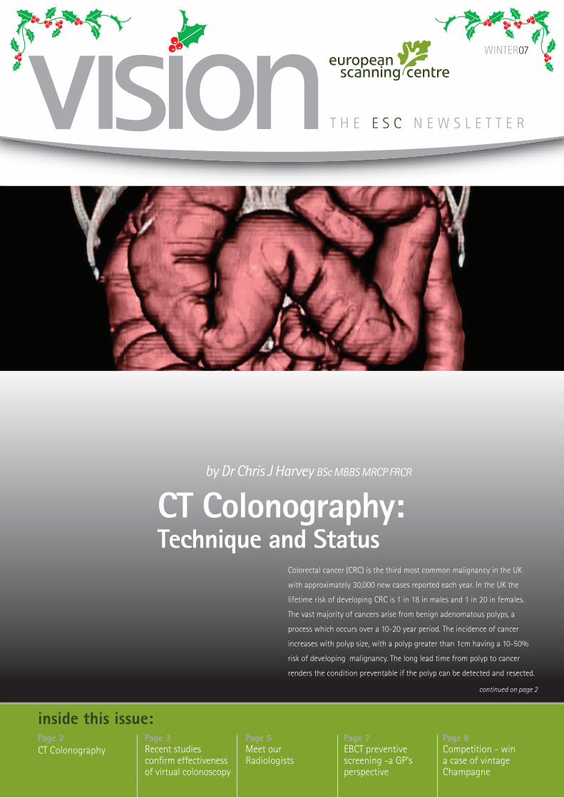
THE ESC NEWSLETTER inside this issue: Page 3 Recent studies confirm effectiveness of virtual colonoscopy Page 2 CT Colonography Page 5 Meet our Radiologists Page 8 Competition - win a case of vintage Champagne Page 7 EBCT preventive screening -a GP’s perspective CT Colonography: Technique and Status Colorectal cancer (CRC) is the third most common malignancy in the UK with approximately 30,000 new cases reported each year. In the UK the lifetime risk of developing CRC is 1 in 18 in males and 1 in 20 in females. The vast majority of cancers arise from benign adenomatous polyps, a process which occurs over a 10-20 year period. The incidence of cancer increases with polyp size, with a polyp greater than 1cm having a 10-50% risk of developing malignancy. The long lead time from polyp to cancer renders the condition preventable if the polyp can be detected and resected. continued on page 2 by Dr Chris J Harvey BSc MBBS MRCP FRCR WINTER07
Transcript of THE ESC NEWSLETTER - European Scanning Centre · colon is confirmed on a CT scanogram before the...
T H E E S C N E W S L E T T E R
inside this issue:Page 3Recent studies confirm effectiveness of virtual colonoscopy
Page 2CT Colonography
Page 5Meet our Radiologists
Page 8Competition - win a case of vintage Champagne
Page 7EBCT preventive screening -a GP’s perspective
CT Colonography: Technique and Status
Colorectal cancer (CRC) is the third most common malignancy in the UK
with approximately 30,000 new cases reported each year. In the UK the
lifetime risk of developing CRC is 1 in 18 in males and 1 in 20 in females.
The vast majority of cancers arise from benign adenomatous polyps, a
process which occurs over a 10-20 year period. The incidence of cancer
increases with polyp size, with a polyp greater than 1cm having a 10-50%
risk of developing malignancy. The long lead time from polyp to cancer
renders the condition preventable if the polyp can be detected and resected.
continued on page 2
by Dr Chris J Harvey BSc MBBS MRCP FRCR
WINTER07

page 2
administration of intravenous contrast. Scanning should be performed in
both supine and prone positions. This is to ensure that all segments of the
colon are well visualised as there may be positional collapse e.g the sigmoid
colon is commonly collapsed in the supine position but is well distended on
the prone view. Prior to the CT the patient follows a low residue diet for 48
hours and then one day before the CT scan patients undergo standard bowel
cleansing with purgatives. Immediately prior to the CT scan an intravenous
injection of 20mg of buscopan is administered to relax the colon and
prevent spasm. Rectal CO2 or air is then insufflated via a small Foley catheter
until a feeling of fullness is experienced. Adequate distension of the whole
colon is confirmed on a CT scanogram before the scan is commenced.
Scanning parameters vary but in a typical study would be performed at
3mm intervals, with overlap of slices. The data is then downloaded to
an independent workstation equipped with appropriate software for 3D
reconstruction. A computer generated retrograde intraluminal ‘fly through’
navigation from rectum to caecum can then be viewed and can be repeated
in the opposite direction.
The virtual colonoscopies (VC) are stored in a ‘cine loop’ format and can be
viewed directly from the workstation monitor. Image analysis is interactive,
and the radiologist can choose to view the rendered mucosa from any angle,
pass through the tightest stricture, and even cross the colonic wall into
adjacent structures. Virtual studies are usually examined in combination
with the standard axial images. The computer can also generate two-
dimensional images at cross sectional and orthogonal angles to the long
axis of the colon to aid interpretation.
Future developments are likely to include the use of oral contrast agents to
‘tag’ or label residual faeces and fluid with the aim of improving sensitivity
and specificity, while reducing the laxative regime. However the optimum
tagging/cathartic regimen is still controversial.
Another area of great interest is computer-aided detection (CAD) to
highlight areas of abnormalities, which the radiologist must then assess.
CAD has been shown to significantly increase sensitivity in the detection of
colonic abnormalities.
CT colonography in symptomatic patients and screening in asymptomatic individuals
Studies show that CT colonography surpasses barium enema and
approaches the sensitivity of conventional colonoscopy (3-5). Meta-analyses
show that CT colonography is both sensitive and specific for the detection
of large and medium polyps (Fig 1) and has a high reported diagnostic
accuracy for symptomatic cancer (Fig 2)(6). Results demonstrate a mean
sensitivity for the detection of cancer of 95.9%, sensitivity and specificity for
polyps greater than 1cm of 92.5% and 97.4%, respectively and for 5-9 mm
polyps of 86.4% and 86.1%, respectively.
Cost effectiveness and patient compliance are major determinants when
considering a feasible screening tool. Studies have shown that these are
Welcome to the Christmas edition of the European Scanning Centre newsletter. All of the team are delighted by the positive response we have received from the inaugural issue and hope you enjoy the latest edition.
In this issue, Dr Chris Harvey, Consultant GI Radiologist from the Hammersmith Hospital, discusses the pros and cons of virtual colonoscopy. Dr Alix Daniel then gives her perspective on using Electron Beam CT for preventive screening. We also put the spotlight on our radiological team, headed by Dr Sarah Howling, as well as our regular Ross’s Riddle.
In response to practitioners’ requests, we are happy to announce our new extended opening hours of 8am – 6pm Monday to Friday. We have also recently obtained recognition by AXA-PPP as a diagnostic imaging centre, which means we are now approved by all private insurance providers.
We welcome your feedback as always, but in the meantime we wish you a Merry Christmas and prosperous New Year.
Dr Paul Jenkins, MA MD FRCPMedical Director
CT Colonography: Technique and Status (continued from front cover)
From the Medical Director...
CT colonography was first described in 1994 and represents a fusion of thin
slice computed tomography of the large bowel with advanced techniques
for rendering three dimensional (3D) images to produce views of the colonic
mucosa, similar to those obtained during conventional colonoscopy, hence
its popular name of virtual colonoscopy. However, CT colonography also
provides exquisite 2D views of the colon in axial, coronal and sagittal planes.
It allows the whole colonic wall to be imaged rather than just the luminal
view as well as depicting extra-colonic abnormalities and the abdominal and
pelvic organs. CT colonography has evolved since 1994 and now has a strong
evidence based role in the investigation of symptomatic patients as well as a
screening tool for colorectal neoplasia in asymptomatic individuals. (1, 2)
Technique
A CT colon scan is performed during a single breath hold of 15-20 seconds
after bowel cleansing, optimal colonic distension, bowel paralysis and

page 3
favourable for CT colonography. Currently flexible sigmoidoscopy and faecal
occult blood tests are favoured as screening tools for the NHS whilst results
of larger CT colonography trials are awaited.
Radiation dose
Radiation dose remains a concern in mass VC screening. The effective dose
equivalent for multidetector (MD) CT colonography varies according to the
protocol performed. A recent study of simulated radiation dose in 64-slice
CT colonography demonstrated a dose range of 2.5-5.7mSv for males and
2.9-6.4mSv for females (a chest x-ray gives a dose of 0.06mSv, the annual
background radiation dose is 2.5-3mSv in the UK and the mean dose for a
barium enema is 7mSv) (7). Preliminary studies have suggested that a protocol
adjustment resulting in a dose reduction to 0.7mSv and 1.7mSv does not
impair sensitivity of detecting significant polyps (≥1cm) because of the
high contrast resolution between luminal air and bowel wall(8, 9). However,
the detection of extracolonic abnormalities will be reduced because of the
decrease in contrast resolution making noise a bigger problem.
Advantages of CT colonography compared to conventional colonoscopy
Although conventional colonoscopy is still considered the gold standard
for the detection of colonic neoplasia, a UK audit showed an overall caecal
intubation rate of only 56%(10) and expert back to back colonoscopies have
been shown to miss 24% of adenomas (11) but the majority of these were
less than 5mm in size. CT colonoscopy offers several advantages over the
conventional approach (Table 1).
continued on page 4
Recent results from two large screening trials demonstrate virtual
colonoscopy (VC) to be as sensitive in colonic polyp detection
compared to more invasive fibre-optic colonoscopy (OC). The US
National CT Colonography Trial performed on 2,531 asymptomatic
participants demonstrated an impressive per-patient sensitivity for
VC of 90% for adenomas 1cm or larger in diameter, a sensitivity
similar to OC.
The second study, published in the New England Journal of Medicine
(October 4, 2007), involving 6,283 subjects found comparable
detection rates between VC and OC for advanced adenomas (3.2%
and 3.4%, respectively) in two demographically similar asymptomatic
patient groups referred by the same primary care providers.
Interestingly, VC detected more cancers compared to OC, and
contrary to expectations, VC was also more effective at detecting
flat colonic polyps.
In both studies, the detection of
significant lesions by VC, which
required subsequent OC was 7.9%
and 8.3%, leading both study
investigators to claim that VC was
a suitable and often preferable
alternative to OC for colorectal
cancer screening.
Principal investigator Dr. Daniel Johnson from the Mayo Clinic hoped
that the results would encourage more individuals over 50 years of
age to get screened. “I think we can say that CT colonography is
similar to the performance of colonoscopy for large adenomas 1cm
or larger, as well as those intermediate adenomas 5-10mm in diameter.
I think it’s reasonable to consider broader application of this relatively
non-invasive imaging modality, which hopefully will enhance
compliance with colorectal cancer screening guidelines.”
Recent studies confirm effectiveness of virtual colonoscopy
Table 1: Advantages and disadvantages of CT virtual colonoscopy over traditional fibreoptic colonoscopy
Advantages• Quicker
• Safer - minimal risk of perforation; 0.03% vs. 0.13%
• No need for sedation
• Non-invasive
• Much higher success rate in visualising the caecum (>95%)
• Allows visualisation of entire colonic mucosa in any plane regardless of strictures/obstruction
• Visualises extracolonic structures
• Allows immediate staging of any cancer
• Non-operator dependent
Disadvantages
• Radiation exposure
• Cannot perform immediate polypectomy/biopsy
• Poor detection of ‘flat’ polyps (also with conventional colonoscopy)

page 4
Conclusion
CT colonography is a non-invasive,
safe, quick, well tolerated and
non-operator dependent sensitive
technique for examining the entire
colon. Its sensitivity and specificity
for colonic neoplasia exceeds that of
barium enema and approaches that
of conventional colonoscopy. It can
accurately indicate which patients
need to proceed onto a conventional
colonoscopy or polypectomy.
Further research is warranted to
fully assess its impact in terms
of a screening tool, acceptability,
availability and cost benefit.
References
1. Harvey CJ et al Eur Radiol 2001; 11: 1612-1625.
2. Tolan D et al. Clin Rad 2007; 62: 819-827.
CT Colonography: Technique and Status (continued from page 3)
Fig 1a.
Fig 1b.
Fig 2a.
3. Rockey D et al. Lancet 2005; 365: 305-311.
4. Pickhardt P et al. NEJM 2003; 349: 2191-200.
5. Cotton P et al. JAMA 2004; 291: 1713-9.
6. Halligan S et al. Radiology 2005; 237: 893-904.
7. Luz O et al. Eur Rad 2007; 17: 616-621.
8. Florie J et al. Eur Radiol 2007 June (Epub).
9. Innaccone R et al. Eur Radiol 2003; 13: 1297-1302.
10. Bowles C et al. Gut 2004; 53: 277-283.
11. Rex D et al. Gastroenterology 1997; 112: 24-28.
12. Yee J et al. Radiology 2005; 236: 519-526.
13. Burling D et al. Radiology 2006; 239: 464-471.
Figure Legends
Fig. 1a. - 64 year-old man with
rectal bleeding. The axial CT shows a
1.5 cm sigmoid polyp (arrow).
Fig 1b. - The same polyp is depicted
in axial, coronal and sagittal
muliplanar reformats as well as
3D. Its position in the colon is also
shown on the ‘fly through’ which
gives its distance from the anal
margin. The polyp was successfully
resected at colonoscopy and was
confirmed as a benign adenoma.
Fig 2a. - 58 year-old man with a
change in bowel habit. A colon
cancer is seen at the hepatic flexure
(arrow on 3D image) and is seen
as an applecore lesion on axial CT
image (bottom left image).
Dr Chris J Harvey BSc MBBS MRCP
FRCR Consultant Radiologist,
Hammersmith Hospital, London
The European Scanning Centre is delighted to announce
that we are now covered by AXA PPP insurance for
diagnostic imaging. We are also covered by all other
major insurers including:
BCWA BUPA
Clinicare Norwich Union
Pru Health WPA
European Scanning covered by AXA PPP
Consulting Room now availableLarge, recently refurbished consulting room available to rent at 68 Harley Street. For further information, please contact Dr Paul Jenkins on:020 7436 5755
New Opening HoursMonday - Friday 8:00am - 6:00pm
We are pleased to announce that the European Scanning Centre has extended its opening hours to better meet the needs of our referring doctors.

page 5
SPOTLIGHT... on our Radiologists
Why did you choose a career in radiology? I was inspired by Dr Mike Reubens who was a radiologist working at the London Chest Hospital and who always seemed so calm and happy sitting in his darkened room. I have also always been a bit of a gadget girl with regard to technology and liked the idea of peeping inside people and trying to piece together a set of clues.
Where did you train and how did your training prepare you for a career in radiology? My medical training was at The Royal Hospital, although I am not sure that this really did prepare me for my radiology career as technology really has marched on. We did not even have MRI scanners back then and a CT head scan took 20 minutes not 3 seconds as it does now. I continued my radiology training at UCLH with a chest fellowship in Vancouver.
What branch of radiology do you specialise in? My specialist interest is chest and cardiac imaging. In the last few years I have also gone a little northwards and now do dedicated thyroid and FNA lists.
What is your position at ESC and how long have you worked there? I am Director of Imaging and have been a Consultant at ESC for the last 4 years, heading up what I think is a fantastic team of radiologists and radiographers. In my NHS capacity, I am also the lead radiologist at the Whittington Hospital.
What role does the radiologist play in a patient’s day at ESC? Although the radiologist is often tucked away in that calm dark room, their responsibility starts with checking the patient is having the appropriate test to answer the clinical question and that it is safe to do so. We detail how we would like CTs and MRIs to be performed and with ultrasounds and biopsies we are hands on. We review the imaging before patients leave to check we have everything we need before composing the final report. If there are any rare complications we deal with them.
Once a patient is scanned, how and where are their images reported? Images are transferred in a few minutes to the powerful workstation
in our office. We then reconstruct them in multiple planes and do fly-throughs as necessary before dictating the final report.
How do the patients and referring clinicians receive the reported images? In essence, in any way they want! Some ask to be rung with the results, others prefer to have their reports faxed and others posted. One learns individual preferences and what each would want to know about fast.
How important is it for the radiographers and radiologists to work as a team? We are so lucky to have such a slick team that work brilliantly together, a bit like a grand prix pit team. It means that the radiographers feel confident to query any of our decisions or to suggest tweaks so as to get the best possible images to report. I remember my first night on call in 1992 was with these same radiographers and it is wonderful to be back with them as a team again.
How do you ensure that you provide a superior clinical service? Simple: We all aim to provide the service that we would wish a relative or colleague to receive.
What are the most technically difficult scans to report on? CT coronary angiograms as we have to look through about 800 images
Interview with ESC’s Director of Imaging, Dr Sarah Howling MB BS MRCP FRCR
and reconstruct along multiple vessels.
What do you enjoy most about working at ESC? It is wonderful to walk into such a happy place where I do not have the same volume of red tape and targets to meet as in the NHS. I can focus on my job and do what I am best at, which is hugely satisfying.
Where do you think radiology is going in the next 5 years? MRI is likely to get more functional and CT will develop in terms of quality of imaging and computer aided review. CT will also have an increasing cardiac role and radiologists will work more closely with cardiologists.
What do you see as the biggest challenges facing the field? Radiation dose certainly. There is increasing concern how this might affect future generations, as some of the doses we are generating on multidetector CT scans are really quite high.
What are the qualities of a good radiologist?Affable, time efficient, experienced, specialised and with a good eye. They need to be a good communicator, available and keen to stay up to date. They also need to give sensible further advice and have a good clear voice so as to keep their secretary happy.
Dr Christopher Harvey BSc MBBS MRCP FRCR
Chris is a Consultant Radiologist at the Hammersmith Hospital where
he leads their ultrasound service. He was integral to the early development of CT colonography in the UK from which he has multiple publications and years of expertise. He is our principle GI specialist but is also adept in all cross-sectional modalities and carotid ultrasound.
“I enjoy working as part of an innovative team”
Dr Christopher Schelvan BSc MBBS MRCP FRCR
Chris is a Consultant Radiologist at St Mary’s Hospital. He has numerous publications and is
Honorary Senior Lecturer at Imperial College. He has particular expertise in body cross-sectional imaging using his extensive experience in CT, MRI and ultrasound.
“ESC has an outstanding collection of staff who provide a friendly, efficient and top notch clinical service to patients and referring doctors. I’m proud to be part of the team”
Dr Jeevan Kumaradevan MB ChB MRCP FRCR
Jeevan is a Consultant Radiologist at the Whittington Hospital where he leads the
urology and vascular intervention radiology services. He trained at the Royal Free Hospital and undertook a prestigious Fellowship in Perth, Australia. He is reputed as being able to guide a needle into virtually anything and is renowned for his helpful nature. He provides our urological expertise.
“It is very satisfying to work as part of a highly select team, that aims to provide the highest possible quality of service to our referring clinicians and their patients.”

page 6
New StaffWe are delighted to announce
the recent appointments of
Julie White and Maria Sweeney
as our new radiology secretaries.
Both Julie and Maria will be
responsible for typing reports and
ensuring they are promptly
delivered to our referring doctors.
We are also happy to welcome
back Inder Bull, our Sales and
Marketing Manager, from
maternity leave. Inder looks
forward to working with you
all again in the near future.
Thursday 24th January 2008 from 6 - 8.30pm
Cavendish Conference Centre, 22 Duchess Mews, London W1
Due to popular demand, we have rescheduled and expanded our symposium programme on CT screening. The symposium will now include five talks on such topics as screening for coronary artery disease, lung cancer, colorectal cancer and functional abdominal imaging.
2 CPD points awarded.
Delegate spaces are available at £25 per person but early registration is advisable as numbers are limited. You can register using our online registration form at www.europeanscanning.com, by faxing the enclosed registration form or by contacting Caroline Metcalf on020 7436 5755.
Symposium on CT screening
Julie White
Maria Sweeney
How to refer patientsMany practitioners have asked for clarification on our booking
process. We hope you find the following guide helpful.
Patients can be referred to us in one of 3 ways:
1 Post/fax us a personalised letter on your headed paper
2 Post/fax us a completed ESC scan request form (available online at www.europeanscanning.com or by request
over the telephone)
3 Call us on 020 7436 5755 A completed scan request form can be faxed or sent with the
patient, by way of written confirmation.
In all cases, we will contact your patient directly to arrange a prompt
and convenient appointment.
Our team will work with you to tailor the ESC service to your
practice and preferences e.g. how, when and where you want
your patients’ reports delivered.

page 7
EBCT scanning has been part of both
my health screening activities and my
daily work as a general practitioner
over the past 4 years. It has helped
me to achieve personalised and
tailored medical prevention at a
level, which goes beyond my initial
expectations.
In terms of the EBCT heart scan, I
have been routinely proposing this
scan to low to medium risk patients
(5-20% risk in non-diabetic patients).
This has helped in several ways: first,
to detect high risk patients (who
were initially considered as mild-
moderate risk); second, to support my
recommendation for treatment with
a statin, when indicated, and third, to
reassure people who were suffering
from atypical chest pain but found to
be at low risk.
High-risk patients have gone on
to undergo further stress testing
and when indicated, subsequent
angioplasty. They are now on
“secondary preventive treatment” and
EBCT preventive health screening - a GP’s perspective
are all participating actively in the
management of their condition. One
of my patients, the husband of a chief
executive of a large company, was
reluctant to take a statin for his high
cholesterol, despite a family history of
coronary heart disease (CHD). He was
reluctant, because he felt well, was
exercising 4 times a week, and really
did not like the idea of taking a tablet
every single day of his life. The EBCT
scan revealed an elevated Agaston
score of 623 and a subsequent
exercise ECG was abnormal.
He underwent an angioplasty with
stenting of the anterior descending
coronary artery. Since then he
remains well, is taking his medicine
on a daily basis and exercises 5 days
a week.
I am also very impressed with the
way in which calcium scoring has
enabled low-medium risk patients to
engage in their preventive treatment
at an individual level. Prior to using
this technology, I was struggling to
persuade patients to take their blood
pressure tablets or statins regularly.
Having had an EBCT scan definitely
helps patients to understand the
link between blood pressure or
cholesterol and risk of heart attacks.
I have also referred low risk patients,
with atypical chest pain. A good
example of this type of referral was
one of my lady patients, aged 52,
who unfortunately lost her husband
the year before from complications
resulting from valve surgery. She
complained of atypical chest pain,
and I suspected this was part of
the bereavement process. She had
an exercise ECG, which came back
normal, but she still complained. I
referred her then for a heart scan.
This revealed a calcium score of zero.
This reassured her, and if I may say,
cured her from those chest pains.
EBCT scan technology can also be
used for screening for colorectal
cancer, or lung disease. I do not
have much experience in lung cancer
detection, and so I will comment only
on colon cancer screening and the
use of EBCT technology.
Confident of the now well-
established high sensitivity and
specificity of EBCT colon scans, I have
been using this technology alongside
conventional colonoscopy. All my
patients over 50 years of age or with
a family history of colon cancer are
offered either of the techniques.
EBCT colonoscopy has been mostly
used for repeat colonoscopy, or
for people on anticoagulants or
because of the patient’s personal and
informed choice.
A few weeks ago, a new patient, who
was asymptomatic, but was willing
to have a full executive medical,
underwent a virtual colonoscopy.
This revealed a large tumour of 3 cm.
He was subsequently colonoscoped
and operated on. Histology revealed
a Dukes A adenocarcinoma of the
sigmoid colon with no spread.
As you will understand, after having
incorporated the use of EBCT
scanning in my activities 4 years
ago, I am still enthusiastic about it. I
have to nonetheless admit that prior
to using this technology I have had
concerns, like many practitioners I
suppose. My main concern was to
end up doing, on occasion, more
harm than good. I do not like for
example to have a result back with an
incidental finding of a lesion in the
lung, kidney or liver, which could be
malignant, but for which the patient
has to wait sometimes 6 months for
a repeat scan and a definitive answer.
Despite my concerns, I have not
received any patient complaint about
this draw back of the technology. I
have indeed felt that my patients
accept this, as a minor inconvenience
which is acceptable as part of the full
process.
Dr Alix DanielGeneral Practitioner99 Harley Street

page 8
Ross’s Riddle
?In each issue 6 bottles of vintage Champagne will be offered to one lucky reader whose correct entry is drawn at random. Entries to be submitted by Friday 29th February, via email to: [email protected] The answer & name of the winner will be announced in the next edition.
Riddle 24 of Santa’s reindeer have to cross a narrow bridge in the dark. They have one torch between them, which must be used on the crossings. One or two can cross at a time but not more than two. One of them can cross in 1 minute, one in 2 minutes, one in 4 minutes and one in 10 minutes. What is the shortest elapsed time for them all to cross the bridge?
Moans without stonesA 21 year old female, engineering student, presented with a 2
year history of severe epigastric pain. The symptoms progressively
worsened, so that she was unable to continue her studies. The pain was
sharp and precipitated by eating. Endoscopy was reported to show a
small ulcer and an initial urease test was positive. Despite appropriate
antibiotic and high dose proton pump therapy, her symptoms persisted.
Further gastroenterological opinions suggested a psychosomatic
disorder, before a general physician investigated for gallbladder
dysmotility. Though previous imaging with abdominal ultrasound, CT
and MRCP confirmed normal anatomy and absence of any abnormality,
the gallbladder dynamic ultrasound stimulation test (DUST) at ESC (a
baseline scan followed by serial ultraound studies of the gallbladder
and common bile duct over an hour, after ingestion of a Mars
bar and 250ml of hot chocolate) demonstrated a markedly atonic
gallbladder (Fig 1). The fat stimulus precipitated a severe episode of her
symptoms, which were completely resolved by subsequent laparoscopic
cholecystectomy. Histology confirmed chronic acalculous cholecystitis.
Case Study • Case Study • Case Study
Electron Beam Computed Tomography (EBCT)
Abdominal & Pelvic Scan
Angiography (carotid artery, coronary and
peripheral)
Bone Mineral Density
Brain
Heart Scan (coronary artery calcium)
Lung Scan
Renal Tract
Sinuses
Virtual Colonoscopy
UltrasoundAbdominal Aorta
Abdomen & Pelvis
Carotid Doppler
Echocardiography
FNA Thyroid
Gallbladder (static and dynamic)
Leg Veins
Prostate (trans-abdominal)
Prostate (trans-rectal)
Renal Tract
Testes
Thyroid Gland
X-rayWe offer a complete
X-ray service
MRIBrain
Carotid Arteries
Elbows
Feet
Fingers
Musculo-skeletal
Neck
Shoulder
Spine
European Scanning Centre68 Harley StreetLondon W1G 7HE
Tel: 020 7436 5755Fax: 020 7436 5756
www.europeanscanning.com
European Scanning Centre
Scans
She is now resuming her
studies and has had no further
abdominal symptoms.
Comment: Mr Marcus Reddy,
Consultant GI surgeon, St.
George’s Hospital. Gallbladder
dysmotility is an under diagnosed
cause of abdominal pain. Patients
in whom routine investigations
have failed to elucidate a cause,
especially when biliary symptoms
are suspected, should undergo a
dynamic ultrasound stimulation
test. This test can demonstrate
the abnormal reponse of the
gallbladder and/or CBD to a large
fatty stimulation and also allows
correlation of symptoms with this
physiological stimulus.
Answer to Ross’s Riddle 1You have two strings, which will burn for one hour each, but they burn in an unpredictable way so that you cannot be sure that half the string would burn, say, in half an hour. Using the strings, how can you time exactly 45 minutes?You light both ends of one string and one end of the second string. When the first string burns out exactly half an hour has passed. You immediately light the other end of the second string and that will complete its burning in another 15 minutes, making 45 minutes in all.
The first correct answer drawn out of a hat was that from Dr Di Holdright, Consultant Cardiologist at The Heart Hospital, London.
Many congratulations from everyone at ESC to our lucky winner!
Patients
Time (minutes)
00 5 10 30 45 60
5
10
15
20
25
30
Gal
lbla
dder
vol
ume
(ml)
Normals (Kishk et al AJR 1987)
Fig 1


![Effect of inherent tibial asymmetry on leg length discrepancy ......limb length [2–5,14] and can be performed with low-dosage radiation protocols [17]. At our institution, a CT scanogram](https://static.fdocuments.us/doc/165x107/6020902ef337624017137e30/effect-of-inherent-tibial-asymmetry-on-leg-length-discrepancy-limb-length.jpg)