The Endocrine System
103
1 The Endocrine System Part A
-
Upload
brennan-chang -
Category
Documents
-
view
70 -
download
0
description
The Endocrine System. Part A. Endocrine System: Overview. Endocrine system – the body’s second great controlling system which influences metabolic activities of cells by means of hormones Endocrine glands – pituitary, thyroid, parathyroid, adrenal, pineal, and thymus - PowerPoint PPT Presentation
Transcript of The Endocrine System

1
The Endocrine System
Part A

Endocrine System: Overview
Endocrine system – the body’s second great controlling system which influences metabolic activities of cells by means of hormones
Endocrine glands – pituitary, thyroid, parathyroid, adrenal, pineal, and thymus
The pancreas and gonads produce both hormones and exocrine products

Endocrine System: Overview
The hypothalamus has both neural functions and releases hormones
Other tissues and organs that produce hormones – adipose cells, pockets of cells in the walls of the small intestine, stomach, kidneys, and heart

Major Endocrine Organs
Figure 16.1

Intercellular communication
Direct communicationThrough gap junctionsIons, small solutes, etcLimited to adjacent cells of the same type
Synaptic communicationNeurotransmittersUsed in crises management

Intercellular communication Autocrine
chemicals that exert effects on the same cells that secrete them
Paracrine locally acting chemicals that affect cells
other than those that secrete themCytokines or local hormonesThese are not considered hormones since
hormones are long-distance chemical signals

Intercellular communication Endocrine
Hormones – chemical substances secreted by cells into the extracellular fluidsRegulate the metabolic function of other
cellsHave lag times ranging from seconds to
hoursTend to have prolonged effects

Types of Hormones Amino acid based
Amines, Thyroxine,ADH Oxytocin GH Prolactin

Types of Hormones
Lipid derivativesSteroids – gonadal and adrenocortical
hormones Eicosanoids – leukotrienes and
prostaglandinsFunction as paracrine and autocrine
factors Function as hormones

Hormone Action Hormones that bind to receptors in the cell
membrane:First and second messengersRegulatory G proteinsWater-soluble hormones
Hormones that bind to intracellular receptors Direct gene activationSteroid and thyroid hormones
The precise response depends on the type of the target cell

Mechanism of Hormone Action
Hormones produce one or more of the following cellular changes in target cellsAlter plasma membrane permeability Stimulate protein synthesis Activate or deactivate enzyme systemsInduce secretory activityStimulate mitosis

Hormones that bind to receptors in the cell membrane: cAMP mechanism
Hormone (first messenger) binds to its receptor, which then binds to a G protein
The G protein is then activated as it binds GTP, displacing GDP
Activated G protein activates the enzyme adenylate cyclase
Adenylate cyclase generates cAMP (second messenger) from ATP
cAMP activates protein kinases, which then cause cellular effects


14
Receptor
Hormone A
ReceptorGTP GTP
GTP GTP GTP GTP
ATP cAMP
Inactive protein kinase A
Active protein kinase A
CatecholaminesACTHFSHLHGlucagonPTHTSHCalcitonin
Triggers responses of targetcell (activates enzymes,stimulates cellularsecretion, opens ionchannels, etc.)
Adenylate cyclase Hormone B
GDPGDP
Extracellular fluid
Cytoplasm
Gs Gi
1
2 34
3 2
1
5
Hormones that bind to receptors in the cell membrane : cAMP mechanism

Hormone binds to the receptor and activates G protein
G protein binds and activates phospholipase Phospholipase splits the phospholipid PIP2 into
diacylglycerol (DAG) and IP3 (both act as second messengers)
DAG activates protein kinases; IP3 triggers release of Ca2+ stores
Ca2+ (second messenger) alters cellular responses
Hormones that bind to receptors in the cell membrane: PIP-Calcium mechanism


GTP PIP2
IP3
ReceptorGTP
GTP
CatecholaminesTRHADHGnRHOxytocin
Triggers responses of target cell
GDP
Extracellular fluid
Cytoplasm
Inactiveprotein kinase C
Activeprotein kinase C
Phospholipase C
Gq
Ca2+ Ca2+- calmodulin
Hormone
Endoplasmicreticulum
DAG1
2 34 5
5
6
Hormones that bind to receptors in the cell membrane: PIP-Calcium mechanism
Catecholamines (α1 receptors)TRHADHGnRHOxytocin

Hormones that bind to intracellular receptors
Steroid HormonesThis interaction prompts DNA transcription
to produce mRNAThe mRNA is translated into proteins, which
bring about a cellular effect

19
Steroidhormone
Steroidhormone
Cytoplasm
Receptor-chaperonincomplex
Molecularchaperones
Receptor-hormonecomplex
Hormoneresponseelements
Binding
Transcription
Chromatin
mRNA
Nucleus
New proteinTranslation
Ribosome
mRNA

Target Cell Specificity
Hormones circulate to all tissues but only activate cells referred to as target cells
Target cells must have specific receptors to which the hormone binds
These receptors may be intracellular or located on the plasma membrane

Target Cell Specificity
Examples of hormone activityACTH receptors are only found on certain
cells of the adrenal cortexThyroxin receptors are found on nearly all
cells of the body

Target Cell Activation Target cell activation depends on three factors
Blood levels of the hormoneRelative number of receptors on the target
cellThe affinity of those receptors for the
hormone

Target Cell Activation
Up-regulation – when a hormone causes the target cells to form more receptors to it. It causes the tissue to become more sensitive to the hormone.
Down-regulation – when a hormone cause the target cells to lose receptors to it. It causes the tissue to become less sensitive to the specific hormone

Hormone Concentrations in the Blood
Hormones circulate in the blood in two forms: free or bound.Bound:
Attached to plasma proteinsBound to their own carriers

Hormone Concentrations in the Blood
Concentrations of circulating hormone reflect: Rate of releaseSpeed of inactivation and removal from the
body Hormones are removed from the blood by:
Degrading enzymesThe kidneysLiver enzyme systems

Interaction of Hormones at Target CellsPermissiveness – one hormone cannot exert its
effects without another hormone being present. Estrogen and thyroid hormone
Synergism – more than one hormone produces the same effects on a target cellGlucagon and epinephrine
Antagonism – one or more hormones opposes the action of another hormone. Glucagon and insulin

Control of Hormone Release
Blood levels of hormones: Are controlled by negative feedback
systemsVary only within a narrow desirable range
Hormones are synthesized and released by glands in response to:Humoral stimuliNeural stimuliHormonal stimuli

Humoral Stimuli
Humoral stimuli – secretion of hormones in direct response to changing blood levels of ions and nutrients
Example: concentration of calcium ions in the bloodDeclining blood Ca2+ concentration
stimulates the parathyroid glands to secrete PTH (parathyroid hormone)
PTH causes Ca2+ concentrations to rise and the stimulus is removed

Humoral Stimuli

Neural Stimuli
Neural stimuli – nerve fibers stimulate hormone releasePreganglionic
sympathetic nervous system (SNS) fibers stimulate the adrenal medulla to secrete catecholamines

Hormonal Stimuli
Hormonal stimuli – release of hormones in response to hormones produced by other endocrine organsThe hypothalamic hormones stimulate the
anterior pituitary In turn, pituitary hormones stimulate targets
to secrete still more hormones

Hormonal Stimuli

Nervous System Modulation
The nervous system modifies the stimulation of endocrine glands and their negative feedback mechanisms

Nervous System Modulation
The nervous system can override normal endocrine controlsFor example, control of blood glucose levels
Normally the endocrine system maintains blood glucose
Under stress, the body needs more glucose
The hypothalamus and the sympathetic nervous system are activated to supply ample glucose

Major Endocrine Organs: Pituitary (Hypophysis)
Pituitary gland – two-lobed organ that secretes nine major hormones
Neurohypophysis – posterior lobe (neural tissue) and the infundibulumReceives, stores, and releases hormones
from the hypothalamus Adenohypophysis – anterior lobe, made up of
glandular tissue Synthesizes and secretes a number of
hormones


Pituitary (Hypophysis)

Pituitary-Hypothalamic Relationships: Posterior Lobe
The posterior lobe is a downgrowth of hypothalamic neural tissue
Has a neural connection with the hypothalamus (hypothalamic-hypophyseal tract)
Nuclei of the hypothalamus synthesize oxytocin and antidiuretic hormone (ADH)
These hormones are transported to the posterior pituitary

Pituitary-Hypothalamic Relationships: Anterior Lobe
The anterior lobe of the pituitary is an outpocketing of the oral mucosa
There is no direct neural contact with the hypothalamus

There is a vascular connection, the hypophyseal portal system, consisting of:The primary capillary plexusThe hypophyseal portal veinsThe secondary capillary plexus
Pituitary-Hypothalamic Relationships: Anterior Lobe

Pituitary-Hypothalamic Relationships: Anterior Lobe

Adenophypophyseal Hormones
The six hormones of the adenohypophysis:Abbreviated as GH, TSH, ACTH, FSH, LH,
and PRLRegulate the activity of other endocrine
glands In addition, pro-opiomelanocortin (POMC):
Has been isolated from the pituitaryIs split into ACTH, opiates, and MSH

Activity of the Adenophypophysis
The hypothalamus sends a chemical stimulus to the anterior pituitaryReleasing hormones stimulate the synthesis
and release of hormonesInhibiting hormones shut off the synthesis
and release of hormones

Activity of the Adenophypophysis
The tropic hormones that are released are:Thyroid-stimulating hormone (TSH) Adrenocorticotropic hormone (ACTH)Follicle-stimulating hormone (FSH) Luteinizing hormone (LH)

Thyroid Stimulating Hormone (Thyrotropin)
Stimulates the normal development and secretory activity of the thyroid
Triggered by hypothalamic peptide thyrotropin-releasing hormone (TRH)
Rising blood levels of thyroid hormones act on the pituitary and hypothalamus to block the release of TSH

Adrenocorticotropic Hormone (Corticotropin)
Stimulates the adrenal cortex to release corticosteroids
Triggered by hypothalamic corticotropin-releasing hormone (CRH) in a daily rhythm
Internal and external factors such as fever, hypoglycemia, and stressors can trigger the release of CRH

Gonadotropins
Gonadotropins – follicle-stimulating hormone (FSH) and luteinizing hormone (LH)Regulate the function of the ovaries and
testesFSH stimulates gamete (egg or sperm)
productionAbsent from the blood in prepubertal boys
and girlsTriggered by the hypothalamic
gonadotropin-releasing hormone (GnRH) during and after puberty

Functions of Gonadotropins
In femalesLH works with FSH to cause maturation of
the ovarian follicleLH works alone to trigger ovulation
(expulsion of the egg from the follicle)LH promotes synthesis and release of
estrogens and progesterone

Functions of Gonadotropins
In malesLH stimulates interstitial cells of the testes to
produce testosteroneLH is also referred to as interstitial cell-
stimulating hormone (ICSH)

Growth Hormone (GH)
Produced by anterior pituitary gland and:Stimulate most cells, but target bone and
skeletal musclePromote protein synthesis and encourage
the use of fats for fuel Most effects are mediated indirectly by
somatomedins or IGFs

Growth Hormone (GH)
Antagonistic hypothalamic hormones regulate GHGrowth hormone–releasing hormone
(GHRH) stimulates GH releaseGrowth hormone–inhibiting hormone (GHIH)
or somatostatin inhibits GH release

Metabolic Action of Growth Hormone
GH stimulates liver, skeletal muscle, bone, and cartilage to produce insulin-like growth factors (IGFs)
Direct action promotes lipolysis and inhibits glucose uptake

Metabolic Action of Growth Hormone (GH)

Prolactin (PRL)
In females, stimulates milk production by the breasts
Triggered by the hypothalamic prolactin-releasing hormone (PRH)
Inhibited by prolactin-inhibiting hormone (PIH) Blood levels rise toward the end of pregnancy Suckling stimulates PRH release and
encourages continued milk production

The Posterior Pituitary and Hypothalamic Hormones
Posterior pituitary – made of axons of hypothalamic neurons, stores antidiuretic hormone (ADH) and oxytocin
ADH and oxytocin are synthesized in the hypothalamus
ADH decreases urine formation Oxytocin stimulates smooth muscle contraction
in breasts and uterus Both use PIP-calcium second-messenger
mechanism

56
The Endocrine System
P A R T B

Oxytocin
Oxytocin is a strong stimulant of uterine contraction
Regulated by a positive feedback mechanism to oxytocin in the blood
This leads to increased intensity of uterine contractions, ending in birth
Oxytocin triggers milk ejection (“letdown” reflex) in women producing milk

Oxytocin
Synthetic and natural oxytocic drugs are used to induce or hasten labor
Plays a role in sexual arousal and satisfaction in males and nonlactating females

Antidiuretic Hormone (ADH) ADH helps to avoid dehydration or water
overloadPrevents urine formation
Osmoreceptors monitor the solute concentration of the blood
With high solutes, ADH preserves water With low solutes, ADH is not released, thus
causing water loss Alcohol inhibits ADH release and causes
copious urine output

Thyroid Gland The largest endocrine gland, located in the
anterior neck, consists of two lateral lobes connected by a median tissue mass called the isthmus
Composed of follicles that produce the glycoprotein thyroglobulin
Colloid (thyroglobulin + iodine) fills the lumen of the follicles and is the precursor of thyroid hormone
Other endocrine cells, the parafollicular cells, produce the hormone calcitonin

Thyroid Gland

Thyroid hormone – major metabolic hormone Consists of two related iodine-containing
compoundsT4 – thyroxine; has two tyrosine molecules
plus four bound iodine atomsT3 – triiodothyronine; has two tyrosines with
three bound iodine atoms
Thyroid Hormone

Effects of Thyroid Hormone TH is concerned with:
Glucose oxidationIncreasing metabolic rate Heat production
TH plays a role in:Maintaining blood pressureRegulating tissue growthDeveloping skeletal and nervous systemsMaturation and reproductive capabilities

Synthesis of Thyroid Hormone Thyroglobulin is synthesized and discharged
into the lumen Iodides (I–) are actively taken into the cell,
oxidized to iodine (I2), and released into the lumen
Iodine attaches to tyrosine, forming T1 (monoiodotyrosine, or MIT), and T2 (diiodotyrosine, or DIT)

Synthesis of Thyroid Hormone
Iodinated tyrosines link together to form T3 and T4
Colloid is then endocytosed and combined with a lysosome, where T3 and T4 are cleaved and diffuse into the bloodstream

66
Iodine
T4
T4
T4
T3
T3
T3
T3T4
Lysosome
Capillary
Iodide(I–)
Thyroid follicle cell
Colloid in lumen of follicle
To peripheraltissues
T4
T3
Lysosomal enzymes cleaveT4 and T3 from thyroglobulincolloid and hormones diffusefrom follicle cell into bloodstream
Thyroglobulin colloidis endocytosed andcombined with alysosome
Iodinated tyrosines arelinked together to formT3 and T4
Thyroglobulincolloid
Iodine is attachedto tyrosine in colloid,forming DIT and MIT
Thyroglobulin is synthesizedand discharged into the follicle lumen
Iodide (I–)is trapped(actively transported in) DIT (T2) MIT (T1)
Colloid
Golgi apparatus
Rough ER
Iodide isoxidized to iodine
1
2 3a
5
6
3b
4

T4 and T3 bind to thyroxine-binding globulins (TBGs) produced by the liver
Both bind to target receptors, but T3 is ten times more active than T4
Peripheral tissues convert T4 to T3
Mechanisms of activity are similar to steroids Regulation is by negative feedback Hypothalamic thyrotropin-releasing hormone
(TRH) can overcome the negative feedback
Transport and Regulation of TH

A peptide hormone produced by the parafollicular, or C cells
Lowers blood calcium levels in children Antagonist to parathyroid hormone (PTH) Regulated by a humoral (calcium ion
concentration in the blood) negative feedback mechanism
Calcitonin

Calcitonin targets the skeleton, where it:Inhibits osteoclast activity (and thus bone
resorption) and release of calcium from the bone matrix
Increases calcium excretion by the kidneysStimulates calcium uptake and incorporation
into the bone matrix
Calcitonin

Parathyroid Glands
Tiny glands embedded in the posterior aspect of the thyroid
Cells are arranged in cords containing oxyphil and chief cells
Chief (principal) cells secrete PTH PTH (parathormone) regulates calcium
balance in the blood

Parathyroid Glands

PTH release increases Ca2+ in the blood as it:Stimulates osteoclasts to digest bone matrix Enhances the reabsorption of Ca2+ and the
secretion of phosphate by the kidneysIncreases absorption of Ca2+ by intestinal
mucosal Rising Ca2+ in the blood inhibits PTH release
Effects of Parathyroid Hormone

Effects of Parathyroid Hormone

Adrenal glands – paired, pyramid-shaped organs atop the kidneys
Structurally and functionally, they are two glands in oneAdrenal medulla – neural tissue that acts as
part of the SNSAdrenal cortex – glandular tissue derived
from embryonic mesoderm
Adrenal (Suprarenal) Glands

Adrenal Cortex Synthesizes and releases steroid hormones
called corticosteroids Different corticosteroids are produced in each
of the three layersZona glomerulosa – mineralocorticoids
(chiefly aldosterone)Zona fasciculata – glucocorticoids
(chiefly cortisol)Zona reticularis – gonadocorticoids
(chiefly androgens)

Adrenal Cortex

Regulate electrolytes in extracellular fluids Aldosterone – most important
mineralocorticoid Maintains Na+ balance by reducing excretion
of sodium from the bodyStimulates reabsorption of Na+ and
secretion of K+ by the kidneys
Mineralocorticoids
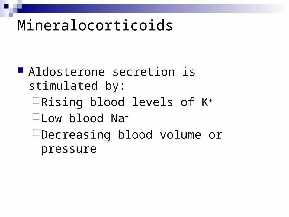
Aldosterone secretion is stimulated by:Rising blood levels of K+
Low blood Na+
Decreasing blood volume or pressure
Mineralocorticoids

The Four Mechanisms of Aldosterone Secretion
Renin-angiotensin mechanism – kidneys release renin, which is converted into angiotensin II that in turn stimulates aldosterone release
Plasma concentration of sodium and potassium – directly influences the zona glomerulosa cells
ACTH – causes small increases of aldosterone during stress
Atrial natriuretic peptide (ANP) – inhibits activity of the zona glomerulosa

Major Mechanisms of Aldosterone Secretion

Glucocorticoids (Cortisol)
Help the body resist stress by:Keeping blood sugar levels relatively
constantMaintaining blood volume and preventing
water shift into tissue Cortisol provokes:
Gluconeogenesis (formation of glucose from noncarbohydrates)
Rises in blood glucose, fatty acids, and amino acids

Excessive Levels of Glucocorticoids
Excessive levels of glucocorticoids:Depress cartilage and bone formationInhibit inflammationDepress the immune systemPromote changes in cardiovascular, neural,
and gastrointestinal function

Gonadocorticoids (Sex Hormones)
Zona Reticularis produces mainly androgens that are converted to testosterone in the testes
Androgens contribute to:The onset of pubertyThe appearance of secondary sex
characteristicsSex drive in females
Androgens can be converted into estrogens after menopause

Adrenal Medulla
Made up of cells that secrete epinephrine and norepinephrine
Secretion of these hormones causes:Blood glucose levels to riseBlood vessels to constrictThe heart to beat fasterBlood to be diverted to the brain, heart, and
skeletal muscle

Adrenal Medulla
Epinephrine is the more potent stimulator of the heart and metabolic activities
Norepinephrine is more influential on peripheral vasoconstriction and blood pressure

Stress and the Adrenal Gland

A triangular gland, which has both exocrine and endocrine cells, located behind the stomach
Acinar cells produce an enzyme-rich juice used for digestion (exocrine product)
Pancreatic islets (islets of Langerhans) produce hormones (endocrine products)
The islets contain two major cell types:Alpha () cells that produce glucagonBeta () cells that produce insulin
Pancreas

Pancreas

Its major target is the liver, where it promotes:Glycogenolysis – the breakdown of
glycogen to glucoseGluconeogenesis – synthesis of glucose
from lactic acid and noncarbohydratesRelease of glucose to the blood from liver
cells
Glucagon

Synthesized as part of proinsulin and then excised by enzymes, releasing functional insulin
Insulin:Lowers blood glucose levelsEnhances transport of glucose into body
cellsCounters metabolic activity that would
enhance blood glucose levels
Insulin

The insulin receptor is a tyrosine kinase enzyme
After glucose enters a cell, insulin binding triggers enzymatic activity that:Catalyzes the oxidation of glucose for ATP
productionPolymerizes glucose to form glycogenConverts glucose to fat (particularly in
adipose tissue)
Effects of Insulin Binding

Regulation of Blood Glucose Levels
The hyperglycemic effects of glucagon and the hypoglycemic effects of insulin

Results from hyposecretion or hypoactivity of insulin
The three cardinal signs of DM are:Polyuria – huge urine outputPolydipsia – excessive thirstPolyphagia – excessive hunger and food
consumption Hyperinsulinism – excessive insulin secretion,
resulting in hypoglycemia
Diabetes Mellitus (DM)

Diabetes Mellitus (DM)

Paired ovaries in the abdominopelvic cavity produce estrogens and progesterone
They are responsible for: Maturation of the reproductive organsAppearance of secondary sexual
characteristicsBreast development and cyclic changes in
the uterine mucosa
Gonads: Female

Testes located in an extra-abdominal sac (scrotum) produce testosterone
Testosterone:Initiates maturation of male reproductive
organsCauses appearance of secondary sexual
characteristics and sex driveIs necessary for sperm productionMaintains sex organs in their functional state
Gonads: Male

Small gland hanging from the roof of the third ventricle of the brain
Secretory product is melatonin Melatonin is involved with:
Day/night cyclesPhysiological processes that show rhythmic
variations (body temperature, sleep, appetite)
Pineal Gland

Thymus
Lobulated gland located deep to the sternum Major hormonal products are thymopoietins
and thymosins These hormones are essential for the
development of the T lymphocytes (T cells) of the immune system

Heart – produces atrial natriuretic peptide (ANP), which reduces blood pressure, blood volume, and blood sodium concentration
Gastrointestinal tract – enteroendocrine cells release local-acting digestive hormones
Placenta – releases hormones that influence the course of pregnancy
Other Hormone-Producing Structures

Kidneys – secrete erythropoietin, which signals the production of red blood cells
Skin – produces cholecalciferol, the precursor of vitamin D
Adipose tissue – releases leptin, which is involved in the sensation of satiety, and stimulates increased energy expenditure
Other Hormone-Producing Structures

Developmental Aspects
Exposure to pesticides, industrial chemicals, arsenic, dioxin, and soil and water pollutants disrupts hormone function
Sex hormones, thyroid hormone, and glucocorticoids are vulnerable to the effects of pollutants
Interference with glucocorticoids may help explain high cancer rates in certain areas

Developmental Aspects
Ovaries undergo significant changes with age and become unresponsive to gonadotropins
Female hormone production declines, the ability to bear children ends, and problems associated with estrogen deficiency (e.g., osteoporosis) begin to occur
Testosterone also diminishes with age, but effect is not usually seen until very old age

Developmental Aspects
GH levels decline with age and this accounts for muscle atrophy with age
Supplemental GH may spur muscle growth, reduce body fat, and help physique
TH declines with age, causing lower basal metabolic rates
PTH levels remain fairly constant with age, and lack of estrogen in women makes them more vulnerable to bone-demineralizing effects of PTH