THE EFFECTS OF SUBSTANCE P AND BACLOFEN ON … · neurones of the cat, for example, Henry et al....
14
J. exp. Biol. (1980), 89, 201-214 2OI With 10 figures Printed in Great Britain THE EFFECTS OF SUBSTANCE P AND BACLOFEN ON MOTONEURONES OF ISOLATED SPINAL CORD OF THE NEWBORN RAT BY MASANORI OTSUKA AND MITSUHIKO YANAGISAWA Department of Pharmacology, Faculty of Medicine, Tokyo Medical and Dental University, Bunkyo-ku, Tokyo 113, Japan SUMMARY The effects of substance P (SP) and baclofen were studied in the isolated spinal cord of newborn rats. Potential changes generated in motoneurones were recorded extracellularly from the ventral root (L3-L5). When SP (8 x io~ 8 M) was introduced into the bath, the depolarization of motoneurones began with a delay of I-I s. A large part of this delay can be explained as a time needed for SP to reach the site of action on spinal neurones. When the preparation was perfused with artificial cerebrospinal fluid (CSF) containing low Ca (o-i ITIM) and high Mg (i - 6-3*5 mia), the spinal reflexes induced by dorsal root stimulation and recorded from the corre- sponding ventral root were completely abolished. The depolarizing action of SP (io~ 7 M) on the motoneurones was potentiated in the low-Ca medium, suggesting that SP acts directly on the motoneurones. Baclofen at io" 6 M depressed the monosynaptic reflex by about 75%. The SP-induced depolarization of motoneurones was greatly depressed by bac- lofen in both normal and o-i mM-Ca mediums. The effects of baclofen (io~ s M) on the responses to various depolarizing agents were compared with that on the response to SP in artificial CSF containing o-i mM-Ca and 1-6-2 mM-Mg. The SP response was reduced by about 80%, whereas the re- sponses to acetylcholine and glycine were not appreciably affected, and those to L-glutamate, GABA and noradrenaline were depressed by 10-22% by baclofen. These results suggest that baclofen blocks transmission at certain primary afferent synapses by antagonizing the action of SP that is released as a transmitter. INTRODUCTION Although the hypothesis that substance P (SP) is an excitatory transmitter of certain primary afferent neurones is supported by abundant neurochemical and immunohistochemical evidence (Cuello et al. 1978; Hokfelt et al. 1975; Otsuka & Takahashi, 1977), relatively little is known about the mode of action of SP at central synapses. As yet, SP-mediated synaptic potentials have not been demonstrated. It is therefore of crucial importance to study the action of SP at the synapses where its transmitter role is anticipated. The isolated spinal cord of the newborn rat (Otsuka & Konishi, 1974) provides an excellent opportunity for studying the action of SP on central neurones, because the peptide can be applied in precisely controlled con- centrations, and its effects on spinal motoneurones can be observed stably with either
Transcript of THE EFFECTS OF SUBSTANCE P AND BACLOFEN ON … · neurones of the cat, for example, Henry et al....

J. exp. Biol. (1980), 89, 201-214 2OIWith 10 figures
Printed in Great Britain
THE EFFECTS OF SUBSTANCE P ANDBACLOFEN ON MOTONEURONES OF ISOLATED SPINAL
CORD OF THE NEWBORN RAT
BY MASANORI OTSUKA AND MITSUHIKO YANAGISAWA
Department of Pharmacology, Faculty of Medicine, Tokyo Medical andDental University, Bunkyo-ku, Tokyo 113, Japan
SUMMARY
The effects of substance P (SP) and baclofen were studied in the isolatedspinal cord of newborn rats. Potential changes generated in motoneuroneswere recorded extracellularly from the ventral root (L3-L5). When SP(8 x io~8 M) was introduced into the bath, the depolarization of motoneuronesbegan with a delay of I-I s. A large part of this delay can be explained as atime needed for SP to reach the site of action on spinal neurones.
When the preparation was perfused with artificial cerebrospinal fluid(CSF) containing low Ca (o-i ITIM) and high Mg (i-6-3*5 mia), the spinalreflexes induced by dorsal root stimulation and recorded from the corre-sponding ventral root were completely abolished. The depolarizing actionof SP (io~7 M) on the motoneurones was potentiated in the low-Ca medium,suggesting that SP acts directly on the motoneurones.
Baclofen at io"6 M depressed the monosynaptic reflex by about 75%. TheSP-induced depolarization of motoneurones was greatly depressed by bac-lofen in both normal and o-i mM-Ca mediums. The effects of baclofen(io~s M) on the responses to various depolarizing agents were compared withthat on the response to SP in artificial CSF containing o-i mM-Ca and1-6-2 mM-Mg. The SP response was reduced by about 80%, whereas the re-sponses to acetylcholine and glycine were not appreciably affected, and thoseto L-glutamate, GABA and noradrenaline were depressed by 10-22% bybaclofen. These results suggest that baclofen blocks transmission at certainprimary afferent synapses by antagonizing the action of SP that is releasedas a transmitter.
INTRODUCTION
Although the hypothesis that substance P (SP) is an excitatory transmitter ofcertain primary afferent neurones is supported by abundant neurochemical andimmunohistochemical evidence (Cuello et al. 1978; Hokfelt et al. 1975; Otsuka &Takahashi, 1977), relatively little is known about the mode of action of SP at centralsynapses. As yet, SP-mediated synaptic potentials have not been demonstrated. It istherefore of crucial importance to study the action of SP at the synapses where itstransmitter role is anticipated. The isolated spinal cord of the newborn rat (Otsuka &Konishi, 1974) provides an excellent opportunity for studying the action of SP oncentral neurones, because the peptide can be applied in precisely controlled con-
centrations, and its effects on spinal motoneurones can be observed stably with either

202 M. OTSUKA AND M. YANAGISAWA
intra- or extracellular recording (Konishi & Otsuka, 1974). Immunohistochemicalstudies have shown that spinal motoneurones are surrounded by SP-containingvaricosities (Barber et al. 1979; Hokfelt et al. 1975, 1977), suggesting that some SP-mediated synapses are located on spinal motoneurones. Furthermore, recent studiesby Jessell et al. (1979) showed that the SP content in the motoneurone area of the ratspinal cord is considerably reduced after dorsal rhizotomy. Therefore, some of theSP-containing terminals on the motoneurones may originate from primary afferentneurones, although other terminals seem to have a supraspinal origin (Hokfelt et al.1977; Kanazawa et al. 1979).
In the present study we therefore studied the effects of SP on the motoneuronesof isolated rat spinal cords. We focused attention particularly on two aspects: thetime course of SP action and the antagonism of baclofen to SP. Many authors usingelectrophoretic application techniques reported that the time course of the action ofSP on central neurones is characteristically slow and long-lasting (Henry, Krnjevic" &Morris, 1975; Krnjevid & Morris, 1974; Phillis & Limacher, 1974). In the spinalneurones of the cat, for example, Henry et al. (1975) reported that the excitant actionof SP appeared with a delay of 15-30 s and lasted for 1-2 min after the applicationwas stopped. In these experiments, however, the slow time course of the SP actionmight reflect the delayed ejection of the peptide from the micropipette (Guyenetet al. 1978). Concerning the effect of baclofen, Saito, Konishi & Otsuka (1975)reported that the SP-induced depolarization of rat spinal motoneurones was antagon-ized by baclofen [y?-(4-chlorophenyl)-y-aminobutyric acid], whereas the glutamate-induced depolarization was affected much less by the drug. However, later studiesusing electrophoretic techniques could not confirm the specificity of antagonism be-tween baclofen and SP (Henry & Ben-Ari, 1976; Phillis, 1976), and it was assumedthat baclofen exerts a general depressant action on central neurones (Johnston, 1978).We have therefore re-examined the effects of baclofen on the actions of SP and otherdrugs. A preliminary report of this work has been published (Otsuka & Yanagisawa,1978).
METHODS
Preparation. Wistar rats of 0-3 days of age were used. Under ether anesthesia, thespinal cord together with the vertebral column was removed and placed in a dish filledwith oxygenated physiological solution at room temperature. The vertebral canal wasopened under a dissecting microscope, and the spinal cord below Th 10 was isolatedtogether with the L3-L5 ventral and dorsal roots. The cord was hemisected and asmuch as possible of the pia mater of the lumbar portion was removed. In manyexperiments the spinal cord was treated with collagenase (Sigma, 0-05 mg/ml) for30 min to facilitate the removal of the pia mater.
Physiological solution. Artificial cerebrospinal fluid (CSF) of the following composi-tion was used (mni): NaCl 138-6; KC1 3-35; CaCla 1-26; MgCl2 1-16; NaHC03 21-0;NaH2PO4 0-58; glucose 10; gassed with 95% 0 2 and 5% C02 (cf. Feldberg &Fleischhauer, i960). In some experiments, CaCl2 and MgCl2 concentrations werechanged as described below. The temperature of perfusion fluid was kept at 27 °C.
Perfusion system. As shown in Fig. 1 A, the isolated spinal cord was placed in a0-2 ml bath and perfused with oxygenated artificial CSF at a rate of 3-8 ml/min. Foa

Substance P and baclofen 203
applying drugs for short periods, the perfusion system shown in Fig. 1C was used innnost experiments. The drug solutions were contained in syringes a and b, which wereconnected to syringe c filled with air and partly with perfusion solution. Since thereservoirs of perfusion fluids were placed at the height of about 1 -3 m, there was aconstant pressure in the syringes a, b and c. By the use of a multi-channel stimulatorand electromagnetic valves, the drug solutions could be applied for a predeterminedduration (O-I-IO s) at constant intervals. When cock d was opened and cock e was
SuctionGas
Fluid in Spinal cordFig. i. Experimental set up. A, Isolated spinal cord of newborn rat and the perfusion bath.B, Extracellular recording electrode. C, Perfusion system for applying drugs. For details seetext.
closed, the drug solution in syringe a flowed into the perfusion system. A quite repro-ducible amount of drug solution could be introduced into the bath and thus theconstant size of responses could be recorded at relatively short intervals (1-6 min)for a long period (Figs. 7-10). When it was necessary to determine exactly the timeat which the test solution entered the bath, an air bubble was placed in the tubemarked / in Fig. 1 C, so that the test solution flow was preceded by the air bubble.Electrical resistance was recorded between the steel tubing g and the bath. When theair bubble passed the steel tubing, the resistance suddenly increased and then, whenthe air bubble entered the chamber, it decreased, thus indicating the time of entranceof the test solution (Fig. 2).
Electrodes. For stimulating the dorsal or ventral root, a suction electrode with a tipinner diameter of about 300/im was used. For extracellular recordings from theventral root, the electrode shown in Fig. 1B was used. The outer barrel of the elec-trode had a tip inner diameter of about 500 fim and was filled with a mixture of liquidparaffin and Vaseline (about 1:1). The level of the liquid paraffin-Vaseline mixturecould be controlled by applying pressure with the syringe connected to the outerbarrel. The inner barrel was a suction electrode filled with physiological solution andconnected through an Ag-AgCl wire to a preamplifier. The electrode was connectedthrough polyethylene tubing to a short piece of glass capillary with a fire-polishedorifice that had an inner diameter of 100-150/tm, depending on the size of thefcentral root. The relative positions of inner and outer barrels could be adjusted by a

204 M. OTSUKA AND M. YANAGISAWA
micromanipulator, and the whole assembly of the recording electrode was mounted!on another micromanipulator. To get good responses to SP, it was important to dissecrthe ventral root close to the surface of the spinal cord and to use a capillary at the tipof the inner barrel that fitted tightly around the ventral root. After the ventral rootwas sucked into the inner barrel, the outer barrel was lowered and a short distance(about i mm or leas) of the ventral root was insulated by the mixture of paraffin andVaseline (Fig. i B). The bath was grounded through a calomel electrode. The pre-amplifier was connected to an oscilloscope, and through a transient memory circuitto a pen recorder.
In a few experiments, intracellular recordings were made from spinal motoneurones.The motoneurones were identified by antidromic action potentials induced by ventralroot stimulation. Microelectrodes filled with 2 M-K-acetate with resistance of 50-120 MQ were used.
5s
2mV
_r\Fig. 2. Time course of actions of 8 x io~*M SP(A), io~* M L-glutamate (B) and 30 mM-KCl (C).Potentials were recorded extracellularly from L4 ventral root of isolated spinal cord of 1-day-old rat. Upper traces show the potential recorded from the ventral root in normal artificialCSF. Upward deflexion shows the depolarization in this and following figures. Lower tracesmonitor the resistance between the steel tubing (g) and the bath as shown in Fig. iC. In eachrecord, the chamber was flushed with the drug solution during the period indicated by hori-zontal black bar, and arrow indicates the beginning of depolarization.
RESULTS
Time course of SP action. When SP in a concentration of 8 x icrB M was introducedinto the chamber, the depolarization of motoneurones began with a delay of I-I +o-i sec (mean + s.E.M., n = 4; Fig. 2 A). Similarly, the depolarizations induced byL-glutamate(io~*M)and KCl(3omM) appeared with delays of 0-4 + 0-1 ando-2±o-i 8,respectively (mean±S.E.M., n = 4; Fig. 2B and C). In the experiment shown inFig. 3 A, a lower concentration of SP (iO"6 M) was applied for about 5 s in artificialCSF containing i-26mM-Ca and 3-5 mM-Mg. The depolarization appeared with adelay of about 3 s. The time from onset to peak and the half decay time were 14 and9 s, respectively.
In order to examine whether the SP-induced depolarization is due to direct or trana-synaptic action on the motoneurones, SP (icr* M) was applied in artificial CSF con-taining o-i mM-Ca and 3-5 mM-Mg, where spinal reflexes induced by dorsal n

Substance P and baclofen 205
Rimulation were completely blocked (cf. Fig. 4). As shown in Fig. 3 B, the size andie time course of SP-induced depolarization were similar to those in the 1-26 mM-
Ca, 3-5 mM-Mg medium. Since the distance between the surface of the spinal cordand the motoneurone area is more than 100 fim, it is likely that the time course of thedepolarization of motoneurones in the low-Ca medium reflects the change of SP
10s
1 mV
Fig. 3. Time course of the depolarizing responses to SP. Extracellular recordings from theventral root in this and all subsequent figures. (A) In artificial CSF containing 1-26 mM-Caand 35 mM-Mg; and (B) in the medium containing o-i mM-Ca and 3-5 mM-Mg. SP solutions(10"* M) were applied to the bath during the periods indicated by horizontal black bars.
- 6
1-5
I 10
M 0-5
>B
Io3
0-1 0-3 0-6 1-26 4
Ca concentration (ITIM)
Fig. 4. Effects of Ca concentrations on the size of monosynaptic reflex ( • ) , SP-induced de-polarization (O), and L-glutamate-induced depolarization (A). Vertical lines represent S.E.M.(n — 2-4). Potential changes were recorded from L4 ventral root and the spinal reflexes wereinduced by a single supramaxima! volley in the corresponding dorsal root with intervals ofu s . SP (io~* M) and L-glutamate (2 x io"4 M) were applied for 30 s periods. Mg concentrationwas kept at 2 mM. Abscissa: logarithmic scale.
concentration at the site of action on motoneurones (Fig. 3 B). The rising phase ofSP-induced depolarization in the 1-26 mM-Ca medium was steeper than that in theo-i mM-Ca medium, suggesting that SP activates excitatory interneurones in additionto its direct action on motoneurones.
That SP exerts a direct depolarizing action on motoneurones is further supported bythe results shown in Fig. 4. When the Ca concentration in the medium was changedfrom 4 to o-i mM, keeping the Mg concentration at 2 mM, the spinal reflexes induced
dorsal root stimulation and recorded from the corresponding ventral root were

206 M. OTSUKA AND M. YANAGISAWA
1-5
9E
1-0
8. 0-5
5
10" 10" 10"1 10"6 10"5
Concentration (u)10" 10"
Fig. 5. Effects of T T X on SP-induced and L-glutamate-induced depolarizations. SP and L-glutamate were applied to the bath during the periods of 30 s in concentrations shown inabscissa. O> • , Responses to SP; A, A. responses to L-glutamate. Open symbols indicatethe responses in artificial CSF containing o-i mM-Ca and i-6 mM-Mg and closed symbolsindicate those after the addition of T T X (1-3x10- ' M).
Baclofen1(T*M 2 X 10"* M
2mV
9 0
Time (min)
Fig. 6. Effects of baclofen on the sizes of monosynaptic reflex. Spinal reflexes were induced by asingle supramaximal volley in L4 dorsal root with intervals of 11 s and recorded extra-cellularly from L4 ventral root. Baclofen was applied to the bath in three different concentra-tions during the periods marked in the figure. Inset shows an example of the record of spinalreflexes.
greatly depressed in o-6 mM-Ca and abolished in 0-3 mM-Ca medium. In contrast,both SP-induced and L-glutamate-induced depolarizations were larger in o-i and0-3 mM-Ca solutions than those in 1-26 mM-Ca solution. To confirm further that thesynaptic transmission in the spinal cord is blocked in low-Ca medium, intracellularrecordings were made from motoneurones. When the perfusion medium was changedfrom normal artificial CSF to the medium containing 0-2 mM-Ca and 2 mM-Mg, theexcitatory postsynaptic potentials (EPSPs) induced by repetitive stimulation (10stimuli at ico Hz) of the dorsal root of the corresponding segment werecompletely abolished in 5-10 min.

Substance P and baclofen 207
^ W e also studied the effects of tetrodotoxin (TTX), which is another means to block™naptic transmission. When the isolated rat spinal cord was perfused with artificialCSF containing TTX (1-3 x io~7 M), the spinal reflexes were completely abolished.Under such conditions, both SP- and glutamate-induced depolarizations were greatlydepressed. The depressant effects of TTX on SP- and glutamate-induced responseswere observed, however, not only in normal artificial CSF but also in the o-i mM-Camedium (Fig. 5), suggesting that TTX acts directly on the motoneurones (seeDiscussion).
1-5
1-0
0-5
Normalartificial CSF
O l mM-Ca, 2 mM-Mg
10 20 30 40Time (min)
50 60 70
Fig. 7. Effects of baclofen (IO~*M) on the SP-induced responses in normal and low-Camediums. SP (6 x io"' M) was applied with pulses of 3 8 duration with intervals of 5 min. Thedepolarizing responses were recorded from L4 ventral root. The preparation was first perfusedwith normal artificial CSF and then with the medium containing o-i mM-Ca and 2 mM-Mg.Baclofen was added during the periods indicated by horizontal black bars.
Effects of baclofen. When one of the dorsal roots (L3-L5) is stimulated by a singleshock, the response recorded from the corresponding ventral root consists of an earlyspike followed by later asynchronous waves as illustrated in the inset in Fig. 6. Webelieve that the initial spike corresponds to the monosynaptic reflex, based on thefollowing observation. When the nerve innervating the medial or lateral gastrocnemiusmuscle was stimulated and the potential was recorded intracellularly from homo-nymous motoneurones of newborn rats, a large synchronous EPSP could be recorded(Konishi & Otsuka, unpublished observation). Fig. 6 illustrates the effect of baclofenon the size of the monosynaptic reflex. In agreement with earlier observations (Pierau& Zimmermann, 1973; Saito et al. 1975) baclofen blocked the spinal monosynapticand polysynaptic reflexes. Baclofen at IO~6M reduced the size of the monosynapticreflex by about 75%. The full effect was reached within a few minutes, and afterwashing, the effect disappeared with a similarly rapid time course. Intracellularrecordings from motoneurones revealed that baclofen (io"6 M) reduced the size ofthe EPSP induced by single-shock stimulation of the dorsal root by about 60%without affecting appreciably the size of the resting potential or the antidromic action
«tential (not illustrated).In the experiment shown in Fig. 7, the responses to SP were induced by applying

208 M. OTSUKA AND M. YANAGISAWA
SP solution (6 x icr7 M) with short pulses of 3 s duration in normal artificial CSH|Addition of io~* M baclofen to the medium reduced the size of SP response by aboH70% and complete recovery occurred within 10 min after washing. The preparationwas then perfused with artificial CSF containing o-i mM-Ca and 2 mM-Mg, wherethe SP response usually became larger. The depressant effect of baclofen on the SPresponse was even more marked in the low-Ca medium, suggesting that baclofen actsdirectly on motoneurones.
co
3
8.
A
1-0
0-5
0
Baclofen_ n
B 1 /
Lc
Baclofenn
ii i
Dihydrc-0-erythroidine
1 1
I Atropine |
\ri i
100s
0-5 mV
20 40 60
Time (min)
80 100 SP ACh
Fig. 8. Effects of baclofen (io~* M), atropine (1-4x10"'M) and dihydro-/?-erytHroidine(4 x io~* M) on the responses to SP and ACh. SP (3 x io"' M) and ACh (3 x io"4 M) were alter-nately applied with pulses of 3 and 2-2 s durations respectively. The interval between SPapplications was 5 min, and ACh was applied 100 s after each SP application. The preparationwas perfused with artificial CSF containing 01 mM-Ca, 2 mM-Mg and 3 x io"1 M prostigmine.Baclofen, artropine and dihydro-/?-crythroidine were added to the medium during the periodsindicated in the figure. (A), O, The response to SP; # , the response to ACh. (B-D) The samplerecords in control solution, after the addition of baclofen, and after the addition of atropine anddihydro-/?-erythroidine; the times when the records (B), (C) and (D) were taken are shown byarrows in (A). In each record the first response was induced by SP and the second wasinduced by ACh.
To test whether the antagonism between baclofen and SP is selective or not, theeffects of baclofen on other agonists were examined. As a routine procedure, eachagonist that produced depolarization of motoneurones was given by pulse applicationwhile the preparation was perfused with artificial CSF containing O'imM-Ca andhigh Mg (i-6-2-omM). In most experiments baclofen was given in a concentrationof io"6 M, and the responses to SP and one of the agonists were alternately recordedat intervals of 1-6 min. In choosing the durations of pulses for applying SP and otheragonists, care was taken so that two alternately given drugs gave approximately samesizes of submaximal responses.
In the experiment shown in Fig. 8, acetylcholine (ACh) and SP were appliedalternately in the presence of prostigmine (3 x io~8 M). After adding baclofen, SPresponse was reduced by about 90% whereas the response to ACh was reduced byonly 6-8%. On the other hand, when the preparation was perfused with the
Substance P and baclofen 209
1-5 - A
1-0co•a
0-5
5 10 15 0 5 10 15Time (min)
10 15 20
Fig. 9. Effects of baclofen (io~* M) on the responses to SP, glycine, GABA and L-glutamate.The responses to SP and to one of the three amino acids were recorded alternately in artificialCSF containing 01 mM-Ca and 2 mM-Mg. Baclofen (10"* M) was added during the periodindicated by horizontal black bars. O, Response to SP; • , response to glycine in A, to GABAin B, and to L-glutamate in C. The concentrations and the periods of application of drugs wereas follows: SP 3 x io~r M, 3 s; glycine io"8 M, 1 s; GABA 2 x io~* M, 1-5 s; and L-glutamateio~* M, 2#8 8. The interval between SP applications was 3 min, and one of the amino acids wasapplied 100 s after each SP application.
2min
BaclofenlmV
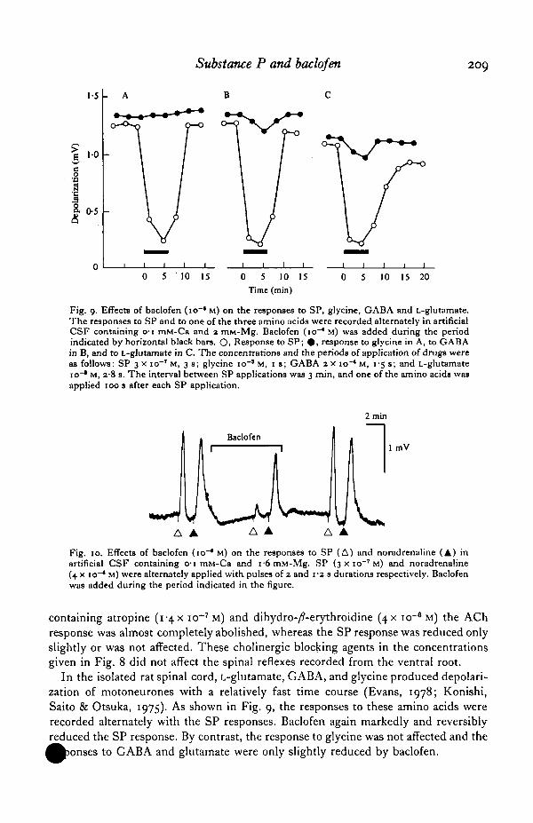
Fig. 10. Effects of baclofen (io~* M) on the responses to SP (A) and noradrenaline (A) inartificial CSF containing 01 mM-Ca and 1-6 mM-Mg. SP (3 x 10"' M) and noradrenaline(4 x io"4 M) were alternately applied with pulses of 2 and 1 -2 s durations respectively. Baclofenwas added during the period indicated in the figure.
containing atropine (i"4X io~7 M) and dihydro-/?-erythroidine (4X io"6 M) the AChresponse was almost completely abolished, whereas the SP response was reduced onlyslightly or was not affected. These cholinergic blocking agents in the concentrationsgiven in Fig. 8 did not affect the spinal reflexes recorded from the ventral root.
In the isolated rat spinal cord, L-glutamate, GABA, and glycine produced depolari-zation of motoneurones with a relatively fast time course (Evans, 1978; Konishi,Saito & Otsuka, 1975). As shown in Fig. 9, the responses to these amino acids wererecorded alternately with the SP responses. Baclofen again markedly and reversiblyreduced the SP response. By contrast, the response to glycine was not affected and the
to GABA and glutamate were only slightly reduced by baclofen.

210 M. OTSUKA AND M. YANAGISAWA
Table i. Effects of baclofen (io"6 M) on responses to SP and variousdepolarizing agents
(Experiments were carried out in artificial CSF containing o-i mM-Ca and 1-6-2-0 mM-Mg.Extracellular recordings from the ventral root. Prostigmine (3 x io~* M) was added when AChresponses were recorded. In most experiments, the responses to SP and one of the depolar-izing drugs were alternately recorded as shown in Figs. 8-10. Intervals of application of drugswere 1—6 min.)
Pulse DepressionConcentration duration by baclofen*
Drug (M) (s) (%) n
Substance PEledoisin-related pep-tide
L-GlutamateGABAGlycineAcetylcholineNoradrenaline
3 x 10-'5 x io~7
10"*2 X IO"4
10"'3X io-*
io-4
•
1-4
3
0-7-3-50-5-1-50-2-1-52-3i - 3
Mean±s.E.M.n, Number of experiments.
83±ro82
i8±2-710+ 1-5
0-7 ±0-45±2-i
22 ±4-4
151
S3335
Fig. 10 shows the effects of baclofen on the responses to noradrenaline and SP.Noradrenaline also produced a depolarizing response of relatively slow time course.Among the depolarizing drugs examined in the present study, noradrenaline was themost susceptible to the depressant action of baclofen. However, the depression ofnoradrenaline response by baclofen was still much less than the depression of SPresponse (cf. Table 1). Phentolamine (7X IO~*M), on the other hand, depressed thenoradrenaline response by about 80% without affecting the SP response and thespinal reflexes.
Table 1 summarizes the effects of baclofen (io"6 M) on the responses to SP andother depolarizing drugs as observed in artificial CSF containing o-i mM-Ca and1-6-2-0 mM-Mg. It is clear that baclofen antagonizes the depolarizing action of SPand eledoisin-related peptide (Lys-Phe-Ile-Gly-Leu-Met-NH2) in a selective manner.However, the specificity of the antagonism between baclofen and SP is only relative.Thus the responses to glutamate and noradrenaline were also depressed by baclofen.
DISCUSSION
The depolarization of spinal motoneurones induced by bath-applied SP and L-glutamate began with delays of 11 and 0-4 s, respectively. In the lumbar spinal cordof 1-day old rats, it is likely that both SP and L-glutamate act on spinal neurones aftertraversing the white matter of more or less 100 /tm thickness. In fact the delay of0-4 s for the response to glutamate probably represents the time needed for theamino acid to reach the site of action, since glutamate, when applied iontophoreti-cally, is known to produce a depolarization of spinal neurones virtually without delay(Takahashi, 1978). Furthermore, the diffusion constant of SP is probably muchsmaller than that of glutamate. These considerations lead to the conclusion that treal delay time for SP-induced depolarization is shorter than several hundred m

Substance P and baclofen 211
this connexion, Vincent & Barker (1979) reported the SP-induced rapidly de->larizing responses of cultured spinal neurones.The effects of SP and baclofen described in the present study can be explained by
the assumption that in the o-i mM-Ca medium the SP-induced depolarization ofmotoneurones results from the direct action of SP, whereas the contribution of SP'stranssynaptic action is minor. In that case the site of action of baclofen in the low-Camedium must also be on the motoneurones. That SP acts directly on motoneuronesis consistent with the presence of SP-containing varicosities around spinal moto-neurones and their dendrites as demonstrated by immunohistochemical studies(Barber et al. 1979; Hokfelt et al. 1975, 1977). These studies have further shown thatmost of the SP-positive terminals in the spinal cord form axo-dendritic synapses.Although the mechanisms of the depressant action of TTX on SP-induced depolar-ization in normal or low-Ca medium are not clear, a possible explanation is that anincrease of Na-permeability is involved in the depolarization produced by SP.
The depolarizing action of SP on spinal motoneurones was markedly depressed bybaclofen. Although the mechanism of the action of baclofen is unknown, the antagon-ism between baclofen and SP showed a considerable degree of selectivity in the low-Ca medium where the synaptic transmission was blocked. Furthermore, there isevidence that baclofen blocks certain types of synapses selectively. According to therecent study of Kato, Waldmann & Murakami (1978), systemic administration ofbaclofen in the cat blocks the primary afferent transmission in the spinal cord withoutaffecting other types of synaptic transmission, such as the excitation of spinal moto-neurones produced by stimulation of the pyramidal tract, reticular formation, andcuneiform nucleus. The excitation of Renshaw cells elicited by antidromic ventralroot stimulation was also unaffected by baclofen (Benecke & Meyer-Lohmann, 1974),which is consistent with the present observation that baclofen did not appreciablyaffect the ACh-induced depolarization of motoneurones. Two other excitatory path-ways were recently reported to be blocked by baclofen. Kanazawa & Yoshida (1978)found that striatal stimulation produced an excitatory effect on nigral cells in the cattreated with picrotoxin and that this excitatory effect was abolished by systemicadministration of baclofen. In contrast, the inhibitory effect on nigral cells producedby striatal stimulation in the normal cat was not affected by baclofen (Olpe et al.1977). Sastry (1978) reported that electrophoretic administration of baclofen blockedthe excitatory effect on the interpeduncular nucleus cells produced by habenularstimulation. There is neurochemical evidence that SP is involved in both striato-nigraland habenulo-interpeduncular pathways (Kanazawa et al. 1977; Mroz, Brownstein &Leeman, 1976). Therefore the blocking action of baclofen on some of the primaryafferent, striato-nigral, and habenulo-interpeduncular synapses may be due to theantagonism of baclofen to SP that is released as a transmitter. However, it is to benoted that the specificity of baclofen is only relative. Furthermore, the drug mighthave multiple sites of action. For example, baclofen may be an antagonist to a certaingroup of transmitters that includes SP. In addition, baclofen might have a presynapticaction (see below).
The antagonism between baclofen and SP was extensively studied using electro-oretic techniques. Although these studies confirmed that baclofen depressed the
effect of SP on central neurones, the effects of ACh and glutamate were

212 M. OTSUKA AND M. YANAGISAWA
also depressed by the drug (Henry & Ben-Ari, 1976; Phillis, 1976). Therefore it wadconcluded that the antagonism of baclofen to SP is not specific, but is due to sPgeneral depressant action (Johnston, 1978). The discrepancies between the results ofelectrophoretic studies and the present results may be due to different modes ofadministration. Fox et al. (1978) reported that the effects of baclofen administered byelectrophoresis are entirely different from those of systemic administration. It ispossible that the local concentration of baclofen is much higher in electrophoreticapplication than in systemic administration.
A possibility that seems remote but cannot be ruled out from the present experi-ments is that the SP-induced depolarization of spinal motoneurones in low-Camedium is mostly due to a release of excitatory transmitters acting on motoneuronesthat is by some unknown reason resistant to the lowering of external Ca concentration.In that case the site of action of baclofen may be postsynaptic on interneurones orpresynaptic on SP-activated nerve terminals. For further elucidation of mechanismsof the actions of SP and baclofen, simple systems such as cultured spinal neuroneswill be useful.
We are grateful to Drs I. Kanazawa and S. Konishi for their helpful advice. Part ofthis work was supported by research grants from the Ministry of Education, Scienceand Culture of Japan.
REFERENCES
BARBER, R. P., VAUGHN, J. E., SLEMMON, R., SALVATEHRA, P. A., ROBERTS, E. & LEEMAN, S. E. (1979).The origin, distribution and synaptic relationships of substance P axons in rat spinal cord. J. comp.Netrrol. 184, 331-351-
BENECKE, R. & MEYER-LOHMANN, J. (1974). Effects of an antispastic drug [/?-(4-chlorophenyl)-y-aminobutyric acid] on Renshaw cell activity. Neuropharmacology 13, 1067-1075.
CUELLO, A. C , EMSON, P., DEL FIACCO, M., GALE, J., IVERSEN, L. L., JESSELL, T. M., KANAZAWA, I.,PAXINOS, G. & QUIK, M. (1978). Distribution and release of substance P in the central nervoussystem. In Centrally Acting Peptides (ed. J. Hughes), pp. 135-155. London: MacMillan.
EVANS, R. H. (1978). The effects of amino acids and antagonists on the isolated hemisected spinal cordof the immature rat. Br. J. Pharmac. 63, 171—176.
FELDBERG, W. & FLEISCHHAUEH, K. (i960). Penetration of bromophenol blue from the perfused cerebralventricles into the brain tissue. J. Physiol., Lond. 150, 451-462.
Fox, S., KRNJEVIC, K., MORRIS, M. E., PUIL, E. & WERMAN, R. (1978). Action of baclofen on mammaliansynaptic transmission. Neuroscience 3, 495-515.
GUYENET, P. G., MROZ, E. A., AGHAJANIAN, G. K. & LEEMAN, S. E. (1978). Delayed iontophoreticejection of substance P from glass micropipettes: correlation with time-course of neuronal excitationin vivo. Neuropharmacology 18, 553—558.
HENRY, J. L. & BEN-ABI, Y. (1976). Actions of the p-chlorophenyl derivative of GABA, Lioresal, onnociceptive and non-nociceptive units in the spinal cord of the cat. Brain Res. 117, 540—544.
HENRY, J. L., KRNJEVI(5, K. & MORRIS, M. E. (1975). Substance P and spinal neurones. Can.J. Physiol.Pharmac. 53, 423-432.
HOKFELT, T., JOHANSSON, O., KELLERTH, J.-O., LJUNGDAHL, A., NILSSON G., NYGARDS, A. & PERNOW,B. (1977). Immunohistochemical distribution of substance P. In Substance P (ed. U.S. von Euler andB. Pernow), pp. 117-145. New York: Raven Press.
HOKFELT, T., KELLERTH, J.-O., NILSSON, G. & PERNOW, B. (1975). Experimental immunohistochemicalstudies on the localization and distribution of substance P in cat primary sensory neurons. Brain Res.100, 235-252.
JESSELL, T., TSUNOO, A., KANAZAWA, I. & OTSUKA, M. (1979). Substance P: depletion in the dorsalhorn of rat spinal cord after section of the peripheral processes of primary sensory neurons. BrainRes. 168, 247-259.

Substance P and baclofen 213fcoHNSTON, G. A. R. (1978). Neuropharmacology of amino acid inhibitory transmitters. A. Rev. Pharmac.W Toxic 18, 269-289.
KANAZAWA, I., EMSON, P. C. & CUELLO, A. C. (1977). Evidence for the existence of substance P-con-taining fibres in striato-nigral and pallido-nigral pathways in rat brain. Brain Res. 119, 447-453.
KANAZAWA, I., SUTOO, D., OSHIMA, I. & SAITO, S. (1979). Effect of transection on choline acetyltrans-ferase, thyrotropin releasing hormone and substance P in the cat cervical spinal cord. Neurosci. Lett.13. 325-33O.
KANAZAWA, I. & YOSHIDA, M. (1978). Substance P and the excitatory input to the substantia nigra.Clinical Neurology 18, 889 (in Japanese).
KATO, M., WALDMANN, U. & MURAKAMI, S. (1978). Effects of baclofen on spinal neurones of cats.Neuropharmacology 17, 827-833.
KONISHI, S. & OTSUKA, M. (1974). Excitatory action of hypothalamic substance P on spinal moto-neurones of newborn rats. Nature, Lond. 252, 734-735.
KONISHI, S., SAITO, K. & OTSUKA, M. (1975). Posttynaptic inhibition and glycine action in isolatedspinal cord of newborn rats. Jap. J. Pharmac. 25, Suppl. 72-73P.
KRNJEVIC, K. & MORRIS, M. E. (1974). An excitatory action of substance P on cuneate neurones. Can.J. Phytiol. Pharmac. 52. 736-744.
MROZ, E. A., BROWNSTEIN, M. J. & LEEMAN, S. E. (1976). Evidence for substance P in the habenulc-interpeduncular tract. Brain Res. 113, 597-599.
OLPE, H. R., KOELLA, W. P., WOLF, P. & HAAS, H. L. (1977). The action of baclofen on neurons ofthe substantia nigra and of the ventral tegmental area. Brain Res. 134, 577-580.
OTSUKA, M. & KONISHI, S. (1974). Electrophysiology of mammalian spinal cord in vitro. Nature, Lond.35a, 733-734-
OTSUKA, M. & TAKAHASHI, T. (1977). Putative peptide neurotransmitters. A. Rev. Pharmac. Toxic. 17,425-439-
OTSUKA, M. & YANAGISAWA, M. (1978). The action of substance P on motoneurons of the isolated ratspinal cord. Advances in Pharmacology and Therapeutics. Proc. yth Int. Congr. Pharmac. vol. 2, pp. 181—190.
PHILLIS, J. W. (1976). Is /?-(4-chlorophenyl)-GABA a specific antagonist of substance P on cerebralcortical neurons? Experientia 3a, 593—594.
PHILLIS, J. W. & LIMACHER, J. J. (1974). Substance P excitation of cerebral cortical Bete cells. BrainRes. 69, 158-163.
PIERAU, F. K. & ZIMMERMANN, P. (1973). Action of a GABA-derivative on postsynaptic potentialsand membrane properties of cat's spinal motoneurones. Brain Res. 54, 376—380.
SAITO, K., KONISHI, S. & OTSUKA, M. (1975). Antagonism between Lioresal and substance P in ratspinal cord. Brain Res. 97, 177—180.
SASTRY, B. R. (1978). Effects of substance P, acetylcholine and stimulation of habenula on rat inter-peduncular neuronal activity. Brain Res. 144, 404-410.
TAKAHASHI, T. (1978). Intracellular recording from visually identified motoneurons in rat spinal cordslices. Proc. R. Soc. Lond. B 202, 417-421.
VINCENT, J.-D. & BARKER, J. L. (1979). Substance P: evidence for diverse roles in neurona] functionfrom cultured mouse spinal neurons. Science 205, 1409—1412.

214
ADDENDUMRecent experiments on mesenteric ganglia of the guinea pig have provided further
support for a transmitter role of SP (S. Konishi, A. Tsunoo & M. Otsuka, Proc.Japan Acad. 55 B (1979), 525). The results are in close parallel to the recent findingsof Kuffler and his colleagues indicating that a LHRH-like peptide is the transmitterthat initiates the late slow EPSP in frog sympathetic ganglia (Kuffler, this volume;Y. N. Jan, L. Y. Jan & S. W. Kuffler, Proc. natn. Acad. Set. U.S.A. 76 (1979), 1501).Our findings are summarized as follows. (1) The inferior mesenteric ganglia of theguinea pig contains a large amount of SP (1300 pg/mg protein). Seven days after theligation of the lumbar splanchnic and intermesenteric nerves, the SP content of theinferior mesenteric ganglia is reduced to a quarter of the control and an accumulationof SP occurs in the proximal segment of the ligated nerves. (2) SP is released from themesenteric ganglia in solutions containing 80 mM potassium. This release is five timesabove the 'resting' release and is calcium-dependent. (3) Bath-application of SP(0-1-5 /JM) induces in mesenteric ganglion cells a depolarization which abolishes theslow EPSP. (4) The SP-induced depolarization of the ganglion cells is accompaniedby changes in membrane conductance as well as in after-hyperpolarization followingaction potentials, and these changes are similar to those associated with the slowEPSP. (5) The action of serotonin which also depolarizes our ganglion cells is antag-onized by methysergide (5 /IM) which, however, does not affect the slow EPSP orthe SP-induced depolarization.
Several of our findings on the mesenteric ganglia, therefore, resemble results in thespinal cord.