The Effects of Omega-3 Fatty Acids in an Ovine Model of … · 2012-04-16 · The Effects of...
115
The Effects of Omega-3 Fatty Acids in an Ovine Model of Anthracycline-induced Non-ischaemic Cardiomyopathy Angelo Carbone BSc Discipline of Medicine Faculty of Health Sciences The University of Adelaide, South Australia & Cardiovascular Research Centre The Royal Adelaide Hospital, South Australia A thesis submitted to the University of Adelaide in candidature for the degree of Master of Medical Science July 2011
Transcript of The Effects of Omega-3 Fatty Acids in an Ovine Model of … · 2012-04-16 · The Effects of...

The Effects of Omega-3 Fatty Acids in an Ovine
Model of Anthracycline-induced Non-ischaemic
Cardiomyopathy
Angelo Carbone BSc
Discipline of Medicine
Faculty of Health Sciences
The University of Adelaide, South Australia
&
Cardiovascular Research Centre
The Royal Adelaide Hospital, South Australia
A thesis submitted to the University of Adelaide
in candidature for the degree of Master of Medical Science
July 2011
2
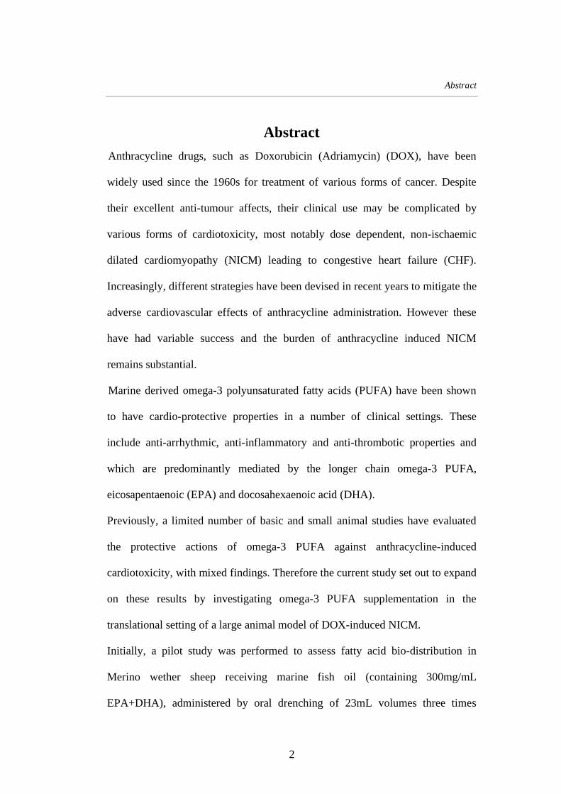
Abstract
Abstract Anthracycline drugs, such as Doxorubicin (Adriamycin) (DOX), have been
widely used since the 1960s for treatment of various forms of cancer. Despite
their excellent anti-tumour affects, their clinical use may be complicated by
various forms of cardiotoxicity, most notably dose dependent, non-ischaemic
dilated cardiomyopathy (NICM) leading to congestive heart failure (CHF).
Increasingly, different strategies have been devised in recent years to mitigate the
adverse cardiovascular effects of anthracycline administration. However these
have had variable success and the burden of anthracycline induced NICM
remains substantial.
Marine derived omega-3 polyunsaturated fatty acids (PUFA) have been shown
to have cardio-protective properties in a number of clinical settings. These
include anti-arrhythmic, anti-inflammatory and anti-thrombotic properties and
which are predominantly mediated by the longer chain omega-3 PUFA,
eicosapentaenoic (EPA) and docosahexaenoic acid (DHA).
Previously, a limited number of basic and small animal studies have evaluated
the protective actions of omega-3 PUFA against anthracycline-induced
cardiotoxicity, with mixed findings. Therefore the current study set out to expand
on these results by investigating omega-3 PUFA supplementation in the
translational setting of a large animal model of DOX-induced NICM.
Initially, a pilot study was performed to assess fatty acid bio-distribution in
Merino wether sheep receiving marine fish oil (containing 300mg/mL
EPA+DHA), administered by oral drenching of 23mL volumes three times

3
weekly for up to 20 weeks. Plasma and erythrocyte fatty acids were monitored
serially and myocardial membrane concentrations were determined at study end.
Systemic and myocardial uptake of long-chain omega-3 PUFA was
demonstrated, with plasma, erythrocyte and myocardial concentrations
increasing by two to three-fold from baseline levels (p<0.05).
For the main study, 17 age and weight-matched Merino wethers received
fortnightly dosing with intracoronary DOX (1.2mg/kg for three doses) to induce
cardiotoxicity. Animals were randomised to oral supplementation with fish oil
(n=8) or olive oil placebo (n=9) commencing two to three weeks before DOX
dosing and continued until 12 weeks after final DOX dose. Comparisons
between the fish oil and placebo groups were made for left ventricular
remodelling and function by cardiac magnetic resonance imaging (CMR),
transthoracic echocardiography and histomorphometric analysis of myocardial
fibrosis burden. Surprisingly, by comparison to placebo animals, sheep in the
fish oil group showed greater decline in left ventricular ejection fraction (LVEF)
(p<0.05), and greater end-diastolic and end-systolic dilatation after DOX
(p<0.05). However, both groups demonstrated similar levels of left ventricular
fibrosis, suggesting that the accentuation of systolic dysfunction observed in the
omega-3 PUFA cohort was not mediated by excess myocardial collagen
deposition.
In summary, this is the first large animal study to evaluate omega-3 PUFA
supplementation in the setting of anthracycline cardiotoxicity. Despite
augmenting circulating and tissue long-chain fatty acid levels, oral intake of fish-
oil exacerbated cardiac remodelling induced by intracoronary DOX.

4
Given these new observational findings, we recommend deferring clinical
investigation until further basic mechanistic studies can better define the
interactions between fatty acids and cardiac biology in the presence of
anthracycline exposure.
5
Declaration
Declaration
I declare that this thesis contains no material that has been accepted for the award
of any other degree or diploma in any university or tertiary institution to Angelo
Carbone. To the best of my knowledge and belief, this thesis contains no material
published or written by another person, except where due reference has been made
in the text.
I give consent to this copy of my thesis, when deposited in the University of
Adelaide Library, being made available for loan and photocopying, subject to the
provisions of the Copyright Act 1968.
I also give permission for the digital version of my thesis to be made available on
the web, via the University’s digital research repository, the Library catalogue, the
Australasian Digital Thesis Program (ADTP) and also through web search engines,
unless permission has been granted by the University to restrict access for a period
of time.
Angelo Carbone, BSc.
July 2011

6
Acknowledgements
Acknowledgements
I am sincerely grateful to the following staff members and departments for their
support and assistance, without which this study would not have been possible.
First and foremost, my principal supervisor, Professor Stephen Worthley
(Department of Medicine, University of Adelaide), who supported my enrolment
into a postgraduate research program and provided extensive resources and
academic support for this study. Professor Worthley has provided extensive and
greatly appreciated support and encouragement towards my career and skills
development in the field of cardiovascular research.
My co-supervisor, Dr Glenn Young, for his collaboration and previous work in
evaluating the clinical effects of dietary omega-3 PUFA supplementation in the
context of cardiovascular disease, that helped formulate the current study.
My great friend Dr Peter Psaltis (Cardiovascular Research Centre (CRC), Royal
Adelaide Hospital (RAH)), who provided such great mentorship, support and
superb interventional skills throughout the study. Peter performed each of the
study’s coronary angiograms and echocardiograms, which continued from his
earlier work to develop and validate the DOX-ovine model. He provided
extensive clinical peri-operative consultancy and study management advice, and
significantly, whilst addressing his own challenges as a final year PhD student
and planning a family move abroad.
To the staff of the Veterinary Services Division, Institute of Medical and
Veterinary Science (IMVS) for their accommodating professional and provision
of all anaesthetic procedures and veterinary management services relating to the
study. In particular, the assistance and support of Melissa Gourlay, Adrian Hines,
Jodie Dier, and Dr Tim Kuchel.

7
Acknowledgements
David Apps and Ella Zielinski of the Food and Nutrition Group, School of
Agriculture, Food and Wine, University of Adelaide for conducting the fatty acid
level analyses.
Dr Robert Metcalf (Rheumatology Unit and Cardiovascular Research Centre,
Royal Adelaide Hospital) for his excellent guidance in relation to current clinical
perspectives of omega-3 PUFA, his input into the study design, and for his
coordination of the fatty acid samples analysis.
To Professor Michael James (Rheumatology Unit, Royal Adelaide Hospital and
Department of Medicine, University of Adelaide) for his study management
advice and insights into omega-3 PUFA uptake and metabolism.
To Ms Kerry Williams (Cardiovascular Investigations Unit (CVIU), RAH) for
providing expert cardiac MR scanning services at all times of day, night and
weekends.
To Professor Tony Thomas, (Pathology Department, Flinders Medical Centre,
Bedford Park SA), for his generous and ready provision of histopathology
consulting and processing services for this study.
To my great friend and colleague, Adam Nelson for volunteering so much of his
own time to assist with after hours procedures and providing helpful advice on
data management, analysis and presentation.
To Michael Weightman for performing LVEF and % Area Fibrosis analyses
which provided pivotal outcome data for this study.
To Dr Michael Worthington (Cardiothoracic Surgery Department, Royal
Adelaide Hospital), for his expert tuition and practical assistance with the
pericardial window procedures.

8
Acknowledgements
To Dr Thomas Sullivan (Lecturer, Statistician, Discipline of Public Health,
University of Adelaide) for providing statistical consultancy and analysis.
To Mr Anthony Brooks (Research Fellow, CRC RAH) for providing additional
statistical analysis support.
To Ms Angie Hooper (Personal Assistant to Professor Worthley), for her helpful
and professional administrative assistance and ongoing support throughout the
study.

9
Table of Contents
Table of Contents
Abstract……………………………………………………………….…...2
Declaration…………………………………………………………….…..5
Acknowledgements……………………………………………….……….6
Introduction
1.1 Cancer and Chemotherapy
1.1.1 Cancer………………………………………….…..…………18
1.1.1.1 Current Treatment Options…………….……...….......18
1.1.2 Anthracyclines……………………………….………….........19
1.1.3 Cytotoxic Effects of Doxorubicin…………………………….19
1.1.3.1 Generation of reactive oxygen species (ROS).…..……20
1.1.3.2 Antioxidant adjuvants to DOX therapy….….…...….....22
1.1.3.3 Mechanisms of anthracycline-induced
anti-tumour activity......................................................22
1.1.3.4 Apoptosis………………………………….…….…….23
1.2 Doxorubicin-induced Cardiomyopathy
1.2.1 Mechanisms of Anthracycline-induced
Cardiomyopathy………..…………………………….……....25
1.2.1.1 Apoptosis and oxidative stress……….….…….….…..25
1.2.1.2 Down-regulation of cardiac specific
muscle proteins……………………………...….……26
1.2.1.3 Release of vasoactive substances…………………….26
1.2.2 Cardiac Monitoring of Patients Receiving Anthracyclines…28
10
1.3 Treatment Strategies to Reduce Anthracycline-induced
NICM
1.3.1.1 Dosing regime…………………..…………….…….29
1.3.1.2 Anthracycline analogues……………………………29
1.3.1.3 Liposomal preparations…………………………….30
1.3.2 Cardioprotective Adjuncts
1.3.2.1 Dexrazoxane…………………………….…………..30
1.3.2.2 Hematopoetic cytokines……………………………..30
1.3.2.3 Antioxidants…………………………………………31
1.3.2.4 Improved prognosis with current generation
heart failure medications……………………………31
1.4 Omega-3 Polyunsaturated Fatty Acids
1.4.1. Fatty acid synthesis…………………….……………………..33
1.4.2 Dietary Sources of Polyunsaturates……..…………..…….….35
1.4.3 Cardioprotective Effects of Omega-3 PUFA…………….……37
1.4.3.1 Absorption of dietary omega-3 PUFA…….…..……..38
1.4.3.2 Anti-inflammatory effects……………………..……...39
1.4.3.3 Anti-arrhythmic effects…………………………..…...40
1.4.3.4 Anti-thrombotic effects…………………………...…..40
1.4.3.5 Other cardiovascular benefits………………..….…...41
1.5 Effect of Omega-3 PUFA on Anthracycline-induced
Cardiomyopathy - Current Perspectives…………………..41

11
1.6 Ovine Model of DOX-induced Cardiomyopathy……........44
1.7 Effects of Omega-3 PUFA in an Ovine Model of
DOX-induced Cardiomyopathy….....……….……..…………45
1.7.1 Thesis Studies Proposal…………………….…………………..45
1.7.1. Study Hypotheses …………………….…..……..………46
Materials and Methods
Animal Ethics Approval …………………….……………..….....….……48
Use of Animals and Study Management………………………………….48
2.1 Omega-3 PUFA Dosing Study
2.1.1 Drenching Protocol…………………….……………...…….….…49
2.1.2 Collection of samples for fatty acid level assessment………….…49
2.1.2.1 Myocardial sample preparation………..……………..50
2.1.2.2 Blood sample preparation……………..………….…..50
2.1.2.3 Separation of phospholipids, preparation of
fatty acid methyl esters (FAMEs) and
identification by gas chromatograph ……...………..50
2.2 Ovine Model of DOX-induced Cardiomyopathy
2.2.1 General Anaesthesia……………………….………………………51
2.2.2 Pericardial Windows…………………………………………........52

12
2.2.3 Cardiac Magnetic Resonance Imaging………………….….…….52
2.2.3.1 Measurement of Left Ventricular Ejection Fraction……53
2.2.4 Transthoracic Echocardiogram…………..…………..…………..54
2.2.5 Blood Samples………………………………….………….….…...54
2.2.6 DOX-Infusion Protocol
2.2.6.1 Establishment of dosage…………………..…….…..…54
2.2.6.2 Group allocation…………………………..………..…55
2.2.6.3 Catheterisation and DOX-infusion……..……….….…55
2.2.7 Retrieval……………………………………………….…….……56
2.2.8 Histopathology Protocol……………………………….……....…57
2.2.9 Histological Assessment of Percent Area Fibrosis…......…….…..57
2.2.10 Sample size calculation………………………….……….…….…58
2.2.11 Statistical analysis…………………………………..………….…58
Results
3.1 Omega-3 PUFA Dosing Study
3.1.1 Omega-3 PUFA Levels
3.1.1.1Omega-3 PUFA Baseline levels…………………….....64
3.1.1.2 Omega-3 PUFA Drenching study - erythrocyte
membrane bound levels ………………..……...………66
3.1.1.3 Omega-3 PUFA Drenching study-Myocardial levels…66
3.1.1.4 Myocardial arachidonic acid levels…………………..68
3.2 Ovine DOX-infusion Study
3.2.1 Clinical Results
3.2.1.1 Mortality rate…………..…………….….…………......69

13
3.2.1.2 Electrocardiographic changes…………….….....……70
3.2.2 Left Ventricular Ejection Fraction and Volume Changes as
Assessed by CMR………….………………..……………....….….71
3.2.3 Fractional Shortening as assessed by TTE………………..……..75
3.2.4 Blood Results
3.2.4.1 Troponin-T post DOX infusion…….………………….77
3.2.4.2 Haemoglobin, WCC, platelets............…..…………….77
3.2.5 Histopathological Assessment
3.2.5.1 Macroscopic Appearances…….….………..………….80
3.2.5.2 Histopathological Findings……..………….….………82
3.2.5.3 Ventricular fibrosis burden ……..……….………..…..82
Discussion
4.1 Study Objectives………………………………….……………………..86
4.2 Uptake of Omega-3 PUFA in Merino Sheep……..……………….86
4.2.1 Elevated Omega-3 PUFA levels at baseline……………….………….87
4.2.2 Implementation of Olive Oil placebo drenching for main study….…..88
4.3 Cardiac effect of Omega-3 PUFA in Ovine model of
DOX-induced NICM…………………………………………….……..88
4.3.1 Possible mechanisms for adverse effects of Omega-3 PUFA on
DOX-induced NICM………………………….……………………..89
4.4 Attrition Rate..........................................................................................90
14
4.5 Study Limitations
4.5.1 Anthracycline Administration and Dosage………………………91
4.5.2 Absence of Neoplasia…………………………………….………..92
4.5.3 General Anaesthesia……………………………………….……...92
4.5.4 Follow up period…………………………………………………..92
4.5.5 Non reporting of some omega-3 PUFA levels and
histopathology samples………………………………………….93
Summary and Future Directions…………..……………………94
References……………………………………………………………..95
Appendix……………………………………………………………..113

15
Abbreviations
AEC Animal Ethics Committee EDD End-diastolic dimension
ALA Alpha-linolenic acid EDV End-diastolic volume
ANOVA Analysis of Variance EPA Eicosapentaenoic acid
ANP Atrial naturetic peptide EPO Erythropoietin
ARA Arachidonic acid ESV End-systolic volume
BSA Body surface area ETE Eicosatrienoic acid
CAM Cell adhesion molecule FOV Field of View
CH3 Methyl group Fr French
CH3CO Acetyl group FS Fractional shortening
CHF Congestive heart failure G-CSF Granulocyte colony
stimulating factors
CI Confidence interval IC Intracoronary
CK Creatine Kinase IV Intravenous
CMR Cardiac magnetic
resonance imaging
LA Left atrium
COOH Carboxyl group LA Linoleic acid
COX-2 Cyclooxygenase-2 LARIF Large Animal Research &
Imaging Facility, IMVS. DGLA dihomo-gamma-linolenic
acid LDH Lactate dehydrogenase
DHA Docosahexaenoic c acid LV Left ventricle
DNA Deoxyribonucleic acid LVEF Left ventricular ejection
fraction
DNR Duanorubicin LVEDD Left ventricular end-
diastolic dimension
DOX Doxorubicin LVESD Left ventricular end-
systolic dimension
DPA Docosapentaenoic acid m2 Metre squared
ECG Electrocardiogram NADH Nicotinamide adenine
dinucleotide hydrogenase

16
Abbreviations (continued)
NICM Nonischaemic
Cardiomyopathy
SD Standard Deviation
PG Prosaglandin SR Sarcoplasmic reticulum
PLA2 Phospolipase A2 TE Echo Time
PUFA Polyunsaturated fatty acid TPO Thrombopoietin
RA Right Atrium TR Repetition Time
RBC Erythrocyte (red blood
cell)
TTE Transthoracic
Echocardiography
ROS Reactive oxygen species VA Ventricular Arrythmia
RV Right ventricle V-CAM Vascular cell adhesion
molecule
SEM Standard Error of the
Mean
WCC White Cell Count

17
Introduction
18
Chapter 1: Introduction
1.1 Cancer and Chemotherapy
1.1.1 Cancer
Cancer is a disease typically characterised by changes in deoxyribonucleic acid
(DNA) leading to the uncontrolled division of abnormal cells. It remains one of the
leading causes of death in all Western industrialised countries1-6. Over 93,000 new
cases of cancer were reported in Australia in 2003 and there is a one in two and one
in three chance of a cancer diagnosis before age 85 in males and females
respectively7.
1.1.1.1 Current treatment options
Current evidence-based treatment options for cancer include surgery, radiotherapy
and chemotherapy. Chemotherapy agents are typically administered intravenously
at regular intervals over weeks or months. Until recently, chemotherapy was more
commonly indicated in advanced cancer stages (III or IV), but its use is now
expanding to earlier stages of disease progression8. Its administration is often
divided into three stages, induction, where the goal is to reduce the number of
cancer cells, consolidation, to achieve complete remission, and finally,
maintenance, given after remission to prevent relapse of the disease9. It has
advantages over surgery and radiotherapy in that delivery of the chemical agent is
via the body’s own blood stream and can therefore be particularly effective against

19
cancers not localised to a specific body area or those areas difficult to access
surgically.
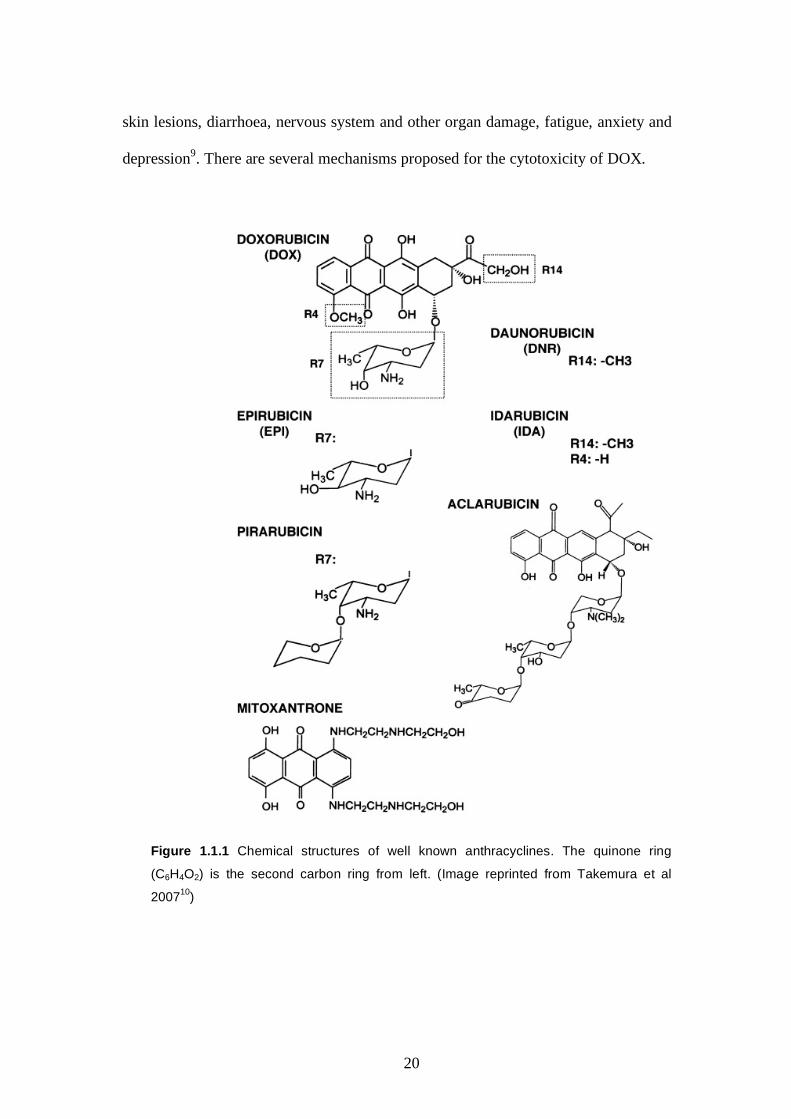
1.1.2 Anthracyclines
Doxorubicin (Adriamycin) (DOX), daunorubicin (DNR) and epirubicin are bacterial
antibiotics widely used as chemotherapy agents in the treatment of various forms of
cancer. They are classed as anthracyclic glucosides (anthracyclines) in that they
target particular parts of the DNA chain and work by impairing DNA replication in
rapidly dividing cells, such as malignant cancer cells. They are administered as single
agents or in combination with systemic adjuvants or other adjunctive agents. They are
isolated from a soil based pigment producing bacterium Streptomyces peucetius, and
have been used extensively since the late 1960’s because of their powerful anti-
tumour effects against several types of cancer. This includes acute leukaemia,
Hodgkin’s and non-Hodgkin’s lymphoma, Kaposi’s sarcoma, soft tissue sarcomas,
osteosarcomas, carcinomas, and breast cancer, all of which can occur in young
patients and have some potential for cure10, 11. Almost 60% of children diagnosed
with cancer receive anthracyclines as part of their treatment12. Doxorubicin is also
referred to as an anthracycline quinone, because of the 1,4 benzoquinone ring
(C6H4O2) which is an integral part of its structure and properties (Figure 1.1.1).
1.1.3 Cytotoxic Effects of Doxorubicin
Chemotherapy agents by nature are cytotoxic. A common side effect of anti-tumour
agents such as DOX is that they also attack healthy cells that are dividing normally,
such as hair follicle, bone marrow and intestinal cells. This is associated with many
of the common adverse effects attributed to chemotherapy such as nausea, alopecia,
20
skin lesions, diarrhoea, nervous system and other organ damage, fatigue, anxiety and
depression9. There are several mechanisms proposed for the cytotoxicity of DOX.
Figure 1.1.1 Chemical structures of well known anthracyclines. The quinone ring
(C6H4O2) is the second carbon ring from left. (Image reprinted from Takemura et al
200710)
21
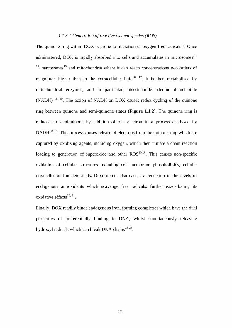
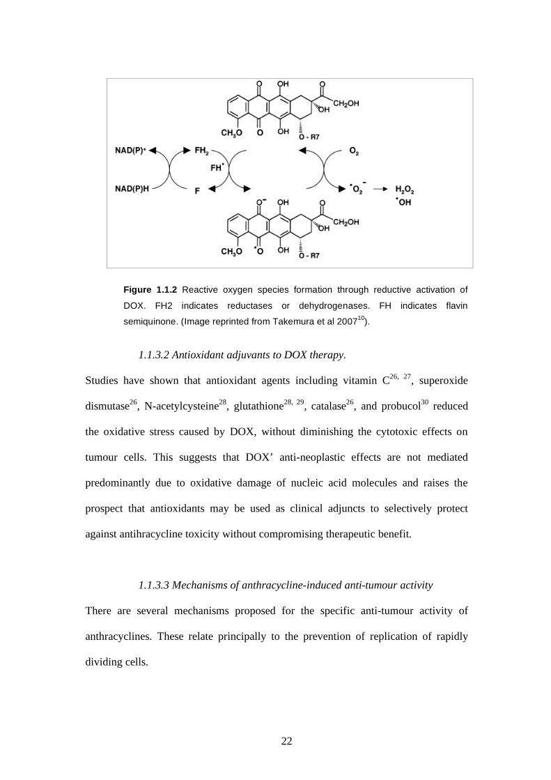
1.1.3.1 Generation of reactive oxygen species (ROS)
The quinone ring within DOX is prone to liberation of oxygen free radicals13. Once
administered, DOX is rapidly absorbed into cells and accumulates in microsomes14,
15, sarcosomes15 and mitochondria where it can reach concentrations two orders of
magnitude higher than in the extracellular fluid16, 17. It is then metabolised by
mitochondrial enzymes, and in particular, nicotinamide adenine dinucleotide
(NADH) 18, 19. The action of NADH on DOX causes redox cycling of the quinone
ring between quinone and semi-quinone states (Figure 1.1.2). The quinone ring is
reduced to semiquinone by addition of one electron in a process catalysed by
NADH10, 18. This process causes release of electrons from the quinone ring which are
captured by oxidizing agents, including oxygen, which then initiate a chain reaction
leading to generation of superoxide and other ROS10,18. This causes non-specific
oxidation of cellular structures including cell membrane phospholipids, cellular
organelles and nucleic acids. Doxorubicin also causes a reduction in the levels of
endogenous antioxidants which scavenge free radicals, further exacerbating its
oxidative effects20, 21.
Finally, DOX readily binds endogenous iron, forming complexes which have the dual
properties of preferentially binding to DNA, whilst simultaneously releasing
hydroxyl radicals which can break DNA chains22-25.
22
Figure 1.1.2 Reactive oxygen species formation through reductive activation of
DOX. FH2 indicates reductases or dehydrogenases. FH indicates flavin
semiquinone. (Image reprinted from Takemura et al 200710).
1.1.3.2 Antioxidant adjuvants to DOX therapy.
Studies have shown that antioxidant agents including vitamin C26, 27, superoxide
dismutase26, N-acetylcysteine28, glutathione28, 29, catalase26, and probucol30 reduced
the oxidative stress caused by DOX, without diminishing the cytotoxic effects on
tumour cells. This suggests that DOX’ anti-neoplastic effects are not mediated
predominantly due to oxidative damage of nucleic acid molecules and raises the
prospect that antioxidants may be used as clinical adjuncts to selectively protect
against antihracycline toxicity without compromising therapeutic benefit.
1.1.3.3 Mechanisms of anthracycline-induced anti-tumour activity
There are several mechanisms proposed for the specific anti-tumour activity of
anthracyclines. These relate principally to the prevention of replication of rapidly
dividing cells.

23
Metabolites of DOX covalently bind with high affinity to DNA bases31, 32. This
interferes with the unlinking of DNA base pairs by helicases32, a process crucial to
DNA replication. It also inhibits synthesis of a new DNA strand by inhibition of
DNA polymerase. A single molecule of DOX is sufficient to irreversibly block
helicase, an enzyme that disrupts the hydrogen bonds that stabilise and hold the two
complementary DNA strands together33. DOX also inhibits topoisomerase II which is
a critical enzyme in the separation of replicated sister chromatids following DNA
replication34-36, 37 This occurs via formation of a DOX topiosomerase II-DNA
complex37. Furthermore, this complex prevents repair of broken DNA strands38-40.
DOX has also been shown to inhibit DNA ligase activity which is crucial in the
joining of DNA strands by a phosphate ester bridge38.
1.1.3.4 Apoptosis
Apoptosis, also termed “programmed cell death”, plays a central role in many normal
biological processes, such as morphogenesis, cell turn over, hormone-dependent
organ atrophy, and immune system function. It is as important to the growth and
development of normal tissue as mitosis. Apoptosis comprises a well-described
cascade-like process that results in progressive changes to cellular components
including nucleic acid fragmentation, chromatin condensation and cytoskeleton
degradation41 42.
Most anticancer drugs induce apoptosis43, with this process affecting normal as well
as tumour cells. It has been suggested that alopecia during chemotherapy is the result
of induced apoptosis of hair follicle cells44. However, the precise mechanisms of
chemotherapy-induced apoptosis are still contentious43.

24
Caspases are a family of cysteine proteases that play essential roles in the apoptosis
signalling pathway. Activated caspases cleave intercellular proteins in a cascade-like
manner leading to release of cytochrome c from mitochondrial lamellae. This process
stimulates further enzymatic activation of caspases and results in cleavage of the
actin cytoskeleton, cell condensation and cell death. Ceramides are a family of lipid
molecules concentrated in cell membranes. They play a crucial role in the signalling
pathway of anthracycline-induced apoptosis by recruitment and activation of caspases
following internalisation of DOX. Apoptosis also involves fragmentation of DNA by
endonucleases45, which in turn contributes to the well-characterised phenomenon of
chromatin condensation. Although apoptosis does not lead to cell lysis, inflammatory
response, or necrosis (which are features typical of uncontrolled cell death and which
further damage adjacent cells), unregulated apoptosis can be equally devastating to
normal tissue structure and function and potentially lead to adverse health effects.
1.2 Doxorubicin-induced Cardiomyopathy
That DOX and other anthracyclines are very potent, effective and widely used anti-
tumour agents, albeit with many of the typical adverse effects attributed to
chemotherapy, is well established. However its major clinical shortcoming is that it
may cause dose-dependent, non-ischaemic dilated cardiomyopathy (NICM) that can
lead to congestive heart failure (CHF). This dose- dependent association between
DOX and onset of cardiomyopathy leading to CHF was first reported by Lefrak et al
in 197346. The authors described a retrospective review of 399 subjects. The
incidence of DOX-induced CHF was more than 4% of patients who received a
cumulative dose of 500-550mg/m2 body surface area (BSA) rising to 18% and 36%

25
for cumulative doses of 550-600mg/m2 and greater than 601mg/m2 respectively.
Patients older than 60 years are at greater risk with a 4.6% incidence reported at only
400mg/m2 DOX cumulative dose47. The prognosis for DOX-induced CHF has
historically been reported as very poor, with up to 20% mortality reported in an early
series48. In a series of 201 young patients, a 23% incidence of cardiac abnormalities
presenting four to 20 years after anthracycline therapy was reported49. These patients
had received a median anthracycline dose of 450mg/m2 BSA. Dose-dependent
anthracycline-induced cardiotoxicity has also been reported in other large patient
cohort clinical studies50, 51. The paediatric population are particularly vulnerable to
the cardiotoxic effects of DOX and other anthracyclines50, 52, 53, with one series
reporting five deaths from a total six cases53. Some studies suggest that enhanced
cardiotoxicty may develop at doses lower than 400mg/m2 BSA where multi-anti
tumour agent therapy is administered51.
The study by Lefrak concluded that the total cumulative dose of DOX should not
exceed 550mg/m2 of body surface area (BSA) 46. Seventy five mg/m2 represents a
typical single dose range for DOX chemotherapy54-56. Therefore the safe threshold for
cardiac complications is exceeded with relatively few doses. In addition, the
cumulative dose refers to that received over a lifetime57, such that repeat courses of
DOX chemotherapy following relapse are likely to be problematic.
Cardiotoxicity is the single most common factor that limits anthracycline use
clinically and this is particularly so for patients with risk factors for increased
toxicity, including those who have received anthracyclines in the past, older patients,
or those who are also to receive other cardiotoxic agents58.

26
1.2.1 Mechanisms of Anthracycline Induced Cardiomyopathy
1.2.1.1 Apoptosis and oxidative stress
Many of the adverse cellular effects of DOX described in the preceding sections,
including non-specific oxidative damage and apoptosis, also relate significantly to the
myocardium18, 19, 59. Redox recycling of anthracyclines by mitochondrial NADH
generates ROS including O2-, H2O2 and OH and which may underlie their
cardiotoxicity18. In addition, exposure of endothelial cells and cardiomyocytes to
DOX causes apoptosis at sub-micromolar concentrations59. Increased oxidative stress
may lead to a variety of changes to cardiomyocyte subcellular structures including
loss of myofibrils and cytoplasmic vacuolization11 (Figure 1.2.1) with the end result
being apoptosis and replacement fibrosis of affected cells10 (Figure 1.2.2) .
Cardiac muscle oxidative injury associated with anthracycline use is marked by
several other sub-cellular abnormalities, including distortion and disruption of
mitochondrial and sarcoplasmic reticulum (SR) membranes46. The myocardium
contains particularly high mitochondrial density to enable its continuous aerobic
respiration during cyclic contraction20, and is poorly endowed with antioxidant
defence mechanisms60. These factors may account for its particular susceptibility to
oxidating agents. However, there are a number of other mechanisms proposed
relating specifically to myocardial susceptibility to anthracyclines.
1.2.1.2 Down -regulation of cardiac specific muscle proteins
DOX down-regulates the expression of a variety of cardiac muscle-specific proteins,
most notably α-actinin61. This is an essential component of the contractile apparatus
in cardiomyocytes. Reduction in myoprotein expression is associated with the
reduced contractility of myocardium and may explain the pathological features of
27
myofibrillar loss in DOX induced cardiomyopathy10 as well as the decreased
contractility. Doxorubicin up-regulates expression of atrial naturetic peptide (ANP), a
polypeptide hormone involved in the homeostasis of atrial myocytes. This up-
regulation can persist constitutively, even following cessation of chemotherapy, and
has been proposed as a factor in those cases of cardiomyopathy presenting months or
years following cessation of therapy62.
1.2.1.3 Release of vasoactive substances
Administration of DOX has also been shown in a canine model to cause profound,
acute, haemodynamic changes involving release of histamine63. This was followed by
the secondary release of catecholamine, notably adrenaline, which caused an increase
in levels of immunoreactive prostaglandins (PG) in the coronary sinus63. Such
induction in the release of vasoactive substances by anthracyclines could be another
pathogenic factor in the onset of cardiomyopathy.

28
Figure 1.2.1 Electron micrograph of normal (upper) and DOX-treated mouse heart
(lower), showing loss of myofibrils in the DOX-treated cardiomyocytes. Bars 1 um.
Figure 1.2.2 Masson’s trichrome stain (upper), and Haemotoxylin-eosin (middle and
lower) of DOX-treated cardiomyocytes showing extensive replacement fibrosis (upper)
vacuolar degeneration (middle) and foci of necrotic cardiomyocytes (lower). Bars
20um. (Figures 1.2.1 and 1.2.2 reprinted from Takemura et al 200710).
NOTE: This figure is included on page 28 of the print copy of the thesis held in the University of Adelaide Library.

29
1.2.2 Cardiac Monitoring of Patients Receiving Anthracyclines
The consequences of heart damage caused by anthracyclines are extensive.
Cardiotoxicity can cause severe morbidity and lead to a reduction in quality of life.
As discussed, it limits further administration of the drug, potentially affecting
chances of patient survival. It requires long term treatment, incurring high medical
costs, and causes premature death12.
Intensive cardiac monitoring including echo and electro- cardiography (ECG) is
routinely conducted during chemotherapy. Further follow-up evaluation may be
indicated for many years following cessation of anthracycline administration49, 64.
Electrocardiogram (ECG) changes associated with DOX-induced cardiomyopathy
include various arrhythmias, most commonly sinus tachycardia (abnormally rapid
heart rate), and anomalies to the ECG wave morphology. Echocardiography, often
combined with exercise stress testing, is used to monitor anatomical and functional
changes including changes to ventricular ejection volumes. Radionuclide cardiac
scintigraphy, utilizing radio-labelled dye injected into the left ventricle (LV) is also
used to measure global ejection fraction, and detect regional wall contractile loss
and myocarditis. However, pathognomic features of anthracycline induced
cardiomyopathy in chemotherapy patients are difficult to diagnose noninvasively11,
and positive findings from these investigations indicate that the disease process is
already established. The greatest specificity and sensitivity for anthracycline-
induced cardiomyopathy is provided by endomyocardial biopsy65, which is used to
demonstrate histopathological changes described earlier, including myofibril loss,
distortion and disruption of mitochondrial and SR membranes and cytoplasmic
vacuolization. However this procedure is particularly invasive, expensive and
available in few centres 66.

30
1.3 Treatment Strategies to Reduce Anthracycline-induced NICM
A number of strategies have been devised and evaluated to reduce the cardiotoxic
effect of anthracyclines. These include modifying dosing strategies, the use of novel
anthracycline derivatives, and co-administration of adjunctive agents.
1.3.1.1 Dosing regime
Different DOX dosing strategies have been compared for their effect on
cardiotoxicity, including continual 48-96 hour infusion and standard bolus
intravenous injection 67, 68. Constant infusion has been associated with a significant
reduction in the number of patients showing severe morphologic changes in
myocardial biopsy specimens67, and an accompanying reduction in the severity of
systemic effects such as nausea and vomiting68.
However, a recent meta-analysis review of five European randomised prospective
clinical trials of anthracycline-induced cardiotoxicty in children, found insufficient
evidence that changing the dosing infusion strategy led to reduced cardiotoxicity in
these patients12.
1.3.1.2 Anthracycline analogues
5-iminodaunodrubicin is an anthracycline analogue which has been shown in a
bovine heart sub-mitochondrial model to have diminished cardiotoxic potential due
to having little or no tendency to undergo oxidation18. To date this has not been
confirmed in clinical studies. In an overview of preclinical and clinical studies, 4'-
epi-doxorubicin, another analogue of DOX, showed qualitatively similar, but
quantitatively less acute cardiac toxicity compared to identical doses of DOX69.
However this may have been at the expense of altered anti-tumour activity69.

31
1.3.1.3 Liposomal preparations
Other clinical and pre-clinical studies have shown that encapsulating conventional
anthracyclines in liposomes (cell membrane vesicles) reduces the incidence and
severity of cumulative dose-related cardiomyopathy while preserving anti-tumour
activity58. However, at present these liposomal equivalents are prohibitively
expensive for routine use compared with conventionally prepared agents.
1.3.2 Cardioprotective Adjuncts
1.3.2.1 Dexrazoxane
As described earlier, DOX readily binds endogenous iron and the resulting
complexes preferentially bind DNA whilst simultaneously releasing hydroxy
radicals which can cause oxidative stress to DNA and adjacent cellular structures.
Dexrazoxane is an iron chelator which has been shown in a canine model to reduce
DOX-induced cardiotoxicity whilst not appreciably limiting its anti-neoplastic
activity70. This has been demonstrated in a study of 101 DOX-treated children with
acute lymphoblastic leukemia71 and in several other clinical trials72. Although
dexrazoxane can increase tolerance to DOX in terms of dose-dependent
cardiomyopathy72, its benefit to long term survival remains unclear73. Adverse
effects associated with the use of dexrazoxane in patients receiving DOX include
increased myelosuppression, infection and fever74. Meta-analysis studies have also
indicated that dexrazoxane may reduce the anti-tumour efficacy of the drug12.
1.3.2.2 Hematopoietic cytokines
More recently, haematopoietic cytokines have been investigated as adjunctive
agents to anthracyclines for their ability to decrease cardiomyopathy and/or other

32
adverse effects. These include granulocyte-colony stimulating factors (G-CSF) and
thrombopoietin (TPO). Granulocyte-colony stimulating factor has been used to
reduce the incidence of granulocytopaenia and infections in elderly patients
receiving DOX75. Thrombopoietin has been shown in a small animal study to
protect against DOX-induced vacuolization, myofibrillar loss and apoptosis in
spontaneously beating cells of primary neonatal rat ventricle, whilst increasing heart
rate, ventricular function and output57. Clinical studies to investigate
supplementation of DOX with G-CSF and TPO in patients with sarcomas are
underway56 but results have not yet been reported. Erythropoietin (EPO) has also
been studied for its cardioprotective effects. This agent appeared to provide
cardiomyocyte protection against acute DOX toxicity in a rat organ bath model76
and in experiments with cultured rat cardiomyocytes77. However, demonstration of
a clinical benefit is once again lacking.
1.3.2.3 Antioxidants
Although antioxidants such as Vitamin E and N-acetylcysteine are prime candidates
for evaluation against anthracycline-induced cardiomyopathy, they have not been
shown to prevent nor delay the onset of cardiomyopathy in clinical studies78, 79.
1.3.2.4 Improved prognosis with current generation heart failure
medications
A recent clinical study reported a significant improvement in left ventricular
ejection fraction (LVEF) in 42% of 201 cases of anthracycline-induced NICM
receiving current generation medications (enalapril and carvedilol) for the treatment
of CHF80. This compares favourably to earlier reported series using contemporary
33
HF medications. The study concluded that LVEF recovery may be achieved when
cardiac dysfunction is detected early and a modern HF treatment is initiated
promptly. However, these agents achieve improvement in LVEF by modulating the
renin-angiotensin hormone systems that regulate blood pressure and fluid balance,
rather than via attenuation of cardiomyopathy onset itself. There remains
considerable scope for further enhancement in the prevention and management of
anthracycline-induced NICM.
To date, the evidence for most of the candidate agents has been restricted to in vitro
and small animal studies and results from the small number of clinical studies have
been disappointing. The emergence of a safe, effective, tolerable and affordable
adjunctive therapy which could attenuate the onset of anthracycline cardiotoxicity
would represent a significant advancement in the management of neoplastic disease.
1.4 Omega-3 Polyunsaturated Fatty Acids Fatty acids are an important source of energy, are integral to many protein structures
and are an essential component of cell membranes. They comprise molecular chains
of between two and 24 carbon atoms with a carboxyl group (COOH) at the terminus,
also known as the “delta” end, and a methyl (CH3) at the opposite, also termed
“omega” end. They are partly classified according to the level of saturation of the
carbon atoms within the chain by hydrogen, (ie whether each carbon has the
maximum number of hydrogen atoms attached). A saturated fatty acid indicates that
all of the available bonds to the carbon atoms within the chain are occupied by
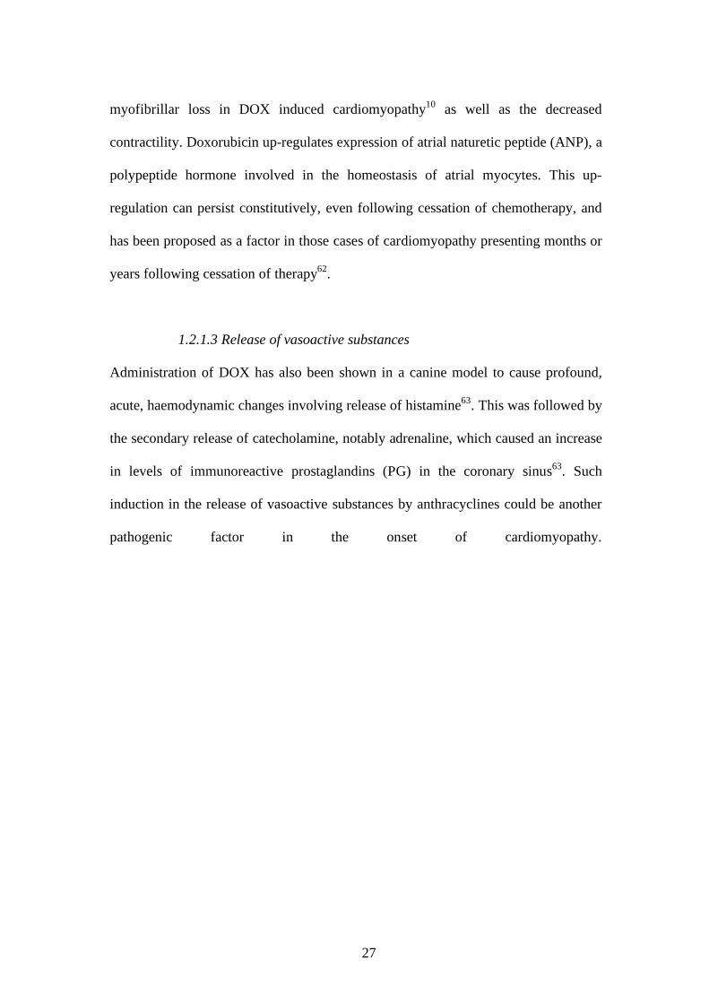
hydrogen (Figure 1.4.1). An unsaturated fatty acid describes a fatty acid chain in
which there are one or more carbon-carbon double bonds and therefore one or more
34
points capable of supporting additional hydrogen atoms that are not currently part of
its structure.
Figure 1.4.1 Stearic acid, an 18 carbon chain saturated fatty acid.
Unsaturated fatty acids comprise mainly 18, 20 or 22 carbon atoms in the chain and
are denoted by the formula “a:bn-c” where “a” is the number of carbon atoms in the
chain, “b” the number of carbon-carbon double bonds, and “c”, the position of the
first double bond in the chain relative to the omega (methyl) end. For example,
linoleic acid (LA), an omega-6 polyunsaturated fatty acid (PUFA) derived from
plants, is denoted as 18:2n-6, where “18” indicates the number of carbon atoms in the
chain, “2” indicates two carbon-carbon double bonds and “6” indicates that the first
of the double bonds occurs between the 6th and 7th carbon from the methyl (or omega)
end. The terms “omega” and “n-“ are inter-changeable.
1.4.1 Fatty acid synthesis
Fatty acids can be synthesised de novo in all organisms. Acetyl-CoA, a dimeric
molecule consisting of an acetyl (CH3CO) group attached to coenzyme A is the
precursor molecule. Coenzyme A can perform dual roles, including oxidation of fatty
acids to generate energy via Krebs citric acid cycle and fatty acid synthesis. In
humans, most fatty acid synthesis occurs in the liver and adipose tissues. Fatty acid
synthase, a large dimeric protein, catalyses the elongation of fatty acid chains from
the acetyl-Co A precursor.

35
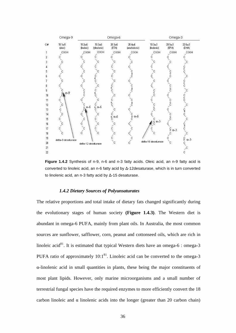
Stearic acid is an 18 carbon saturated fatty acid (18:0), synthesised by all organisms.
It is the major precursor for PUFA, which are formed by the insertion of carbon-
carbon double bonds by desaturase enzymes. Desaturase enzymes are named from
the first carbon atom from the delta (carboxyl) end of the molecule which has a
double bond. Hence stearic acid (18:0) is converted to oleic acid (18:1n-9) by Δ-9
desaturase, as it catalyses the insertion of a double bond to the 9th carbon atom in the
chain from the delta end. (Figure 1.4.2). Oleic acid is further converted by Δ-12
desaturase to LA (18:2n-6), as it now has two carbon to carbon double bonds, with
the first located at the 6th carbon from the omega end.
Further desaturation by Δ-15 desaturase converts LA to α-linolenic acid (ALA,
18:3n-3), now with the first double bond located at the 3rd carbon from the omega
end. Animals do not possess either the Δ-12 desaturase or the Δ-15 desaturase
enzymes. Therefore, both the omega-6 and omega-3 PUFAs are classified as essential
fatty acids as they must be obtained from dietary sources. Both LA and ALA can
undergo further desaturation and elongation (ie addition of acetyl group to alpha-
end), as depicted in Figure 1.4.2.
36
Figure 1.4.2 Synthesis of n-9, n-6 and n-3 fatty acids. Oleic acid, an n-9 fatty acid is
converted to linoleic acid, an n-6 fatty acid by Δ-12desaturase, which is in turn converted
to linolenic acid, an n-3 fatty acid by Δ-15 desaturase.
1.4.2 Dietary Sources of Polyunsaturates
The relative proportions and total intake of dietary fats changed significantly during
the evolutionary stages of human society (Figure 1.4.3). The Western diet is
abundant in omega-6 PUFA, mainly from plant oils. In Australia, the most common
sources are sunflower, safflower, corn, peanut and cottonseed oils, which are rich in
linoleic acid81. It is estimated that typical Western diets have an omega-6 : omega-3
PUFA ratio of approximately 10:181. Linoleic acid can be converted to the omega-3
α-linolenic acid in small quantities in plants, these being the major constituents of
most plant lipids. However, only marine microorganisms and a small number of
terrestrial fungal species have the required enzymes to more efficiently convert the 18
carbon linoleic and α linolenic acids into the longer (greater than 20 carbon chain)

37
omega-6 and omega-3 PUFA derivatives, arachidonic (ARA) and eicosapentaenoic
acid (EPA) respectively (Figure 1.4.4). Arachidonic acid and EPA can be further
converted by these organisms to 22 carbon chain docosapentaenoic (DPA) and
docosahexaenoic acid (DHA). These fatty acids are assimilated up the marine food
chain and ultimately into the seafood products consumed by man.
Figure 1.4.3 Hypothetical scheme of the relative percentages of fat and different fatty acid
families in human nutrition as extrapolated from cross-sectional analyses of contemporary
hunter-gatherer populations and from longitudinal observations and their putative changes
during the preceding 100 y. trans Fatty acids, the result of the hydrogenation process, have
increased dramatically in the food supply during this century (reprinted from Simopoulos A.
Am J Clin Nutr, 1999;70(3);560S-569S).
NOTE: This figure is included on page 37 of the print copy of the thesis held in the University of Adelaide Library.
38
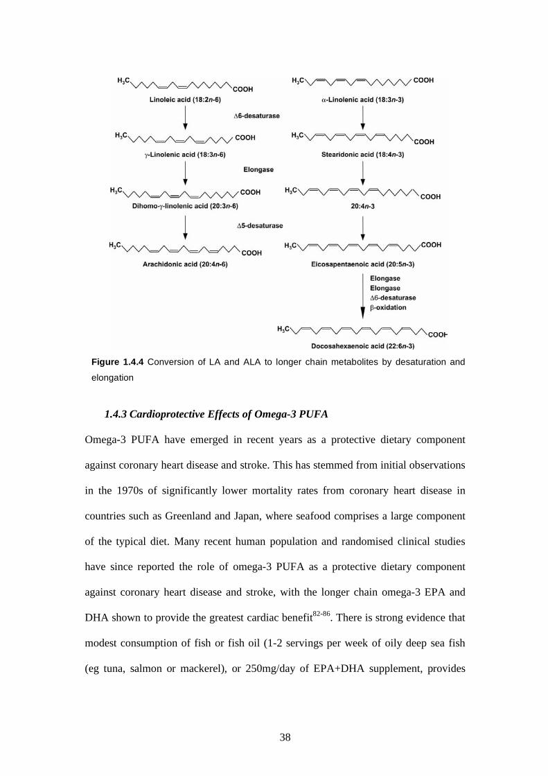
Figure 1.4.4 Conversion of LA and ALA to longer chain metabolites by desaturation and
elongation
1.4.3 Cardioprotective Effects of Omega-3 PUFA
Omega-3 PUFA have emerged in recent years as a protective dietary component
against coronary heart disease and stroke. This has stemmed from initial observations
in the 1970s of significantly lower mortality rates from coronary heart disease in
countries such as Greenland and Japan, where seafood comprises a large component
of the typical diet. Many recent human population and randomised clinical studies
have since reported the role of omega-3 PUFA as a protective dietary component
against coronary heart disease and stroke, with the longer chain omega-3 EPA and
DHA shown to provide the greatest cardiac benefit82-86. There is strong evidence that
modest consumption of fish or fish oil (1-2 servings per week of oily deep sea fish
(eg tuna, salmon or mackerel), or 250mg/day of EPA+DHA supplement, provides
39
sufficient levels to reduce the risk of sudden cardiac death by 36%, regardless of
presence or absence of established CVD 82.
1.4.3.1 Absorption of dietary omega-3 PUFA
Following ingestion, EPA and DHA are rapidly absorbed through the gut and into the
blood plasma. If sufficient intake levels are maintained over several days, they
incorporate into cell membrane phospholipid bilayers, where they substitute
arachidonic acid (ARA), a 20 carbon chain omega-6 PUFA, in a process involving
phospholipase A2 (PLA2) (Figure 1.4.4). Incorporation of PUFAs into membranes is
a result of their continual turnover, and membrane levels of PUFAs reflect the
relative proportions of PUFAs available. Membrane levels also reflect the degree to
which tissues conserve omega-3 PUFA already present, ie, some tissues selectively
release omega-6 before omega-3 PUFAs. Incorporation of EPA and DHA causes a
decrease in membrane bound ARA. The incorporation of omega-3 PUFA into the
membrane phospholipid bilayer is a prerequisite for its biological activity and imparts
numerous cellular effects, including changes to membrane polarisation properties,
protein activities, matrix remodelling and modulation of gene expression87.
40
1.4.3.2 Anti-inflammatory effects
A number of mechanisms have been proposed to describe the cardio-protective
effects of omega-3 PUFA. Following dietary supplementation, EPA and DHA have
been shown to incorporate into the membranes of human cardiomyocytes88. As
discussed, incorporation of EPA and DHA into cell membrane phospholipids is via
substitution of omega-6 PUFAs of which ARA is the major component. Both ARA
and EPA are precursors for eicosanoids, including prostaglandins and thromboxanes
Figure 1.4.4
Incorporation of Omega-3 fatty
acids.
Ingestion of omega-3 causes a
rapid rise in omega-3 plasma
levels. If sufficient ingestion
levels are maintained, Omega-
3’s incorporate into cell
membrane phospholipid bilayer
by substituting arachidonic acid,
an omega-6 fatty acid.
Membrane incorporation and
maintenance of membrane
bound levels of omega-3 are
prerequisite for cellular changes
in response to omega-3’s and
impart numerous cellular effects,
including modulation of bioactive
lipid mediators and signalling
molecules, altered membrane
protein activity and transduction
of signalling pathways within the
cell leading to modified gene and
ultimately modified protein
expression. Image reprinted from
Surette, M.E.CMAJ 2008; 178:
177-180.
NOTE: This figure is included on page 40 of the print copy of the thesis held in the University of Adelaide Library.

41
(via the lipoxygenase pathway) and leukotrienes (via the lipoxygenase pathway) and
act as signalling control molecules for inflammatory and immune processes. It is
widely regarded that eicosanoid derivatives from omega-6 PUFA such as ARA tend
to be pro-inflammatory, whilst those derived from omega-3 PUFA are significantly
less so89. Membrane phospholipid-bound ARA, released by phospolipase A, forms
the predominant precursor of pro-inflammatory eicosanoids. Substitution of
membrane bound ARA with EPA/DHA therefore reduces the availability of this
omega-6 PUFA for conversion to pro-inflammatory eicosanoids, and thereby reduces
the response to inflammatory stimuli in tissues, including myocardium.
1.4.3.3 Anti-arrhythmic effects
It is now well established that incorporation of omega-3 PUFA into cardiomyocyte
membranes alters their voltage conduction properties such that they demonstrate
more controlled sinus rhythm and resistance to anti-arrhythmia90, 91. Enhancement
of the conduction properties of cardiomyocyte cell membranes following
incorporation of EPA and DHA is believed to be a predominant mechanism
underlying the cardioprotective properties of omega-3 PUFA90-92. The anti-
arrhythmic effects of omega-3 PUFA were well characterised in a study by Billman
et al93, in a canine model of inducible ventricular fibrillation (VF), where infusion
of omega-3 PUFA in these animals delayed onset of VF in response to exercise
stress compared with baseline values.
1.4.3.4 Anti-thrombotic effects
Several studies have demonstrated the anti-thrombotic properties of omega-3 fatty
acids. Human consumption of 3-4g/day of EPA/DHA has been shown to reduce

42
thrombosis by decreasing platelet aggregation induced by collagen and platelet
activation factor94. Cell adhesion molecules (CAM) are trans-membrane proteins
that play several key roles in tissue homeostasis and development. They function by
modulating binding of particular cell types. Certain CAMs such as vascular cell
adhesion molecules (VCAM-1) also play a role in disease processes such as
inflammation and atherogenesis. It is difficult to measure membrane levels of
adhesion molecules in the human vasculature following DHA supplementation,
however it has been shown in cultured human saphenous vein endothelial cells to
reduce membrane surface expression of these molecules95. Thromboxane A2 is a
pro-thrombotic eicosanoid synthesized in platelets via cyclooxygenase action on its
precursor molecule ARA. Omega-3 PUFA reduces production in platelets. These
properties are likely to be relevant to the anti-thrombotic properties of omega-
PUFA.
1.4.3.5 Other cardiovascular benefits
Other studies have shown a modest but dose-dependent improvement in endothelial
function95, with a resulting decrease in blood pressure96, and lowering of plasma
triglyceride levels97 in response to dietary omega-3 PUFA supplementation.
1.5 Effect of Omega-3 PUFA on Anthracycline-induced NICM –
Current Perspectives
Few studies have investigated the effects of omega-3 PUFA supplementation in the
presence of anthracycline agents, with all of these conducted in vitro or in small
animal neoplastic models. Germain et al, reported that dietary supplementation of
43
omega-3 PUFA increased rat mammary tumour sensitivity to epirubicin without
altering its cardiac toxicity98. All animal cells, including tumour cells, can
potentially incorporate EPA and DHA into their phospholipid membranes during
dietary supplementation. In addition, EPA and DHA, due to their lower level of
saturation compared with ARA and other membrane phospholipids, may also be
more prone to lipid peroxidation. This was proposed as the mechanism underlying
the sensitisation of neoplastic cells by omega-3 PUFA in the rat mammary study98.
Since lipid peroxidation may also mediate anthracycline induced cardiotoxicity, it is
important to determine whether omega-3 PUFA supplementation may exacerbate or
reduce this serious side effect. A more recent study monitored cardiac function in
omega-3 PUFA fed Sprague Dawley rats during co-administration of epirubicin99.
They showed no additive nor preventative effect of omega-3 PUFA on
cardiotoxicity. Dietary fish oil supplementation in mice has also been shown to
augment the efficacy of DOX against breast cancer xenografts without increasing
non-cardiac drug toxicity100. Although these findings are reassuring and raise the
potential of omega-3 PUFA as a tumour sensitising agent, conflicting evidence also
exists to suggest that dietary omega-3 PUFA may diminish cellular anti-oxidant
defences, which in turn may accentuate cardiomyocyte susceptibility to lipid
peroxidation after exposure to DOX101. Table 1.5.1 summarises the outcomes of
several small animal and in vitro models investigating the effects of omega-3 PUFA
supplementation with co-administered anthracyclines.
As discussed, all published findings to date are confined to small animal or in vitro
neoplasia models with many not assessing cardiac function directly. Further, some
results indicate that dietary supplementation of omega-3 PUFA in association with
anthracycline administration may exacerbate cardiotoxicity. Clearly there is a need

44
to evaluate the cardio-protective effects of omega-3 PUFA in a large DOX treated
in vivo model comparable to human cardiovascular anatomy and physiology, before
a clinical trial evaluating its safety and effectiveness in patients receiving
anthracyclines can be considered.
Detrimental Effect
No Change or
Indeterminant
Benefit
Matsui et al, CanJCard 101. Rat DOX model +
omega-3 PUFA
Germain et al, Pharm Res99.
Rat epirubicin model +
omega-3 PUFA
Germain et al, Lipids98.
Rat mammary tumor
model + epirubicin +
DHA
Selting et al, Am J Vet
Res102. Dog lymphoma
study + DOX + Hi/Low
dose omega-3 PUFA
Colas et al, Clin Canc
Res103. Rat mammary
tumor model +
epirubicin + DHA
Rudra et al, Anti Canc
Res104. Cancer Cell lines +
DOX + DHA
Hardman et al, Clin
Canc Res100. Mouse
cancer xenograft model
+ DOX + omega-3
PUFA
Table 1.5.1. Summary of conclusions from several small animal and in vitro studies
investigating the effects of omega-3 PUFA on anthracycline induced cytotoxicity. There is
no clear consensus regarding it’s benefit or otherwise.

45
1.6 Ovine Model of DOX-induced Cardiomyopathy
Previous work undertaken by this department has validated a sheep model of DOX-
induced, non-ischaemic cardiomyopathy (NICM)105. In this study 10 healthy
Merino wethers received fortnightly intra-coronary (IC) infusions of DOX for a
mean cumulative dose of 3.6mg/kg. Dosing was targeted to the coronary arteries to
minimise systemic adverse effects (e.g. myelosuppression, diarrhoea, anorexia) and
to cause reproducible cardiotoxicity. The development of toxic cardiomyopathy was
validated by cardiac magnetic resonance imaging (CMR), including late uptake of
gadolinium to assess for scar tissue formation. Magnetic resonance imaging is the
current “gold standard” for non-invasive assessment of cardiac volumes and
function106 and enabled high resolution assessment of the myocardium. Additional
assessment was performed with transthoracic echocardiography, haemodynamic
measurements and histology. The doxorubicin dosing regimen was generally well
tolerated with systemic toxicity consisting mainly of reversible myelosuppression.
Premature attrition rate was 20%, with both deaths due to cardiac arrhythmia, but
which was a substantial improvement compared to previous studies with reported
mortality rates of up to 60%107, 108. Importantly, reproducible, moderate-severe
biventricular systolic dysfunction was induced within six weeks of completion of
DOX dosing. LV and RV EF declined from 46.2 + 4.7% (baseline) to 31.3 + 8.5%
(final) (p<0.01) and from 39.5 + 5.6% to 28.9 + 9.6%, (p<0.05), respectively.
Histological changes comprised cardiomyocyte degradation, vacuolisation, Purkinje
fibre damage, small vessel injury and significantly, interstitial and replacement
fibrosis of both ventricles and which were typical of pathological features
associated with non-ischaemic cardiomyopathy109-111.

46
1.7 Effects of Omega-3 PUFA in an Ovine Model of DOX-induced
Cardiomyopathy
Based on the validation study findings, sheep may represent a suitable large animal
model for the assessment of the cardioprotective effects of omega-3 PUFA
supplementation in response to Doxorubicin administration. Ruminants have been
shown to consume omega-3 PUFA supplemented feed (including fish products) and
absorb these into skeletal muscle and other tissues112-114. These studies were
undertaken to investigate increasing the omega-3 PUFA content of skeletal muscle
as a dietary source for human consumption. Some reduction in absorption compared
with humans due to hydrogenation by ruminal bacteria has been reported115, but this
may be countered by administering higher doses. Modest increases in tissue omega-
3 PUFA have been demonstrated in sheep in response to dietary supplementation
with fish oil, and with factors such as dose and breed of sheep affecting the level of
absorption of omega-3 PUFA into tissues113, 114. Merino sheep have not been
previously assessed for their uptake of dietary fish oil, nor have any ovine studies
characterised the biodistribution of long chain fatty acids in the myocardium after
supplemental therapy.
1.7.1 Thesis Studies Proposal
This thesis therefore consisted of two sub-studies:
Study 1 was designed to verify the feasibility of increasing plasma and myocardial
phopholipid membrane bound uptake of EPA and DHA in Merino sheep following
oral ingestion of fish oil. In turn, this enabled us to investigate the cardiac effects of

47
dietary omega-3 PUFA on in sheep also receiving intracoronary doxorubicin dosing
(Study 2).
1.7.1.1 Study Hypotheses
• Study 1: “Oral ingestion of fish oil results in a significant increase of
omega-3 PUFA levels into plasma, and erythrocyte and cardiomyocyte cell
membranes”
• Study 2, “Dietary supplementation of omega-3 PUFAs will protect
myocardium from DOX-induced cardiomyopathy, and this will manifest as
less reduction in Left Ventricular Ejection Fraction compared with control
group”.

48
Materials and Methods

49
Chapter 2: Materials and Methods
Animal ethics approval and study management
The full study proposal was submitted to the Institute of Medical & Veterinary
Science and The University of Adelaide Animal Ethics Committees (AEC).
Approval was granted by the IMVS (Approval No. 105/07, 13th August) and
University AEC’s (No. M-043-2007, 5th September 2007) for the use of 26 animals
to conduct the proposed studies. The use of additional scavenge myocardial tissue
from four healthy weight-matched animals was also approved to provide
myocardium for comparative histology.
Use of Animals and Study Management
The studies were undertaken over a 20 month period from October 2007 to June
2009. Individual sheep were identified by a colour coded and numbered ear tag. The
first 10 animals were used in a pilot study to further refine the DOX infusion
protocol. The pilot study assessed the feasibility of further reducing the number of
DOX dosings (compared with the earlier validaton study), and accordingly, the
number of animal interventions, whilst maintaining induction of moderate levels of
cardiac dysfunction with acceptable mortality rate. Five of these animals firstly
received Omega-3 PUFA drenching to verify uptake of omega-3 PUFA into plasma
and tissues. The remaining five sheep were fed normal diet.
Once the infusion protocol was established, a further 16 animals, in two batches of
eight, were used for the subsequent main study. These were randomly allocated to
fish oil (omega-3 group) or olive oil placebo drenching (control group) for the study
duration. Sheep with poor baseline ventricular function as assessed by
50
echocardiography and CMR were not included into the DOX infusion protocol.
DOX-infusion data is reported from the remaining 17 animals (eight omega-3 and
nine control group sheep) and includes data from some animals in the pilot as well
as the main study.
2.1 Omega-3 PUFA Dosing Study
2.1.1 Drenching Protocol
Five skeletally mature Merino wether sheep (weight range 71 to 76kg) were
drenched three times per week for up to 20 weeks (50mL Mini Drenching Gun)
with 23mLs of fish oil containing 300mg/mL EPA + DHA. This equates to
approximately 10g of fish oil and comprises 3g of long chain omega-3 PUFA
(EPA+DHA) intake per day. For the main study, weight and age matched control
sheep were drenched with an identical volume and frequency with olive oil placebo.
Lucerne chaff and water was supplied ad lib to both groups.
2.1.2 Collection of samples for fatty acid level assessment
All fatty acid analyses were conducted by the Food and Nutrition Group, School of
Agriculture, Food and Wine, University of Adelaide. Blood samples were taken
from each sheep prior to commencement of drenching, then weekly for four weeks
and then at monthly intervals thereafter and finally, at time of euthanasia, to monitor
changes in erythrocyte membrane-bound levels of omega-3 PUFA. Following
euthanasia, the sheep were exsanguinated and the heart removed intact.
Approximately 3mm3 samples of myocardium were taken from each of the left and
right ventricles adjacent to the apical tip, and left and right atrial appendage and
stored at -20C for assessment of myocardial fatty acid levels88.

51
2.1.2.1 Myocardial sample preparation Myocardial samples were cleaned of adipose tissue and clotted blood.
Approximately 0.2 g tissue was homogenized in 2mL saline before mixing with 3
mL methanol. Chloroform (6mL) was added, the samples were centrifuged (1560 x
g, 10 min, room temperature), and the chloroform phase was transferred to a 20-mL
glass vial and evaporated to dryness
2.1.2.2 Blood sample preparation
Whole blood was centrifuged at 1800g for 10 minutes, and the plasma removed and
stored at -70oC until analysis. The remaining red blood cells (RBC) were washed 3
times with normal saline, then 1 ml of RBCs was added to 0.5 ml saline and 2 ml
propan-2-ol, vortex mixed and allowed to stand for 5 minutes, 4 ml chloroform was
then added and mixed thoroughly. Following centrifugation, the lower chloroform
layer containing the total lipid fraction was removed and stored in a glass vial at
-20oC until analysis. Total lipids were extracted from plasma with chloroform/
methanol (2:1, by volume).
2.1.2.3 Separation of phospholipids, preparation of fatty acid methyl
esters and identification by gas chromatography
Phospholipids were separated from total lipid extracts from each of the myocardial,
plasma and RBC preparations by thin-layer chromatography (TLC Silica gel 60H
Merck Darmstadt, Germany). The TLC solvent system was petroleum spirit/diethyl
ether/glacial acetic acid (180:30:2, by volume). Lipid classes were visualised with
fluorescein 5-isothiocyanate against TLC standard 18-5 (NuChek Prep Inc: Elysian,
52
MN, USA). All solvents contained the anti-oxidant butylated hydroxyl anisole at
0.005% (wt/vol). Phospholipid fractions were transesterified by methanolysis (1%
H2SO4 in methanol) for 3 h at 70° C. After cooling, the resulting fatty acid methyl
esters (FAME) were extracted with n-heptane and transferred into gas
chromatography vials containing anhydrous Na2SO4. FAME were separated and
quantified with a Hewlett-Packard 5880 gasliquid chromatograph using a capillary
column equipped with flame ionisation detection and Hewlett-Packard Chem-
Station data system (Avondale PA, USA). Separation was achieved on a 50 m x
0.33 mm ID BPX-70 column (SGE, Melbourne, Australia). Helium was the carrier
gas at a column flow rate of 35 cm/s. The inlet split ratio was set at 30:1. The oven
temperature at injection was set at 140°C and programmed to rise to 220°C at
5C/min. The injector and detector temperatures were set at 250°C and 300°C,
respectively. FAME were identified by comparison of retention times to authentic
lipid standards (NuChek Prep Inc: Elysian, MN. USA)116.
2.2 Ovine Model of DOX-induced Cardiomyopathy
2.2.1 General Anaesthesia
Animals were fasted for 24 hours prior to general anaesthesia, which was carried
out for pericardial window, CMR, TTE, DOX infusion protocols and prior to
euthanasia. Anaesthetic induction was achieved via masking with a mixture of
isoflurane (5%) in 100% oxygen by endotracheal intubation. Anaesthesia was
maintained by inhalation of a mixture of isoflurane (2-3%) in 100% oxygen.
Animals were mechanically ventilated with a tidal volume of 10mL/kg to maintain a
partial pressure of expired carbon dioxide of approximately 40mmHg. Additional

53
monitoring under anaesthesia included pulse oximetry, continuous
electrocardiography, and intra-arterial assessment of blood pressure.
2.2.2 Pericardial Windows
Sheep received a pericardial window procedure prior to intra-coronary DOX-
infusion protocol to avoid inflammatory pericardial effusion in response to DOX117.
Following anaesthetic induction, sheep were placed in left lateral recumbency and a
20cm2 area over the left rib cage was shaved and scrubbed (Betadine sponge and
70% alcohol solution). A 15cm length incision was made over the 6th intercostal
space and dissection proceeded through the underlying fascia and muscle layers.
Manual ventilation was taken over during initial dissection. Haemostasis of
subcutaneous tissues continued as the intercostal membrane was perforated. A 3cm
diameter segment of the pericardium was then resected. Four x 2.0 Polysorb sutures
were used to re-appose the intercostal margins. Just prior to final closure of the
thoracic cavity by tensioning the final Polysorb suture, the lungs were maximally
inflated to reinstate normal baro-physiology. Subcutaneous and epidermal layers
were then closed using 0.0 Polysorb. Animals were recovered quickly without
insertion of a chest tube. One millilitre ketoprofen (Ilium Co. NSW, Australia) and
1mL/10kg Teramycin (Pfizer Co. Pty Ltd, Sao Paulo, Brasil) was administered IM
for analgesia and antibiotic prophylaxis respectively. IM Ketoprofen was continued
once daily for 2-3 days.
2.2.3 Cardiac Magnetic Resonance Imaging
The primary assessment of cardiac contractile function was via three lead
electrogram ECG-gated CMR to assess global and regional left ventricular ejection

54
fraction (LVEF). Sheep underwent CMR two weeks following pericardial window
and then at 12 weeks following final DOX infusion to establish baseline and final
LVEF and end-diastolic and end-systolic volumes (EDV, ESV). Prior to CMR,
sheep were anaesthetised and the wool over the left para-sternal region shaved and
then wiped with 70% ethanol solution to improve adherence of the ECG leads. The
sheep were then transferred to the MRI suite and positioned in supine recumbency
inside the scanner (Siemens Sonata 1.5 Tesla MR Imaging system, Siemens,
Erlangen, Germany). Cine images were ECG-gated and consisted of steady state
free precession (SSFP) sequences with the following parameters: repetition time
(T/R)/echo time (TE) 52.05 ms/1.74ms; flip angle 70o; matrix 256 x 150; 25 phases
per cardiac cycle; FOV 380mm with slice thickness of 6mm with a 4mm inter-slice
gap.
2.2.3.1 Measurement of Left Ventricular Ejection Fraction
Ventricular ejection fractions and chamber volumes were determined in blinded
fashion, with Argus software (Siemens Medical Solutions, Erlangen, Germany).
End-diastolic and end-systolic images were chosen as the maximal and minimal,
mid-ventricular, cross-sectional areas in a cinematic display. Short axis endocardial
and epicardial borders were traced manually for each slice in end-diastole and end-
systole. As per modified Simpson’s rule, these areas were multiplied by the slice
thickness (10 mm) and added together to obtain the EDV and ESV, respectively.
Papillary muscles were included for the volume measurements. Atrial slices at end-
systole secondary to apical movement of the base of the heart during LV contraction
were not included. Ejection fraction was calculated by the formula:
EF (%) = (EDV-ESV) / (EDV x100)
55
2.2.4 Transthoracic Echocardiogram
Transthoracic echocardiogram (TTE) was performed prior to each DOX infusion
and then at time of euthanasia, to enable serial monitoring of changes to contractile
function during the study period. Echocardiography was performed 30 minutes after
anaesthetic induction and the commencement of mechanical ventilation. Sheep were
positioned in right lateral recumbency and two dimensional-guided M-mode
measurements of the LV end-diastolic diameter (EDD) and end-systolic diameter
(ESD) were taken from right parasternal short axis views, just basal to the insertion
of papillary muscles. Data was averaged from three separate consecutive cardiac
cycles. Left ventricular fractional shortening (FS) was derived from the equation;
FS (%) = (EDD – ESD)
EDD x 100
2.2.5 Blood Samples
Peripheral venous blood samples were taken immediately before and then 24 hours
following each DOX infusion, and then just prior to euthanasia to quantify full
blood count, renal and liver function and cardiac biomarkers (troponin-T and
creatinine kinase).
2.2.6 DOX-Infusion Protocol
2.2.6.1 Establishment of dosage
The DOX dosing strategy was established during the pilot study. Administration of
0.75mg/kg per dose was used in accordance with previously published data117.
Administration of three and up to four fortnightly doses of 0.75mg/kg DOX did not
achieve the intended degree of cardiac dysfunction in the pilot study control animals
as based on echocardiography and CMR findings. Further validation revealed that

56
the optimal cumulative dose to result in moderate cardiac dysfunction and
acceptable animal mortality and morbidity comprised three doses of 1.2mg/kg/dose
at fortnightly intervals, (cumulative dose of 3.6mg/kg), followed by a 12 week
period prior to euthanasia. This was the adopted DOX regimen for the study. In
total, 17 Merino wether sheep (mean weight 55.6 + 7.1kg) received this DOX-
infusion protocol.
2.2.6.2 Group allocation
For the main study, sheep were randomly allocated to receive either 23mls olive oil
(placebo) or fish oil (treatment group), 23mLs three times per week via drenching
throughout the DOX dosing and follow up period, until time of euthanasia. Both
groups were matched for body size and weight. Results from the pilot drenching
study demonstrated that erythrocyte membrane bound concentrations of long chain
fatty acids increased by 2-3 fold within two to three weeks of dietary fish oil
supplementation. Therefore, omega-3 PUFA drenching was commenced two to
three weeks before the start of the DOX protocol, and continued throughout the
dosing and follow-up period. A higher concentration (60% - 600mg/mL
EPA+DHA) of omega-3 PUFA was used during the first two weeks drenching
period to expedite elevation of phospholipd membrane bound EPA+DHA prior to
commencement of DOX infusions. A 30% concentration (300mg/mL EPA+DHA
was then used for the remainder of the study period.
2.2.6.3 Catheterisation and DOX-infusion
Following anaesthetic induction, sheep were placed in supine recumbency and the
groin region shaved, sterile scrubbed and draped. Peripheral arterial access was

57
obtained via percutaneous cannulation of the right femoral artery using 5-French
introducer and sheath (Cordis Corp, Miami, USA). Heparin (100 IU/kg) Heparin
was administered via 5mL bolus into the arterial sheath. An Amplatz diagnostic
catheter (AL1 Cordis Corp, Miami, USA) was used to catheterise the left-sided
coronary arteries under fluoroscopic guidance (Phillips BV25 Fluoroscopic Imager,
Bloomfield CT, USA). Coronary artery engagement was confirmed by bolus
administration of iodinated contrast media (Ultra-vist 360 Bayer Ltd, NSW
Australia). The left anterior descending and circumflex arteries were engaged
selectively, with half of the DOX administered to each artery in sequence. The total
dose of DOX (1.2mg/kg made up in 50mL 0.9% saline) was infused over a 30
minute period. During dosing, the catheter position was repeatedly visualised by
fluoroscopy and records taken of heart rate, intra-arterial blood pressure, and
electrogram appearance.
When dosing had been completed and the catheter removed, femoral artery
haemostasis was achieved by manual compression for at least 10 minutes. Animals
received post-operative antibiotic prophylaxis and analgaesia for 72 hours. Sheep
were monitored in a small (1m x 2m) housing pen for the duration of the DOX
dosing protocol. Two weeks after the final DOX infusion they were returned to the
field station for a further 10 weeks until final CMR and euthanasia. Animals were
monitored twice daily for health status and condition.
2.2.7 Retrieval
At 12 weeks following final DOX infusion, and within 3-4 days of final CMR,
sheep were anaesthetised and final blood samples, TTE and haemodynamic
measurements obtained. Euthanasia was then performed by intravenous

58
administration of 120mg/kg pentobarbital. The heart was removed intact and the
pericardial fat layer removed. Surface areas of pallor or other notable features were
documented. Approximately 3mm3 samples of ventricular and atrial appendage
myocardium were taken and stored at -20C for subsequent assessment of omega-3
PUFA levels. The heart was then weighed intact and immediately perfusion fixed
with 3-4L of 4% paraformaldehyde, before storage in 10% buffered formalin. For
histological comparison, hearts were also obtained and processed identically from
four healthy control sheep.
2.2.8 Histopathology Protocol
Transverse slices of the fixed heart were made at 10mm intervals starting at the LV
apex. Slices from the basal, mid and distal ventricular levels were divided into six,
six and four segments respectively. Segments were labelled according to their
anatomical orientation; anterior, posterior, lateral and septal aspects, and then
processed for paraffin embedding. The apical segment was processed whole.
Sections (10um) were taken from each paraffin block and stained with
haematoxylin and eosin for assessment of morphologic cellular changes and van
Gieson stain for identification and quantification of fibrotic tissue.
2.2.9 Histological Assessment of Percent Area Fibrosis
The van Gieson stained slide sections were photographed with a NanoZoomer
Digital Pathology system (Hamamatsu Photonics, Hamamtsu, Japan). Complete
image sets were exported from each slide section at 5x magnification into Image
Pro-Plus software (ver 5.2.1.59, Media Cybernetics Inc. Bethesda, MD USA). The
percentage area of fibrosis occupying each segment was calculated using “colour

59
cube-based segmentation” algorithm to select and quantify fibrotic tissue (staining
red) against normal myocardium (brown). The epicardium and surface coronary
vessels contain abundant collagen in normal healthy myocardium, and were
therefore removed from the analysis region of interest. Data are presented as
segmental and mean total % fibrosis area for each of the Omega-3, placebo control,
and healthy group.
2.2.10 Sample size calculation
A study group totaling 16 animals achieves a statistical power of 80% to detect a
12% absolute difference in decline in mean global left ventricular ejection fraction
on CMR between sheep in each group, (with 8% variance (SD)) and a p value <
0.05. Allowance was made for two animals per group not surviving the disease
process.
2.2.11 Statistical Analysis
For the Omega-3 PUFA dosing study, statistical analysis was performed using
Prism Graph ver. 5.03 (Graph Pad Software, La Jolla, Ca, USA). Normality of data
was assessed by Shapiro-Wilk test and normally distributed data presented as mean
+ S.D. Intergroup differences at baseline were assessed using Students unpaired t-
test. Comparisons between groups over time were performed by two way ANOVA.
Statistical significance was established at a two-tailed p value <0.05. Where a
significant difference was encountered, this was post-tested using Tukey-Kramer
Multiple comparisons test.
For the DOX-infusion study, statistical analysis was performed using SPSS ver 15
(IBM Corporation, Route 100, Somers NY USA). Normally distributed variables

60
are reported as mean ± SD. Intergroup baseline comparisons were analysed by using
Student’s unpaired, two-tailed t-test. Skewed datasets are reported as median and
inter-quartile range. Categorical variables are reported as number and percentage.
Statistical significance was set at two-tailed p<0.05.
Continuous variables were assessed using linear mixed effects models to examine
the effects of time (pre, post or baseline, dox1, dox2, dox3), group (placebo,
Omega-3) and their interaction (group*time). A random effect of sheep ID was
fitted to the model to account for the repeated observations (time). The residuals
were visually assessed for normality to ensure appropriate model fit. If the residuals
were skewed, the data was log transformed and the model re-run. If the interaction
term was significant, appropriate post-hoc tests were performed within groups. If
mixed effects model Hessian matrix was not satisfied because of limited follow-up
data (due to unexpected deaths or technical issues) statistical analysis was not
performed, but raw data presented. Simple inter or intra-group comparisons were
performed using t-tests for normally distributed data, Mann-Whitney for skewed
data or Chi-squared for count data.
Intraclass agreement for the LVEF and % area of fibrosis measurements were
assessed via intraclass correlation and 95% Bland Altman confidence limits of
agreement (Appendix).
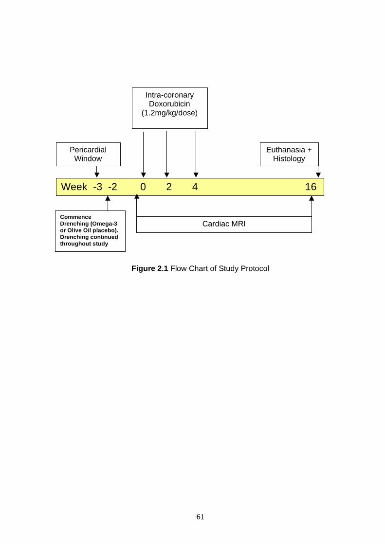
61
Figure 2.1 Flow Chart of Study Protocol
Intra-coronary Doxorubicin
(1.2mg/kg/dose)
Pericardial Window
Euthanasia + Histology
Week -3 -2 0 2 4 16
Cardiac MRI
Commence Drenching (Omega-3 or Olive Oil placebo). Drenching continued throughout study
Commence Drenching (Omega-3 or Olive Oil placebo). Drenching continued throughout study

62
Table 2.2 Equipment and Supplier
Equipment Specifications and Supplier Drenching
Drenching Gun
Mini 50mL Drenching Gun –
Farmers Mailbox, Victoria, Australia
Omega-3 Fatty Acid (Fish Oil) 60%
preparation
Berg Lipidtech, Eidsnes, Norway
Omega-3 PUFA (Fish Oil) 30%
preparation
Melrose Laboratories Pty Ltd.
Mitcham, Victoria, Australia
Olive Oil (Placebo)
Melrose Laboratories Pty Ltd.
Mitcham, Victoria, Australia
Fatty Acid Level Analysis
Silica gel 60H Merck, Darmstadt Germany
TLC standard 18-5 NuChek Prep Inc: Elysian, MN,
USA
BPX-70 column SGE, Melbourne, Australia
Lipid standards NuChek Prep Inc: Elysian, MN, USA Gasliquid Chromatograph and Chem-
Station data system
Avondale PA, USA
Imaging and Analysis
Magnetic Resonance Imaging
Scanner
Siemens Sonata 1.5 Tesla MR
Imaging system, Siemens, Erlangen,
Germany
Echocardiography Machine Siemens Acuson console, Erlangen,
Germany
Fluoroscopic Imager Phillips BV 25, Bloomfield CT, USA
Argus software Leonardo workstation, Siemens
Medica Solutions, Erlangen,
Germany
NanoZoomer Digital Pathology
system
Hamamatsu Photonics, Hamamatsu
Japan
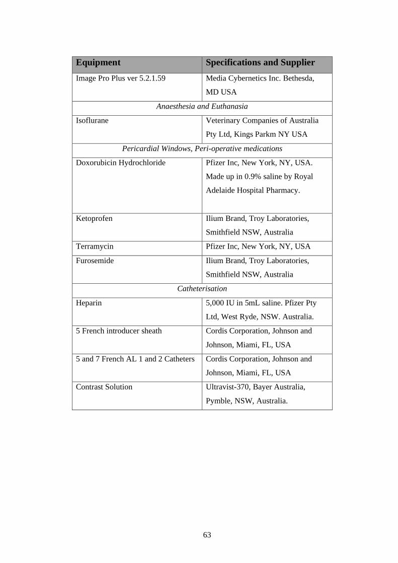
63
Equipment Specifications and Supplier Image Pro Plus ver 5.2.1.59 Media Cybernetics Inc. Bethesda,
MD USA
Anaesthesia and Euthanasia
Isoflurane Veterinary Companies of Australia
Pty Ltd, Kings Parkm NY USA
Pericardial Windows, Peri-operative medications
Doxorubicin Hydrochloride Pfizer Inc, New York, NY, USA.
Made up in 0.9% saline by Royal
Adelaide Hospital Pharmacy.
Ketoprofen Ilium Brand, Troy Laboratories,
Smithfield NSW, Australia
Terramycin Pfizer Inc, New York, NY, USA
Furosemide Ilium Brand, Troy Laboratories,
Smithfield NSW, Australia
Catheterisation
Heparin 5,000 IU in 5mL saline. Pfizer Pty
Ltd, West Ryde, NSW. Australia.
5 French introducer sheath Cordis Corporation, Johnson and
Johnson, Miami, FL, USA
5 and 7 French AL 1 and 2 Catheters Cordis Corporation, Johnson and
Johnson, Miami, FL, USA
Contrast Solution Ultravist-370, Bayer Australia,
Pymble, NSW, Australia.

64
Results

65
Chapter 3: Results
3.1 Omega-3 PUFA Dosing Study
Omega PUFA level data is presented as a percentage of total fatty acid content
within the plasma, erythrocyte (RBC) or myocardial membrane bound fatty acid as
indicated.
3.1.1 Omega-3 PUFA Levels
3.1.1.1 Omega-3 PUFA Baseline levels
At baseline, the plasma levels of total omega-3 long chain PUFA were significantly
higher in the dosing study sheep assigned to the control group than in the omega-3
group, 14.89 ± 1.70 % Control Group Dosing study (n=4) vs 7.26 + 0.94% Omega-3
dosing study sheep (n=4) (p<0.05 ANOVA, with post test Tukey Multiple
Comparison Test)), Table 3.1.1.
The baseline erythrocyte (RBC) membrane bound levels of total omega-3 long
chain PUFA were also significantly higher in the dosing study sheep assigned to the
control group, compared with those in the dosing study omega-3 group; 2.08 ±
0.41% Control Group Dosing study (n=4) vs 1.57 ± 0.19% omega-3 group dosing
study (n=4) (p<0.05 ANOVA, with post test Tukey Multiple Comparison Test)),
Table 3.1.1.

66
Omega-3 Long Chain fatty acid
(n-3 LC PUFA)
Omega-3 Group Dosing Study
(n=4)
Control Group Dosing Study
(n=4) Baseline Plasma Phospholipids Levels
18:3n-3 (ALA)1
5.95 ± 0.94a
2.32 ± 0.20b
20:5n-3 (EPA)1
1.71 ± 0.22a
2.76 ± 0.56bc
22:5n-3 (DPA)2
2.76 ± 0.21a
5.52 ± 1.00c
22:6n-3 (DHA)
2.79 ± 0.59a
6.61 ± 0.96c
Total n-32
13.78 ± 0.73a
17.64 ± 1.53b
Total n-3 LC PUFA1 7.26 ± 0.94a 14.89 ± 1.70b
Baseline RBC Levels
18:3n-3 (ALA)2
0.97 ± 0.14ab
1.15 ± 0.21a
20:5n-3 (EPA)2
0.65 ± 0.08a
0.63 ± 0.19ab
22:5n-3 (DPA)2
0.56 ± 0.07a
0.95 ± 0.21b
22:6n-3 (DHA) 2
0.36 ± 0.09a
0.50 ± 0.08a
Total n-32
2.78 ± 0.31a
3.52 ± 0.55b
Total n-3 LC PUFA2
1.57 ± 0.19ab
2.08 ± 0.41c
Table 3.1.1 Plasma and erythrocyte bound (RBC) baseline levels of long chain omega-3
PUFA. Plasma and RBC total long chain omega-3 PUFA were higher at baseline in the
Dosing study control compared with the omega-3 group sheep (p<0.05). Values within a row
with a different superscript are significantly different (p<0.05, ANOVA with post test with
Tukey-Kramer Multiple Comparisons Test).

67
3.1.1.2 Omega-3 PUFA drenching study – erythrocyte membrane bound
levels
Drenching sheep with 23mLs fish oil containing 300mg/mL EPA+DHA three times
per week caused a significant increase in erythrocyte membrane bound levels of
omega-3 long chain PUFA compared with olive oil fed placebo controls, (p<0.05
ANOVA, with post test Tukey Multiple Comparison Test), (Figure 3.1.2). The
increase in omega-3 PUFA levels reached significance between three to four weeks
of drenching with fish oil and then stabilized following an additional three to four
weeks of drenching. The plasma levels of omega-3 PUFA did not change in the
control sheep (receiving olive oil placebo) over the duration of the study (p>0.05).
3.1.1.3 Omega-3 PUFA drenching study - myocardial levels
Samples of left and right ventricular (LV/RV) and right atrial tissue (RA) were
harvested from the sheep hearts at time of sacrifice for assessment of myocardial
phospholipid bound levels of omega-3 PUFA. There was a significant,
approximately two-fold increase in the myocardial levels of omega-3 PUFA in
myocardium taken from the omega-3 group sheep compared with controls (p
<0.05), (Figure 3.1.3).
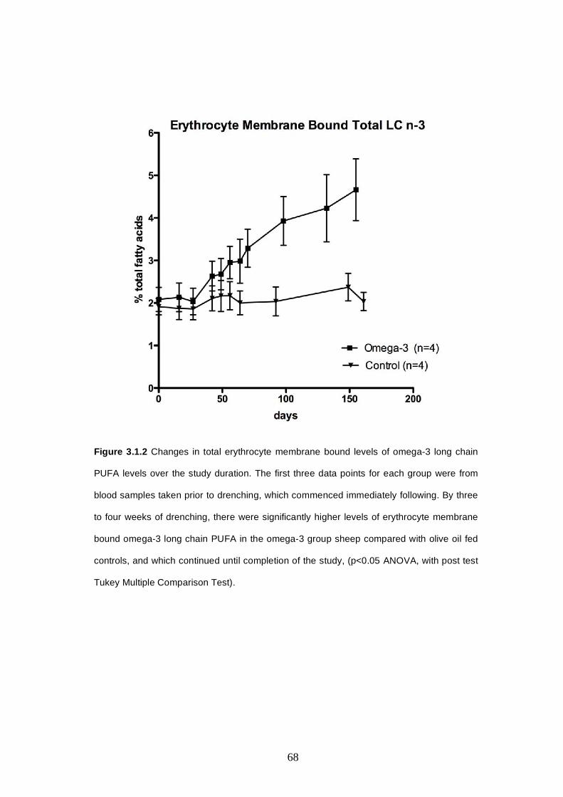
68
Figure 3.1.2 Changes in total erythrocyte membrane bound levels of omega-3 long chain
PUFA levels over the study duration. The first three data points for each group were from
blood samples taken prior to drenching, which commenced immediately following. By three
to four weeks of drenching, there were significantly higher levels of erythrocyte membrane
bound omega-3 long chain PUFA in the omega-3 group sheep compared with olive oil fed
controls, and which continued until completion of the study, (p<0.05 ANOVA, with post test
Tukey Multiple Comparison Test).
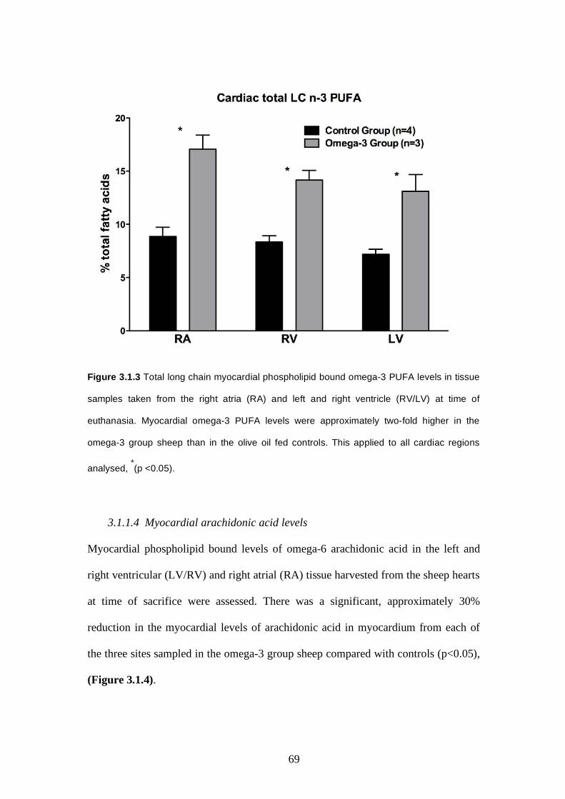
69
Figure 3.1.3 Total long chain myocardial phospholipid bound omega-3 PUFA levels in tissue
samples taken from the right atria (RA) and left and right ventricle (RV/LV) at time of
euthanasia. Myocardial omega-3 PUFA levels were approximately two-fold higher in the
omega-3 group sheep than in the olive oil fed controls. This applied to all cardiac regions
analysed, *(p <0.05).
3.1.1.4 Myocardial arachidonic acid levels
Myocardial phospholipid bound levels of omega-6 arachidonic acid in the left and
right ventricular (LV/RV) and right atrial (RA) tissue harvested from the sheep hearts
at time of sacrifice were assessed. There was a significant, approximately 30%
reduction in the myocardial levels of arachidonic acid in myocardium from each of
the three sites sampled in the omega-3 group sheep compared with controls (p<0.05),
(Figure 3.1.4).

70
Figure 3.1.4 Myocardial bound omega-6 arachidonic acid levels from right atria and left
and right ventricular samples taken from sheep myocardium at time of euthanasia. There
was a significant, approximately 30% reduction in arachidonic acid levels in myocardium
from omega-3 group sheep compared with controls at each of the three sites sampled
(*p<0.05).
3.2 Ovine DOX-infusion Study
3.2.1 Clinical Results
3.2.1.1 Mortality rate
A total of 17 animals (weight range 41 to 62.5kg) received the intra-coronary DOX
infusion protocol (eight omega-3 and nine placebo fed controls). The baseline
parameters for these animals are shown in Table 3.2.1. There were three premature
deaths in each group, (omega-3 treated (37.5%) and control groups (33%)). The
control group deaths occurred between three and 12 weeks following the final
scheduled DOX dosing. One animal died suddenly in its holding pen 12 weeks
following the final DOX infusion and hours prior to the final scheduled CMR. This

71
animal had appeared well in the weeks following DOX dosing. The sudden nature
of its death implicated an arrhythmic cause. The second sheep that died early
appeared in good condition during the weeks following its final DOX dose, but died
suddenly in its field pen at six weeks post DOX 3. The absence of preceding signs
and the sudden nature of its death also suggested ventricular arrhythmia (VA) as
cause of death. The heart and lungs were retrieved for histopathology assessment.
The third placebo sheep developed malaise at three weeks following its final DOX
dose. The animal appeared in discomfort and ceased feeding. A provisional
diagnosis of fluid retention secondary to cardiac failure was made. Fluid was
withheld overnight and 3 doses of furosemide (50mg) were administered throughout
the following day to diurese the animal of excess fluid. The sheep’s condition
showed initial improvement, but 24 hours later the animal suffered a witnessed
cardiac arrest and could not be revived. The heart was removed intact for
histological assessment.
In the omega-3 group, two animals died suddenly, one at six and the other at seven
weeks following final DOX dose. In both cases, there were no preceding signs of
cardiac failure nor non-cardiac pathology, indicating sudden cardiac arrhythmia as
the likely cause of death. The third premature death in the omega-3 cohort occurred
as a result of procedural complications during the final DOX dosing.
3.2.1.2 Electrocardiographic changes
Electrocardiogram changes were frequently observed during intra-coronary DOX
infusion in sheep in both groups. Most frequent changes consisted of transient ST
segment elevation or depression. Infusions were never associated with ectopic beats

72
nor arrhythmia and ECG changes always resolved prior to completion of the
angiographic procedure.
Control Group
Omega-3 Group
n
9 8
Weight Range
41 to 62.5kg
41 to 62kg
Mean B/L Weight (kg)
(p=NS)
58.0 + 14
53.0 + 12
Mean B/L LVEF (%) (p=NS)
42.5 + 2.3
48.6 + 2.6
Mortality Rate
3/9 (33%)
3/8 (37.5%)
Likely cause of early
deaths
Arrhythmia : (n=2 sheep) Congestive Heart Failure:
(n=1)
Arrhythmia: (n=2). Procedural: (n=1)
Mean F/U Period for Survivors from DOX 1
(days)
89 + 7 days
90 + days
(6.03 SEM)
Table 3.2.1 Baseline and follow up parameters for sheep in DOX-infusion study. At
baseline there was no difference in mean Weight, nor LVEF (p=NS) between groups.
B/L = Baseline, F/U = Follow Up.
3.2.2 Left Ventricular Ejection Fraction and Volume Changes as
Assessed by CMR
The LV EF was similar between omega-3 and control group animals at baseline
(p=NS). DOX-infusion resulted in a significant decline in LV EF for both omega-3
and control groups (p<0.05), however, the Omega 3 group declined to a greater
extent (19.4% absolute reduction; (from 48.6 + 7.4% baseline to 29.2 + 6.9% final))
compared to the placebo controls (8.1% absolute reduction; (from 42.5 + 6.8%
baseline to 34.4 + 7.5)), (p<0.05 for intergroup comparison), (Figure 3.2.2A).

73
The LV EF changes were accompanied by a significant increase in LV End-Diastolic
volume (EDV) from baseline (72.9 + 18.0mLs) to final (110.3 + 38.5mLs) in the
Omega-3 group (p<0.05), with no change in the control EDV over time (baseline
75.3 + 13.0 to final 73.3 + 11.0), (p=NS). There was a significant difference in final
EDV between groups (p<0.05), however there was no group effect on EDV change
over time by two-way ANOVA (p=NS), (Figure 3.2.2B).
Left ventricular ESV showed a significant increase in the omega-3 group (38.1 +
12.8mLs baseline to 79.1 + 33.0mLs final) (p<0.05) with no change over time in the
control group (43.7 + 10.7mLs baseline to 49.4 + 9.7mLs final), (p=NS). Although
there was a significant difference in final ESV between groups (p<0.05), there was no
group effect on ESV over time, using two-way ANOVA (p=NS), (Figure 3.2.2C).
Figure 3.2.3 shows CMR generated long and short axis TrueFISP images of the heart
in a control sheep pre- and post DOX infusion, showing wall thinning and dilatation
changes typical of DOX-induced cardiomyopathy.
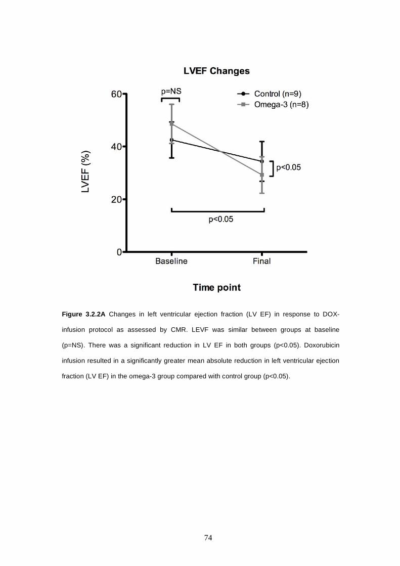
74
Figure 3.2.2A Changes in left ventricular ejection fraction (LV EF) in response to DOX-
infusion protocol as assessed by CMR. LEVF was similar between groups at baseline
(p=NS). There was a significant reduction in LV EF in both groups (p<0.05). Doxorubicin
infusion resulted in a significantly greater mean absolute reduction in left ventricular ejection
fraction (LV EF) in the omega-3 group compared with control group (p<0.05).

75
B
C
Figure 3.2.2 B and C. Changes in left ventricular end diastolic volume (EDV) (B) end systolic
volume (ESV) (C) in response to DOX-infusion protocol. EDV increased significantly in the
Omega-3 group, (p<0.05), with no change in the control EDV over time (p=NS). There was a
significant difference in final EDV between groups (p<0.05). The LV End-Systolic volume
(ESV) increased significantly in the omega-3 group with no change over time in the control
group (p<0.05). There was a significant difference in final ESV between groups (p<0.05).

76
Figure 3.2.3 Long axis (panels AB, EF) and short axis (CD, GH) TrueFISP CMR images of
the heart at baseline (A-D) and final assessment (E-F) in a sheep with severe LV dilation and
systolic dysfunction following DOX infusion protocol.
3.2.3 Fractional Shortening, End Diastole and End Systole Diameter
Changes as assessed by Trans Thoracic Echocardiogram
Short axis subvalvular mean LV fractional shortening (SAX FS) was measured via
echocardiogram prior to each DOX infusion and then immediately prior to sacrifice.
Fractional shortening decreased significantly in response to DOX infusion in both
groups (baseline FS 33.3 + 4.5% to 20.1 + 4.5% final in control group, and from
baseline 34.4 + 5.0% to 26.2 + 7.7%, final in omega-3 group, (p<0.01). Significant
deterioration of FS was not observed until after the final DOX infusion. There were
no group nor time effect differences on FS when assessed by two-way ANOVA
(p=NS). The changes in SAX FS following each DOX infusion are shown in Figure
3.2.4.

77
Figure 3.2.4. Left Ventricular Transthoracic echocardiography short axis Fractional
Shortening Changes (FS). FS decreased significantly in response to DOX infusion in both the
control and omega-3 groups, (p<0.01)). The decrease did not reach significance until
following the third (final) DOX infusion. There were no group nor time effect differences in FS
(p=NS).
3.2.4 Blood Results
Due to quarantine restrictions arising at the Large Animal Research Facility at Gilles
Plains (LARIF) during the study, and where several of the study euthanasias were
performed, final blood samples from one omega-3 group and five control group sheep
could not be released for cell and serum marker levels analysis. Statistical analyses

78
was performed using endpoint data from the remaining animals, however the affect of
the missing final data points in terms of statistical significance is unknown.
3.2.4.1 Troponin-T post DOX infusion
Elevations of serum troponin-T titer were generally mild following each DOX
infusion, ranging from 0.03 to 0.66ug/L in the control group and 0.03 to 0.18ug/L in
the omega-3 group animals. (Figure 3.2.6A). Four (44%), five (56%) and four (44%)
sheep in the Control group returned positive troponin-T levels following DOX 1, 2
and 3 infusions respectively. Six of eight sheep (75%) in the omega-3 group showed
mild to moderate troponin-T rises following each of the three DOX infusions (Figure
3.2.6B). Of these, five animals showed a troponin-T rise following each of the three
DOX infusions.
3.2.4.2 Haemoglobin, white cell count and platelets
There were no group nor time effects on Haemoglobin levels in response to DOX
infusion, p=NS, (Figure 3.2.7A).
There were no group effects on White Cell Count (WCC), (p=NS). The WCC in both
groups showed a non-significant decline following the first DOX infusion (p=NS),
with a further significant decline following the second DOX infusion (p<0.05). The
WCC levels then stabilised following the third and final DOX dose. The decline in
WCC from baseline to final in both groups (time effect) was significant (p<0.01),
(Figure 3.2.7B).
The platelet count in both groups showed a significant decline following the first
DOX infusion (p=<0.05), which then stabilised following the second and third DOX

79
infusion. The change in platelet count from baseline to final in both groups (time
effect) was not significant (p=NS), (Figure 3.2.7C).
A
B
Figure 3.2.6 Mean Troponin-T levels post each DOX infusion for each group (A) and %
positive Troponin-T rises following each DOX infusion for each group (B).
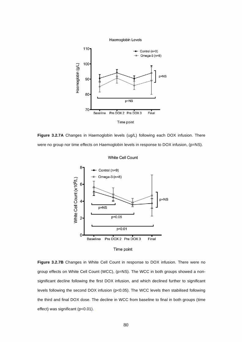
80
Figure 3.2.7A Changes in Haemoglobin levels (ug/L) following each DOX infusion. There
were no group nor time effects on Haemoglobin levels in response to DOX infusion, (p=NS).
Figure 3.2.7B Changes in White Cell Count in response to DOX infusion. There were no
group effects on White Cell Count (WCC), (p=NS). The WCC in both groups showed a non-
significant decline following the first DOX infusion, and which declined further to significant
levels following the second DOX infusion (p<0.05). The WCC levels then stabilised following
the third and final DOX dose. The decline in WCC from baseline to final in both groups (time
effect) was significant (p<0.01).
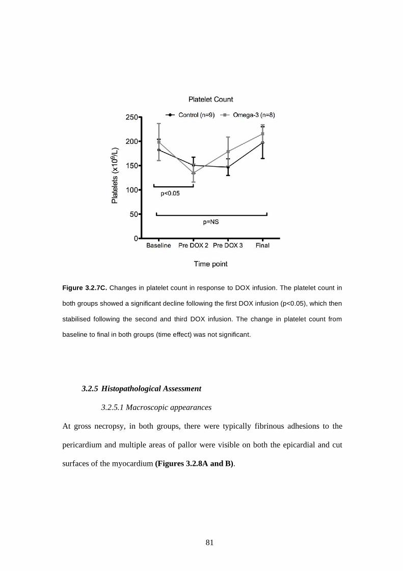
81
Figure 3.2.7C. Changes in platelet count in response to DOX infusion. The platelet count in
both groups showed a significant decline following the first DOX infusion (p<0.05), which then
stabilised following the second and third DOX infusion. The change in platelet count from
baseline to final in both groups (time effect) was not significant.
3.2.5 Histopathological Assessment
3.2.5.1 Macroscopic appearances
At gross necropsy, in both groups, there were typically fibrinous adhesions to the
pericardium and multiple areas of pallor were visible on both the epicardial and cut
surfaces of the myocardium (Figures 3.2.8A and B).

82
Figure 3.2.8A A formalin fixed intact ovine heart retrieved 12 weeks after three intra-coronary
doses of 1.2mg/kg Doxorubicin. Large areas of surface pallor are clearly visible on the lateral
surface of the left ventricle.
Figure 3.2.8B. Ventricular cut surface taken at basal (sub mitral valve) level, of Doxorubicin
treated formalin fixed heart. This image shows the slicing protocol used for the mid and basal
slices, ie: commencing clockwise from top segment; anterior, antero-lateral, postero-lateral,
posterior, posterior-septal, anterior-septal. Foci of midmural pallor are clearly visible in each
segment, particularly the anterior, antero-lateral and postero-lateral segments.

83
3.2.5.2 Histopathological findings
Histological review of Haematoxylin and Eosin stained slide sections from both
groups revealed several pathological features typical of DOX-induced tissue damage.
These included vacuolation within cardiomyocyte cytoplasm, loss of myofibrillar
striations, areas of fibrosis with associated fibroblast proliferation and histiocyte
infiltrate, vacuolization of purkinje fibre cells, occasional foci of necrotic cells and
areas of perivascular oedema. Further changes included vasculitis, pyknosis of
endothelial cells, suggestive of apoptosis, and mural lymphocyte infiltrates.
3.2.5.3 Ventricular fibrosis burden
Doxorubicin treatment resulted in the presence of both interstitial and replacement
fibrosis distributed heterogeneously throughout the ventricular myocardium in both
omega-3 and placebo animals. Foci of replacement fibrosis were typically small-
moderate in size and were most frequently located in the sub-epicardial and/or mid-
wall layers of the myocardium.
Quantification of the percent (%) area of fibrosis was determined from the van
Gieson stained sections by using a colour cube based segmentation algorithm within
Image Pro Plus software (ver 5.2.1) to select fibrotic tissue against normal
myocardium, (Figure 3.2.9).
The mean % area of fibrosis was calculated for each segment within each of the four
myocardial levels sectioned from each heart ie: basal, mid, distal and apical levels.
Mean % area of fibrosis was then calculated for each level, and also combined for all
levels, and compared with normal healthy untreated myocardium. For the mean %
area of fibrosis for all levels combined, there was a significantly greater % area of
fibrosis in the placebo (3.73 + 0.62%) and omega-3 (3.87 + 1.91%) groups compared
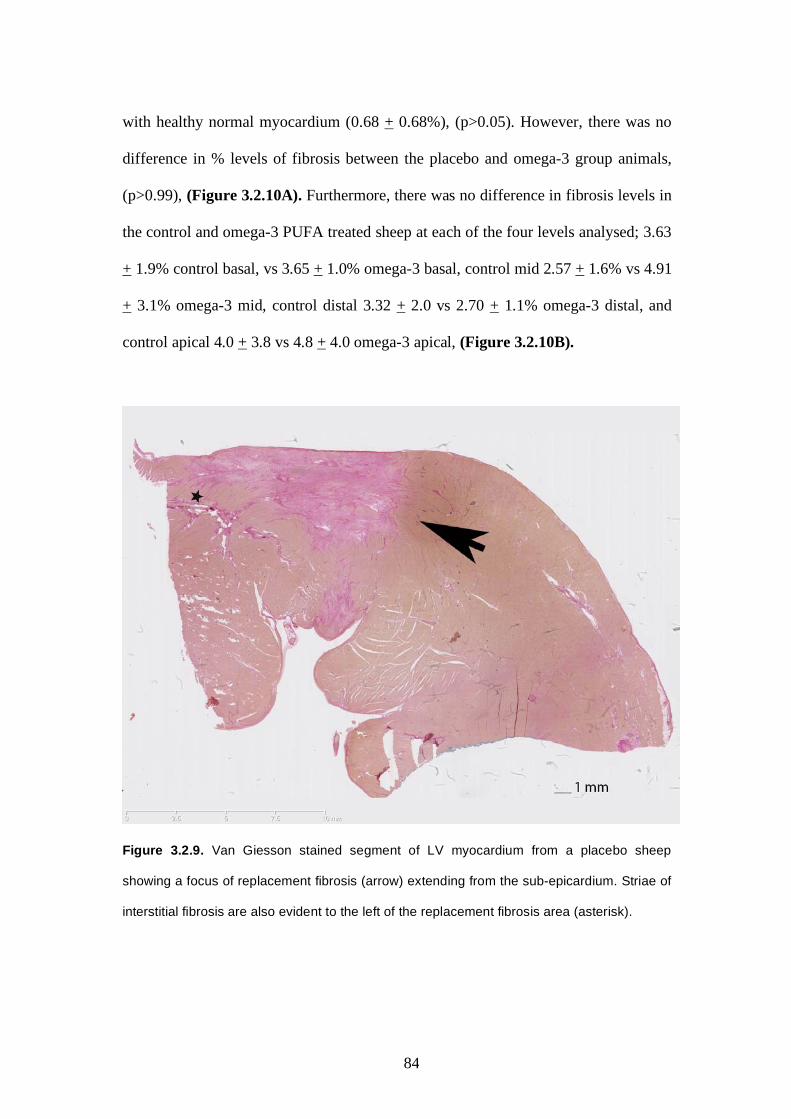
84
with healthy normal myocardium (0.68 + 0.68%), (p>0.05). However, there was no
difference in % levels of fibrosis between the placebo and omega-3 group animals,
(p>0.99), (Figure 3.2.10A). Furthermore, there was no difference in fibrosis levels in
the control and omega-3 PUFA treated sheep at each of the four levels analysed; 3.63
+ 1.9% control basal, vs 3.65 + 1.0% omega-3 basal, control mid 2.57 + 1.6% vs 4.91
+ 3.1% omega-3 mid, control distal 3.32 + 2.0 vs 2.70 + 1.1% omega-3 distal, and
control apical 4.0 + 3.8 vs 4.8 + 4.0 omega-3 apical, (Figure 3.2.10B).
Figure 3.2.9. Van Giesson stained segment of LV myocardium from a placebo sheep
showing a focus of replacement fibrosis (arrow) extending from the sub-epicardium. Striae of
interstitial fibrosis are also evident to the left of the replacement fibrosis area (asterisk).
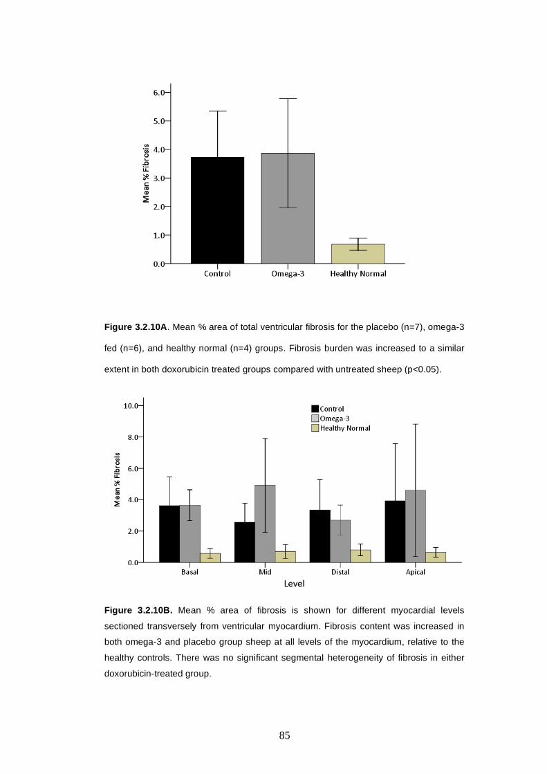
85
Figure 3.2.10A. Mean % area of total ventricular fibrosis for the placebo (n=7), omega-3
fed (n=6), and healthy normal (n=4) groups. Fibrosis burden was increased to a similar
extent in both doxorubicin treated groups compared with untreated sheep (p<0.05).
Figure 3.2.10B. Mean % area of fibrosis is shown for different myocardial levels
sectioned transversely from ventricular myocardium. Fibrosis content was increased in
both omega-3 and placebo group sheep at all levels of the myocardium, relative to the
healthy controls. There was no significant segmental heterogeneity of fibrosis in either
doxorubicin-treated group.

86
Discussion

87
Chapter 4: Discussion
4.1 Study Objectives Despite a number of treatment regimens devised to mitigate the adverse
cardiovascular effects of anthracyclines, chemotherapy-induced cardiomyopathy
remains an important cause of patient morbidity and mortality. Recently, omega-3
PUFA have been considered for their adjunctive protective actions against
anthracycline cardiotoxicity. Previous studies utilising either small animal or in vitro
model systems have yielded conflicting findings, such that further translational
research is required before progression to clinical investigation. We therefore
conducted a novel, large animal study to evaluate the effects of dietary omega-3
PUFA supplementation on cardiac function and remodelling in a validated model of
anthracycline-induced cardiomyopathy. This provided further translational preclinical
data to complement the pre-existing basic scientific literature. The study utilised
CMR, which is considered the current “gold standard” imaging modality for non-
invasive assessment of cardiac volumes and function and state of the art histological
analysis to quantify and compare levels of myocardial fibrosis between groups.
4.2 Uptake of Omega-3 PUFA in Merino Sheep
This study demonstrated that dietary supplementation of fish oil containing omega-3
PUFA in Merino wethers caused an elevation in erythrocyte and myocardial-bound
levels of long chain omega-3 PUFA. An accompanying and significant reduction in
the myocardial levels of arachidonic acid (ARA) in the myocardium in the omega-3
group sheep compared with controls (p<0.05), was also observed, suggesting that the

88
long chain omega-3 PUFA displaced phospholipid-bound ARA within cell membrane
phospholipids. As indicated previously, incorporation of omega-3 fatty acids into the
membrane phospholipid bilayer is a prerequisite for its biological activity87. These
findings confirm that dehydrogenation of omega-3 PUFA in the sheep rumen
following oral ingestion did not preclude its uptake into plasma and ultimately,
phospholipid bound cell membranes. This has not been previously reported in the
Merino sheep and demonstrates that this strain is a suitable preclinical large animal
surrogate for multi-disciplinary studies assessing effects of dietary omega-3 PUFA
supplementation.
4.2.1 Elevated Omega-3 PUFA levels at baseline
At baseline, the plasma and phospholipid bound levels of total omega-3 long chain
PUFA were significantly higher in the dosing study sheep assigned to the control
group compared with the omega-3 group. During the weeks leading up to the dosing
study, all sheep were housed in large outdoor field pens. They received standard feed,
lucerne chaff, however being housed outdoors, may also have had varying access to
fresh pasture feed. Lucerne chaff contains negligible omega-3 PUFA, however fresh
pasture feed may have contained sufficient levels of omega-3 PUFA to elevate the
plasma and erythrocyte bound omega-3 PUFA levels in the control animals at
baseline.
Changes were implemented following the initial dosing study in an attempt to address
baseline variations in plasma and erythrocyte phospholipid bound omega-3 PUFA
levels. These included transferring the animals from outdoor to indoor pen enclosures
two weeks prior to the commencement of drenching, thereby removing all access to
fresh pasture feed. However, some study animals in the second main study batch

89
nevertheless returned elevated baseline omega-3 levels. It is likely that levels of
circulating and tissue bound omega-3 PUFA persisted in these sheep, even following
two weeks abstinence from fresh pasture feed. Significantly however, omega-3
PUFA levels in all sheep receiving dietary supplementation of fish oil increased
above baseline levels within 2-3 weeks of commencement of drenching, and
stabilised to approximately two- to three-fold that of placebo group animals.
4.2.2 Implementation of Olive Oil placebo drenching for main study
The fish oil drenching protocol resulted in sheep receiving a high oil dietary intake
compared with their normal diet, the effects of which were unknown in ruminant
animals. The olive-oil placebo drenching was therefore implemented for the main
study animals to account for any adverse or other systemic effects relating solely to a
high oil intake diet.
4.3 Cardiac effect of Omega-3 PUFA in Ovine model of DOX-
induced NICM
Dietary supplementation of omega-3 PUFA was associated with a greater decline in
LV EF as assessed by CMR in response to intra-coronary anthracycline infusion
compared with control animals receiving olive oil placebo. Furthermore, there was a
significant increase in ESV and EDV as measured by CMR in the omega-3 group
compared with placebo controls. This indicates that omega-3 PUFA resulted in
augmented cardiac remodelling and systolic dysfunction in the setting of DOX
exposure, a finding that rejects our primary hypothesis and contradicts the many other
purported benefits of omega-3 PUFA on cardiac health. Notably, the similar levels of

90
fibrosis observed in both the control and omega-3 PUFA treated animals suggest that
the greater adverse effect of DOX on cardiac function in the omega-3 group may be
mediated by mechanism/s independent of fibrosis.
4.3.1 Possible mechanisms for adverse effects of Omega-3 PUFA on DOX-
induced NICM
Administration of DOX induces cyclooxygenase-2 (COX-2) expression118, which
catalyzes the first step in the conversion of ARA to Prostglandins (PG).
Prostaglandins are a group of 20 carbon lipid compounds synthesized ubiquitously
within nucleated cells in response to injury. Though they are generally regarded as
mediators of several pro-inflammatory effects, certain types such as PGE1 and
prostacyclin (PGI2) also confer tissue benefits following injury, including suppression
of apoptosis119, suppression of necrosis and release of cytotoxic factors120, and
stimulation of neutrophil activation by dilating vasculature, particularly in disease
conditions mediated by ROS121.
In the rat model of cardiac injury by Dowd119, DOX administered to rat neonatal
cardiomyocytes induced cardiac cell injury, as demonstrated by an increase in plasma
cardiac troponin-T, serum lactate dehydrogenase (LDH) and apoptosis. Blocking the
release of PG via a specific COX-2 inhibitor (SC236) further aggravated the DOX-
induced injury. In contrast, prior administration of a PG analogue (iloprost)
attenuated the degree of injury. Thus, the release of PG by cardiomyocytes may have
attenuated their sensitivity to DOX insult, largely via a reduction in apoptosis.
As discussed previously, incorporation of EPA and DHA into cell membranes occurs
via substitution of ARA. In our study we observed a 30% reduction in myocardial
bound ARA levels in the omega-3 PUFA fed animals compared with the placebo

91
group. EPA is an n-3 homologue of ARA and as such, it competitively inhibits COX-
2 metabolism of ARA to PG. It acts as a competitive inhibitor. This reduction of
COX-2-generated PGs may partly account for the mechanism by which omega-3
PUFA supplementation aggravated cardiotoxicity.
The cellular pathways which mediate anthracycline-induced cardiotoxicity are
multiple and complex. The finding of similar fibrosis levels between the omega-3 and
control groups in the current study may suggest that the greater decline in LV EF in
the omega-3 group animals compared with control animals, was mediated by
pathological pathways independent of fibrosis. As discussed previously,
anthracyclines also induce apoptosis, down regulate cardiomyocyte contractile
proteins and effect other changes leading to cardiomyopathy. One or more of these
processes may be amplified in the context of omega-3 enrichment, which also exerts
numerous cellular changes, manifesting greater decline in cardiac function.
Further assessment of histological markers such as apoptosis, contractile protein
status, CMR parameters (including diastology and wall thickness changes between
groups), as well as RNA micro-array analyses of myocardial samples are planned as a
follow-on to this thesis to provide alternative mechanistic insights.
4.4 Attrition Rate
The attrition rate in this study was higher than anticipated. Six of 17 (35%) sheep
died prematurely, compared with our previously observed mortality rate of 20%109.
One reason for this difference in mortality rate may have been a subtle change to the
DOX dosing regimen. Our previous protocol consisted of administering 3-4 doses of
DOX at 0.75 and up to 1mg/kg, with a mean cumulative dose of 3.6 mg/kg109. In
order to ensure consistent dosing amongst all animals in the current placebo-
92
controlled study, we opted to standardise the regimen to three doses of 1.2mg/kg.
This also enabled an ethically favourable reduction in the number of procedural
interventions, whilst achieving our optimal cumulative DOX dose to induce moderate
and reproducible cardiomyopathy. Although there was some individual variation in
response to doxorubicin, all sheep in the current study showed deterioration in global
LV function by 12 weeks follow up. This follow-up period was longer than the six-
week duration used in our validation study109. Indeed, two animals (one from each
group) died from suspected cardiac arrhythmia between six and twelve weeks post-
DOX, which also contributed to the high mortality rate observed in the current study.
Anthracycline-induced cardiomyopathy was characterised in this study by moderate
decline of LV EF and FS from baseline levels, and increases in EDV and ESV
(omega-3 group only) as measured by CMR. It was also characterised by mild
elevations in Troponin-T levels, accompanying changes in WCC and platelet levels,
and significant myocardial fibrosis.
4.5 Study Limitations
4.5.1 Anthracycline Administration and Dosage
This study utilised intra-coronary administration of anthracyclines, with the objective
of minimising systemic toxicity, and associated attrition rate, by achieving higher
first pass concentration of DOX in the heart. In the clinical setting, chemotherapy
agents are typically administered intra-venously. However in animal models of IV
administered anthracycline, attrition rates of greater than 50% have been reported122.
The model’s strength is that it enables reproducible cardiotoxicity with acceptable
animal mortality and overt morbidity. However, its limitation, is that it does not
simulate the systemic administration of DOX in clinical chemotherapeutic practice.
93
Myocardial uptake of DOX might be less predictable by the IV route, although the
difference between the two delivery strategies is unknown. Changes in somatic
effects of DOX by adopting the intra-coronary compared with IV route are also
unknown.
4.5.2 Absence of Neoplasia
The animals included in this study were young adults free of any known neoplastic or
other adverse health conditions. The influence of neoplastic disease in this model is
therefore unknown, including the pharmacological effects of DOX-infusion on
tumour cells during dietary supplementation of omega-3 PUFA. Since the proposed
model excluded neoplasia, the findings relate specifically to the effects on the
myocardium of Omega-3 PUFA in sheep receiving DOX.
4.5.3 General Anaesthesia
All CMR and TTE measurements were performed under general anaesthesia. Though
the anaesthetic protocol was standardised for each procedure throughout the study,
the influence of anaesthesia on the cardiac outcomes assessed is unknown.
4.5.4 Follow-up Period
The 12 week follow up period in this study was selected to allow sufficient
progression of DOX-induced NICM. It is believed that a longer follow up period in
this model may have resulted in advancing disease progression and further escalation
of the study attrition rate. In the clinical setting, patients may continue dietary
supplementation of omega-3 PUFA for extended periods following chemotherapy.

94
The affects of long term omega-3 PUFA supplementation in the context of
anthracycline-induced NICM is unknown.
4.5.5 Non reporting of some histopathology samples
Mean % area ventricular fibrosis were not reported for one of the omega-3 and two of
the control group sheep due to quarantine restrictions placed at the IMVS field station
at LARIF prohibiting the removal of their samples for omega-3 PUFA and fibrosis
level analysis following the scheduled euthanasia. However, given the high level of
variance of these levels between the placebo and omega-3 group sheep in the samples
analysed, it is unlikely that the inclusion of myocardial and blood samples from the
one additional placebo and two omega-3 group sheep hearts would have altered the
outcome reported, which showed no statistically significant difference between these
groups.
95
Summary and Future Directions
NICM contributes to approximately one third of cases of clinical heart failure and
comprises various underlying aetiologies, including idiopathic, genetic, infectious,
metabolic, iatrogenic and toxic processes123.
In an ovine model of DOX-induced NICM, we found no beneficial effects of dietary
supplementation with fish oil derived omega-3 long chain PUFA. This is despite
confirming incorporation of omega-3 PUFA into the myocardial tissue. In fact, we
found evidence of a detrimental effect with this therapy. Therefore, based on findings
from this model, we would not recommend co-administration of omega-3 PUFA for
the prevention of anthracycline-induced myocardial injury.
Basic mechanistic studies to better define the interactions between fatty acids and
cardiac biology in the presence of anthracycline exposure are required before clinical
evaluation can take place. This should include gene expression microarray, biomarker
and histological analyses to further explore response to anthracycline and omega-3
PUFA co-administration, and with particular attention to inflammatory, oxidative
stress and apoptotic pathways. Further, sub cellular structural changes should be
evaluated to determine whether omega-3 PUFA attenuates or in fact potentiates these
adverse effects.
The ovine model of DOX-induced cardiomyopathy described in this study has many
potential applications for the investigation of novel therapies in the management of
NICM, and has distinct advantages over small animal and in vitro models of DOX-
induced cardiotoxicity.
Though this study was undertaken essentially in the context of cancer patients
receiving anthracyclines, the model has proved particularly useful in investigating
novel therapies in the setting of non-ischaemic heart disease.

96
References
1. ABS. ABS National Health Survey. 2004.
2. Forman D, Stockton D, Moller H, Quinn M, Babb P, De Angelis R,
Micheli A. Cancer prevalence in the UK: results from the
EUROPREVAL study. Ann Oncol. 2003;14(4):648-654.
3. Mariotto AB, Rowland JH, Ries LA, Scoppa S, Feuer EJ. Multiple cancer
prevalence: a growing challenge in long-term survivorship. Cancer
Epidemiol Biomarkers Prev. 2007;16(3):566-571.
4. Edwards BK, Brown ML, Wingo PA, Howe HL, Ward E, Ries LA,
Schrag D, Jamison PM, Jemal A, Wu XC, Friedman C, Harlan L, Warren
J, Anderson RN, Pickle LW. Annual report to the nation on the status of
cancer, 1975-2002, featuring population-based trends in cancer treatment.
Journal of the National Cancer Institute. 2005;97(19):1407-1427.
5. Ries LA, Wingo PA, Miller DS, Howe HL, Weir HK, Rosenberg HM,
Vernon SW, Cronin K, Edwards BK. The annual report to the nation on
the status of cancer, 1973-1997, with a special section on colorectal
cancer. Cancer. 2000;88(10):2398-2424.
6. Wingo PA, Ries LA, Parker SL, Heath CW, Jr. Long-term cancer patient
survival in the United States. Cancer Epidemiol Biomarkers Prev.
1998;7(4):271-282.
7. The Cancer Council of New South Wales. Cancer in Australia: an
overview, 2006. 2006.
8. Belani CP. The ANITA trial seals the deal for adjuvant therapy in non-
small-cell lung cancer. Clin Lung Cancer. 2005;6(6):331-332.
97
9. Chemotherapy for Blood-Related Cancers. National Marrow Donor
Program. 2008.
10. Takemura G, Fujiwara H. Doxorubicin-induced cardiomyopathy from the
cardiotoxic mechanisms to management. Prog Cardiovasc Dis.
2007;49(5):330-352.
11. Pawan K SD, Iliskovic N. Doxorubicin Induced Cardiomyopathy. NEJM.
1998;339(13):900-905.
12. van Dalen EC, Caron HN, Kremer LC. Prevention of anthracycline-
induced cardiotoxicity in children: the evidence. Eur J Cancer.
2007;43(7):1134-1140.
13. Sinha BK, Katki AG, Batist G, Cowan KH, Myers CE. Adriamycin-
stimulated hydroxyl radical formation in human breast tumor cells.
Biochemical pharmacology. 1987;36(6):793-796.
14. Goodman J, Hochstein P. Generation of free radicals and lipid
peroxidation by redox cycling of adriamycin and daunomycin.
Biochemical and biophysical research communications. 1977;77(2):797-
803.
15. Bachur NR, Gordon SL, Gee MV. Anthracycline antibiotic augmentation
of microsomal electron transport and free radical formation. Molecular
pharmacology. 1977;13(5):901-910.
16. Sarvazyan N. Visualization of doxorubicin-induced oxidative stress in
isolated cardiac myocytes. The American journal of physiology.
1996;271(5 Pt 2):H2079-2085.
17. Konorev EA, Kennedy MC, Kalyanaraman B. Cell-permeable superoxide
dismutase and glutathione peroxidase mimetics afford superior protection

98
against doxorubicin-induced cardiotoxicity: the role of reactive oxygen
and nitrogen intermediates. Archives of biochemistry and biophysics.
1999;368(2):421-428.
18. Davies KJ, Doroshow JH. Redox cycling of anthracyclines by cardiac
mitochondria. I. Anthracycline radical formation by NADH
dehydrogenase. The Journal of biological chemistry. 1986;261(7):3060-
3067.
19. Doroshow JH, Davies KJ. Redox cycling of anthracyclines by cardiac
mitochondria. II. Formation of superoxide anion, hydrogen peroxide, and
hydroxyl radical. The Journal of biological chemistry. 1986;261(7):3068-
3074.
20. Doroshow JH, Locker GY, Myers CE. Enzymatic defenses of the mouse
heart against reactive oxygen metabolites: alterations produced by
doxorubicin. The Journal of clinical investigation. 1980;65(1):128-135.
21. Singal PK, Siveski-Iliskovic N, Hill M, Thomas TP, Li T. Combination
therapy with probucol prevents adriamycin-induced cardiomyopathy.
Journal of molecular and cellular cardiology. 1995;27(4):1055-1063.
22. Muindi JR, Sinha BK, Gianni L, Myers CE. Hydroxyl radical production
and DNA damage induced by anthracycline-iron complex. FEBS letters.
1984;172(2):226-230.
23. Muindi J, Sinha BK, Gianni L, Myers C. Thiol-dependent DNA damage
produced by anthracycline-iron complexes. The structure-activity
relationships and molecular mechanisms. Molecular pharmacology.
1985;27(3):356-365.
99
24. Eliot H, Gianni L, Myers C. Oxidative destruction of DNA by the
adriamycin-iron complex. Biochemistry. 1984;23(5):928-936.
25. Myers C, Gianni L, Zweier J, Muindi J, Sinha BK, Eliot H. Role of iron
in adriamycin biochemistry. Federation proceedings. 1986;45(12):2792-
2797.
26. Cervantes A, Pinedo HM, Lankelma J, Schuurhuis GJ. The role of
oxygen-derived free radicals in the cytotoxicity of doxorubicin in
multidrug resistant and sensitive human ovarian cancer cells. Cancer
letters. 1988;41(2):169-177.
27. Shimpo K, Nagatsu T, Yamada K, Sato T, Niimi H, Shamoto M,
Takeuchi T, Umezawa H, Fujita K. Ascorbic acid and adriamycin
toxicity. The American journal of clinical nutrition. 1991;54(6
Suppl):1298S-1301S.
28. Yoda Y, Nakazawa M, Abe T, Kawakami Z. Prevention of doxorubicin
myocardial toxicity in mice by reduced glutathione. Cancer research.
1986;46(5):2551-2556.
29. Myers C, Bonow R, Palmeri S, Jenkins J, Corden B, Locker G,
Doroshow J, Epstein S. A randomized controlled trial assessing the
prevention of doxorubicin cardiomyopathy by N-acetylcysteine. Seminars
in oncology. 1983;10(1 Suppl 1):53-55.
30. Siveski-Iliskovic N, Hill M, Chow DA, Singal PK. Probucol protects
against adriamycin cardiomyopathy without interfering with its antitumor
effect. Circulation. 1995;91(1):10-15.

100
31. Sinha BK, Chignell CF. Binding mode of chemically activated
semiquinone free radicals from quinone anticancer agents to DNA.
Chemico-biological interactions. 1979;28(2-3):301-308.
32. Graves DE, Krugh TR. Adriamycin and daunorubicin bind in a
cooperative manner to deoxyribonucleic acid. Biochemistry.
1983;22(16):3941-3947.
33. Bachur NR, Lun L, Sun PM, Trubey CM, Elliott EE, Egorin MJ, Malkas
L, Hickey R. Anthracycline antibiotic blockade of SV40 T antigen
helicase action. Biochemical pharmacology. 1998;55(7):1025-1034.
34. Szulawska A, Czyz M. [Molecular mechanisms of anthracyclines action].
Postepy higieny i medycyny doswiadczalnej (Online). 2006;60:78-100.
35. Muller I, Niethammer D, Bruchelt G. Anthracycline-derived
chemotherapeutics in apoptosis and free radical cytotoxicity (Review).
International journal of molecular medicine. 1998;1(2):491-494.
36. Suzuki H, Tarumoto Y, Ohsawa M. Topoisomerase II inhibitors fail to
induce chromosome-type aberrations in etoposide-resistant cells:
evidence for essential contribution of the cleavable complex formation to
the induction of chromosome-type aberrations. Mutagenesis.
1997;12(1):29-33.
37. Holm C, Stearns T, Botstein D. DNA topoisomerase II must act at mitosis
to prevent nondisjunction and chromosome breakage. Mol Cell Biol.
1989;9(1):159-168.
38. Ciarrocchi G, Lestingi M, Fontana M, Spadari S, Montecucco A.
Correlation between anthracycline structure and human DNA ligase
inhibition. Biochem J. 1991;279 ( Pt 1):141-146.

101
39. Tewey KM, Rowe TC, Yang L, Halligan BD, Liu LF. Adriamycin-
induced DNA damage mediated by mammalian DNA topoisomerase II.
Science. 1984;226(4673):466-468.
40. Capranico G, Tinelli S, Zunino F. Formation, resealing and persistence of
DNA breaks produced by 4-demethoxydaunorubicin in P388 leukemia
cells. Chemico-biological interactions. 1989;72(1-2):113-123.
41. Carson DA, Ribeiro JM. Apoptosis and disease. Lancet.
1993;341(8855):1251-1254.
42. Kerr JF, Wyllie AH, Currie AR. Apoptosis: a basic biological
phenomenon with wide-ranging implications in tissue kinetics. British
journal of cancer. 1972;26(4):239-257.
43. Hickman JA. Apoptosis induced by anticancer drugs. Cancer metastasis
reviews. 1992;11(2):121-139.
44. Cece R, Cazzaniga S, Morelli D, Sfondrini L, Bignotto M, Menard S,
Colnaghi MI, Balsari A. Apoptosis of hair follicle cells during
doxorubicin-induced alopecia in rats. Laboratory investigation; a journal
of technical methods and pathology. 1996;75(4):601-609.
45. Maestre N, Tritton TR, Laurent G, Jaffrezou JP. Cell surface-directed
interaction of anthracyclines leads to cytotoxicity and nuclear factor
kappaB activation but not apoptosis signaling. Cancer research.
2001;61(6):2558-2561.
46. Lefrak EA, Pitha J, Rosenheim S, Gottlieb JA. A clinicopathologic
analysis of adriamycin cardiotoxicity. Cancer. 1973;32(2):302-314.
102
47. Kimmick GG, Fleming R, Muss HB, Balducci L. Cancer chemotherapy
in older adults. A tolerability perspective. Drugs & Aging. 1997;10(1):34-
49.
48. Bristow MR, Mason JW, Daniels JR. Monitoring of anthracycline
cardiotoxicity. Cancer treatment reports. 1978;62(10):1607-1608.
49. Steinherz LJ, Steinherz PG, Tan CT, Heller G, Murphy ML. Cardiac
toxicity 4 to 20 years after completing anthracycline therapy. Jama.
1991;266(12):1672-1677.
50. Von Hoff DD, Rozencweig M, Layard M, Slavik M, Muggia FM.
Daunomycin-induced cardiotoxicity in children and adults. A review of
110 cases. The American journal of medicine. 1977;62(2):200-208.
51. Watts RG. Severe and fatal anthracycline cardiotoxicity at cumulative
doses below 400 mg/m2: evidence for enhanced toxicity with multiagent
chemotherapy. American journal of hematology. 1991;36(3):217-218.
52. Kobrinsky NL, Ramsay NK, Krivit W. Anthracycline cardiomyopathy.
Pediatric cardiology. 1982;3(3):265-272.
53. Dearth J, Osborn R, Wilson E, Kelly D, Mantle J, Rogers E, Malluh A,
Crist W. Anthracycline-induced cardiomyopathy in children: a report of
six cases. Medical and pediatric oncology. 1984;12(1):54-58.
54. Speth PA, van Hoesel QG, Haanen C. Clinical pharmacokinetics of
doxorubicin. Clin Pharmacokinet. 1988;15(1):15-31.
55. Tranchand B, Catimel G, Lucas C, Sarkany M, Bastian G, Evene E,
Guastalla JP, Negrier S, Rebattu P, Dumortier A, Foy M, Grossin F,
Mazier B, Froudarakis M, Barbet N, Clavel M, Ardiet C. Phase I clinical
and pharmacokinetic study of S9788, a new multidrug-resistance reversal

103
agent given alone and in combination with doxorubicin to patients with
advanced solid tumors. Cancer chemotherapy and pharmacology.
1998;41(4):281-291.
56. Patel SR, Vadhan-Raj S, Burgess MA, Plager C, Papadopolous N,
Jenkins J, Benjamin RS. Results of two consecutive trials of dose-
intensive chemotherapy with doxorubicin and ifosfamide in patients with
sarcomas. American journal of clinical oncology. 1998;21(3):317-321.
57. Li K, Sung RY, Huang WZ, Yang M, Pong NH, Lee SM, Chan WY,
Zhao H, To MY, Fok TF, Li CK, Wong YO, Ng PC. Thrombopoietin
protects against in vitro and in vivo cardiotoxicity induced by
doxorubicin. Circulation. 2006;113(18):2211-2220.
58. Ewer MS, Martin FJ, Henderson C, Shapiro CL, Benjamin RS, Gabizon
AA. Cardiac safety of liposomal anthracyclines. Seminars in oncology.
2004;31(6 Suppl 13):161-181.
59. Kalyanaraman B, Joseph J, Kalivendi S, Wang S, Konorev E, Kotamraju
S. Doxorubicin-induced apoptosis: implications in cardiotoxicity.
Molecular Cell Biochemistry. 2002;234-235(1-2):119-124.
60. Thayer WS. Adriamycin stimulated superoxide formation in
submitochondrial particles. Chemico-biological interactions.
1977;19(3):265-278.
61. Lewis W, Galizi M, Puszkin S. Compartmentalization of adriamycin and
daunomycin in cultured chick cardiac myocytes. Effects on synthesis of
contractile and cytoplasmic proteins. Circulation research.
1983;53(3):352-362.

104
62. Boucek RJ, Jr., Miracle A, Anderson M, Engelman R, Atkinson J, Dodd
DA. Persistent effects of doxorubicin on cardiac gene expression. Journal
of molecular and cellular cardiology. 1999;31(8):1435-1446.
63. Bristow MR, Sageman WS, Scott RH, Billingham ME, Bowden RE,
Kernoff RS, Snidow GH, Daniels JR. Acute and chronic cardiovascular
effects of doxorubicin in the dog: the cardiovascular pharmacology of
drug-induced histamine release. Journal of cardiovascular
pharmacology. 1980;2(5):487-515.
64. Steinherz LJ, Steinherz PG, Tan C. Cardiac failure and dysrhythmias 6-
19 years after anthracycline therapy: a series of 15 patients. Medical and
pediatric oncology. 1995;24(6):352-361.
65. Mason JW, Bristow MR, Billingham ME, Daniels JR. Invasive and
noninvasive methods of assessing adriamycin cardiotoxic effects in man:
superiority of histopathologic assessment using endomyocardial biopsy.
Cancer treatment reports. 1978;62(6):857-864.
66. Ganz WI, Sridhar KS, Ganz SS, Gonzalez R, Chakko S, Serafini A.
Review of tests for monitoring doxorubicin-induced cardiomyopathy.
Oncology. 1996;53(6):461-470.
67. Legha SS, Benjamin RS, Mackay B, Ewer M, Wallace S, Valdivieso M,
Rasmussen SL, Blumenschein GR, Freireich EJ. Reduction of
doxorubicin cardiotoxicity by prolonged continuous intravenous infusion.
Ann Intern Med. 1982;96(2):133-139.
68. Legha SS, Benjamin RS, Mackay B, Yap HY, Wallace S, Ewer M,
Blumenschein GR, Freireich EJ. Adriamycin therapy by continuous
105
intravenous infusion in patients with metastatic breast cancer. Cancer.
1982;49(9):1762-1766.
69. Ganzina F. 4'-epi-doxorubicin, a new analogue of doxorubicin: a
preliminary overview of preclinical and clinical data. Cancer Treat Rev.
1983;10(1):1-22.
70. Herman EH, Ferrans VJ, Myers CE, Van Vleet JF. Comparison of the
effectiveness of (+/-)-1,2-bis(3,5-dioxopiperazinyl-1-yl)propane (ICRF-
187) and N-acetylcysteine in preventing chronic doxorubicin
cardiotoxicity in beagles. Cancer research. 1985;45(1):276-281.
71. Lipshultz SE, Rifai N, Dalton VM, Levy DE, Silverman LB, Lipsitz SR,
Colan SD, Asselin BL, Barr RD, Clavell LA, Hurwitz CA, Moghrabi A,
Samson Y, Schorin MA, Gelber RD, Sallan SE. The effect of
dexrazoxane on myocardial injury in doxorubicin-treated children with
acute lymphoblastic leukemia. The New England journal of medicine.
2004;351(2):145-153.
72. Hasinoff BB, Hellmann K, Herman EH, Ferrans VJ. Chemical, biological
and clinical aspects of dexrazoxane and other bisdioxopiperazines.
Current medicinal chemistry. 1998;5(1):1-28.
73. Wiseman LR, Spencer CM. Dexrazoxane. A review of its use as a
cardioprotective agent in patients receiving anthracycline-based
chemotherapy. Drugs. 1998;56(3):385-403.
74. Seymour L, Bramwell V, Moran LA. Use of dexrazoxane as a
cardioprotectant in patients receiving doxorubicin or epirubicin
chemotherapy for the treatment of cancer. The Provincial Systemic
Treatment Disease Site Group. Cancer Prev Control. 1999;3(2):145-159.

106
75. Osby E, Hagberg H, Kvaloy S, Teerenhovi L, Anderson H, Cavallin-
Stahl E, Holte H, Myhre J, Pertovaara H, Bjorkholm M. CHOP is
superior to CNOP in elderly patients with aggressive lymphoma while
outcome is unaffected by filgrastim treatment: results of a Nordic
Lymphoma Group randomized trial. Blood. 2003;101(10):3840-3848.
76. Ramond A, Sartorius E, Mousseau M, Ribuot C, Joyeux-Faure M.
Erythropoietin pretreatment protects against acute chemotherapy toxicity
in isolated rat hearts. Experimental biology and medicine (Maywood, N.J.
2008;233(1):76-83.
77. Fu P, Arcasoy MO. Erythropoietin protects cardiac myocytes against
anthracycline-induced apoptosis. Biochemical and biophysical research
communications. 2007;354(2):372-378.
78. Minotti G, Cairo G, Monti E. Role of iron in anthracycline cardiotoxicity:
new tunes for an old song? Faseb J. 1999;13(2):199-212.
79. Ladas EJ, Jacobson JS, Kennedy DD, Teel K, Fleischauer A, Kelly KM.
Antioxidants and cancer therapy: a systematic review. J Clin Oncol.
2004;22(3):517-528.
80. Cardinale D, Colombo A, Lamantia G, Colombo N, Civelli M, De
Giacomi G, Rubino M, Veglia F, Fiorentini C, Cipolla CM.
Anthracycline-induced cardiomyopathy: clinical relevance and response
to pharmacologic therapy. Journal of the American College of
Cardiology. 2010;55(3):213-220.
81. Kris-Etherton PM, Taylor DS, Yu-Poth S, Huth P, Moriarty K, Fishell V,
Hargrove RL, Zhao G, Etherton TD. Polyunsaturated fatty acids in the

107
food chain in the United States. The American journal of clinical
nutrition. 2000;71(1 Suppl):179S-188S.
82. Mozaffarian D. Fish and n-3 fatty acids for the prevention of fatal
coronary heart disease and sudden cardiac death. The American journal of
clinical nutrition. 2008;87(6):1991S-1996S.
83. Leaf A. Historical overview of n-3 fatty acids and coronary heart disease.
The American journal of clinical nutrition. 2008;87(6):1978S-1980S.
84. Hansen SN, Harris WS. New evidence for the cardiovascular benefits of
long chain omega-3 fatty acids. Current atherosclerosis reports.
2007;9(6):434-440.
85. Dietary supplementation with n-3 polyunsaturated fatty acids and vitamin
E after myocardial infarction: results of the GISSI-Prevenzione trial.
Gruppo Italiano per lo Studio della Sopravvivenza nell'Infarto
miocardico. Lancet. 1999;354(9177):447-455.
86. Burr ML, Fehily AM, Gilbert JF, Rogers S, Holliday RM, Sweetnam PM,
Elwood PC, Deadman NM. Effects of changes in fat, fish, and fibre
intakes on death and myocardial reinfarction: diet and reinfarction trial
(DART). Lancet. 1989;2(8666):757-761.
87. Bordoni A, Astolfi A, Morandi L, Pession A, Danesi F, Di Nunzio M,
Franzoni M, Biagi P, Pession A. N-3 PUFAs modulate global gene
expression profile in cultured rat cardiomyocytes. Implications in cardiac
hypertrophy and heart failure. FEBS letters. 2007;581(5):923-929.
88. Metcalf RG, James MJ, Gibson RA, Edwards JR, Stubberfield J, Stuklis
R, Roberts-Thomson K, Young GD, Cleland LG. Effects of fish-oil

108
supplementation on myocardial fatty acids in humans. The American
journal of clinical nutrition. 2007;85(5):1222-1228.
89. Cleland LG, James MJ. Marine oils for antiinflammatory effect -- time to
take stock. The Journal of rheumatology. 2006;33(2):207-209.
90. Berecki G, Den Ruijter HM, Verkerk AO, Schumacher CA, Baartscheer
A, Bakker D, Boukens BJ, van Ginneken AC, Fiolet JW, Opthof T,
Coronel R. Dietary fish oil reduces the incidence of triggered arrhythmias
in pig ventricular myocytes. Heart Rhythm. 2007;4(11):1452-1460.
91. Ninio DM, Murphy KJ, Howe PR, Saint DA. Dietary fish oil protects
against stretch-induced vulnerability to atrial fibrillation in a rabbit
model. Journal of cardiovascular electrophysiology. 2005;16(11):1189-
1194.
92. Metcalf RG, Sanders P, James MJ, Cleland LG, Young GD. Effect of
dietary n-3 polyunsaturated fatty acids on the inducibility of ventricular
tachycardia in patients with ischemic cardiomyopathy. The American
journal of cardiology. 2008;101(6):758-761.
93. Billman GE, Hallaq H, Leaf A. Prevention of ischemia-induced
ventricular fibrillation by omega 3 fatty acids. Proceedings of the
National Academy of Sciences of the United States of America.
1994;91(10):4427-4430.
94. Mori TA, Beilin LJ, Burke V, Morris J, Ritchie J. Interactions between
dietary fat, fish, and fish oils and their effects on platelet function in men
at risk of cardiovascular disease. Arteriosclerosis, thrombosis, and
vascular biology. 1997;17(2):279-286.
109
95. De Caterina R, Liao JK, Libby P. Fatty acid modulation of endothelial
activation. The American journal of clinical nutrition. 2000;71(1
Suppl):213S-223S.
96. Theobald HE, Goodall AH, Sattar N, Talbot DC, Chowienczyk PJ,
Sanders TA. Low-dose docosahexaenoic acid lowers diastolic blood
pressure in middle-aged men and women. The Journal of nutrition.
2007;137(4):973-978.
97. Skulas-Ray AC, West SG, Davidson MH, Kris-Etherton PM. Omega-3
fatty acid concentrates in the treatment of moderate hypertriglyceridemia.
Expert opinion on pharmacotherapy. 2008;9(7):1237-1248.
98. Germain E, Lavandier F, Chajes V, Schubnel V, Bonnet P, Lhuillery C,
Bougnoux P. Dietary n-3 polyunsaturated fatty acids and oxidants
increase rat mammary tumor sensitivity to epirubicin without change in
cardiac toxicity. Lipids. 1999;34 Suppl:S203.
99. Germain E, Bonnet P, Aubourg L, Grangeponte MC, Chajes V,
Bougnoux P. Anthracycline-induced cardiac toxicity is not increased by
dietary omega-3 fatty acids. Pharmacol Res. 2003;47(2):111-117.
100. Hardman WE, Avula CP, Fernandes G, Cameron IL. Three percent
dietary fish oil concentrate increased efficacy of doxorubicin against
MDA-MB 231 breast cancer xenografts. Clin Cancer Res.
2001;7(7):2041-2049.
101. Matsui H, Morishima I, Hayashi K, Kamiya H, Saburi Y, Okumura K.
Dietary fish oil does not prevent doxorubicin-induced cardiomyopathy in
rats. Can J Cardiol. 2002;18(3):279-286.
110
102. Selting KA, Ogilvie GK, Gustafson DL, Long ME, Lana SE, Walton JA,
Hansen RA, Turner AS, Laible I, Fettman MJ. Evaluation of the effects
of dietary n-3 fatty acid supplementation on the pharmacokinetics of
doxorubicin in dogs with lymphoma. Am J Vet Res. 2006;67(1):145-151.
103. Colas S, Maheo K, Denis F, Goupille C, Hoinard C, Champeroux P,
Tranquart F, Bougnoux P. Sensitization by dietary docosahexaenoic acid
of rat mammary carcinoma to anthracycline: a role for tumor
vascularization. Clin Cancer Res. 2006;12(19):5879-5886.
104. Rudra PK, Krokan HE. Cell-specific enhancement of doxorubicin
toxicity in human tumour cells by docosahexaenoic acid. Anticancer Res.
2001;21(1A):29-38.
105. Psaltis P, Carbone A, Nelson A, Lau DH, Manavis J, Finnie J, Teo K,
Sanders P, Gronthos S, Zannettino A, Worthley SG. An ovine model of
toxic, non-ischaemic cardiomyopathy - assessement by cardiac magnetic
resonance imaging. J Card Fail. 2008;In Press.
106. Teo KS, Carbone A, Piantadosi C, Chew DP, Hammett CJ, Brown MA,
Worthley SG. Cardiac MRI assessment of left and right ventricular
parameters in healthy Australian normal volunteers. Heart, lung &
circulation. 2008;17(4):313-317.
107. Monnet E, Orton EC. A canine model of heart failure by intracoronary
adriamycin injection: hemodynamic and energetic results. J Card Fail.
1999;5(3):255-264.
108. Bright JM, Buss DD. Effects of verapamil on chronic doxorubicin-
induced cardiotoxicity in dogs. Journal of the National Cancer Institute.
1990;82(11):963-964.
111
109. Psaltis PJ, Carbone A, Nelson A, Lau DH, Manavis J, Finnie J, Teo KS,
Mackenzie L, Sanders P, Gronthos S, Zannettino AC, Worthley SG. An
ovine model of toxic, nonischemic cardiomyopathy--assessment by
cardiac magnetic resonance imaging. J Card Fail. 2008;14(9):785-795.
110. Psaltis PJ, Carbone A, Leong DP, Lau DH, Nelson AJ, Kuchel T, Jantzen
T, Manavis J, Williams K, Sanders P, Gronthos S, Zannettino AC,
Worthley SG. Assessment of myocardial fibrosis by endoventricular
electromechanical mapping in experimental nonischemic
cardiomyopathy. The international journal of cardiovascular imaging.
111. Psaltis PJ, Carbone A, Nelson AJ, Lau DH, Jantzen T, Manavis J,
Williams K, Itescu S, Sanders P, Gronthos S, Zannettino AC, Worthley
SG. Reparative effects of allogeneic mesenchymal precursor cells
delivered transendocardially in experimental nonischemic
cardiomyopathy. Jacc.3(9):974-983.
112. Yanez-Ruiz DR, Williams S, Newbold CJ. The effect of absence of
protozoa on rumen biohydrogenation and the fatty acid composition of
lamb muscle. The British journal of nutrition. 2007;97(5):938-948.
113. Demirel G, Wachira AM, Sinclair LA, Wilkinson RG, Wood JD, Enser
M. Effects of dietary n-3 polyunsaturated fatty acids, breed and dietary
vitamin E on the fatty acids of lamb muscle, liver and adipose tissue. The
British journal of nutrition. 2004;91(4):551-565.
114. Wachira AM, Sinclair LA, Wilkinson RG, Enser M, Wood JD, Fisher
AV. Effects of dietary fat source and breed on the carcass composition, n-
3 polyunsaturated fatty acid and conjugated linoleic acid content of sheep

112
meat and adipose tissue. The British journal of nutrition. 2002;88(6):697-
709.
115. Chikunya S, Demirel G, Enser M, Wood JD, Wilkinson RG, Sinclair LA.
Biohydrogenation of dietary n-3 PUFA and stability of ingested vitamin
E in the rumen, and their effects on microbial activity in sheep. The
British journal of nutrition. 2004;91(4):539-550.
116. Hodge AM, Simpson JA, Gibson RA, Sinclair AJ, Makrides M, O'Dea K,
English DR, Giles GG. Plasma phospholipid fatty acid composition as a
biomarker of habitual dietary fat intake in an ethnically diverse cohort.
Nutr Metab Cardiovasc Dis. 2007;17(6):415-426.
117. Borenstein N, Bruneval P, Behr L, Laborde F, Montarras D, Daures JP,
Derumeaux G, Pouchelon JL, Chetboul V. An ovine model of chronic
heart failure: echocardiographic and tissue Doppler imaging
characterization. J Card Surg. 2006;21(1):50-56.
118. Adderley SR, Fitzgerald DJ. Oxidative damage of cardiomyocytes is
limited by extracellular regulated kinases 1/2-mediated induction of
cyclooxygenase-2. The Journal of biological chemistry.
1999;274(8):5038-5046.
119. Dowd NP, Scully M, Adderley SR, Cunningham AJ, Fitzgerald DJ.
Inhibition of cyclooxygenase-2 aggravates doxorubicin-mediated cardiac
injury in vivo. The Journal of clinical investigation. 2001;108(4):585-
590.
120. Mizoguchi Y, Tsutsui H, Miyajima K, Sakagami Y, Seki S, Kobayashi K,
Yamamoto S, Morisawa S. The protective effects of prostaglandin E1 in

113
an experimental massive hepatic cell necrosis model. Hepatology
(Baltimore, Md. 1987;7(6):1184-1188.
121. Yamanka H SA, Brown GE, Yamaguchi Y, Hofbauer B, Steer ML
Protective effects of prostaglandin E1 on acute lung njury of caerulin-
induced acute pancreatitis in rats. Gastrointestinal Liver Physiology.
1997;272(35):G23 - G30.
122. Schwarz ER, Pollick C, Dow J, Patterson M, Birnbaum Y, Kloner RA. A
small animal model of non-ischemic cardiomyopathy and its evaluation
by transthoracic echocardiography. Cardiovascular research.
1998;39(1):216-223.
123. Follath F. Ischemic versus non-ischemic heart failure: should the etiology
be determined? Heart failure monitor. 2001;1(4):122-125.
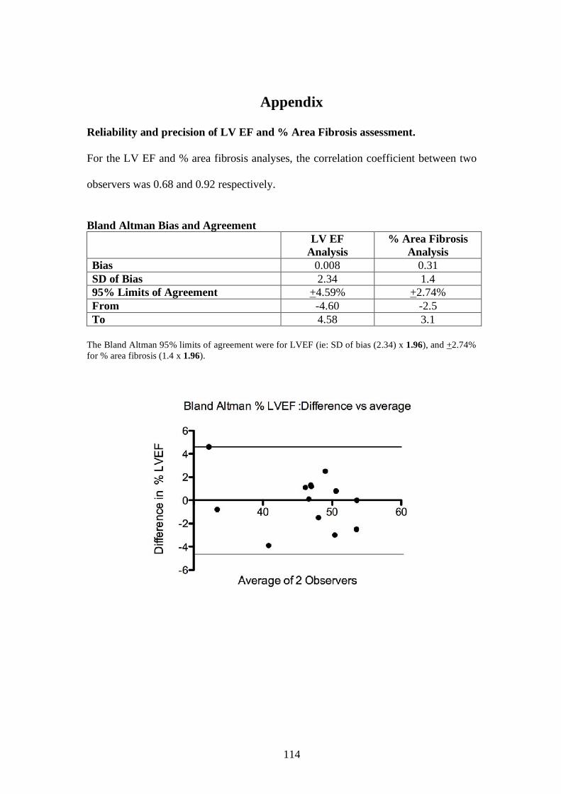
114
Appendix
Reliability and precision of LV EF and % Area Fibrosis assessment.
For the LV EF and % area fibrosis analyses, the correlation coefficient between two
observers was 0.68 and 0.92 respectively.
Bland Altman Bias and Agreement LV EF
Analysis % Area Fibrosis
Analysis Bias 0.008 0.31 SD of Bias 2.34 1.4 95% Limits of Agreement +4.59% +2.74% From -4.60 -2.5 To 4.58 3.1
The Bland Altman 95% limits of agreement were for LVEF (ie: SD of bias (2.34) x 1.96), and +2.74% for % area fibrosis (1.4 x 1.96).

115


![Universitas Brawijaya€¦ · contains omega-3 fatty acid which is equivalent to omega 3 fatty acid in cod liver oil t l], Omega-3 fatty acid product of tuna canning has limited stability](https://static.fdocuments.us/doc/165x107/5feaff47d35ceb2fbf6799fa/universitas-brawijaya-contains-omega-3-fatty-acid-which-is-equivalent-to-omega-3.jpg)