
The effects of 5-HT on sensory, central and motor neurons driving
18
Comparative Biochemistry and Physiology Part B 127 (2000) 533 – 550 The effects of 5-HT on sensory, central and motor neurons driving the abdominal superficial flexor muscles in the crayfish J.R. Strawn a , W.S. Neckameyer b , R.L. Cooper a, * a 101 Thomas Hunt Morgan School of Biological Sciences, Uni6ersity of Kentucky, Lexington, KY 40506 -0225, USA b Department of Pharmacology and Physiological Sciences, St Louis Uni6ersity, St Louis, MO 63104, USA Received 12 January 2000; received in revised form 1 August 2000; accepted 8 August 2000 Abstract Serotonin (5-HT) induces a variety of physiological and behavioral effects in crustaceans. However, the mechanisms employed by 5-HT to effect behavorial changes are not fully understood. Among the mechanisms by which these changes might occur are alterations in synaptic drive and efficacy of sensory, interneurons and motor neurons, as well as direct effects on muscles. We investigated these aspects with the use of a defined sensory-motor system, which is entirely contained within a single abdominal segment and consists of a ‘cuticular sensory neurons – segmental ganglia – abdominal superficial flexor motor neurons – muscles’ circuit. Our studies address the role of 5-HT in altering (1) the activity of motor neurons induced by sensory stimulation; (2) the inherent excitability of superficial flexor motor neurons; (3) transmitter release properties of the motor nerve terminal and (4) input resistance of the muscle. Using en passant recordings from the motor nerve, with and without sensory stimulation, and intracellular recordings from the muscle, we show that 5-HT enhances sensory drive and output from the ventral nerve cord resulting in an increase in the firing frequency of the motor neurons. Also, 5-HT increases transmitter release at the neuromuscular junction, and alters input resistance of the muscle fibers © 2000 Elsevier Science Inc. All rights reserved. Keywords: Serotonin; Neurotransmission; Crayfish; Neuromuscular junction; Synapse; Quantal www.elsevier.com/locate/cbpb 1. Introduction Neuromodulators are well recognized as impor- tant signaling molecules in all animals examined because of the wide variety of behavioral effects which they might elicit. They may serve as both neurotransmitters and paracrine factors in central and peripheral nervous systems (Guillemin, 1978; Jan and Jan, 1982; O’Shea and Schaffer, 1985). One neuromodulator in particular, serotonin (5- HT), has received much attention both in the news media and in the academic literature because it has been implicated in the behavioral expression of dominance, aggression, and assertiveness in many animals, including humans (Linnoilia and Virkkunen, 1982; Winberg et al., 1992; Cases et al., 1995; Coccaro, 1995; Yeh et al., 1996). The distribution of 5-HT in the peripheral nervous systems of invertebrates implies that the transmit- ter is not directed at a discrete target tissue but * Corresponding author. Tel.: +1-859-2575950; fax: +1- 859-2571717. E-mail address: [email protected] (R.L. Cooper). 0305-0491/00/$ - see front matter © 2000 Elsevier Science Inc. All rights reserved. PII:S0305-0491(00)00287-X
Transcript of The effects of 5-HT on sensory, central and motor neurons driving

Comparative Biochemistry and Physiology Part B 127 (2000) 533–550
The effects of 5-HT on sensory, central and motor neuronsdriving the abdominal superficial flexor muscles in the
crayfish
J.R. Strawn a, W.S. Neckameyer b, R.L. Cooper a,*a 101 Thomas Hunt Morgan School of Biological Sciences, Uni6ersity of Kentucky, Lexington, KY 40506-0225, USA
b Department of Pharmacology and Physiological Sciences, St Louis Uni6ersity, St Louis, MO 63104, USA
Received 12 January 2000; received in revised form 1 August 2000; accepted 8 August 2000
Abstract
Serotonin (5-HT) induces a variety of physiological and behavioral effects in crustaceans. However, the mechanismsemployed by 5-HT to effect behavorial changes are not fully understood. Among the mechanisms by which these changesmight occur are alterations in synaptic drive and efficacy of sensory, interneurons and motor neurons, as well as directeffects on muscles. We investigated these aspects with the use of a defined sensory-motor system, which is entirelycontained within a single abdominal segment and consists of a ‘cuticular sensory neurons–segmental ganglia–abdominalsuperficial flexor motor neurons–muscles’ circuit. Our studies address the role of 5-HT in altering (1) the activity ofmotor neurons induced by sensory stimulation; (2) the inherent excitability of superficial flexor motor neurons; (3)transmitter release properties of the motor nerve terminal and (4) input resistance of the muscle. Using en passantrecordings from the motor nerve, with and without sensory stimulation, and intracellular recordings from the muscle, weshow that 5-HT enhances sensory drive and output from the ventral nerve cord resulting in an increase in the firingfrequency of the motor neurons. Also, 5-HT increases transmitter release at the neuromuscular junction, and alters inputresistance of the muscle fibers © 2000 Elsevier Science Inc. All rights reserved.
Keywords: Serotonin; Neurotransmission; Crayfish; Neuromuscular junction; Synapse; Quantal
www.elsevier.com/locate/cbpb
1. Introduction
Neuromodulators are well recognized as impor-tant signaling molecules in all animals examinedbecause of the wide variety of behavioral effectswhich they might elicit. They may serve as bothneurotransmitters and paracrine factors in centraland peripheral nervous systems (Guillemin, 1978;
Jan and Jan, 1982; O’Shea and Schaffer, 1985).One neuromodulator in particular, serotonin (5-HT), has received much attention both in thenews media and in the academic literature becauseit has been implicated in the behavioral expressionof dominance, aggression, and assertiveness inmany animals, including humans (Linnoilia andVirkkunen, 1982; Winberg et al., 1992; Cases etal., 1995; Coccaro, 1995; Yeh et al., 1996). Thedistribution of 5-HT in the peripheral nervoussystems of invertebrates implies that the transmit-ter is not directed at a discrete target tissue but
* Corresponding author. Tel.: +1-859-2575950; fax: +1-859-2571717.
E-mail address: [email protected] (R.L. Cooper).
0305-0491/00/$ - see front matter © 2000 Elsevier Science Inc. All rights reserved.PII: S 0305 -0491 (00 )00287 -X

J.R. Strawn et al. / Comparati6e Biochemistry and Physiology, Part B 127 (2000) 533–550534
over a widespread area, perhaps interacting withreceptors on several different target cells. Conse-quently, this could result in relatively fast, aswell as slow, long-lasting modifications of theefficacy in transmission at synapses where othertransmitters are released (Kupfermann, 1979).
The crustacean nervous system is experimen-tally tractable, and previous studies have facili-tated correlation of the physiology, fromrelatively few neurons and muscles, to particularbehaviors. The advantage of neuromuscular sys-tems in the crayfish is that entire muscles areinnervated by one to no more than a dozenexcitatory axons (LaFramboise et al., 2000;Sohn et al., 2000). Moreover, the drive of themotor neurons can readily be examined by stim-ulation of various known sensory neurons,which enables one to assess their role in alteringmotor neuron activity. In addition, the integra-tion of sensory information in the ventral nervecord (VNC) which effects the motor output isaccessible for study. Lastly, the biophysicalproperties of muscle fibers that can effect thepostsynaptic response are readily approachable.Thus, the crustacean nervous system enables oneto investigate each component of a sensory–ganglia–motor neuron–muscle pathway whichelicits that behavior.
In lobsters and crayfish, it has been shownthat injections of serotonin (5-HT) into the cir-culatory system cause the animals to assume asemi-dominant posture (a raised extension of thechelipeds, although with a flexed state of theirabdomen, Livingstone et al., 1980). The raisedchelipeds is thought to be a visual, social-statussignal. Injection of a different neuromodulator,octopamine, produces a submissive behavior,with lowering of the chelipeds but the same timewith extension of the animal’s extremities (Liv-ingstone et al., 1980; Harris-Warrick and Krav-itz, 1984). The actions of 5-HT in eliciting thesebehaviors were shown to be selective within theVNC to enhance excitation of excitatory flexormotor neurons and decrease excitation of theinhibitor flexor motor neurons. The converse isobserved for the exciter and inhibitor motorneurons to the abdominal extensors. Sincecrayfish with higher levels of 5-HT have beenshown to be more assertive (Huber et al., 1997;Huber and Delago, 1998), one might expect themore aggressive individuals to have flexed abdo-mens; however, this behavioral posture remains
to be quantified during social interactions. 5-HTis excitatory at the presynaptic terminals of mo-tor neurons (Dudel, 1965; Glusman and Kravitz,1982; Livingstone et al., 1980; Griffis et al.,1999; Strawn et al., 1999), suggesting that thegreater mass of flexor muscle on which 5-HTcan act, relative to the extensor muscles andnumber of terminals, may account for the flexedstate during injection of high concentrations of5-HT. In addition, the motor nerves to the ab-dominal superficial flexor muscles are sponta-neously active in the animal, as well as inisolated ventral nerve cords. Thus, if 5-HT en-hances this activity, one would expect the ani-mal to be in a flexed state. Previousinvestigations did not address whether sensoryneuron activity could be altered by 5-HT, andwhether the alterations in sensory neuron activ-ity could have an effect on the activity of thesuperficial motor neurons. It is well establishedin the Aplysia nervous system that 5-HT sensi-tizes a gill-withdrawal reflex (Siegelbaum et al.,1982) and it appears from the results presentedherein in the crayfish system that 5-HT can alsoenhance sensory drive.
The aims of this study were to examine therole of 5-HT on each component within acrayfish ‘sensory–ganglia–motor nerve–muscle’consisting of specific, identified cells within thesecond abdominal segment. Experiments weredesigned to assess the role of 5-HT in altering(1) the activity of the sensory neurons whichdrive the motor neurons; (2) the activity of su-perficial flexor motor neurons; (3) vesicular re-lease properties of the superficial motor nerveterminals and (4) direct input resistance of themuscle. Furthermore, we attempted to measure5-HT levels in hemolymph and within the ven-tral nerve cord of crayfish. The few values of5-HT reported in the literature for crustaceansvary considerably.
This study illustrates the importance of deter-mining the sensory contribution to the intrinsicactivity of motor neurons, and demonstratesthat 5-HT modulates not only the motor neu-rons but also sensory input to the pathway.This abdominal superficial flexor preparation al-lows one to address issues fundamental to allcircuits and chemical synapses.
Preliminary results of this study have beenpresented in abstract form (Strawn et al., 1999,2000).
J.R. Strawn et al. / Comparati6e Biochemistry and Physiology, Part B 127 (2000) 533–550 535
2. Methods
2.1. Animals
All experiments were performed using malefreshwater crayfish, Procambarus clarkii, measur-ing 6–10 cm in body length (Atchafalaya Biologi-cal Supply, Raceland, LA). All animals werehoused as individuals for at least 2 weeks andwere fed dried fish food weekly.
2.2. Dissection
All experiments were carried out in a modifiedVan Harreveld’s solution (205 mM NaCl; 5.3 mMKCl; 13.5 mM CaCl2 · 2H2O; 2.45 mMMgCl2 · 6H2O; 0.5 mM HEPES adjusted to pH7.4. Cooper et al., 1998). The animals were sac-rificed in less than 5 s by rapid decapitationfollowed by removal of the abdomen. The abdo-men was separated from the thorax by cuttingthrough the carapace between the thorax andabdomen. The VNC was transected between T5and A1. The abdomen was then pinned ventralside up in a sylgard dish. Exposure of the VNCwith the nerve roots and superficial flexor muscleentailed cutting along the midline of the ventralarticular membrane (anterior to posterior) andthen along the rib laterally from the midline. Thissection of articulating membrane was thenreflected rostrally and removed so as to expose thesuperficial flexors, VNC and segmental ganglia tothe bath solution.
For recording of synaptic currents from theneuromuscular junction, the dorsal carapace ofthe abdomen was cut away so as to provide accessfor the removal of the deep flexor. The third rootwas cut near the VNC and the VNC was re-moved. The preparation was repinned ventral sideup and the superficial flexor and third root ex-posed as described above.
2.3. Intracellular and en passant recordings
All recordings were performed from the secondabdominal segment. For intracellular recordings,a microelectrode (2 M potassium acetate, 20–30MV) was inserted into a medial muscle fiber ofthe superficial flexor. Extracellular recordings ofnerve activity in the third root were made by asuction electrode. All data were recorded on aVHS tape (Vetter) and to a Macintosh computer
(Power Macintosh, 300 MHz G3) via MacLab/4sA/D converter (ADInstruments). For cuticularstimulations, a wooden dowel lined with severaltoothbrush bristles was placed on a mircomanipu-lator which was positioned along the lateral sideof the second abdominal segment. The manipula-tor was then moved back and forth for a setdistance of 1 cm along the cuticle of the segmentin which the nerve root was being monitored.Subsequent experiments, using electrical stimula-tion (50 Hz, 300 ms) of the second root wereperformed to circumvent problems with standard-izing the mechanical stimulation and adaptationof the sensory endings. The extracellular stimula-tion was applied to the proximal end of thesecond root by the use of a suction electrode.
2.4. Analysis of synaptic currents
Focal macropatch recording was used to mea-sure synaptic currents at the neuromuscular junc-tions. The synaptic currents were obtained usingthe loose patch technique by lightly placing a10–20 mm diameter, fire-polished, glass electrodedirectly over a spatially isolated varicosity along avital dye-visualized nerve terminal. To visualizethe nerve terminals, living preparations werefluorescently stained for 2–5 min with 2–5 mM4-[4-(diethylamino)styryl]-N-methylpyridinium io-dide (4-di-2-Asp; Molecular Probes, Eugene, OR)in crayfish saline. The preparation was exposed tothe dye solution for 2–5 min, followed by wash-ing in crayfish saline. The synaptic transmissionremained unaltered by this dye (Cooper et al.,1995b). The macropatch electrode is specific forthe current recording within the region of theelectrode lumen. The lumen of the patch electrodewas filled with the same solution as the bathingmedium. The electrode seal resistance was in therange from 100 KV to 1 MV. Since the seal couldbe easily lost if the muscle twitched under theelectrode, stimulation was restricted to 1 Hz.Evoked excitatory postsynaptic currents (EPSCs)and miniature excitatory postsynaptic currents(mEPSCs) were recorded and analyzed to deter-mine the mean quantal content (m), the numberof release sites (n), and the probability of releaseat the sites (p) (Cooper et al., 1995c, 1996a,b; delCastillo and Katz, 1954). In each synaptic currentrecording, a trigger artifact and a nerve spike werevisualized, indicating nerve stimulation. This wasdone to determine if a stimulation failed, or if

J.R. Strawn et al. / Comparati6e Biochemistry and Physiology, Part B 127 (2000) 533–550536
synaptic release failed, in spite of nerve terminaldepolarization. Mean quantal content was deter-mined by direct counts (m):
direct counts (m)
=�%(0)(c )+ (1)(c )+ (2)(c ) …
n,
the total number of trials
By direct counts of the evoked quantal events,estimates of n and p were obtained. In some cases,there were no evoked events that followed thenerve terminal spikes; such a failure in evokedrelease was counted as a zero. If only one singleevent occurred after the spike, it was counted asone; when double events occurred, they were re-ferred to as two (Table 1). Quantal release overthe time was monitored by examining the area ofthe evoked current (a measure of charge). Thetime of peak evoked events varied due to latencyjitter, so that when multiple events occurred, themeasurements of peak amplitude were not asreliable. The change in the charge measures beforeand after application of 5-HT (100 nM, Sigma) tothe nerve terminals, and the frequency in oc-curence of charge of the evoked events were calcu-lated. Additionally, the data sets were tested for abest-fit approximation based on assumptions dis-cussed in earlier reports (Dudel and Kuffler, 1961;Cooper et al., 1995c). Binomial distributions rep-resent the quantal nature of release in crayfishneuromuscular junctions (Wojtowicz et al., 1991).To test for non-uniform binomial distributions,the procedures described earlier were used (Woj-towicz et al., 1991). The chi-squared statistic (x2)and a modified Akaike information criterion(AIC) were used to estimate the distribution thatbest fits the observed distribution of events. Abootstrap method, as previously described(Cooper et al., 1995c), was employed to estimatethe standard error of the estimated n and p values.The bootstrap method consisted of drawing 1000random samples from the original data set basedon an open scheme (Cooper et al., 1995c). Sinceexposure to 5-HT produced gradual changes in allof the quantal parameters, sample sets of data forevery 200 events were used and were found to besufficient to obtain statistically significant valuesfor quantal predictions. The 1000 sampledgrouped data and the 200 event-group sets werethen compared (Table 1).
2.5. Immunofluorescence
Whole mount preparations of the isolated su-perficial flexor muscle were pinned to a Sylgarddish with the muscle in a stretched position. Theywere fixed with 2.5% (v/v) glutaraldehyde, 0.5%(v/v) formaldehyde dissolved in a PBS buffer (9.5ml of NaH2PO4 · 2H2O of 0.2 M stock, 40.5 mlNa2HPO4 · 7H2O of 0.2 M stock, 4 g sucrose in a100 ml volume) for 1 h with two changes ofsolution. The preparation was then placed intovials and washed in PBS buffer containing 0.2%(v/v) Triton X-100 and 1% (v/v) normal goatserum (Gibco/BRL, Grand Island, NY) for 1 hwith three changes at room temperature. Thetissue was then incubated with primary antibodyto GABA (Sigma, 1:1000 in PBS buffer) on ashaker at 4°C for 12 h. The tissue was washedthree times and incubated in secondary antibody(goat, anti-rabbit IgG conjugated with Texas Red,Sigma) diluted 1:200 with PBS buffer at roomtemperature for 2 h, followed by two washes inbuffer. The synaptic locations were observed byimmunocytochemistry as previously shown innerve terminals (Cooper et al., 1995a; Msghinaand Atwood, 1997; Cooper, 1998). Fluorescentimages of the nerve terminals were viewed with aLeica DM IRBE inverted fluorescent microscopeusing a 63X (1.2 NA) water immersion objectivewith appropriate illumination. The composite im-ages of Z-series were collected with a Leica TCSNT confocal microscope for illustration.
2.6. HPLC analysis
Quantification of 5-HT concentrations, both inthe hemolymph and in the abdominal ganglia,was performed using high performance liquidchromatography with electrochemical detection(HPLC-ED). The animals were agitated andhemolymph was extracted using a chilled syringeand needle. For extraction of 5-HT from theganglia, the VNC was quickly excised in coldcrayfish saline and the T1–A1 as well as A2–A6ganglia were removed and homogenized in 0.1 Mperchloric acid. Both the hemolymph and homog-enized ganglia were first deproteinated; the crudeextract was then passed through a 0.22 mm filter(Ultra-Free, Millipore). The samples were injectedinto the HPLC system which consisted of an ESApump (model 580), a manual injector (model9125), and an HR-80 3 mm particle size column.

J.R. Strawn et al. / Comparati6e Biochemistry and Physiology, Part B 127 (2000) 533–550 537
Tab
le1
Influ
ence
of5-
HT
onqu
anta
lre
leas
epa
ram
eter
s:m
ean
quan
tal
cont
ent
(m),
num
ber
ofsi
tes
rele
asin
g(n
),an
dpr
obab
ility
ofre
leas
eat
the
site
s(P
)a
401–
600
Eve
nts
601–
800
801–
1000
All
Swee
ps1–
200
201–
400
Obs
Pre
dO
bsP
red
Obs
Pre
dO
bsP
red
Obs
Pre
dO
bsP
red
Pre
p.1
910
2626
1483
149
2525
0Sa
line
910
189
187
186
186
173
174
184
184
172
177
190
490
45
31
02
22
32
32
2013
0.98
0.87
0.94
0.89
0.97
0.93
m2
(0)
2(0
)n
2(0
)2
(0)
2(0)
2(0
)0.
49(0
.009
)0.
438(
0.01
2)0.
47(0
.009
)0.
445(
0.12
4)p
0.48
3(0.
008)
0.46
5(0.
005)
10
10
02
28
03
66
05-
HT
661
6115
6767
6566
7676
335
335
6613
717
512
912
912
512
612
311
515
123
628
629
210
31
102
210
06
629
3110
10
00
00
00
00
41
01.
711.
651.
721.
62m
1.68
1.71
4(0
)3
(0)
3(0
)3
(0)
3(0
)4
(0)
n0.
428
(0.0
09)
0.55
2(0
.012
)0.
575
(0.0
1)0.
54(0
.013
)p
0.42
1(0.
004)
0.57
(0.0
13)
Pre
p.2
3838
7678
4418
144
Salin
e0
00
181
2323
128
128
136
136
8188
135
135
171
171
651
651
148
4526
2643
4021
2129
2916
716
72
00
00
00
10
01
11
30.
50.
260.
430.
210.
290.
34m
2(0
)2
(0)
2(0
)2
(0)
3(0
)n
3(0
)0.
47(0
.002
)0.
418(
0.03
2)0.
443(
0.02
)0.
570(
0.01
3)p
0.32
9(0.
006)
0.37
8(0.
014)
00
00
00
00
5-H
T0
00
00
03
00
00
00
10
00
013
413
413
413
391
9115
488
287
155
600
602
423
6363
5860
9898
9884
357
339
404
43
08
711
1111
2538
575
23
42
5 61
10
2.35
2.37
2.6
2.65
2.25
m2.
44n
4(0
)4
(0)
4(0
)6
(0)
6(0
)4
(0)
0.56
2(0
.008
)0.
592
(0.0
1)0.
592
(0.0
1)0.
65(0
.01)
0.44
2(0
.008
)p
0.41
0(0
.003
2)
aA
seri
esof
evok
edev
ents
indu
ced
at1
hzst
imul
atio
nto
the
mot
orne
rve
befo
rean
daf
ter
appl
icat
ion
of5-
HT
(100
nM).
The
tim
ese
ries
isdi
vide
din
tobi
nsof
200
tria
lsfo
ras
sess
men
tof
quan
tal
para
met
ers:
mea
nqu
anta
lco
nten
t(m
),th
enu
mbe
rof
rele
ase
site
s(n
),an
dth
epr
obab
ility
ofre
leas
eat
the
site
s(p
).T
hest
anda
rder
ror
ofes
tim
atin
gn
and
pw
ere
obta
ined
bya
boot
stra
ppin
gpr
oced
ure
(Coo
per
etal
.,19
95c)
and
are
show
nw
ithi
nth
epa
rent
hese
s.T
hepr
edic
ted
(pre
d)di
stri
buti
ons
wer
eob
tain
edfr
omfit
ting
the
obse
rved
(obs
)di
stri
buti
onby
am
axim
umlik
elih
ood
esti
mat
ion.
Num
ber
ofth
edi
scre
teev
ents
indi
cate
das
:0,
failu
res;
1,on
es;
2,tw
os;
etc.
are
give
n.
J.R. Strawn et al. / Comparati6e Biochemistry and Physiology, Part B 127 (2000) 533–550538
Detection and quantifying were accomplished us-ing a Coulochem detector (ESA model 5200A), ananalytical cell (model 5011, channel 1 set to −50mV and channel 2 set at 300 mV) and a guard cell(model 5020, set at 400 mV). The mobile phaseconsisted of 75 mM NaH2PO4, 1.5 mM SDS, 100ml/l triethylamine, 15% acetonitrile, 12.5%methanol. Under these conditions, serotonineluted at 12 minutes.
The average weight of the ganglia (T1–A1),0.42 mg, was dissolved in 200 ml of perchloric acid.Of this amount, only 20 ml reaches the column.Considering the MW of 5-HT at 212.7 g/mol, forthe 5-HT used in the calibration curves, this calcu-lates to 94 nM 5-HT for the entire starting tissue.
3. Results
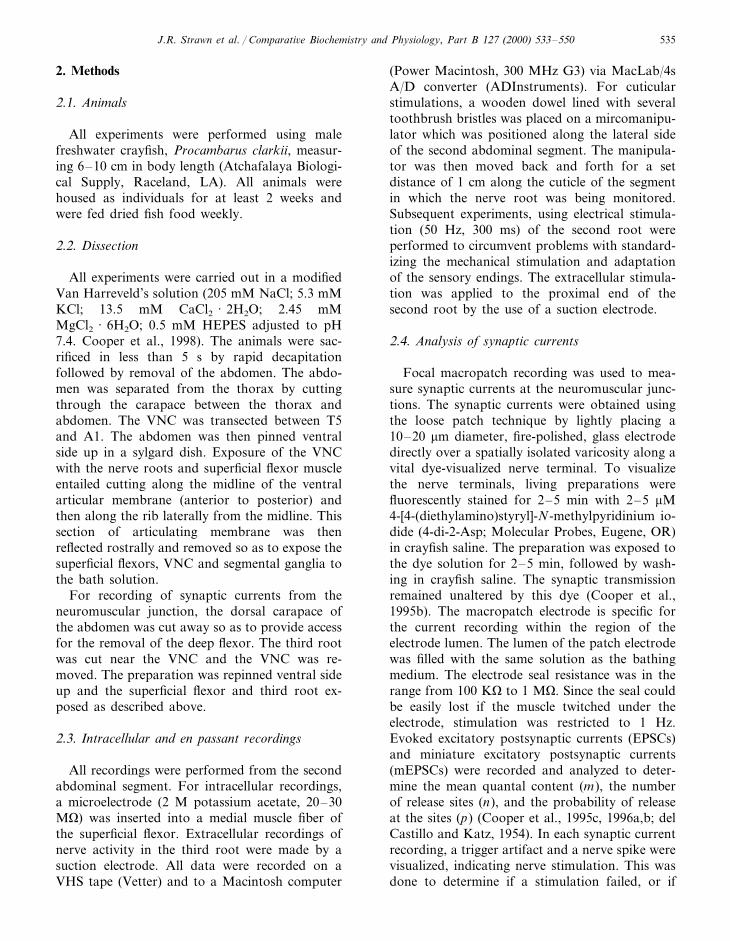
To record from the ventral surface of the su-perficial flexor muscles or from the superficialflexor motor nerve, the articular membrane of thesegment was trimmed and removed to allowaccess to the entire ventral surface of the muscle(Fig. 1A). The connectives to adjoining gangliaand the 1st and 2nd roots were transected todetermine their influence on the intrinsic ac-tivity measured from the superficial flexormotor nerve. Intracellular recordings from themedial fibers revealed the intrinsic activityassociated with the motor neurons to themuscle.
Fig. 1. (A) Ventral dissection of the 2nd abdominal segment stained with methylene blue. The articulating membrane (arrow heads) hasbeen reflected so as to expose the slow superficial flexors (right) and the fast deep flexors are shown on the left. (B) The 3rd root withterminals and varicosities which have been stained with 4-Di-2-Asp. (C, D) Enlarged view of the clusters of varicosities on the musclefiber surface of medial fibers. (E) GABA immunocytochemical analysis of the inhibitory axon terminal with varicosities. Scale bar: A,3.6 cm; B–D, 135mm; E, 33 mm.

J.R. Strawn et al. / Comparati6e Biochemistry and Physiology, Part B 127 (2000) 533–550 539
3.1. 5-HT increases firing frequency of thesuperficial flexor motor neurons
In each of the five preparations, application of5-HT (100 nM) to the ventral nerve cord contain-ing only segments A1–A3, with the 1st and 2ndroots severed, resulted in an increased firing fre-quency of the superficial flexor motor neurons asassessed by intracellular recordings from the me-dial superficial flexor muscle (Fig. 2A). The rateof change in the frequency of excitatory postsy-naptic potentials (EPSPs) by exposure to 5-HTwas determined by counting the number of eventsfor 1 s every 10 s over the 10 min recordingperiod. The change in firing frequency for a repre-sentative preparation is shown (Fig. 2B). Sincethere was substantial variation in the degree ofbasal activity among preparations, the mean fre-quency of the five preparations before and afterexposure to 5-HT is shown to illustrate the rangeof activities. The mean frequency of 5-HT effectswas determined after the maximum response oc-curred during the exposure to 5-HT (Fig. 2C).Preparations that had a high initial firing fre-quency showed a lower percent change as com-pared to preparations with a low basal rate (Fig.2D). These rates are representative only of theactivity on the individual fiber being monitored.Neighboring fibers exhibited lower or higher ratesof basal activity depending on the number ofexcitatory motor neurons innervating the fiber.
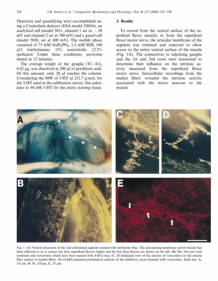
The overall activity of the entire 3rd root wasmonitored after application of 5-HT by en pas-sant recordings of the extracellular spikes (Fig.3A). Variations in the rate of increasing activityamongst different preparations was also observedfrom the intracellular monitoring of EPSPs. Apreparation which demonstrated a slow rise inactivity while exposed to 5-HT is shown in Fig.3B. The increase in firing frequency associatedwith the application of 5-HT was observed in fiveof the six preparations (Fig. 3C). The mean andstandard error of the percentage change, 48.62915.16, clearly demonstrates an increase drive ofthe intrinsic spontaneous activity of these motorneurons. It became apparent that when sensoryneurons were left intact, exposure to 5-HT re-sulted in a larger change to the frequency of the3rd root motor neurons. Therefore, the influenceof sensory neuron activity on the drive of the 3rdroot, and the effects of 5-HT, were assessed.
Fig. 2. (A) Effects of 5-HT on firing frequency of the motorneurons as recorded with an intracellular electrode within amedial muscle fiber to monitor excitatory postsynaptic poten-tials (EPSPs). (B), Representative preparation treated with 100nM 5-HT which demonstrates the time frame of action andthe persistence of 5-HT’s effect in maintaining a higher fre-quency of EPSPs. 5-HT was added at the 600 s mark in therecording. (C) Mean frequency of EPSPs (9S.E.M.) in fiveanimals with an intact CNS as well as the 1st and 2nd roots.(D) The percentage change for preparations in B are shown.Note the preparations that demonstrating the lowest initialactivity increased the most upon exposure to 5-HT.
J.R. Strawn et al. / Comparati6e Biochemistry and Physiology, Part B 127 (2000) 533–550540
3.2. 5-HT enhances sensory dri6e of superficialflexor motor neurons
While recording from the third root, the lateralcuticle of the animal was stimulated as illustratedin Fig. 4. Stimulation of cuticular sensory neu-rons increased the firing frequency of the motorneurons (Fig. 5A). The basal activity monitoredin the 3rd root, with sensory neurons intact, alsoshowed a increase upon exposure to 5-HT. Theheightened effects of 5-HT on sensory drive ofthe superficial motor neurons was observed in allpreparations examined (n=12). The mean values
reported are an average of five subsequent trialsin saline and in 5-HT. The saline bath was ex-changed with one containing 100 nM of 5-HTand incubated in the dark for 5 min before addi-tional stimulation. In order to bypass the activa-tion of the sensory endings to address the effectsof 5-HT on sensory neuron drive, standardizedstimulation was applied with an electrode placedon a severed distal end of the second root whilerecording from the third root in saline and in5-HT. Again, 5-HT increased the baseline activity(n=7, Fig. 5B) even in the absence of alteredsensory drive. With the stimulation paradigm, theassociated increases in activity were quantified bytaking the response 1 s before stimulation (50 Hz,300 ms), and 1 s after stimulation (Fig. 5B). Thisenhancement of the stimulated-associated drive ofspontaneous activity was observed in seven out ofseven preparations (Wilcoxon rank sum test, PB0.02, Fig. 5C). Because the increases in activityrecorded from the third root could be the resultof the 5-HT’s effects at the primary sensory affer-ent neurons themselves or effects within theVNC, we recorded directly from the second rootin the presence and absence of cuticular stimula-tion. Although we did not measure the latencyfor the effect of 5-HT on the integration withinthe VNC, the diffusion of 5-HT across the gan-glion sheath to act on the connections within theneuropil would likely take longer then the directactions on the more readily exposed sensory neu-rons.
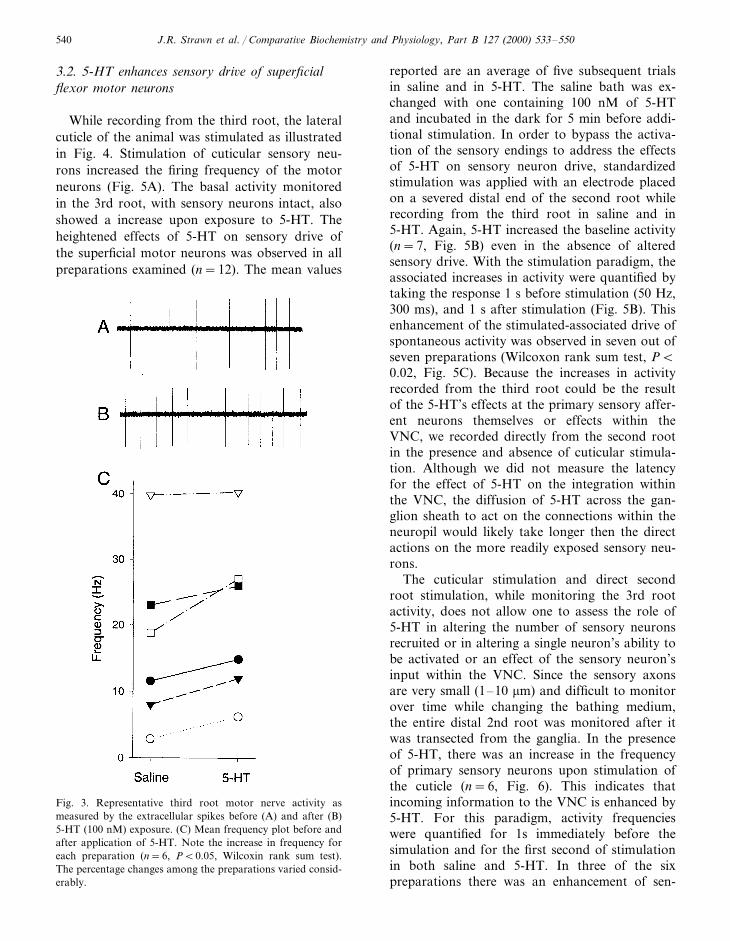
The cuticular stimulation and direct secondroot stimulation, while monitoring the 3rd rootactivity, does not allow one to assess the role of5-HT in altering the number of sensory neuronsrecruited or in altering a single neuron’s ability tobe activated or an effect of the sensory neuron’sinput within the VNC. Since the sensory axonsare very small (1–10 mm) and difficult to monitorover time while changing the bathing medium,the entire distal 2nd root was monitored after itwas transected from the ganglia. In the presenceof 5-HT, there was an increase in the frequencyof primary sensory neurons upon stimulation ofthe cuticle (n=6, Fig. 6). This indicates thatincoming information to the VNC is enhanced by5-HT. For this paradigm, activity frequencieswere quantified for 1s immediately before thesimulation and for the first second of stimulationin both saline and 5-HT. In three of the sixpreparations there was an enhancement of sen-
Fig. 3. Representative third root motor nerve activity asmeasured by the extracellular spikes before (A) and after (B)5-HT (100 nM) exposure. (C) Mean frequency plot before andafter application of 5-HT. Note the increase in frequency foreach preparation (n=6, PB0.05, Wilcoxin rank sum test).The percentage changes among the preparations varied consid-erably.

J.R. Strawn et al. / Comparati6e Biochemistry and Physiology, Part B 127 (2000) 533–550 541
Fig. 4. Schematic of ventral view in a dissected second segment of the crayfish abdomen. The cuticular stimulation occurred along thelateral side of the segment. The mechanically stimulation was given at approximately 0.5 Hz (shown at top). The third ganglion (A3)of the ventral nerve cord for the next caudal segment is shown.
sory neuron activity upon exposure to 5-HT with-out cuticular stimulation.
3.3. 5-HT alters synaptic transmission
5-HT is known to alter synaptic transmissionby enhancing the EPSP amplitudes on a numberof neuromuscular junctions in crayfish: openermuscle (Crider and Cooper 1999, 2000; Southardet al., 2000) leg extensor muscle (Shearer andCooper, 2000) and abdominal extensors (Griffis etal., 1999). To assess the effects of 5-HT on synap-tic transmission of the motor neuron terminals,the severed third root was stimulated at 1 Hzwhile recording from the superficial flexor. Toensure that only a single motor neuron was beingrecruited, a single sized postsynaptic event wasmonitored while the preparation was exposed tosaline prior to the addition of 5-HT. Upon theaddition of 5-HT, a rapid increase in the EPSPamplitude was observed (Fig. 7, n=5, PB0.05Wilcoxon Rank sum test). However, the increasein EPSP amplitude could be due to several mech-anisms. First, 5-HT could increase input resis-tance of the muscle which would increase theamplitude of the EPSP. 5-HT could also increasequantal release, which could also account for anincrease in the EPSP amplitude. To determine theindividual contributions of these mechanisms,both quantal release from the presynaptic termi-nal and input resistance of the postsynaptic mus-cle fiber were measured.
3.4. 5-HT increases quantal release
Presynaptic efficacy of synaptic transmission isreadily assessed by counting the number of quan-tal events in evoked synaptic currents. 5-HTcauses a direct effect on the presynaptic terminalby altering the number of evoked events. A focalmacropatch electrode was used to record thesynaptic currents before and after application of5-HT (Fig. 8A, Table 1). To record from varicosi-ties, the terminals were visualized by applicationof the vital dye (4-Di-2-ASP) and the lumen of themacropatch electrode was placed directly over avaricosity. The stained terminals on a medial mus-cle fiber along with five individual axons caneasily be seen in Fig. 1B. Enlarged images ofterminals illustrates the clusters of varicositiesalong the terminal (Fig. 1C, D). The distinctterminal morphology of varicosities along the ter-minal lengths is indicative of tonic motor nerveterminals as compared to phasic nerve terminalsas has been described for other crustacean neuro-muscular junctions (Bradacs et al., 1997). Theinhibitory terminals can be visualized with im-munocytochemistry (Fig. 1E). The arrangement ofterminals and multiple varicosities within clustershinders location of isolated varicosities uncontam-inated by currents from neighboring varicosities.
In cases when multiple evoked events were ob-tained, discrete quantal events could not bequantified, although the entire synaptic event wasstill monitored and quantified by measuring the

J.R. Strawn et al. / Comparati6e Biochemistry and Physiology, Part B 127 (2000) 533–550542
charge (current x time) or the current area underthe trace. We have demonstrated that 5-HT in-creases mean quantal content (m), the number ofrelease sites (n), and the probability of release atthe sites (p). To assess the rate of this modulatoreffect on the quantal parameters, every 200 and1000 evoked events were grouped for quantalanalysis (Table 1). To obtain estimates of thequantal parameters (n and p), each set of 1000trials and the 200 trial subsets were examined andanalyzed to determine the statistical distributionwhich would best describe its profile. To obtain
the n and p values and the standard errors of theestimation, this analysis was followed by boot-strapping procedures. In addition, the standarderrors (shown in parentheses in Table 1) for theestimations are relatively low, suggesting reason-able fits to the calculated quantal parameters. Theresults presented in Table 1, also indicate thatapplications of 100 nM 5-HT resulted in an in-crease in p and n during a 1 Hz stimulation. In thefirst preparation, the sites of release (i.e. n) gener-ally increased from 2 sites to 3 or 4, where as inthe second preparation, n increased from 2 or 3 to
Fig. 5. Activity of the 3rd root before and during cuticular stimulation in saline (A1) and in 100 nM 5-HT (A2). The time during cuticlestimulation is indicated by the bar. Note the enhanced activity before and after stimulation when the preparation is bathed in 5-HT.Representative third root recording before and after electrical stimulation of the second root in saline (B1) and during exposure to 100nM 5-HT (B2). (C) Mean frequencies are plotted for the 3rd root activity before and after electrical stimulation during saline and 5-HTexposure. The mean frequencies are also indicated for the activity resulting from the stimulations while exposed to saline or 5-HT. Theasterisk (*) indicates the three preparations which demonstrate very similar firing frequencies.
J.R. Strawn et al. / Comparati6e Biochemistry and Physiology, Part B 127 (2000) 533–550 543
Fig. 6. (A) Recording from primary sensory afferents contained within the second root of the 2nd abdominal segment. Extracellularrecording illustrates the activity before cuticular stimulation, but note the large increase in the number of different size units activatedduring the stimulation. (B) Plots of the mean activity before and during cuticular stimulation in six preparations while bathed in salineor 5-HT (100 nM) indicates that 5-HT enhances primary sensory activity when the neurons are stimulated. This is best observed in theoverall frequency of activity during the stimulation in saline and 5-HT for the same preparations.
4 or larger. In both cases, the average probabilityof release at any one site (i.e. p) also increased,which is clearly evident in the subsets of 200 trials.When the data is grouped into the 1000 trials thep values are lower after 5-HT application becausen has doubled. Overall, there is an increase in m inall cases after exposure of the terminals to 5-HT.The average of 1000 evoked events before andafter the application of 5-HT show the overalleffect on synaptic currents (Fig. 8B). To betterexamine the change, a scatter plot of the synapticcharge for one of the preparations is provided fora representative case in Fig. 8C. The larger cur-rents are due to a greater number of events,indicating that 5-HT has a direct role in enhanc-ing synaptic transmission by acting at the presy-naptic terminal. This observation is supported bythe direct quantal counts in Table 1. Note theincrease in all the quantal parameters and alsothat the samples of every 200 events appear tobe sufficient for obtaining statistically significantvalues for quantal predictions as well as a1000 events. The larger EPSP amplitudes re-corded with intracellular electrodes are not onlydue to an enhancement of vesicular release butalso to a slight increase in input resistance ofmuscle fibers.
3.5. Input resistance is slightly increased
The effect of 5-HT on muscles input resistancecaused a significant increase in the EPSP ampli-tude. The slight increase in the input resistancewas observed in five out of five preparations(mean9S.E.M: Saline, 29.0295.39; 5-HT,38.4895.43, PB0.05).
3.6. 5-HT quantification
High performance liquid chromatography withelectrochemical detection was used to determinethe concentration of 5-HT within the hemolymphand the ventral nerve cord. Since it has previouslybeen reported that serotonergic neurons are local-ized in the thoracic ganglia and within the firstabdominal ganglion (A1) for lobsters (Beltz andKravitz, 1982), these six ganglia were removed foranalysis. In two preparations we obtained 94 and120 nM per combined ganglia. The additionalabdominal ganglia (A2–A6) did not show anydetectable 5-HT peaks. The hemolymph samplesproved problematic since a very broad responsewas observed. Dilution of the sample, to eliminatethe broad background, eliminated the peak at thetime a standard of 5-HT would normally elute.

J.R. Strawn et al. / Comparati6e Biochemistry and Physiology, Part B 127 (2000) 533–550544
4. Discussion
Since the initial discovery of neuromodulatorsin crustaceans, an increasing number of neuro-hemoral modulators (buccalin, histamine, red pig-ment concentrating hormone, choleystokin,crustacean cardioactive peptide, and allatostatin)have been identified which regulate development,
central pattern generators, and behavior (Christieet al., 1995; Kreissl et al., 1999). It has beensuggested that the 5-HT and octopaminergic neu-rons may function as ‘gain-setters’ in altering theoutput of neuronal circuits (Ma et al., 1992;Schneider et al., 1996; Horner et al., 1997). Muchwork remains to be done before we can fullyunderstand the effects of neuromodulators on in-
Fig. 7. (A) Average evoked post-synaptic potentials (EPSP) recorded in the superficial flexor muscle bathed in saline and after 5 minof exposure to 100 nM of 5-HT demonstrates the enhancement in the response. (B) A representative scatter plot of EPSP amplitudesevoked at 1 Hz stimulation rate before and during 5-HT exposure illustrates the rapid rise upon exposure to 5-HT. (C) Histogramsshowing the mean EPSP amplitudes before and after 5-HT treatment in each of five preparations (mean amplitudes9S.E.M.). Notethat there are very small deviations in the mean values within a preparation but significant differences among preparations. Thepercentage changes are indicated above each bar graph.

J.R. Strawn et al. / Comparati6e Biochemistry and Physiology, Part B 127 (2000) 533–550 545
Fig. 8. Influence of 5-HT on synaptic currents as recorded with a focal macropatch electrode from a spatially isolated varicosity.Representative single traces in the presence of saline (A1) and 100 nM 5-HT (A2) are shown. Note in the evoked excitatory postsynapticcurrents (EPSC’s) individual quanta can be counted (arrows � ). A miniature excitatory postsynaptic current (mEPSC) is shown in A1.Also note that the asterisk (*) indicates the extracellular recording of an action potential (i.e. spike). Averaged responses for 1000 trialsare shown during saline (B1) and 5-HT exposure (B2). (C) A representative scatter plot showing the influence of 5-HT on EPSC chargebefore and during the addition of 5-HT. (D) Histograms of the responses shown in the above scatter plot of charge (current× time)for saline and 5-HT treatment. Note the rightward shift in the histogram indicating larger evoked responses, likely due to the releaseof more vesicles as indicated with the direct quantal counting procedure.

J.R. Strawn et al. / Comparati6e Biochemistry and Physiology, Part B 127 (2000) 533–550546
dividual target cells. Given that different neuro-modulators may work in concert with one another,analysis of their mixed action is an area for forfuture research. In addition, few studies, particu-larly in vertebrates, address the effects of neuro-modulators on entire pathways which can regulatea specific behavior. In this study, we have used thewell established crustacean superficial abdominalflexor preparation to examine the influence of bothsensory input and 5-HT on the activity of the motorneurons.
Little is known about the direct effects of 5-HTapplication on crustacean neurons, with the excep-tion of a few studies that have shown that 5-HTexerts its effects through an inositol triphosphate(IP3) second-messenger system (Dixon and Atwood1989a,b; Delaney et al., 1991). Few analyses to datehave addressed the influence of known modulatorsdirectly at the sites of release from presynapticneurons to quantify on a relative basis the sites ofrelease (n) and the influence on the probability ofvesicle release (p) at active synapses (Cooper andRuffner, 1998; Crider and Cooper, 1999, 2000; Heet al., 1999). The present work on the superficialflexor muscle and another recent study (Southardet al., 2000) support the hypothesis that silentsynaptic sites are recruited in the presence of 5-HT,thus raising the quantal parameter, n. The proba-bility of release at the initial sites of release alsoincrease after application of 5-HT. Prior studiesexamining the effects of 5-HT have been accom-plished by whole cell measures of muscle potentialsarising from thousands of synapses (Grundfest andReuben, 1961; Dudel, 1965; Kravitz et al., 1976;Wheal and Kerkut, 1976; Florey and Rathmayer1978; Fisher and Florey, 1983). With the focalmacropatch techniques, direct measurement ofsynaptic parameters in presynaptic motor nerveterminals that influence release can be implemented(Cooper et al., 1995b,c; Southard et al., 2000). Inaddition the direct actions of neuromodulators onsensory neurons, such as effects on transductionand spike frequency to the CNS, can be readilyassessed in crayfish systems (Li et al., 1997).
A substantial amount of work on the neuromod-ulation of sensory-motor control has been con-ducted on the well-characterized serotoninergicsensory-motor circuit in Aplysia. This system hasbeen shown to increase intracellular calcium levelsin postsynaptic cells as well as effect a change inionic conductances leading to spike broadening inrested synapses (Byrne and Kandel, 1996). Unlike
crayfish, calcium entry is enhanced at the releasesites in Aplysia presynaptic neurons in which facil-itation is induced by 5-HT (Delaney et al., 1991;Eilot et al., 1993). From intracellular recordings incrayfish motor axons, there were no indications ofspike broadening (Dixon and Atwood, 1985), al-though this may happen within the nerve terminals(Dudel, 1965). With the use of calcium sensitiveindicators it was determined that 5-HT does notenhance an intracellular rise in calcium within theneuron (Delaney et al., 1991). Perhaps more sensi-tive temporal measures of calcium influx at activezones might better address this issue. In Aplysiasensory neurons, 5-HT can activate both PKA andPKC, which in part explains the temporal differ-ences of the effects of a slow K+ channel and anincrease in Ca2+ influx (Braha et al., 1993). Phar-macological evidence indicates 5-HT1A and 5-HT2
receptors are present in the crayfish nervous system(Yeh et al., 1996). The use of a variety of receptorsubtypes offers an advantage to the animal byutilizing alternate, intracellular biochemical path-ways for modulating synaptic transmission.
The drive to maintain the intrinsic spontaneousneural activity of the superficial flexor motor neu-rons has received much attention, but withoutmuch understanding as to the exact nature of thelocation of the input from within the ventral nervecord (Eckert, 1961; Kennedy and Takeda 1965a,b).Although it was shown by Eckert (1961) that theabdominal stretch receptors of the abdomen(MROs) were not contributing to the intrinsicactivity of the superficial flexor motor neurons,Kennedy and Takeda (1965a) did report that‘natural’ stimuli to the carapace increased activityof the nerve, but they were not informative to thelocation of the stimulus and the type of stimulus.No reports previous to this study have describedthe influence of neuromodulators in altering sen-sory drive of the motor neurons. Since the activitystill remains in isolated segmental ganglia it isassumed that it arises within the ganglion and canbe driven to higher firing rates by sensory axonstimulation as well as by application of 5-HT. Inan isolated ventral nerve cord with several gangliaintact, a higher rate of activity is common as wellas an increase in occurences in activity of all sixaxons within the 3rd root to these muscles. It is wellknown that the six motor axons innervate thesemuscles and that their cell bodies reside in thesegmental ganglia for both lobsters (Harris-War-rick and Kravitz, 1984) and crayfish (Kennedy andTakeda 1965a,b).
J.R. Strawn et al. / Comparati6e Biochemistry and Physiology, Part B 127 (2000) 533–550 547
Since it has been postulated that 5-HT plays arole in regulating the behavioral state of thecrayfish, lobsters, and crabs (Livingstone et al.,1980; Sneddon et al., 2000), several attempts havebeen made to determine its concentration in theVNC, the hemolymph, and in isolated ganglia oflobsters (Livingstone et al., 1980; Harris-Warrickand Kravitz 1984; Fadool et al., 1988). However,there has been considerable variation in therecorded measurements. Harris-Warrick andKravitz (1984) utilized HPLC-ED to measure 5-HT concentrations only within the first two ab-dominal ganglia. Their findings, that 5-HT ispresent at 90970 fmol/mg wet weight in the firsttwo abdominal ganglia, suggest that 5-HT isavailable for release from the VNC. Our measure-ments of 5-HT in the VNC indicated a range of94–120 nM per combined T1–A1 ganglia. Inaddition, we found it extremely difficult to mea-sure 5-HT concentrations in the hemolymph dueto broad signal arising from unknown contami-nants in the samples. Upon dilution of thehemolymph, the background problems were re-duced but no signal could be detected at theelution time expected for 5-HT. Our inability tomeasure 5-HT levels in the hemolymph of thecrayfish is likely a common technical problemsince reports are not available in the literature forcrayfish.
As pointed out in Livingstone, et al. (1980), theexcitatory direct effects of 5-HT on muscle couldresult in flexion of their abdomen. This seemsfeasible, since the flexor muscles are much largerthan the extensors. If a mass action on the mus-cles and neural systems were equal, then flexionwould be the postural state the animal wouldassume. Later work by Harris-Warrick and Krav-itz (1984) showed that 5-HT also has some directeffects in regulating the activity of the motorneuron cell bodies associated with flexion andextension. They reported that the cell bodies ofmotor neurons to the flexors were more activewhile the extensors were inhibited, resulting inflexion as the preferred state when exposed to5-HT. We have now shown that 5-HT even en-hanced the sensory input that drives the flexormotor neurons. In addition, we have demon-strated that 5-HT has a direct effect on the presy-naptic nerve terminals of the flexor motorneurons. Even if there was no 5-HT-induced in-crease in activity in the VNC and the spontaneousintrinsic activity to the superficial flexor muscles
remained the same, the transmitter release wouldbe enhanced due to 5-HT’s action at the presy-naptic motor nerve terminals, thus promotingflexion of the abdomen.
The state of abdominal flexion in crayfish doesnot appear to be the posture that dominantcrayfish, within a pair, exhibit during the socialinteractions or while maintaining a dominant hi-erarchical status. Submissive crayfish will eventuck their abdomens under themselves as theycower to an aggressor. These behaviors have beenreadily observed in the field and in laboratorysettings (Bovbjerg, 1953, 1956; Bruski and Dun-ham, 1987; Li et al., 2000; Listerman et al., 2000).Interestingly, the behavioral postures noted inlobsters (Livingstone et al., 1980) are reversed for5-HT and octopamine injections in the Australiancrayfish, Cherax destructor (McRae, 1996). Possi-bly, entirely different responses would be ob-served in the superficial flexor preparation in theAustralian crayfish. In addition, since dominanceis generally size related among crayfish, onewould expect a very plastic response system forrapidly altered social conditions. For instance, adominant male in one dyad would likely changequickly to being submissive in another dyad pair-ing with the arrival of a much larger opponent.This suggests rapid modulation within an individ-ual’s social state, and if such states can be hor-monally altered, this may in part regulate thecrayfish’s ‘fight or flight’ response. Quantificationof abdominal positions during interactive behav-iors have not been reported in the literature. Thebehaviors readily observed for dominant crayfishare meral spread of the chelipeds and abdominalextension of the solitary animal as well as duringan interaction with another crayfish. It remains tobe quantified if abdominal flexion is actually adominant posture that can be linked to 5-HTlevels in the hemolymph.
Acknowledgements
Funding was provided by NSF grants IBN-9808631 (RLC), NSF-ILI-DUE 9850907 (RLC)and NSF-IBN 9423616 (WSN) as well as anundergraduate training fellowship from HHMIand an NSF-REU to (JRS). Appreciation is givento Mr Joseph Shearer for editorial comments.
J.R. Strawn et al. / Comparati6e Biochemistry and Physiology, Part B 127 (2000) 533–550548
References
Beltz, B.S., Kravitz, E.A., 1982. Mapping of serotonin-like immunoreactivity in the lobster nervous system.J. Neurosci. 3, 585–602.
Bruski, C.A., Dunham, D.W., 1987. The importance ofvision in agonistic communication of the crayfishOrconectes rusticus, I. an analysis of bout dynamics.Behaviour 63, 83–107.
Bovbjerg, R.V., 1953. Dominance order in the crayfishOrconectes 6irilis (Hagen). Physiol. Zool. 26, 173–178.
Bovbjerg, R.V., 1956. Some factors affecting aggressivebehavior in crayfish. Physiol. Zool. 29, 127–136.
Bradacs, H., Cooper, R.L., Msghina, M., Atwood,H.L., 1997. Differential physiology and morphologyof phasic and tonic motor axons in a crayfish limbextensor muscle. J. Exp. Biol. 200, 677–691.
Braha, O., Edmonds, B., Sacktor, T., Kandel, E.R.,Klein, M., 1993. The contributions of protein kinaseA and protein Kinase C to the actions of 5-HT onthe L-type Ca2+ current of sensory neurons inAplysia. J. Neurosci. 13, 1839–1851.
Byrne, J.H., Kandel, E.R., 1996. Presynaptic facilita-tion revisited: state and time dependence. J. Neu-rosci. 16, 425–435.
Cases, O., Seif, I., Grimsby, J., Gaspar, P., Chen, K.,Pournin, S., Muller, U., Aguet, M., Babinet, C.,Shih, J.C., De Maeyer, E., 1995. Aggressive behav-ior and altered amounts of brain serotonin andnorepinephrine in mice lacking MAOA. Science268, 1763–1766.
Christie, A.E., Skiebe, P., Marder, E., 1995. Matrix ofneuromodulators in neurosecretory structures of thecrab Cancer borealis. J. Exp. Biol. 198, 2431–2439.
Coccaro, E.F., 1995. Impulsive aggression and centralserotonergic system function in humans: an exampleof a dimensional brain–behavior relationship. Int.Clin. Psychopharmacol. 7, 3–12.
Cooper, R.L., Hampson, D., Atwood, H.L., 1995a.Synaptotagmin-like expression in the motor nerveterminals of crayfish. Brain Res. 703, 214–216.
Cooper, R.L., Marin, L., Atwood, H.L., 1995b. Synap-tic differentiation of a single motor neuron: conjointdefinition of transmitter release, presynaptic calciumsignals, and ultrastructure. J. Neurosci. 15, 4209–4222.
Cooper, R.L., Stewart, B.A., Wojtowicz, J.M., Wang,S., Atwood, H.L., 1995c. Quantal measurement andanalysis methods compared for crayfish andDrosophila neuromuscular junctions and rathippocampus. J. Neurosci. Meth. 61, 67–79.
Cooper, R.L., Harrington, C.C., Marin, L., Atwood,H.L., 1996a. Quantal release at visualized terminalsof a crayfish motor axon: intraterminal and regionaldifferences. J. Comp. Neurol. 375, 583–600.
Cooper, R.L., Winslow, J., Govind, C.K., Atwood,H.L., 1996b. Synaptic structural complexity as afactor enhancing probability of calcium-mediatedtransmitter release. J. Neurophysiol. 75, 2451–2466.
Cooper, R.L., 1998. Development of sensory processesduring limb regeneration in adult crayfish. J. Exp.Biol. 201, 1745–1752.
Cooper, R.L., Ruffner, M.E., 1998. Depression ofsynaptic efficacy at intermolt in crayfish neuromus-cular junctions by 20-Hydroxyecdysone, a moltinghormone. J. Neurophysiol. 79, 1931–1941.
Cooper, R.L., Warren, W.M., Ashby, H.E., 1998. Ac-tivity of phasic motor neurons partially transformsthe neuronal and muscle phenotype to a tonic-likestate. Muscle Nerve 21, 921–931.
Crider, M.E., Cooper, R.L., 1999. The importance ofthe stimulation paradigm in determining facilitationand effects of neuromodulation. Brain Res. 842,324–331.
Crider, M.E., Cooper, R.L., 2000. Differentially facili-tation of high- and low-output nerve terminals froma single motor neuron. J. Appl. Physiol. 88, 987–996.
Delaney, K., Tank, D.W., Zucker, R.S., 1991. Presy-naptic calcium and serotonin-mediated enhance-ment of transmitter release at crayfish neuro-muscular junction. J. Neurosci. 11, 2631–2643.
del Castillo, J., Katz, B., 1954. Quantal components ofthe end-plate potential. J. Physiol. 124, 560–573.
Dixon, D., Atwood, H.L., 1985. Crayfish motor nerveterminal’s response to serotonin examined by intra-cellular microelectrode. J. Neurobiol. 16, 409–424.
Dixon, D., Atwood, H.L., 1989a. Adenyate cyclasesystem is essential for long-term facilitation at thecrayfish neuromuscular junction. J. Neurosci. 9,4246–4252.
Dixon, D., Atwood, H.L., 1989b. Conjoint action ofphosphoinositol and adenylate cyclase systemsin serotonin-induced facilitation at the crayfish neu-romuscular junction. J. Neurophysiol. 62, 1251–1259.
Dudel, J., 1965. Potential changes in the crayfish motornerve terminal during repetitive stimulation.Pfluegers Arch. 282, 323–337.
Dudel, J., Kuffler, S.W., 1961. The quantal nature oftransmission and spontaneous miniature potentialsat the crayfish neuromuscular junction. J. Physiol.155, 514–529.
Eckert, R.O., 1961. Reflex relationships of the abdomi-nal stretch receptors of the crayfish. J. Cell. Comp.Physiol. 57, 149–162.
Eilot, L.S., Kandel, E.R., Siegelbaum, S.A., Blumen-feld, H., 1993. Imaging terminals of Aplysia sensoryneurons demonstrates role of enhanced Ca2+ influxin presynaptic facilitation. Nature 361, 634–637.
J.R. Strawn et al. / Comparati6e Biochemistry and Physiology, Part B 127 (2000) 533–550 549
Fadool, D.A., Cobb, S.J., Kass-Simon, G., Brown,P.R., 1988. Liquid chromatographic procedures forthe analysis of compounds in the serotonergic andoctopamine pathways of lobster hemolymph. J.Chromatogr. 452, 491–501.
Florey, E., Rathmayer, M., 1978. The effects of octo-pamine and other amines on the heart and on theneuromuscular transmission in decapod crus-taceans: further evidence for a role as a neurohor-mone. J. Comp. Biochem. Physiol. 61C, 229–237.
Fisher, L., Florey, E., 1983. Modulation of synaptictransmission and excitation-contraction coupling inthe opener muscle of the crayfish, Astacus lepto-dactylus, by 5-hydroxytryptamine and octopamine.J. Exp. Biol. 102, 187–198.
Glusman, S., Kravitz, E.A., 1982. The action of sero-tonin on the excitatory nerve terminals in lobsternerve-muscle preparations. J. Physiol. 325, 223–241.
Griffis, B., Bonner, P.H., Cooper, R.L., 1999. Increasedsensitivity of transformed (phasic to tonic-like) mo-toneurons to the neuromodulator 5-HT. Abst. Soc.Neurosci. 25, 792.13.
Grundfest, H., Reuben, J.P., 1961. Neuromuscularsynaptic activity in lobster. In: Florey, E. (Ed.),Nervous Inhibition. Pergamon Press, Oxford, pp.92–104.
Guillemin, R., 1978. Peptides in the brain. Science 202,390–402.
He, P., Southard, R.C., Whiteheart, S.W., Cooper,R.L., 1999. Role of a-SNAP in promoting efficientneurotransmission at the crayfish neuromuscularjunction. J. Neurophysiol. 82, 3406–3416.
Horner, M., Weiger, W.A., Edwards, D.H., Kravitz,E.A., 1997. Excitation of identified serotonergicneurons by escape command neurons in lobsters. J.Exp. Biol. 200, 2017–2033.
Harris-Warrick, R.M., Kravitz, E.A., 1984. Cellularmechanisms for modulation of posture by octo-pamine and serotonin in the lobster. J. Neurosci. 4,1976–1993.
Huber, R., Delago, A., 1998. Serotonin alters decisionsto withdraw in fighting crayfish, Astacus astacus :the motivational concept revisited. J. Comp. Phys-iol. 182, 573–583.
Huber, R., Orzeszyna, M., Pokorny, N., Kravitz, E.A.,1997. Biogenic amines and aggression: experimentalapproaches in crustaceans. Brain Behav. Evol. 50,60–68.
Jan, L.Y., Jan, Y.N., 1982. Peptidergic neurotransmis-sion in the sympathetic ganglion of the frog. J.Physiol. 327, 219–246.
Kennedy, D., Takeda, K., 1965a. Reflex control ofabdominal flexor muscles in the crayfish: the twitchsystem. J. Exp. Biol. 43, 211–227.
Kennedy, D., Takeda, K., 1965b. Reflex control of theabdominal flexor in the crayfish: the tonic system. J.Exp. Biol. 43, 229–246.
Kravitz, E.A., Batelle, B.A., Evans, P.D., Talamo,B.R., Wallace, B.G., 1976. Octopamine neurons inlobsters. Neurosci. Symp. 1, 67–81.
Kupfermann, I., 1979. Moduatory actions of neuro-transmitters. Ann. Rev. Neurosci. 2, 447–465.
Kreissl, S., Weiss, T., Djokaj, S., Balezina, O., Rath-mayer, W., 1999. Allatostatin modulates skeletalmuscle performance in crustaceans through pre-and postsynaptic effects. Eur. J. Neurosci. 11,2519–2530.
LaFramboise, W.A., Griffis, B., Bonner, P., Warren,W., Scalise, D., Guthrie, R.D., Cooper, R.L., 2000.Muscle type-specific myosin isoforms in crustaceanmuscles. J. Exp. Zool. 286, 36–48.
Li, H., Ward, E., Bradacs, H., Cooper, R.L., 1997.Neuromodulator effects on primary sensory neu-rons: rapidly and slowly adapting proprioceptors.Abst. Soc. Neurosci. 23, 313.8.
Li, H., Listerman, L.R., Doshi, D., Cooper, R.L., 2000.Heart rate measures in blind cave crayfish duringenvironmental disturbances and social interactions.Comp. Biochem. Physiol. 127A, 55–70.
Linnoilia, V.M., Virkkunen, M., 1982. Aggression, sui-cidality, and serotonin. J. Clin. Psychiatry 53, 46–51.
Listerman, L., Deskins, J., Bradacs, H., Cooper, R.L.,2000. Measures of heart rate during social interac-tions in crayfish and effects of 5-HT. Comp.Biochem. Physiol. A 125, 251–263.
Livingstone, M.S., Harris-Warrick, R.M., Kravitz,E.A., 1980. Serotonin and octopamine produce op-posite postures in lobsters. Science 208, 76–79.
Ma, P.M., Beltz, B.S., Kravitz, E.A., 1992. Serotonin-containing neurons in lobsters: their role as ‘gain-setters’ in postural control mechanisms. J.Neurophysiol. 68, 36–54.
McRae, T., 1996. On the postural effects induced infemale Cherax destructor (Clark) by serotonin andoctopamine. Freshwater Crayfish 11, 293–298.
Msghina, M., Atwood, H.L., 1997. Distribution andmorphology of inhibitory innvervation in crayfish(Procambarus clarkii ) limb and abdominal muscles.Cell. Tissue Res. 290, 111–118.
O’Shea, M., Schaffer, M., 1985. Neuropeptide function:the invertebrate contribution. Ann. Rev. Neurosci.8, 171–198.
Schneider, H., Budhiraja, P., Walter, I., Beltz, B.S.,Peckol, E., Kravitz, E.A., 1996. Developmental ex-pression of the octopamine phenotype in lobsters. J.Comp. Neurol. 371, 3–14.
Shearer, J., Cooper, R.L., 2000. The differential effectsof 5-HT on tonic and phasic motor nerve terminals.Am. Zool. 39, 244A.
J.R. Strawn et al. / Comparati6e Biochemistry and Physiology, Part B 127 (2000) 533–550550
Siegelbaum, S.A., Camardo, J.S., Kandel, E.R., 1982.Serotonin and cyclic AMP close single K+ channelsin Aplysia sensory neurons. Nature 299, 413–417.
Sneddon, L.U., Taylor, A.C., Huntingford, F.A., Wat-son, D.G., 2000. Agonistic behavior and biogenicamines in shore crabs Carcinus maenas. J. exp. Biol.203, 537–545.
Sohn, J., Mykles, D.L., Cooper, R.L., 2000. Theanatomical, physiological and biochemical charac-terization of muscles associated with the articulatingmembrane in the dorsal surface of the crayfishabdomen. J. Exp. Zool. 287, 353–377.
Southard, R.C., Haggard, J., Crider, M.E., Whiteheart,S.W., Cooper, R.L., 2000. Influence of serotonin onthe kinetics of vesicular release. Brain Res. 871,16–28.
Strawn, J.R., Bonner, P.H., Cooper, R.L., 1999. Motorcommand and synaptic transmission: roles of CNS,sensory systems, and neuromodulation. Abst. Soc.Neurosci. 25, 792.12.
Strawn, J.R., Neckameyer, W.S., Cooper, R.L., 2000.The effects of 5-HT on sensory neurons, CNScommand, and neuromuscular junctions of thecrayfish abdominal superficial flexor. Am. Zool. 39,245A.
Wheal, H.V., Kerkut, G.A., 1976. The pre-and post-synaptic actions of 5-HT in Crustacea. Comp.Biochem. Physiol. 54C, 67–70.
Winberg, S., Nisson, G.E., Olsen, K., 1992. Changes inbrain serotonergic activity during hierarchic behav-ior in Arctic Charr (Sal6elinus alpinus L.) are so-cially induced. J. Comp. Physiol. 170A, 93–100.
Wojtowicz, J.M., Smith, B.R., Atwood, H.L., 1991.Activity-dependent recruitment of silent synapses.Ann. NY Acad. Sci. 627, 169–179.
Yeh, S.-R., Fricke, R.A., Edwards, D.H., 1996. Theeffect of social experience on serotonergic modula-tion of the escape circuit of crayfish. Science 271,366–369.
.


![Identification and Characterization of Pleural Neurons ......dulin, sensory neuron, motor neuron, inhibition, neural cir- cuit, Aplysia] The sensory and motor neurons that mediate](https://static.fdocuments.us/doc/165x107/5fc497a9642d1777a877bb71/identification-and-characterization-of-pleural-neurons-dulin-sensory-neuron.jpg)