Cellular Respiration Cellular Respiration: Harvesting Chemical Energy.

Phil. Trans. 1894.B. Plate
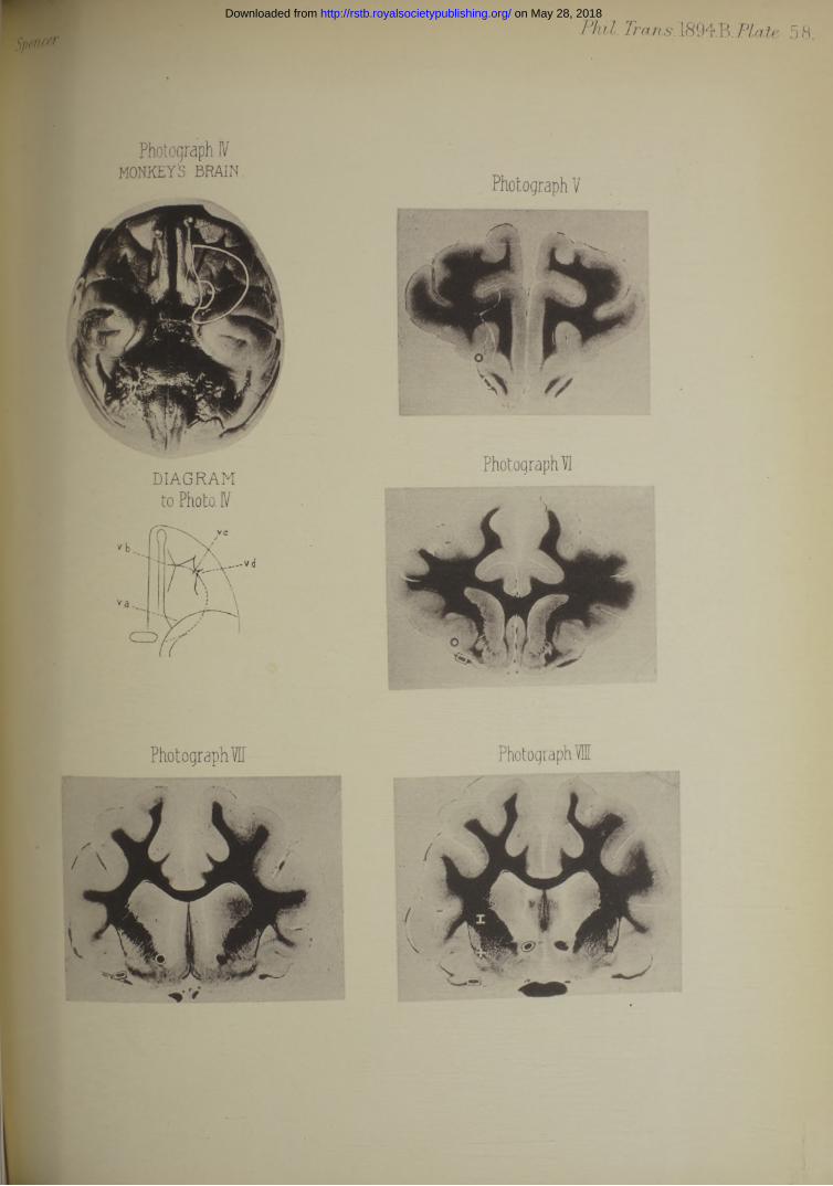
Photograph Ic a t 's b r a i n
Photograph Lb.RABBIT'S BRAIN
Photograph IIIDOG'S BRAIN
DIAGRAMto Photo. 11
DIAGRAMto Photo 111
XiX ’
on May 28, 2018http://rstb.royalsocietypublishing.org/Downloaded from
Phil. Trans.1894.B.Plate 58.m '
Photograph IVMONKEY'S. BRAIN. n, r
rhotographV
Photograph VIDIAGRAM
on May 28, 2018http://rstb.royalsocietypublishing.org/Downloaded from
Phil. Trans. 1894.B. 5 9.
Photograph 1
Photograph E l
on May 28, 2018http://rstb.royalsocietypublishing.org/Downloaded from
[ 609 ]
The Effect produced upon Respiration by Faradic Excitation o f the Cerebrumin the Monkey, Dog,Cat, and Rabbit.
/ W . G. S p e n c e r , M.S., M .B., Assistant Surgeon to the Westminster Hospital.
Communicated by Professor V ictor H orsley, F.R.S.
Received December 15, 1893,—Read January 25, 1894.
[ P l a t e s 57-59.]
C o n t e n t s .P age.
iminary . . . . . V . . . . . . . . . . . . . . . . . . . . v 609Introduction . ...................................... . ...................................................... • • • 610General experimental method .................................• . • • • • ............................ 611Historical retrospect. . . . . . . . . . . . . . . . . , . . . . 613Description of photographs of cortex of brains . . .. . . ...................................... 61/Description of photographs of brain sections . . . . . . . . . . . . . 618
lils op Results..................................... ..... . . ........................... ..... • • • • ■ • • •' • 619Diminution of Action ..................................... ..... 619—640'
(a.) Slowing and arrest obtained from the cerebral cortex ................................................ 619The necessary state of anaesthesia ..................................................................................... 619Description of tracings from the c o r t e x ...................................... ................ . * • * 620Causes preventing slowing and arrest from being obtained . . . . . . . . 629
(f>.) Slowing and arrest obtained from cerebral se c t io n s ..................................... ..... 632Description of tracings from cerebral sections . . • . . . • • • • • • • 635
. Increased Action . ..................................................... . V .......................... ..... • • • • 640—653(1.) Increased action—Acceleration . . . . . . . . . . . . • • • • 640
Description of tracings ..................................... • • • • • • • • . • 644(2.) Increased action—Over-inspiratory Clonus . . . . • • • * • • • • •
Description of tracings . . . ................................ . . . • • • • • 649(3.) Increased action—Over-inspiratory Tonus ..................................................................... 649
Description of tracings.......................................................................................................... 650Increased action—General C onsiderations.......................... ..... • • . . • • •
Note of the circulatory changes produced concurrently . .....................................................nclusions . . . . . . . . . . . . . . . . . . . . 654-656MLCCCXCIY.— B. 4 I 14.9.94
on May 28, 2018http://rstb.royalsocietypublishing.org/Downloaded from
610 MR. W. G. SPENCER ON RESPIRATION AFFECTED BY FARADIC EXCITATION
L ist op P hotographs.
Photograph. I. (a) and (6). Rabbits brain . . . . . .Photograph II. Cat’s b r a in ....................................Photograph III. Dog’s brain .......................... ..... . •Photograph IV. Monkey’s brain . ....................................Photographs V. to XV. Sections of cat’s brain . . . .
Plate.575758 58
58,
Tracings I. to V. Slowing and Arrest from Cortex........................................ 621, 626, 627, 630, 63]Tracings VI. to XIII. Slowing and Arrest from Cerebral Sections . 630, 633, 636, 637, 639, 642Tracings XIV. to XVIII. Acceleration ............................................................................ 643, 645, 65CTracings XIX. to XXIII. Over-inspiratory Clonus......................................... 646, 64<Tracings XXIV. to XXVIII. Over-inspiratory Tonus....................................................... 651, 65i
Diagram I., p. 622.Diagram II., see Conclusions, p. 654.
INTRODUCTION.
The object of the following investigation was to elucidate the character of tb representation of respiration in the highest nerve centres. For this purpose fe excitation method was employed, so that the results of the research are embodied the effects produced on the movements of respiration when various regions of tiecerebral hemisphere are excited. .. .. '^Hfj
By a careful regulation of the anaesthetic state in the species of animal used, by recognition of the exact spot excited, and by employing a suitable stimulus, a constat effect upon respiration can be obtained from the cerebral cortex according to the poit stimulated. And the same result can also be obtained along a line joining the andiscovered in the cortex with the medulla oblongata.
I t is absolutely necessary, in order to get constant results, that the degree! anaesthesia employed should be accurately recognized. This has apparently not be. done before. While some previous observers have, in fact, not used an anaestheticS all, others have employed considerable doses of morphine or chloral. Experiments different observers on non-amesthetized animals have yielded variable results, due . the fact that other disturbing factors have not been taken into account. n® these is apnoea : for instance, any excitation of a sensitive part may cause acce ^ of the respiratory rhythm, and, as a consequence of this acceleration, slowing, or even short arrest. During the prevalence of the apnoea the excitability of the much lowered, so that the same stimulus re-applied to the same spot pro®||^ different change in the respiratory rhythm. Other conditions produce or a . anaesthetic state, such as loss of blood, exposure of the brain, extravasated
on May 28, 2018http://rstb.royalsocietypublishing.org/Downloaded from
esing on the brain, general exhaustion of the animal, or some disease present in fanimal. On the other hand, morphine or chloral in considerable doses completely Iterates some of the respiratory effects.a the four species of animals, monkey, dog, cat, and rabbit, it is easy to form a *ment as to the state of anaesthesia—at least, after paying a little attention to the u xhe rapidity of the corneal reflex may act as a guide, or, better still, the lability of the second division of the 5tli nerve in the orbit, or of the dura mater.
GENERAL EXPERIMENTAL METHOD.
he essential preliminary to every experiment has been a regular respiratory fchm, obtained by employing such a stage of anaesthesia as proved by experience ,e best suited for the particular experiment proposed. A tracing of the respiration circulation before, during, and after the application of the electrical stimulus was
3n in each experiment, and from the tracings alone have the conclusions furnished i drawn.Lll the experiments have been repeated many times in each species of animal, hough, of course, only comparatively few. tracings can be included in this paper, re is no point mentioned of which I have not many illustrations amongst the ;ings in my possession.he Age o f the Animals.—In accordance with what has been known for many years gallois,# S emon and H orsley,t &c.), the age of the animal is an important ure in the innervation of respiration., I have, therefore, in all cases used adultsf*Record o f the Respiratory Movements.—After anaesthetizing the animal a tube was 3rted into the trachea, so as to exclude any mechanical obstruction to the respira- n, or any complication of the result by a simultaneous excitation of the larynx, list, at the same time, by this means the anaesthetic can be more readily regulated. Paul B ert’s transmission apparatus, connected with a Marey’s receiving tambour, ® fixed by an inextensile band round the lower third of the thorax, where the eumference enlarges most during ordinary respiration. The tracings, therefore, in 3 present research record the variations in the circumference of the lower third of 3 chest. In all cases the tracings are to be read from left to right, the upstroke presenting the enlargement in inspiration of the chest from the position of equili- ium, and the downstroke the return during expiration to the position of rest.The following terms applied to the respiratory tracing will be made use of in order distinguish between the very different degrees of respiratory movement observed.
* Legallois, ‘ CEuvres.’ Paris, 1830. Yol. 1, 1.t Semon and H orsley, “ The Central Motor Innervation of the Larynx.” ‘ Philosophical Transactions,’ 590> B, p. 187.
OF THE CEREBRUM IN THE MONKEY, DOG, CAT, AND RABBIT. 611.
4 I 2
on May 28, 2018http://rstb.royalsocietypublishing.org/Downloaded from
612 MR. W. GK SPENCER ON RESPIRATION AFFECTED BY FARADIC EXCITATION
Expiration.—The line drawn through the lowest points of the normal tracing.Active Expiration.—Any line below expiration.Inspiration.—A line drawn through the tops of the normal respiratory curve.Over-inspiration.—Any line above inspiration.Record o f the Circulation.—The cannula was placed in the carotid of the dog, ca
and rabbit, and in the common iliac in the case of the monkey, and connected wif a mercury manometer.
Although this was regularly done, I propose not to refer at length to the changes the circulation obtained simultaneously with the respiratory ones, but to reserve n observations under this heading for a future communication. I have noted, howevt on page 653, two well marked effects which may be found.
The Stimulus.—This was obtained through a Du B ois-B eymond Inductorium frc. two Grenet’s cells. The current was just sufficiently strong to be perceived by tf| tip of the tongue when the secondary coil was 12 centime, from covering the primar The numbers 12, 10, 8, 6, 4, 2, 0 will be used as abbreviations to express the streng. of the faradic stimulus when the secondary coil was 12, 10, 8, 6, 4, 2, 0 centin distant from covering the primary.
The electrodes were of platinum, about 1 millim. apart.An electromagnet in the primary circuit served to indicate the application ai
duration of the stimulus.The rate o f movement was marked by a metronome beating seconds, and recordii
by an electromagnet.The recording points of respiration, circulation, and stimulus were kept vertically s
line.The recording apparatus used was the Kymographion of H urthle.The Course o f an Experiment.—The animal being suitably anaesthetized, and ti
point to be stimulated exposed and gently dried, the electrodes were placed again the surface of the brain. A tracing was first taken with the electrodes in positic before the stimulus, secondly, during the excitation, and thirdly, it was continiu after the cessation of the stimulus until the respiration returned to its previo condition.
A current less than sufficient to produce any effect was first employed and then oi of increased strength. The stimulus was applied for six seconds or more, and often repeated at the same spot at short intervals.
Exposure" o f the Brain.—The skull was always removed so as to sufficiently expo* the part to be excited, the bleeding being stopped by soft wax and amadou. Tl) dura mater was raised as occasion required, and the brain kept warm throughout the experiment by salt solution at blood heat, save that the dried where the electrodes were to be applied. fMg-
The contents of the orbit were removed, or that of the eyeball onlysclerotic drawn forwards.
on May 28, 2018http://rstb.royalsocietypublishing.org/Downloaded from
613
J j^mi-sections or in sections of both hemispheres, the plane of the surface 3 made perpendicular to that of the under-surface of the brain at the point of tin and also at right angles to the longitudinal axis. In hemisections, particular etvas taken to reach up to the middle line and not beyond.pertain amount of haemorrhage inevitably ensued in making such hemisections,,} was controlled by pressing the vessels against the base of the skull with sponges binadou.
HISTORICAL RETROSPECT.
derations in respiration have been frequently noticed by various investigators r g experiments upon the cerebral cortex and upon the basal ganglia, but the results ,ned have not agreed together, or have been negative.Lm disposed to think th a t the variations in the results have been due to the use n-ansesthetized animals in which the effects upon the respiration were complicated •her sensorimotor effects. The negative results followed from not stimulating ;:able spots, or because the animal had been too deeply anaesthetized by drugs or cher ways.[inilew sk y* used young cats and dogs slightly morphinized, and recorded the jration by a tracheal cannula. He obtained two kinds of results. Cortical ration in the region of the facial centre of H it z ig produced a t first a slowing of respiration with increased amplitude. W ith a stronger stimulus a deep inspiration followed by a longer expiration and a pause. These phenomena only occurred ijtimes, and often took place after the stimulus had ceased, even as much as 10 rads after in one case. A sufficiently strong excitation applied towards the base ihe brain, especially when it affected the cerebral peduncles, caused marked deration of the respiration similar to the result of stimulating the dura mater and (peripheral nerves. The arrest of respiration in these experiments may, from my ovations, have possibly been due to the morphine, for slowing of respiration is >;rved to occur without any stimulus in dogs and cats well under the influence of (phine, and arrest may be produced in such animals by the excitation of any sensory
iEPlNEtlaid stress upon the increased frequency of the respiratory movements. >ochefontaine produced rapid movements of inspiration, followed by spasms of
aying intensity,, and finally convulsions. The experiments were made on non- tftsthetized animals.Iichet§ employed dogs so deeply chloralized that the limbs did not react to cortical
Banilewsey, “ Experimentelle Beitrage zur Physiologie des Greliirnes.” * Archiv fur Physiologie, [Jiger,’ 1875, vol. 11, p. 128, see Table VI. and VII.
bHipiNE—quoted by F r a n c is F ranck, see below.1 Bochefontaine, ‘Archives de Physiologie,’ B rown-S^quaru, ’ 1876, vol. 3, series 2, p. 168.
Richet—quoted by F rancois F ranck, see below.
OF THE CEREBRUM IN THE MONKEY, DOG, CAT, AND RABBIT.
on May 28, 2018http://rstb.royalsocietypublishing.org/Downloaded from
614 MR. W. G. SPENCER ON RESPIRATION AFFECTED BY FA RADIO EXCITATION
excitations. He then found that stimulation of the sigmoid gyrus arrested respira. The same result occurred not only at many other points of the cortex, but also obtained from the sciatic nerve.
Both chloral and morphine, according to my own observation, tend to products irregular rhythm in which pauses occur at uncertain intervals. This change may times be induced by any sensory stimulus.
Munk’s* observations in this respect were published in 1883, and reprintefe 1890. Other experimenters have not confirmed them. When the electrodes placed upon the frontal lobe, some millims. in front of the frontal, i.e., the cniaj sulcus, and somewhat lateral to its median end (just where a shallow longituciaj depression runs from before backwards), respiration was arrested with the second coil at 7 or 6. The arrest was in deepest inspiration, and the diaphragm in extr tetanic contraction. Very often acceleration of the respiration preceded the ins]r£ tory tetanus, during which, by deeper inspiration and less expirations, the thoraxui diaphragm came to occupy gradually the maximal inspiratory position, and were fi# fixed in that position. The abdominal muscles during this time remained relaal When the electrodes were placed on the under-surface of the frontal lobe, aboutil middle, there followed from stimulus 7 or 6 either a powerful tetanus of the abdomi muscles in a maximal expiratory phase, or the latter contracted with great freqmc but only to a very small extent, and returned with short jerks to the position of si If the electrodes approached the bulbus or tractus olfactorius sneezing or coughs produced.
In the ape along the horizontal limb of the prsecentral sulcus, and between it u the middle line, with the stimulus at 7 or 6, the thorax and diaphragm assumes inspiratory position; further outwards than this fissure excitation produced a tetn of the abdominal muscles.
As far as they go these observations are in the main confirmed by my own.Francois Franck! made a further examination of the subject and criticized I
work of previous observers. He pointed out that the arrest of respiration prodne by excitation of the cortex was frequently due to apnoea following accelene respiration or was produced by the severe tetanic convulsions into which the imi was thrown. He further showed that under deep morphine or ether respite frequently became irregular with pauses which were very likely to coincide electrical stimulus. He concluded that excitation of the motor area with a sufficiel strong and prolonged stimulus produces modifications in the respiratory moveme and that excitation of other parts of the cortex does not produce respiratory react except in the case of convulsions, i.e.,when the effect of the stimulus spreadsjto motor area or tracts. The respiratory rate quickens or slows without any corresponds between the spot stimulated and the effect produced. I t is according to him rat
* Munk, c Ueber die Formationen der Grosshirnrinde/ Berlin, 1890, p. 164. f F rancois F ranck, ‘ Lemons sur les Fonctions motrices du Cerveau,’ 1887.
on May 28, 2018http://rstb.royalsocietypublishing.org/Downloaded from
OF THE CEREBRUM IN THE MONKEY, DOG, CAT, AND RABBIT, 615
jgree of intensity of the excitation or the degree of excitability of the cerebrum ^appears to influence the frequency. The strongest stimulus produced slowing Ldiptionally arrest, so th a t one cannot discuss centres for acceleration or forjkf.ixange in amplitude, augmentation, or diminution may be observed both with leased and a decreased rate. The inspiratory state coincides with the greater lude and the expiratory state with the less. Special points for inspiration .miration cannot be found upon the cortex, each of the points of the motor zone poduce the respiratory modifications indicated above.FERRICHT* experimented upon dogs anaesthetised with morphine and recorded aspiration by means of an oesophageal cannula. W ith a faradic current less than mt to produce muscular contractions when applied to the * motor ’ area, he fed in some of the experiments slowing of the respiration by excitation of an a the third external convolution outside the orbicularis centre.E20BRASCHENSKY+ experimented with dogs under morphine and with cats under form and ether. One-third of the experiments were negative. In the dog he ied arrest of respiration for two or three seconds immediately after opening the [using a very weak faradic current, but he never saw an active contraction of the atory muscles. In some cats he obtained arrest in inspiration, in others no i His two last experiments were negative. The area stimulated was the same ct investigated by U nverricht, viz., on the anterior part of the third external lution.[is well known tha t centres influencing respiration have been inferred from ition experiments upon the cerebrum, and have been called “ Cerebral Respi- w Centres.”■ristiani,! experimenting upon rabbits, concluded th a t an inspiratory centre was ied in the lateral wall of the third ventricle, in the optic thalamus, just in front b corpora quadrigemina and the commencement of the aqueduct. The area was wuely limited, only 1 millim. square, from which an electrical, mechanical, or taal stimulation evoked an arrest of the diaphragm in inspiration, or a remarkable •eration of the respiratory rate, together with an increase in amplitude. The 1e was symmetrical on each side. Excitation of the corpora quadrigemina, just
or to one side of the aqueduct of S y l v i u s , caused an arrest in expiration. & latter experiments were made after the removal of the hemispheres, corpora ha, and optic thalami by vertical transverse section through the commencement
aqueduct. Thus the ‘ inspiratory’ centre had been removed before the ’ ex :°ry ’ one was excited.Unverricht, “ Ueber die Innervation der Athembewegungen,” 4 Kongress fur innere Mediein,’
7, p. 237.TPreobraschensky, “ Ueber Atbmungscentren in der Hirnrinde,” ‘ Wiener klinische Woohen-. b!t,’ 1890, p. 833.tiiRisTiANi, ‘Zur Physiologie des Gehirnes.’ Berlin, L885, chapter 1.
on May 28, 2018http://rstb.royalsocietypublishing.org/Downloaded from
616 MR. W. G. SPENCER ON RESPIRATION AFFECTED BY FARADIC EXCITATKj
Martin and Booker* stimulated the corpora quadrigemina in rabbits by plu- the electrodes into the substance of the mesencephalon. When the points were " the iter, acceleration was produced, which was changed into inspiratory arre applying a strong stimulus. By plunging the electrodes deep into the pons|£ obtained arrest in expiration. They experimented on two cats only and obtained same inspiratory results as in the case of the rabbit.
The last-named observers, by plunging in the electrodes, appear to have stimi,Mj the same points as myself on the surface of vertical transverse sections. W experimental results varied, perhaps because they did not suitably anaesthetize hi animals. Christiani, no doubt, stimulated in the line of the tract which I shallyi on describe as causing acceleration and over-inspiratory tonus. Again, the shoj | the rabbit of making a vertical transverse section of the brain through the beghijj of the aqueduct, renders the animal deeply anaesthetized. Christiani, under circumstances, obtained arrest of respiration probably in the line of the tract of bij to which I shall refer as causing arrest when stimulated.
In view of the importance of this subject, I have repeated their experimen | the way the observers aforenamed performed them, and have obtained the uj results. But I fail to see why the presence of “ respiratory centres” should;® been assumed from these experiments, if the term “ respiratory centre*” be usi: anything like the same sense as when applied to the representation of respirati-i j the floor of the 4th ventricle. In fact, excitation experiments of this kinder results which are equally well or even better attributed to the results of stimuli] fibres than of stimulating central mechanisms.
Knoll,t experimenting more recently, does not say anything about the use s anaesthetic. He found that excitation of the optic thalamus and corpora quadrigei: caused quickening of the respiration. A strong current applied to the under-si/a of the anterior corpora quadrigemina caused spasm, especially of the eyes, taihi flanks, the respiration becoming shallower or arrested. He never obtained expiratory arrest. He concluded, from his experiments, that there was no respire centre in either the optic thalamus or in the corpora quadrigemina, but that the « of excitation of these regions was to produce impulses in psychical or sensory t»( leading to true respiratory centres in the medulla and cord.
His experimental results and the conclusions drawn seem to me to follow using non-ansesthetized animals.
* Martin and B ooker, ‘ Journal of Physiology,’ vol. 1, p. 370.t K noll, ‘ Sitzungsberichte der Akademie der Wissenschaften,’ Wien, 1885, vol. 92, sec. 3, p. 32
on May 28, 2018http://rstb.royalsocietypublishing.org/Downloaded from

DESCRIPTION OF PHOTOGRAPHS.
> mature consideration, I think it best to give a description of the photographs f all, in order that the position of the points stimulated may be the more easilyestood.> the photographs of brains and sections of brains have been indicated by marks oints where the several effects to be afterwards described in detail, were obtained.
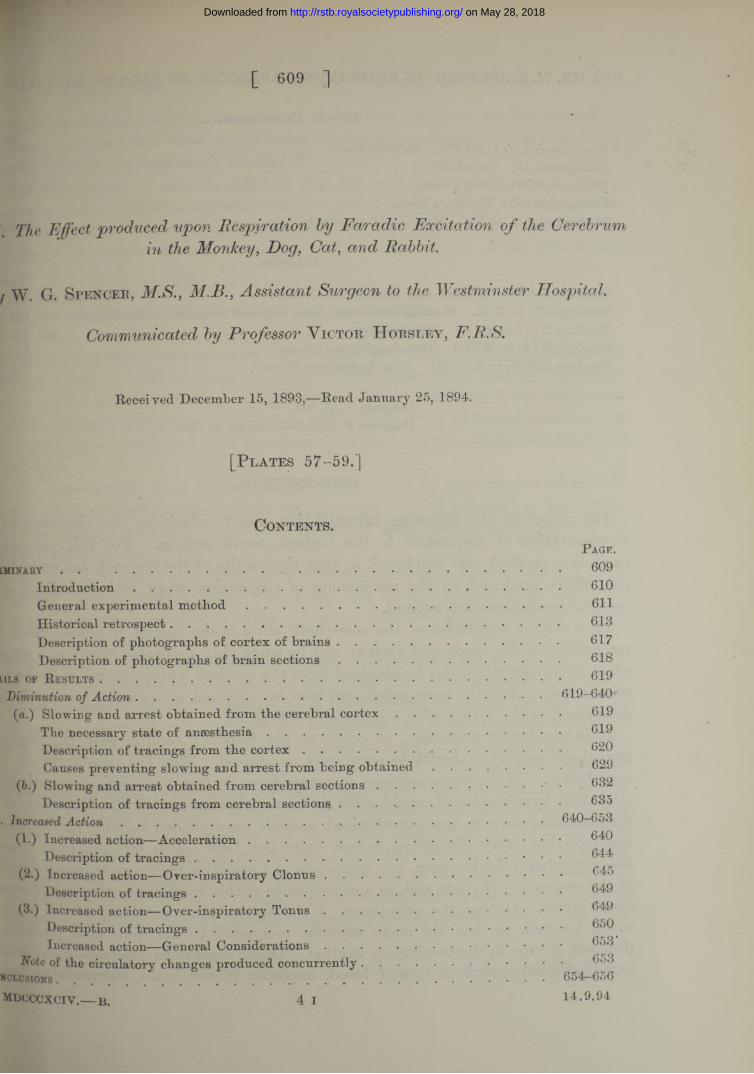
Rabbit’s B rain (see P late 57).
Dtograph la. A photograph of the under-surface of a rabbit’s brain, ring has been placed upon the strip of cortex between the outer margin of factory tract and the rhinal fissure ju st in front of the point where the sylvian r crosses the fissure. The ring marks the point which will be referred to as pot, on the application of the stimulus to which respiration can be arrested. I t shows the junction of the olfactory bulb and tract, marked by a square, the , from which over-inspiratory clonus was obtained.otograph I b.A photograph of the upper surface of a rabbit’s brain, slightly£ed.e cross marks the point a t which a branch from the anterior cerebral artery is over from the mesial to the convex aspect. This is the place where the most ed acceleration can be obtained by the faradic stimulus.
Cat’s Brain (see P late 57).
!otograph II. A photograph of the antero-inferior and outer aspect.(small ring is placed upon the apex of the olfactory lobe, i.e.} the antero- aal end of the triangular convolution formed by the outer border of the olfactory ; the rhinal fissure and the furrow made by the sylvian artery. This is the place te arrest can be obtained.k outer line delimits a semicircular area, a t the border of which respiration #• only be arrested by the strongest current used. The border line com- *68 at the junction of the olfactory lobe and tract, crosses the supraorbital >'e some 3 to 4 millims. from its lower end and also the anterior composite lobe ^ut extending as far as the anterior end of the coronary fissure. The line ftes the anterior end of the anterior suprasylvian fissure, runs backwards parallel ;(d from 2 to 3 millims. above the rhinal fissure, nearly to the anterior end of the dor ectosylvian fissure. The limit then turns downwards and joins the rhinal
"re about the point where the sylvian artery crosses. The outer part of the dory tract forms the lower border of the area.fig ' •le uPpe>r and inner end of the supraorbital fissure just beyond the limit of the
A ograph is the spot where acceleration is most marked, he junction of the olfactory bulb and tract is also shown.^cccxciv.—b. 4 k
OF THE CEREBRUM IN THE MONKEY, DOG, CAT, AND RABBIT. 617
on May 28, 2018http://rstb.royalsocietypublishing.org/Downloaded from

618 MR. W. Gr. SPENCER ON RESPIRATION AFFECTED BY FARAD1C EXCITATlOf
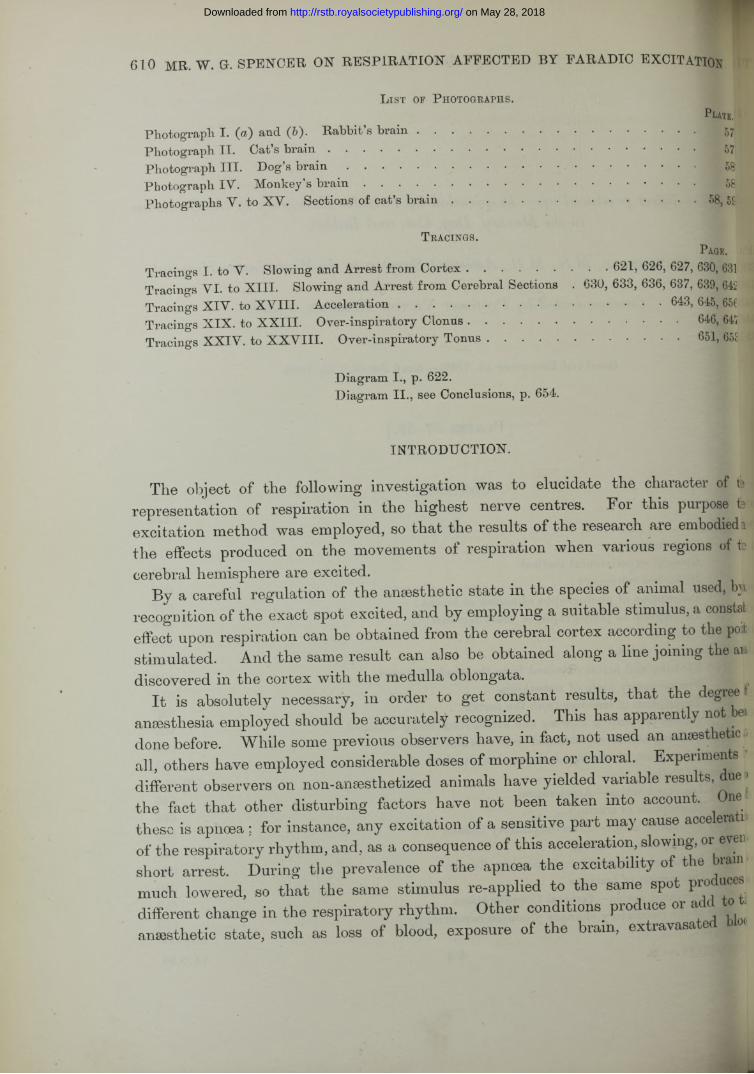
Dog's Brain* (see Plate 57).
Photograph III. A photograph of a dog’s brain in the same position as that of h cat’s.
A small ring has been drawn upon the olfactory lobe between the junction the supraorbital with the rhinal fissures in front, and the sylvian artery behind indicate the centre of the area by the excitation of which arrest of respiration ca' produced. An outer line marks the limit where respiration could be arrested onl$» the strongest current used. This line commences on the olfactory lobe a t 4 millims. in front of the junction of the supraorbital and rhinal fissures, it croeg the rhinal fissure about the point where there is a small notch to be seen, andh orbital lobe, to the supraorbital fissure, some 4 millims. from its lower end. Bead this point it passes backwards and downwards to the point where the sylvian ar traverses the olfactory lobe and rhinal fissure. The outer part of the olfactory bd is the lower border.
The upper and inner end of the supraorbital fissure is just beyond the photogrsh it is the point where the faradic stimulus causes the most acceleration.
The junction of the olfactory bulb and tract is shown.
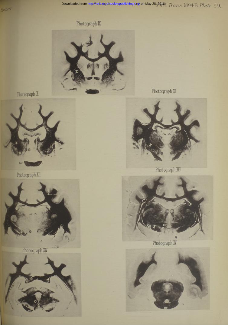
Monkey’s Brain (see Plate 58).
Photograph IV. A photograph of the under-surface of a monkey’s brain.A small semicircle placed just external to the olfactory tract in front of >1
sylvian fissure indicates the centre of the area for respiratory arrest. A larger sa circle marks the limit within which arrest could be obtained with the stroip current used. Commencing at the outer border of the olfactory tract about! middle of its length, the limit passes directly outwards to the transverse portio the H-like orbital fissure, thence along the edge of the shallow concavity whirfl orbital lobe forms, across the sylvian fissure, and turning inwards on the temp sphenoidal lobe reaches the outer side of the optic commissure, some 2 millims. f behind the sylvian fissure.
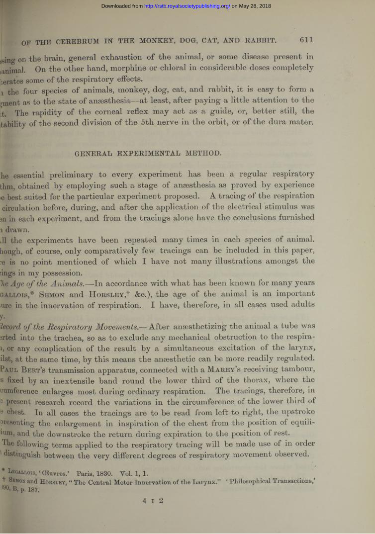
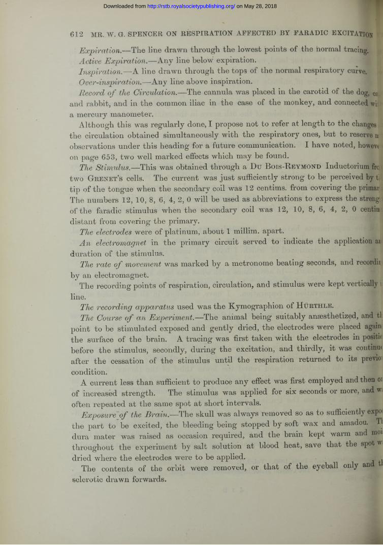
P hotographs of V ertical S ections of the Cerebrum.
Upon the photographs have been placed the following marksA Small Ring. —This indicates the point on each half of the section, by the str*
lation of which arrest was obtained. >A Cross.—This indicates the point on each half of the section, by the stimu a1
of which marked acceleration was caused.
* For the names applied to the cat’s and dog s brain, see L angley, The Structure Brain,” ‘ Journal of Physiology,’ 1883-1884, vol. 4, p. 248.
on May 28, 2018http://rstb.royalsocietypublishing.org/Downloaded from
OF THE CEREBRUM IN THE MONKEY, DOG, CAT, AND RABBIT. 619
Square.:—This indicates the point on each half of the section, by the stimulation vlch over-inspiratory clonus was produced.ne Letter I.—This indicates the point on each half of the section, by the stimulation t»iich well-marked over-inspiratory tonus and tetanus was most easily obtained.
Vertical Sections o f Cat's Cerebrum (see Plates 58 and 59).
>otographs V. to XV. A series of photographs, taken from Dr. H oward xa’s microscopical preparations of a cat’s brain, upon which marks have been 3 as above.jring the course of the research, I made use of a series of photographs taken iDr. H oward Tooth’s microscopical preparation of a monkey’s brain, and also iDgraphs made from sections of the brain of the rabbit and dog. The points were ied in on these photographs, but, on account of the similarity shown to the ;bs obtained in the cat, these photographs have not been reproduced.
I DETAILS OF THE EFFECTS OBTAINED BY EXCITATION OF THE CORTEX REBRI AND OF THE SURFACES OF VERTICAL TRANSVERSE HEMISECTIONS
SECTIONS MADE THROUGH THE CEREBRUM.
A. D iminution of A ction.
fc.) Slowing and Arrest o f the Respiration obtained fro m the Cerebral Cortex.
tie Necessary State o f Anaesthesia in Experiments upon the Cortex.—In order stain arrest of respiration by excitation of the area shown in the photographs, the :&1 must be narcotized so th a t no voluntary or reflex movements are evoked by excitation. But a slight corneal reflex must still exist, and a strong stimulus me able to disturb the respiration through the dura mater, fifth, or sciatic nerves, fen these effects have been abolished, the animal is too deeply anaesthetized, and Cortex will be found no longer excitable to the strongest current used. But the lability quickly returns if the animal be allowed to take a few breaths of air free A ether. is necessary to use ether. Morphine, except in very small doses, tends to
*ish the excitability of the cortex, to render the respiratory rhythm irregular, and, teover, any excessive dose is not so quickly got rid of as ether, he cat may easily be maintained in the suitable stage of anaesthesia, especially n the experiment is confined to the arrest area, so th a t it is not awakened by
station of the sensori-motor region, nor on excitation of the dura mater, he rabbit presents somewhat greater difficulties, especially as there is only one
^ United point on the cortex whence arrest of respiration can be evoked.and monkeys are not so easily maintained in the same state as the cat, they
4 K 2
on May 28, 2018http://rstb.royalsocietypublishing.org/Downloaded from

620 MR. W. G. SPENCER ON RESPIRATION AEEECTED BY FARADIC EXCITATION
are apt to quickly pass into a too superficial or a too deep anaesthesia. But if an j tremely small dose of morphine be injected subcutaneously half an hour before i commencement of the experiment, the animals can be kept in a more constant sti, of anaesthesia, resembling the condition produced in the cat by ether alone. The d<5 of morphine in this case must not exceed 0*01 gr. per 1 lb. (0*0014 grm. per 1 ki| live weight, injected subcutaneously, i.e., 1 minim per 1 lb. live weight of the 1 -t cent, morphine hydrochlorate solution.
The rabbit however cannot be given morphine even in very small doses with| depressing the excitability of the cortex so that no arrest can be obtained. In first experiments I found the arrest area in the dog and monkey without morphg and I have not used any morphine at all in the cat.
The Necessary State o f Ancesthesia in Experiments upon VerticalSections.—The deepest ether anaesthesia in which the animal will breathe regularly required when exciting the surface of sections in order to obtain arrest and to obliter© all the effects of the stimulus upon other fibres, ., those sensori-motor and coma, sural ones which increase the activity of the respiratory movements. Babbits arefe most difficult to get to breathe regularly m deep ether anaesthesia. Here als*a minute dose of morphine, 0*01 grm. per 1 lb. (0*0014 grm. per 1 kilo.) live wei|, may be tried in such animals as do not breathe freely in deep ether anaesthesia. morphine aids in keeping the animal deeply anaesthetized with a smaller quantity! ether.
Slowing and Arrest o f Respiration by Excitation o f the Cortex Cerebri of the (t See Photograph II.
The Strength o f the Faradic Stimulus.—At the centre of the arrest area the stimij required to arrest respiration was generally 7, or 6, sometimes 8. Weaker currea were not quite strong enough to stop respiration hut caused slowing. weaker still had no effect. The further away the point stimulated was from * centre of the area, the stronger the stimulus required until the limit was reached wl| 0 was necessary to check the breathing.' Beyond the limit no arrest occurred altholslowing of the rhythm might be observed.
The form o f the Arrest.—This was generally in full inspiration, or at some P<f midway between inspiration and expiration. At the centre of the area arrest °ccul sometimes in active expiration or in expiration. JBH
The arrest, especially when the stimulus was sufficiently strong and applied the centre of the area, could be maintained for 6-10 seconds, and after the cessaff of the stimulus respiration commenced with a slow rhythm and only gradually regan its former rate. The experiment could he repeated again and again either j same place, or at various points of the area.
on May 28, 2018http://rstb.royalsocietypublishing.org/Downloaded from
Tracing I.
65
55
5
on May 28, 2018
http://rstb.royalsocietypublishing.org/D
ownloaded from

Description o f Tracings showing Slowing and Arrest in the Cat.
Tracing 1, p. 621. The point of the cortex excited was the central point of tl area. See Plate 57, Photograph II.
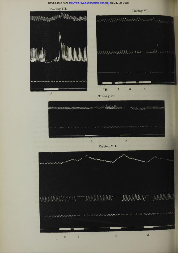
The first line is the manometer tracing, the second that of the respiration, tl third marks the rate of the travelling surface, the space between each vertical strol being traversed in a second, the fourth the incidence, duration, and amount of tl stimulus. W ith 6 the respiration was slowed or incompletely arrested, with 5 am took place in full inspiration, and this was repeated four times at short intervals.
Arrest in expiration could not be obtained so commonly in the cat as in the dt The arrest in active expiration appeared to be limited to the centre of the area al was evoked by a current which did not influence any part beyond the exact sp, mentioned. Moreover for this result it is essential that the effects of shock should !* avoided as far as possible.
I will now give a table formed from tracings taken from most of the points of t area within which arrest of respiration can be obtained. All the tracings were tab from the same cat. Other excitation experiments made on the cortex of the sae animal beyond the “ arrest ” area were negative. The arrest took place in full inspv tion throughout, near the olfactory bulb the arrest was broken by the over-inspiratf clonus to be described later on. I also add a diagram made by enlarging Photogrdi II on which the numbers indicate the points stimulated.
Diagram I.
o
622 MR. W. G. SPENCER ON RESPIRATION AFFECTED BY FARADIC EXCITATION
I t is not maintained that there is a sharply defined limit beyond which a stng stimulus has no effect, but that the area described is the most extensive met withn favourable cases, the more deeply anaesthetized or the more exhausted the animal more does the area contract.
on May 28, 2018http://rstb.royalsocietypublishing.org/Downloaded from

OF THE CEREBRUM IN THE MONKEY, DOG, CAT, AND RABBIT. 623
T a b l e .— See Diagram I. for points.
Amount of Faradic
Points resulting in
current. Arrest. Slowing.
8 8 incomplete9 10(a)
26 10 (b)27 25
7 6789
6 23
10 1610 (a) 30 (6)10 (6) 47112528293030 (a) 49
48
4 45 14
10 (a)12 191516171830 (6) 48
46
2 14 10 (c)46 13
30 (c)
0 10 (c) incomplete 2013 4424 incomplete 30 (c) incomplete 45
50 (a)
50
on May 28, 2018http://rstb.royalsocietypublishing.org/Downloaded from

624 MR. W. G. SPENCER ON RESPIRATION AFEECTED BY FARADIC EXCITATION
Points. Amount of current. Result.
2 6 Arrest, “ clonus.”3 6 “ Clonus,” then arrest.4 4 “ Clonus,” then arrest.5 4 Arrest.6 7 Arrest.7 8 Slowing.7 7 Arrest.8 8 Arrest, incomplete.8 7 Arrest.9 8 Arrest, incomplete.9 7 Arrest.
10 7 Slowing.10 6 Arrest.10 (a) 8 Slowing.10 (a) 6 Arrest, incomplete.10 ( ) 4 Arrest.10 (6) 8 Slowing.10 (») 6 Arrest.10(c) 2 . Slowing.10 (c) 0 Arrest, incomplete.11 6 Arrest.11 4 “ Clonus.”12 4 Arrest.13 2 Slowing.13 0 Arrest.14 4 Slowing.14 2 Arrest.15 4 Arrest.16 6 Slowing.16 4 Arrest.17 4 Arrest.18 4 Arrest.19 4 Slowing.20 0 Slowing.24 0 Arrest, incomplete.25 8 Slowing.25 6 Arrest.26 8 Arrest.27 8 Arrest.28 6 Arrest.29 6 Arrest.30 6 Arrest.30(a) 6 '* Arrest.30 (b) 6 Slowing.30 (6) 4 Arrest.30 (c) 2 Slowing.30 (c) 0 Arrest, incomplete.44 0 Slowing.45 0 Arrest.46 4 Slowing.46 2 Arrest.47 6 Slowing.47 4 Arrest.48 6 Slowing.48 4 Arrest.49 6 Arrest.50 0 Arrest.50 (a) 0 Slowing.
1
on May 28, 2018http://rstb.royalsocietypublishing.org/Downloaded from

OF THE CEREBRUM IN THE MONKEY, DOG, CAT, AND RABBIT. 625
fig and Arrest o f Respiration by Excitation o f the Cortex Cerebri o f the Dog,lee Photograph III .
\ Necessary Stage o f Anesthesia. See p. 619.en the animal was strong a little chloroform was mixed with the first ether but not afterwards. In any case ether has to be given in a more concentrated to dogs than to other animals. The employment of the small doses of morphine, oned on p. 620, is not absolutely necessary, and I did not use it until I had the area, but it is certainly convenient for the prevention of sudden variations anaesthesia.
> Strength o f the Stimulus.—At the centre of the area arrest was generally best ied in the dog with 6, but in some instances 7 proved sufficient, others ed 5. Along the outer limit 0 was necessary, and there were intermediate i where arrest took place with 4 and 2. Ju s t outside the border slowing could )duced with 0.j Form o f Arrest.—This was generally in active expiration, particularly a t the ) of the area. Less often the arrest was in expiration. In the stage of hesia employed it was only sometimes that arrest in any degree of inspiration 'ed, but the tendency was more marked the further the distance from the centre area.
oing II., pp. 626 and 627. For the three points excited see Photograph I l ia , b, c. *est in active expiration was produced from three central points of the area sd in succession. In each case 8 produced slight slowing, but 6 was required to ;. Some irregularity of rhythm with an increase in amplitude took place after Bssation of the excitation before the respiration resumed its regular character, icing III., p. 630. For the point stimulated see Photograph III. e experiment in this case was made at the anterior border of the area and t was only produced at 0. The arrest did not last the whole time of the ilus, but during the latter part the breathing started again slowly.
m9 Mid Arrest o f Respiration by Excitation o f the Cortex Cerebri o f the Rabbit. See Photograph la.
ie Necessary Stage o f Anesthesia. See p. 619.•her alone was, as a rule, used in this animal in sufficient amount to prevent fttary and reflex movements from occurring when the spot was excited, but no 3* Even minute doses of morphine reduced the excitability enough to prevent arrest of respiration from being obtained.' Amount o f the Stimulus.—At only one spot could arrest of respiration be'lue(b and that with 8 or 6. Stronger currents applied around this spot did not ie arrest.®CCCXCIV.— B. 4 L
on May 28, 2018http://rstb.royalsocietypublishing.org/Downloaded from

Tracing II. (a) 1,
Tracing II. (a) 2.
on May 28, 2018http://rstb.royalsocietypublishing.org/Downloaded from

Tracing II. (6).
Tracing II. (c).
10 8 . 64 l 2
on May 28, 2018http://rstb.royalsocietypublishing.org/Downloaded from

628 MR. W. G. SPENCER ON RESPIRATION AFFECTED BY FARADIC EXCITATION
The Form o f the Arrest.—This was nearly always in full inspiration, but as in t> cat arrest in expiration occasionally happened.
Tracing IV., p. 630. The point excited is marked on Photograph Ice.The respiratory rate was slowed with 10, and arrested in full inspiration at 8.
Slowing and Arrest o f Respiration by Excitation o f the Cortex Cerebri in the Monk;{Macacus Rhesus).
The Suitable State o f Anaesthesia. See p. 619.The Exposure o f the Orbital Surface o f the Frontal Lobe.—The animal beg
placed on its back in a semi-recumbent position with the head fully extended, oe orbital surface of the frontal lobe appeared facing forwards and upwards when tie frontal bone including the orbital plate was removed.
The Blood-pressure Tracing— The carotids were not interfered with in ordeito avoid complications from the effects illustrated by the experiments contained i & i former paper.* Therefore the cannula in the monkey was invariably placed in ne common iliac artery outside the peritoneum.
The Arrest Area.—This has been marked out in Photograph IY. I do not aavt that the effect cannot be obtained at points internal to the chord of the arc dim But there are two difficulties in trying to decide this point, firstly, the liabfiitjof wounding the internal carotid artery and its branches, and secondly, the impossib ly of preventing the spread of the excitation to the meninges. I prefer, therefore, ofc to absolutely delimit the inner border of this region.
The Degree o f the Stimulus.—A current of 7 or 6 arrests respiration when apped just external to the junction of the olfactory tract with the uncinate convolute. But exciting points 1 mlllim. apart along any radius of the semicircle indicated in he photograph, arrest of respiration occurred with 4, 2, or 0, according to the dist&ce from the centre. Just beyond the border 0 caused slowing, but not arrest.
The Character o f the Arrest.— The arrest in the monkey was nearly alwaym expiration, but only rarely was any active expiration seen. Beyond the disappn ance of the respiratory curves no change was noted in the circulation from excfteonof the cortex only. S
Tracing V., p. 631. The points excited are in connection with Photograph 1Y(а) R e s p i r a t i o n which was not arrested by 8 was stopped by 7. This w»a
the centre of the area. H*(б) 0 was required in this experiment, the point excited being at the an
border of the area.(c) A little within the limit 2 was necessary.(d) Just outside the border there was no arrest with 0.
* S pencer and H orsley, “ The Control of Haemorrhage from the Middle Cerebral Artery, Bl Medical Journal,’ 1889, vol. 1, p. 457.
on May 28, 2018http://rstb.royalsocietypublishing.org/Downloaded from

629
s w orthy o f note tha t Tracing V., a, b, c, d were from the same monkey, some similar experiments intervening.
The Occurrence o f the
ias, therefore, been shown that the respiration can be slowed and arrested by tion of a certain spot and a limited area around it. This spot is situated in all limals examined to the outer side of the olfactory tract ju st in front of the in of the tract with the uncinate. And this arrest can be constantly obtained ie experiment repeated again and again under certain conditions.[he animal must be in such a stage of ansesthesia, th a t whilst the other effects respiration hereafter to be described are excluded, the excitability of the cortex 3 arrest effect shall not be lost.[he animal must be in a normally active stage, and the brain to be excited must ive been injured in any way.
sufficiently strong faradic stimulus is required.
rsely the Causes which prevent the Occurrence o f the Arrest.a) Insufficient Ancesthesia.—Over-inspiration and over-inspiratory tonus with ig or acceleration of rhythm are excited when the narcosis is not of the right 3 and arrest is prevented. In many cases the tracing thus obtained strongly sted the struggle between the conflicting effects, c.g.t excitation sometimes 3ed a short arrest, which then became arrest in over-inspiration—and near factory tract a clonic effect occurred instead of arrest. I f the animal were id to awake sufficiently there might ensue reflex arrest for a second or two on ng with a weak current, but a repetition of the excitation under these circum- bs caused a different response.(b.) Too Deep Ancesthesia.—W ith ether sufficient to abolish the corneal reflex, ortex of this region was rendered inexcitable. A t the centre of the area 0 produce slowing but no arrest, even this was lost in deeper anaesthesia, hine, when given in larger doses than those mentioned, likewise abolished the ability, the respiratory rhythm tended to become irregular, showing the omena of periodic respiration, of spontaneous pauses in expiration, and of visions. This was especially the case in the dog, but was also observed in rabbit and monkey. W hen the animal was subjected to sufficient morphine pontaneous arrests to occur, arrest of respiration, as others have noted, could Y be obtained by faradizing any sensory surface, including the convex surface of 3rain, but was equally well got from a nerve such as the sciatic.The cortex of animals not in a normal state of health before the experiment more easily inhibited by the ether, and also the cortex was less excitable, slowing, but no arrest, might be obtained. In other words, the depression due to srfect health was added to the ether effect, and in some cases was sufficient
OF THE CEREBRUM IN THE MONKEY, BOG, CAT, AND RABBIT.
on May 28, 2018http://rstb.royalsocietypublishing.org/Downloaded from
Tracing III. Tracing YI.
Tracing VIL
on May 28, 2018http://rstb.royalsocietypublishing.org/Downloaded from

Tracing V. (a).
§
Tracing V. (&). Tracing Y7 (c). Tracing V. (d).
on May 28, 2018http://rstb.royalsocietypublishing.org/Downloaded from
632 MR. W. G. SPENCER ON RESPIRATION AFFECTED BY FARADIC EXCITATION
of itself to abolish the excitability. Such instances occurred in monkeys wit’ diarrhoea, in dogs with tape-worms, in rabbits with parasites in the liver, and i pregnant cats. The latter condition was not sufficiently advanced in any case to b noted before the experiment, and there was, of course, no mechanical obstruction t the respiratory movements. Some of the pregnancies noted were very early.
Exhaustion of the animal from exposure of the brain, from loss of blood, fr0! repetition of the experiment, tended to diminish the excitability, and this ensued a the more rapidly the deeper the primary anaesthesia, whether from ether or from tl, prior causes above noted.
3. No arrest could be obtained except when the secondary coil was at least 8 or centims. from the primary, although slowing of the rhythm might occur with slightly weaker stimulus.
(b.) On the connection with the Medulla Oblongata of the area op thi Cortex Cerebri for Slowing and Arrest of R espiration.
I will now proceed to describe the results of the excitation method I have employ to trace the fibres connecting the cortex with the central mechanism in the medul oblongata.
On this point I need not describe in full detail the results obtained in each of tl species of animals examined. Since the tract, connecting the area on the cortex wit the medulla, runs through the ventral portion of the brain, i.e., through a part which the difference between the four species is very slightly marked, and al; because the results were practically identical. But although I have combined tl description and inserted in this paper only a few tracings, yet I have, in n experiments, assumed nothing. I have stimulated every hemisection in each speci of animal many times, and have tracings sufficient to afford many illustrations every point.
Method o f making Hemisections or Sections o f the Brain. See p. 613.
The necessary state o f Anaesthesia. See p. 619.The deepest ether anaesthesia consistent with regular respiration was found to
the best. Of course great watchfulness was required to prevent an overdose. - some cases, when once arrested, the respiration did not begin until after some artifici respiration had been employed, but usually the respiration started again spontaneous! Only in some rabbits did the respiration tend to stop before they were sufficient! anaesthetized. I used, in these exceptional cases, the small dose of morphine alread noted, viz., 0*01 grain per 1 lb. live weight, and so obtained the necessary anaesthes without an excess of ether. As already mentioned, the dog required the ether vapoi in a more concentrated form than other animals.
on May 28, 2018http://rstb.royalsocietypublishing.org/Downloaded from
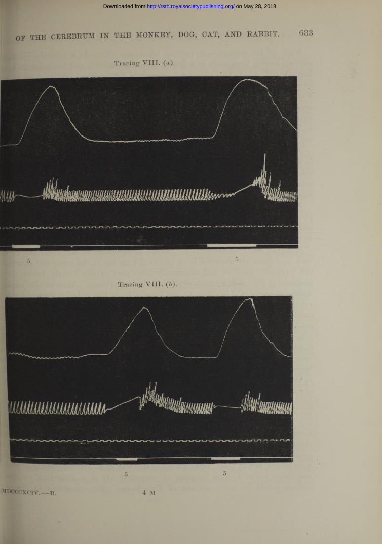
OF THE CEREBRUM IN THE MONKEY, DOG, CAT, AND RABBIT. 633
Tracing VIII.
Tracing VIII.
on May 28, 2018http://rstb.royalsocietypublishing.org/Downloaded from
634 MR. W. a . SPENCER ON RESPIRATION AFFECTED BY FARADIC EXCITATION
Mode o f Experiment.—A vertical transverse (frontal) liemisection (sometimes section) having been made in a deeply narcotized animal, and the point to he excit located and dried, the electrodes were held in position whilst a tracing 0f circulation and respiration were being taken. The current was then applied |6 seconds or more ; generally current 6, sometimes 7 or 5, was required to prodn. arrest. The record was continued until the recovery of the rhythm to the sta existing before the experiment. When the electrodes were placed on the right sp and arrest obtained, the electrodes being held in situ, the experiment could | repeated again and again for six times or more with the same result.
The Form o f the Arrest.—Arrest generally took place in full inspiration, less 0% in a position midway between inspiration and expiration. I t was only in the deep! stages of anaesthesia that arrest took place in expiration, especially in monkeys. | was difficult to keep the other animals in this sufficiently deep stage whilst breath® regularly, or, if arrested by excitation, respiration required artificial aid to start aga.
The Strand o f Fibres which, when excited, produced Arrest.—If the seriesrf photographs taken from Dr. Tooth’s sections of the brain of the cat be examine and the fibres enclosed in the ring noted (see Plates 58 and 59), it will then be possite to trace the following description :—
Froth the centre of the cortical “ arrest” area, in each animal fibres may be foMi back, and these are observed to be gathered together at the lower and anterior en<bf the lenticular nucleus, and apparently form part of the bundle known in hur.n anatomy as the olfactory limb of the anterior commissure. This bundle passes bat- wards, upwards, and inwards, to the inner side of the anterior cornu of the latri ventricle. These fibres tend to the middle line, at which point they form the me anterior fibres of the anterior commissure. Evidence that these fibres actually de<s- satew ith those of the opposite side will be referred to directly. Behind the anteor commissure, the tract passes downwards and outwards from the middle line, closto the infundibulum, above the optic commissure, and then above the inner end of ie optic tract. Behind this,- it runs above and just internal to the crusta. At M beginning of the aqueduct, the strand runs backwards in the tegmentum, just exteial to the fibres of the 3rd nerve as they pass towards the ventral surface. At the 1« of exit of the 3rd nerve the fibres lie vertically above the point of exit of the ne^ being an equal distance below and outside of the aqueduct in the structure know*! the red nucleus. The strands on either side are here parallel; as far as the ex& the 3rd nerve the bundle of fibres can be traced microscopically, but, beyond, t * 3 become lost amongst the other fibres of the tegmentum. But evidence oanW obtained experimentally of their parallel course as far back as the upper border 0 W pons. Beyond this I have not traced them. The animals used in the presentees were adults, and in them, when a hemisection was made involving the pons fibres,® respiration either became irregular or very slow. I t is possible that young# ^ viduals would be more tolerant of disturbance. Two variations in
on May 28, 2018http://rstb.royalsocietypublishing.org/Downloaded from

»eriment went to show that the strand on each side really decussates at the or commissure. Thus (l) if the brain be hemisected frontally and removed ( b a c k on one side as the origin of the 3rd nerve, the other half of the brain ,s exposed on its mesial aspect, so tha t either the cortical mesial surface or the. arface can be excited. There is one point, and one point only, on the mesial
where arrest can be produced by excitation, and that is the cut surface anterior commissure, and immediately behind and below this structure. The which have come from the cortex of the side removed, are in this way
d immediately after decussation. In the monkey the decussation of the so- . olfactory limb of the anterior commissure can be easily perceived to take place at, and can be clearly distinguished from the rest of the anterior commissure i joins the temporo-sphenoidal lobes. Again, if after hemisection and extirpation > level of the 3rd nerve the cortex of the opposite side be excited, no arrest can fcained, nor can any arrest be obtained from hemisections made in this remaining a front of the anterior commissure. By using very strong currents some slight ig may occasionally be observed.
OF THE CEREBRUM IN THE MONKEY, DOG, CAT, AND RABBIT. 635
iplion o f Tracings to Shotv the Result o f Excitation o f Cerebral Hemisections in Producing Slowing and Arrest o f Respiration.
icing VI., p. 630. Monkey. From a hemisection immediately behind the cortical t area. See Photograph V. Coil 6 caused slowing with diminished am plitude; rest in expiration.the dog, arrest in active expiration and in expiration was obtained with a
ency towards an inspiratory phase when a stronger stimulus was used, acing YU., p. 63Q, Cat. From a hemisection a t level of Photograph V II. :e the “ olfactory limb of the anterior commissure ” first appears as a distinct le. Arrest occurred in inspiration.racing VIII,, p. 633. Babbit. From a transverse section through the anterior comure, t.e., the electrodes were applied to the anterior commissure in the middle
See Photograph IX. The respiration was arrested four times in succession 5. There was each time a great rise of blood pressure. I t is to be noted
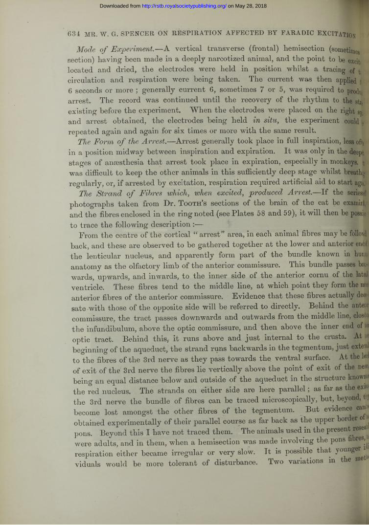
the rabbit could not, without risk, be always so deeply anaesthetized as the other >ials. On the contrary, in the monkey this is easily accomplished, and then it will sen that no rise of blood pressure occurred. See Tracing IX.1 acing IX., p. 636. Monkey. From the mesial surface of the left hemisphere c the removal of the right half behind the level of exit of the 3rd nerve. The Erodes were applied just behind and below the cut surface of the anterior
issure. See Photographs IX. and X. Arrest ’was obtained in expiration twice 14 ® ail( six times in succession with 5.
4 M 2
on May 28, 2018http://rstb.royalsocietypublishing.org/Downloaded from
Trac
ing
IX.
(a).
on
May
28,
201
8ht
tp://
rstb
.roy
also
ciet
ypub
lishi
ng.o
rg/
Dow
nloa
ded
from
on May 28, 2018http://rstb.royalsocietypublishing.org/Downloaded from
638 MR. W. G. SPENCER ON RESPIRATION AFFECTED BY FARADIC EXCITATION
Tracings VIII., IX., and XI. illustrate an important point, viz., that the result-described can be obtained many times in succession by keeping the electrodes to th same spot, and making use of a stimulus sufficiently strong, yet not so great as to quickly induce exhaustion, provided always that the animal be kept in the same l of anaesthesia.
In the case of the experiments illustrated by the next tracing, a variation K procedure was adopted, in order to show the influence of the etherisation. I B animal was first allowed to come out a little from the state of deep ansesdHand then excitation was repeated whilst ether vapour was being administered, iH
m
the animal was in a more deeply anaesthetized state with each experiment than it been in the previous one.
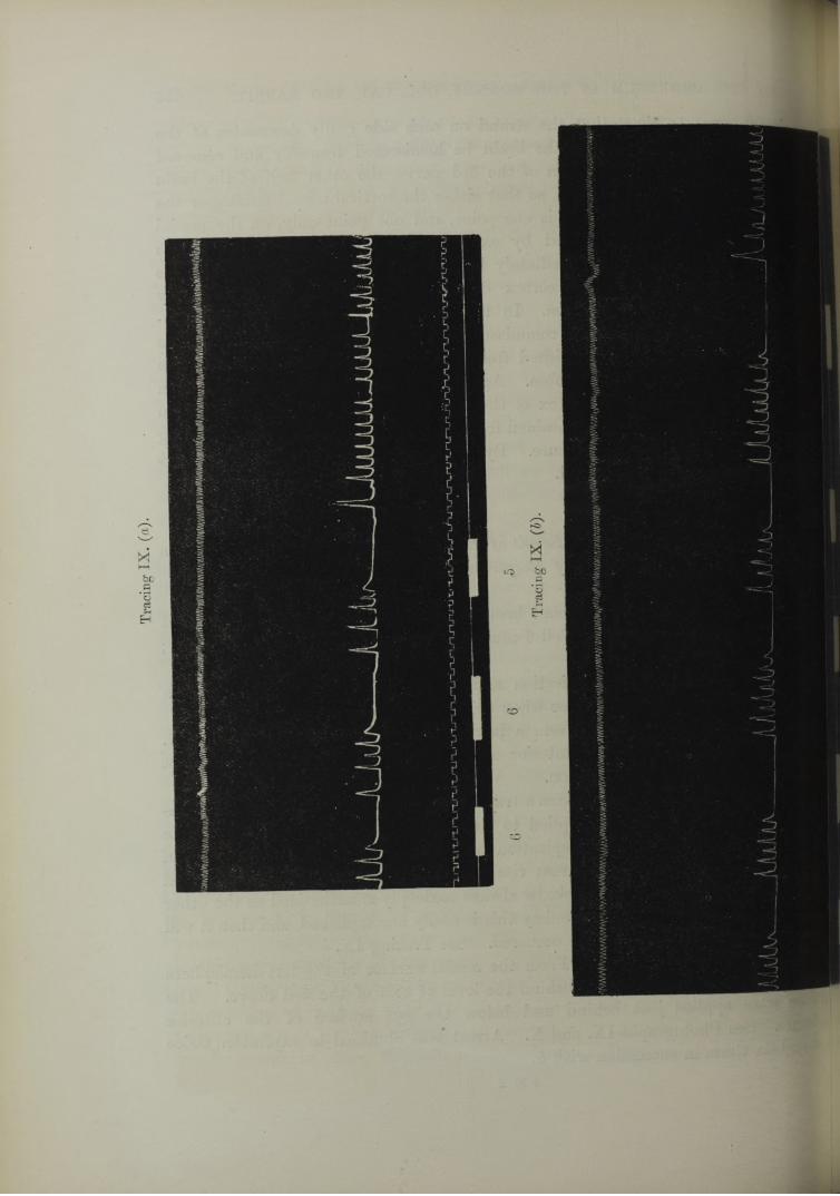
Tracing X., p. 637. From a hemisection in a dog at the level where the optic is cut longitudinally in its course to the occipital cortex. See Photograph XI. animal during the time that the record was being taken was breathing ethefj becoming more deeply anaesthetized. Arrest was produced eleven times in success at the same spot in an animal which was at the commencement in a superficial std of anaesthesia, but, by breathing ether, gradually passed into a deep stage. first experiment the arrest was complicated by the over-inspiratory tonus, will be referred to later on. In the second experiment the over-inspiratory to] tending to complicate the arrest was again present, but weaker. The tracing illustrates a commonly observed point, that with the deepening of the ansesthflj some increase in the strength of the stimulus was required. Thus the first experi^L required 8 and the last 4, but allowance must be also made for exhaustion Si to the repetition of the experiment at short intervals.
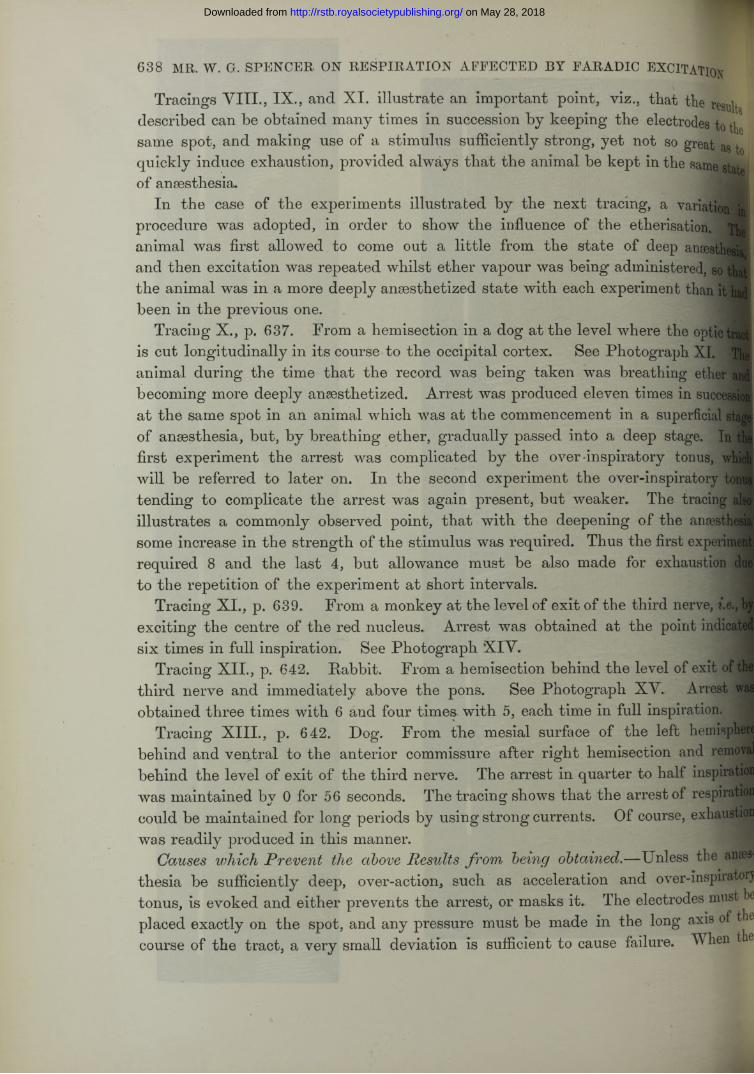
Tracing XI., p. 639. From a monkey at the level of exit of the third nerve, i.e< exciting the centre of the red nucleus. Arrest was obtained at the point indicate six times in full inspiration. See Photograph X IV .
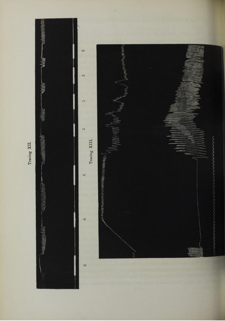
Tracing XII., p. 642. Rabbit. From a hemisection behind the level of exit of® third nerve and immediately above the pons. See Photograph XV. Arrest H obtained three times with 6 and four times, with 5, each time in full inspiration.
Tracing XIII., p. 642. Dog. From the mesial surface of the left hemisptH behind and ventral to the anterior commissure after right hemisection and remfflB behind the level of exit of the third nerve. The arrest in quarter to half inspiration was maintained by 0 for 56 seconds. The tracing shows that the arrest of respiratid could be maintained for long periods by using strong currents. Of course, exhaus^JJ was readily produced in this manner.
Causes which Prevent the above Results from being .—Unless the anseithesia be sufficiently deep, over-action, such as acceleration and over-inspiratorj tonus, is evoked and either prevents the arrest, or masks it. The electrodes must e placed exactly on the spot, and any pressure must be made in the long axis of t 6 course of the tract, a very small deviation is sufficient to cause failure. When 6
on May 28, 2018http://rstb.royalsocietypublishing.org/Downloaded from
LJwO
uiiu\A
----
OF THE CEREBRUM IN THE MONKEY, DOG, CAT, AND RABBIT. 639
on May 28, 2018http://rstb.royalsocietypublishing.org/Downloaded from

640 MR. W. G. SPENCER ON RESPIRATION AFFECTED BY FARADIC EXCITATION
electrodes tire applied exactly to the right place and held steadily there, the expg] ment may be repeated many times in succession. Exhaustion with slow or irreguh rhythm does not abolish the excitability, but the arrest being produced, it tends, continue even after the cessation of the stimulus, and artificial respiration may required to start the rhythm.
THE DETAILS OF THE EFFECTS OBTAINED BY EXCITATION OF THE CORT] CEREBRI, AND OF THE SURFACE OF VERTICAL SECTIONS THROUGH Tf CEREBRUM, IN THE CAT, DOG, AND RABBIT.
B. An Increased Action.
(1.) Acceleration of the Rhythm.
General.—-The excitation of any sensitive part in a non-ansesthetized animal tei; to disturb the respiration in the direction of increased action, the rhythm is more*: less accelerated, irregular, and there are muscular movements of the limbs In a slightly anaesthetized animal a strong stimulus produces this effect overawe extent of the cerebral surface. Under ether anaesthesia sufficient to abolish fi excitability of the cortex so far that no movements of the limbs are evoked by I stimulus, acceleration of respiration is not caused by faradic excitation except y excitation of one spot on the cortex. Even if the faradic current be so strong call forth automatic movements, any acceleration of the rhythm which then ny be obtained elsewhere is much less pronounced than the rapid rhythm exciting this particular spot.
On stimulating hemisections of the brain from before backwards this acceleratn area on the cortex can be found to be connected with the medulla along a define line, excitation of any point along this line producing marked acceleration.
So long, therefore, as the animal is sufficiently under the anaesthetic, acceleration rhythm only occurs on the stimulus being applied at the one spot on the cortex ft along the course of the tract through the cerebrum. jj^H j
The Suitable Stage o f Anaesthesia.—In all three species of animals on which e following observations were made, sufficient ether was given to abolish the of movements of the limbs, and to suppress the over-inspiratory tendency which’ be later on referred to. This applies both to the excitation of the cortex and t of the surface of the sections of the hemisphere. A deep stage of anaesthesia abobs all cortical excitability for acceleration, as well as that of the sections.
One source of error had always to be carefully excluded, viz., the alteration mjj respiratory centre due to apnoea produced by the acceleration. I t was there«IJM®*i advisable to only allow the stimulus to act for about six seconds, otherwjp respiratory rhythm began to get slower. I t was also necessary to allow u w ^ a return to the normal respiratory rate before repeating the expenmen , o « the excitability was found to have been lowered by the apncea.
on May 28, 2018http://rstb.royalsocietypublishing.org/Downloaded from
[rea on the Cortex Cerebri where the most marked Acceleration was obtained on Excitation.
rthe Doq and Cat.— The effect was best marked at the upper end of the supra- tl sulcus. From a limited area around the acceleration could be obtained, but in s marked degree. On increasing the anaesthesia this was the last place where cceleration could be obtained with 0, and it was at this point tha t acceleration be first obtained in an animal recovering from deep anaesthesia.
ithe Rabbit.—If, when viewing the dorsal aspect of the rabbit’s brain, the eye is back from the olfactory bulb along the inner margin of the hemisphere, an |r is seen coming up between the edge of the hemisphere and the falx cerebri, hen turning over on the convex surface and running outwards in a line which *sts the position of the crucial sulcus in the cat and dog. When the pia mater refully stripped off, a groove remains. This vessel serves to indicate the place 3 marked acceleration can be obtained in this animal. Wdien the electrodes are d astride of the vessel near the margin marked acceleration is obtained. An of 2 millims. in diameter overlapping the margin, i.e.} extending a little way on ]Q mesial surface, is about the limit within which this phenomenon is markedly jsented. p
Connection with the Medulla Oblongata o f the Area on the Cortex which causes Acceleration when Excited.
he course which the fibres leading from the cortex cerebri take towards the ulla oblongata is indicated upon the accompanying photographs by a cross. I t ends in the corona radiata to the lenticular nucleus where this structure blends i the lower and external part of the internal capsule as seen in frontal sections, i band of fibres then passes below the internal capsule as the latter changes into crusta, and appears to slope towards the inner portion of the tegmentum. The nd on each side reaches the middle line in the same frontal plane as the exit of 3rd nerve. The spot where acceleration is at this level obtained is thus in the 'r-peduncular grey matter, rather nearer to the ventral margin than to the floor of L aqueduct.ihe acceleration tract on either side appears to decussate in the plane of the t of the 3rd nerve. I have already shown that hemisection and extirpation [one side behind the anterior commissure, but in front of the plane of exit of
3rd nerve, removes the “arrest” effect; but marked acceleration can still be Gained from the spot on the cortex of the remaining hemisphere. On the other ad, removal of one hemisphere immediately in front of the pons does away with the ];cial acceleration from the cortex of the remaining hemisphere. Some slight 1 toleration may still be obtained from the cortex and internal capsule if the anaes- mdcccxciv.— b . 4 N
OF THE CEREBRUM IN THE MONKEY, DOG, CAT, AND RABBIT. 641
on May 28, 2018http://rstb.royalsocietypublishing.org/Downloaded from
Trac
ing
XII
.
Trac
ing
XII
I.
on
May
28,
201
8ht
tp://
rstb
.roy
also
ciet
ypub
lishi
ng.o
rg/
Dow
nloa
ded
from

Tracing XVI.
Tracing XV.
on May 28, 2018http://rstb.royalsocietypublishing.org/Downloaded from
thesia become superficial, but this is the general effect mentioned on page 640, is only very small, and is connected with the medulla through the descending sensor motor tract, i.e., does not decussate above the pons. Finally, when one-half of tl cerebrum has been removed no marked acceleration can be obtained from the mesi surface of the remaining half, except from a point immediately dorsal to and behir. the exit of the 3rd nerve from the crus.
644 MR, W. G. SPENCER ON RESPIRATION AFFECTED BY FARAD1C EXCITATION
The Description o f Tracings showing Acceleration.
The respiratory rate was counted by comparing a period of 6 seconds before at during the stimulus.
Tracing XIV., p. 643. Rabbit. The spot on the cortex cerebri of the right sh was excited (see Photograph I b). Before any excitation the respiratory rate was 80 p minute. Stimulation with 8 caused the rate to become 100 per minute, i.e., < increase of 0*25. W ith 7 the rate became 120 per minute, i.e., an increase of 0‘5.
Tracing XV., p. 643. Dog. Excitation of the upper end of the supraorbital fissu on the left side, after hemisection and extirpation of the right half behind the level ’ the anterior commissure. Excitation with 6 caused acceleration three times in sum sion, the first time from 50 to 110, an increase of 1*2, the second time from 40 to IS an increase of 2, and the third time from 35 to 95, an increase of 1*7. The slow rate’ respiration before the stimulus marks a deeper stage of anaesthesia than Tracing XI Further, also, an opposite hemisection having been made in this case, it is clear th. the fibres connecting the cortex with the medulla cannot cross above the levelf hemisection.
Identical effects were obtained from the cat.Tracing XVI., p, 643. Cat. For point of excitation see Photograph VIII. Wi!
8 no acceleration occurred, but with 6 the rate changed from 70 to 170, an increa of 1 '4. There was a marked rise of blood-pressure; in other experiments wideeper anaesthesia no rise in blood-pressure occurred (see Tracing XVIII.), and t. respiration being slower at the commencement, even greater acceleration folio we I have a tracing from a dog, taken from the same spot. 8 produced a fraction increase of 3*4, viz., from 25 to 110 ; the absolutely greater rate occurs in the slight stages of anaesthesia, e.g., 170 in this Tracing.
Tracing XVII., p. 645. Babbit. Right hemisection through the plane of the ei of the 3rd nerve, see Photograph XIV. W ith 8 the increase was from 60 to 120, u an increase of 1 on the first experiment and an increase from 45 to 150 on the seco* experiment, i.e., an increase of 2*3. The second experiment was thus better than t first. I t has often been found that an almost imperceptible movement of t electrodes may, on the one hand, improve the reaction and, on the other hand, iinpfc it. This indicates the necessity of exciting the exact point if the maximal effect5® f be obtained.
1
on May 28, 2018http://rstb.royalsocietypublishing.org/Downloaded from

OF THE CEREBRUM IN' THE MONKEY, DOG, CAT, AND RABBIT. 645
Tracing XVII.
8 *• 8
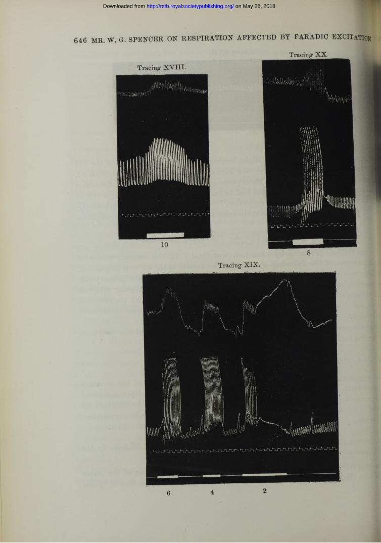
:,cing XVIII., p. 646. Dog. Right hemisection at the plane of exit of the 3rd See Photograph XIV. The rate increased from to 110, i.e., 1, with 10.
was practically no variation in blood-pressure. In this region where the electrodes so close to the crusta, the animal had to be well anaesthetized, although not so v as required for the arrest of respiration, for if not, over-inspiratory tonus compli- ithe acceleration, and there was a t the same time a marked rise in blood-pressure.
(2.) Qver-insjpivatory Clonus.
s movement produced by excitation was similar in the several species of Is and was of the following character. The animal made an over-inspiration, ihen several sharp over-inspirations were superimposed upon the primary one. Dver-inspiratory jerks were peculiar in following one another at regular intervals hythmic manner and in not ceasing exactly a t the same time with the stimulus, ,i.wo, or three more of these over-inspirations taking place after the cessation of timulus.e result could be obtained from a definite area of the cortex, and along a tract i to the medulla.
Area on the Cortex fo r Over-inspiratory Clonus.
lie was invariably and most easily obtained from the junction of the olfactory and tract. By excitation of the portion of the frontal lobe, (< prorean lobe,”
t immediately above and behind this spot the same effect could be obtained by of a stronger stimulus. But the typical effect with the weakest excitation
^obtained at the junction referred to, both on the dorsal and lateral aspect.
Apparent Course o f the Fibres Connecting the Cortex with the Medulla by the Excitation of which Over-inspiratory Clonus is produced.ids was followed in the vertical sections commencing with those of the outer °f the olfactory tract, and continuing backwards past the furrow formed by the
on May 28, 2018http://rstb.royalsocietypublishing.org/Downloaded from
646 MR. W, G. SPENCER ON RESPIRATION AFFECTED BY FARADIC EXCITATION
6 4 2
on May 28, 2018http://rstb.royalsocietypublishing.org/Downloaded from
OF THE CEREBRUM IN THE MONKEY, DOG, CAT, AND RABBIT. 647
Tracing XXI.
0 0
Tracing XXII.
6
Tracing XXIII.
10 8
on May 28, 2018http://rstb.royalsocietypublishing.org/Downloaded from
sylvian artery to the temporo-sphenoidal lobe at its mesial part, viz., the uncina convolution. The effect obtained by excitation was traced along the uncinate conv lution to the uncus. In the vertical section the over-inspiratory clonus is obtained from the region outside the optic tract as it passes backwards from t chiasma, whereas all the other respiratory effects are represented in this secti internal to the tract. In the next section, behind where the optic tract has extendi to the occipital lobe, the over-inspiratory clonus is to be obtained from the outer si? of the peduncle, external to the crusta. Immediately behind the exit of the f nerve transverse pontine fibres begin to separate the crusta from the ventral surfa,, and over-inspiratory clonus can here be obtained beneath the pyramidal tra;. Behind the posterior perforated spot, at the naked-eye margin of the pons, the clots can be obtained in the middle line at the ventral margin.
The course of the fibres which seem to subserve this reaction was easy to tracer far backwards as the section through the optic tract, see Photography Sfcf Naturally, however, the connection between the uncinate gyrus and the region! the crusta, &c., was involved in greater difficulty. I repeated the ex p erin ^ il this point whilst constantly referring, as I have always done throughout this reseaap to Dr. Tooth’s microscopical preparations of the cat’s brain and to the made from them. In the section from which Photograph XIV. was taken transvee or oblique fibres are already apparent beneath the crusta, although the 3rd nerva not yet passed, and the posterior perforated spot still forms the ventral margin mmmiddle line. ’ t ,,
The excitation of the under-surface of the anterior border of the pons was theaespot where the over-inspiratory clonus was obtained in the middle line. Ia#gfj ment with this, the removal of one hemisphere through the plane of exit of thejc nerve did not abolish the effect obtained by excitation of the remaining hemispbe although the arrest and acceleration effect disappeared.
648 MR. W. G. SPENCER ON RESPIRATION AFFECTED BY FARADIC EXCITATION
The Degree of Stimulus.8 and 6 may be taken as the usual amounts required. If the a n im a l were i
very supetficial stage of anesthesia, less was needed. Currents of increasing SO® up to 0 produced a similar but less marked result in the deeper stages of an esth e ...
Causes which Prevented the Effect from being Obtained.As the animal became exhairI t disappeared in deep ansesthesia even with 0.
the effect was more and more incomplete. . •Over-inspiratory clonus might be complicated with arrest or with over-inspn
tonus. I t was liable to be complicated with arrest when the °” tex 'v fj owing to spreading of the stimulus or the effect of the same from the junctio olfactory bulb and tract to the adjacent arrest area. Over-inspiratory
on May 28, 2018http://rstb.royalsocietypublishing.org/Downloaded from

OP THE CEREBRUM IN THE MONKEY, DOG, CAT, AND RABBIT. 649
J e t"the result in the sections behind the optic tract, where the strand passes il to the centre of the crusta. This was avoided by keeping the electrodes close B under-surface of the crus, by not employing any stronger current than was 4tely necessary, and by using sufficient ether to diminish the tendency to over- iiitory tonus.
Option o f Tracings showing Over-inspiratory Clonus.
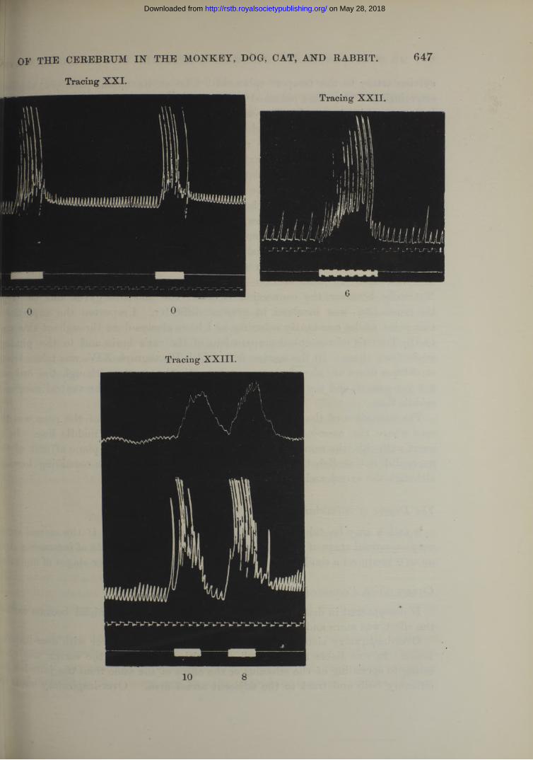
ticing XIX., p. 646. Cat. Junction of olfactory bulb and tract, right side, see tgraph II. Over-inspiratory clonus was produced by 6 and 4, a t first, also i2, but afterwards arrest occurred from the extension of the stimulus to the $ area. W ith this tracing may be compared the table on p. 623, as showing feet of exciting the points marked 2, 3, 4, and 11 on Diagram I. icing XX., p. 646. Dog. Junction of left olfactory bulb and tract, see Photo- j III. The right hemisphere had been excluded by section between the plane :lt of the 3rd nerve and the anterior end of the pons. 8 produced the effect, icing XXI., p. 647. Rabbit. Junction of left olfactory bulb and tract, see ograph 16. The right hemisphere had been excluded by section at the plane h optic tract. The clonus was twice elicited by a current of 0. The animal was deeper stage of anaesthesia than in the preceding cases. In a stage deeper still eaction is lost.icing XXII., p. 647. Dog. Tip of left uncinate convolution, see Photograph XL til., and just external to the optic tract, after section of the right side between lane of exit of the 3rd nerve, and the anterior end of the pons. The clonus was ciced with 6..acing XXIII., p. 647. Cat. The middle line at the ventral margin of the rior end of the pons, see Photograph XV., after a right hemisection and removal ae hemisphere at this level, the clonus was produced with 10 and 8.
(3.) Over-inspiratory Tonus.
ns is the most widely generalized effect of any stimulus upon the respiration.' non-ansesthetized or partly anaesthetized animal, all previous observers have l7n that a stimulus, if strong enough, will influence the respiration in this Rstion when applied to any sensory surface.tie greater relative influence of anaesthesia upon the over-inspiratory tonus has ved of the other effects being largely or entirely freed from its complicating fence.0 noubt greater degrees of over-inspiratory tonus might be most easily produced n°n-&naesthetized animal, but all my experiments have been with anaesthetized
l)aals; and I may recall what I have mentioned before, viz., that there are other C6S of anaesthesia than drugs, and in order to produce the same amount of over-
1 dcccxci v. —b . 4 o
on May 28, 2018http://rstb.royalsocietypublishing.org/Downloaded from

650 MR. W. a. SPENCER ON RESPIRATION APFECTED BY FARADIC EXCITATION
inspiration in the same animal with the same stimulus applied to the same place, it necessary that the animal should be in a definite stage of anaesthesia, whether caused] ether, shock, haemorrhage, &c. I have examined the production of over-inspir^p tonus from three points of view, by excitation of the 5th nerve and meninges, q£K sciatic nerve after the removal of the cerebrum, as well as the cortical effects.
Over-inspiratory Tonus from Excitation o f the 5th Nerve and Meninges.In animals under sufficient ether to abolish voluntary movements, I only obtH
by faradic excitation of the 5th nerve and meninges over-inspiratory tonus, { never observed any arrest of the respiratory rhythm. Slight excitation of | terminal ends of the 5th nerve in the nose, by chloroform or tobacco smoke,» been described as instances of the causation of arrest by excitation of this nJB The well-known fact that every child or animal forced to inspire an anaesthetic, ‘ 9 its breath,” is an example of this, and it requires a distinct effort of the adult hurf will to resist the tendency. This arrest through the peripheral terminations of le 5th nerve disappears when there is sufficient anaesthesia to abolish voluntaryism ments. Arrest has been produced in a like fashion by exciting the trunkJKfl nerve or its branches in the dura mater in a non-anaesthetized animal with a vy weak current But an animal may be made “ to hold its breath by gentle excitaln of many other sensory surfaces, such as the cerebral cortex or the sciatic nerve, i these cases, however, the arrest usually ends in a general convulsion, and ^ i repetition of such an experiment the convulsion is just as likely to occur primal;instead of an arrest. <
When the anaesthesia is sufficient to abolish voluntary movements, then excitaoi of the 5th nerve root, of the Gasserian ganglion, of the divisions of the 5th nerve, v of the dura mater, results only in over-inspiratory tonus, and not in absolute of the rhythm. If excessive currents are employed, the rhythm may be arrenHj spreading from the dura mater to the arrest area of the cortex either immediately direct contact, or mediately through the cerebrospinal fluid, or blood clot. J p also spread to the roots of the vagus. ; The amount of current required to pro* over-inspiratory tonus varies with the amount of anaesthesia. In deep ether there was not the slightest effect produced upon the respiration even with the stronf current used. When the current was strong relative to the amount of anaesthesia, thorax assumed an extreme degree of expansion, and the lever of the Mats tambour no longer recorded any thing but fine tetanic movements, but on direct o vation the respiratory rhythm was seen to continue although small in amplitude masked by the extreme expansion.
Description o f Tracings from the 5th , showing Over-inspiratory Tonus
Tracing XXIV., p. 651. Rabbit. Excitation of the 2nd division of the 5th the orbit. The result was over-inspiratory tonus, with a continuance of the
on May 28, 2018http://rstb.royalsocietypublishing.org/Downloaded from
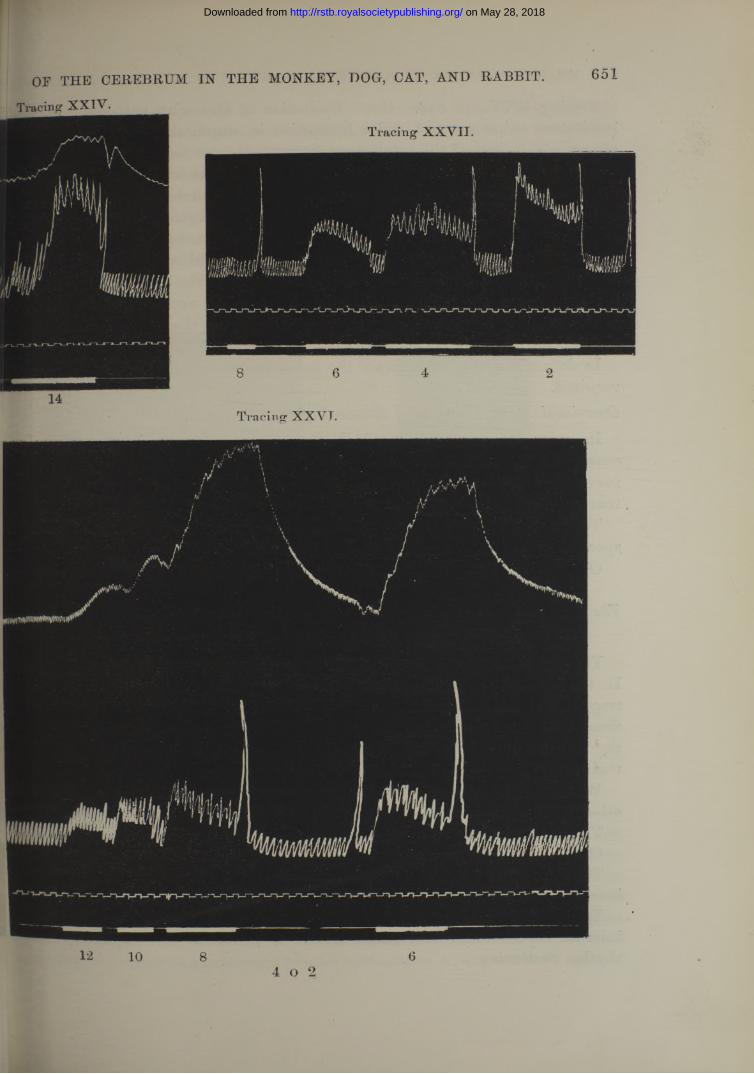
651OF THE CEREBRUM IN THE MONKEY, DOG, CAT, AND RABBIT.
Tracing XXVT.
12 10 84 o 2
6
on May 28, 2018http://rstb.royalsocietypublishing.org/Downloaded from

Tracing XXV., p. 647. Cat. Excitation of Gasserian ganglion. Marked ove- inspiratory tonus occurred, and a diminution in amplitude from the expansion of t} thorax which masked the respiratory movements.
In Tracings XXIV. and XXV. the same current was used ; and other tracings fr0! the dog and monkey, with the same strength of stimulus, show similar effects, on rather less marked in the case of the monkey. In comparing a number of experimen upon the 5th nerve in the various species, it is to be noted that when the ansesttieli was insufficient acceleration of rhythm took place, and continued after the cessati* of the stimulus, until the animal was given more of the drug, when, on repetition f the experiment, the rate was not altered. Also, in exciting the 5th nerve, or du, mater, near the olfactory bulb, care had to he taken that the current did not spies through the olfactory nerves to the bulb, and set up “ clonus.”
In deeper stages of anaesthesia the same result occurs, but a stronger excitation!required.Over-inspiratory Tonus after Removal o f the Cerebrum.
In, order to show that over-inspiratory tonus is a general effect of exciting a, sensory nerve, and has no special connection with the cerebrum, I excited the ||Bia? nerve after the complete removal of the cerebrum by incision at the level of h
. tentorium cerebelli. „Tracing XXVI., p. 651. Cat. Excitation of the left sciatic nerve in the poplitl
space, after the removal of the whole of the cerebrum immediately above the pons. Over-inspiratory tonus occurred with 12, 10, 8, and 6.
The Area on the Cortex where the most marked Over-inspiratory Tonus isand the apparent course o f the Fibres connecting this Area with the Medulla.
The greatest effect is to be obtained in the centre of the “ sensori-motor” ar, In the sections the best effect is obtained from the centre of the descending mol tract in the internal capsule, and lower down, in the middle of the crusta. Tk fibres do not decussate early, for if the opposite hemisphere be removed just beh« the plane of exit of the 3rd nerve, the effect may still be obtained by stimulate their course downwards in the vertical sections of the remaining half.
Whether there is one spot in the sensori-motor area more easily affected ths| aj other, I cannot say, so readily is the result influenced by the anaesthesia.
Tracing XXVII., p. 651. Cat. Excitation of the “ sensori-motor” area.Great over-inspiratory tonus was produced with 6, 4, and 2. > ,The same effect followed excitation of this area when the opposite hemisphere
been completely removed down to the tentorium cerebelli. . VTracing XXVIII., p. 646. Dog. Excitation of the crusta just
internal capsule, see Photograph XIII. 8 produced over-inspiratory tonus,rhythm continuing.
652 MR. W. G. SPENCER ON RESPIRATION AFFECTED B Y FARADIC EXCITATION
on May 28, 2018http://rstb.royalsocietypublishing.org/Downloaded from

Sparing a number of tracings of over-inspiratory tonus, I find that, although . parked in the sensori-motor area of the cortex, and in the line of the descending ctract, yet it is produced outside this and complicates the “ a rrest” or “ accele- o ” effect, unless removed by the anaesthesia.'lis on the ‘‘arrest” area, arrest in over-inspiratory tonus follows excitation a the anaesthesia is insufficient. Conversely it may be obtained from the rst” area, uncomplicated by any arrest after opposite hemisection behind the 5tor commissure, and from the “ acceleration ” area uncomplicated after a similar ilection behind the third nerve exit.
OF THE CEREBRUM IN THE MONKEY, DOG, CAT, AND RABBIT. 053
Tracing XXV.
14
Tracing XXVIII.
8
Increased Action o f the Respiration in the Monkey.have been forced to distinguish the monkey from the rabbit, cat, and dog,
eause in it an increased action of the respiration is so much less marked. There pears to be no fundamental difference, for I have obtained the same reactions, iceleration,” “ over-inspiratory clonus,” and “ over-inspiratory tonus,” a t the points responding to those of the other animals, but only to a much smaller degree, fe monkey reacts like the other three species do when very exhausted. I have not ' J succeeded in artificially producing a greater excitability of the monkey’s brain so
on May 28, 2018http://rstb.royalsocietypublishing.org/Downloaded from
as to obtain marked increased action of the respiration. The result of such attea has been to excite irregularity of the rhythm and general convulsions. This lessee representation of the other phenomena, or the relatively greater sensitiveness to§ effects of anaesthesia, on the part of the monkey, allows slowdng and arrest tea obtained all the more readily, and the localisation of the points of representation be more easily recognised, since the efiect is not so liable to be complicated m simultaneous calling forth of increased action.
On the cortex over-inspiratory tonus and acceleration are to be obtained atfe| anterior end of the sulcus known as X (sulcus frontalis superior).
A Note o f the Concurrent Effects upon the Circulation.
The stimulus directed to producing respiratory changes sometimes influenced circulation, and notably in two ways :—
(1.) A marked rise of blood pressure occurred, especially when the sensori-m,or area of the cortex was excited and the sections in the course of the descending tract. But this happened only when the animal was not deeply anaesthetic. W ith deeper anaesthesia a very slight rise of blood-pressure took place, or no clug& at all.
(2.) In the region of the “ arrest” area, especially in the neighbourhood of he sylvian artery, a fall of blood-pressure with a slower heart rate was met with.
But the exact conditions under which the circulatory effects can constantl be obtained has not yet been worked out.
654 MR. W. Gr. SPENCER ON RESPIRATION AFFECTED BY FARADIC EXCITATION
CONCLUSION. (See Diagram II.)
I t has proved itself an easy matter to fail in obtaining an effect upon the respirciOti by excitation of the cerebrum, either from experimenting upon inexcitable part oi owing to excessive anaesthesia however produced.
Again, in all animals, when too slightly anaesthetized, complex and variable resits are liable to occur, such as combinations of slowing and over-inspiratory teas, acceleration changing to slowing, arrest interrupted by over-inspirations.
But, by careful regulation of the anaesthetic state, the following definite resits follow constantly from faradic excitations. (See Diagram II.)
A. D iminution of Action.
Slowing and Arrest.
The cortical area, where this result was obtained, is situated just ou^ 6 olfactory tract in front of the point where the tract joins the temporo-sphenoidal J *
on May 28, 2018http://rstb.royalsocietypublishing.org/Downloaded from
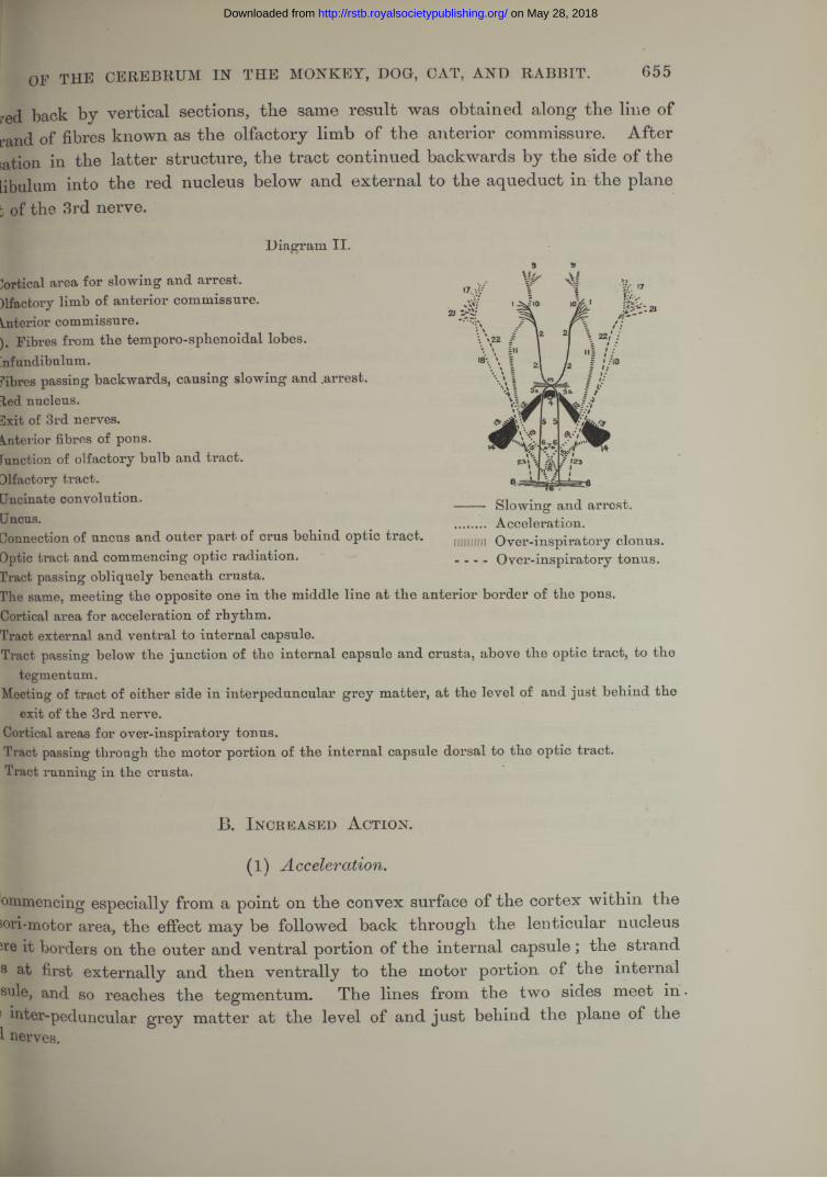
655
red back by vertical sections, the same result was obtained along the line of rand of fibres known as the olfactory limb of the anterior commissure. After sation in the latter structure, the tract continued backwards by the side of the libulum into the red nucleus below and external to the aqueduct in the plane b of the 3rd nerve.
OF THE CEREBRUM IN THE MONKEY, DOO, CAT, AND RABBIT.
Diagram II.
lortical area for slowing and arrest, fifacfcory limb of anterior commissure, interior commissure.|. Fibres from the temporo-sphenoidal lobes, nfundibulum.fibres passing backwards, causing slowing and .arrest, ted nucleus. ilyit of 3rd nerves.Interior fibres of pons.Function of olfactory bulb and tract.Difactory tract.Jncinate convolution.Uncus.Donnection of uncus and outer part of crus behind optic tract.Uptic tract and commencing optic radiation, rract passing obliquely beneath crusta. rhe same, meeting the opposite one in the middle line at the anterior border of the pons.Cortical area for acceleration of rhythm.Tract external and ventral to internal capsule.Tract passing below the junction of the internal capsule and crusta, above the optic tract, to the
tegmentum.Meeting of tract of either side in interpeduncular grey matter, at the level of and just behind the
exit of the 3rd nerve.Cortical areas for over-inspiratory tonus.Tract passing through the motor portion of the internal capsule dorsal to the optic tract.Tract running in the crusta.
Acceleration. Over-inspiratory clonus. Over-inspiratory tonus.
B. Increased A ction.
(1 ) Acceleration.
ommencing especially from a point on the convex surface of the cortex within the Jori-motor area, the effect may be followed back through the lenticular nucleus ire it borders on the outer and ventral portion of the internal capsule ; the strand s at first externally and then ventrally to the motor portion of the internal sale, and so reaches the tegmentum. The lines from the two sides meet in . ! inter-peduncular grey matter at the level of and just behind the plane of the 1 nerves.
on May 28, 2018http://rstb.royalsocietypublishing.org/Downloaded from
(2) Over-inspiratory Clonus.
This effect was obtained from the junction of the olfactory bulb and tract, and o continuing to apply the stimulus backwards along the olfactory tract was traced ini the uncinate convolution of the temporo-sphenoidal lobe. Followed to the uncus passed behind the optic tract to the crus, and then pointing obliquely inwards ventral] to the crusta the effect on each side converged to the middle line at the upper bord* of the pons.
(3) Over-inspiratory Tonus.
The descending motor tract yielded this result, so did the 5th nerve and du mater, as well as the sciatic nerve after complete removal of the cerebrum at the lev of the tentorium cerebelli.
So far as the anatomical arrangement of the tracts above described is concerned, may say that medullated fibres are to be seen in Dr. Tooth’s preparations of the cal and monkey’s brain, running in the same course as that indicated by faradic excitati* of living sections. An exception to this statement occurs in the case of the ove inspiratory clonus, in which I have not made out the connection between the unci and the pontine fibres lying ventral to the crusta.
I t is manifest that the respiratory alterations described above as capable of beii induced by the faradic current are such as can be produced in the conscious state 1 volitional effort, and any explanation of the results described above must depend up*, the acceptation or refusal of the general doctrine concerning the sensori-motor funetio: of the cortex, and that concerning the mechanism of the respiratory centre in t medulla.
I sum up therefore, the contents of this paper, when I say that whilst the effect up* respiration of exciting the cerebrum in a non-anaesthetized animal is probably a compl one, yet, by careful regulation of the anaesthetic state, four constant effects can obtained upon respiration by stimulation' of the cortex cerebri, and these can be trad down each in a course of its own from the cortex to the medulla oblongata.
D escription of P lates.
PLATE 57.
Photograph I A.—Under-surface of a Rabbit’s Brain, the small ring marks the P01 for slowing and arrest, the square that for inspiratory Tonus.
Photograph Ib.—Dorsal surface of a Rabbits Brain, the cross marks the poifit acceleration.
656 MR. W. G. SPENCER ON RESPIRATION AFFECTED BY FARADIC EXCITATION
on May 28, 2018http://rstb.royalsocietypublishing.org/Downloaded from

657
foraph II., with diagram.*—Ventral and lateral surface of a Cat’s Brain, the inner ring marks the centre, the outer the boundary of the area for slowing
tgraph III., with diagram*.—Ventral and lateral surface of a Dogs Brain, the ^ inner ring marks the centre, the outer the boundary of the area for slowing
and arrest.
Of THE CEBEBEtTM IN THE MONKEY, DOG, CAT, AND BABBIT.
PLATE 58.
ograph IV., with diagram.*—Ventral surface of a Monkey’s Brain, the inner ring marks the centre, the outer the boundary of the area for slowing andarrest
itographs V. to V III.—Vertical transverse sections of Cat’s Brain.
PLA TE 59.
tographs IX. to XV.—Vertical transverse sections of Cat’s Brain.
On the Diagrams appended to Photographs II., HI., and IV., the Roman figures refer to the ber of the tracings, and the dotted line from each number leads to the point of the brain, by nation of which the tracing so numbered was obtained.The following marks have been placed on Photographs V. to XV., indicating the point on each of the Bection by the stimulation of which was obtained . *
(1.) Slowing and arrest . . . . Marked by a small ring.(2.) A cce le r a tio n ................................. » ” cross.(3.) Over-inspiratory Clonus . . »» »> square.(4.) Over-inspiratory Tonus. . . » >> letter I.
toDCCCXCIV.— B. 4 P
on May 28, 2018http://rstb.royalsocietypublishing.org/Downloaded from