
The Effect of Splenectomy on the Reversal of Cirrhosis: a ...
11
Research Article The Effect of Splenectomy on the Reversal of Cirrhosis: a Prospective Study Dao-Bing Zeng , 1 Liang Di, 1 Rui-Chi Zhang, 2 Qing-Liang Guo, 1 Bin-Wei Duan, 1 Cui-Yu Jia, 2 Feng Chen, 2 Dong-Dong Lin, 1 Yun-jin Zang, 1 and Shi-Chun Lu 3 1 General Surgery Department, Beijing You’an Hospital, Capital Medical University, Beijing 100069, China 2 Department of Radiology, Beijing You’an Hospital, Capital Medical University, Beijing 100069, China 3 Institute & Hospital of Hepatobiliary Surgery of Chinese PLA, Chinese PLA Medical School, Chinese PLA General Hospital, Beijing 100853, China Correspondence should be addressed to Dao-Bing Zeng; [email protected] and Shi-Chun Lu; [email protected] Received 21 November 2018; Revised 13 February 2019; Accepted 21 February 2019; Published 8 April 2019 Academic Editor: Anastasios Koulaouzidis Copyright © 2019 Dao-Bing Zeng et al. This is an open access article distributed under the Creative Commons Attribution License, which permits unrestricted use, distribution, and reproduction in any medium, provided the original work is properly cited. Background. Studies have demonstrated that liver fibrosis can be reversed by medication treatments. After splenectomy, cirrhosis patients have short-term changes in several serum markers for cirrhosis and liver stiffness. Aims. To investigate the effect of splenectomy on the severity of cirrhosis. Methods. A total of 62 patients with cirrhosis and portal hypertension receiving splenectomy from December 2014 to July 2017 were enrolled. The degree of cirrhosis was preoperatively and postoperatively evaluated by serum markers, including hyaluronan (HA), laminin, amino-terminal propeptide of type III procollagen (PIIINP), type IV collagen (C-IV), liver stiffness (FibroScan), and liver volume. Results. HA levels significantly increased at 1 week and 1 month postoperation (both P <0 05), whereas the levels of PIIINP and C-IV significantly decreased from 1 month to 12 months postoperation (all P <0 05). In addition, elastography examination demonstrated that the FibroScan score significantly reduced from 1 month to 24 months postoperation as compared with the baseline level (all P <0 05). CT scan showed that the liver volume significantly increased at 6 months postoperation (P <0 05). Furthermore, the alteration trends of these serum markers and the FibroScan score were further confirmed by the multivariate linear regression. Conclusions. These observations suggested that splenectomy may result in long-term reversal of cirrhosis. 1. Introduction Cirrhosis is a pathological state of the liver characterized by fibrosis and morphologic conversion of normal liver tissue into abnormal nodules, which is the final stage of various chronic hepatic disorders [1]. There are a variety of causes of liver cirrhosis, including viral, alcoholic, autoimmune alcoholic, and fatty liver diseases [1]. Cirrhosis was tradition- ally considered as an irreversible disease. Nevertheless, in the past decade, both preclinical [2, 3] and clinical studies [4–8] have provided evidence demonstrating that liver fibrosis can be reversed to some extent, and even cirrhosis can be his- tologically reversed. For example, Chang et al. have demon- strated that long-term entecavir therapy in patients with chronic hepatitis B induces substantial histological improvement and regression of fibrosis or cirrhosis [8]. Kim et al. have reported that candesartan (an angiotensin- blocking agent) treatment results in significant improvement of fibrosis in histological and quantitative assessments in alcoholic hepatitis [6]. A retrospective study on (n = 87) by Czaja and Carpenter have revealed that corticosteroid ther- apy improves fibrosis in 53% of patients with autoimmune hepatitis [5]. All these observations strongly suggest that medication treatments may have the beneficial effect of reversal of cirrhosis. The current “gold standard” for evaluating the degree of liver fibrosis or cirrhosis remains ultrasound-guided liver biopsy. However, liver biopsy is invasive and may induce complications such as pain, pneumothorax, hemor- rhage, and perforation, which limit its clinical application Hindawi Gastroenterology Research and Practice Volume 2019, Article ID 5459427, 10 pages https://doi.org/10.1155/2019/5459427
Transcript of The Effect of Splenectomy on the Reversal of Cirrhosis: a ...

Research ArticleThe Effect of Splenectomy on the Reversal of Cirrhosis:a Prospective Study
Dao-Bing Zeng ,1 Liang Di,1 Rui-Chi Zhang,2 Qing-Liang Guo,1 Bin-Wei Duan,1
Cui-Yu Jia,2 Feng Chen,2 Dong-Dong Lin,1 Yun-jin Zang,1 and Shi-Chun Lu 3
1General Surgery Department, Beijing You’an Hospital, Capital Medical University, Beijing 100069, China2Department of Radiology, Beijing You’an Hospital, Capital Medical University, Beijing 100069, China3Institute & Hospital of Hepatobiliary Surgery of Chinese PLA, Chinese PLA Medical School, Chinese PLA General Hospital,Beijing 100853, China
Correspondence should be addressed to Dao-Bing Zeng; [email protected] and Shi-Chun Lu; [email protected]
Received 21 November 2018; Revised 13 February 2019; Accepted 21 February 2019; Published 8 April 2019
Academic Editor: Anastasios Koulaouzidis
Copyright © 2019 Dao-Bing Zeng et al. This is an open access article distributed under the Creative Commons Attribution License,which permits unrestricted use, distribution, and reproduction in any medium, provided the original work is properly cited.
Background. Studies have demonstrated that liver fibrosis can be reversed by medication treatments. After splenectomy, cirrhosispatients have short-term changes in several serum markers for cirrhosis and liver stiffness. Aims. To investigate the effect ofsplenectomy on the severity of cirrhosis. Methods. A total of 62 patients with cirrhosis and portal hypertension receivingsplenectomy from December 2014 to July 2017 were enrolled. The degree of cirrhosis was preoperatively and postoperativelyevaluated by serum markers, including hyaluronan (HA), laminin, amino-terminal propeptide of type III procollagen (PIIINP),type IV collagen (C-IV), liver stiffness (FibroScan), and liver volume. Results. HA levels significantly increased at 1 week and 1month postoperation (both P < 0 05), whereas the levels of PIIINP and C-IV significantly decreased from 1 month to 12 monthspostoperation (all P < 0 05). In addition, elastography examination demonstrated that the FibroScan score significantly reducedfrom 1 month to 24 months postoperation as compared with the baseline level (all P < 0 05). CT scan showed that the livervolume significantly increased at 6 months postoperation (P < 0 05). Furthermore, the alteration trends of these serum markersand the FibroScan score were further confirmed by the multivariate linear regression. Conclusions. These observations suggestedthat splenectomy may result in long-term reversal of cirrhosis.
1. Introduction
Cirrhosis is a pathological state of the liver characterized byfibrosis and morphologic conversion of normal liver tissueinto abnormal nodules, which is the final stage of variouschronic hepatic disorders [1]. There are a variety of causesof liver cirrhosis, including viral, alcoholic, autoimmunealcoholic, and fatty liver diseases [1]. Cirrhosis was tradition-ally considered as an irreversible disease. Nevertheless, in thepast decade, both preclinical [2, 3] and clinical studies [4–8]have provided evidence demonstrating that liver fibrosiscan be reversed to some extent, and even cirrhosis can be his-tologically reversed. For example, Chang et al. have demon-strated that long-term entecavir therapy in patients withchronic hepatitis B induces substantial histological
improvement and regression of fibrosis or cirrhosis [8].Kim et al. have reported that candesartan (an angiotensin-blocking agent) treatment results in significant improvementof fibrosis in histological and quantitative assessments inalcoholic hepatitis [6]. A retrospective study on (n = 87) byCzaja and Carpenter have revealed that corticosteroid ther-apy improves fibrosis in 53% of patients with autoimmunehepatitis [5]. All these observations strongly suggest thatmedication treatments may have the beneficial effect ofreversal of cirrhosis.
The current “gold standard” for evaluating the degreeof liver fibrosis or cirrhosis remains ultrasound-guidedliver biopsy. However, liver biopsy is invasive and mayinduce complications such as pain, pneumothorax, hemor-rhage, and perforation, which limit its clinical application
HindawiGastroenterology Research and PracticeVolume 2019, Article ID 5459427, 10 pageshttps://doi.org/10.1155/2019/5459427

[9]. In addition, sampling bias and variability may lead toinaccurate staging in liver biopsy [10]. Currently, severalnoninvasive diagnostic tools have been developed for theassessment of liver fibrosis, including serum markers,imaging examinations, and liver stiffness measurements[11, 12]. Serum cirrhosis markers include hyaluronan (HA)[13], laminin (LN), amino-terminal propeptide of type IIIprocollagen (PIIINP) [14], type IV collagen (C-IV) [15],matrix metalloproteinases (MMPs) [16], tissue inhibitor ofmetalloproteinases-1 (TIMP-1) [17], and aspartate amino-transferase to alanine aminotransferase ratio (AST/ALT)[18]. Imaging diagnostic methods for liver cirrhosis includeabdominal ultrasonography (US), computed tomography(CT), and magnetic resonance imaging (MRI) [19]. Tran-sient elastography, such as FibroScan, is a method developedfor measurement of liver stiffness and diagnosis of fibrosisand cirrhosis [20].
Our previous study found that after cirrhosis patientsreceiving splenectomy, there are short-term changes inseveral serum markers for cirrhosis and liver stiffness(FibroScan value) [21], indicating that splenectomy has ashort-term effect on serum fibrosis markers and liver stiff-ness in cirrhosis patients. This phenomenon raises thepossibility that splenectomy might be able to induce re-versal of cirrhosis. To verify this hypothesis, this pros-pective study is aimed at investigating the long-termeffect of splenectomy on the degree of cirrhosis by usingserum markers, imaging examinations, and liver stiffnessmeasurement.
2. Materials and Methods
2.1. Participants. A total of 62 patients with cirrhosis andportal hypertension who underwent splenectomy with/with-out esophagogastric devascularization in our hospital fromDecember 2014 to July 2017 were enrolled in this prospectivestudy. The inclusion criteria were (1) age of 20-65 years, (2)clinically or pathologically confirmed cirrhosis and portalhypertension (including viral hepatitis, alcoholic hepatitis,and autoimmune hepatitis), (3) diameter of splenic artery> 5 19mm or the ratio of splenic artery diameter to properhepatic artery diameter > 1 4, and (4) preoperative evalua-tion which showed stable vital signs, Child-Pugh grades Aand B; patients can tolerate the abdominal surgery undergeneral anesthesia. The exclusion criteria are (1) idiopathicportal hypertension, (2) Budd-Chiari syndrome, and (3) poorcompliance, preoperative evaluation which showed vitalsigns instability, need to use vasoactive drugs to maintainblood pressure, severe hepatic encephalopathy symptoms,and severe coagulation dysfunction. This study was approvedby the institutional review board of the Beijing You’anHospital, Capital Medical University. Written informedconsent was obtained from each patient.
2.2. Surgical Management. Patients with a history of uppergastrointestinal bleeding (n = 47) underwent splenectomycombined with esophagogastric devascularization as previ-ously described [22]. For patients without a history of upper
gastrointestinal bleeding (n = 15), after splenectomy, itwas determined whether esophagogastric devascularizationshould be carried out based on the intraoperative portal pres-sure [23]. Five patients did not receive esophagogastricdevascularization because the portal pressure had beenreduced to normal level after splenectomy. Two patientscombined with hepatocellular carcinoma simultaneouslyunderwent partial hepatectomy. All hepatitis B patients(n = 47) received antiviral therapy before and after surgery.
2.3. Determining the SerumMarkers of Cirrhosis. For evaluat-ing the degree of cirrhosis, the serum markers (HA, LN,PIIINP, and C-IV) were determined. At preoperation,1 week, 1 month, 3 months, 6 months, and 12 months post-operation, peripheral blood samples were collected and thelevels of the above serum markers were determined usingthe sensitized chemiluminescence immunoassay detectionsystem (JETLIA 96/2; China Medical Technologies, Beijing,China) on the same day.
2.4. Assessment of Liver Stiffness by FibroScan. Liver stiffnessmeasurement was performed at preoperation, 1 month, 3months, 6 months, 12 months, and 24 months postoperationusing continuous FibroScan (Echosens, Paris, France)according to the manufacturer’s protocol. Briefly, the patientwas placed in the supine position, and the scanning was con-ducted in the region encompassing the 7th, 8th, and 9thintercostal spaces between the anterior axillary and midaxil-lary lines. The number of successful detections should belarger than 10, and the median value of FibroScan value(expressed in kPa) was recorded as the final FibroScan score.In addition, the interquartile range/median FibroScan scoreshould be smaller than 33% and the detection success rateshould be greater than 60%. The ultrasound was performedusing the Aixplorer diagnostic ultrasound system (Super-sonic Imagine, France).
2.5. Evaluation of Liver Volume. Imaging examinations wereconducted at preoperation, 1 month, 3 months, 6 months,12 months, and 24 months postoperation. Liver volumescanning was conducted using a LightSpeed VCT 64-sliceCT scanner (GE. Healthcare, USA). The image was acquiredusing a three-phase enhanced scanner (arterial phase 20-25 s,portal venous phase 65-70 s, and equilibrium phase 180 s),with a scanning range from the dome of the diaphragm tothe lower edge of the liver and spleen. A nonionic contrastmedium was injected into the elbow vein using a high-pressure syringe. After image acquisition, the data wasused for liver volume reconstruction and assessment usingthe Advantage Workstation 4.3 software (GE Healthcare)according to the manufacturer’s protocol.
2.6. Statistical Analysis. Continuous data were expressed asmean ± standard deviation (SD) and compared by Student’spaired t-test. If normality was not assumed, the Wilcoxonsum-of-rank test would be used for comparisons betweendependent variables. Categorical data were indicated bynumber and percentage (%). One-way repeated measure-ment ANOVA was used for the comparisons among timepoints (from preoperation to 2 years), and using Fisher’s
2 Gastroenterology Research and Practice

LSD comparisons for the post hoc test. Univariate and mul-tivariate generalized estimating equation (GEE) and linearregression models were used to investigate the change amongtime points to the results of markers (HA, LN, PIIINP, C-IV,FibroScan, and liver volume). A first-order autoregressiveworking correlation matrix was adopted for the repeatedmeasures data. Patients’ age, sex, and Child-Pugh score werecontrolled as covariates in multivariate models. The signifi-cant level of all analyses was set at a P value <0.05, two-tailed. All analyses were performed using IBM SPSS Version20 (SPSS Statistics V20, IBM Corporation, Somers, NewYork, USA).
3. Results
3.1. Patients’ Clinical Features. A total of 62 eligible patients(mean age = 46 90 ± 9 58 years) were enrolled in this study,including 34 (54.84%) male and 28 (45.16%) female.Patients’ demographic and clinical characteristics, as wellas the details of the operation, are summarized in Table 1.Hepatitis B virus (HBV) infection (n = 47, 75.81%) was themost common cause of cirrhosis. Other causes of cirrhosisincluded hepatitis C virus (HBC) infection (n = 9, 14.52%),alcoholic fatty liver (n = 10, 16.13%), primary biliary cirrho-sis (PBC, n = 3, 4.84%), primary sclerosing cholangitis (PSC,n = 1, 1.61%), and autoimmune hepatitis (AIH, n = 2,3.23%). The mean surgical time was 214 25 ± 49 28 min.The amounts of operative bleeding, transfusion of red bloodcells, and plasma were 201 64 ± 187 56mL, 91 80 ± 257 10mL, and 170 49 ± 287 14mL, respectively. Following sple-nectomy, 4 patients developed postoperative intraperitonealhemorrhage, which was resolved by emergency exploratorylaparotomy to stop bleeding (n = 3) or conservative drugtreatment (n = 1). One case had upper gastrointestinalbleeding at 8 days after operation, which were resolvedby drug therapy for hemostasis and portal hypertension.One patient with pancreatic leakage (grade I) was treatedwith conservative treatment. One case with abdominal infec-tion (Staphylococcus epidermidis) was treated with tienamand vancomycin.
3.2. The Change of Serum Markers, FibroScan Score, andLiver Volume. The median follow-up time was 12 months(range: 1-24 months). Five patients did not return to our hos-pital for follow-up due to living in remote areas. During thefollow-up, one patient died of an accidental fall, and onepatient died of multiple autoimmune diseases at 16 monthspostoperation. One patient was diagnosed with HCC at oneyear postoperation and was withdrawn from the study. Aftersplenectomy, the patient’s portal vein pressure was signifi-cantly decreased (34 15 ± 5 03 vs. 25 70 ± 4 19, P < 0 001).In addition, platelet counts significantly increased at all thetime points after splenectomy (Table 2, all P < 0 05). In addi-tion, the Child-Pugh score was significantly decreased at 1, 3,6, 12, and 24months after splenectomy (Table 2, all P < 0 05).To investigate if splenectomy has an effect on the degree ofcirrhosis, the serum markers of cirrhosis (HA, LN, PIIINP,and C-IV) were determined. Compared to the corresponding
baseline (preoperation) levels, HA was significantly increasedat 1 week and 1 month postoperation (Figure 1(a), both P <0 05). However, no significance was found in LN(Figure 1(b), all P > 0 05). PIINP was significantly decreasedfrom 1 month to 12 months postoperation (Figure 1(c), allP < 0 05), and C-IV was significantly reduced from 1 weekto 12 months postoperation (Figure 1(d), all P < 0 05).
Meanwhile, elastography examination demonstratedthat the FibroScan score was significantly reduced from 1month to 24 months postoperation as compared with thebaseline level (all P < 0 05, Figure 2(a)). CT scan revealedthat the liver volume was only significantly increased at6 months postoperation (P < 0 05, Figure 2(b)).
3.3. Multivariate Linear Regression Results with GEE Models.To further confirm the change trends of the serum andimaging markers, the multivariate linear regression withGEE models adjusted for patients’ sex, age, and Child-
Table 1: Patients’ demographic and clinical characteristics (n = 62).
Parameters Mean ± SD or N (%)
Sex
Male 34 (54.84)
Female 28 (45.16)
Age, year 49 60 ± 9 58Hemorrhage, times 1 63 ± 1 96Child-Pugh score 6 34 ± 1 23Child-Pugh rank
A 36 (58.06)
B 24 (38.71)
C 2 (3.23)
History of disease
HBV 47 (75.81)
HCV 9 (14.52)
Alcoholic fatty liver 10 (16.13)
PBC 3 (4.84)
PSC 1 (1.61)
AIH 2 (3.23)
Surgery time (minutes) 214 25 ± 49 28Operative bleeding (ml) 201 64 ± 187 56Transfusion of RBC (ml) 91 80 ± 257 10Transfusion of plasma (ml) 170 49 ± 287 14Preoperation
HA (μg/l) 126 34 ± 93 52LN (μg/l) 62 71 ± 130 87PIIINP (μg/l) 12 14 ± 10 13C-IV (μg/l) 98 74 ± 84 31FibroScan (kPa) 22 95 ± 15 54Liver volume (cm3) 1055 88 ± 306 72
HBV: hepatitis B virus; HCV: hepatitis C virus; PBC: primary biliarycirrhosis; PSC: primary sclerosing cholangitis; AIH: autoimmune hepatitis.
3Gastroenterology Research and Practice
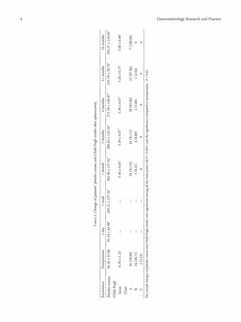
Table2:Changeof
patients’p
lateletcoun
tsandChild-Pughresults
aftersplenectom
y.
Param
eters
Preop
eration
1day
1week
1mon
th3mon
ths
6mon
ths
12mon
ths
24mon
ths
Plateletcoun
ts56
26±37
0691
94±44
98∗
2932
1±1371
0∗3029
0±1379
2∗2808
2±1331
0∗2737
8±1408
7∗2557
9±78
79∗
2555
7±1100
5∗
Child-Pugh
Score
634±
123
——
546
±065
∗549
±087
∗538
±067
∗526
±075
∗50
0±00
0∗
Class A
36(58.06)
——
54(91.53)
41(91.11)
38(95.00)
33(97.06)
7(100.00)
B24
(38.71)
——
5(8.47)
4(8.89)
2(5.00)
1(2.94)
0
C2(3.23)
——
00
00
0
The
overallchanges
ofplateletcoun
tsandChild-Pughresults
weresignificant
amon
gallthe
timepo
ints(allP<0001),and
thesignificancecomparedto
preoperation
,∗P<00
5.
4 Gastroenterology Research and Practice

Pugh score was performed. As shown in Table 3, comparedto their corresponding reference time points (preoperation),HA was significantly elevated at 1 week and 1 month (both P< 0 05), while PIINP and C-IV were significantly reducedfrom1month to 12months (allP < 0 05). However, no signif-icance was found in LN (all P > 0 05).
As shown in Table 4, the FibroScan score was significantlydecreased from 1 month to 24 months postoperation as
compared with the baseline (all P < 0 05), and the livervolume was only significantly increased at 24 monthspostoperation (P < 0 05).
4. Discussion
Even though previous studies have demonstrated that medi-cation treatments can result in reversal of cirrhosis, however,
Pre-operation
1 week
800
600
400
200
0
�휇g/
L
1 month 3 months
Time
HA
⁎
⁎
6 months 12 months
(a)
250200150100
500
�휇g/
L
LN
Pre-operation
1 week 1 month 3 months
Time
6 months 12 months
(b)
25201510
50
�휇g/
L
PIIINP
⁎ ⁎ ⁎⁎
Pre-operation
1 week 1 month 3 months
Time
6 months 12 months
(c)
�휇g/
L
C-IV⁎
⁎
⁎ ⁎ ⁎
Pre-operation
1 week 1 month 3 months
Time
0
100
200
6 months 12 months
(d)
Figure 1: The changes in patients’ serum markers, including HA (a), LN (b), PIIINP (c), and C-IV (d). ∗P < 0 05 compared to preoperation.The numbers of compared pairs of all serum markers at 1 week, 1 month, 3 months, 6 months, and 12 months were 48, 39, 30, 27, and 15,respectively. The sample sizes of HA were 51, 50, 40, 32, 29, and 16 to preoperation, 1 week, 1 month, 3 months, 6 months, and 12 months,respectively. The sample sizes of LN were 51, 50, 40, 32, 29, and 16 to preoperation, 1 week, 1 month, 3 months, 6 months, and 12 months,respectively. The sample sizes of PIIINP were 51, 50, 40, 32, 29, and 16 to preoperation, 1 week, 1 month, 3 months, 6 months, and 12 months,respectively. The sample sizes of C-IV were 51, 50, 40, 32, 29, and 16 to preoperation, 1 week, 1 month, 3 months, 6 months, and 12 months,respectively.
⁎
⁎⁎⁎
Pre-operation
3 months1 month0
10
20
30
40
6 months
Time
FibroScan
12 months 24 months
kPa
⁎
(a)
⁎
0500
100015002000
Liver volume
Pre-operation
3 months1 month 6 months
Time
cm3
12 months 24 months
(b)
Figure 2: The change of patients’ FibroScan (a) and liver volume (b). ∗P < 0 05 compared to preoperation. The numbers of compared pairs ofFibroScan at 1 month, 3 months, 6 months, 12 months, and 24months were 45, 37, 30, 31, and 6, respectively. The numbers of compared pairsof liver volume at 1 month, 3 months, 6 months, 12 months, and 24 months were 37, 28, 28, 22, and 5, respectively. The sample sizes ofFibroScan were 55, 48, 40, 34, 32, and 6 to preoperation, 1 month, 3 months, 6 months, 12 months, and 24 months, respectively. Thesample sizes of liver volume were 50, 44, 36, 35, 30, and 6 to preoperation, 1 month, 3 months, 6 months, 12 months, and 24 months,respectively.
5Gastroenterology Research and Practice
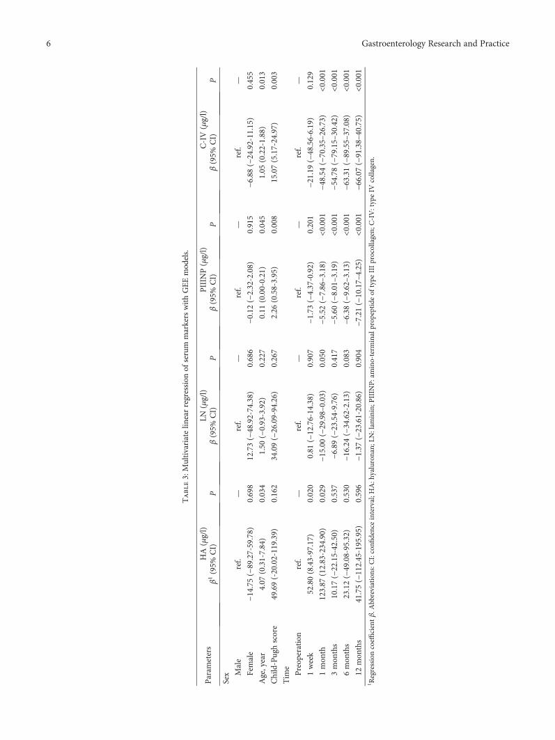
Table3:Multivariatelin
earregression
ofserum
markers
withGEEmod
els.
Param
eters
HA(μg/l)
LN(μg/l)
PIIIN
P(μg/l)
C-IV(μg/l)
β1(95%
CI)
Pβ(95%
CI)
Pβ(95%
CI)
Pβ(95%
CI)
P
Sex Male
ref.
—ref.
—ref.
—ref.
—
Female
−14.75
(−89.27-59.78)
0.698
12.73(−48.92-74.38)
0.686
−0.12(−2.32-2.08)
0.915
−6.88(−24.92-11.15)
0.455
Age,year
4.07
(0.31-7.84)
0.034
1.50
(−0.93-3.92)
0.227
0.11
(0.00-0.21)
0.045
1.05
(0.22-1.88)
0.013
Child-Pughscore
49.69(-20.02-119.39)
0.162
34.09(−26.09-94.26)
0.267
2.26
(0.58-3.95)
0.008
15.07(5.17-24.97)
0.003
Tim
e
Preop
eration
ref.
—ref.
—ref.
—ref.
—
1week
52.80(8.43-97.17)
0.020
0.81
(−12.76-14.38)
0.907
−1.73(−4.37-0.92)
0.201
−21.19
(−48.56-6.19)
0.129
1mon
th123.87
(12.83-234.90)
0.029
−15.00
(−29.98–0.03)
0.050
−5.52(−7.86–3.18)
<0.001
−48.54
(−70.35–26.73)
<0.001
3mon
ths
10.17(−22.15-42.50)
0.537
−6.89(−23.54-9.76)
0.417
−5.60(−8.01–3.19)
<0.001
−54.78
(−79.15–30.42)
<0.001
6mon
ths
23.12(−49.08-95.32)
0.530
−16.24
(−34.62-2.13)
0.083
−6.38(−9.62–3.13)
<0.001
−63.31
(−89.55–37.08)
<0.001
12mon
ths
41.75(−112.45-195.95)
0.596
−1.37(−23.61-20.86)
0.904
−7.21(−10.17–4.25)
<0.001
−66.07
(−91.38–40.75)
<0.001
1 Regressioncoeffi
cientβ.A
bbreviations:C
I:confi
denceinterval;H
A:h
yaluronan;
LN:lam
inin;P
IIIN
P:amino-term
inalprop
eptide
oftype
IIIprocollagen;
C-IV:typeIV
collagen.
6 Gastroenterology Research and Practice

studies on the effect of splenectomy on the degree of cirrhosisare limited [24]. In this study, we investigated the effect ofsplenectomy on the reversal of cirrhosis. The results showedthat compared to the baseline level, HA levels significantlyincreased at 1 week and 1 month postoperation, whereasthe levels of PIIINP and C-IV significantly decreased from1 month to 12 months postoperation. In addition, theFibroScan score significantly reduced from 1 month to 24months postoperation. CT scan showed that the liver volumesignificantly increased at 6 months postoperation. Further-more, the alteration trends of these serum markers andFibroScan score were further confirmed by multivariate lin-ear regression. Taken together, these observations suggestedthat splenectomy may result in reversal of cirrhosis.
Our previous study showed that the optimal cutoffs forabnormal splenic artery internal diameter and S/P ratio incirrhosis-induced portal hypertension are >5.19mm and>1.40, respectively, which could be a marker for splanchnichemodynamic disturbances [25]. Therefore, only patientswith a splenic artery internal diameter > 5 19mm and S/Pratio > 1 40were enrolled in this study. Liver fibrosis is a con-sequence of disorganization of extracellular matrix (ECM)components, which cause loss of normal liver cell function[26, 27]. In fibrotic liver, the ECM metabolites are signifi-cantly increased so that the serum levels of ECM compo-nents, such as HA, LN, PIIINP, and C-IV, can be used asmarkers for the stage and progression of cirrhosis [28]. Forinstance, HA reflects the liver fibrogenesis and liver injury[29], while PIIINP and C-IV indicate the metabolism of col-lagens [30]. The elevated serum HA level in the cirrhosispatient is due to the fact that the liver sinusoidal endothelialcells reduce the uptake of HA [31]. It has been shown thatfollowing partial hepatectomy, hepatic stellate cells synthe-sized large amounts of HA during liver regeneration [32],suggesting that liver regeneration is associated with elevatedHA level. In this study, serum HA level was significantly ele-vated at 1 week and 1 month postoperation. Our previousstudy showed that serumHA level does not alter immediately
postoperation, but significantly increases at 2 days and 1week postoperation. Meanwhile, although the serum HAlevels were slightly higher at 3, 6, and 12 months postopera-tion than at preoperation, the differences did not reach signif-icance. These observations implied that reversal of cirrhosismight be started early at 2 days after splenectomy. However,further evidence is necessary to support this suggestion.
Collagens are synthesized by hepatic stellate cells asprecursor molecules, followed by cleaving at both N- andC-terminal ends by proteinases, and the mature collagen isthen integrated into the ECM. Hence, both the procollagenand the propeptide can reflect the synthesis of ECM [28].PIIINP is a well-studied marker of liver fibrosis [33, 34]. Ithas been shown that PIIINP has a high sensitivity and spec-ificity to detect cirrhosis [33]. C-IV plays important roles inthe pathogenesis of fibrosis disease, and the serum levels ofC-IV can be used for predicting the state of liver fibrosis[28]. In this study, the serum level of PIINP was significantlydecreased from 1month to 12 months after splenectomy, andthe C-IV level was significantly reduced from 1 week to 12months postoperation.
FibroScan (transient elastography) is a method for theassessment of liver fibrosis through measuring liver stiffnessby a monodimensional ultrasound [35]. A meta-analysis byShaheen et al. has reported an excellent diagnostic accuracyof FibroScan for HCV-related cirrhosis with an area underthe curve of the receiver operating characteristic (AUROC)of 0.95 [36]. Moreover, FibroScan can provide better diag-nostic performance for predicting liver fibrosis than serummarkers [35]. In the current study, the preoperative FibroS-can values of all patients were greater than 21 kPa, suggestingsevere cirrhosis. After splenectomy, the FibroScan valuesexhibited a continuous decrease trend, and all the postopera-tive FibroScan values were significantly lower than the preop-erative ones. At 24 months postoperation, the FibroScanvalues could be reduced to 10 kPa. Taken together, our serummarkers and liver stiffness measurement suggested that aftersplenectomy, reversal of cirrhosis might be started early at 1
Table 4: Multivariate linear regression of imaging markers with GEE models.
ParametersFibroScan (kPa) Liver volume (cm3)
β1 (95% CI) P β (95% CI) P
Sex
Male ref. — ref. —
Female −3.09 (−8.27-2.09) 0.242 −115.41 (−251.37-20.55) 0.096
Age (years) 0.18 (−0.12-0.49) 0.235 1.11 (−4.71-6.92) 0.708
Child-Pugh score 3.94 (0.33-7.56) 0.032 −19.22 (−86.99-48.56) 0.578
Time
Preoperation ref. — ref. —
1 month −3.11 (−5.82–0.41) 0.024 21.07 (−16.41-58.55) 0.270
3 months −4.07 (−6.62–1.52) 0.002 25.32 (−16.24-66.88) 0.232
6 months −5.90 (−8.85–2.94) <0.001 32.34 (−14.75-79.43) 0.178
12 months −7.29 (−10.67–3.91) <0.001 25.83 (−24.75-76.40) 0.317
24 months −10.63 (−14.82–6.44) <0.001 111.02 (17.87-204.18) 0.0191Regression coefficient β. CI: confidence interval.
7Gastroenterology Research and Practice
week and can last for at least 2 years. To our best knowledge,this is the first study reporting the long-term effect of sple-nectomy on the reversal of cirrhosis. However, it is worthto further elucidate the mechanism underlyingsplenectomy-induced reversal of cirrhosis. It should bepointed out that the reduced portal hypertension followingsplenectomy may also decrease the liver stiffness.
Accumulating evidence has suggested that there is asignificant volume reduction in cirrhotic livers as comparedwith normal livers [37, 38]. In addition, long-term oralnucleos(t)ide analogue therapy in patients with HBV-related liver compensated and decompensated cirrhosis leadsto a significant increase in liver volume [39]. In this study, wefound that although the liver volume was increased at all thetime points after splenectomy, only the difference at 6months postoperation reaches statistical significance, whichmay be attributed to the small sample size of this study. Astudy with a large sample size should be conducted to furthervalidate this issue.
Liver fibrosis frequently causes portal hypertension [40],in turn leading to hypersplenism [41]. Portal hypertension-induced hypersplenism causes thrombocytopenia [41]. Sple-nectomy can effectively reduce portal pressure and correcthypersplenism and improve hypersplenism-induced throm-bocytopenia [42, 43]. In this study, patients’ portal veinpressure was significantly decreased after splenectomy. Inaddition, platelet counts significantly increased at all the timepoints after splenectomy. Since the progression of fibrosisparallels the increase in portal pressure [44], it is worth inves-tigating if reduction in the portal pressure after splenectomycontributes to the reversal of cirrhosis.
In addition to splenectomy, several nonsurgical methodshave been utilized to treat hypersplenism.Microwave ablationcan improve hypersplenism in cirrhosis patients, but multipleablations are required, with mean ablation times of 8 8 ± 1 3,and the therapeutic effect is not as good as splenectomy [45].Although high-intensity focused ultrasound improvedhypersplenism, both white blood cells (WBCs) and plateletcounts did not return to normal levels [46]. Studies haveshown that in children with chronic liver disease with hypers-plenism [47] and cirrhosis in adults [48], after propranololtreatment for 1-4 weeks, platelet counts significantly increase.However, the follow-up durations on these two studies areboth short, and their long-termefficacy is uncertain. Althoughsplenic artery embolization can improveWBCs and platelet inpatients with hypersplenism, it may cause severe compli-cations [49]. The incidence of the postembolization syn-drome is high to 77.8-100% [50]. The incidence ofmorbidity and complications after splenic embolizationwas higher than that of splenectomy, and the 1-year effectiverate was only 16% [51]. Radiotherapy for hypersplenism canonly increase the platelet counts but not increase WBCs andred blood cells (RBCs) [52]. By contrast, in a study of 226patients with splenectomy and followed up for 3-96months (mean = 63), splenectomy effectively increasedWBCs, platelet counts, and RBCs [53], indicating that sple-nectomy improves spleen function with long-term efficacy.
Several limitations of the current study should be pointedout. First, we only adopted noninvasive methods to assess the
degree of cirrhosis. The improvement in markers of fibrosisand liver stiffness may be attributed to the reduced portalhypertension following splenectomy, and reversal of cirrho-sis should be further supported by pathological evidence.Nevertheless, the pathological examination cannot be repeat-edly carried out in a short time due to its invasiveness andcomplications. Hence, we chose to adopt serum markersand liver stiffness measurement for assessment of the degreeof cirrhosis in this trial. In addition, the sample size of thisstudy was relatively small, and two patients having under-gone hepatectomy may cause heterogeneity of the enrolledpopulation. Furthermore, the mechanism of splenectomy-induced reversal of cirrhosis remains to be investigated. Allthese limitations should be addressed in the following study.
5. Conclusions
In summary, our findings provide evidence of serummarkersand liver stiffness measurement suggesting that splenectomymay induce long-term reversal of cirrhosis. Further investi-gation of the mechanism is necessary.
Data Availability
The data used to support the findings of this study areincluded within the article.
Ethical Approval
All procedures performed in studies involving human partic-ipants were in accordance with the ethical standards of theinstitutional and/or national research committee and withthe 1964 Helsinki declaration and its later amendments orcomparable ethical standards.
Conflicts of Interest
The authors declare that they have no conflict of interest.
Acknowledgments
This study was supported by the Beijing Municipal Science &Technology Commission (Z151100004015064).
References
[1] R. G. Romanelli and C. Stasi, “Recent advancements in diagno-sis and therapy of liver cirrhosis,” Current Drug Targets,vol. 17, no. 15, pp. 1804–1817, 2016.
[2] J. Jiao, D. Sastre, M. I. Fiel et al., “Dendritic cell regulation ofcarbon tetrachloride-induced murine liver fibrosis regression,”Hepatology, vol. 55, no. 1, pp. 244–255, 2012.
[3] T. Kisseleva, M. Cong, Y. Paik et al., “Myofibroblasts revert toan inactive phenotype during regression of liver fibrosis,” Pro-ceedings of the National Academy of Sciences of the UnitedStates of America, vol. 109, no. 24, pp. 9448–9453, 2012.
[4] C. Corpechot, F. Carrat, A. Bonnand, R. Poupon, andR. Poupon, “The effect of ursodeoxycholic acid therapy onliver fibrosis progression in primary biliary cirrhosis,” Hepa-tology, vol. 32, no. 6, pp. 1196–1199, 2000.
8 Gastroenterology Research and Practice

[5] A. J. Czaja and H. A. Carpenter, “Decreased fibrosis duringcorticosteroid therapy of autoimmune hepatitis,” Journal ofHepatology, vol. 40, no. 4, pp. 646–652, 2004.
[6] M. Y. Kim, M. Y. Cho, S. K. Baik et al., “Beneficial effects ofcandesartan, an angiotensin-blocking agent, on compensatedalcoholic liver fibrosis - a randomized open-label controlledstudy,” Liver International, vol. 32, no. 6, pp. 977–987,2012.
[7] S. L. George, B. R. Bacon, E. M. Brunt, K. L. Mihindukulasur-iya, J. Hoffmann, and A. M. Di Bisceglie, “Clinical, virologic,histologic, and biochemical outcomes after successful HCVtherapy: a 5-year follow-up of 150 patients,” Hepatology,vol. 49, no. 3, pp. 729–738, 2009.
[8] T.-T. Chang, Y.-F. Liaw, S.-S. Wu et al., “Long-term entecavirtherapy results in the reversal of fibrosis/cirrhosis and contin-ued histological improvement in patients with chronic hepati-tis B,” Hepatology, vol. 52, no. 3, pp. 886–893, 2010.
[9] S. Kose, G. Ersan, B. Tatar, P. Adar, and B. Erturk Sengel,“Evaluation of percutaneous liver biopsy complications inpatients with chronic viral hepatitis,” The Eurasian Journal ofMedicine, vol. 47, no. 3, pp. 161–164, 2015.
[10] D. Nguyen and T. JA, “Noninvasive assessment of liver fibro-sis,” Hepatology, vol. 53, no. 6, pp. 2107–2110, 2011.
[11] C. Lucero and R. S. Brown Jr, “Noninvasive measures of liverfibrosis and severity of liver disease,” Gastroenterology &hepatology, vol. 12, no. 1, pp. 33–40, 2016.
[12] C. Li, R. Li, andW. Zhang, “Progress in non-invasive detectionof liver fibrosis,” Cancer Biology & Medicine, vol. 15, no. 2,pp. 124–136, 2018.
[13] M. A. El Serafy, A. M. Kassem, H. Omar, M. S. Mahfouz, andM. E. S. E. L. Raziky, “APRI test and hyaluronic acid as non-invasive diagnostic tools for post HCV liver fibrosis: systematicreview and meta-analysis,” Arab Journal of Gastroenterology,vol. 18, no. 2, pp. 51–57, 2017.
[14] W. M. C. Rosenberg, M. Voelker, R. Thiel et al., “Serummarkers detect the presence of liver fibrosis: a cohort study,”Gastroenterology, vol. 127, no. 6, pp. 1704–1713, 2004.
[15] S. S. Veidal, M. A. Karsdal, A. Nawrocki et al., “Assessment ofproteolytic degradation of the basement membrane: a frag-ment of type IV collagen as a biochemical marker for liverfibrosis,” Fibrogenesis & Tissue Repair, vol. 4, no. 1, 2011.
[16] V. Leroy, F. Monier, S. Bottari et al., “Circulating matrix metal-loproteinases 1, 2, 9 and their inhibitors TIMP-1 and TIMP-2as serum markers of liver fibrosis in patients with chronichepatitis C: comparison with PIIINP and hyaluronic acid,”The American Journal of Gastroenterology, vol. 99, no. 2,pp. 271–279, 2004.
[17] R. Flisiak, P. Maxwell, D. Prokopowicz, P. M. Timms, andA. Panasiuk, “Plasma tissue inhibitor of metalloproteinases-1and transforming growth factor beta 1–possible non-invasivebiomarkers of hepatic fibrosis in patients with chronic B andC hepatitis,” Hepatogastroenterology, vol. 49, no. 47,pp. 1369–1372, 2002.
[18] G. J. Park, B. P. Lin, M. C. Ngu, D. B. Jones, and P. H. Katelaris,“Aspartate aminotransferase: alanine aminotransferase ratioin chronic hepatitis C infection: is it a useful predictor of cir-rhosis?,” Journal of Gastroenterology and Hepatology, vol. 15,no. 4, pp. 386–390, 2000.
[19] S. K. Yeom, C. H. Lee, S. H. Cha, and C.M. Park, “Prediction ofliver cirrhosis, using diagnostic imaging tools,” World Journalof Hepatology, vol. 7, no. 17, pp. 2069–2079, 2015.
[20] M. Fernandez, E. Trépo, D. Degré et al., “Transient elastogra-phy using Fibroscan is the most reliable noninvasive methodfor the diagnosis of advanced fibrosis and cirrhosis in alcoholicliver disease,” European Journal of Gastroenterology & Hepa-tology, vol. 27, no. 9, pp. 1074–1079, 2015.
[21] D. Kong, X. Chen, S. Lu et al., “Short-term effects of splenec-tomy on serum fibrosis indexes in liver cirrhosis patients,”International Journal of Clinical and Experimental Pathology,vol. 8, no. 11, pp. 15260–15264, 2015.
[22] Experts Group from the Ministry of Health Special,“Expert consensus on technical specifications of pericardialdevascularization (2013 edition),” Chinese Journal of Diges-tive Surgery, vol. 13, pp. 19–21, 2014.
[23] J. Cao, S. Lu, D. Zeng, D. Lin, J. Wu, and Q. Guo, “Clinicalanalysis of splenectomy and selective devascularization basedon intra-operative free portal pressure,” Chinese Journal ofHepatobiliary Surgery, vol. 20, pp. 648–651, 2014.
[24] Y. Nomura, M. Kage, T. Ogata et al., “Influence of splenectomyin patients with liver cirrhosis and hypersplenism,”HepatologyResearch, vol. 44, no. 10, pp. E100–E109, 2014.
[25] D.-B. Zeng, C.-Z. Dai, S.-C. Lu, N. He, W. Wang, and H.-J. Li,“Abnormal splenic artery diameter/hepatic artery diameterratio in cirrhosis-induced portal hypertension,”World Journalof Gastroenterology, vol. 19, no. 8, pp. 1292–1298, 2013.
[26] R. C. Benyon and J. P. Iredale, “Is liver fibrosis reversible?,”Gut, vol. 46, no. 4, pp. 443–446, 2000.
[27] N. Ueki, T. Taguchi, M. Takahashi et al., “Inhibition of hyalur-onan synthesis by vesnarinone in cultured human myofibro-blasts,” Biochimica et Biophysica Acta (BBA) - Molecular CellResearch, vol. 1495, no. 2, pp. 160–167, 2000.
[28] T. Liu, X. Wang, M. A. Karsdal, D. J. Leeming, andF. Genovese, “Molecular serum markers of liver fibrosis,” Bio-marker Insights, vol. 7, pp. 105–117, 2012.
[29] L. Yao, Z. M. Yao, and T. Yu, “Influence of BOL on hyaluronicacid, laminin and hyperplasia in hepatofibrotic rats,” WorldJournal of Gastroenterology, vol. 7, no. 6, pp. 872–875, 2001.
[30] J. P. Iredale, R. C. Benyon, J. Pickering et al., “Mechanisms ofspontaneous resolution of rat liver fibrosis. Hepatic stellate cellapoptosis and reduced hepatic expression of metalloproteinaseinhibitors,” The Journal of Clinical Investigation, vol. 102,no. 3, pp. 538–549, 1998.
[31] L. D. DeLeve, “Liver sinusoidal endothelial cells in hepaticfibrosis,” Hepatology, vol. 61, no. 5, pp. 1740–1746, 2015.
[32] D. Vrochides, V. Papanikolaou, H. Pertoft, A. A. Antoniades,and P. Heldin, “Biosynthesis and degradation of hyaluronanby nonparenchymal liver cells during liver regeneration,”Hepatology, vol. 23, no. 6, pp. 1650–1655, 1996.
[33] J. P. Teare, S. M. Greenfield, R. P. H. Thompson et al., “Com-parison of serum procollagen III peptide concentrations andPGA index for assessment of hepatic fibrosis,” Lancet,vol. 342, no. 8876, pp. 895–898, 1993.
[34] C. Trocme, V. Leroy, N. Sturm et al., “Longitudinal evaluationof a fibrosis index combining MMP-1 and PIIINP comparedwith MMP-9, TIMP-1 and hyaluronic acid in patients withchronic hepatitis C treated by interferon-alpha and ribavirin,”Journal of Viral Hepatitis, vol. 13, no. 10, pp. 643–651,2006.
[35] B. K. Kim, H. S. Kim, J. Y. Park et al., “Prospective validation ofELF test in comparison with fibroscan and fibrotest to predictliver fibrosis in Asian subjects with chronic hepatitis B,” PLoSOne, vol. 7, no. 7, article e41964, 2012.
9Gastroenterology Research and Practice
[36] A. A. M. Shaheen, A. F. Wan, and R. P. Myers, “FibroTest andFibroScan for the prediction of hepatitis C-related fibrosis: asystematic review of diagnostic test accuracy,” The AmericanJournal of Gastroenterology, vol. 102, no. 11, pp. 2589–2600,2007.
[37] Y. M. Li, F. Lv, X. Xu et al., “Evaluation of liver functionalreserve by combining D-sorbitol clearance rate and CT mea-sured liver volume,” World Journal of Gastroenterology,vol. 9, no. 9, pp. 2092–2095, 2003.
[38] P. Liu, P. Li, W. He, and L.-Q. Zhao, “Liver and spleen volumevariations in patients with hepatic fibrosis,” World Journal ofGastroenterology, vol. 15, no. 26, pp. 3298–3302, 2009.
[39] C. H. Lee, I. H. Kim, J. C. Moon et al., “3-Dimensional livervolume assessment in patients with hepatitis B virus-relatedliver cirrhosis during long-term oral nucleos(t)ide analoguestherapy,” World Journal of Gastroenterology, vol. 23, no. 2,pp. 297–305, 2017.
[40] K. T. Suk and D. J. Kim, “Staging of liver fibrosis or cirrho-sis: the role of hepatic venous pressure gradient measure-ment,” World Journal of Hepatology, vol. 7, no. 3,pp. 607–615, 2015.
[41] Y. Lv, W. Y. Lau, Y. Li et al., “Hypersplenism: history andcurrent status,” Experimental and Therapeutic Medicine,vol. 12, no. 4, pp. 2377–2382, 2016.
[42] X. L. Zhan, Y. Ji, and Y. D. Wang, “Laparoscopic splenectomyfor hypersplenism secondary to liver cirrhosis and portalhypertension,” World Journal of Gastroenterology, vol. 20,no. 19, pp. 5794–5800, 2014.
[43] M. M. A. Zaitoun, M. A. A. Basha, A. Raafat, T. Rushdy, andW. A. Mawla, “Splenectomy for hypersplenism with orwithout preoperative splenic artery embolisation,” EuropeanRadiology Experimental, vol. 2, no. 1, p. 23, 2018.
[44] B. Procopet and A. Berzigotti, “Diagnosis of cirrhosis andportal hypertension: imaging, non-invasive markers of fibrosisand liver biopsy,” Gastroenterology Report, vol. 5, no. 2,pp. 79–89, 2017.
[45] X. W. Jiang, F. Gao, Y. Ma, S. F. Feng, X. L. Liu, and H. K.Zhou, “Percutaneous microwave ablation in the spleen fortreatment of hypersplenism in cirrhosis patients,” DigestiveDiseases and Sciences, vol. 61, no. 1, pp. 287–292, 2016.
[46] J. Zhu, H. Zhu, Z. Mei et al., “High-intensity focused ultra-sound ablation: an effective and safe treatment for secondaryhypersplenism,” The British Journal of Radiology, vol. 87,no. 1043, article 20140374, 2014.
[47] U. Poddar, U. Shava, S. K. Yachha et al., “β-blocker therapyameliorates hypersplenism due to portal hypertension in chil-dren,” Hepatology International, vol. 9, no. 3, pp. 447–453,2015.
[48] K. Sakai, T. Iwao, K. Oho, A. Toyonaga, and M. Sata,“Propranolol ameliorates thrombocytopenia in patients withcirrhosis,” Journal of Gastroenterology, vol. 37, no. 2,pp. 112–118, 2002.
[49] G. N’Kontchou, O. Seror, V. Bourcier et al., “Partial splenicembolization in patients with cirrhosis: efficacy, toleranceand long-term outcome in 32 patients,” European Journal ofGastroenterology & Hepatology, vol. 17, no. 2, pp. 179–184,2005.
[50] X. H. He, J. J. Gu, W. T. Li et al., “Comparison of total splenicartery embolization and partial splenic embolization forhypersplenism,” World Journal of Gastroenterology, vol. 18,no. 24, pp. 3138–3144, 2012.
[51] P. G. Tarazov, A. A. Polykarpov, and P. G. Tarazov, “Hypers-plenism in liver cirrhosis: is conservative treatment still best?,”HPB Surgery, vol. 9, no. 1, 1995.
[52] M. T. Liu, C. Y. Hsieh, T. H. Chang, J. P. Lin, and C. C. Huang,“Radiotherapy for hypersplenism from congestive splenomeg-aly,” Annals of Saudi Medicine, vol. 24, no. 3, pp. 198–200,2004.
[53] Y. F. Lu, X. Q. Li, X. Y. Han, X. G. Gong, and S. W. Chang,“Peripheral blood cell variations in cirrhotic portal hyperten-sion patients with hypersplenism,” Asian Pacific Journal ofTropical Medicine, vol. 6, no. 8, pp. 663–666, 2013.
10 Gastroenterology Research and Practice

Stem Cells International
Hindawiwww.hindawi.com Volume 2018
Hindawiwww.hindawi.com Volume 2018
MEDIATORSINFLAMMATION
of
EndocrinologyInternational Journal of
Hindawiwww.hindawi.com Volume 2018
Hindawiwww.hindawi.com Volume 2018
Disease Markers
Hindawiwww.hindawi.com Volume 2018
BioMed Research International
OncologyJournal of
Hindawiwww.hindawi.com Volume 2013
Hindawiwww.hindawi.com Volume 2018
Oxidative Medicine and Cellular Longevity
Hindawiwww.hindawi.com Volume 2018
PPAR Research
Hindawi Publishing Corporation http://www.hindawi.com Volume 2013Hindawiwww.hindawi.com
The Scientific World Journal
Volume 2018
Immunology ResearchHindawiwww.hindawi.com Volume 2018
Journal of
ObesityJournal of
Hindawiwww.hindawi.com Volume 2018
Hindawiwww.hindawi.com Volume 2018
Computational and Mathematical Methods in Medicine
Hindawiwww.hindawi.com Volume 2018
Behavioural Neurology
OphthalmologyJournal of
Hindawiwww.hindawi.com Volume 2018
Diabetes ResearchJournal of
Hindawiwww.hindawi.com Volume 2018
Hindawiwww.hindawi.com Volume 2018
Research and TreatmentAIDS
Hindawiwww.hindawi.com Volume 2018
Gastroenterology Research and Practice
Hindawiwww.hindawi.com Volume 2018
Parkinson’s Disease
Evidence-Based Complementary andAlternative Medicine
Volume 2018Hindawiwww.hindawi.com
Submit your manuscripts atwww.hindawi.com


![Application for inclusion of tenofovir disoproxil fumarate ... · et al AASLD 2014; Corsa et al, AASLD 2014] o Regression of liver fibrosis and reversal of cirrhosis [Marcellin et](https://static.fdocuments.us/doc/165x107/5c4aa6de93f3c34c50653abc/application-for-inclusion-of-tenofovir-disoproxil-fumarate-et-al-aasld-2014.jpg)


