Skeletal Fixation Devices. External fixation Casts Traction.

The effect of paraformaldehyde fixation and PBS storage on thewater content of the human lens
Robert C Augusteyn,1,2 Gijs Vrensen,3 Ben Willekens4
1Vision Cooperative Research Centre, Sydney, Australia; 2Department of Biochemistry and Molecular Biology, La Trobe University,Bundoora Australia; 3Department of Ophthalmology, Leiden University Medical Center, Leiden, The Netherlands; 4Research TeamRetinal Signal Processing, Netherlands Institute for Neurosciences, Amsterdam, The Netherlands
Purpose: Fixation and phosphate buffered saline (PBS) storage are frequently used before studies of the morphological,biochemical, and optical properties of the human lens begin. It is assumed that this does not alter the properties beingexamined. The present study was undertaken to determine the effects of fixation and PBS storage on the human lens wetweight.Methods: Human donor lenses were incubated in a buffered paraformaldehyde (PF) solution or in PBS and their wetweights were monitored for up to 44 and 13 days, respectively.Results: PF fixation resulted in a large decrease in wet weight, averaging 25%±2.3% at 30 days for 14 human donorlenses, aged 49–80 years. The loss was essentially complete by 21 days. Out of the 10 lenses, aged 52–71 years, whichwere incubated in PBS alone, six of them increased in weight by an average of 38% over 13 days and four ruptured withinfour days. Comparison of literature data for a fixed eight-year-old lens with those for an unfixed seven-year-old lensindicated that the decrease in wet weight was due mainly to a loss of water from the cortex, which resulted in virtualdisappearance of the water/protein gradient and the formation of a plateau containing 58% water in over 90% of the lens.Conclusions: Fixation substantially alters the amount and distribution of water in the human lens. Caution should beexercised when interpreting data on water and protein distributions as well as cell dimensions obtained with lenses whichhave been fixed. In addition, prolonged storage of a lens in PBS will result in substantial water uptake, which may affectmeasurements of their dimensions and optical properties.
The ocular lens grows continuously throughout life bymitotic division of the epithelial cells in the anterior pre-equatorial region. The post-mitotic cells subsequentlydifferentiate and elongate to form new cohorts of fiber cells,which cover their predecessors. Unlike in other tissues ofectodermal origin, the ‘older’ cells are not shed and retain themajority of their proteins (crystallins). Instead, they areprogressively compacted and form the different parts of thelens nucleus. This compaction is thought to be due to the lossof water. As a consequence, the concentration of protein and,hence, the refractive index gradually increases. This generatesa refractive index gradient, increasing from the outside toinside, which is essential for reducing spherical aberration ofthe lens.
One of the key requirements for understanding thebiologic origin of lens optical properties, such as the refractiveindex, and how these properties change during thedevelopment of cataracts and presbyopia is a detailedknowledge of the content and distribution of water and proteinin the lens. Several attempts have been made to obtain such
Correspondence to: Professor Robert C. Augusteyn, Vision CRC,Dynamic Vision, 30 Melcombe Rd, Ivanhoe, Victoria, 3079,Australia; Phone: 03 9499 1838; email: [email protected] addresses of other authors: Gijs Vrensen, [email protected]
information, but most have been complicated by post-mortemchanges in the lens and the inability of the techniques used tocollect data from discrete locations in the lens. Many of thedifficulties appeared to have been overcome through theapplication of micro-autoradiography of freeze-dried lensslices [1], a microdissection technique [2], and Ramanmicrospectrometry [3,4]. This has yielded detailed data on thedistribution of the crystallins and water in human lenses, thechanges of these lenses with age [1–5], and on theconformation of the proteins [6].
In a recent re-analysis of the human lens growth usingboth published and unpublished data [7], it was recognizedthat the lens wet weights reported by Willekens, Kappelhof,and Vrensen [8] fell well below the weights reported fromother laboratories. Therefore, these were not included in there-analysis. It was noted that these lenses had been fixed in acacodylate buffered solution of glutaraldehyde andparaformaldehyde as a prerequisite for (ultra)structural andRaman studies referred to above [3–6,8]. It has to beconsidered that the fixation process might have resulted in asubstantial loss of water. If correct, this could have importantramifications for the interpretation of the local proteindistribution data such as those obtained in the above Ramanstudies.
Fixation is commonly used to stabilize the lens beforeultrastructural analyses such as studies on the sizes and
Molecular Vision 2008; 14:90-94 <http://www.molvis.org/molvis/v14/a11>Received 1 October 2007 | Accepted 29 December 2007 | Published 17 January 2008
© 2008 Molecular Vision
90
compaction of fiber cells in different regions of the lens andtheir changes with age [9–11]. If the fixation and thesubsequent embedding procedure had resulted in a significantwater loss from these lenses, some of the conclusions aboutlocalized compaction and cell sizes may have to bereconsidered.
Therefore, we have examined the effects ofparaformaldehyde (PF) fixation and phosphate buffered saline(PBS) storage on human lens weights. Our observationsindicate that water loss is indeed considerable during fixationwhile incubation in PBS results in substantial water uptake.
METHODSThe human eye lenses were obtained from the Cornea Bankof Amsterdam (Dr. E. Pels, Head) after the dissection ofcorneas for transplantation purposes. The research on thismaterial adhered to the Declaration of Helsinki and is incompliance with the Dutch Law for the use of humans post-mortem and donor material, respectively. The donor eyeballswere collected in the hospital and sent to the eye bank in amoist chamber at a temperature of approximately 4 °C. Thetime between death of the donor and dissection of the corneaand lens was recorded. This ranged from 4.5 to 36 h. A totalof 24 lenses with no visible opacities were collected. For thestudy of the effect of PF fixation, 14 lenses (six pairs and twosingle lenses), ranging in age between 61 and 74 years, weredissected, freed from zonular fibers, blotted dry on filterpaper, and immediately weighed on a Mettler high precisionbalance. Subsequently, the lenses were placed in numberedvials containing 1.5% phosphate buffered paraformaldehyde(pH 7.3, 340 mOsm) at room temperature (20 °C). At dailyintervals, except on the weekends and a few free days, thelenses were re-weighed after blotting dry on filter paper. Thiswas continued for at least 30 days and at most 44 days forsome lenses. For the study of the effect of PBS on lens wetweight, the same procedure was followed. Ten lenses (fivepairs), ranging in age between 52 and 71 years, were used.Because of lens ruptures, the PBS measurements were stoppedafter 13 days.
The water distribution of an unfixed seven-year-old lenswas calculated from the published refractive index (RI)distribution [12] by first calculating the protein concentrationusing refractive increments as determined by Pierscionek,Smith, and Augusteyn [13] and then calculating the watercontent using a value of 0.74 for the partial specific volumeof the proteins. This was compared with the water distributionof an eight-year-old lens as measured using Ramanmicrospectroscopy [5].
Lens diameter and axial thickness values obtained frompublished in vivo [14] and in vitro [8,15,16] measurementswere used to calculate lens volumes, using the equation for anellipsoid, assuming perfect rotational symmetry about theoptical axis. This may overestimate volumes by a maximumof 5% [15].
RESULTSFourteen human lenses, ranging in age from 61 to 74 years,were placed in the fixative solution (1.5% PF in PBS), andtheir weights were monitored for up to 44 days (Figure 1A).
Lens weight decreased rapidly with over 12% lost in thefirst day. Thereafter, the rate gradually decreased, and theweight loss was virtually complete around 21 days. The other10 lenses, placed in PBS only, served as controls (Figure 1B).In contrast to the lenses in a fixative, these rapidly took upwater. Six of the lenses increased in weight by almost 40%over 13 days and appeared to be still slowly accumulatingwater. The remaining four lenses ruptured within four days.
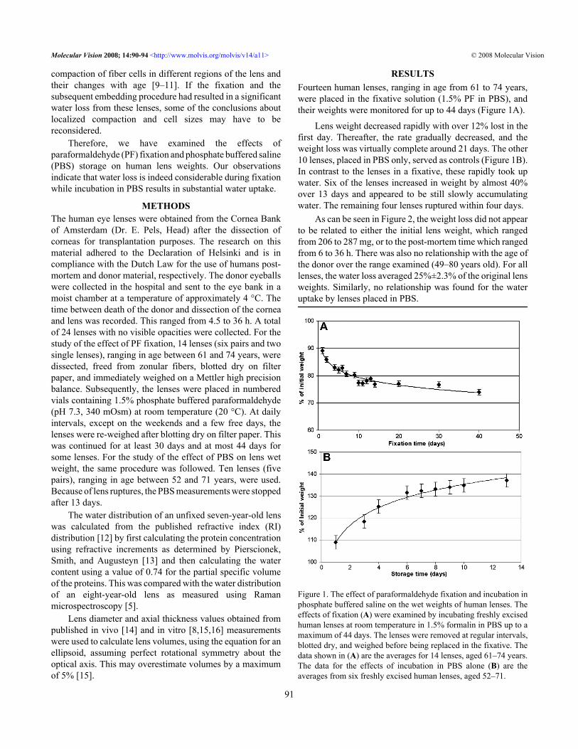
As can be seen in Figure 2, the weight loss did not appearto be related to either the initial lens weight, which rangedfrom 206 to 287 mg, or to the post-mortem time which rangedfrom 6 to 36 h. There was also no relationship with the age ofthe donor over the range examined (49–80 years old). For alllenses, the water loss averaged 25%±2.3% of the original lensweights. Similarly, no relationship was found for the wateruptake by lenses placed in PBS.
Figure 1. The effect of paraformaldehyde fixation and incubation inphosphate buffered saline on the wet weights of human lenses. Theeffects of fixation (A) were examined by incubating freshly excisedhuman lenses at room temperature in 1.5% formalin in PBS up to amaximum of 44 days. The lenses were removed at regular intervals,blotted dry, and weighed before being replaced in the fixative. Thedata shown in (A) are the averages for 14 lenses, aged 61–74 years.The data for the effects of incubation in PBS alone (B) are theaverages from six freshly excised human lenses, aged 52–71.
Molecular Vision 2008; 14:90-94 <http://www.molvis.org/molvis/v14/a11> © 2008 Molecular Vision
91

DISCUSSIONThe present study was undertaken because of a recent report[7] that the wet weights of fixed lenses described by Willekenset al. [8] were substantially lower than those of unfixed lensesdescribed by other laboratories. No significant differenceshave been observed between the dry weights of fixed andunfixed lenses of the same age indicating that no solids arelost from the lens during fixation (Augusteyn, unpublishedobservations). Therefore, the data presented here show clearlythat the low weight can be attributed to the substantial (25%)loss of water during fixation.
Willekens et al. [8] determined lens weights after sevendays of fixation at which time a 19.7% decrease in weightwould have occurred according to our current measurements.Correcting the previously published data for this loss yieldsvalues for wet weights statistically indistinguishable fromunfixed lenses freshly collected for the present study and fromlenses used in the recent re-analysis of lens growth [7].
These observations suggest that there could be substantialdecreases in the dimensions and volume of the lens on
Figure 2. Relationship between maximum weight loss duringfixation and initial lens weight and post-mortem time. Human lenses,ranging in age from 61 to 74 years, were incubated for up to 44 h in1.5% paraformaldehyde in PBS and the maximum amount of weightlost was compared with the initial lens wet weight (A) and with thepost-mortem (PM) time (B). No trends can be observed for eitherplot and the average weight loss is 25%±2.3%.
fixation. This was examined by comparing the published datafor fixed lens dimensions with those of unfixed lenses andcalculating volumes from these. From the best fit of the datapresented by Willekens et al. [8], fixed lens thickness rangedaround 3–4 mm between the ages of 20 and 80 years. Thismay be contrasted with the 4.2–5 mm reported for unfixedlenses [15,16] and 4–5 mm for the fully accommodated in vivolenses [14] over the same age range. To avoid errors due toshape changes in the lenses, volumes were calculated wheredimensions were available. Figure 3 shows the volumecomparisons for fixed, unfixed, and in vivo lenses.
Although there is some variability in the data, it wouldappear that the volumes of unfixed in vitro and in vivo lensesare comparable, ranging between 150 and 230 µl over the 20–80 year age range. Any difference can probably be attributedto the uptake of water by the post-mortem lens [17]. Bycontrast, the fixed lenses are substantially smaller at 110–176 µl, almost exactly 25% lower, in close agreement with theobserved weight loss in the present study.
To determine from where the water was lost, the waterdistribution in a fixed eight-year-old lens described bySiebinga et al. [5] was compared with that calculated from therefractive index gradient observed in an unfixed seven-year-old lens [12]. The distributions along the sagittal (optical) axesare shown in Figure 4. Essentially the same differences werefound for the equatorial axis.
The fixed lens had lower water contents almost along thewhole axis. Between 0.06 and 0.97 of the normalized distance,the water content in the fixed lens averaged 58% (w/w). In theunfixed lens, the lowest water content was around 68%, butthis was limited to the central 0.4–0.6 of the normalizeddistance. Much larger differences are observed in the
Figure 3. Comparison of the volumes of fixed and unfixed lenses.The volumes of fixed (blue diamond) [8] and unfixed (greendiamond) [12] in vitro lenses and fully accommodated in vivo lenses(red diamond) [14] are shown. Volumes of unfixed lenses werecalculated using the published dimensions for individual lenses,assuming an ellipsoid shape. Fixed lens volumes were calculatedusing values calculated from the published regression equations forthickness and diameter [8].
Molecular Vision 2008; 14:90-94 <http://www.molvis.org/molvis/v14/a11> © 2008 Molecular Vision
92

periphery. As can be seen from Figure 4, the outer 0.2 of thenormalized axis contains the water/refractive index gradientin the unfixed lens. Following fixation, this region has becomemuch narrower (0.03–0.06) with the virtual disappearance ofthe gradient. In terms of actual dimensions in the sagittal planeof the eight-year-old fixed lens, the width of the gradient hadreduced from the unfixed 1.2 mm [12,18] to an average of0.2 mm while in the equatorial plane it decreased from 1.2 mmto 0.6 mm.
Calculation of the total water content in the inner region(from 0.2 to 0.8 along the normalized axes) and the outer (from0 to 0.2 plus from 0.8 to 1.0 of the normalized axes) revealedthat >90% of the water lost probably came from the cortex.
It was also reported from the Raman measurements thatthe nuclear water content increases with age from 58% to near70% around the age of 80 [5]. This is inconsistent with thetrends in protein concentrations determined bymicroautoradiography [1] and microdissection [2], with thewater content determined using thermogravimetric analysis[19], and with the development of a plateau of refractive indexobserved with MRI [18]. The reason for this apparent increasewith the fixed lenses is not obvious, but it seems unlikely thatthe normal nucleus could accommodate such a large influx ofwater without an obvious disruption of its ordered structure.The likely explanation of this observation is that there is anage-related decrease in the amount of water that could be lostfrom the lens center during fixation.
Several ultrastructural studies on lens cell sizes indifferent regions of the lens at different ages have beenconducted with fixed lenses [9–11]. It is not possible to assesshow much water was lost since dimensions were not reportedfor the fixed tissues. Our data suggest that water loss from the
Figure 4. The distribution of water along the sagittal axis in fixed andunfixed human lenses. Comparison of the distributions of wateralong the sagittal axis in an unfixed seven-year-old lens (greendiamond), which was calculated from the RI gradient obtained withMRI [12], and in a fixed eight-year-old lens (red diamond) asdetermined by the use of Raman microspectroscopy [5] is shown.The dimensions were normalized to eliminate differences in thethickness of the lenses due to possible shrinkage of the fixed lens.
lens during fixation is not uniform and, as suggested by theRaman observations, may even vary with age. How this mightaffect cell dimensions in lens layers, such as the embryonicand the fetal, juvenile, and adult nuclei as well as differentregions of the cortex, cannot be gauged without furtherinformation. However, the possibility of significant changecannot be ruled out.
At first, the loss of water during fixation appears rathersurprising given that the osmolality of the fixing solution isessentially the same as that of PBS, which resulted in wateruptake. The lowest water content observed in the fixed seven-year-old lens was around 58%, and this encompassed almostall of the lens [5]. This suggests that free water is lost andcompaction of human cells may be limited to this finalconcentration of water because the residual water is tightlybound to the crystallins and can only be removed by moreforcing conditions such as heating or vacuum drying.However, it is not clear what the driving force for such waterloss may be. More likely, it may not be the free water whichis lost.
Fixation of the lens proteins modifies hydrophilic sidechains and generates crosslinked aggregates. This wouldreduce their osmolality and hydration shells with aconcomitant release of water, which would move out of thelens. In the nucleus, the proteins have already beensubstantially modified through age-related processes[reviewed in 20] so there is less water loss from this region.It can be calculated from the data presented by Bettelheim andcoworkers [21] that bound (nonfreezable) water in a 50-year-old lens represents around 25% of the lens weight, remarkablyclose to the weight loss observed in the current study. Thissuggests that the water lost from the lens may actually havecome from the bound fraction in the crystallins.
Storage in PBS leads to a significant increase in wetweight and even to the rupture of some lenses. The observationthat lenses swell when placed in an apparently isotonicmedium is not new. Uptake of water and/or an increase in lensthickness have frequently been observed in lenses left in theeye for prolonged periods or in culture media and saltsolutions [17,22,23]. This can probably be attributed to afailure of the volume-regulating systems due to a lack ofnutrients. It was noted by Augusteyn et al. [17] that lensswelling along the optic axis and capsular separation couldoften be detected within hours of a lens being placed in simplesalt solutions. Incubation in nutrient-rich media such asDMEM or TC199 reduced the incidence of lens swelling butdid not prevent it completely. Schachar found lens thicknessstarted to increase within two days of storage in Optisol,leading to a 10% increase in lens thickness in 10 days [23].Such increases could have serious implications for in vivostudies on lens optical and physical properties.
Although systematic studies have not been undertaken onother species, it has been observed in our laboratory that
Molecular Vision 2008; 14:90-94 <http://www.molvis.org/molvis/v14/a11> © 2008 Molecular Vision
93

baboon, cow, dunnart, kangaroo, rabbit, rhesus monkey,sheep, and toad lenses also shrink when fixed inparaformaldehyde. Incubation of cow, cynomolgus monkey,kangaroo, rhesus monkey, rat, and sheep lenses in PBS resultsin swelling. This suggests that the phenomena described forhuman lenses may be universal.
The observations presented here indicate that cautionneeds to be exercised when interpreting data obtained fromlenses, which have been fixed or stored in PBS. This wouldinclude data on the distribution of water [3–5] as well as dataon protein conformation [6] and on fiber cell dimensions [9–11]. It would appear that the properties of the lens, andespecially the cortex, have been grossly altered by fixation sonone of the data should be considered as representative of thenative in vivo state.
ACKNOWLEDGMENTSThis work was supported by NIH consortium grant,R01EY014225, and the Cooperative Research Centre Schemeof Australia. We are grateful to the Cornea Bank Amsterdam(Dr. E. Pels, Head) for providing the tissues used in this study.
REFERENCES1. Fagerholm PP, Philipson BT, Lindstrom B. Normal human lens
– the distribution of protein. Exp Eye Res 1981; 33:615-20.[PMID: 7318958]
2. Bours J, Wegener A, Hofmann D, Fodisch HJ, Hockwin O.Protein profiles of microsections of the fetal ands adult lensduring development and ageing. Mech Ageing Dev 1990;54:13-27. [PMID: 2195251]
3. Huizinga A, Bot AC, de Mul FF, Vrensen GF, Greve J. Localvariations in absolute water content of human and rabbit eyelenses measured by Raman microspectroscopy. Exp Eye Res1989; 48:487-96. [PMID: 2714410]
4. Bot ACC, Huizinga A, de Mul FFM, Vrensen GJM, Greve J.Raman microspectroscopy of fixed human and rabbit eyelenses and lens slices; new potentialities. Exp Eye Res 1989;49:161-9. [PMID: 2767164]
5. Siebinga I, Vrensen GF, De Mul FF, Greve J. Age-relatedchanges in local water and protein content of human eyelenses measured by Raman microspectroscopy. Exp Eye Res1991; 53:233-9. [PMID: 1915680]
6. Siebinga I, Vrensen GF, Otto K, Puppels GJ, De Mul FF, GreveJ. Ageing and changes in protein conformation in the humanlens: a Raman microspectroscopic study. Exp Eye Res 1992;54:759-67. [PMID: 1623961]
7. Augusteyn RC. Growth of the human eye lens. Mol Vis 2007;13:252-7. [PMID: 17356512]
8. Willekens B, Kappelhof J, Vrensen G. Morphology of the aginghuman lens. Lens Res 1987; 4:207-30.
9. Taylor VL, al-Ghoul KJ, Lane CW, Davis VA, Kuszak JR,Costello MJ. Morphology of the normal human lens. InvestOphthalmol Vis Sci 1996; 37:1397-410. [PMID: 8641842]
10. Al-Ghoul KJ, Costello MJ. Light microscopic variation of fibercell size, shape and ordering in the equatorial plane in bovineand human lenses. Mol Vis 1997; 3:2.http://www.emory.edu/molvis/v3/al-ghoul [PMID: 9238091]
11. Al-Ghoul KJ, Nordgren RK, Kuszak AJ, Freel CD, Costello MJ,Kuszak JR. Structural evidence of human nuclear fibercompaction as a function of ageing and cataractogenesis. ExpEye Res 2001; 72:199-214. [PMID: 11180969]
12. Jones CE, Atchison DA, Meder R, Pope JM. Refractive indexdistribution and optical properties of the isolated human lensmeasured using magnetic resonance imaging (MRI). VisionRes 2005; 45:2352-66. [PMID: 15979462]
13. Pierscionek B, Smith G, Augusteyn RC. The refractiveincrements of bovine α-, β- and γ-crystallins. Vision Res1987; 27:1539-41. [PMID: 3445487]
14. Strenk SA, Semmlow JL, Strenk LM, Munoz P. Groniund-Jacob Jm DeMarco JK. Age-related changes in human ciliarymuscle and lens: A magnetic resonance imaging study. InvestOphthalmol Vis Sci 1999; 40:1162-9. [PMID: 10235549]
15. Rosen AM, Denham DB, Fernandez V, Borja D, Ho A, MannsF, Parel JM, Augusteyn RC. In vitro dimensions andcurvatures of human lenses. Vision Res 2006; 46:1002-9.[PMID: 16321421]
16. Schachar RA. Growth patterns of fresh human crystalline lensesmeasured by in vitro photographic biometry. J Anat 2005;206:575-80. [PMID: 15960767]
17. Augusteyn RC, Rosen AM, Borja D, Ziebarth NM, Parel JM.Biometry of primate lenses during immersion in preservationmedia. Mol Vis 2006; 12:740-7. [PMID: 16865087]
18. Augusteyn RC, Jones C, Pope JM. Development of a refractiveindex plateau in the nucleus of the human lens. Clin ExpOptom. 2008In press
19. Heys KR, Cram SL, Truscott RJW. Massive increase in stiffnessof the human lens nucleus with age: the basis for presbyopia?Mol Vis 2004; 10:956-63. [PMID: 15616482]
20. Augusteyn RC, Stevens A. Macromolecular structure of the eyelens. Prog Polym Sci 1998; 23:375-413.
21. Lahm D, Lee LK, Bettleheim FA. Age dependence of freezableand nonfreezable water content of normal human lenses.Invest Ophthalmol Vis Sci 1985; 26:1162-5. [PMID:4019108]
22. Augusteyn RC, Cake MA. Post mortem uptake of water bysheep lenses left in the eye. Mol Vis 2005; 11:749-51. [PMID:16179906]
23. Schachar RA. Central surface curvature of postmortem-extracted intact human crystalline lenses. Ophthalmology2004; 111:1699-704. [PMID: 15350325]
Molecular Vision 2008; 14:90-94 <http://www.molvis.org/molvis/v14/a11> © 2008 Molecular Vision
The print version of this article was created on 17–January–2008. This reflects all typographical corrections and errata to thearticle through that date. Details of any changes may be found in the online version of the article.
94