THE EFFECT OF MOUTHGUARD DESIGN ON RESPIRATORY FUNCTION … · THE EFFECT OF MOUTHGUARD DESIGN ON...
56
1 THE EFFECT OF MOUTHGUARD DESIGN ON RESPIRATORY FUNCTION IN ATHLETES. AND CAN CEPHALOMETRIC AIRWAY ANALYSIS PREDICT VENTILATORY FUNCTION IN ELITE ATHLETES? This thesis is presented for the degree of Masters of Clinical Research at the University of Western Australia Dieter Gebauer Bachelor of Dental Science Bachelor of Medicine and Surgery (Honours) Graduate Diploma in Dental Studies FRACDS (OMS) School of Medicine and Pharmacology School of Dentistry School of Sport Science, Exercise and Health 2014
Transcript of THE EFFECT OF MOUTHGUARD DESIGN ON RESPIRATORY FUNCTION … · THE EFFECT OF MOUTHGUARD DESIGN ON...

1
THE EFFECT OF MOUTHGUARD DESIGN ON RESPIRATORY FUNCTION IN ATHLETES.
AND
CAN CEPHALOMETRIC AIRWAY ANALYSIS PREDICT
VENTILATORY FUNCTION IN ELITE ATHLETES?
This thesis is presented for the degree of Masters of Clinical Research at the University of Western
Australia
Dieter Gebauer Bachelor of Dental Science
Bachelor of Medicine and Surgery (Honours) Graduate Diploma in Dental Studies
FRACDS (OMS)
School of Medicine and Pharmacology School of Dentistry
School of Sport Science, Exercise and Health
2014

2
Publication Arising from this Thesis
Gebauer D, Williamson R, Wallman K, Dawson B (2011). The Effect of Mouthguard
Design on Respiratory Function in Athletes. Clin J Sport Med Vol 21(2):95-100
Gebauer D, Williamson R, Wallman K (2014). Do Craniofacial Abnormalities Impair
Elite Athlete’s Ventilatory Performance during Submaximal and Maximal Exercise?
J of Dent, Med and Med Science Vol 4(2):22-27.
Peer-Reviewed Conference Proceedings
Gebauer D, Williamson R, Wallman K, Dawson B (2011). The Effect of Mouthguard
Design on Respiratory Function in Athletes.
a) Annual conference of the Australian and New Zealand Society of Oral and
Maxillofacial Surgery (ANZAOMS) Gold Coast Australia 27th-30th October
2009. (Oral Presentation)
b) 20th Royal Australian College of Dental Surgeons (RACDS) Convocation
Perth Australia 11th-14th March 2010. (Oral Presentation)
Gebauer D, Williamson R, Wallman K (2014). Do Craniofacial Abnormalities Impair
Elite Athlete’s Ventilatory Performance during Submaximal and Maximal Exercise?
a) Annual conference of the Australian and New Zealand Society of Oral and
Maxillofacial Surgery (ANZAOMS) Perth Australia 19-21st September 2013.
(Oral Presentation)

3
Table of Contents
Publications Arising from this Thesis 2
Peer-Reviewed Conference Proceedings 2
Table of Contents 3
Acknowledgements 4
Statement of Originality 5
List of Tables 6
List of Figures 7
List of Abbreviations 8
CHAPTER ONE
Introduction
CHAPTER TWO Study One: THE EFFECT OF MOUTHGUARD DESIGN ON
RESPIRATORY FUNCTION IN ATHLETES. CHAPTER THREE
Linking Statements
CHAPTER FOUR
Study Two: DO CRANIOFACIAL ABNORMALITIES IMPAIR ELITE ATHELETS VENTILATORY PERFORMANCE DURING SUBMAXIMAL AND MAXIMAL EXERCISE?
CHAPTER FIVE
Discussion
CHAPTER SIX
Appendices

4
Acknowledgements
Most sincere thanks and appreciation go to all of whom were involved and
contributed to the completion of this thesis, in particular:
Associate Professor Karen Wallman, for being so friendly and accommodating in
making me feel part of the School of Sport Science, Exercise and Health. Much can
be learnt from the way your department conducts its research and your help and
advice will never be forgotten.
Mr Michael Phillips, biostatistician at the West Australian Institute of Medical
Research, for making a difficult task easy by his clear advice and sense of humour.
Mr Adam Surjan, honours student at the School of Sport Science, Exercise and
Health, for his assistance in conducting the testing protocols.
Professor Raymond Williamson, as the director of training of the West Australian
Oral and Maxillofacial Surgery Programme. Without your leadership, I would
never have had a career that I enjoy.
Research Participants, for your cooperation and involvement. These studies would
not have been possible without your participation.
Mr Barry McGee and Mr Sean Brown, resident prosthetist at the Oral Health
Centre of West Australia for their kind assistance in fabricating the mouthguards.
Mr Peter Herring prosthetist from ERKODENT who donated the blank
mouthguard templates that were used to fabricate the mouthguards used in the
trial.

5
Statement of Originality
This thesis describes original research conducted by the author at the School of
Dentistry and School of Sport Science, Exercise and Health at the University of
Western Australia from Nov 2007- Dec 2013.
The author, under the guidance and assistance of Professor Raymond Williamson
and Associate Professor Karen Wallman is responsible for the research concept
and design. Participant recruitment, data collection, and data analysis were
carried out by the candidate, as well as the implementation of the experiments.
The candidate drafted the thesis, and the papers which have been accepted
and/or are currently being considered for publication, with assistance in writing
and submission processes by both Professor Raymond Williamson and Associate
Professor Karen Wallman. Feedback on the thesis was provided by Professor
Raymond Williamson and Associate Professor Karen Wallman.
Signature:
Dr Dieter Gebauer (Candidate)
Signature:
Associate Professor Raymond Williamson
Signature:
Associate Professor Karen Wallman (Supervisor)

6
List of Tables
Chapter 2
Table 1 - Demographic Characteristics of Male Team Sport Players (n=27).
Table 2- Mouthguard Comparison of Normal Palatal Design Mouthguard (NPD) vs
Palate Free Design (PFD). (n=27).
Table 3 - Minute Ventilation ( e,) Oxygen Uptake ( O2) and Heart Rate (HR)
Whilst Exercising at 10 km·h-1, 12 km.h-1 and Maximal Intensity Levels of Exercise,
Wearing No Mouthguard (NM), Regular Palate Design Mouthguard (NPD) and
Palate Free Design (PFD). (n = 27).
Chapter 4
Table 1 – Cephalometric landmarks.
Table 2 - Cephalometric measurements.
Table 3 - Demographic characteristics of male team sport players (n=27).
Table 4 - Mean, standard deviation and range of cephalometric values.
Table 5- Posterior airway space (PAS), soft palate length (PNS-P), hyoid to
mandibular plane. Length (MP-H) and skeletal classification when compared to
ventilation ( E L·min-1).
V

7
List of Figures
Chapter 2
Figure 1: Palate free and palate covering mouthguard design.
Figure 2: Face mask applied correctly.
Figure 3: An athlete engaged in a testing protocol.
Chapter 4
Figure 1: Lateral cephalometric radiograph with cephalometric parameters
illustrated.

8
List of Abbreviations
ANB A point-Nasion-B point
ECG electrocardiography
EEG electroencephalography
EMG electromyography
EOG electrooculography
Gn Gnathion
Go Gonion
GTX graded exercise test
MP-H hyoid to mandibular plane
length
Na nasion
NPD normal palatal design
NM no mouthguard
OSA obstructive sleep apnoea
PAS posterior airway space
PFD palatal free design
PNS-P soft palate length
PNS posterior nasal spine
E ventilation
O2 oxygen uptake
O2peak maximal effort

9
CHAPTER ONE
Introduction

10
The author of this thesis has previously been an Australian Institute of Sport
Scholarship holder in the sport of water polo and had played this sport on a
regular basis over many years. During this time, many weekends were spent
repairing craniofacial trauma injuries sustained by both friends and elite athletes
that could have been prevented if the athlete had worn a mouthguard.
Surprisingly, some of these athletes had the means to wear a mouthguard but
made a deliberate decision not to wear one, citing perceived respiratory
difficulties as the reason. Notably, the author had difficulty in comprehending this
as he had never encountered such difficulty.
The craniofacial skeleton provides a framework for the upper respiratory tract.
New and emerging scientific evidence has demonstrated the beneficial effect of
jaw advancement procedures on the patency of the upper airway. If airway
features associated with upper airway collapse in obstructive sleep apnoea (OSA)
are shown to influence ventilation in elite athletes during exercise states, jaw
advancement procedures may benefit the performance of susceptible athletes.
Therefore, the aim of study one was to determine whether a custom made
mouthguard of a particular design had an effect on ventilation and/or the work of
breathing during exercise when compared to wearing no mouthguard.
Furthermore, the aim of study two was to determine if there was a correlation
with poor ventilatory performance and an OSA susceptible upper respiratory tract
during moderate and maximal exercise states.
Elite athletes are role models to others within their sport, and the use of
protective equipment, in particular, the wearing of mouthguards, is very
important. Many athletes choose not to wear a mouthguard because they believe
that it interferes with their ventilation and hence their exercise performance.
Consequently, it is important to determine whether mouthguards actually do

11
occlude the upper respiratory tract because if it can be established that
mouthguards do not impair ventilation, then hopefully this will encourage elite
athletes to wear mouthguards. This should also influence this behaviour at an
amateur level. As a medical practitioner, the practice of preventative health
should always be a consideration where possible. Preventable dento-alveolar and
facial trauma is over represented in emergency departments and a reduction in
admissions can reduce hospital workload. Conversely, if improvement in an elite
athlete’s performance can be achieved by the performance of a surgical
procedure that assists the occlusive airway, surgeons can assist that athlete in
reaching their full potential and consequently enhance their earning capacity if
they are in the professional arena. They will also have additional health benefits
of the individual being less susceptible to the effects of OSA later in life if weight
gain occurs.

12
CHAPTER TWO
Study One

13
The effect of mouth guard design on respiratory function in
athletes.
This paper has been published by
The Clinical Journal of Sports Medicine
Vol. 21(2) 2011 pp: 95-100.
Presented here in the Journal submission format

14
Abstract
Objective: To test the hypothesis that two types of custom made mouthguards
will have no effect on ventilation ( E: L∙min-1), oxygen uptake ( O2: mL∙kg-1·min-
1) and heart rate (HR beats per min) at varying exercise intensities (10 km·h-1, 12
km·h-1 and at subjective maximal effort: O2peak) in male field hockey and water
polo players.
Design: A randomised, prospective crossover study.
Setting: The Physiology Testing Laboratory, School of Sports Science, Exercise and
Health at the University of Western Australia, a tertiary educational institution.
Participants: Twenty-seven male, team-sport athletes.
Interventions: Each athlete participated in three experimental exercise sessions
separated by one week intervals. Testing involved a graded exercise test (GXT)
performed on a treadmill wearing either a custom laminated mouthguard with
normal palatal surface (NPD), a custom laminated mouthguard with palatal
coverage up to the gingival margin (PFD), or no mouthguard (NM). The
experimental trials were performed in a random, counterbalanced order.
Main Outcome Measurements: E (L·min-1) and O2 (mL∙kg-1·min-1) were
measured during the GXT at intensities that equated to 10 km·h-1, 12 km·h-1 and
subjective maximal effort ( O2peak).
Results: There were no significant differences between trials for E (L·min-1) and
O2 (mL∙kg-1·min-1) at any of the intensities assessed (p < 0.05).

15
Conclusions: The wearing of two different custom made mouthguards during a
GXT did not impair E or O2 during varying levels of exercise intensity in team
sport athletes.
Key words: Mouthguard; Ventilation; Oxygen Uptake; Respiration; Athlete.
Introduction
Many contact sports risk either deliberate or accidental contact to the head and
neck region.1 Occasionally, contact to the head of an athlete results in injury
ranging from a closed head injury such as concussion, dento-alveolar injury, facial
lacerations or fracture of the facial skeleton. Mouth guards provide protection
from concussion, dento-alveolar injury, and fractures of the facial skeleton2-8. The
protective capability of a mouthguard stems from its ability to distribute and
dissipate a point of impact force over a larger area9-14. The severity of peri-oral
lacerations may be reduced by the use of a custom fabricated mouthguard.
Currently there are three main types of mouthguard available to provide
protection to athletes4, 9, 10, 15, 16. These include a stock mouthguard, a boil and
bite type, or a professionally fitted custom made mouthguard. The professionally
fitted mouthguard is considered the gold standard as it has the greatest area of
surface contact between the protective surface and the dentition, hence the best
ability to distribute forces.4, 10, 12, 13
Many presentations of facial and dento-alveolar trauma resulting from sporting
activity could have been prevented or minimised by the use of appropriately
fitted mouthguards2, 17-20. Unfortunately many professional sports administrators
do not mandate the use of a protective mouthguard, and many elite athletes
refrain from wearing mouth guards during contact sports because they have an

16
underlying belief that mouthguards reduce ventilation and oxygen uptake and
therefore negatively impact upon their exercise performance. A study performed
by Sports Medicine Australia21 on sport injury in Western Australia during the
1997 and 1998 sporting seasons, reported that only 34% of Australian rules
football players wore mouthguards. These percentages were even lower in
hockey (23%), basketball (~6%) and netball players (~2%). This is a major concern
when considering both the physical and emotional cost of oro-facial injury.21
Therefore, the aim of this study was to assess the effects of mouthguards on
ventilation ( E L·min-1) and oxygen uptake ( O2 mL∙kg-1·min-1) during an
incremental exercise test to exhaustion. Two different types of custom made
mouthguards, normal palatal design (NPD) and palate free design (PFD) were
compared to a control condition where no mouthguard (NM) was worn.
It was hypothesised that use of a custom made mouth guard (either NPD or PFD
design) would have no effect on E (L·min-1) or O2 (mL∙kg-1·min-1) during
exercise performed at 10 km·h-1, 12 km·h-1 or at subjective maximal effort, when
compared to a control (no mouth guard) trial.
Methods
Prior to recruitment of participants, a power analysis was performed on O2
data obtained from the Australian Institute of Sport. This analysis demonstrated
that 30 participants (paired comparison) were required in order to detect an
equivalent measurement of +/- 2% difference in O2max to α=0.01 with a power
of 95%.

17
Thirty-one males who participated in team sports (hockey or water polo) either
at first division, state level or national level were recruited into the study. Four
participants withdrew during the trials for reasons associated with injury (4)
leaving 27 subjects (mean (SD): age - 23.5 yr (3.8), body mass index - 24.6 kg∙m-2
(2.1), ht – 1.82 m (0.08), body-mass – 81.7 kg (8.6)). Participants were informed
of the study requirements, benefits and risks before giving written informed
consent. The Research Ethics Committee of the University of Western Australia
(UWA) granted approval for the study’s procedures. Participants were excluded
if they had any respiratory pathology (defined by disclosure by the participant of
any lifetime medical history of lung disease), were anaemic (defined by having a
haemoglobin level of less than 135 g∙L-1), were smokers (defined as having a
history of consumption of a tobacco product), wore a removable dental
appliance, or had fixed orthodontic appliances.
Experimental Overview
Prior to participating in the experimental trials, all participants attended the Oral
Health Centre of Western Australia’s (OHCWA) oral surgery clinic where they
completed a questionnaire that provided information about their mouthguard
attitudes and utilization, as well of any history of facial trauma. During this
session, two alginate dental impressions were made of the maxillary teeth
(Kromopan 100 – Lascod Firenze Italy). Plaster casts of the impressions were
made on the same day using yellow stone (Ainsworth Buffstone Sydney Australia)
and were trimmed to standard specifications.

18
The two mouth guard designs are illustrated below in Figure 1.
Figure 1: Full Palate and Palate Free Mouth Guard Design
Each mouthguard was extended to cover the first maxillary molar, was 4 mm
thick and fabricated using a laminated vacuum technique.
The NPD mouthguard was made with the palatal margin extending approximately
4 mm past the gingival margin. The PFD mouthguard only extended to the free
gingival margin of the palate.
One plaster cast was used to make the NPD and the other for PFD. The
mouthguards were a laminated design using 2 mm thickness layered plastic
sheeting to obtain a 4 mm even thickness using a vacuum machine. (Erkoflex -
Erkodent Pfalzgrafenweiler Germany). The volume of each mouthguard was then
measured by a displacement technique. The volume of the water displaced was
measured and recorded (Sartorius GMBH, Model E1200 Goltingen Germany).

19
Finally, each participant had a 4 mL sample of blood taken and analysed for
haemoglobin in a Beckman Coulter LH750 haematology analyser (California USA)
in order to screen for anaemia.
Exercise testing was performed at the Exercise Physiology Laboratory in the
School of Sport Science, Exercise and Health at UWA. Participants participated in
three experimental trials that were undertaken in a randomised, counter-
balanced order and performed at the same time of day, approximately one week
apart. Use of a counter-balanced design ensured that participants were evenly
distributed across the three experimental trials in order to avoid a learning effect
associated with one particular trial. The three trials consisted of exercise with no
mouthguard (NM), exercise wearing a custom laminated mouthguard with
normal palatal surface (NPD), or exercise wearing a custom laminated
mouthguard with palatal coverage up to the gingival margin (PFD). Participants
were required to maintain their normal diet and training throughout the study,
and were required to fast (other than water) during the 2-h period prior to
testing. Additionally, participants were asked not to perform vigorous exercise in
the 24-h period prior to testing.
During the first session, prior to the exercise test, each participant’s height was
measured using a stadiometer, while body mass was ascertained using Sauter
scales (August Sauter GmbH D-7470 Albstadt 1 Ebingen, West Germany). A
fifteen minute familiarisation session then took place where the athletes were
required to warm-up on the treadmill as if they were preparing for a team game.
During this warm-up session, participants wore a heart rate monitor (Polar,
Finland) and breathed into a computerised gas analysis system (Meta 2000,
UWA, Australia) through a face mask that covered their mouth and nose (Figure
2).

20
Figure 2: Face Mask Design
This system consisted of a ventilometer (Morgan, Kent, United Kingdom) that
measured the volume of inspired air and oxygen and carbon dioxide. Analysers
(Ametek Applied Electrochemistry S-3A/1 and CD-3A, AEI Technologies,
Pittsburgh, USA) measured the percentage of oxygen and carbon dioxide in
expired air. The carbon dioxide analyser uses the principle of infra-red
absorption for measurement, while measurement by the oxygen analyser is
based on the principle of high temperature zirconia crystal conductivity. The gas
analysers were calibrated before each test using a beta (ß) standard reference
gas, while the ventilometer was calibrated using a one litre syringe, as per
manufacturer specifications. After this familiarisation session E (L·min-1), O2
(mL∙kg-1·min-1) and HR (beats per min: bpm) were measured during a GXT

21
performed on the treadmill. The protocol consisted of the participant running for
one minute at 9 km·h-1. This was followed by two plateau stages of 5 min each,
with the speed of the treadmill progressively increased following the plateau
stages until volitional exhaustion was reached. The first plateau stage occurred
at 10 km·h-1, while the second plateau stage occurred at 12 km·h-1. (Figure 3)
Figure 3: Athlete on Testing Equipment
The two plateau stages were separated by a 1 min increment of 11 km·h-1. The
last minute of testing during which volitional exhaustion occurred was
considered to represent O2peak.

22
Upon completion of each exercise test the participants were asked to rate the
mouthguard with a score out of ten on a visual analogue scale, with 10
representing the most comfortable score. Participants were also asked to rate
their current mouthguard if they wore one prior to the trial. The ratings were
compared to detect if there was a preferred mouthguard design.
Statistical Analysis
A repeated measure ANOVA was used to analyse HR, E, O2 recorded during
the two plateau periods of the GXT, as well as O2max. Where appropriate, post
hoc comparisons were also used. Statistical significance was set at p<0.05 for all
analyses and data was analysed using Stata Version 9. (StataCorp 2005. Statistical
Software Release 9. College Station TX: Stata Corp LP.) Student t tests were used
to determine if there were any significant differences between the ratings and
volumes of mouthguards NPD and PFD.
Results
The physical characteristics and general demographics of the participant
population are included in Table 1.
Table 1. Demographic Characteristics of Male Team Sport Players (n=27)
Variable Mean SD
Age (y) 23.5 3.8
Height (m) 1.82 0.08
Body mass (kg) 81.7 8.6
Haemoglobin (gL-1) 150 6.8
Body Mass Index (kg.m-2) 24.6 2.1
Eight water polo players and nineteen hockey players participated in this trial.
23
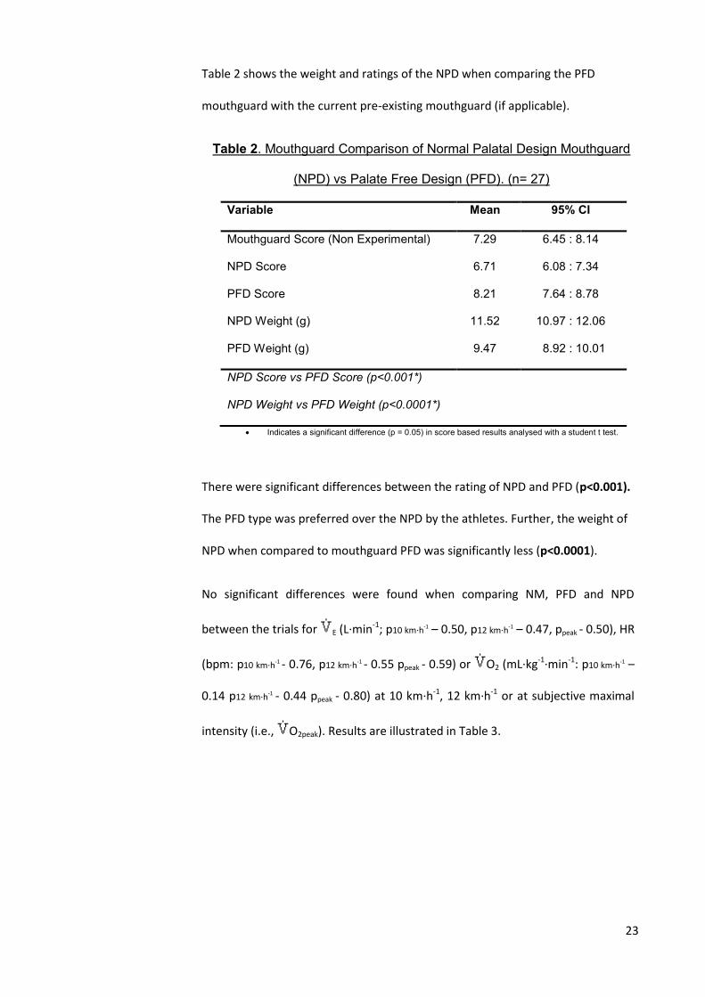
Table 2 shows the weight and ratings of the NPD when comparing the PFD
mouthguard with the current pre-existing mouthguard (if applicable).
Table 2. Mouthguard Comparison of Normal Palatal Design Mouthguard
(NPD) vs Palate Free Design (PFD). (n= 27)
Variable Mean 95% CI
Mouthguard Score (Non Experimental) 7.29 6.45 : 8.14
NPD Score 6.71 6.08 : 7.34
PFD Score 8.21 7.64 : 8.78
NPD Weight (g) 11.52 10.97 : 12.06
PFD Weight (g) 9.47 8.92 : 10.01
NPD Score vs PFD Score (p<0.001*)
NPD Weight vs PFD Weight (p<0.0001*)
Indicates a significant difference (p = 0.05) in score based results analysed with a student t test.
There were significant differences between the rating of NPD and PFD (p<0.001).
The PFD type was preferred over the NPD by the athletes. Further, the weight of
NPD when compared to mouthguard PFD was significantly less (p<0.0001).
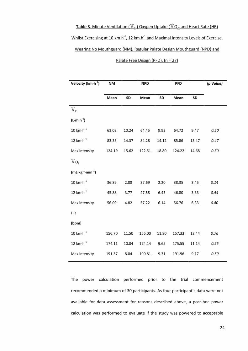
No significant differences were found when comparing NM, PFD and NPD
between the trials for E (L·min-1; p10 km·h-1 – 0.50, p12 km·h-1 – 0.47, ppeak - 0.50), HR
(bpm: p10 km·h-1 - 0.76, p12 km·h-1 - 0.55 ppeak - 0.59) or O2 (mL∙kg-1·min-1: p10 km·h-1 –
0.14 p12 km·h-1 - 0.44 ppeak - 0.80) at 10 km·h-1, 12 km·h-1 or at subjective maximal
intensity (i.e., O2peak). Results are illustrated in Table 3.
24
Table 3. Minute Ventilation ( e,) Oxygen Uptake ( O2) and Heart Rate (HR)
Whilst Exercising at 10 km·h-1, 12 km.h-1 and Maximal Intensity Levels of Exercise,
Wearing No Mouthguard (NM), Regular Palate Design Mouthguard (NPD) and
Palate Free Design (PFD). (n = 27)
Velocity (km·h-1
) NM NPD PFD (p Value)
Mean SD Mean SD Mean SD
E
(L∙min-1
)
10 km·h-1
63.08 10.24 64.45 9.93 64.72 9.47 0.50
12 km·h-1
83.33 14.37 84.28 14.12 85.86 13.47 0.47
Max intensity 124.19 15.62 122.51 18.80 124.22 14.68 0.50
O2
(mL∙kg-1∙min
-1)
10 km·h-1
36.89 2.88 37.69 2.20 38.35 3.45 0.14
12 km·h-1
45.88 3.77 47.58 6.45 46.80 3.33 0.44
Max intensity 56.09 4.82 57.22 6.14 56.76 6.33 0.80
HR
(bpm)
10 km·h-1
156.70 11.50 156.00 11.80 157.33 12.44 0.76
12 km·h-1
174.11 10.84 174.14 9.65 175.55 11.14 0.55
Max intensity 191.37 8.04 190.81 9.31 191.96 9.17 0.59
The power calculation performed prior to the trial commencement
recommended a minimum of 30 participants. As four participant’s data were not
available for data assessment for reasons described above, a post-hoc power
calculation was performed to evaluate if the study was powered to acceptable

25
standards. These calculations revealed that the group sample size of 27 resulted
in a 97% power to detect a difference of +/- 3% in O2peak, with the α level being
0.05, which is within an acceptable range for power calculation.
Discussion
This study found no significant differences between respiratory function, oxygen
uptake and heart rate results between the NM, NPD and PFD mouthguards trials
during exercise of varying intensities. Although not significant, the NM condition
resulted in the lowest O2peak values compared to the two other trials (2% and
1.2 % lower than the RPD and the PFD trials, respectively). Anecdotal complaints
by athletes that mouthguards “affect breathing” should not be used as a valid
excuse to refrain from wearing one.
To date, only eight published papers known to the authors have explored the
relationship between respiratory function, oxygen uptake and mouthguard use22-
29. These studies resulted in equivocal outcomes which most likely relate to the
use of varying study designs, methodology and cohort size.
Of previous study designs, only studies by Delaney et al.24, Bourdin et al.23 and
Francis et al.25 randomised the testing protocols to minimise the effect of
familiarisation bias in the data23-25. No previously published study had performed
a power analyses in order to determine the number of participants required to
obtain statistically significant data.
Further, comparison of previous studies demonstrated a large amount of
variability in testing protocols, and only four examined the effect of custom
mouthguards, which are considered the gold standard in protective oro-facial
care22, 23, 26, 29. Also, previous studies have used mouthpieces instead of full face

26
masks during the GXT, to measure ventilatory and oxygen uptake parameters. A
mouthpiece would eliminate the effect of the dentition and lip musculature on
airflow dynamics22, 24, 28 and significantly affect the results. Finally, the current
study is the only study known to the authors that assessed respiratory function
and oxygen uptake at various exercise intensities (i.e. 10 km·h-1, 12 km·h-1, or at
subjective maximal effort; i.e., O2peak).
A limitation to this study was that each mouthguard was fabricated from a
different impression and that subtle differences between the two mouthguards
may have been normalised by the use of non-identical casts. In addition, testing
undertaken in laboratory conditions does not emulate field conditions during
competition. However analyses of respiratory, oxygen uptake and heart rate data
at varying exercise intensities represents a strength of this study.
Women were excluded from this trial as the primary target group primarily
because males are over represented in hospital emergency departments being
admitted with injuries sustained when not wearing protective equipment during
sporting games21. Possibly this can be speculated to be due to contact sport being
generally male dominant, combined with macho tendencies for not wanting to
wear protective equipment. Additionally, males are on average, heavier and
more capable of transmitting greater force during collisions. However, as
respiratory physiology trends in women are similar to males, these conclusions
may also be applicable to women. Formal testing would be required to test this
conjecture.
Non custom fit mouthguards such as boil and bite type have similar thickness to
the laminates used to fabricate custom made mouthguards. The amount of
contact between the mouthguards, teeth and gums is less and therefore the total

27
volume of the mouthguard is greater causing a reduction in airway patency. We
speculate that this will cause a reduction of ventilatory function.
Additionally the fit of non-custom mouthguards is usually performed at home by
non-professionals. The extension of the periphery of the mouthguard can be
incorrectly trimmed, compromising its protective capability. If the periphery of a
mouthguard is overextended it can unnecessarily encroach on the airway space.
The participants enrolled in this trial rated the PFD mouthguard significantly
higher to the NPD type. This design is more popular and likely to be worn without
compromising its protective capability30-32, and the researchers recommend
fabricating this design to encourage utilisation in elite athletes who complain
about mouthguards interfering with exercise performance.
Two members of our research team are experienced with dealing in facial and
dentoalveolar trauma and reconstruction of lost dentition. This type of
preventable facial and dentoalveolar injury is still overrepresented in tertiary
hospitals in Western Australia21. Therefore, the results of this study have the
potential to benefit a significant number of sports participants in the professional
and amateur arena if governing bodies mandate the use of a mouthguard.
These findings could help professional sports health bodies and dental
association’s lobby health insurers to subsidise mouthguards so that cost would
not be a factor in preventing access to professionally fitted mouthguards. As
respiratory performance is not affected by the wearing of a mouthguard, it could
be proposed that injuries sustained as the result of not wearing a mouthguard
should not be covered by the insurer or sporting body.
As a result of this study’s outcome, the researchers recommend that sporting
bodies mandate the use of mouthguards during professional sporting activities

28
that are classified as contact sports. This should encourage and motivate the
general population, particularly children, who view elite athletes as role models,
to change their behaviour and to routinely use mouthguards. Consequently, this
would reduce the general incidence of dento-alveolar trauma, facial fracture and
concussion rates that result in visits to emergency departments.
Conclusion
This study demonstrated that use of a regular palate design (NPD) or a palate
free mouthguard (PFD) mouthguard by male team sport athletes had no negative
impact on E, O2 or HR during exercise of varying intensities compared to a no
mouthguard trial. This has important implications for athletes in encouraging
behaviour that will reduce the incidence of dento-alveolar trauma, concussion
and jaw fracture.

29
References
1. Kumamoto DP, Maeda Y, Kumamoto DP, et al. A literature review of sports-
related orofacial trauma. Gen Dent. 2004; 52:270-280.
2. Ada Council on Access P, Interprofessional R, Affairs ADACoS. Using
mouthguards to reduce the incidence and severity of sports-related oral injuries. J
Am Dent Assoc. 2006; 137:1712-1720.
3. Hendrick K, Farrelly P, Jagger R, et al. Oro-facial injuries and mouthguard use in
elite female field hockey players. Dent Traumatol. 2008; 24:189-192.
4. Knapik JJ, Marshall SW, Lee RB, et al. Mouthguards in sport activities : history,
physical properties and injury prevention effectiveness. Sports Med. 2007; 37:117-
144.
5. Labella CR, Smith BW, Sigurdsson A, et al. Effect of mouthguards on dental
injuries and concussions in college basketball. Med Sci Sports Exerc. 2002; 34:41-
44.
6. Lieger O, von Arx T, Lieger O, et al. Orofacial/cerebral injuries and the use of
mouthguards by professional athletes in Switzerland. Dent Traumatol. 2006; 22:1-
6.
7. Mihalik JP, McCaffrey MA, Rivera EM, et al. Effectiveness of mouthguards in
reducing neurocognitive deficits following sports-related cerebral concussion.
Dent Traumatol. 2007; 23:14-20.
8. Wisniewski JF, Guskiewicz K, Trope M, et al. Incidence of cerebral concussions
associated with type of mouthguard used in college football. Dent Traumatol.
2004; 20:143-149.
9. Bemelmanns P, Pfeiffer P. Shock absorption capacities of mouthguards in
different types and thicknesses. Int J Sports Med. 2001; 22:149-153.
10. Craig RG, Godwin WC. Properties of athletic mouth protectors and materials. J
Oral Rehabil. 2002; 29:146-150.
11. Duhaime CF, Whitmyer CC, Butler RS, et al. Comparison of forces transmitted
through different EVA mouthguards. Dent Traumatol. 2006; 22:186-192.
12. Lim D, Robinovitch S, Goodman D, et al. Effect of mouthguards on the
transmission of force across the human jaw. Clin J Sport Med. 2005; 15:313-319.
13. Westerman B, Stringfellow PM, Eccleston JA. Forces transmitted through EVA
mouthguard materials of different types and thickness. Aust Dent J. 1995; 40:389-
391.
14. Westerman B, Stringfellow PM, Eccleston JA. The effect on energy absorption
of hard inserts in laminated EVA mouthguards. Aust Dent J. 2000; 45:21-23.

30
15. Low D. Mouthguard protection and sports-related dental trauma. Ann R
Australas Coll Dent Surg. 2002; 16:153-155.
16. Newsome PR, Tran DC, Cooke MS. The role of the mouthguard in the
prevention of sports-related dental injuries: a review. Int J Paediatr Dent. 2001;
11:396-404.
17. Berry DC, Miller MG, Leow W, et al. Attitudes of Central Collegiate Hockey
Association ice hockey players toward athletic mouthguard usage. J Public Health
Dent. 2005; 65:71-75.
18. Braham RA, Finch CF. Do community football players wear allocated
protective equipment? Descriptive results from a randomised controlled trial. J Sci
Med Sport. 2004; 7:216-220.
19. Chatterjee M, Hilton I, Chatterjee M, et al. A comparison of the attitudes and
beliefs of professional rugby players from one club and parents of children playing
rugby at an adjacent amateur club to the wearing of mouthguards. Prim Dent
Care. 2007; 14:111-116.
20. Gardiner DM, Ranalli DN. Attitudinal factors influencing mouthguard
utilization. Dent Clin North Am. 2000; 44:53-65.
21. Sports Medicine Australia (WA Branch). Western Australia Sports Injury Study.
Perth Western Australia: Sports Medicine Australia; 2001.
22. Amis T, Di Somma E, Bacha F, et al. Influence of intra-oral maxillary sports
mouthguards on the airflow dynamics of oral breathing. Med Sci Sports Exerc.
2000; 32:284-290.
23. Bourdin M, Brunet-Patru I, Hager PE, et al. Influence of maxillary
mouthguards on physiological parameters. Med Sci Sports Exerc. 2006; 38:1500-
1504.
24. Delaney JS, Montgomery DL. Effect of noncustom birnolar mouthguards on
peak ventilation in ice hockey players. Clin J Sport Med. 2005; 15:154-157.
25. Francis KT, Brasher J. Physiological effects of wearing mouthguards. Br J
Sports Med. 1991; 25:227-231.
26. Kececi AD, Cetin C, Eroglu E, et al. Do custom-made mouth guards have
negative effects on aerobic performance capacity of athletes? Dent Traumatol.
2005; 21:276-280.
27. Luke R, Taylor G, Kaplan R. The effect of a mouthguard on airflow. Diastema.
1982; 10:56-57.
28. Schwartz R, Collins BJ, Fong C. Effects of a single and double commercial
athletic mouthpiece on expiratory peak flow: A pilot study. Cranio. 2000; 18:23-
29.

31
29. von Arx T, Flury R, Tschan J, et al. Exercise capacity in athletes with
mouthguards. Int J Sports Med. 2008; 29:435-438.
30. Yamanaka T, Ueno T, Oki M, et al. Study on the effects of shortening the distal
end of a mouthguard using modal analysis. J Med Dent Sci. 2002; 49:129-133.
31. Yamada J, Maeda Y, Satoh H, et al. Anterior palatal mouthguard margin
location and its effect on shock-absorbing capability. Dent Traumatol. 2006;
22:139-144.
32. Maeda Y, Machi H, Tsugawa T. Influences of palatal side design and finishing
on the wearability and retention of mouthguards. Br J Sports Med. 2006; 40:1006-
1008.

32
CHAPTER THREE
Linking Statements

33
The recurrent theme of this thesis is one of upper respiratory tract patency.
Occlusion due to anatomical or introduced means may have a detrimental effect
on an elite athlete’s performance. Study one demonstrated that introduced
occlusion due to the wearing of a mouthguard had no effect on ventilation during
a graded exercise test compared with not wearing a mouthguard. Furthermore,
any improvement in ventilatory capacity in an elite athlete engaged in sport may
have a significant affect in the athletes’ sensation of air hunger and fatigability.
Many trials have focused on ventilatory capacity during rest as part of obstructive
sleep apnoea research. Airway patency and its effect on ventilatory capacity
during exercise states, with a particular slant on orthognathic cephalometrics has
not been previously reported in the literature and is being introduced as a new
academic concept in this trial. The principals of orthognathic surgical examination
and workup include three important parameters, known as FAB - the facial
balance, the airway and the bite. Historically, emphasis has been placed on the
bite and facial aesthetic parameters. However with the advent of new findings
demonstrating the benefit of airway advancement procedures on obstructive
sleep apnoea patients, the airway has become an important consideration. In a
non-elite athlete population seeking advice for their dento-facial deformity, many
patients present in their teens to early twenty’s for treatment, and considerations
are made regarding their life long risks of airway patency. Weight gain and
associated metabolic syndrome is increasing in prevalence in western society and
those with a dento-facial deformity are placed at additional risk of developing
obstructive sleep apnoea. This time of life represents a unique preventative
health opportunity to improve the airway and consequently avoid years of
detrimental health effects of obstructive sleep apnoea, medication and
ventilatory support treatments prior to being offered a surgical procedure which
is considered by many respiratory physicians to be a last resort option. In many

34
instances the underlying dento-facial deformity is a main contributing factor.
Similar logic can be applied to an elite athlete, many of whom suffer weight gain
post completion of their professional careers. If this procedure is applied at the
time of the athlete’s development in their teens to early twenty’s, this may have a
significant effect on their exercise performance should a correlation be
discovered.

35
CHAPTER FOUR
Study Two

36
Can Cephalometric Airway Analysis Predict Ventilatory Function in Elite
Athletes?
This paper has been published as
“Do craniofacial abnormalities impair elite athletes’ ventilatory performance
during submaximal and maximal exercise?”
in the
Journal of Dentistry, Medicine and Medical Sciences Vol 4 (2) 2014: pp22-27
Presented here in the journal submission format

37
Abstract
-Objective: To determine if there is any difference in ventilatory performance in
individuals with craniofacial cephalometric landmarks associated with obstructive
sleep apnoea during sub-maximal and maximal exertion in elite athletes.
-Design: Prospective cohort study
-Setting: The Physiology Testing Laboratory, School of Sports Science, Exercise and
Health at the University of Western Australia, a tertiary educational institution.
-Participants: Twenty-seven, male, team-sport athletes.
-Interventions: Each athlete underwent facial cephalometric assessment and was
graded into Class 1 (N = 14), Class 2 (N = 10) or Class 3 (N= 3) facial skeletal profile.
Posterior airway space (PAS), soft palate length (PNS-P) and hyoid to mandibular
plane length (MP-H) were also measured. Each athlete then completed a graded
exercise test (GTX) to exhaustion on a treadmill.
-Main Outcome Measures: Minute ventilation (L· Min-1) was measured during the
GTX at intensities that equated to 10km.h-1, 12km.hr-1 and subjective maximal
effort. A comparison of cephalometric landmarks and ventilation was made.
-Results: No interpretable statistically significant results were found.
Conclusion: There was no relationship between airway cephalometric parameters
and ventilatory performance.
Key Words: Airway, Cephalometrics, Orthognathic Landmarks, Respiration,
Ventilation.

38
Introduction
The craniofacial skeleton provides a framework for the oropharyngeal soft tissue
to form the upper respiratory tract. Morphological craniofacial variables are
recorded by cephalometric (anthropometric) measurements.1-6 Four craniofacial
variables have been chosen which have been associated with upper airway
obstruction, as defined in obstructive sleep apnoea literature.2-7 It is unknown if
cephalometric airway features associated with upper airway collapse at rest can
influence ventilation during exercise states. Oral and maxillofacial surgeons
prescribe jaw advancement procedures to treat upper airway obstruction and to
improve ventilation.5,8-14 If a correlation did exist, elite athletes who demonstrate
these cephalometric parameters may be prescribed treatment such as a surgical
procedure used in obstructive sleep apnoea (OSA) to improve ventilation during
exercise states.
Methods
Thirty-one elite male athletes who participated in team sports (hockey/ water
polo) were recruited into this study. Four participants withdrew during the trials
for reasons associated with injury leaving 27 participants (mean (SD): age - 23.5 y
(3.8), body mass index - 24.6 kg∙m-2 (2.1), ht – 1.82 m (0.08), body-mass – 81.7 kg
(8.6)). Following verbal and written explanation of the trial, all participants
provided written informed consent. Individuals were excluded if they had any
respiratory pathology (disclosed by a positive medical history of lung disease) or
were smokers (defined as having a history of consumption of a tobacco product).
No participants met these exclusion clauses. The Research Ethics Committee of
the University of Western Australia (UWA) granted approval for the study’s
procedures.

39
Experimental Overview
All participants attended the Oral Health Centre of Western Australia and
completed a medical history questionnaire. They each had an end expiratory
lateral cephalometric radiograph taken and standard airway landmarks were
measured.1-5,7 (Method of Ricketts 1972, 1979) The cephalometric landmarks
recorded are listed in Table 1.
Table 1 – Cephalometric Landmarks
A Point (A) The deepest point on the concavity of the bony surface anterior
to the roots of the maxillary incisors. This point represents the
anterior limit of maxillary basal bone at the junction of basal
alveolar bone.
B Point (B) The deepest point on the concavity of the bony surface anterior
to the roots of the lower incisors. This point represents the
anterior limit of the mandible basal bone and lies at the junction
of basal and alveolar bone.
Nasion (Na) The intersection of the inter-nasal and fronto-nasal sutures. This
usually appears as a notch just above the maximum concavity of
the fronto-nasal outline of the bony nasal bridge.
Gonion (Go) The lowest and most posterior point on the curvature of the
gonial angle of the mandible.
Posterior
Nasal Spine
(PNS)
The most posterior projection of the image of the hard palate
and defines the posterior landmark of the maxillary plane.
Gnathion
(Gn)
The most inferior point of the mandible in the midline. The
midpoint between the most anterior and inferior point on the
bony chin, measured at the intersection of the mandibular
baseline and the nasion-pogonion line.

40
The cephalometric measurements are listed in Table 2.
Table 2 - Cephalometric Measurements
Posterior Airway Space
(PAS)
A line is drawn from B point to Gonion into the
pharyngeal space. A measurement is made of the
airway space length at this level.
Soft Palate Lengths
(PNS-P)
Measurement of the length of the soft palate from
the posterior nasal spine to the lowest point of the
curvature of the soft palate.
Hyoid to Mandibular
Plane Length (MP-H)
The Mandibular Plane is the line joining the menton
and gonion. A measurement is made perpendicular
from this plane to the highest point of the hyoid bone.
A-Na-B (ANB) The difference between the angles S-N-A and S-N-B. It
is a measure of the horizontal difference between
maxillary and mandibular basal positions often
termed the skeletal base relationship.
Facial profiles of the participants are classified into 3 skeletal relationships
defining various jaw projections. Class 1 skeletal relationship is where the ANB
angle is 2 +/- 2 degrees, and denotes a normal jaw projection. Class 2 skeletal
relationship is where the ANB > 4 degrees, where the lower jaw projection is
reduced relative to the upper jaw. Class 3 skeletal relationship has an ANB angle
of < 0, where the lower jaw is greater relative to the upper jaw (Fig 1).

41
Figure 1: Cephalometric Measurements
Exercise testing was performed at the Exercise Physiology Laboratory in the
School of Sport Science, Exercise and Health at UWA. Participants were
instructed to prepare for the tests as if they were preparing for game day.
Additionally, participants were asked not to perform vigorous exercise in the 24-
h period prior to testing.
Each participant’s height was measured using a stadiometer, while body mass
was ascertained using Sauter scales (August Sauter GmbH D-7470 Albstadt 1
Ebingen, West Germany). A fifteen minute familiarisation session then took
place. Minute Ventilation ( E: L·min-1) was measured during a graded exercise
test (GXT) performed on the treadmill. The protocol consisted of the participant

42
running for one minute at 9 km·h-1. This was followed by two plateau stages of 5
min each, with the speed of the treadmill progressively increasing following the
plateau stages until volitional exhaustion was reached. The first plateau stage
occurred at 10 km·h-1, while the second plateau stage occurred at 12 km·h-1. The
two plateau stages were separated by a 1 min increment of 11 km·h-1.
Participants wore a heart rate monitor (Polar, Finland) and breathed into a
computerised gas analysis system (Meta 2000, UWA, Australia) through a face
mask which covered their mouth and nose (Fig 1). This system consisted of a
ventilometer (Morgan, Kent, United Kingdom) that measured the volume of
inspired air and oxygen and carbon dioxide. Analysers (Ametek Applied
Electrochemistry S-3A/1 and CD-3A, AEI Technologies, Pittsburgh, USA) measured
the percentage of oxygen and carbon dioxide in expired air. The carbon dioxide
analyser uses the principle of infra-red absorption for measurement, while
measurement by the oxygen analyser is based on the principle of high
temperature zirconia crystal conductivity. The gas analysers were calibrated
before each test using a beta (ß) standard reference gas, while the ventilometer
was calibrated using a one litre syringe, as per manufacturer specifications.
Statistical Analysis
Simple and multiple linear regression analysis were made of the ventilatory
parameters ( E :L·min-1) and the cephalometric measurements (Skeletal
classification / PAS / PNS-P and MP - H). Statistical significance was set at P<0.05
for all analysis, and data were analysed using Stata Version 9 (StataCorp 2005,
Statistical Software Release 9; Stata Corp LP, College Station Texas).

43
Results
Table 3 - Demographic Characteristics of Male Team Sport Players (n=27)
VARIABLE
MEAN SD
Age (y)
23.5
3.8
Height (m) 1.82 0.08
Body mass (kg) 81.7 8.6
Body Mass Index (kg.m-2) 24.6 2.1
Table 4 - Mean, Standard Deviation and Range of Cephalometric Values.
Comparison made with previously published data5* (Previously published data)
__________________________________________________________________ VARIABLE AVERAGE SD RANGE __________________________________________________________________ Posterior airway Space (PAS) (mm) 11.5 *(9.2) 3.1 *(2.7) 5-20 *(2-16) Soft palate length (PNS-P) (mm) 39.2 *(36.3) 4.7 *(5.8) 30-48*(11-49) Hyoid perpendicular To mandibular plane 18.4 *(21.8) 6.3 *(6) 5-27 *(8-41) (MP-H) (mm) __________________________________________________________________
Table 5 displays p values where cephalometric variables were compared to
ventilatory variables. Significance was set at P<0.05.

44
The cohort of individuals enrolled in the trial had similar demographics to
previously published data. Age, height, body mass and BMI were all within the
range expected of elite athletes. Posterior airway space (PAS) and MP-H had
similar means and standard deviations. Soft palate length PNS-P has a similar
mean but a reduced standard deviation with the range skewed towards the
higher end of the spectrum.
Table 5 - Posterior Airway Space (PAS), Soft Palate Length (PNS-P), Hyoid to
Mandibular Plane Length (MP-H) and Skeletal Classification when compared to
Ventilation E (L·Min-1)
(* P<0.05 statistically significant) _________________________________________________________________________
E Min E Mod E Max (p value) (p value) (p value) _________________________________________________________________________ Soft palate length 0.232 0.674 0.669 Posterior airway space 0.644 0.557 0.546 Hyoid perpendicular to mandibular plane 0.619 0.169 0.702
_________________________________________________________________________
Skeletal classification
Class 1 (n=14) 0.82 *0.037 0.604
Class 2 (n=10) 0.754 0.372 0.940
_________________________________________________________________________ Class 3 (not done) n = 3
Individual comparisons of isolated cephalometric parameters to individual
ventilatory parameters failed to reveal any statistically significant results apart
from significant result (P= 0.037) recorded for Class 1 individuals exercising at
moderate intensity (12 km·h-1).
V
V V V

45
Discussion
Craniofacial dysmorphology and abnormal cephalometric parameters have long
been associated with abnormal airway patency. In those individuals that display
reduced airway patency there is an increased likelihood of suffering from
obstructive sleep apnoea. Of relevance, an occlusive airway, as demonstrated by
craniofacial cephalometric assessment, may potentially affect an elite athlete’s
ventilation during exercise as it is known to do so in obstructive sleep apnoea
patients during rest states. 1-5,7 In the current study four main cephalometric
parameters were chosen for analysis as these represent the most frequently
referred to in the OSA literature and the ones used in our surgical department
when prescribing surgical procedures to those patients suffering from OSA.
Surgical procedures improve these parameters by:
a) Improving the projection of the facial skeleton on the cranial base.
b) Reducing the length of the soft palate.
c) Reducing the distance from the mandibular plane to hyoid bone.
d) Increasing posterior airway space.
Importantly, surgical advancement of the facial skeleton, namely the maxilla and
mandible, has been shown to improve airway patency under resting conditions in
OSA patients. 5,9-14,19 This is achieved as the facial bony skeleton provides the
framework for attachment of the oropharyngeal peri-airway soft tissues. Jaw
advancement procedures place these soft tissues under tension and provide
improved airway patency by impairing the ability of the airway to collapse.
Consequently, the purpose of this study was to identify whether elite athletes
demonstrating craniofacial dysmorphology consistent with a narrow airway were
at risk of poor ventilatory performance during an incremental exercise test to

46
maximal exertion. Importantly, if ventilation was found to be compromised in
these individuals, subsequent jaw advancement procedures could potentially
benefit this condition.
Results from this study failed to demonstrate statistical correlations between PAS,
PNS-P, MP-H and skeletal classification when compared to ventilation assessed at
various points during a graded exercise test, apart from one isolated parameter,
being a correlation (p value of 0.037) between Class 1 skeletal classification and
E Mod. The authors are unable to explain this significant result and believe that
it is due to the generation of many p values for comparison (15). It was expected
that significant results would have occurred in the class 2 group with smaller
mandibles, which are a known risk for OSA as the mandible provides the
foundation for the peri-mandibular connective tissue which supports the airway.
When comparing our data with previously published data, the PNS – P results
were not similar. 5,8 Our data showed an elongated soft palate length with a
reduced standard deviation and a skewed distribution at a higher range. In order
to avoid inter observer error in this trial; one radiographer performed all
radiography, while another investigator recorded measurements from the lateral
cephalometric radiographs. Differences between the current study and
previously published data may be due to the cohort assessed, as only elite
athletes were measured in this trial. These differences may also be due to
measurement error, as soft tissue landmarks are more difficult to define on lateral
cephalometric radiographs than hard tissue examples.
Furthermore, only three athletes with a Class 3 skeletal profile were recruited into
the trial representing a limitation to this study. In order to optimally power a
study of this nature due to the population expression of class 3 malocclusion, a
multicentre trial would be required.
V

47
Airway cephalometric assessment and patency information has been obtained
from obstructive sleep apnoea data. 1-5,7 In the current study, minute ventilation
was chosen as a primary outcome variable as it is most likely to be affected by
surgical procedures to improve the patency of the upper respiratory tract and
influence airflow due to the changing of upper airway diameter. Assessment of
ventilation during exercise states by formal polysomnography as required in OSA
literature would be difficult to measure and interpret especially with regards to
electroencephalography, electrooculography and electromyography. Lateral
radiographic cephalometric analysis was chosen as the preferred airway
assessment tool in the current study as it is a simple and cost effective method of
assessment, which is already in common use in the dental office. Furthermore
this form of assessment has been referenced in the obstructive sleep apnoea
literature. Inter examiner reliability assessments were not required as one person
performed all of the cephalometric tracings. A potential but unlikely source of bias
may have been introduced as no intra examiner reliability studies were
performed.
Regarding airway patency, there are a number of alternative ways airway patency
can be measured apart from standard cephalometric assessment 1,6,10 These
include 3-D volumetric data of airways via cone beam computerised tomography,
magnetic resonance imaging or low dose computer tomography, either supine or
erect. 6,9 Direct nasendoscopy via fibre optic scope can also provide important
dynamic information during respiration of airway behaviour. 15-17 In the West
Australian population, it is difficult to obtain MRI scanner access, with testing
being extremely costly. Computer tomography scanning does offer 3-dimensional
information, however a large radiation dose is absorbed in a young population
and the assessment is performed supine which affects airway patency. 6,18,19
Furthermore, nasendoscopy is an invasive assessment, which is not popular with

48
those participating and does not provide quantitative data for statistical
assessment. 16,17 Furthermore, at the time of the trial, low dose cone beam CT was
not available for research purposes.
Future research should replicate this study in an elite athlete population known to
have obstructive sleep apnoea. It would also be of interest to perform the same
assessments on individuals who are having jaw advancement procedures prior to
and 6 months after this procedure. Jaw advancement procedures do improve
airway patency, but it is still unclear if this is accompanied with an improvement
in minute ventilation. Additionally, improved minute ventilation may not
necessarily result in improved exercise performance. Specifically, at peak exercise
capacity, air movement in and out of the thoracic inlet via the upper respiratory
tract may not be at a maximal level and therefore not relevant to improved
athletic performance. Nonetheless, information on these parameters is of interest
to populations where improved ventilation is of importance.
Conclusion
No statistically significant relationship exists between cephalometric landmarks
and ventilatory performance in elite athletes. There is no reason to screen elite
athlete’s airways via cephalometric analysis and recommend treatment on the
grounds that it will improve ventilation.

49
References
1. Tangugsorn V, Krogstad O, Espeland L, Lyberg T. Obstructive sleep
apnoea: multiple comparisons of cephalometric variables of obese and
non-obese patients. Journal of Cranio-Maxillofacial Surgery. 2000;28:204-
212.
2. Lyberg T, Krogstad O, Djupesland G. Cephalometric analysis in patients
with obstructive sleep apnoea syndrome: I Skeletal morphology. The
Journal of Laryngology and Otology. 1989;103:287-292.
3. DeBerry-Borowiecki B, Kukwa A, Blanks RHI. Cephalometric analysis for
diagnosis and treatment of obstructive sleep apnea. Laryngoscope.
1988;98:226-234.
4. Strelzow VV, Blanks RHI, Basile A, Strelzow AE. Cephalometric airway
analysis in obstructive sleep apnea syndrome. Laryngoscope.
1988(98):1149-1158.
5. Hochban W, Brandenburg U. Morphology of the viscerocranium in
obstructive sleep apnoea syndrome - cephalometric evaluation of 400
patients. Jourrnal of cranio-maxillo-facial surgery. 1994;22:205-213.
6. Baik UB, Suzuki M, Ikeda K, Sugawara J, Mitani H. Relationship between
cephalometric characteristics and obstructive sites in obstructive sleep
apnea syndrome. Angle Orthodontist. 2002;72(2):124-133.
7. Lyberg T, Krogstad O, Djupesland G. Cephalometric analysis in patients
with obstructive sleep apnoea syndrome: II Soft tissue morphology. The
Journal of Laryngology and Otology. 1989;103:293-297.
8. Cillo JE, Thayer S, Dasheiff RM, Finn R. Relations between obstructive
sleep apnea syndrome and specific cephalometric measurements, body
mass index, and apnea-hypopnea index. Journal of Oral and Maxillofacial
Surgery. 2012;70:e278-e283.

50
9. Fairburn SC, Waite PD, Vilos G, et al. Three-dimensional changes in upper
airways of patients with obstructive sleep apnea following
maxillomandibular advancement. Journal of Oral and Maxillofacial
Surgery. 2007;65:6-12.
10. Fleisher KE, Krieger AC. Current trends in the treatment of obstructive
sleep apnea. Journal of Oral and Maxillofacial Surgery. 2007;65:2056-
2068.
11. Hochban W, Conradt R, Brandenburg U, Heitmann J, Peter PJH. Surgical
maxillofacial treatment of obstructive sleep apnea. Plastic and
Reconstructive Surgery. 1997;99(3):619-626.
12. Li KK, Riley RW, Powell NB, Troell R, Guilleminault C. Overview of phase II
surgery for obstructive sleep apnea syndrome. Ear, Nose and Throat
Journal. 1999;78(11):851-857.
13. Li KK, Riley RW, Powell NB, Guilleminault C. Maxillomandibular
advancement for persistent obstructive sleep apnea after phase I surgery
in patients without maxillomandibular deficiency. The Laryngoscope.
2000;110:1684-1688.
14. Pirklbauer K, Russmeuller G, Stiebellehner L, et al. Maxillomandibular
advancement for treatment of obstructive sleep apnea syndrome: A
systematic review. Journal of Oral and Maxillofacial Surgery.
2011;69:e165-e176.
15. Leighton S, Drake AF. Airway considerations in craniofacial patients. Oral
and maxillofacial surgery clinics of North America. 2004(16):555-566.
16. Li KK, Guilleminault C, Riley RW, Powell NB. Obstructive sleep apnea and
maxillomandibular advancement: An assessment of airway changes using
radiographic and nasopharyngoscopic examinations. Journal of Oral and
Maxillofacial Surgery. 2002(60):526-530.
51
17. Li KK. Maxillomandibular advancement for obstructive sleep apnea.
Journal of Oral and Maxillofacial Surgery. 2011;69:687-694.
18. Pae E-K, Lowe AA, Fleetham JA. A role of pharyngeal length in obstructive
sleep apnea patients. American Journal of Orthodontics and Dentofacial
Orthopedics. 1997;111(1):12-17.
19. Costello BJ, Posnick JC. The role of maxillofacial osteotomies in the
treatment of obstructive sleep apnea. Current opinion in otolaryngology
& head and neck surgery. 2003;11:267-274.

52
CHAPTER FIVE
Discussion

53
This thesis investigated the relationship between an elite athlete’s upper airway
patency and ventilation and/or work of breathing. Ventilatory comparisons were
made in two trials. Study one, a trial examining the effects of occlusion of the
upper airway, compared the effects of wearing a mouthguard and not wearing a
mouthguard on ventilation during exercise of different intensities in athletes.
Study two, a trial interested in anatomic obstruction, compared athletes’
ventilatory performance during exercise of different intensities with known
cephalometric parameters of obstructive sleep apnoea.
The results of study one demonstrated that professionally fitted mouthguards do
not interfere with the ventilation of elite athletes during various exercise states.
Notably, there were no significant differences between trials for E (L·min-1) and
O2 (mL∙kg-1·min-1) at any of the exercise intensities assessed (p > 0.05).
In study two, athletes who had upper airways that were susceptible to OSA, as
measured by cephalometric parameters, were not also susceptible to poor
ventilatory performance during exercise of different intensities, as no
interpretable statistically significant results were found (p > 0.05).
There were limitations of this thesis. These include, in study one, each
mouthguard was fabricated from a different dental impression and that subtle
differences between the two mouthguards may have been normalised by the use
of non-identical casts. In addition, testing was undertaken in laboratory
conditions which do not emulate field conditions during competition.
In study two, the value of individual cephalometric evaluation in the assessment
of an occlusive airway has been questioned by Miles et al (1996) in recent
publications. 3-D airway imaging was not possible in this cohort due to the
expense and availability of this technology. Ethics committee approval for

54
protocols using 3-D CT scanning, due to increases in absorbed radiation dose, is
difficult to obtain. Furthermore, the statistical assessment of this trial was
underpowered.
The conclusions of study one is that despite the perception that mouthguards
occlude the airway, they have no effect on ventilation or work of breathing,
independent of the type of mouthguard worn and the level of exercise. Notably,
palate free mouthguards are rated higher by athletes and more likely to be worn.
In study two, no statistically significant relationship existed between
cephalometric landmarks known to demonstrate airway occlusion at rest and
ventilatory performance during exercise of various intensities. Screening and
treatment of athletes’ airways to improve performance via cephalometrics is not
recommended as it is unlikely that those athletes who demonstrate occlusive
airways will benefit from jaw advancement procedures.
The findings of this thesis should support action where sport administrators and
sporting bodies mandate the use of mouthguards in those playing contact sport at
elite level. Furthermore, health insurers should change policy to heavily subsidise
mouthguards and consider not covering athletes who fail to wear them. These
recommendations should have taxpayer benefit in Australia by reducing
craniofacial trauma rates from contact sport where the current costs are managed
by the publically funded health system.
Further research is needed to expand the principles of study two to patients
undergoing orthognathic surgery. Longitudinal assessment of ventilatory
parameters for pre and post orthognathic advancement procedures can be
measured to investigate if ventilatory performance is improved.

55
CHAPTER 6
Appendices

56