The effect of lifelong endurance exercise on ...
43
This is the author manuscript accepted for publication and has undergone full peer review but has not been through the copyediting, typesetting, pagination and proofreading process, which may lead to differences between this version and the Version of Record. Please cite this article as doi: 10.1113/JP278503. This article is protected by copyright. All rights reserved. The Effect of Lifelong Endurance Exercise on Cardiovascular Structure and Exercise Function in Women Graeme Carrick-Ranson 1,4 , Nikita M. Sloane 2 , Erin J. Howden 3,4 , Paul S. Bhella 5 , Satyam Sarma 4 , Shigeki Shibata 4 ; Naoki Fujimoto 4 , Jeffrey L. Hastings 4 , and Benjamin D. Levine 4 1 School of Health Sciences, the University of South Australia, Adelaide, Australia 2 Department of Exercise Sciences, the University of Auckland, Auckland, New Zealand 3 Baker Heart and Diabetes Institute, Melbourne, Australia 4 Institute for Exercise and Environmental Medicine, Texas Health Presbyterian Dallas, and the University of Texas Southwestern Medical Center, Dallas, TX, USA 5 Division of Cardiology, John Peter Smith Health Network, Fort Worth, Texas and Department of Internal Medicine, TCU and UNT School of Medicine, Fort Worth, Texas Correspondence should be addressed to: Benjamin D. Levine, MD 25 Institute for Exercise and Environmental Medicine 7232 Greenville Ave, Suite 435, Dallas, TX 75231. Telephone: (214) 345-4619 28 Fax: (214) 345-4618 E-mail: [email protected]
Transcript of The effect of lifelong endurance exercise on ...

This is the author manuscript accepted for publication and has undergone full peer review but has not
been through the copyediting, typesetting, pagination and proofreading process, which may lead to
differences between this version and the Version of Record. Please cite this article as doi:
10.1113/JP278503.
This article is protected by copyright. All rights reserved.
The Effect of Lifelong Endurance Exercise on Cardiovascular
Structure and Exercise Function in Women
Graeme Carrick-Ranson1,4
, Nikita M. Sloane2, Erin J. Howden
3,4, Paul S. Bhella
5, Satyam
Sarma4, Shigeki Shibata
4; Naoki Fujimoto
4, Jeffrey L. Hastings
4, and Benjamin D. Levine
4
1School of Health Sciences, the University of South Australia, Adelaide, Australia
2Department of Exercise Sciences, the University of Auckland, Auckland, New Zealand
3Baker Heart and Diabetes Institute, Melbourne, Australia
4 Institute for Exercise and Environmental Medicine, Texas Health Presbyterian Dallas, and
the University of Texas Southwestern Medical Center, Dallas, TX, USA
5Division of Cardiology, John Peter Smith Health Network, Fort Worth, Texas and
Department of Internal Medicine, TCU and UNT School of Medicine, Fort Worth, Texas
Correspondence should be addressed to:
Benjamin D. Levine, MD
25 Institute for Exercise and Environmental Medicine
7232 Greenville Ave, Suite 435, Dallas, TX 75231.
Telephone: (214) 345-4619 28 Fax: (214) 345-4618
E-mail: [email protected]

This article is protected by copyright. All rights reserved.
KEY POINTS:
The beneficial effects of sustained or lifelong (>25 years) endurance exercise on
cardiovascular structure and exercise function have been largely established in men.
The current findings indicate that committed (≥ 4 weekly exercise sessions) lifelong
exercise results in substantial benefits in exercise capacity (V̇O2max), cardiovascular
function at submaximal and maximal exercise, left ventricular mass and compliance,
and blood volume compared to similarly aged or even younger (middle-age) untrained
women.
Endurance exercise training should be considered a key strategy to prevent
cardiovascular disease with aging in women as well as men.
This article is protected by copyright. All rights reserved.
ABSTRACT
This study was a retrospective, cross-sectional analysis of exercise performance and left
ventricular (LV) morphology in 70 women to examine whether women who have performed
regular, lifelong endurance exercise acquire the same beneficial adaptations in cardiovascular
structure and function and exercise performance that have been reported previously in men.
Three groups of women were examined: 1) 35 older (>60 years) untrained women (older
untrained, OU), 2) 13 older women who had consistently performed 4 or more endurance
exercise sessions weekly for at least 25 years (older trained, OT) , and 3) 22 middle-aged
(range 35-59 years) untrained women (middle-age untrained, MU) as a reference control for
the appropriate age-related changes. Oxygen uptake (V̇O2) and cardiovascular function
[cardiac output (Q̇); stroke volume (SV)] (acetylene rebreathing) were examined at rest,
steady-state submaximal exercise, and maximal exercise (maximal oxygen uptake, V̇O2max).
Blood volume (CO rebreathing) and LV mass (cardiac MRI), plus invasive measures of static
and dynamic chamber compliance were also examined. V̇O2max (p <0.001) and maximal
exercise Q̇ and SV were larger in older trained women compared to the two untrained groups
(~17% and ~27% for Q̇ and SV respectively versus MU; ~40% and ~38% versus OU, all p
<0.001). Blood volume (ml.kg-1
) and LV mass index (g.m2) were larger in OT versus OU
(~11% and ~16% respectively, both p ≤ 0.015) Static LV chamber compliance was greater in
OT compared to both untrained groups (median (25 - 75%): MU: 0.065(0.049 - 0.080); OU:
0.085(0.061 - 0.138); OT: 0.047(0.031 - 0.054), p ≤ 0.053). Collectively, these findings
indicate that lifetime endurance exercise appears to be extremely effective at preserving or
even enhancing cardiovascular structure and function with advanced age in women.
Keywords: women, aging, lifelong exercise, exercise capacity, left ventricular function,
cardiovascular function

This article is protected by copyright. All rights reserved.
INTRODUCTION
Healthy aging is associated with a progressive decline in maximal exercise capacity as
indicated by maximal oxygen uptake (V̇O2max) (Astrand et al., 1973; Fleg et al., 2005).
There is considerable epidemiological evidence that demonstrates that a low V̇O2max
increases the risk of functional disability and loss of independence, as well as cardiovascular-
related and all-cause mortality in men and women (Blair et al., 1989; Paterson et al., 2004);
thus, exercise capacity represents an important health-related risk factor with aging (Ross et
al., 2016).
Our laboratory (Carrick-Ranson et al., 2014) and several others (Ogawa et al., 1992;
Seals et al., 1994; Schulman et al., 1996; Hagberg et al., 1998; McCole et al., 1999; Gates et
al., 2003; Dogra et al., 2012) have reported a substantially higher V̇O2max in late middle-
aged and older (>50 years) adults who have performed vigorous and sustained endurance
exercise compared to age-similar untrained controls. This higher exercise capacity results
from a larger maximal cardiac output (Q̇) due primarily to a larger stroke volume (Ogawa et
al., 1992; Seals et al., 1994; Schulman et al., 1996; McCole et al., 1999, 2000; Dogra et al.,
2012; Carrick-Ranson et al., 2014). The primary mechanism that underpins the superior
exercise stroke volume in trained older adults is enhanced left ventricular (LV) diastolic
filling (Seals et al., 1994; Schulman et al., 1996; Hagberg et al., 1998; Wiebe et al., 1999).
Increased cardiac size and eccentric remodeling, a larger blood volume and greater LV
chamber compliance are important training-related adaptations that collectively enhance the
recruitment of the Frank-Starling mechanism during exercise in endurance trained individuals
(Levine, 1993).
The majority of the beneficial effects of lifelong endurance exercise on cardiovascular
function has largely been established in men (Hagberg et al., 1985; Fleg et al., 1994; Seals et
al., 1994; Schulman et al., 1996; Hagberg et al., 1998) or mixed cohorts of men and women
This article is protected by copyright. All rights reserved.
(Arbab-Zadeh et al., 2004; Shibata et al., 2008; Shibata & Levine, 2012b; Bhella et al., 2014;
Carrick-Ranson et al., 2014; Hieda et al., 2018; Shibata et al., 2018), with relatively fewer
studies focused on women only (Stevenson et al., 1994; McCole et al., 1999; Wiebe et al.,
1999; McCole et al., 2000; Dogra et al., 2012). Given the evidence to suggest distinct sex-
related differences in the type and magnitude of functional and structural cardiovascular
adaptations to endurance exercise training (Spina et al., 1993; Spina et al., 1996; Howden et
al., 2015), there is a need for a comprehensive description of the cardiovascular adaptations
in women who have performed lifelong endurance exercise.
Accordingly, the purpose of the current study was to examine in women the effect of
lifelong endurance training on LV structure and function at rest and during upright exercise.
We hypothesized that V̇O2max, exercise stroke volume, LV mass and blood volume would
be significantly larger and LV chamber compliance more “youthful” in women who had
performed lifelong exercise compared to older untrained women and would reflect those
observed in the middle-aged untrained women.
METHODS
Ethical Approval
All participants signed an informed consent approved by the institutional review
boards of the University of Texas Southwestern and Texas Health Resources Presbyterian
Hospital of Dallas and performed in accordance with the Declaration of Helsinki.
Participants Recruitment and Screening
This current study is a retrospective, cross-sectional analysis of 70 women previously
recruited for several studies examining the effect of healthy aging and endurance exercise
training on LV compliance in our laboratory including baseline data from a randomized
control trial (http:// www.clinicaltrials.gov identifier: NCT01014572)(Fujimoto et al., 2012;
This article is protected by copyright. All rights reserved.
Fujimoto et al., 2013; Bhella et al., 2014). Participants were recruited from three primary
sources; the Dallas Heart Study (Victor et al., 2004), the Cooper Center Longitudinal Study
(Chen et al., 2010), and a random sample of employees of Texas Health Resources, the third
largest employer in the Dallas-Fort Worth metroplex and a diverse health care company.
These women included: 1) 22 early-to-late middle-aged (35-59 years) untrained
women (middle-age untrained), 2) 35 older (>60 years) untrained women (Older untrained),
and 3) 13 older women who had performed at least 4 weekly endurance exercise sessions (for
at least 30 minutes per session) for most of their adult lives (>25 years) (Older trained).
Untrained middle-aged and older women were performing ≤ 3 exercise session weekly. Eight
of the older trained women had performed near daily exercise and were competitive Masters
athletes, while the remaining five were “committed” (4-5 sessions per week) but not
competitive lifelong exercisers (Bhella et al., 2014; Carrick-Ranson et al., 2014). The
middle-aged untrained group were included to demonstrate the “healthy” age-related changes
in exercise capacity and cardiovascular variables, while the selection of the lifelong exercise
cohort was based on our previous works that demonstrated that 4-to-7 exercise sessions over
25 years resulted in a significant improvement in exercise capacity, cardiovascular function
during exercise and LV chamber compliance in a mixed group of men and women (Bhella et
al., 2014; Carrick-Ranson et al., 2014). The recruitment process for both untrained and
trained women have been described in previous reports (Arbab-Zadeh et al., 2004; Fujimoto
et al., 2012; Fujimoto et al., 2013; Bhella et al., 2014).
All participants were rigorously screened for comorbidities and were excluded for any
of the following: obesity (BMI ≤ 30 kg/m2), taking cardiovascular medications, chronic lung
disease, regular cigarette smoking within the previous 10 years, untreated thyroid disorders,
diabetes mellitus, systemic arterial hypertension (24-hour blood pressure >140/90 mmHg),
ECG changes suggestive of ischemic coronary artery disease or left bundle-branch block,

This article is protected by copyright. All rights reserved.
atrial flutter/fibrillation, atrioventricular block greater than first degree, or structural heart
disease by an exercise stress test and echocardiogram.
Assessment of 24-hour blood pressures
24-hour blood pressures were measured with a clinical ambulatory blood pressure
monitor that uses a microphone over the brachial artery to detect Korotkoff sounds gated to
the ECG to minimize noise (Accutracker II or Oscar 2, Suntech Medical Instruments,
Morrisville, NC). These devices have been validated according to the international protocols
for the validation of blood pressures measuring devices (Taylor et al., 1993; Jones et al.,
2004). For 24-hour monitoring, each participant chose their bedtime and wake time, and
measurements were made every 30 minutes during the scheduled wake period and every 60
minutes during the scheduled sleep period. Actual bedtime and wake times were confirmed
prior to analysis.
Assessment of Exercise LV Function
Oxygen uptake (V̇O2), hemodynamics, and blood pressures were assessed at the
following standardized treadmill conditions as previously reported (Carrick-Ranson et al.,
2014; Carrick-Ranson et al., 2016): 1) quiet rest, 2) low-intensity (~30–45% of V̇O2max)
steady-state submaximal exercise, 3) moderate-intensity (~60–75% of V̇O2max) steady-state
submaximal exercise, and 4) maximal exercise. Two older trained participants were tested on
an upright cycle because of orthopedic concerns. Gas fractions were analyzed by mass
spectrometry and ventilatory volumes by a Tissot spirometer, as previously reported (Arbab-
Zadeh et al., 2004). V̇O2max was defined as the highest V̇O2 at exhaustive exercise measured
from at least a 30-second Douglas bag. Q̇ was measured during exercise with the acetylene
rebreathing method (Triebwasser et al., 1977), which has been validated previously in this
This article is protected by copyright. All rights reserved.
laboratory (Jarvis et al., 2007). Heart rate was measured via a 12-lead ECG, and stroke
volume was calculated as Q̇/heart rate, while systemic arteriovenous oxygen content
difference (a-vDO2) was calculated from the Fick equation [a-vDO2 = V̇O2/Q̇]. As body size
and composition influences the size of cardiovascular variables (Carrick-Ranson et al., 2012),
stroke volume was scaled relative to body surface area (BSA) (ml.m2) and fat-free mass
(FFM; SV-FFM). We have previously reported typical error of measurement expressed as a
coefficient of variation (%) for test-retest reproducibility in our laboratory for V̇O2max and
maximal Q̇, stroke volume and heart rate are 4.4%, 11.1%, 11.9% and 3.5% respectively
(Carrick-Ranson et al., 2014). Maximal cardiac power (CPO), a global index of ventricular
pump function, was calculated using the formula: CPO = Q̇ x mean arterial pressure (MAP) x
K, in which K is the conversion factor into watts (Cooke et al., 1998).
Resting and exercise blood pressures were measured on the left arm by ECG gated
electrosphygmomanometry (Tango, SunTech Medical, NC, United States of America). Pre-
exercise standing resting BP was collected with the participant’s left arm hanging relaxed.
During exercise, the arm was allowed to swing freely during measurement. All participants
were instructed not to hang onto and/or tightly grip the treadmill handrail during the
measurement of exercise blood pressure.
Systemic vascular resistance (SVR) was calculated as: MAP/Q̇ × 80, where 80 is a
conversion factor to dyn·s·cm−5 (Hagberg et al., 1983). Effective arterial elastance (Ea), an
index of total arterial load on the LV, was estimated as ESP/stroke volume, in which ESP
represents end-systolic blood pressure, by multiplying brachial systolic blood pressure by 0.9
(Chen et al., 2001). Ea was scaled relative to BSA, as previously reported (Redfield et al.,
2005), while systemic arterial compliance (SAC) was estimated by SI/pulse pressure (Chemla
et al., 1998).

This article is protected by copyright. All rights reserved.
Invasive Assessment of LV Function and Compliance
The assessment of LV chamber compliance in these participants has been previously
described (Arbab-Zadeh et al., 2004; Fujimoto et al., 2012; Fujimoto et al., 2013; Bhella et
al., 2014). Briefly, a 6Fr balloon-tipped fluid-filled catheter (Edwards Lifesciences,
California) was placed using fluoroscopic guidance through an antecubital vein into the
pulmonary artery. The catheter was connected to a pressure transducer with the zero-
reference point set 5.0 cm below the sternal angle. Resting supine measures (baseline 1) were
collected, and then LV filling was decreased using lower body negative pressure (LBNP) of -
15 mmHg and -30 mmHg. Mean pulmonary capillary wedge pressure (PCWP) and LV end-
diastolic volume (LVEDV) were collected after 5 minutes of each level of LBNP. After
release of LBNP and confirmed return to hemodynamic baseline (baseline 2), LV filling was
then increased through a rapid infusion of warm (37°C) isotonic saline solution at 200 ml per
minute to achieve total volume infusion of 10-15 ml/kg and 20-30 ml/kg.
Echocardiographic images were digitally acquired using an iE33 (Philips,
Netherlands) and ATL HDI5000 (Advanced Technology Laboratories, Bothell, Washington)
and were measured offline in Xcelera cardiovascular image management system (Philips,
Netherlands). LVEDV was determined using a modified Simpson’s method (Arbab-Zadeh et
al., 2004) and scaled relative to BSA (LVEDVI). As LV volumes by echocardiography have
been reported to underestimate those by MRI (Jenkins et al., 2007), a correction factor was
determined as the ratio of LVEDV by echocardiography at baseline 1 to that by cardiac MRI
(described below) for each participant. For those participants in which an MRI assessment
was not performed, a mean group correction factor was used. This individual correction
factor was used to correct LV volumes by echocardiography during loading and unloading
conditions to limit errors due to foreshortening or suboptimal echo images.

This article is protected by copyright. All rights reserved.
Frank-Starling (stroke volume/PCWP) and the preload-recruitable stroke work
(PRSW = [stroke volume × MAP]/LVEDV) relationships were constructed for the
assessment of global systolic LV function. For LV diastolic function assessment, the
pressure-volume (PCWP/LVEDVI) relationship was constructed. For the present study, we
characterized two different but related properties of the heart during diastole: 1) static
stiffness or overall chamber stiffness (or its inverse compliance) referred to as the stiffness
constant, S, of the exponential equation describing the pressure-volume curve (see below);
and 2) dynamic compliance is defined as the instantaneous change in LV filling pressure
relative to the change in LVEDVI (∆mmHg/∆ml/m2). To characterize the LV pressure-
volume relationship, we modelled each individual in the present experiment according to the
equation (Mirsky, 1984): P = P∞ (expa(V−V0)
− 1), where P is PCWP, P∞ is pressure asymptote
of the curve, V is LVEDV index and V0 is equilibrium volume or the volume at which P= 0
mmHg pressure as previously cited in our laboratory (Fujimoto et al., 2012; Fujimoto et al.,
2013; Bhella et al., 2014). The averages of the individual LV stiffness constants for all
participants within each group are reported. In addition, myocardial pressure-volume curves
were also calculated using the difference between PCWP and right atrial (RA) pressure
(PCWP - RA) as an index of transmural filling pressure (Belenkie et al., 2002) to assess the
contribution of pericardial constraint.
Assessment of LV Mass
Cardiac MRI was performed on a 1.5-T Philips NT MRI scanner. Short-axis,
gradient-echo, cine MRI sequences were obtained to calculate LV mass (LVM), which was
computed as the difference between epicardial and endocardial areas multiplied by the
density of heart muscle, 1.05 g/ml (Arbab-Zadeh et al., 2004). For LV volume determination,
the endocardial border of each slice was identified manually at end-diastole and end-systole

This article is protected by copyright. All rights reserved.
respectively. LV volumes were calculated by use of the Simpson rule technique as previously
described (Peshock et al., 1996). LV mass and LVEDV were scaled to BSA and FFM as
previously reported (Whalley et al., 2004; Bhella et al., 2014) and the LV mass-to-volume
ratio was used as a global indication of LV remodeling. Cardiac MRI was not performed in
two middle-aged untrained and an older trained woman due to claustrophobia.
Assessment of Body Composition
Body density and composition were determined by underwater weighing with
correction for residual lung volume (Wilmore & Behnke, 1969). Each participant performed
at least three adequate measurements defined as a definite plateau in underwater weight, and
the mean value was calculated. In a small number of participants (middle-age untrained
women (n=1) and trained older women (n=2)) who could not tolerate being underwater, a
seven-site skinfold measurement was used to determine body fat percentage.
Assessment of Blood Volume and Plasma Volume
Total blood volume (BV) was determined using a modified carbon monoxide
rebreathing method (Burge & Skinner, 1995) as previously described (Carrick-Ranson et al.,
2014). We have previously reported a typical error of measurement expressed as a coefficient
of variation (%) for test-retest reproducibility for hemoglobin mass is ~3% for repeated
measures (Gore et al., 2006). BV was not assessed in six trained older women (all master
athletes) as this method was not routinely performed at the time of study recruitment. BV and
plasma volume were scaled to total body mass and FFM as previously described by our
laboratory and others (Davy & Seals, 1994; Stevenson et al., 1994; Carrick-Ranson et al.,
2012).

This article is protected by copyright. All rights reserved.
Statistical Analysis
A 1-way analysis of variance (ANOVA) was used to determine group differences in
participant characteristics, LV structure and function and BV variables and for parametric
data Bonferroni’s t-test post hoc testing was used when the ANOVA resulted in a p <0.05
result. Non-parametric data were analyzed via Kruskal-Wallis ANOVA on ranks, with
Dunn’s post hoc testing. Repeated-measures (RM) ANOVAs were used to determine group
differences in exercise metabolic and hemodynamics variables. When the main group effect
achieved p <0.05, Bonferroni post hoc tests were performed to examine group differences at
the different experimental conditions. All statistical analyses were performed using
SigmaStat (Systat Software) and p <0.05 was considered statistically significant. Data are
presented as means ± SD or median (25% - 75%) in Tables 1-5 and means ± SD in Figs 1-4.
RESULTS
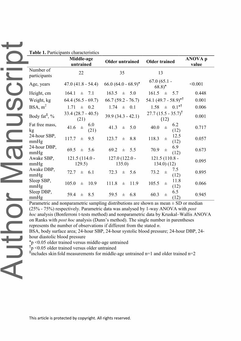
Participants Characteristics and Maximal Exercise Capacity
Older trained women were lighter, had a smaller BSA, and were leaner, but had
similar FFM. 24-hour systolic blood pressure was higher in the older untrained women,
though this result was variable with an ANOVA p value of 0.057 while diastolic blood
pressure was not different among groups (ANOVA p =0.673) (Table 1).
Resting and Exercise Metabolic and Hemodynamics Variables
Rest: V̇O2 and Q̇ in absolute levels were not significantly different among groups
(ANOVA p ≥0.169); however, the older trained women had a lower heart rate and a larger
absolute and scaled stroke volume (versus middle-aged and older untrained all p ≤ 0.050;
ANOVA p ≤ 0.006). CPO was not significantly different among groups (ANOVA p =0.111).
Ea was lower in older trained compared to age-similar untrained women (p <0.001), while

This article is protected by copyright. All rights reserved.
SAC was higher with sedentary aging (older untrained versus middle-age untrained p =0.008;
ANOVA p ≤ 0.006). MAP and SVR were unaltered with aging or lifelong endurance exercise
(ANOVA p ≥ 0.406) (Table 2).
Exercise: V̇O2max relative to total body mass and FFM was higher in older trained
women compared to the untrained older (~58% and ~34% respectively, p <0.001) and
middle-age women (~31% and ~19% respectively, p ≤0.015) (ANOVA p <0.001; Table 3).
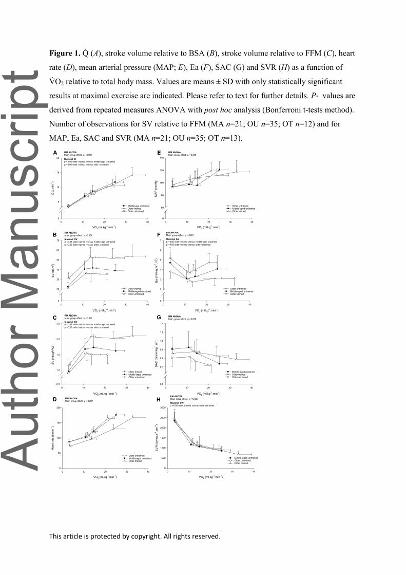
The mean group ΔQ̇ /ΔV̇O2 relation during incremental exercise was basically
superimposable among groups (Fig. 1A; Middle-aged untrained 5.8±1.0 L.L-1
; Older
untrained 5.7±0.9 L.L-1
; Older trained 6.3±0.9 L.L-1
; ANOVA p =0.235).
At maximal exercise, Q̇ was larger in the older trained compared to older (~40%, p
<0.001) and middle-aged (~17%, p <0.001) untrained women (RM ANOVA group main
effect p <0.001; Fig, 1A). Likewise, CPO at maximal exercise was significantly higher in
trained older women compared to the untrained groups (p <0.001 versus middle-aged and
older untrained; RM ANOVA group main effect p <0.001). Irrespective of unit, stroke
volume was shifted upward during all exercise conditions in the trained older women
compared to the middle-aged and older untrained women (RM ANOVA group main effect p
<0.001; Figs. 1B-C for stroke volume relative to BSA and FFM). Heart rate was shifted
rightward and down, reflecting a lower heart rate for any V̇O2 in the older trained women but
the overall heart rate response was not different among the groups (RM ANOVA group main
effect p =0.287; Fig. 1D).
Ea was shifted downward during submaximal exercise and was significantly lower at
maximal exercise in the older trained women compared to both middle-aged and older
untrained women (27% and 34% respectively p ≤ 0.012; RM ANOVA group main effect p
<0.001; Fig. 1F). There were no significant differences in MAP across the experimental
conditions, due in part to the variable exercise responses between groups (RM ANOVA

This article is protected by copyright. All rights reserved.
group main effect p =0.358; Fig. 1E). SVR was ~31% lower (p =0.023) in trained versus
untrained older women at maximal exercise (RM ANOVA group main effect p =0.035; Fig.
1H). Likewise, SAC was higher at rest and exercise in trained older women compared to their
age-similar untrained peers (RM ANOVA group main effect p =0.006); however, due to a
variable response this effect did not achieve statistical significance at maximal exercise (p
=0.450; Fig. 1G).
Systemic a-vDO2 was significantly higher (p ≤ 0.004 versus middle-age untrained and
older trained) at low-intensity, steady-state exercise in the older untrained women but there
were no group differences at moderate-intensity, steady-state or maximal exercise (all p ≥
0.109; RM ANOVA group main effect p =0.011; Table 3 for maximal exercise values).
Invasive Assessment of LV Function and Cardiac Compliance
Group-averaged Frank-Starling relations were not different among groups (ANOVA,
p =0.228; Fig. 2A); however, the preload recruitable stroke work relation was less steep in
older trained women compared to middle-aged and older untrained women (ANOVA p
<0.001; slope of the preload recruitable stroke work relation (ml x mmHg.ml.m2); median (25
- 75%) middle-aged untrained: 149.0 (132.0 - 220.0); older untrained 177.0 (144.0 - 265.0);
older trained; 117.0 (98.0 - 139.5); Fig. 2B; p ≤ 0.039 between groups)
The LV pressure-volume relationship was steeper and shifted upward and left in older
untrained compared to middle-age untrained women, indicating a stiffer (less
compliant/flexible) ventricle with sedentary aging (p =0.018; Table 4). In contrast, this
relationship was less steep and shifted rightward in older trained women, resulting in a
significantly greater ventricular chamber compliance compared to older untrained women (p
<0.001), while it was likely greater in older trained women compared to middle-aged
untrained women (p =0.053; Cohen’s d = 0.899) (Fig. 3A, median (25 - 75%): middle-age

This article is protected by copyright. All rights reserved.
untrained: 0.065 (0.049 - 0.080); older untrained: 0.085 (0.061 - 0.138); older trained: 0.047
(0.031 - 0.054)). Dynamic LV compliance during increased LV preload via saline infusion
was lower with sedentary aging (middle-aged untrained versus older untrained, p <0.001) but
was preserved in older trained women (p =1.000 versus middle-age untrained, ANOVA p
<0.001; Table 4).
When examined by the LV transmural pressure-volume relationship, which is more
representative of myocardial compliance without the effects of external constraint, the effect
of sedentary aging was less convincing (p =0.106) but remained significantly enhanced in the
trained versus untrained older women (p =0.007; ANOVA p =0.005; Fig. 3B).
Blood Volume and MRI measurements
Absolute levels of BV and plasma volume were not different among groups (ANOVA
p ≥ 0.868 for both); however, these variables were significantly larger in trained women
compared to their age-similar untrained peers when scaled relative to total body mass and
FFM (all p ≤ 0.041, ANOVA p ≤ 0.046; Table 5).
Irrespective of age, LVM index (g.m2) was significantly larger in trained women (p ≤
0.020 versus both untrained groups, ANOVA p =0.002); however, this difference was
eliminated when examined in absolute levels or scaled relative to FFM (ANOVA p > 0.338).
LVEDVI was significantly different between trained and untrained older women (p =0.004,
ANOVA p =0.005), while the LV mass-to-volume ratio was not significantly different among
groups (ANOVA p =0.637; Table 5).
DISCUSSION
There are two novel findings from this study. First, vigorous endurance exercise four
or more times per week for at least 25 years is associated with a larger maximal exercise

This article is protected by copyright. All rights reserved.
capacity, exercise stroke volume, LV size and BV, and greater ventricular chamber
compliance compared to age-similar untrained women, and in most instances, compared to
untrained women that were on average ~20 years younger. Second, there were also notable
favorable effects of lifelong exercise on arterial function (Ea, SAC) and heart rate during
exercise. Collectively, these findings highlight lifelong endurance exercise training as an
effective strategy for preserving or even enhancing cardiovascular structure and function with
aging in women.
Effect of Prolonged or Lifelong Endurance Exercise on V̇O2max in Women
Several investigators have previously reported the beneficial effect of lifelong or
sustained endurance exercise training on V̇O2max in middle-aged and older women
compared to age-similar controls (Ogawa et al., 1992; Stevenson et al., 1994; Jones et al.,
1997; Parker Jones et al., 1999; McCole et al., 2000; Dogra et al., 2012). In this current
study, V̇O2max scaled relative to total body mass or FFM in the older trained women was
substantially larger compared to untrained older and middle-aged women, reinforcing the
powerful effect of being physically active over several decades on reducing the unfavorable
effects of aging on cardiovascular structure and function and overall physical function.
Effect of Prolonged or Lifelong Exercise on Hemodynamics and Systemic a-vDO2 in
Women
There was a remarkably consistent V̇O2-Q̇ relationship during exercise in the current
younger and older cohorts. Similar observations have been reported in endurance trained
young and older male and female athletes (Proctor et al., 1998) and sedentary and endurance
trained young men and women (Fu & Levine, 2005). These findings suggest that the complex
integration of cardiac and non-cardiac regulatory mechanisms that impact central

This article is protected by copyright. All rights reserved.
hemodynamics during exercise are remarkably well preserved even after several decades of
biological aging irrespective of training status.
The current findings support previous investigations (Ogawa et al., 1992; McCole et
al., 2000; Dogra et al., 2012) demonstrating a substantially larger sub-maximal and maximal
exercise stroke volume in endurance trained older women compared to age-similar controls.
A larger LVEDV and thus greater recruitment of the Frank-Starling mechanism is recognized
as the primary factor that facilitates the larger exercise stroke volume in highly trained older
men (Fleg et al., 1994; Seals et al., 1994; Schulman et al., 1996; Hagberg et al., 1998).
Unfortunately, we did not assess ventricular volumes or dynamic diastolic filling during
exercise in these cohorts; however, we speculate that a larger more compliant ventricle
coupled with an expanded blood volume observed in the older trained women is a key
mechanism to enhance ventricular filling during exercise (Hagberg et al., 1998).
Enhanced ventricular-arterial coupling with increased preload (Shibata et al., 2008;
Hieda et al., 2018) and potentially more vigorous ventricular suction with exercise (Popovic
et al., 2006) could also contribute to a larger stroke volume response in endurance trained
older women. Ea, an index of total arterial load on the LV, was lower at rest and during
exercise in the trained older women compared to middle-aged and older untrained women.
An observation of a lower Ea during exercise in healthy older adults has been previously
reported with lifelong exercise (Carrick-Ranson et al., 2014) and in response to a 12-month
endurance training program in previously untrained older men and women (Shibata &
Levine, 2012b; Carrick-Ranson et al., 2016). From a physiological and clinical perspective,
these findings are important as Ea during exercise is adversely altered with healthy aging
(Najjar et al., 2004) and is abnormal in women with cardiovascular disease (systolic arterial
hypertension) (Chantler et al., 2008; Park et al., 2008).

This article is protected by copyright. All rights reserved.
Based on the current findings, the lower Ea in middle-aged and older trained women
likely reflects primarily a lower SVR and to a smaller degree a higher SAC during
submaximal and maximal exercise. Several reports from our laboratory have reported that
invasive and non-invasive indices of ventricular-arterial coupling (v-a coupling) during
physiologic stress are greater with long-term training in older adults (Shibata et al., 2008;
Shibata & Levine, 2012a; Hieda et al., 2018; Shibata et al., 2018). However, Shibata &
Levine (Shibata & Levine, 2012b) previously demonstrated that training-related changes in
Ea and SAC in previously untrained older men and women were largely reflective of a larger
stroke volume, as Ea and SAC were restored to pre-training levels when stroke volume was
reduced by lower body negative pressure; highlighting the critical role of stroke volume on
training-related changes in these indices of effective elastance and LV afterload.
A larger stroke volume during exercise with lifelong or prolonged training is an
important observation (Ogawa et al., 1992; McCole et al., 1999, 2000; Dogra et al., 2012), as
stroke volume is reported to be only modestly increased with relatively short- (12 weeks)
(Murias et al., 2010) and long-term (9-12 months) (Spina et al., 1993) endurance exercise
training in previously sedentary older women. These findings are supported by reports of
unaltered LV systolic and diastolic function during various physiological provocations after
several months of exercise training in older women (Spina et al., 1996; Spina et al., 2000).
Collectively, these findings reinforce the importance of engaging in committed levels of
endurance exercise throughout adulthood on cardiovascular function and exercise
performance during latter life in women (Bhella et al., 2014; Carrick-Ranson et al., 2014;
Hieda et al., 2018; Shibata et al., 2018).
Systemic a-vDO2 increased during exercise in all groups and was unaffected by
training status at maximal exercise in the current cohorts. This finding is consistent with
some (McCole et al., 2000; Dogra et al., 2012) but not all previous studies (Ogawa et al.,

This article is protected by copyright. All rights reserved.
1992) in endurance trained and untrained women, which may reflect measurement techniques
or potentially the age or previous training history of the trained participants. The importance
of systemic a-vDO2 to exercise performance is particularly pertinent in older women, as this
characteristic may represent the principal mechanism for an increased V̇O2max after several
months of exercise training in this population (Spina et al., 1993; Murias et al., 2010).
Improved redistribution of blood flow to contracting skeletal muscle, increased skeletal
muscle capillarization, and proliferation of mitochondrial size and number and well as
aerobic metabolism enzymes may contribute to a higher systemic and regional oxygen
extraction and utilization after exercise training in older women; however, to date, these
factors have rarely been examined with relatively short- or long-term training in women
(Coggan et al., 1992).
Effects of Prolonged or Lifelong Exercise on LV Morphology in Women
While the “athletes heart syndrome” has been reported in young male (Scharhag et
al., 2002; Utomi et al., 2013) and female endurance athletes (Riley-Hagan et al., 1992), and
male endurance Masters athletes (Heath et al., 1981; Seals et al., 1994; Bohm et al., 2016),
there is only very limited information on the effect of lifelong endurance exercise on cardiac
morphology in Master women athletes. Hagmar and colleagues (Hagmar et al., 2005)
reported a larger LV chamber size and LVEDV in former elite endurance athletes who were
now recreational endurance athletes compared to controls; however, neither absolute LVM
nor LVMI were significantly different between athletes and controls. A extremely large
LVMI (mean: 165.1 g/m2) was reported in post-menopausal marathon runners (mean age ~55
years, range 45-69 years); however, no control group was examined, making it difficult to
interpret these findings (Knebel et al., 2014).

This article is protected by copyright. All rights reserved.
In this current study, we found a ~16% larger LVM when scaled relative to BSA.
Likewise, LVEDVI was larger in trained older women while the LV mass-to-volume ratio
was not significantly different between groups. These findings suggest that eccentric and
balanced physiologic hypertrophy is the primary mechanism of adaptation to lifelong
endurance exercise in women.
Effect of Prolonged or Lifelong Exercise on Invasive Measures of LV Performance and
Compliance in Women
We have previously shown that sedentary but otherwise healthy aging is associated
with a reduction in LV chamber compliance and distensibility (Arbab-Zadeh et al., 2004;
Fujimoto et al., 2012); however, this stiffening was prevented in male and female Masters
athletes (Arbab-Zadeh et al., 2004). An important observation of the present study is that
habitual and vigorous endurance exercise over several decades enhances LV compliance and
distensibility in older women to a degree that is similar or even surpasses that observed in
women with a mean age that is ~20 years younger. The enhanced cardiac compliance
observed in lifelong exercising women compared to middle-aged sedentary individuals
reinforces the powerful effect of lifelong exercise on cardiac compliance with senescence
(Arbab-Zadeh et al., 2004; Bhella et al., 2014).
In contrast to the marked effect of lifelong exercise, 1-year of dynamic exercise in
previously sedentary older (>60 years) men and women did not improve cardiac compliance
(Fujimoto et al., 2010) even when combined with an advanced glycation end-product cross-
link breaker (Alagebrium) (Fujimoto et al., 2013). However, a recent investigation showed
that sustained (2 years) endurance exercise significantly improved LV chamber compliance
in middle-aged adults (Howden et al., 2018). Collectively, these findings suggest that

This article is protected by copyright. All rights reserved.
exercise may need to be initiated earlier in life and performed over a longer duration to
enhance or preserve cardiac compliance with aging in women as well as men.
The preservation of cardiac compliance with lifelong endurance training may be
attributable to changes in the intrinsic viscoelastic properties of the myocardium, cardiac
morphology or pericardial constraint. In the present study, compliance remained superior in
trained compared to untrained older women after accounting for pericardial constraint
suggesting other myocardial factors contribute to the enhanced cardiac compliance in these
participants.
Effect of Prolonged or Lifelong Exercise on Blood Volume in Women
Previous cross-sectional studies have reported that absolute and body size and
composition relative levels of BV and plasma volume are larger in recreationally active older
females (Jones et al., 1997), and middle-aged and older women distance runners (Stevenson
et al., 1994) and swimmers (Parker Jones et al., 1999) compared to their untrained peers. In
this present study, only BV and plasma volume relative to BSA and FFM were significantly
larger in the trained compared to untrained older women. This current finding likely reflects
smaller absolute levels but comparable BSA and FFM relative levels of BV and plasma
volume to those reported in middle-aged and older women athletes (Stevenson et al., 1994).
These findings of a larger BV in trained older women may be important given that
BV does not appear to increase with relatively short-term training (3 months) in previously
sedentary older women (Stachenfeld et al., 1998; Katyal et al., 2003). These observations
cannot be explained by a suboptimal training stimulus as there was a relatively large increase
(14 - 17%) in V̇O2max relative to total body mass post-training in these investigations. The
mechanism(s) that underpin this blunted adaptation in BV in older women are not well
defined. Based on findings in older men, changes in the regulation of the plasma protein
This article is protected by copyright. All rights reserved.
albumin, which causes a fluid shift from the interstitial to intravascular fluid space, is
implicated in the diminished adaptation in plasma volume after exercise training in older
adults (Zappe et al., 1996; Okazaki et al., 2002). Given the tightly coupled relationship
between BV expansion and exercise stroke volume with endurance exercise (Bonne et al.,
2014), these findings potentially indicate that a significant period of intensive exercise may
be required to elicit favorable blood volume adaptations in women particularly if exercise is
initiated later in life.
Effect of Menopausal Status and Menopausal Hormone Therapy on CV Structure and
Function during Exercise
Given the board chronological ages of our untrained middle-aged group, this cohort
included women who were premenopausal, perimenopausal and postmenopausal. Alterations
in autonomic nervous system activity, endothelial function and cardiovascular tissue stiffness
with menopause could all negatively impact central and peripheral hemodynamics during
exercise (Taddei et al., 1996; Staessen et al., 2001; Barnes et al., 2014). Several recent
elegant studies in premenopausal and recently postmenopausal women reported no
differences in V̇O2max relative to total body mass (Nyberg et al., 2017) and LV mass and
ventricular dimensions (Egelund et al., 2017) prior to commencing a 12-week exercise
training program. In premenopausal and postmenopausal women, Q̇ and stroke volume were
not significantly different at submaximal exercise workloads (50 watts and 60% of V̇O2max)
(Green et al., 2002). Unfortunately, we did not examine menopausal status comprehensively
in our middle-aged women at the time of study recruitment; however, there were no
significant difference in primary and secondary outcome measures (maximal Q̇ and stroke
volume, LV mass and LVEDV, and LV compliance) when the middle-aged women were
split into two chronological age ranges; aged 35-49 (mean age: 42.1 years) and 50-59 years

This article is protected by copyright. All rights reserved.
(mean age: 55.4 years) (data not shown). Nevertheless, we cannot completely exclude the
possibility that menopausal status did not influence our exercise and LV function and
compliance results.
We are unable to comment on the potential modulating effects of menopausal
hormone replacement therapy (HRT) or hormonal contraceptives on our findings, as these
factors were not extensively examined in the current cohorts of women. While limited to only
small cohorts of women, cross-sectional investigations have reported that short or long-term
HRT did not elicit notable effects on V̇O2, Q̇, stroke volume or blood pressures during
submaximal or maximal exercise in asymptomatic untrained or highly-trained women (Fleg
et al., 1995; McCole et al., 1999, 2000; Kirwan et al., 2004). In response to exercise training,
adaptations in submaximal exercise Q̇ and stroke volume were not significantly influenced by
HRT (O'Donnell et al., 2009).
Several pieces of evidence indicate the important role of oestrogens in endothelial-
mediated macrovascular function in previously sedentary postmenopausal women (Pierce et
al., 2011; Moreau et al., 2013) and more recently for micro- and macrovascular function in
response to habitual exercise (Santos-Parker et al., 2017). However, it is unclear whether
these adaptations confer an improvement in central circulatory function during large muscle
mass exercise (Kirwan et al., 2004). Future research is required to establish the role of HRT
and hormonal contraceptives on CV structure and function and how this information could be
used in the development of exercise-based strategies to optimize CV health in women.
Study Limitations
There are several limitations that should be acknowledged. Similar to our previous
investigation in a larger cohort (Carrick-Ranson et al., 2014), LV volumes during exercise

This article is protected by copyright. All rights reserved.
were not collected, which would have allowed for a more comprehensive description of aging
and lifelong exercise-related changes in LV filling function and v-a coupling during exercise.
A relatively small sample size combined with the mixture of committed exercisers
and Master athletes could be viewed as a limitation. However, comprehensive physiological
investigations in trained and untrained older women are rare and therefore the current
findings are novel. Nevertheless, the small sample size combined with a reduced number of
observations for certain outcomes measures (BV and LVM for example) may have resulted in
a type-2 error.
The lack of a younger endurance trained group could be considered a limitation;
however, the primary purpose of this analysis was to examine the effects of sedentary aging
and lifelong exercise on cardiovascular structure and function and thus we do not believe this
addition would have significantly strengthened the current findings.
Finally, we did not examine factors such as genetics, HRT, hormonal contraceptives,
menopausal status or other lifestyle factors such as diet that may influence cardiovascular
structure and function in the current cohorts; therefore, we cannot exclude the possibility that
the improved exercise in our trained older women may be related to factors other than
lifelong exercise.
Conclusion
In summary, the current findings suggest that lifelong exercise of four or more
exercise sessions weekly is associated with substantial effects on maximal exercise capacity,
exercise hemodynamics and heart rate control, LV mass and compliance, and total blood
volumes in women. Exercise training should be considered a key strategy to prevent
cardiovascular disease with aging in women as well as men.

This article is protected by copyright. All rights reserved.
Additional Information
Disclosures: The authors have no conflicts to disclose.
Funding support: This project was supported by the National Institutes of Health
(ref#AG17479).
Authors Contributions: Exercise function, blood volume and catheterization experiments
were performed at the Institute for Exercise and Environmental Medicine (IEEM) in Dallas,
TX; MRI scans were performed at the University of Texas Southwestern Medical Center in
Dallas, TX. GCR and BDL designed the experiments; GCR, PSB, SS, SS, NF, JLH and BDL
performed the experiments; GCR, NMS, EJH and SS analyzed the data; GCR, NMS and EJH
drafted the manuscript and BDL edited and revised the manuscript. All authors have
approved the final version of the manuscript and agree to be accountable for all aspects of the
work. All persons designated as authors qualify for authorship, and all those who qualify for
authorship are listed.
\

This article is protected by copyright. All rights reserved.
Arbab-Zadeh A, Dijk E, Prasad A, Fu Q, Torres P, Zhang R, Thomas JD, Palmer D & Levine BD.
(2004). Effect of aging and physical activity on left ventricular compliance. Circulation 110,
1799-1805.
Astrand I, Astrand PO, Hallback I & Kilbom A. (1973). Reduction in maximal oxygen uptake with
age. J Appl Physiol 35, 649-654.
Barnes JN, Hart EC, Curry TB, Nicholson WT, Eisenach JH, Wallin BG, Charkoudian N & Joyner
MJ. (2014). Aging enhances autonomic support of blood pressure in women. Hypertension
63, 303-308.
Belenkie I, Kieser TM, Sas R, Smith ER & Tyberg JV. (2002). Evidence for left ventricular constraint
during open heart surgery. Can J Cardiol 18, 951-959.
Bhella PS, Hastings JL, Fujimoto N, Shibata S, Carrick-Ranson G, Palmer MD, Boyd KN, Adams-
Huet B & Levine BD. (2014). Impact of lifelong exercise "dose" on left ventricular
compliance and distensibility. J Am Coll Cardiol 64, 1257-1266.
Blair SN, Kohl HW, 3rd, Paffenbarger RS, Jr., Clark DG, Cooper KH & Gibbons LW. (1989).
Physical fitness and all-cause mortality. A prospective study of healthy men and women.
JAMA 262, 2395-2401.
Bohm P, Schneider G, Linneweber L, Rentzsch A, Kramer N, Abdul-Khaliq H, Kindermann W,
Meyer T & Scharhag J. (2016). Right and Left Ventricular Function and Mass in Male Elite
Master Athletes: A Controlled Contrast-Enhanced Cardiovascular Magnetic Resonance
Study. Circulation 133, 1927-1935.
Bonne TC, Doucende G, Fluck D, Jacobs RA, Nordsborg NB, Robach P, Walther G & Lundby C.
(2014). Phlebotomy eliminates the maximal cardiac output response to six weeks of exercise
training. Am J Physiol Regul Integr Comp Physiol 306, R752-760.
Burge CM & Skinner SL. (1995). Determination of hemoglobin mass and blood volume with CO:
evaluation and application of a method. J Appl Physiol (1985) 79, 623-631.
Carrick-Ranson G, Fujimoto N, Shafer KM, Hastings JL, Shibata S, Palmer MD, Boyd K & Levine
BD. (2016). The effect of 1 year of Alagebrium and moderate-intensity exercise training on
left ventricular function during exercise in seniors: a randomized controlled trial. J Appl
Physiol (1985) 121, 528-536.
Carrick-Ranson G, Hastings JL, Bhella PS, Fujimoto N, Shibata S, Palmer MD, Boyd K, Livingston
S, Dijk E & Levine BD. (2014). The effect of lifelong exercise dose on cardiovascular
function during exercise. J Appl Physiol (1985) 116, 736-745.
Carrick-Ranson G, Hastings JL, Bhella PS, Shibata S, Fujimoto N, Palmer D, Boyd K & Levine BD.
(2012). The Effect of Age-related Differences in Body Size and Composition on
Cardiovascular Determinants of VO2max. J Gerontol A Biol Sci Med Sci.

This article is protected by copyright. All rights reserved.
Chantler PD, Melenovsky V, Schulman SP, Gerstenblith G, Becker LC, Ferrucci L, Fleg JL, Lakatta
EG & Najjar SS. (2008). The sex-specific impact of systolic hypertension and systolic blood
pressure on arterial-ventricular coupling at rest and during exercise. American Journal of
Physiology-Heart and Circulatory Physiology 295, H145-H153.
Chemla D, Hebert JL, Coirault C, Zamani K, Suard I, Colin P & Lecarpentier Y. (1998). Total arterial
compliance estimated by stroke volume-to-aortic pulse pressure ratio in humans. Am J
Physiol 274, H500-505.
Chen CH, Fetics B, Nevo E, Rochitte CE, Chiou KR, Ding PA, Kawaguchi M & Kass DA. (2001).
Noninvasive single-beat determination of left ventricular end-systolic elastance in humans. J
Am Coll Cardiol 38, 2028-2034.
Chen J, Das S, Barlow CE, Grundy S & Lakoski SG. (2010). Fitness, fatness, and systolic blood
pressure: data from the Cooper Center Longitudinal Study. Am Heart J 160, 166-170.
Coggan AR, Spina RJ, King DS, Rogers MA, Brown M, Nemeth PM & Holloszy JO. (1992). Skeletal
muscle adaptations to endurance training in 60- to 70-yr-old men and women. J Appl Physiol
(1985) 72, 1780-1786.
Cooke GA, Marshall P, al-Timman JK, Wright DJ, Riley R, Hainsworth R & Tan LB. (1998).
Physiological cardiac reserve: development of a non-invasive method and first estimates in
man. Heart 79, 289-294.
Davy KP & Seals DR. (1994). Total blood volume in healthy young and older men. J Appl Physiol
(1985) 76, 2059-2062.
Dogra S, Spencer MD & Paterson DH. (2012). Higher cardiorespiratory fitness in older trained
women is due to preserved stroke volume. J Sport Sci Med 11, 745-750.
Egelund J, Jorgensen PG, Mandrup CM, Fritz-Hansen T, Stallknecht B, Bangsbo J, Nyberg M &
Hellsten Y. (2017). Cardiac Adaptations to High-Intensity Aerobic Training in
Premenopausal and Recent Postmenopausal Women: The Copenhagen Women Study.
Journal of the American Heart Association 6.
Fleg J, Morrell C, Bos A, Brant L, Talbot L, Wright J & Lakatta E. (2005). Accelerated longitudinal
decline of aerobic capacity in healthy older adults. Circulation 112, 674-682.
Fleg J, O'Connor F, Gerstenblith G, Becker L, Clulow J, Schulman S & Lakatta E. (1995). Impact of
age on the cardiovascular response to dynamic upright exercise in healthy men and women. J
Appl Physiol 78, 890-900.
Fleg J, Schulman S, O'Connor F, Gerstenblith G, Becker L, Fortney S, Goldberg A & Lakatta E.
(1994). Cardiovascular responses to exhaustive upright cycle exercise in highly trained older
men. J Appl Physiol 77, 1500-1506.

This article is protected by copyright. All rights reserved.
Fu Q & Levine BD. (2005). Cardiovascular response to exercise in women. Med Sci Sports Exerc 37,
1433-1435.
Fujimoto N, Hastings JL, Bhella PS, Shibata S, Gandhi NK, Carrick-Ranson G, Palmer D & Levine
BD. (2012). Effect of ageing on left ventricular compliance and distensibility in healthy
sedentary humans. J Physiol 590, 1871-1880.
Fujimoto N, Hastings JL, Carrick-Ranson G, Shafer KM, Shibata S, Bhella PS, Abdullah SM, Barkley
KW, Adams-Huet B, Boyd KN, Livingston SA, Palmer D & Levine BD. (2013).
Cardiovascular effects of 1 year of alagebrium and endurance exercise training in healthy
older individuals. Circ Heart Fail 6, 1155-1164.
Fujimoto N, Prasad A, Hastings JL, Arbab-Zadeh A, Bhella PS, Shibata S, Palmer D & Levine BD.
(2010). Cardiovascular effects of 1 year of progressive and vigorous exercise training in
previously sedentary individuals older than 65 years of age. Circulation 122, 1797-1805.
Gates PE, Tanaka H, Graves J & Seals DR. (2003). Left ventricular structure and diastolic function
with human ageing. Relation to habitual exercise and arterial stiffness. Eur Heart J 24, 2213-
2220.
Gore CJ, Rodriguez FA, Truijens MJ, Townsend NE, Stray-Gundersen J & Levine BD. (2006).
Increased serum erythropoietin but not red cell production after 4 wk of intermittent
hypobaric hypoxia (4,000-5,500 m). J Appl Physiol (1985) 101, 1386-1393.
Green JS, Stanforth PR, Gagnon J, Leon AS, Rao DC, Skinner JS, Bouchard C, Rankinen T &
Wilmore JH. (2002). Menopause, estrogen, and training effects on exercise hemodynamics:
the HERITAGE study. Med Sci Sports Exerc 34, 74-82.
Hagberg J, Allen W, Seals D, Hurley B, Ehsani A & Holloszy J. (1985). A hemodynamic comparison
of young and older endurance athletes during exercise. J Appl Physiol 58, 2041-2046.
Hagberg J, Goldberg A, Lakatta L, O'Connor F, Becker L, Lakatta E & Fleg J. (1998). Expanded
blood volumes contribute to the increased cardiovascular performance of endurance-trained
older men. J Appl Physiol 85, 484-489.
Hagberg JM, Ehsani AA & Holloszy JO. (1983). Effect of 12 months of intense exercise training on
stroke volume in patients with coronary artery disease. Circulation 67, 1194-1199.
Hagmar M, Hirschberg AL, Lindholm C, Schenck-Gustafsson K & Eriksson MJ. (2005). Athlete's
heart in postmenopausal former elite endurance female athletes. Clin J Sport Med 15, 257-
262.
Heath GW, Hagberg JM, Ehsani AA & Holloszy JO. (1981). A physiological comparison of young
and older endurance athletes. J Appl Physiol Respir Environ Exerc Physiol 51, 634-640.

This article is protected by copyright. All rights reserved.
Hieda M, Howden E, Shibata S, Fujimoto N, Bhella PS, Hastings JL, Tarumi T, Sarma S, Fu Q,
Zhang R & Levine BD. (2018). Impact of Lifelong Exercise Training Dose on Ventricular-
Arterial Coupling. Circulation 138, 2638-2647.
Howden EJ, Perhonen M, Peshock RM, Zhang R, Arbab-Zadeh A, Adams-Huet B & Levine BD.
(2015). Females have a blunted cardiovascular response to one year of intensive supervised
endurance training. J Appl Physiol (1985) 119, 37-46.
Howden EJ, Sarma S, Lawley JS, Opondo M, Cornwell W, Stoller D, Urey MA, Adams-Huet B &
Levine BD. (2018). Reversing the Cardiac Effects of Sedentary Aging in Middle Age-A
Randomized Controlled Trial: Implications For Heart Failure Prevention. Circulation 137,
1549-1560.
Jarvis SS, Levine BD, Prisk GK, Shykoff BE, Elliott AR, Rosow E, Blomqvist CG & Pawelczyk JA.
(2007). Simultaneous determination of the accuracy and precision of closed-circuit cardiac
output rebreathing techniques. J Appl Physiol (1985) 103, 867-874.
Jenkins C, Bricknell K, Chan J, Hanekom L & Marwick TH. (2007). Comparison of two- and three-
dimensional echocardiography with sequential magnetic resonance imaging for evaluating left
ventricular volume and ejection fraction over time in patients with healed myocardial
infarction. Am J Cardiol 99, 300-306.
Jones PP, Davy KP, DeSouza CA, van Pelt RE & Seals DR. (1997). Absence of age-related decline in
total blood volume in physically active females. Am J Physiol 272, H2534-2540.
Jones SC, Bilous M, Winship S, Finn P & Goodwin J. (2004). Validation of the OSCAR 2
oscillometric 24-hour ambulatory blood pressure monitor according to the International
Protocol for the validation of blood pressure measuring devices. Blood Press Monit 9, 219-
223.
Katyal S, Freeman M, Miller JA & Thomas SG. (2003). Short-term aerobic training and circulatory
function in women: age and hormone-replacement therapy. Clin Sci (Lond) 104, 267-273.
Kirwan LD, MacLusky NJ, Shapiro HM, Abramson BL, Thomas SG & Goodman JM. (2004). Acute
and chronic effects of hormone replacement therapy on the cardiovascular system in healthy
postmenopausal women. J Clin Endocrinol Metab 89, 1618-1629.
Knebel F, Spethmann S, Schattke S, Dreger H, Schroeckh S, Schimke I, Hattasch R, Makauskiene R,
Kleczka J, Sanad W, Lock J, Brechtel L, Baumann G & Borges AC. (2014). Exercise-induced
changes of left ventricular diastolic function in postmenopausal amateur marathon runners:
assessment by echocardiography and cardiac biomarkers. Eur J Prev Cardiol 21, 782-790.
Levine BD. (1993). Regulation of central blood volume and cardiac filling in endurance athletes: the
Frank-Starling mechanism as a determinant of orthostatic tolerance. Med Sci Sports Exerc 25,
727-732.

This article is protected by copyright. All rights reserved.
McCole SD, Brown MD, Moore GE, Zmuda JM, Cwynar JD & Hagberg JM. (1999). Cardiovascular
hemodynamics with increasing exercise intensities in postmenopausal women. J Appl Physiol
(1985) 87, 2334-2340.
McCole SD, Brown MD, Moore GE, Zmuda JM, Cwynar JD & Hagberg JM. (2000). Enhanced
cardiovascular hemodynamics in endurance-trained postmenopausal women athletes. Med Sci
Sports Exerc 32, 1073-1079.
Mirsky I. (1984). Assessment of diastolic function: suggested methods and future considerations.
Circulation 69, 836-841.
Moreau KL, Stauffer BL, Kohrt WM & Seals DR. (2013). Essential role of estrogen for
improvements in vascular endothelial function with endurance exercise in postmenopausal
women. J Clin Endocrinol Metab 98, 4507-4515.
Murias JM, Kowalchuk JM & Paterson DH. (2010). Mechanisms for increases in V O2max with
endurance training in older and young women. Med Sci Sports Exerc 42, 1891-1898.
Najjar SS, Schulman SP, Gerstenblith G, Fleg JL, Kass DA, O'Connor F, Becker LC & Lakatta EG.
(2004). Age and gender affect ventricular-vascular coupling during aerobic exercise. J Am
Coll Cardiol 44, 611-617.
Nyberg M, Egelund J, Mandrup CM, Andersen CB, Hansen K, Hergel IF, Valbak-Andersen N,
Frikke-Schmidt R, Stallknecht B, Bangsbo J & Hellsten Y. (2017). Leg vascular and skeletal
muscle mitochondrial adaptations to aerobic high-intensity exercise training are enhanced in
the early postmenopausal phase. J Physiol 595, 2969-2983.
O'Donnell E, Kirwan LD & Goodman JM. (2009). Aerobic exercise training in healthy
postmenopausal women: effects of hormone therapy. Menopause-the Journal of the North
American Menopause Society 16, 770-776.
Ogawa T, Spina R, Martin W, Kohrt W, Schechtman K, Holloszy J & Ehsani A. (1992). Effects of
Aging, Sex and Physical Training on Cardiovascular Responses to Exercise. Circulation 86,
494-503.
Okazaki K, Kamijo Y, Takeno Y, Okumoto T, Masuki S & Nose H. (2002). Effects of exercise
training on thermoregulatory responses and blood volume in older men. J Appl Physiol (1985)
93, 1630-1637.
Park S, Ha JW, Shim CY, Choi EY, Kim JM, Ahn JA, Lee SW, Rim SJ & Chung N. (2008). Gender-
related difference in arterial elastance during exercise in patients with hypertension.
Hypertension 51, 1163-1169.

This article is protected by copyright. All rights reserved.
Parker Jones P, Davy KP, Desouza CA & Tanaka H. (1999). Total blood volume in endurance-trained
postmenopausal females: relation to exercise mode and maximal aerobic capacity. Acta
Physiol Scand 166, 327-333.
Paterson DH, Govindasamy D, Vidmar M, Cunningham DA & Koval JJ. (2004). Longitudinal study
of determinants of dependence in an elderly population. J Am Geriatr Soc 52, 1632-1638.
Peshock RM, Willett DL, Sayad DE, Hundley WG, Chwialkowski MC, Clarke GD & Parkey RW.
(1996). Quantitative MR imaging of the heart. Magn Reson Imaging Clin N Am 4, 287-305.
Pierce GL, Eskurza I, Walker AE, Fay TN & Seals DR. (2011). Sex-specific effects of habitual
aerobic exercise on brachial artery flow-mediated dilation in middle-aged and older adults.
Clin Sci (Lond) 120, 13-23.
Popovic ZB, Prasad A, Garcia MJ, Arbab-Zadeh A, Borowski A, Dijk E, Greenberg NL, Levine BD
& Thomas JD. (2006). Relationship among diastolic intraventricular pressure gradients,
relaxation, and preload: impact of age and fitness. Am J Physiol Heart Circ Physiol 290,
H1454-1459.
Proctor D, Beck K, Shen P, Eickhoff T, Halliwill J & Joyner M. (1998). Influence of age and gender
on cardiac output-VO2 relationships during submaximal cycle ergometer. J Appl Physiol 84,
599-605.
Redfield MM, Jacobsen SJ, Borlaug BA, Rodeheffer RJ & Kass DA. (2005). Age- and gender-related
ventricular-vascular stiffening: a community-based study. Circulation 112, 2254-2262.
Riley-Hagan M, Peshock RM, Straygundersen J, Katz J, Ryschon TW & Mitchell JH. (1992). Left-
Ventricular Dimensions and Mass Using Magnetic-Resonance-Imaging in Female Endurance
Athletes. Am J Cardiol 69, 1067-1074.
Ross R, Blair SN, Arena R, Church TS, Despres JP, Franklin BA, Haskell WL, Kaminsky LA, Levine
BD, Lavie CJ, Myers J, Niebauer J, Sallis R, Sawada SS, Sui XM, Wisloff U, Hlth CLC,
Cardiology CC, Prevention CE, Nursing CCS, Genomics CF & Council S. (2016).
Importance of Assessing Cardiorespiratory Fitness in Clinical Practice: A Case for Fitness as
a Clinical Vital Sign A Scientific Statement From the American Heart Association.
Circulation 134, E653-E699.
Santos-Parker JR, Strahler TR, Vorwald VM, Pierce GL & Seals DR. (2017). Habitual aerobic
exercise does not protect against micro- or macrovascular endothelial dysfunction in healthy
estrogen-deficient postmenopausal women. J Appl Physiol (1985) 122, 11-19.
Scharhag J, Schneider G, Urhausen A, Rochette V, Kramann B & Kindermann W. (2002). Athlete's
heart: right and left ventricular mass and function in male endurance athletes and untrained
individuals determined by magnetic resonance imaging. J Am Coll Cardiol 40, 1856-1863.

This article is protected by copyright. All rights reserved.
Schulman SP, Fleg JL, Goldberg AP, Busby-Whitehead J, Hagberg JM, O'Connor FC, Gerstenblith
G, Becker LC, Katzel LI, Lakatta LE & Lakatta EG. (1996). Continuum of cardiovascular
performance across a broad range of fitness levels in healthy older men. Circulation 94, 359-
367.
Seals D, Hagberg J, Spina R, Rogers M, Schechtman K & Ehsani A. (1994). Enhanced left ventricular
performance in endurance trained older men. Circulation 89, 198-205.
Shibata S, Fujimoto N, Hastings JL, Carrick-Ranson G, Bhella PS, Hearon CM & Levine BD. (2018).
The effect of lifelong exercise frequency on arterial stiffness. Journal of Physiology-London
596, 2783-2795.
Shibata S, Hastings JL, Prasad A, Fu Q, Okazaki K, Palmer MD, Zhang R & Levine BD. (2008).
'Dynamic' Starling mechanism: effects of ageing and physical fitness on ventricular-arterial
coupling. J Physiol 586, 1951-1962.
Shibata S & Levine BD. (2012a). Biological aortic age derived from the arterial pressure waveform. J
Appl Physiol 110, 981-987.
Shibata S & Levine BD. (2012b). Effect of exercise training on biologic vascular age in healthy
seniors. Am J Physiol Heart Circ Physiol 302, H1340-1346.
Spina R, Ogawa T, Kohrt W, Holloszy J & Ehsani A. (1993). Differences in cardiovascular
adaptations to endurance exercise training between older men and women. J Appl Physiol 75,
849-855.
Spina R, Rashid S, Davila-Roman V & Ehsani A. (2000). Adaptations in beta-adrenergic
cardiovascular responses to training in older women. J Appl Physiol 89, 2300-2305.
Spina RJ, Miller TR, Bogenhagen WH, Schechtman KB & Ehsani AA. (1996). Gender-related
differences in left ventricular filling dynamics in older subjects after endurance exercise
training. J Gerontol A Biol Sci Med Sci 51, B232-237.
Stachenfeld NS, Mack GW, DiPietro L, Morocco TS, Jozsi AC & Nadel ER. (1998). Regulation of
blood volume during training in post-menopausal women. Med Sci Sports Exerc 30, 92-98.
Staessen JA, van der Heijden-Spek JJ, Safar ME, Den Hond E, Gasowski J, Fagard RH, Wang JG,
Boudier HA & Van Bortel LM. (2001). Menopause and the characteristics of the large
arteries in a population study. J Hum Hypertens 15, 511-518.
Stevenson ET, Davy KP & Seals DR. (1994). Maximal aerobic capacity and total blood volume in
highly trained middle-aged and older female endurance athletes. J Appl Physiol (1985) 77,
1691-1696.

This article is protected by copyright. All rights reserved.
Taddei S, Virdis A, Ghiadoni L, Mattei P, Sudano I, Bernini G, Pinto S & Salvetti A. (1996).
Menopause is associated with endothelial dysfunction in women. Hypertension 28, 576-582.
Taylor R, Chidley K, Goodwin J, Broeders M & Kirby B. (1993). Accutracker-Ii (Version 30/23)
Ambulatory Blood-Pressure Monitor - Clinical Validation Using the British-Hypertension-
Society and Association-for-the-Advancement-of-Medical-Instrumentation-Standards. J
Hypertens 11, 1275-1282.
Triebwasser JH, Johnson RL, Burpo RP, Campbell JC, Reardon WC & Blomqvist CG. (1977).
Noninvasive Determination of Cardiac-Output by a Modified Acetylene Rebreathing
Procedure Utilizing Mass-Spectrometer Measurements. Aviat Space Environ Med 48, 203-
209.
Utomi V, Oxborough D, Whyte GP, Somauroo J, Sharma S, Shave R, Atkinson G & George K.
(2013). Systematic review and meta-analysis of training mode, imaging modality and body
size influences on the morphology and function of the male athlete's heart. Heart 99, 1727-
1733.
Victor RG, Haley RW, Willett DL, Peshock RM, Vaeth PC, Leonard D, Basit M, Cooper RS,
Iannacchione VG, Visscher WA, Staab JM, Hobbs HH & Dallas Heart Study I. (2004). The
Dallas Heart Study: a population-based probability sample for the multidisciplinary study of
ethnic differences in cardiovascular health. Am J Cardiol 93, 1473-1480.
Whalley GA, Doughty RN, Gamble GD, Oxenham HC, Walsh HJ, Reid IR & Baldi JC. (2004).
Association of fat-free mass and training status with left ventricular size and mass in
endurance-trained athletes. J Am Coll Cardiol 44, 892-896.
Wiebe CG, Gledhill N, Jamnik VK & Ferguson S. (1999). Exercise cardiac function in young through
elderly endurance trained women. Med Sci Sports Exerc 31, 684-691.
Wilmore JH & Behnke AR. (1969). An anthropometric estimation of body density and lean body
weight in young men. J Appl Physiol 27, 25-31.
Zappe DH, Bell GW, Swartzentruber H, Wideman RF & Kenney WL. (1996). Age and regulation of
fluid and electrolyte balance during repeated exercise sessions. Am J Physiol 270, R71-79.
Graeme Carrick-Ranson has a great interest in the age-related changes in cardiovascular
physiology and how these alterations can be positively influenced by endurance exercise
training. Graeme completed his PhD at the University of Auckland (2004–2009) examining
the effects of healthy aging and endurance exercise training on left ventricular diastolic filling
during exercise. To extend his skills and knowledge in the assessment of cardiovascular
function, he did a Post-Doctoral research fellowship at the Institute for Exercise and
Environmental Medicine in Dallas (2009–2012). Currently, he is working at the University of

This article is protected by copyright. All rights reserved.
South Australia (2018-current), exploring his original research interests of aging and
exercise training.
This article is protected by copyright. All rights reserved.
Figure 1. Q̇ (A), stroke volume relative to BSA (B), stroke volume relative to FFM (C), heart
rate (D), mean arterial pressure (MAP; E), Ea (F), SAC (G) and SVR (H) as a function of
V̇O2 relative to total body mass. Values are means ± SD with only statistically significant
results at maximal exercise are indicated. Please refer to text for further details. P‐ values are
derived from repeated measures ANOVA with post hoc analysis (Bonferroni t-tests method).
Number of observations for SV relative to FFM (MA n=21; OU n=35; OT n=12) and for
MAP, Ea, SAC and SVR (MA n=21; OU n=35; OT n=13).
VO2 (ml.kg
-1.min
-1)
0 10 20 30 40
SV
(m
l.kg
FF
M-1
)
0.0
1.0
1.5
2.0
2.5
Older trained Middle-aged untrainedOlder untrained
VO2 (ml.kg
-1.min
-1)
0 10 20 30 40
SA
C (
ml.m
mH
g-1
.m2)
0.0
0.4
0.6
0.8
1.0
1.2
1.4
Middle-aged untrainedOlder trained Older untrained
VO2 (ml.kg
-1.min
-1)
0 10 20 30 40
SV
R (
dyne
s.s
-1.c
m-5
)
0
500
1000
1500
2000
2500
3000
Middle-aged untrainedOlder untrained Older trained
VO2 (ml.kg
-1.min
-1)
0 10 20 30 40
Ea
(m
mH
g.m
l-1.m
2)
0
2
3
4
5
6
7
Older untrained Middle-aged untrainedOlder trained
VO2 (ml.kg
-1.min
-1)
0 10 20 30 40
MA
P (
mm
Hg
)
0
60
80
100
120
140
Older untrained Middle-aged untrainedOlder trained
VO2 (ml.kg
-1.min
-1)
0 10 20 30 40
He
art
ra
te (
b.m
in-1
)
0
50
100
150
200
Older untrained Middle-aged untrainedOlder trained
VO2 (ml.kg
-1.min
-1)
0 10 20 30 40
SV
(m
l.m2)
0
20
30
40
50
60
70
Older trained Middle-aged untrainedOlder untrained
VO2 (ml.kg
-1.min
-1)
0 10 20 30 40
Q (
L.m
in-1
)
0
5
10
15
20
Middle-age untrainedOlder trained Older untrained
A
B
C
E
F
G
D
RM ANOVAMain group effect, p <0.001
RM ANOVAMain group effect, p =0.358
RM ANOVAMain group effect, p <0.001
RM ANOVAMain group effect, p <0.001
RM ANOVAMain group effect, p <0.001
RM ANOVAMain group effect, p =0.006
RM ANOVAMain group effect, p =0.287
RM ANOVAMain group effect, p =0.035 H
Maximal Qp <0.05 older trained versus middle-age untrained
p <0.05 older trained versus older untrained
Maximal SVp <0.05 older trained versus middle-age untrained
p <0.05 older trained versus older untrained
Maximal SVp <0.05 older trained versus middle-age untrained
p <0.05 older trained versus older untrained
Maximal Eap <0.05 older trained versus middle-age untrained
p <0.05 older trained versus older untrained
Maximal SVRp <0.05 older trained versus older untrained

This article is protected by copyright. All rights reserved.
Figure 2. Group-averaged Frank-Starling (A) and preload recruitable stroke work
relationship (B). P‐ values are derived from Kruskal–Wallis ANOVA on Ranks with post
hoc analysis (Dunn’s method).

This article is protected by copyright. All rights reserved.
Figure 3. Group-averaged LV pressure-volume relationship (A) and LV transmural pressure-
volume relationship (B). P‐ values are derived from Kruskal–Wallis ANOVA on Ranks
with post hoc analysis (Dunn’s method). Note the different scale used for Fig. 3A and B.
This article is protected by copyright. All rights reserved.
Table 1. Participants characteristics
Middle-age
untrained Older untrained Older trained
ANOVA p
value
Number of
participants 22 35 13
Age, years 47.0 (41.8 - 54.4) 66.0 (64.0 - 68.9)* 67.0 (65.1 -
68.8)* <0.001
Height, cm 164.1 ± 7.1 163.5 ± 5.0 161.5 ± 5.7 0.448
Weight, kg 64.4 (56.5 - 69.7) 66.7 (59.2 - 76.7) 54.1 (49.7 - 58.9)*† 0.001
BSA, m2 1.71 ± 0.2 1.74 ± 0.1 1.58 ± 0.1*† 0.006
Body fat‡, %
33.4 (28.7 - 40.5)
(21) 39.9 (34.3 - 42.1)
27.7 (15.5 - 35.7)†
(12) 0.001
Fat free mass,
kg 41.6 ±
6.0
(21) 41.3 ± 5.0 40.0 ±
6.2
(12) 0.717
24-hour SBP,
mmHg 117.7 ± 9.5 123.7 ± 8.8 118.3 ±
12.5
(12) 0.057
24-hour DBP,
mmHg 69.5 ± 5.6 69.2 ± 5.5 70.9 ±
6.9
(12) 0.673
Awake SBP,
mmHg
121.5 (114.0 -
129.5)
127.0 (122.0 -
135.0)
121.5 (110.8 -
134.0) (12) 0.095
Awake DBP,
mmHg 72.7 ± 6.1 72.3 ± 5.6 73.2 ±
7.5
(12) 0.895
Sleep SBP,
mmHg 105.0 ± 10.9 111.8 ± 11.9 105.5 ±
11.8
(12) 0.066
Sleep DBP,
mmHg 59.4 ± 8.5 59.5 ± 6.8 60.3 ±
6.5
(12) 0.945
Parametric and nonparametric sampling distributions are shown as mean ± SD or median
(25% - 75%) respectively. Parametric data was analysed by 1-way ANOVA with post
hoc analysis (Bonferroni t-tests method) and nonparametric data by Kruskal–Wallis ANOVA
on Ranks with post hoc analysis (Dunn’s method). The single number in parentheses
represents the number of observations if different from the stated n.
BSA, body surface area; 24-hour SBP, 24-hour systolic blood pressure; 24-hour DBP, 24-
hour diastolic blood pressure
*p <0.05 older trained versus middle-age untrained †p <0.05 older trained versus older untrained
‡includes skin
fold measurements for middle-age untrained n=1 and older trained n=2

This article is protected by copyright. All rights reserved.
Table 2. Resting upright metabolic and hemodynamic variables
Middle-age
untrained Older untrained Older trained
ANOVA
p value
V̇O2, ml.kg-
1.min
-1
3.1 (2.8 - 3.5) (21) 3.0 (2.9 - 3.3) 3.3 (2.8 - 3.7) 0.420
V̇O2,
ml.kgFFM-
1.min
-1
4.9 (4.5 - 5.0) (20) 4.8 (4.5 - 5.4) 4.6 (4.0 - 5.5) (12) 0.663
Q̇, L.min-1
3.3 (2.9 - 3.7) 3.1 (2.8 - 3.4) 3.4 (3.0 - 3.8) 0.169
QI, L.m2.min
-
1
1.90 (1.68 - 2.13) 1.80 (1.60 - 1.95) 2.20 (1.95 - 2.45)† <0.001
Q̇, ml.kgFFM-
1.min
-2
84.3 (67.4 - 88.7)
(21) 76.0 (70.0 - 83.0) 86.5 (77.3 - 91.8) (12) 0.048
CPO, watts 0.68 (0.59 - 0.74)
(20) 0.60 (0.50 - 0.70) 0.70 (0.60 - 0.80)
† 0.111
Heart rate,
b.min-1
86.8 ± 12.7 85.8 ± 10.1 72.2 ± 7.1*†
<0.001
SV, ml 37.8 (33.6 - 43.3) 36.2 (31.6 - 41.8) 46.1 (39.6 - 59.9)*† 0.006
SV, ml.m2 22.0 (19.9 - 25.0) 21.0 (18.4 - 24.2) 30.1 (25.7 - 38.2)*†
<0.001
SV,
ml.kgFFM-1
0.96 ± 0.18 0.90 ± 0.18 1.18 ±
0.18*† (12)
<0.001
MAP, mmHg 89.8 (86.4 - 99.7)
(20)
89.0 (84.3 -
101.0) 93.7 (90.6 - 99.7) 0.406
Ea,
mmHg.ml-
1.m
2
4.7 ± 1.1
(20) 5.2 ± 1.1 3.8 ± 0.7
† <0.001
SAC,
ml.mmHg-
1.m
2
0.8 (0.6 - 1.0) (20) 0.6 (0.4 - 0.7)* 0.7 (0.6 - 0.9) 0.006
SVR, dynes.s-
1.cm
-5
2243.2 (1964.7 -
2769.6) (20)
2353.4 (2054.6 -
2774.0)
2161.5 (2049.6 -
2592.4) 0.605
a-vDO2,
ml.100 ml-1
6.1 ± 1.0 6.7 ± 1.2 5.5 ± 0.9
† 0.003
Parametric and nonparametric sampling distributions are shown as mean ± SD or median
(25% - 75%) respectively. Parametric data was analysed by 1-way ANOVA with post
hoc analysis (Bonferroni t-tests method) and nonparametric data by Kruskal–Wallis ANOVA
on Ranks with post hoc analysis (Dunn’s method). The single number in parentheses
represents the number of observations if different from the stated n in Table 1.
V̇O2, oxygen uptake; Q̇, cardiac output; CPO, cardiac power; SV, stroke volume; MAP, mean
arterial pressure; Ea, effective arterial elastance; SAC, systemic arterial compliance; SVR,
systemic vascular resistance; a-vDO2, arterial-venous oxygen content difference
*p <0.05 older trained versus middle-age untrained †p <0.05 older trained versus older untrained

This article is protected by copyright. All rights reserved.
Table 3. Maximal exercise variables
Middle-age
untrained Older untrained Older trained
ANOVA p
value
V̇O2max,
L.min-1
1.6 ± 0.3 1.4 ± 0.2* 1.8 ± 0.2
† <0.001
V̇O2max,
ml.kg-1
.min-1
25.9 (21.6 - 27.8) 21.5 (18.5 - 24.3)* 34.0 (29.2 - 38.9)*†
<0.001
V̇O2max,
ml.kgFFM-
1.min
-1
39.1 ± 4.7
(21) 34.6 ± 5.4* 46.4 ±
6.5*† (12)
<0.001
Q̇I, L.m-2
.min-1
6.8 ± 1.0 5.8 ± 0.8* 8.9 ± 1.8*†
Q̇, ml.kgFFM-
1.min
-2
284.1 ± 38.1
(21) 245.1 ± 39.6* 349.8 ±
43.2*†
(12)
CPO, watts 3.1 ± 0.5
(21) 2.6 ± 0.5* 3.7 ±
0.9*† (12)
Systemic a-
vDO2, ml.100
ml
13.8 ± 1.8 14.3 ± 2.0 13.2 ± 2.1
Parametric and nonparametric sampling distributions are shown as mean ± SD or median
(25% - 75%) respectively. Parametric data was analysed by 1-way ANOVA with post
hoc analysis (Bonferroni t-tests method) and nonparametric data by Kruskal–Wallis ANOVA
on Ranks with post hoc analysis (Dunn’s method). Q̇I, Q̇, CPO, and systemic a-vDO2 results
are from repeated measures ANOVA’s with post hoc analysis (Bonferroni t-tests method).
The single number in parentheses represents the number of observations if different from the
stated n in Table 1.
V̇O2max, maximal oxygen uptake; Q̇ cardiac output; Q̇I, cardiac index; CPO, cardiac power;
systemic a-vDO2, systemic arteriovenous oxygen content difference
*p <0.05 older trained versus middle-age untrained †p <0.05 older trained versus older untrained
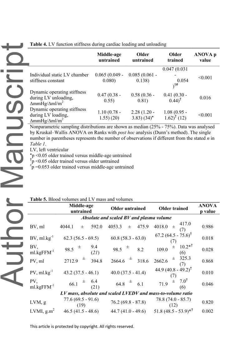
This article is protected by copyright. All rights reserved.
Table 4. LV function stiffness during cardiac loading and unloading
Middle-age
untrained
Older
untrained
Older
trained
ANOVA p
value
Individual static LV chamber
stiffness constant
0.065 (0.049 -
0.080)
0.085 (0.061 -
0.138)
0.047 (0.031
-
0.054
)†#
<0.001
Dynamic operating stiffness
during LV unloading,
∆mmHg/∆ml/m2
0.47 (0.38 -
0.55)
0.58 (0.36 -
0.81)
0.41 (0.30 -
0.44)†
0.016
Dynamic operating stiffness
during LV loading,
∆mmHg/∆ml/m2
1.10 (0.78 -
1.55) (20)
2.28 (1.20 -
3.83) (34)*
1.08 (0.95 -
1.62)† (12)
<0.001
Nonparametric sampling distributions are shown as median (25% - 75%). Data was analysed
by Kruskal–Wallis ANOVA on Ranks with post hoc analysis (Dunn’s method). The single
number in parentheses represents the number of observations if different from the stated n in
Table 1.
LV, left ventricular
*p <0.05 older trained versus middle-age untrained †p <0.05 older trained versus older untrained
#p =0.053 older trained versus middle-age untrained
Table 5. Blood volumes and LV mass and volumes
Middle-age
untrained Older untrained Older trained
ANOVA
p value
Absolute and scaled BV and plasma volume
BV, ml 4044.1 ± 592.0 4053.3 ± 475.9 4018.0 ± 417.0
(7) 0.986
BV, ml.kg-1
62.3 (56.5 - 69.5) 60.8 (58.3 - 63.0) 67.2 (64.5 - 75.6)
†
(7) 0.018
BV,
ml.kgFFM-1
98.5
± 9.4
(21) 98.5
± 8.2 109.0
± 10.2*†
(6) 0.028
PV, ml 2712.9 ±
394.8 2664.6 ±
318.6 2662.6 ± 325.3
(7) 0.868
PV, ml.kg-1
43.2 (37.5 - 46.1) 40.0 (37.5 - 41.4) 44.9 (40.8 - 49.2)
†
(7) 0.010
PV,
ml.kgFFM-1
66.1
± 6.4
(21) 64.8
± 6.1 71.9
± 7.0†
(6) 0.046
LV mass, absolute and scaled LVEDV and mass-to-volume ratio
LVM, g 77.6 (69.5 - 91.6)
(19) 76.2 (69.8 - 87.8)
78.8 (74.0 - 85.7)
(12) 0.820
LVMI, g.m2 46.5 (41.5 - 48.6) 44.7 (41.0 - 49.6) 51.8 (48.5 - 53.9)*
† 0.002
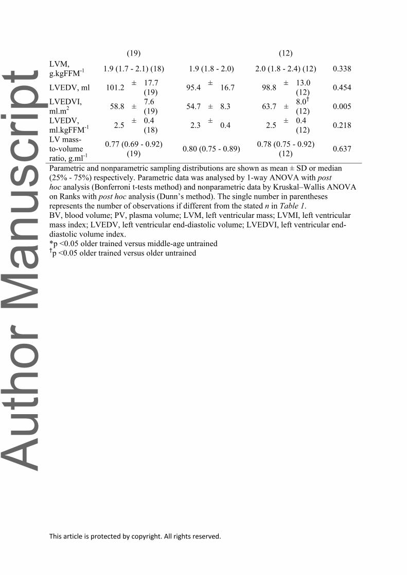
This article is protected by copyright. All rights reserved.
(19) (12)
LVM,
g.kgFFM-1 1.9 (1.7 - 2.1) (18) 1.9 (1.8 - 2.0) 2.0 (1.8 - 2.4) (12) 0.338
LVEDV, ml 101.2 ± 17.7
(19) 95.4
± 16.7 98.8
± 13.0
(12) 0.454
LVEDVI,
ml.m2
58.8 ± 7.6
(19) 54.7 ± 8.3 63.7 ±
8.0†
(12) 0.005
LVEDV,
ml.kgFFM-1
2.5
± 0.4
(18) 2.3
± 0.4 2.5
± 0.4
(12) 0.218
LV mass-
to-volume
ratio, g.ml-1
0.77 (0.69 - 0.92)
(19) 0.80 (0.75 - 0.89)
0.78 (0.75 - 0.92)
(12) 0.637
Parametric and nonparametric sampling distributions are shown as mean ± SD or median
(25% - 75%) respectively. Parametric data was analysed by 1-way ANOVA with post
hoc analysis (Bonferroni t-tests method) and nonparametric data by Kruskal–Wallis ANOVA
on Ranks with post hoc analysis (Dunn’s method). The single number in parentheses
represents the number of observations if different from the stated n in Table 1.
BV, blood volume; PV, plasma volume; LVM, left ventricular mass; LVMI, left ventricular
mass index; LVEDV, left ventricular end-diastolic volume; LVEDVI, left ventricular end-
diastolic volume index.
*p <0.05 older trained versus middle-age untrained †p <0.05 older trained versus older untrained

Minerva Access is the Institutional Repository of The University of Melbourne
Author/s:
Carrick-Ranson, G; Sloane, NM; Howden, EJ; Bhella, PS; Sarma, S; Shibata, S; Fujimoto, N;
Hastings, JL; Levine, BD
Title:
The effect of lifelong endurance exercise on cardiovascular structure and exercise function in
women.
Date:
2020-07
Citation:
Carrick-Ranson, G., Sloane, N. M., Howden, E. J., Bhella, P. S., Sarma, S., Shibata, S.,
Fujimoto, N., Hastings, J. L. & Levine, B. D. (2020). The effect of lifelong endurance
exercise on cardiovascular structure and exercise function in women.. J Physiol, 598 (13),
pp.2589-2605. https://doi.org/10.1113/JP278503.
Persistent Link:
http://hdl.handle.net/11343/275775
File Description:
Accepted version