The effect of high molecular phospholipase A2 inhibitors on 3T6 fibroblast proliferation
6
The effect of high molecular phospholipase A 2 inhibitors on 3T6 fibroblast proliferation Teresa Sanchez, Juan J. Moreno* Department of Physiology, Faculty of Pharmacy, Barcelona University, E-08028 Barcelona, Spain Received 24 March 2000; accepted 27 September 2000 Abstract Recently, we suggested that arachidonic acid and/or its cyclooxygenase pathway metabolites may be involved in regulating 3T6 fibroblast proliferation. In the present study we evaluate the role of high-molecular phospholipase A 2 (PLA 2 ) enzymes in the 3T6 fibroblast growth. Our results demonstrate that the cytosolic PLA 2 inhibitor, arachidonyl trifluoromethylketone and the cytosolic calcium-independent PLA 2 (iPLA 2 ) inhibitor, bromoenol lactone, decrease arachidonic acid release and prostaglandin E 2 production in 3T6 fibroblast cultures stimulated by fetal calf serum. These effects were correlated with the impairment of 3T6 fibroblast proliferation and DNA synthesis at the S/G 2 boundary, which prolongs the S phase. These data suggest a role of iPLA 2 in the control of 3T6 fibroblast growth. © 2001 Elsevier Science Inc. All rights reserved. Keywords: Cell growth; Phospholipase A 2 ; Arachidonic acid; Prostaglandins; Bromoenol lactone; Arachidonyl trifluoromethylketone 1. Introduction Considerable amounts of arachidonic acid are found es- terified in the membranes of mammalian cells and later released via phospholipase A 2 (E.C. 3.1.1.4, PLA 2 hydro- lysis of the acyl bond at the sn-2 position [1]. Then, free intracellular arachidonic acid (AA) can be metabolized by prostaglandin H endoperoxide synthases (cyclooxygenases) [2] to produce prostanoids, the predominant AA metabolites synthesized by fibroblasts [3]. PLA 2 comprises a large superfamily of distinct enzymes that exhibit different substrate specificity, cofactor require- ment, and subcellular localization [4]. Type II (14-kDa), a secreted PLA 2 form is well characterized and known to exist in both an extracellular form in inflammatory fluids [5] and in a cell-associated form [6]. The cytosolic 85-kDa PLA 2 (cPLA 2 ) is structurally distinct and, unlike the 14-kDa PLA 2 , shows a preference for AA in the sn-2 position of cellular phospholipids and is regulated by physiological intracellular calcium concentrations and phosphorylation [7,8]. Recently, an 80-kDa calcium-independent cytosolic PLA 2 (iPLA 2 ) was identified in macrophages. This enzyme shares some characteristics with secreted PLA 2 and others with cPLA 2 and may serve as a housekeeping enzyme involved in the remodeling of membrane phospholipids [9]. These enzymes are induced by inflammatory cytokines and growth factors, and they influence cellular AA release and the subsequent eicosanoid production [10]. However, their contribution to the regulation of cell proliferation has not been examined. The AA metabolites appear to have an important but yet undefined role in growth-dependent signaling pathways. Moreover, AA and eicosanoids serve as intermediates in growth factor signaling pathways, and AA and eicosanoids are effectors and targets for second-messenger interaction [11]. Furthermore, most tumor cells produce AA metabo- lites, which modulate a wide range of biological effects that induce growth and invasiveness of tumors [12]. Thus, we recently suggested that AA and/or its cyclooxygenase path- way metabolites may be involved in regulating cellular proliferation of 3T6 fibroblasts [13–15]. The present study attempted to evaluate the role of high-molecular PLA 2 en- zyme inhibitors in 3T6 fibroblast proliferation. Our results demonstrate that a cPLA 2 and/or iPLA 2 inhibitors decreases * Corresponding author. Tel.: 134 93 402 4505; fax: 134 93 402 18 96. E-mail address: [email protected] (J.J. Moreno). Abbreviations: AA, arachidonic acid; PLA 2 , phospholipase A 2 ; cPLA 2 , cytosolic phospholipase A 2 ; iPLA 2 , calcium-independent phospholipase A 2 ; FCS, fetal calf serum; BEL, bromoenol lactone; AACOCF 3 , arachido- nyl trifluoromethylketone; PGE 2 , prostaglandin E 2 Biochemical Pharmacology 61 (2001) 811– 816 0006-2952/01/$ – see front matter © 2001 Elsevier Science Inc. All rights reserved. PII: S0006-2952(01)00555-X
-
Upload
teresa-sanchez -
Category
Documents
-
view
212 -
download
0
Transcript of The effect of high molecular phospholipase A2 inhibitors on 3T6 fibroblast proliferation

The effect of high molecular phospholipase A2 inhibitors on 3T6fibroblast proliferation
Teresa Sanchez, Juan J. Moreno*Department of Physiology, Faculty of Pharmacy, Barcelona University, E-08028 Barcelona, Spain
Received 24 March 2000; accepted 27 September 2000
Abstract
Recently, we suggested that arachidonic acid and/or its cyclooxygenase pathway metabolites may be involved in regulating 3T6fibroblast proliferation. In the present study we evaluate the role of high-molecular phospholipase A2 (PLA2) enzymes in the 3T6 fibroblastgrowth. Our results demonstrate that the cytosolic PLA2 inhibitor, arachidonyl trifluoromethylketone and the cytosolic calcium-independentPLA2 (iPLA2) inhibitor, bromoenol lactone, decrease arachidonic acid release and prostaglandin E2 production in 3T6 fibroblast culturesstimulated by fetal calf serum. These effects were correlated with the impairment of 3T6 fibroblast proliferation and DNA synthesis at theS/G2 boundary, which prolongs the S phase. These data suggest a role of iPLA2 in the control of 3T6 fibroblast growth. © 2001 ElsevierScience Inc. All rights reserved.
Keywords:Cell growth; Phospholipase A2; Arachidonic acid; Prostaglandins; Bromoenol lactone; Arachidonyl trifluoromethylketone
1. Introduction
Considerable amounts of arachidonic acid are found es-terified in the membranes of mammalian cells and laterreleased via phospholipase A2 (E.C. 3.1.1.4, PLA2 hydro-lysis of the acyl bond at the sn-2 position [1]. Then, freeintracellular arachidonic acid (AA) can be metabolized byprostaglandin H endoperoxide synthases (cyclooxygenases)[2] to produce prostanoids, the predominant AA metabolitessynthesized by fibroblasts [3].
PLA2 comprises a large superfamily of distinct enzymesthat exhibit different substrate specificity, cofactor require-ment, and subcellular localization [4]. Type II (14-kDa), asecreted PLA2 form is well characterized and known toexist in both an extracellular form in inflammatory fluids [5]and in a cell-associated form [6]. The cytosolic 85-kDaPLA2 (cPLA2) is structurally distinct and, unlike the 14-kDaPLA2, shows a preference for AA in the sn-2 position of
cellular phospholipids and is regulated by physiologicalintracellular calcium concentrations and phosphorylation[7,8]. Recently, an 80-kDa calcium-independent cytosolicPLA2 (iPLA2) was identified in macrophages. This enzymeshares some characteristics with secreted PLA2 and otherswith cPLA2 and may serve as a housekeeping enzymeinvolved in the remodeling of membrane phospholipids [9].These enzymes are induced by inflammatory cytokines andgrowth factors, and they influence cellular AA release andthe subsequent eicosanoid production [10]. However, theircontribution to the regulation of cell proliferation has notbeen examined.
The AA metabolites appear to have an important but yetundefined role in growth-dependent signaling pathways.Moreover, AA and eicosanoids serve as intermediates ingrowth factor signaling pathways, and AA and eicosanoidsare effectors and targets for second-messenger interaction[11]. Furthermore, most tumor cells produce AA metabo-lites, which modulate a wide range of biological effects thatinduce growth and invasiveness of tumors [12]. Thus, werecently suggested that AA and/or its cyclooxygenase path-way metabolites may be involved in regulating cellularproliferation of 3T6 fibroblasts [13–15]. The present studyattempted to evaluate the role of high-molecular PLA2 en-zyme inhibitors in 3T6 fibroblast proliferation. Our resultsdemonstrate that a cPLA2 and/or iPLA2 inhibitors decreases
* Corresponding author. Tel.:134 93 402 4505; fax:134 93 402 1896.
E-mail address:[email protected] (J.J. Moreno).Abbreviations:AA, arachidonic acid; PLA2, phospholipase A2; cPLA2,
cytosolic phospholipase A2; iPLA2, calcium-independent phospholipaseA2; FCS, fetal calf serum; BEL, bromoenol lactone; AACOCF3, arachido-nyl trifluoromethylketone; PGE2, prostaglandin E2
Biochemical Pharmacology 61 (2001) 811–816
0006-2952/01/$ – see front matter © 2001 Elsevier Science Inc. All rights reserved.PII: S0006-2952(01)00555-X

AA release and prostaglandin E2 production in 3T6 fibro-blast cultures. These effects were correlated with the im-pairment of DNA synthesis and 3T6 growth stimulated byfetal calf serum.
2. Materials and methods
2.1. Materials
RPMI 1640, fetal calf serum (FCS), penicillin G, strep-tomycin and trypsin/EDTA were purchased from Life Tech-nologies. [5,6,8,9,11,12,14,15-3H]AA (180–240 Ci/mmol)and [methyl-3H]thymidine (20 Ci/mmol) were obtainedfrom Du Pont-New England Nuclear. Propidium iodide,RNase and manoalide were supplied by Sigma ChemicalCo. Bromoenol lactone (BEL), an iPLA2 inhibitor, andarachidonyl trifluoromethylketone (AACOCF3), an non-se-lective inhibitor of high molecular weight PLA2, were ac-quired from Alexis Corp. Antibodies directed againstcPLA2 and iPLA2 were from Santa Cruz Biotechnology Inc.and Cayman Chemicals Co., respectively. Flosulide, an cy-clooxygenase-2 inhibitor, was kindly provided by Dr. J.Queralt. All other reagents were of analytical grade.
2.2. Cell culture
Murine 3T6 fibroblasts (ATCC CCL96) were grown andmaintained as previously described [15]. Thus, fibroblastswere grown in RPMI 1640 containing 10% FCS, and pen-icillin (100 U/mL) and streptomycin (100mg/mL). Cellswere harvested with trypsin/EDTA and passed to tissue-culture plates with a surface area of 5 cm2/well (tissue-culture cluster 12; Costar). Cell cultures were maintained ina temperature- and humidity-controlled incubator at 37°with 95% air-5% CO2. Cell viability tests were performedusing the trypan blue exclusion test.
2.3. SDS-polyacrylamide gelelectrophoresis/immunoblotting
Proteins from cell lysates (105 cells) were resolved byelectrophoresis on SDS-PAGE (7.5%) gels under reducingconditions. The separated proteins were electroblotted ontonitrocellulose membranes using a Miniprotean II system(BioRad), according to the manufacturer’s instructions. Themembranes were probed with cPLA2 and iPLA2 antibodies(1:2000) and visualized using the ECL Western blot anal-ysis system (Amersham Pharmacia, Biotech).
2.4. Incorporation and release of [3H]AA
After a period of fibroblast replication (3–4 days) and aperiod of FCS starvation (6 hr), the medium was removedand replaced with 0.5 mL RPMI 1640 containing 0.1% fattyacid-free BSA and 0.1mCi [3H]AA for 24 hr. [3H]AA
incorporation was performed in preconfluent cultures(1000–3000 cells/cm2.). Cells were then washed three timeswith medium containing 0.5% BSA to remove unincorpo-rated [3H]AA. After a study period, the medium was re-moved for analysis of radioactivity released. The amount of[3H]AA released into the medium was expressed as a per-centage of cell-incorporated [3H]AA, which was determinedin solubilized cells. Background release from untreated cells(8 6 1% of [3H]AA incorporated) was subtracted from alldata.
2.5. Measurement of prostaglandin E2 levels
An aliquot of culture medium (0.25 mL) was acidifiedwith 1 mL of 1% formic acid. Prostaglandin E2 was ex-tracted in ethyl acetate (5 mL), and, after the aqueous phasewas discarded, the organic phase was evaporated under astream of nitrogen. The overall recovery for the extractionprocedure was established by including [3H]PGE2 and wasfound to be 876 2%. PGE2 levels were determined usinga PGE2-monoclonal enzyme immunoassay kit (CaymanChemicals Co.), following the manufacturer’s protocol.
2.6. Cell growth
The influence of treatments was assessed on 3T6 fibro-blasts plated at 3000 cells/well in 12-well plates and cul-tured for 3 days in RPMI 1640-supplemented with 10%FCS in presence of different treatments. Finally, the cellswere washed, trypsinized, and counted. Medium plus treat-ments were changed each day to avoid the possible break-down of the drugs.
2.7. Analysis of DNA synthesis
DNA synthesis was measured by a [3H]thymidine incor-poration assay. This involved culturing 3T6 fibroblasts in96-well plates (Costar) in RPMI 1640 with 10% FCS at adensity of 400 cells/well. Six hours later, cells were incu-bated with the drugs and [3H]thymidine (1mCi/well) for 24hr. Then, [3H]thymidine-containing media were aspirated,and cells were washed three times with medium containing0.5% BSA to remove unincorporated [3H]thymidine. Fi-nally, cells were overlaid with 1% Triton X-100, and thencells were scraped off the dishes. Finally, radioactivitypresent in the cell fraction was measured by scintillationcounting, using a Packard Tri-Carb 1500 counter.
2.8. Cell cycle analysis
For analysis of cell cycle distribution, treated 3T6 fibro-blasts, were trypsinized, fixed in ethanol, and suspended ina solution containing 0.1% sodium citrate, 0.1% TritonX-100, and 50mg/mL propidium iodide and treated for 30min at 37° with 10mg of RNase A. The stained cells wereanalyzed by a FACScan flow cytometer (Becton Dickin-
812 T. Sanchez, J.J. Moreno / Biochemical Pharmacology 61 (2001) 811–816

son), and the distribution of cells at each stage of the cyclewas determined.
2.9. Statistical analysis
Results are expressed as means6 SEM. Differencesbetween control cultures and treated cultures were tested byusing either Student’st-test or one-way analysis of variancefollowed by the least significant difference test as appropri-ate.
3. Results
In order to determine the presence of cPLA2 and iPLA2
in 3T6 fibroblasts, we performed a Western-blotting analy-sis using specific antibodies. As shown in Fig. 1, antibodiesrecognized the presence of both enzymes.
Exposure to serum produced activation of PLA2 that wasmanifested by [3H]AA release (Fig. 2). AACOCF3, a trim-ethylketone analogue of AA recently shown to stronglyinhibit 85 kDa PLA2 but not the 14-kDa form [16], signif-icantly inhibited this [3H]AA mobilization in 3T6 fibroblastcultures (IC50 ; 1 mM) (Fig. 2). Recently, a selective iPLA2inhibitor BEL has been developed [17]. This drug alsosignificantly reduced [3H]AA release induced by serum(IC50 ; 2.5 mM) (Fig. 2). Serum also increased the forma-tion of PGE2 by 3T6 fibroblasts. As indicated both thecPLA2 inhibitor, AACOCF3 and the iPLA2 inhibitor, BELproduced significant decreases in PGE2 in serum-supple-mented media (Fig. 3), suggesting that both enzymes,
cPLA2 and iPLA2, contribute to AA release and the subse-quent prostaglandin formation induced by FCS. However,these effects were significantly attenuated by the exogenousaddition of AA (20mM) to the media (Table 1).
Fig. 1. Western blot analysis of cPLA2 and iPLA2 in cultured murine 3T6fibroblast in absence (24 hr) or presence of FCS (10%). Western blotshown is representative of three experiments with similar results.
Fig. 2. Effect of AACOCF3 (0.1–10 mM) and BEL (0.3–10mM) on[3H]AA release induced by FCS. Fibroblasts prelabeled with [3H]AA wereincubated with FCS (10%) in absence or presence of PLA2 inhibitors for 2hr. Data are the mean6 SEM (N 5 5–6). *P , 0.05 vsnon-treated cells.
Fig. 3. Effect of AACOCF3 (0.1–10mM) and BEL (0.3–10mM) on PGE2
production stimulated by FCS. Fibroblasts were incubated with FCS (10%)in absence or presence of PLA2 inhibitors for 2 hr. Data are the mean6SEM (N 5 4–5). *P , 0.05 vsnon-treated cells.
813T. Sanchez, J.J. Moreno / Biochemical Pharmacology 61 (2001) 811–816
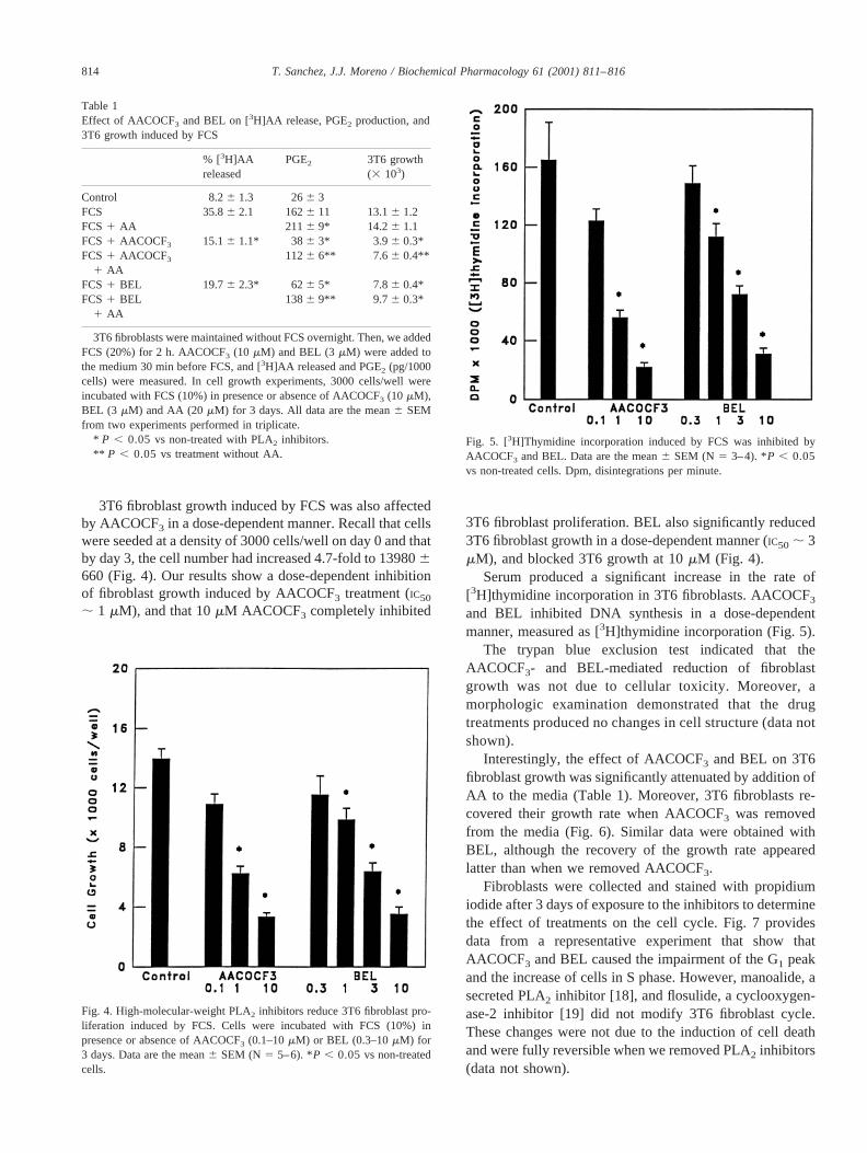
3T6 fibroblast growth induced by FCS was also affectedby AACOCF3 in a dose-dependent manner. Recall that cellswere seeded at a density of 3000 cells/well on day 0 and thatby day 3, the cell number had increased 4.7-fold to 139806660 (Fig. 4). Our results show a dose-dependent inhibitionof fibroblast growth induced by AACOCF3 treatment (IC50
; 1 mM), and that 10mM AACOCF3 completely inhibited
3T6 fibroblast proliferation. BEL also significantly reduced3T6 fibroblast growth in a dose-dependent manner (IC50 ; 3mM), and blocked 3T6 growth at 10mM (Fig. 4).
Serum produced a significant increase in the rate of[3H]thymidine incorporation in 3T6 fibroblasts. AACOCF3
and BEL inhibited DNA synthesis in a dose-dependentmanner, measured as [3H]thymidine incorporation (Fig. 5).
The trypan blue exclusion test indicated that theAACOCF3- and BEL-mediated reduction of fibroblastgrowth was not due to cellular toxicity. Moreover, amorphologic examination demonstrated that the drugtreatments produced no changes in cell structure (data notshown).
Interestingly, the effect of AACOCF3 and BEL on 3T6fibroblast growth was significantly attenuated by addition ofAA to the media (Table 1). Moreover, 3T6 fibroblasts re-covered their growth rate when AACOCF3 was removedfrom the media (Fig. 6). Similar data were obtained withBEL, although the recovery of the growth rate appearedlatter than when we removed AACOCF3.
Fibroblasts were collected and stained with propidiumiodide after 3 days of exposure to the inhibitors to determinethe effect of treatments on the cell cycle. Fig. 7 providesdata from a representative experiment that show thatAACOCF3 and BEL caused the impairment of the G1 peakand the increase of cells in S phase. However, manoalide, asecreted PLA2 inhibitor [18], and flosulide, a cyclooxygen-ase-2 inhibitor [19] did not modify 3T6 fibroblast cycle.These changes were not due to the induction of cell deathand were fully reversible when we removed PLA2 inhibitors(data not shown).
Table 1Effect of AACOCF3 and BEL on [3H]AA release, PGE2 production, and3T6 growth induced by FCS
% [3H]AAreleased
PGE2 3T6 growth(3 103)
Control 8.26 1.3 266 3FCS 35.86 2.1 1626 11 13.16 1.2FCS1 AA 211 6 9* 14.26 1.1FCS1 AACOCF3 15.16 1.1* 386 3* 3.9 6 0.3*FCS1 AACOCF3
1 AA1126 6** 7.6 6 0.4**
FCS1 BEL 19.76 2.3* 626 5* 7.8 6 0.4*FCS1 BEL
1 AA1386 9** 9.7 6 0.3*
3T6 fibroblasts were maintained without FCS overnight. Then, we addedFCS (20%) for 2 h. AACOCF3 (10 mM) and BEL (3mM) were added tothe medium 30 min before FCS, and [3H]AA released and PGE2 (pg/1000cells) were measured. In cell growth experiments, 3000 cells/well wereincubated with FCS (10%) in presence or absence of AACOCF3 (10 mM),BEL (3 mM) and AA (20 mM) for 3 days. All data are the mean6 SEMfrom two experiments performed in triplicate.
* P , 0.05 vsnon-treated with PLA2 inhibitors.** P , 0.05 vstreatment without AA.
Fig. 4. High-molecular-weight PLA2 inhibitors reduce 3T6 fibroblast pro-liferation induced by FCS. Cells were incubated with FCS (10%) inpresence or absence of AACOCF3 (0.1–10mM) or BEL (0.3–10mM) for3 days. Data are the mean6 SEM (N 5 5–6). *P , 0.05 vsnon-treatedcells.
Fig. 5. [3H]Thymidine incorporation induced by FCS was inhibited byAACOCF3 and BEL. Data are the mean6 SEM (N 5 3–4). *P , 0.05vs non-treated cells. Dpm, disintegrations per minute.
814 T. Sanchez, J.J. Moreno / Biochemical Pharmacology 61 (2001) 811–816

4. Discussion
AA release is an early event in the mitogenic process ofa number of growth factors and the subsequent cell prolif-eration. This mobilization of AA from membrane phospho-lipids occurs mainly by two pathways: hydrolytic cleavage
of the sn-2 position of the glycerophospholipid backbonecatalyzed by PLA2, or activation of phospholipase C, whichcleaves the phosphate-ester bond, and subsequent hydroly-sis of diacylglycerol produced by diacylglycerol lipase. In3T6 fibroblasts, stimulated AA release through PLA2 hasbeen shown to be required for mitogenesis. Moreover, theseprevious studies demonstrated that nonselective PLA2 in-hibitors reduce 3T6 fibroblast proliferation [13,15].
Owing to the central role of cPLA2 in AA signaling, thedesign of cPLA2 inhibitors is an area of great interest.AACOCF3 is a reversible cPLA2 inhibitor that also inhibitscyclooxygenases [16]. Unfortunately, AACOCF3 also po-tently inhibits iPLA2 [20] because both cPLA2 and iPLA2
appear to use a central Ser for catalysis and, probably,similar catalytic mechanisms. There is one way to ascertainwhether the inhibitory effects of AACOCF3 result fromcPLA2 or iPLA2. This involves the parallel use of BEL, aselective inhibitor of iPLA2 [17]. Thus, if a given responseis inhibited by AACOCF3 and by BEL, this would indicatethe involvement of iPLA2 [21].
Our results suggest that BEL-sensitive iPLA2 may be, atleast partially, implicated in the AA release, PGE2 genera-tion and cell growth stimulated by FCS in 3T6 fibroblastcultures, in agreement with recent studies with iPLA2 in-hibitors including BEL that suggested the contribution ofiPLA2 to stimulus-induced AA liberation [22,23]. However,further studies will be necessary to understand the preciserole of cPLA2 and iPLA2 on FCS-stimulated AA release and3T6 fibroblast growth.
Interestingly, FACS-scan analysis suggests that the re-duction in cell growth following AACOCF3 and BEL treat-ments results from an impairment of the percentage of cellsin G1 and the enhancement of cells in S phase whereasmanoalide and flosulide, treatments that partially reduced3T6 growth [15], did not induce changes.
The mechanism by which high-molecular-weight PLA2
influences 3T6 fibroblast proliferation remains to be deter-mined. However, cyclooxygenase inhibitors such as keto-profen or indomethacin had effect on 3T6 fibroblast growth[15,24], indicating that the high-molecular-weight PLA2-dependent effect on proliferation may be mediated by AAand/or prostaglandins. Thus, when fibroblasts were treatedwith exogenous AA, the reduction in growth rate producedby AACOCF3 or BEL was significantly attenuated. Thisaction may be related to the capacity of AA to stimulatec-fos, c-jun, and/or mitogen-activated protein kinase [25,26]. Additionally, recent reports indicate that the release ofAA by PLA2 is accompanied by the formation of lysolipids,and lysophosphatidic acid, lysophosphatidylinositol and ly-sophosphatidylcholine stimulate mitogen-activated proteinkinases and proliferation [27,28]. Moreover, these lysolip-ids are precursors of glycerophosphoinositides, which showevidence of being accumulated in ras-transformed cells.
In summary, we have demonstrated that AACOCF3 andBEL block 3T6 fibroblast proliferation and DNA synthesisat the S/G2 boundary, thus prolonging the S phase. These
Fig. 6. The inhibition of AACOCF3 or BEL on fibroblast growth werereversible. Cells were treated with AACOCF3 (10mM, �) or BEL (10mM,■) for 7 days or removed after 2 days (ƒ, h) in presence of 10% FCS.Control cells were cultured in presence of 10% FCS (E). Data are the meanof 3–4 results.
Fig. 7. Distribution of 3T6 fibroblast with respect to DNA content in cellscultured with FCS (10%) for 2 days or FCS in presence of AACOCF3 (10mM), BEL (10 mM), manoalide (0.5mM) or flosulide (10 mM). Thisexperiment is representative of three experiments with similar results.
815T. Sanchez, J.J. Moreno / Biochemical Pharmacology 61 (2001) 811–816

data suggest a role of iPLA2 in the control of 3T6 fibroblastgrowth.
Acknowledgments
T. Sanchez is a recipient of a predoctoral fellowship fromthe Autonomous Government of Catalonia. This study wassupported by the Spanish Ministry of Education (DGESPM97-0110 and PM98-0191). FACS-analyses were per-formed at the Serve´is Cientıfic-tecnics of the BarcelonaUniversity. We are very grateful to Robin Rycgroft forvaluable assistance in the preparation of the manuscript.
References
[1] Waite M. Phospholipases In: Vance DE, Vance JE, editors. Biochem-istry of lipids, lipoproteins and membranes. New York: ElsevierScience Publishing Co., 1996. p. 211–36.
[2] Smith WL, DeWitt DL. Biochemistry of prostaglandin endoperoxideH synthase-1 and synthase-2 and their differential susceptibility tononsteroideal anti-inflammatory drugs. Semin Nephrol 1995;15:179–94.
[3] Mayer B, Rauter Z, Zenzmaier E, Gleispack H, Esterbauer H. Char-acterization of lipoxygenase metabolites of arachidonic acid in cul-tured human fibroblasts. Biochim Biophys Acta 1984;795:151–61.
[4] Dennis EA. The growing phospholipase A2 superfamily of signaltransduction enzymes. Trends Biochem Sci 1997;22:13057–60.
[5] Kramer RM, Hession C, Johansen B, Hayes G, McGray P, Chow EP,Tizard R, Pepinsky RB. Structure and properties of a human non-pancreatic phospholipase A2. J Biol Chem 1989;264:5768–75.
[6] Kramer RM, Johansen B, Hession C, Pepinsky R. Structure andproperties of a secretable phospholipase A2 from human platelets In:Wong PYK, Dennis EA, editors. Phospholipase A2. New York:Plenum Publishing Corp., 1990. p. 35–53.
[7] Clark JD, Milona N, Knopf JL. Purification of a 110-kilodaltoncytosolic phospholipase A2 from the human monocytic cell lineU937. Proc Natl Acad Sci USA 1990;87:7708–12.
[8] Clark JD, Schievella AR, Nalefski EA, Lin LL. Cytosolic phospho-lipase A2. J Lipid Mediat Cell Signal 1995;12:83–117.
[9] Balsinde J, Dennis EA. Function and inhibition of intracellular cal-cium independent phospholipase A2. J Biol Chem 1997;272:16069–72.
[10] Murakami M, Kambe T, Shimbara S, Kudo I. Functional couplingbetween various phospholipases A2 and cyclooxygenases in immedi-ate and delayed prostanoid biosynthetic pathways. J Biol Chem 1999;274:3103–15.
[11] Di Marzo V. Arachidonic acid and eicosanoids as targets and effec-tors in second messenger interactions. Prostaglandins LeukotrienesEssent Fatty Acids 1995;53:239–54.
[12] Reich R, Martin GR. Identification of arachidonic acid pathwaysrequired for the invasive and metastatic activity of malignant tumorcells. Prostaglandins 1996;51:1–17.
[13] Lloret S, Torrent M, Moreno JJ. Proliferation-dependent changes inarachidonic acid mobilization from phospholipids of 3T6 fibroblasts.Pflugers Archiv Eur J Physiol 1996;432:655–62.
[14] Moreno JJ. Regulation of arachidonic acid release and prostaglandinformation by cell-cell adhesive interactions in wound-repair. Pflu¨gersArchiv Eur J Physiol 1997;433:351–6.
[15] Martinez J, Sanchez T, Moreno JJ. Role of prostaglandin H synthase-2-mediated conversion of arachidonic acid in controlling 3T6 fibro-blast growth. Am J Physiol 1997;273:C1466–71.
[16] Riendeau D, Guay J, Weech PK, Laliberte F, Yergey J. Arachidonyltrifluoromethylketone, a potent inhibitor of 85-kDa phospholipaseA2, blocks production of arachidonate and 12-hydroxyeicosatetra-enoic acid by calcium ionophore-challenged platelets. J Biol Chem1994;269:15619–24.
[17] Hazen SL, Zupan LA, Weiss RH, Getman DP, Gross RW. Suicideinhibition of canine myocardial cytosolic calcium-independent phos-pholipase A2. Mechanism-based discrimination between calcium-de-pendent and -independent phospholipase A2. J Biol Chem 1991;266:7227–32.
[18] Jacobs RS, Culver P, Langdon R, O’Brien T, White S. Some phar-macological observations in marine natural products. Tetrahedron1985;41:981–4.
[19] Klein TN, Sing RM, Pfeilschifter J, Ullrich V. Selective inhibition ofcyclooxygenase 2. Biochem Pharmacol 1994;48:1605–10.
[20] Lio YC, Reynolds LJ, Balsinde J, Dennis EA. Irreversible inhibitionof Ca21-independent phospholipase A2 by methyl arachidonyl flu-orophosphonate. Biochim Biophys Acta 1996;1302:55–60.
[21] Balsinde J, Dennis EA. Distinct roles in signal transduction for eachof the phospholipase A2 enzymes present in P388D1 macrophages.J Biol Chem 1996;271:6758–65.
[22] Gross RW, Rudolph AE, Wang J, Sommers CD, Wolf MJ. Nitricoxide activates the glucose-dependent mobilization of arachidonicacid in macrophages-like cell line (RAW 264.7) that is largely me-diated by calcium-independent phospholipase A2. J Biol Chem 1995;270:14855–8.
[23] Atsumi G, Tajima M, Hadano A, Nakatani Y, Murakami M, Kudo I.Fas-induced arachidonic acid release is mediated by Ca21-indepen-dent phospholipase A2 but not cytosolic phospholipase A2, whichundergoes proteolytic inactivation. J Biol Chem 1998;273:13870–7.
[24] Sanchez T, Moreno JJ. Ketoprofen S(1) enantiomer inhibits prosta-glandin production and cell growth in 3T6 fibroblast cultures. EurJ Pharmacol 1999;370:63–7.
[25] Falasca M, Iurisci C, Carvelli A, Sacchetti A, Corda D. Release of themitogen lysophosphatidylinositol from H-Ras-transfected fibroblasts:a possible mechanism of autocrine control of cell proliferation. On-cogene 1998;16:2357–65.
[26] Sellmayer A, Danesch U, Weber PC. Effects of different polyunsat-urated fatty acids on growth-related early gene expression and cellgrowth. Lipids 1996;31:537–40.
[27] Corda D, Falasca M. Glycerophosphoinositols as potential markers ofras-induced transformation and novel second messengers. AnticancerRes 1996;16:1341–50.
[28] Wojtaszek PA, Van Putten V, Nemenoff RA. Activation of a novelform of phospholipase A2 during liver regeneration. FEBS Lett 1995;367:228–32.
816 T. Sanchez, J.J. Moreno / Biochemical Pharmacology 61 (2001) 811–816