The discovery and early study of cells progressed with the invention and improvement of microscopes...
53
• The discovery and early study of cells progressed with the invention and improvement of microscopes in the 17th century. • In a light microscope (LM) visible light passes through the specimen and then through glass lenses. – The lenses refract light such that the image is magnified into the eye or onto a video screen. Microscopes provide windows to the world of the cell Copyright © 2002 Pearson Education, Inc., publishing as Benjamin Cummings
-
Upload
joleen-palmer -
Category
Documents
-
view
216 -
download
2
Transcript of The discovery and early study of cells progressed with the invention and improvement of microscopes...
• The discovery and early study of cells progressed with the invention and improvement of microscopes in the 17th century.
• In a light microscope (LM) visible light passes through the specimen and then through glass lenses.– The lenses refract light such that the image is magnified
into the eye or onto a video screen.
Microscopes provide windows to the world of the cell
Copyright © 2002 Pearson Education, Inc., publishing as Benjamin Cummings

• Microscopes vary in magnification and resolving power.
• Magnification is the ratio of an object’s image to its real size.
• Resolving power is a measure of image clarity.– It is the minimum distance two points can be
separated by and still be viewed as two separate points.
– Resolution is limited by the shortest wavelength of the source, in this case light.
Copyright © 2002 Pearson Education, Inc., publishing as Benjamin Cummings

• The minimum resolution of a light microscope is about 2 microns, the size of a small bacterium
• Light microscopes can magnify effectively to about 1,000 times the size of the actual specimen.– At higher magnifications,
the image blurs.
Copyright © 2002 Pearson Education, Inc., publishing as Benjamin Cummings
Fig. 7.1
• While a light microscope can resolve individual cells, it cannot resolve much of the internal anatomy, especially the organelles.
• To resolve smaller structures we use an electron microscope (EM), which focuses a beam of electrons through the specimen or onto its surface.– Because resolution is inversely related to wavelength
used, electron microscopes with shorter wavelengths than visible light have finer resolution.
– Theoretically, the resolution of a modern EM could reach 0.1 nanometer (nm), but the practical limit is closer to about 2 nm.
Copyright © 2002 Pearson Education, Inc., publishing as Benjamin Cummings

• Transmission electron microscopes (TEMs) are used mainly to study the internal ultrastructure of cells.– A TEM aims an electron beam through a thin
section of the specimen.– The image is focused
and magnified by electromagnets.
– To enhance contrast, the thin sections are stained with atoms of heavy metals.
Copyright © 2002 Pearson Education, Inc., publishing as Benjamin Cummings
Fig. 7.2a

• Scanning electron microscopes (SEMs) are useful for studying surface structures.– The sample surface is covered with a thin film
of gold.– The beam excites electrons on the surface.– These secondary electrons are collected and
focused on a screen.
• The SEM has great depth of field, resulting in an image that seems three-dimensional.
Copyright © 2002 Pearson Education, Inc., publishing as Benjamin Cummings
Fig. 7.2b

Cell Theory• All living things are composed of cells
• Cells are the basic unit of structure and function
• Cell have to come from preexisting cells.
• All cells are surrounded by a plasma membrane.
• The semifluid substance within the membrane is the cytoplasm, containing the organelles.
• All cells contain chromosomes which have genes in the form of DNA.
• All cells also have ribosomes, tiny organelles that make proteins using the instructions contained in DNA.
Prokaryotic and eukaryotic cells differ in size and complexity
Copyright © 2002 Pearson Education, Inc., publishing as Benjamin Cummings
Copyright © 2002 Pearson Education, Inc., publishing as Benjamin Cummings
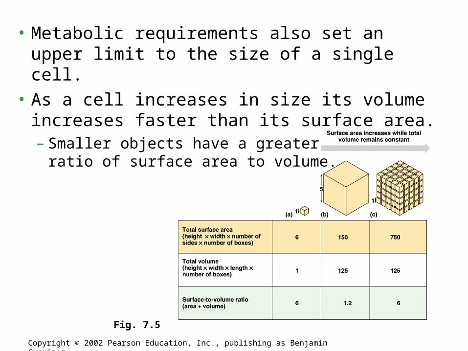
Fig. 7.5
• Metabolic requirements also set an upper limit to the size of a single cell.
• As a cell increases in size its volume increases faster than its surface area.– Smaller objects have a greater
ratio of surface area to volume.

Plasma (Cell) Membrane
All Membranes are Phospholipid Bilayers

• The plasma membrane separates the living cell from its nonliving surroundings.
• This thin barrier, 8 nm thick, controls traffic into and out of the cell.
• Like other membranes, the plasma membrane is selectively permeable, allowing some substances to cross more easily than others.
•Copyright © 2002 Pearson Education, Inc., publishing as Benjamin Cummings

• The main macromolecules in membranes are lipids and proteins, but include some carbohydrates.
• The most abundant lipids are phospholipids.
• Phospholipids and most other membrane constituents have both hydrophobic regions and hydrophilic regions.
• The phospholipids and proteins in membranes create a unique physical environment, described by the fluid mosaic model.– A membrane is a fluid structure with proteins
embedded or attached to a double layer of phospholipids.
•Copyright © 2002 Pearson Education, Inc., publishing as Benjamin Cummings

Figure 5.12 The structure of a phospholipid
•Copyright © 2002 Pearson Education, Inc., publishing as Benjamin Cummings
•Fig. 8.1b
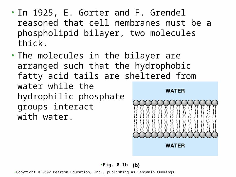
• In 1925, E. Gorter and F. Grendel reasoned that cell membranes must be a phospholipid bilayer, two molecules thick.
• The molecules in the bilayer are arranged such that the hydrophobic fatty acid tails are sheltered from water while the hydrophilic phosphate groups interact with water.

• The plasma membrane functions as a selective barrier that allows passage of oxygen, nutrients, and wastes for the whole volume of the cell.
• The volume of cytoplasm determines the need for this exchange.
• Rates of chemical exchange may be inadequate to maintain a cell with a very large cytoplasm.
• The need for a surface sufficiently large to accommodate the volume explains the microscopic size of most cells.
• Larger organisms do not generally have larger cells than smaller organisms - simply more cells.
Copyright © 2002 Pearson Education, Inc., publishing as Benjamin Cummings

• A major difference between prokaryotic and eukaryotic cells is the location of chromosomes.
• In a eukaryotic cell, chromosomes are contained in a membrane-enclosed organelle, the nucleus.
• In a prokaryotic cell, the DNA is concentrated in the nucleoid without a membrane separating it from the rest of the cell.
Copyright © 2002 Pearson Education, Inc., publishing as Benjamin Cummings

Copyright © 2002 Pearson Education, Inc., publishing as Benjamin Cummings
Fig. 7.4 The prokaryotic cell is much simpler in structure, lacking a nucleus and the other membrane-enclosed organelles of the eukaryotic cell.

• In Eukaryote cells, the chromosomes are contained within a membranous nuclear envelope.
• The region between the nucleus and the plasma membrane is the cytoplasm.– All the material within the plasma membrane of a
prokaryotic cell is cytoplasm.
• Within the cytoplasm of a eukaryotic cell is a variety of membrane-bounded organelles of specialized form and function.– These membrane-bounded organelles are absent in
prokaryotes.
Copyright © 2002 Pearson Education, Inc., publishing as Benjamin Cummings

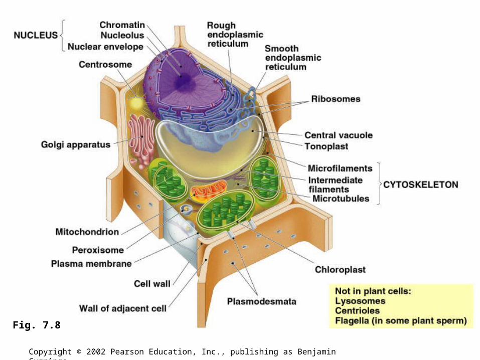
• A eukaryotic cell has internal membranes, which partition the cell into compartments.
• These membranes also participate in metabolism as many enzymes are built into membranes.
• The barriers created by membranes provide different local environments that facilitate specific metabolic functions.
• The general structure of a biological membrane is a double layer of phospholipids (phospholipid bilayer) with other lipids and diverse proteins.
Internal membranes compartmentalize the functions of a eukaryotic cell
Copyright © 2002 Pearson Education, Inc., publishing as Benjamin Cummings

Copyright © 2002 Pearson Education, Inc., publishing as Benjamin Cummings
Fig. 7.7

Copyright © 2002 Pearson Education, Inc., publishing as Benjamin Cummings
Fig. 7.8

• The nucleus contains most of the DNA in a eukaryotic cell.– Some DNA is located in mitochondria and chloroplasts.
• The nucleus is separated from the cytoplasm by a double membrane.
• Where the double membranes are fused, a pore allows large macromolecules and particles to pass through.
The nucleus contains a eukaryotic cell’s genetic library
Copyright © 2002 Pearson Education, Inc., publishing as Benjamin Cummings

• The nuclear side of the envelope is lined by the nuclear lamina, a network of intermediate filaments that maintain the shape of the nucleus.
Copyright © 2002 Pearson Education, Inc., publishing as Benjamin Cummings
Fig. 7.9
• Within the nucleus, the DNA and associated proteins are organized into fibrous material, chromatin.
• In a normal cell they appear as a diffuse mass.
• However when the cell prepares to divide, the chromatin fibers coil up to be seen as separate structures, chromosomes.
• Each eukaryotic species has a characteristic number of chromosomes.– A typical human cell has 46 chromosomes, but
sex cells (eggs and sperm) have only 23 chromosomes.
Copyright © 2002 Pearson Education, Inc., publishing as Benjamin Cummings
• In the nucleus is a region of densely stained fibers and granules adjoining chromatin, the nucleolus. – In the nucleolus, ribosomal RNA (rRNA) is
synthesized and assembled with proteins from the cytoplasm to form ribosomal subunits.
• The nucleus directs protein synthesis by synthesizing messenger RNA (mRNA).– The mRNA travels to the cytoplasm and
combines with ribosomes to translate its genetic message proteins.
Copyright © 2002 Pearson Education, Inc., publishing as Benjamin Cummings

• Ribosomes contain rRNA and protein.
• A ribosome is composed of two subunits that combine to carry out protein synthesis.
Ribosomes build a cell’s proteins
Copyright © 2002 Pearson Education, Inc., publishing as Benjamin Cummings
Fig. 7.10

• Some ribosomes, free ribosomes, are suspended in the cytoplasm and synthesize proteins that function within the cytoplasm.
• Other ribosomes, bound ribosomes, are attached to the outside of the endoplasmic reticulum.– These synthesize proteins that are either
included into membranes or for export from the cell.
• Ribosomes can shift between roles depending on the polypeptides they are synthesizing.
Copyright © 2002 Pearson Education, Inc., publishing as Benjamin Cummings

• Many of the internal membranes in a eukaryotic cell are part of the endomembrane system.
• These membranes are either in direct contact or connected via transfer of vesicles, sacs of membrane.
• In spite of these links, these membranes have diverse functions and structures.
• The endomembrane system includes the nuclear envelope, endoplasmic reticulum, Golgi apparatus, lysosomes, vacuoles, and the plasma membrane.
Endomembranes
Copyright © 2002 Pearson Education, Inc., publishing as Benjamin Cummings

• The endoplasmic reticulum (ER) accounts for half the membranes in a eukaryotic cell.
• The ER membrane is continuous with the nuclear envelope.– Smooth ER looks smooth because
it lacks ribosomes.– Rough ER looks rough because
ribosomes (bound ribosomes) are attached to the outside, including the outside of the nuclear envelope.
Copyright © 2002 Pearson Education, Inc., publishing as Benjamin Cummings
Fig. 7.11
• The smooth ER is rich in enzymes and plays a role in a variety of metabolic processes.
• Enzymes of smooth ER synthesize lipids, including oils, phospholipids, and steroids.
• The smooth ER also catalyzes a key step in the mobilization of glucose from stored glycogen in the liver.– An enzyme removes the phosphate group from
glucose phosphate, a product of glycogen hydrolysis, permitting glucose to exit the cell.
Copyright © 2002 Pearson Education, Inc., publishing as Benjamin Cummings
• Rough ER is especially abundant in those cells that secrete proteins.– As a polypeptide is synthesized by the ribosome, it is
threaded into the space of the ER through a pore formed by a protein in the ER membrane.
– Many of these polypeptides are glycoproteins, polypeptides to which an oligosaccharide is attached.
• These secretory proteins are packaged in transport vesicles that carry them to their next stage.
Copyright © 2002 Pearson Education, Inc., publishing as Benjamin Cummings

• Rough ER is also a membrane factory.– Membrane bound proteins are synthesized
directly into the membrane.– Enzymes in the rough ER also synthesize
phospholipids from precursors in the cytosol.– As the ER membrane expands, parts can be
transferred as transport vesicles to other components of the endomembrane system.
Copyright © 2002 Pearson Education, Inc., publishing as Benjamin Cummings

• Many transport vesicles from the ER travel to the Golgi apparatus for modification of their contents.
• The Golgi is a center of manufacturing, warehousing, sorting, and shipping.
• The Golgi apparatus is especially extensive in cells specialized for secretion.
Copyright © 2002 Pearson Education, Inc., publishing as Benjamin Cummings

• The Golgi apparatus consists of flattened membranous sacs - cisternae - looking like a stack of pita bread.– The membrane of each cisterna separates its internal
space from the cytosol
Copyright © 2002 Pearson Education, Inc., publishing as Benjamin Cummings
Fig. 7.12
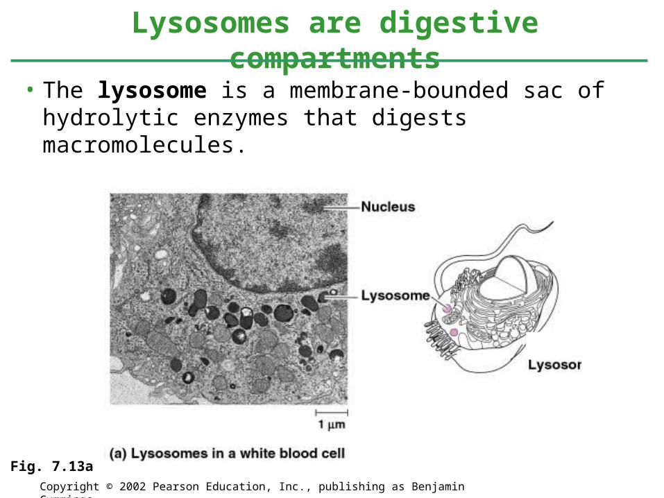
• The lysosome is a membrane-bounded sac of hydrolytic enzymes that digests macromolecules.
Lysosomes are digestive compartments
Copyright © 2002 Pearson Education, Inc., publishing as Benjamin Cummings
Fig. 7.13a
• Lysosomal enzymes can hydrolyze proteins, fats, polysaccharides, and nucleic acids.
• These enzymes work best at pH 5.– Proteins in the lysosomal membrane pump
hydrogen ions from the cytoplasm to the inside of the lysosomes.
• While rupturing one or a few lysosomes has little impact on a cell, massive leakage from lysosomes can destroy an cell by autodigestion.
• The lysosomes create a space where the cell can digest macromolecules safely.
Copyright © 2002 Pearson Education, Inc., publishing as Benjamin Cummings
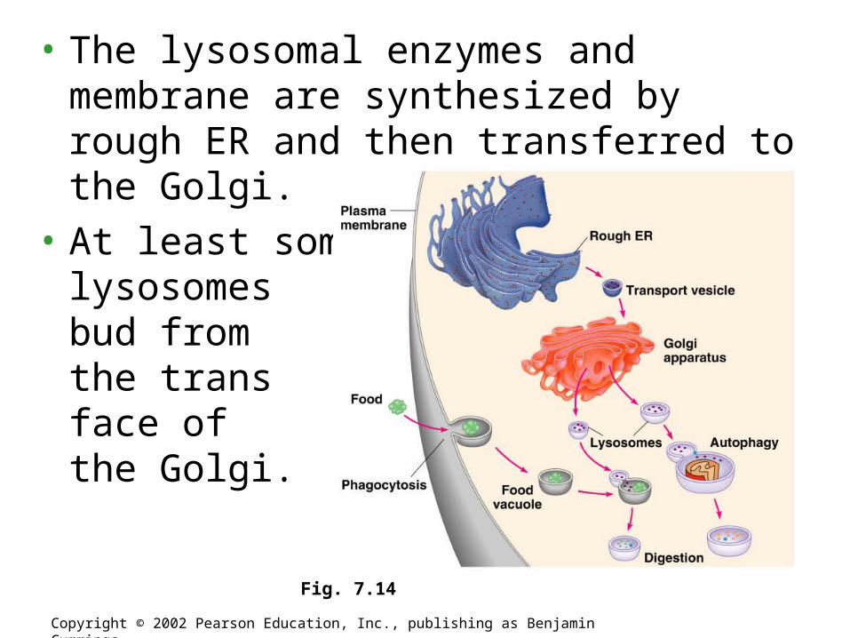
• The lysosomal enzymes and membrane are synthesized by rough ER and then transferred to the Golgi.
• At least some lysosomes bud from the trans face of the Golgi.
Copyright © 2002 Pearson Education, Inc., publishing as Benjamin Cummings
Fig. 7.14
Copyright © 2002 Pearson Education, Inc., publishing as Benjamin Cummings
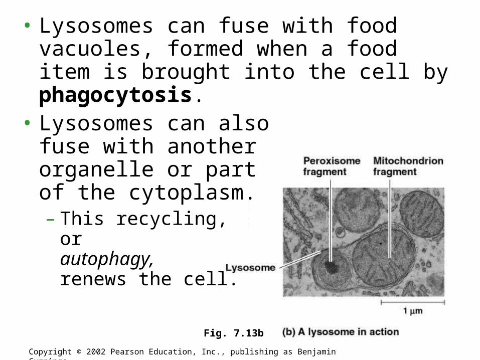
Fig. 7.13b
• Lysosomes can fuse with food vacuoles, formed when a food item is brought into the cell by phagocytosis.
• Lysosomes can also fuse with another organelle or part of the cytoplasm.– This recycling,
or autophagy,renews the cell.

• The lysosomes play a critical role in the programmed destruction of cells in multicellular organisms.– This process allows reconstruction during the
developmental process.
• Several inherited diseases affect lysosomal metabolism.– These individuals lack a functioning version of
a normal hydrolytic enzyme.– These diseases include Pompe’s disease in
the liver and Tay-Sachs disease in the brain.
Copyright © 2002 Pearson Education, Inc., publishing as Benjamin Cummings

• The endomembrane system plays a key role in the synthesis (and hydrolysis) of macromolecules in the cell.
• The various components modify macromolecules for their various functions.
Copyright © 2002 Pearson Education, Inc., publishing as Benjamin Cummings
Fig. 7.16

• Vesicles and vacuoles (larger versions) are membrane-bound sacs with varied functions.– Food vacuoles, from phagocytosis, fuse with
lysosomes.– Contractile vacuoles, found in freshwater
protists, pump excess water out of the cell.– Central vacuoles are found in many mature
plant cells.
Vacuoles have diverse functions in cell maintenance
Copyright © 2002 Pearson Education, Inc., publishing as Benjamin Cummings
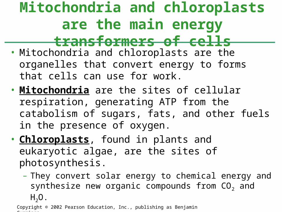
• Mitochondria and chloroplasts are the organelles that convert energy to forms that cells can use for work.
• Mitochondria are the sites of cellular respiration, generating ATP from the catabolism of sugars, fats, and other fuels in the presence of oxygen.
• Chloroplasts, found in plants and eukaryotic algae, are the sites of photosynthesis.– They convert solar energy to chemical energy and
synthesize new organic compounds from CO2 and H2O.
Mitochondria and chloroplasts are the main energy transformers of cells
Copyright © 2002 Pearson Education, Inc., publishing as Benjamin Cummings
•Both organelles have small quantities of DNA that direct the synthesis of the polypeptides produced by these internal ribosomes.
•Mitochondria and chloroplasts grow and reproduce as semiautonomous organelles.

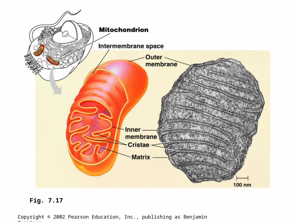
• Almost all eukaryotic cells have mitochondria.– There may be one very large mitochondrion or
hundreds to thousands of individual mitochondria.
– The number of mitochondria is correlated with aerobic metabolic activity.
– A typical mitochondrion is 1-10 microns long.– Mitochondria are quite dynamic: moving,
changing shape, and dividing.
Copyright © 2002 Pearson Education, Inc., publishing as Benjamin Cummings
Copyright © 2002 Pearson Education, Inc., publishing as Benjamin Cummings
Fig. 7.17

• The chloroplast is one of several members of a generalized class of plant structures called plastids.– Leucoplasts (Amyloplasts) store starch in roots and
tubers.– Chromoplasts store pigments for fruits, flowers and give
leaves their fall colors.
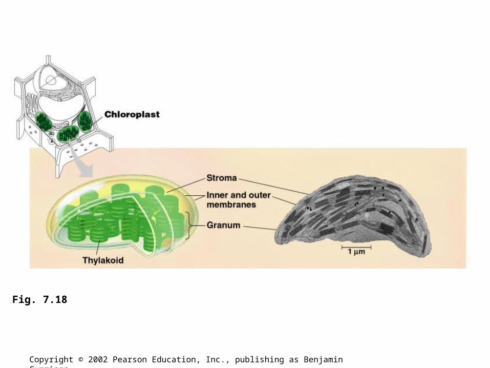
• The chloroplast produces sugar by capturing light energy and converting it into chemical energy (Photosynthesis).– Chloroplasts gain their color from high levels of the
green pigment chlorophyll.
• Chloroplasts are found in leaves and other green structures of plants and in eukaryotic algae.
Copyright © 2002 Pearson Education, Inc., publishing as Benjamin Cummings
Copyright © 2002 Pearson Education, Inc., publishing as Benjamin Cummings
Fig. 7.8
Copyright © 2002 Pearson Education, Inc., publishing as Benjamin Cummings
Fig. 7.18

• The cytoskeleton is a network of fibers extending throughout the cytoplasm.
• The cytoskeleton organizes the structures and activities of the cell.
Copyright © 2002 Pearson Education, Inc., publishing as Benjamin Cummings
Fig. 7.20
• Cytoskeleton
• The cytoskeleton provides mechanical support and maintains shape of the cell.
• The cytoskeleton provides anchorage for many organelles and cytosolic enzymes.
• The cytoskeleton is dynamic, dismantling in one part and reassembling in another to change cell shape.
Copyright © 2002 Pearson Education, Inc., publishing as Benjamin Cummings

Copyright © 2002 Pearson Education, Inc., publishing as Benjamin Cummings

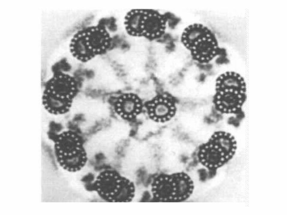
•Cillia and Flagella contain microtubules in a 9+2 arrangement.• Nine pairs of microtubules surrounding two microtubles