The Diagnostic Dilemma of Neurolymphomatosis - thejcn.com · Gaurav Prakash: e: a Departments of...
8
274 Copyright © 2016 Korean Neurological Association Neurolymphomatosis (NL) defined as infiltration of the central nervous system or the pe- ripheral nervous system (PNS) by malignant lymphoma cells is a rare clinical entity. Howev- er, the increasing use of fluorodeoxyglucose positron-emission tomography (FDG-PET) and magnetic resonance imaging in evaluating PNS disorders is resulting in; this condition being recognized more frequently. Here; we report five NL patients and review the current litera- ture. We report five patients with non-Hodgkin’s lymphoma (NHL) and NL, all of whom were men aged 47–69 years. e clinical presentation varied from symmetrical peripheral neuropathy to mononeuropathy. Peripheral neuropathy was the presenting manifestation of a systemic lymphoma in two patients (40%). Neuroimaging as well as whole-body FDG-PET helped in determining the correct diagnosis in all of the patients. NL is an unusual presenta- tion of NHL resulting from infiltration of the PNS by malignant lymphomatous cells. While evaluating peripheral neuropathy, a high degree of suspicion of NL is required since the pre- senting symptoms vary, conventional radiology has only modest sensitivity, and a pathologi- cal diagnosis is oſten difficult. FDG-PET helps in the early diagnosis and treatment of this condition. Key Wordszz neurolymphomatosis, diffuse large-B-cell lymphoma, mononeuropathy, fluorodeoxyglucose positron-emission tomography. e Diagnostic Dilemma of Neurolymphomatosis INTRODUCTION Neurolymphomatosis (NL), defined as invasion of the peripheral nervous system (PNS) (cranial nerves, peripheral nerves, roots, and plexus) by lymphoma cells, is a rare clinical entity; 1 being the least common way in which lymphoma can involve the PNS. It is impor- tant to differentiate NL, the common mode of PNS involvement in lymphoma, from other causes of PNS involvement in lymphoma (e.g., paraneoplastic PNS involvement, and radia- tion-and chemotherapy-induced damage to the PNS), since their treatment modalities dif- fer. 2 e increasing use of positron emission tomography (PET) and magnetic resonance imaging (MRI) in evaluating PNS disorders is resulting in increasing numbers of NL cases being recognized. Here we report five NL patients and review the current literature regarding the diagnosis and management of this rare condition. PATIENT 1 Mr. A, a 69-year-old male, presented to us with a two month history of shooting pains in the bilateral gluteal regions and the back of the thigh, followed 1 month later by pin prick paresthesias in both upper limbs and the right half of the face, along with asymmetrical quadriparesis affecting the legs more than the arms, low-grade fever, dry cough, orthop- Ritu Shree a Manoj Kumar Goyal a Manish Modi a Balan Louis Gaspar b Bishan Dass Radotra b Chirag Kamal Ahuja c Bhagwant Rai Mittal d Gaurav Prakash e a Departments of Neurology b Histopathology, c Radiodiagnosis, d Nuclear Medicine and e Clinical Hematology, Postgraduate Institute of Medical Education and Research (PGIMER), Chandigarh, India pISSN 1738-6586 / eISSN 2005-5013 / J Clin Neurol 2016;12(3):274-281 / http://dx.doi.org/10.3988/jcn.2016.12.3.274 Received January 29, 2016 Revised February 22, 2016 Accepted February 25, 2016 Correspondence Manoj Kumar Goyal, MD, DM Department of Neurology, Postgraduate Institute of Medical Education and Research (PGIMER), Chandigarh, India Tel +917087008271 E-mail [email protected] cc is is an Open Access article distributed under the terms of the Creative Commons Attribution Non-Com- mercial License (http://creativecommons.org/licenses/by-nc/3.0) which permits unrestricted non-commercial use, distribution, and reproduction in any medium, provided the original work is properly cited. JCN Open Access REVIEW
Transcript of The Diagnostic Dilemma of Neurolymphomatosis - thejcn.com · Gaurav Prakash: e: a Departments of...

274 Copyright © 2016 Korean Neurological Association
Neurolymphomatosis (NL) defined as infiltration of the central nervous system or the pe-ripheral nervous system (PNS) by malignant lymphoma cells is a rare clinical entity. Howev-er, the increasing use of fluorodeoxyglucose positron-emission tomography (FDG-PET) and magnetic resonance imaging in evaluating PNS disorders is resulting in; this condition being recognized more frequently. Here; we report five NL patients and review the current litera-ture. We report five patients with non-Hodgkin’s lymphoma (NHL) and NL, all of whom were men aged 47–69 years. The clinical presentation varied from symmetrical peripheral neuropathy to mononeuropathy. Peripheral neuropathy was the presenting manifestation of a systemic lymphoma in two patients (40%). Neuroimaging as well as whole-body FDG-PET helped in determining the correct diagnosis in all of the patients. NL is an unusual presenta-tion of NHL resulting from infiltration of the PNS by malignant lymphomatous cells. While evaluating peripheral neuropathy, a high degree of suspicion of NL is required since the pre-senting symptoms vary, conventional radiology has only modest sensitivity, and a pathologi-cal diagnosis is often difficult. FDG-PET helps in the early diagnosis and treatment of this condition.Key Wordszz neurolymphomatosis, diffuse large-B-cell lymphoma, mononeuropathy,
fluorodeoxyglucose positron-emission tomography.
The Diagnostic Dilemma of Neurolymphomatosis
INTRODUCTION
Neurolymphomatosis (NL), defined as invasion of the peripheral nervous system (PNS) (cranial nerves, peripheral nerves, roots, and plexus) by lymphoma cells, is a rare clinical entity;1 being the least common way in which lymphoma can involve the PNS. It is impor-tant to differentiate NL, the common mode of PNS involvement in lymphoma, from other causes of PNS involvement in lymphoma (e.g., paraneoplastic PNS involvement, and radia-tion-and chemotherapy-induced damage to the PNS), since their treatment modalities dif-fer.2 The increasing use of positron emission tomography (PET) and magnetic resonance imaging (MRI) in evaluating PNS disorders is resulting in increasing numbers of NL cases being recognized.
Here we report five NL patients and review the current literature regarding the diagnosis and management of this rare condition.
PATIENT 1
Mr. A, a 69-year-old male, presented to us with a two month history of shooting pains in the bilateral gluteal regions and the back of the thigh, followed 1 month later by pin prick paresthesias in both upper limbs and the right half of the face, along with asymmetrical quadriparesis affecting the legs more than the arms, low-grade fever, dry cough, orthop-
Ritu Shreea Manoj Kumar Goyala Manish Modia Balan Louis Gasparb Bishan Dass Radotrab Chirag Kamal Ahujac Bhagwant Rai Mittald Gaurav Prakashe
a Departments of Neurology b Histopathology, cRadiodiagnosis, dNuclear Medicine and e Clinical Hematology, Postgraduate Institute of Medical Education and Research (PGIMER), Chandigarh, India
pISSN 1738-6586 / eISSN 2005-5013 / J Clin Neurol 2016;12(3):274-281 / http://dx.doi.org/10.3988/jcn.2016.12.3.274
Received January 29, 2016Revised February 22, 2016Accepted February 25, 2016
CorrespondenceManoj Kumar Goyal, MD, DMDepartment of Neurology, Postgraduate Institute of Medical Education and Research (PGIMER), Chandigarh, IndiaTel +917087008271E-mail [email protected]
cc This is an Open Access article distributed under the terms of the Creative Commons Attribution Non-Com-mercial License (http://creativecommons.org/licenses/by-nc/3.0) which permits unrestricted non-commercial use, distribution, and reproduction in any medium, provided the original work is properly cited.
JCN Open Access REVIEW
www.thejcn.com 275
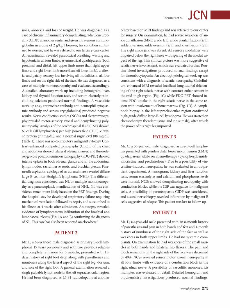
Shree R et al. JCNnoea, anorexia and loss of weight. He was diagnosed as a case of chronic inflammatory demyelinating radiculoneurop-athy (CIDP) at another center and given intravenous immuno-globulin in a dose of 2 g/kg. However, his condition contin-ued to worsen, and he was referred to our tertiary-care center. An examination revealed paradoxical breathing, wasting and hypotonia in all four limbs, asymmetrical quadriparesis (both proximal and distal, left upper limb more than right upper limb, and right lower limb more than left lower limb), areflex-ia, and patchy sensory loss involving all modalities in all four limbs and on the right side of the face. He was diagnosed as a case of multiple mononeuropathy and evaluated accordingly. A detailed laboratory work-up including hemogram, liver, kidney and thyroid function tests, and serum electrolytes in-cluding calcium produced normal findings. A vasculitic work-up (e.g., antinuclear antibody, anti-neutrophil cytoplas-mic antibody and serum cryoglobulins) produced negative results. Nerve conduction studies (NCSs) and electromyogra-phy revealed motor-sensory axonal and demyelinating poly-neuropathy. Analysis of the cerebrospinal fluid (CSF) showed 60 cells (all lymphocytes) per high power field (HPF), elevat-ed protein (79 mg/dL), and a normal sugar level (88 mg/dL) (Table 1). There was no contributory malignant cytology. Con-trast-enhanced computed tomography (CECT) of the chest and abdomen showed bilateral adrenal masses, and fluorode-oxyglucose positron-emission tomography (FDG-PET) showed intense uptake in both adrenal glands and in the abdominal lymph nodes, sacral nerve roots, and brachial plexus. Fine-needle aspiration cytology of an adrenal mass revealed diffuse large-B-cell non-Hodgkin’s lymphoma (NHL). The differen-tial diagnosis considered was NL or multiple mononeuropa-thy as a paraneoplastic manifestation of NHL. NL was con-sidered much more likely based on the PET findings. During the hospital stay he developed respiratory failure requiring mechanical ventilation followed by sepsis, and succumbed to his illness at 4-weeks after admission. An autopsy revealed evidence of lymphomatous infiltration of the brachial and lumbosacral plexus (Fig. 1A and B) confirming the diagnosis of NL. This case has also been reported on elsewhere.1
PATIENT 2
Mr. B, a 68-year-old male diagnosed as primary B-cell lym-phoma 15 years previously and with two previous relapses and complete remission for 5 months, presented with 15-days history of right foot drop along with paresthesias and numbness along the lateral aspect of the right leg, dorsum, and sole of the right foot. A general examination revealed a single palpable lymph node in the left supraclavicular region. He had been diagnosed as L5-S1-radiculopathy at another
center based on MRI findings and was referred to our center for surgery. On examination, he had severe weakness of an-kle dorsiflexion (MRC grade 1/5), ankle plantar flexion (2/5), ankle inversion, ankle eversion (2/5), and knee flexion (3/5). The right ankle jerk was absent. All sensory modalities were impaired below the right knee with sparing of the medial as-pect of the leg. This clinical picture was more suggestive of sciatic nerve involvement, which was evaluated further. Rou-tine blood investigations produced normal findings except for thrombocytopenia. An electrophysiological work-up was consistent with a diagnosis of sciatic neuropathy. Gadolini-um-enhanced MRI revealed localized longitudinal thicken-ing of the right sciatic nerve with contrast enhancement in the mid-thigh region (Fig. 1C) while FDG-PET showed in-tense FDG uptake in the right sciatic nerve in the same re-gion with involvement of bone marrow (Fig. 1D). A lymph-node biopsy in the left supraclavicular region confirmed high-grade diffuse large-B-cell lymphoma. He was started on chemotherapy (bendamustine and rituximab), after which the power of his right leg improved.
PATIENT 3
Mr. C, a 56-year-old male, diagnosed as pre-B-cell lympho-ma presented with painless distal lower motor neuron (LMN) quadriparesis while on chemotherapy (cyclophosphamide, vincristine, and prednisolone). Due to a possibility of vin-cristine-induced neuropathy, he was evaluated in an outpa-tient department. A hemogram, kidney and liver function tests, serum electrolytes and calcium and phosphorus levels were normal. NCSs showed demyelinating neuropathy with conduction blocks, while the CSF was negative for malignant cells. A possibility of paraneoplastic CIDP was considered, and a sural nerve biopsy revealed infiltration by malignant B cells suggestive of relapse. This patient was lost to follow-up.
PATIENT 4
Mr. D, 62-year-old male presented with an 8-month history of paresthesias and pain in both hands and feet and 1-month history of numbness of the right side of the face as well as weakness in both upper limbs. He had no systemic com-plaints. On examination he had weakness of the small mus-cles in both hands and bilateral hip flexors. The pain and touch sensations on the right side of the face were decreased by 40%. NCSs revealed sensorimotor axonal neuropathy in all four limbs with evidence of a conduction block in the right ulnar nerve. A possibility of vasculitic mononeuritis multiplex was evaluated in detail. Detailed hemogram and biochemistry investigations produced normal findings.

276 J Clin Neurol 2016;12(3):274-281
Short Review of NeurolymphomatosisJCNTa
ble
1. C
linic
al a
nd in
vest
igat
iona
l pro
file
of th
e ca
ses
Char
acte
ristic
sPa
tient
1Pa
tient
2Pa
tient
3Pa
tient
4Pa
tient
5
Age
(yea
rs)
6968
5662
47
Sex
Mal
eM
ale
Mal
eM
ale
Mal
e
Refe
rral
dia
gnos
is at
t
ime
of p
rese
ntat
ion
CIDP
DLBC
-NH
L w
ith p
rola
psed
in
terv
erte
bral
disc
an
d ra
dicu
lopa
thy
Pre
B-ce
ll ly
mph
oma
with
ch
emot
hera
py-i
nduc
ed
neur
opat
hyCI
DPU
ndiff
eren
tiate
d sa
rcom
a
In re
miss
ion
-Ye
s N
o; o
n ch
emot
hera
py-
No
Clin
ical
feat
ures
Feve
r, w
eigh
t los
s, co
ugh,
as
ymm
etric
al q
uadr
ipar
esis,
pa
rest
hesia
sRi
ght f
oot d
rop
LMN
-typ
e qu
adrip
ares
isM
onon
eurit
is m
ultip
lex
with
out
syst
emic
sym
ptom
sFe
ver,
wei
ght l
oss,
head
ache
, pa
rapa
resis
, dip
lopi
a
CSF
Clea
r, 88
mg/
dL s
ugar
; 79
mg/
dL p
rote
in; 6
0 ce
lls
(all
lym
phoc
ytes
), no
mal
igna
nt c
ells
Not
ana
lyze
dN
ot a
naly
zed
Clea
r, 55
mg/
dL s
ugar
; 104
mg/
dL
prot
ein;
10
calls
(all
lym
phoc
ytes
) no
mal
igna
nt c
ells;
lym
phom
atou
s c
ells
whe
n re
peat
ed a
fter
3 m
onth
s
Hem
orrh
agic
, 560
mg/
dL p
rote
in,
11 m
g/dL
sug
ar; n
o m
alig
nant
cel
ls
Radi
olog
ical
i
nves
tigat
ions
CECT
reve
aled
: bi
late
ral a
dren
al m
asse
s
Gad
olin
ium
-enh
ance
d M
RI re
veal
ed: t
hick
enin
g of
the
right
sci
atic
ner
ve
with
enh
ance
men
t
Not
don
eN
ot d
one
Gad
olin
ium
enh
ance
d M
RI th
e sp
ine
and
brai
n re
veal
ed: i
nten
se d
ural
en
hanc
emen
t in
the
lum
bosa
cral
spi
ne,
lept
omen
inge
al e
nhan
cem
ent i
n th
e br
ain,
th
icke
ning
and
enh
ance
men
t of b
ilate
ral
5th
and
3rd
cran
ial n
erve
s
FDG
-PET
find
ings
Inte
nse
upta
ke in
bot
h ad
rena
l gl
ands
, abd
omin
al ly
mph
no
des,
sacr
al n
erve
root
s, an
d br
achi
al p
lexu
s
Inte
nse
upta
ke in
the
right
sc
iatic
ner
ve w
ith in
volv
emen
t of
bon
e m
arro
w
(bila
tera
l fem
ur a
nd ti
bia)
Not
don
e
Inte
nse
upta
ke in
pal
atin
e to
nsils
, le
ft b
reas
t, m
edia
stin
al, a
xilla
ry a
nd h
ilar
lym
ph n
odes
, bila
tera
l hum
erus
, an
d le
ft te
stis
Inte
nse
upta
ke in
the
cere
bellu
m,
spin
al c
ord,
pso
as m
uscl
e,
lum
bar p
lexu
s, st
ernu
m, a
nd li
ver
NCS
sM
otor
-sen
sory
axo
nal a
nd
dem
yelin
atin
g po
lyne
urop
athy
Axon
al d
egen
erat
ion
of ri
ght t
ibia
l and
co
mm
on p
eron
eal n
erve
s
Dem
yelin
atin
g ne
urop
athy
w
ith c
ondu
ctio
n bl
ocks
Sens
orim
otor
axo
nal n
euro
path
y in
all
four
lim
bs w
ith e
vide
nce
of c
ondu
ctio
n bl
ock
in th
e rig
ht
ulna
r ner
ve
Not
don
e
Biop
sy/F
NAC
FNAC
of a
dren
al
mas
s re
veal
ed: D
LBC-
NH
L
Lym
ph-n
ode
biop
sy o
f the
le
ft s
upra
clav
icul
ar re
gion
co
nfirm
ed D
LBC-
NH
L
Sura
l ner
ve b
iops
y re
veal
ed:
infil
trat
ion
of th
e ne
rve
by m
alig
nant
B c
ells
Axill
ary
lym
ph n
ode
biop
sy re
veal
ed:
DLBC
; sur
al n
erve
bio
psy
reve
aled
: in
filtr
atio
n of
the
nerv
e by
ly
mph
omat
ous
cells
Biop
sy re
veal
ed
high
-gra
de ly
mph
oma
CECT
: con
tras
t-en
hanc
ed c
ompu
ted
tom
ogra
phy,
CIDP
: chr
onic
infla
mm
ator
y de
mye
linat
ing
poly
radi
culo
neur
opat
hy, C
SF: c
ereb
rosp
inal
flui
d, D
LBC:
diff
use
larg
e B-
cell,
FDG
-PET
: fluo
rode
oxyg
luco
se
posit
ron-
emiss
ion
tom
ogra
phy,
FNAC
: fine
-nee
dle
aspi
ratio
n cy
tolo
gy, M
RI: m
agne
tic re
sona
nce
imag
ing,
NCS
s: n
erve
con
duct
ion
stud
ies,
NH
L: n
on-H
odgk
in’s
lym
phom
a, L
MN
: low
er m
otor
neu
ron.

www.thejcn.com 277
Shree R et al. JCNFDG-PET revealed intense uptake in the palatine tonsils, left breast, mediastinal, axillary and hilar lymph nodes, bilateral humerus and left testis (Fig. 2A). A CSF examination showed 10 cells/HPF, protein at 59 mg/dL, and sugar at 104 mg/dL. A axillary lymph node biopsy revealed diffuse large-B-cell lymphoma, and a sural-nerve biopsy revealed infiltration by lymphomatous cells (Fig. 3). He was started on chemothera-py with rituximab, cyclophosphamide, vincristine, Adriamy-cin and prednisone (the R-CHOP regimen). He improved initially but returned after 3 months with difficulty walking, at which time he was found to have cerebellar ataxia. Gado-linium-enhanced MRI of the brain produced normal find-ings. The CSF contained lymphomatous cells suggestive of
central nervous system (CNS) involvement by lymphoma cells. He was further treated with whole-body radiation and chemotherapy, but succumbed to his illness. An autopsy was not performed since his relatives did not give consent.
PATIENT 5
Mr. E, a 47-year-old male, was apparently normal until Sep-tember 2014, when he started experiencing low-grade fever, anorexia, weight loss and headache. He was evaluated at an-other center and diagnosed as undifferentiated sarcoma based on a biopsy of liver lesions. He received six cycles of paclitaxel, cyclophosphamide and Adriamycin from January
Fig. 1. Nerve bundle infiltration (patient 1), FDG-PET and MRI of right sciatic nerve (patient 2). A: A nerve bundle with infiltration by lymphoma cells (patient 1). B: CD 20 stain highlights diffuse infiltration by diffuse large B cells (patient 1). C: PET CT showing intense FDG uptake in right sci-atic nerve (yellow arrow) (patient 2). D: intense enhancement of right sciatic nerve (arrow) on T1 weighted contrast MRI (patient 2).
A
C D
B

278 J Clin Neurol 2016;12(3):274-281
Short Review of NeurolymphomatosisJCNto April 2015 which did not provide relief. His headaches be-came much more intense and holocranial. He subsequently developed severe low-back ache, paraparesis, and diplopia, and was referred to our hospital. On examination he had bi-lateral involvement of the 5th, 6th, 7th, 9th, and 10th cranial nerves, (LMN) weakness in both lower limbs with sensation at the T11 spinal level, and the LMN type of sphincteric in-volvement. He was evaluated for the possibility of neurosar-comatosis. Gadolinium-enhanced MRI of the brain and whole spine revealed intense dural enhancement in the lum-
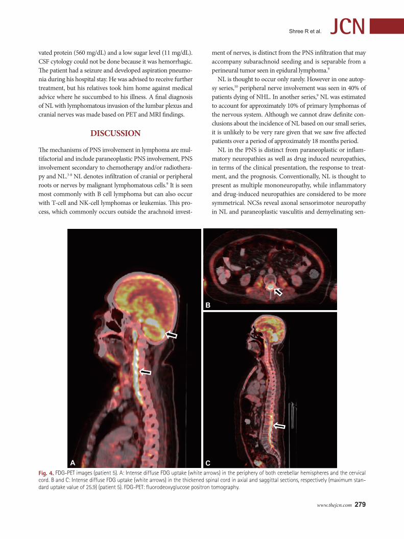
bosacral spine, leptomeningeal enhancement over the cere-bral convexities, and enhancement along multiple cranial nerves with thickening and enhancement of the bilateral tri-geminal nerves (Fig. 2B and C). FDG-PET revealed intense uptake in the cerebellum, spinal cord (cervical, T1 to T3 and T11 to S1 segments) (Fig. 4), psoas muscles, lumbar plexus, sternum and liver. An independent review of the biopsy specimens produced findings consistent with a diagnosis of high grade lymphoma (the exact classification was not possi-ble for technical reasons). The CSF was characterized by ele-
Fig. 2. FDG-PET images (patient 4) and MRI Brain images (patient 5). A: FDG-PET showing intense uptake (white arrow) in the left testis (patient 4). B and C: Thickened and enhancing trigeminal nerves (yellow arrows) on fluid-attenuated inversion recovery and T1-weighted MRI, respectively (patient 5). FDG-PET: fluorodeoxyglucose positron tomography, MRI: magnetic resonance imaging.
A B C
Fig. 3. Nerve Biopsy staining (patient 4). A: Hematoxylin and eosin-stained nerve biopsy sample that appears relatively normal at low magnifica-tion. B: High-power view of the same sample showing a few large atypical lymphoid cells (yellow arrows) amidst Schwann cells. C: CD20 immu-nostaining highlights these lymphoma cells (red arrows). D: The same cells were negative for CD3 immunostain (black arrows) (patient 4).
A B
C D
www.thejcn.com 279
Shree R et al. JCN
Fig. 4. FDG-PET images (patient 5). A: Intense diffuse FDG uptake (white arrows) in the periphery of both cerebellar hemispheres and the cervical cord. B and C: Intense diffuse FDG uptake (white arrows) in the thickened spinal cord in axial and saggittal sections, respectively (maximum stan-dard uptake value of 25.9) (patient 5). FDG-PET: fluorodeoxyglucose positron tomography.
A
B
C
vated protein (560 mg/dL) and a low sugar level (11 mg/dL). CSF cytology could not be done because it was hemorrhagic. The patient had a seizure and developed aspiration pneumo-nia during his hospital stay. He was advised to receive further treatment, but his relatives took him home against medical advice where he succumbed to his illness. A final diagnosis of NL with lymphomatous invasion of the lumbar plexus and cranial nerves was made based on PET and MRI findings.
DISCUSSION
The mechanisms of PNS involvement in lymphoma are mul-tifactorial and include paraneoplastic PNS involvement, PNS involvement secondary to chemotherapy and/or radiothera-py and NL.3-8 NL denotes infiltration of cranial or peripheral roots or nerves by malignant lymphomatous cells.9 It is seen most commonly with B cell lymphoma but can also occur with T-cell and NK-cell lymphomas or leukemias. This pro-cess, which commonly occurs outside the arachnoid invest-
ment of nerves, is distinct from the PNS infiltration that may accompany subarachnoid seeding and is separable from a perineural tumor seen in epidural lymphoma.9
NL is thought to occur only rarely. However in one autop-sy series,10 peripheral nerve involvement was seen in 40% of patients dying of NHL. In another series,9 NL was estimated to account for approximately 10% of primary lymphomas of the nervous system. Although we cannot draw definite con-clusions about the incidence of NL based on our small series, it is unlikely to be very rare given that we saw five affected patients over a period of approximately 18 months period.
NL in the PNS is distinct from paraneoplastic or inflam-matory neuropathies as well as drug induced neuropathies, in terms of the clinical presentation, the response to treat-ment, and the prognosis. Conventionally, NL is thought to present as multiple mononeuropathy, while inflammatory and drug-induced neuropathies are considered to be more symmetrical. NCSs reveal axonal sensorimotor neuropathy in NL and paraneoplastic vasculitis and demyelinating sen-
280 J Clin Neurol 2016;12(3):274-281
Short Review of NeurolymphomatosisJCNsorimotor neuropathy in paraneoplastic neuropathy that arise due to humoral factors.4 However our series suggests that NL can present as a symmetrical neuropathy (patient 3) as well as pure demyelinating (patient 3) or a mixed axonal and demyelinating neuropathy (patients 1 and 2). One large se-ries9 found four main patterns of PNS involvement in NL: painful polyradiculopathy or neuropathy (31%), painless neu-ropathy (28%), cranial neuropathy (21%), and mononeurop-athy (15%); the corresponding prevalence rates in our series were 40%, 20%, 20%, and 20%, respectively. Our findings are consistent with the above study9 and broadly similar to those of Tomita et al.11 The clinical findings that suggest that NL to be a cause of multiple mononeuropathy include severe pain, particularly when it affects all four limbs and has an asym-metric distribution, and rapid evolution.9
NL is frequently misdiagnosed. All of the patients in the current series had been diagnosed with other conditions be-fore a detailed evaluation was performed in our center. This was mainly due to the need for a biopsy to confirm the diag-nosis, which is often difficult to perform since not all of the nerves are accessible in a biopsy procedure and there is a possibility of a permanent large deficit. Sural nerve biopsies were performed in two patients (patients 3 and 4), and the diagnostic yield was 100%. Furthermore, the diagnostic yield of CSF in NL is low [20% in the current series (only-patient 4) and 29.1% (21 of 72 patients) in a large series9], in contrast to meningeal lymphoma in which the yield is typically >95%.12 Imaging studies (MRI and FDG-PET) often help in making the correct diagnosis. MRI findings that are sugges-tive of NL include enlargement or enhancement of nerves or nerve roots beyond the root sleeve. Since such findings can also occur with inflammatory neuropathies, they need to be interpreted in the context of clinical data and other investiga-tions, MRI showed enlarged and thickened peripheral and/or cranial nerves in 40% (2 of 5) of the patients in the current study. Our results are similar to another series,9 in which nerve or root enlargement or enhancement on MRI was seen in 29 of 72 (40.3%) patients.
FDG-PET is currently the most sensitive and specific im-aging technique for lymphoma. In the present study FDG-PET helped in making the diagnosis in three out of the four patients in which NL was diagnosed. FDG-PET exhibited 100% (4 of 4) sensitivity for the identification of lymphoma and its utility in the diagnosis of NL is also suggested by sev-eral case reports.13-17 However, FDG-PET may also provide negative findings even in patients with proven NL. One study,10 found that whole-body FDG-PET was negative in a patient who was eventually diagnosed as NL. Also, its speci-ficity in the diagnosis of NL compared to CIDP is unclear.10 The current diagnostic yield of PET and CECT is estimated
to be 84%.8 Since the prognosis of NL has improved with modern treatments, the diagnostic criteria for diagnosing NL need to be relaxed. In one series,9 45% of patients were diagnosed as NL only after autopsy. There have been no case reports of nerve thickening due to inflammatory or infec-tious disease processes in patients with lymphoma. Thus, al-though biopsy continues to be the gold standard for the diag-nosis of NL, this can be diagnosed even in the absence of a histopathological confirmation and chemotherapy can be started empirically. In the absence of histological confirma-tion, the diagnosis of NL requires the appropriate integration of clinical and investigation data with histopathological data obtained from non-neural tissue and the CSF. Occasionally, only a good clinical response to empirical chemotherapy may lead the clinician to the correct diagnosis.9
Most cases of NL involve large B cell lymphoma (100% in our series with the grading of NHL possible in all of them). Though NL may antedate systemic lymphoma, it was previ-ously found that eventually 73% of patients showed evidence of systemic disease.9 The evaluations performed at the time of presentation showed systemic disease in all of the cases in the current series which may be related to the relatively late presentation of the patients in our series.
TREATMENT
The treatment of NL is similar to that of primary CNS lym-phoma. Before starting treatment it is essential to accurately estimate the extent of disease, and we suggest that staging should include examination of the vitreous, contrast-en-hanced MRI images of the whole neuraxis, and whole-body FDG-PET. Since NL involves roots beyond the borders of the subarachnoid space;9 intrathecal drugs and traditional “cra-nio-spinal” radiation fields might not be effective. Thus, the mainstay of treatment includes systemic chemotherapy with or without intrathecal chemotherapy or radiotherapy. The response rate to combined treatment regimens in one series was 82%.9 It is difficult to comment on the exact treatment or prognosis based on our small series. The poor prognosis seen in our cases may be related to their late presentation, ad-vanced stage as well as the diagnosis of diffuse large-B-cell lymphomas in all of the cases. The most effective regimen is unknown, and regimen selection is often based on protocols used to treat malignant CNS lymphoma. High dose metho-trexate or; rituximab alone or as part of the R-CHOP regi-men have been used. In one series; the best results were ob-tained with methotrexate.9
www.thejcn.com 281
Shree R et al. JCNCONCLUSION
To conclude, NL should be considered in the differential di-agnosis of any neuropathy especially one that is painful and rapidly evolving. Future studies of NL may help in establish-ing clear diagnostic and therapeutic protocols for this rare condition.
Conflicts of InterestThe authors have no financial conflicts of interest.
REFERENCES1. Petluri G, Goyal MK, Singla V, Mittal BR, Nahar U, Modi M, et al.
Neurolymphomatosis: a rare cause of multiple mononeuropathy. World J Neurosci 2014;4:190-193.
2. Peterson J, Caliskan B, Bonyadlou S. Positron emission tomography/computerized tomography imaging of multiple focus of neurolym-phomatosis. Indian J Nucl Med 2014;29:252-253.
3. Vallat JM, De Mascarel HA, Bordessoule D, Jauberteau MO, Tabaraud F, Gelot A, et al. Non-Hodgkin malignant lymphomas and peripher-al neuropathies--13 cases. Brain 1995;118(Pt 5):1233-1245.
4. Viala K, Béhin A, Maisonobe T, Léger JM, Stojkovic T, Davi F, et al. Neuropathy in lymphoma: a relationship between the pattern of neu-ropathy, type of lymphoma and prognosis? J Neurol Neurosurg Psy-chiatry 2008;79:778-782.
5. Vital C, Vital A, Julien J, Rivel J, deMascarel A, Vergier B, et al. Pe-ripheral neuropathies and lymphoma without monoclonal gammop-athy: a new classification. J Neurol 1990;237:177-185.
6. Briani C, Vitaliani R, Grisold W, Honnorat J, Graus F, Antoine JC, et al. Spectrum of paraneoplastic disease associated with lymphoma.
Neurology 2011;76:705-710.7. Baehring JM, Batchelor TT. Diagnosis and management of neuro-
lymphomatosis. Cancer J 2012;18:463-468.8. Grisariu S, Avni B, Batchelor TT, van den Bent MJ, Bokstein F, Schiff
D, et al. Neurolymphomatosis: an International Primary CNS Lym-phoma Collaborative Group report. Blood 2010;115:5005-5011.
9. Baehring JM, Damek D, Martin EC, Betensky RA, Hochberg FH. Neurolymphomatosis. Neuro Oncol 2003;5:104-115.
10. Jellinger K, Radiaszkiewicz T. Involvement of the central nervous sys-tem in malignant lymphomas. Virchows Arch A Pathol Anat Histol 1976;370:345-362.
11. Tomita M, Koike H, Kawagashira Y, Iijima M, Adachi H, Taguchi J, et al. Clinicopathological features of neuropathy associated with lymphoma. Brain 2013;136(Pt 8):2563-2578.
12. Young RC, Howser DM, Anderson T, Fisher RI, Jaffe E, DeVita VT Jr. Central nervous system complications of non-Hodgkin’s lympho-ma. The potential role for prophylactic therapy. Am J Med 1979;66:435-443.
13. Cheson BD. Role of functional imaging in the management of lym-phoma. J Clin Oncol 2011;29:1844-1854.
14. Salm LP, Van der Hiel B, Stokkel MP. Increasing importance of 18F-FDG PET in the diagnosis of neurolymphomatosis. Nucl Med Com-mun 2012;33:907-916.
15. Jung YH, Woo IS, Han DJ, Han CW. (18)F-fluoro-2-deoxy-D-glu-cose positron emission tomography findings of neurolymphomato-sis. Blood Res 2014;49:83.
16. Cheung C, Lopes D, Hung KN, Chan T, Chan KW, Kwong YL. Neu-rolymphomatosis: role of positron emission tomography in diagno-sis. Ann Hematol 2012;91:1313-1314.
17. Hong CM, Lee SW, Lee HJ, Song BI, Kim HW, Kang S, et al. Neuro-lymphomatosis on F-18 FDG PET/CT and MRI findings: a case re-port. Nucl Med Mol Imaging 2011;45:76-78.