The cyclin D1 carboxyl regulatory domain controls …The cyclin D1 carboxyl regulatory domain...
23
The cyclin D1 carboxyl regulatory domain controls the division and differentiation of hematopoietic cells Miguel Chaves-Ferreira, Gerald Krenn, Florence Vasseur, Aleksandr Barinov, Pedro Gon¸calves, Orly Azogui, Ana Cumano, Zhi Li, Sandra Pellegrini, Benedita Rocha, et al. To cite this version: Miguel Chaves-Ferreira, Gerald Krenn, Florence Vasseur, Aleksandr Barinov, Pedro Gon¸calves, et al.. The cyclin D1 carboxyl regulatory domain controls the division and differentiation of hematopoietic cells. Biology Direct, 2015, 11 (1), pp.21. <10.1186/s13062-016-0122-9>. <inserm-01322560> HAL Id: inserm-01322560 http://www.hal.inserm.fr/inserm-01322560 Submitted on 27 May 2016 HAL is a multi-disciplinary open access archive for the deposit and dissemination of sci- entific research documents, whether they are pub- lished or not. The documents may come from teaching and research institutions in France or abroad, or from public or private research centers. L’archive ouverte pluridisciplinaire HAL, est destin´ ee au d´ epˆ ot et ` a la diffusion de documents scientifiques de niveau recherche, publi´ es ou non, ´ emanant des ´ etablissements d’enseignement et de recherche fran¸cais ou ´ etrangers, des laboratoires publics ou priv´ es.
Transcript of The cyclin D1 carboxyl regulatory domain controls …The cyclin D1 carboxyl regulatory domain...

The cyclin D1 carboxyl regulatory domain controls the
division and differentiation of hematopoietic cells
Miguel Chaves-Ferreira, Gerald Krenn, Florence Vasseur, Aleksandr Barinov,
Pedro Goncalves, Orly Azogui, Ana Cumano, Zhi Li, Sandra Pellegrini,
Benedita Rocha, et al.
To cite this version:
Miguel Chaves-Ferreira, Gerald Krenn, Florence Vasseur, Aleksandr Barinov, Pedro Goncalves,et al.. The cyclin D1 carboxyl regulatory domain controls the division and differentiationof hematopoietic cells. Biology Direct, 2015, 11 (1), pp.21. <10.1186/s13062-016-0122-9>.<inserm-01322560>
HAL Id: inserm-01322560
http://www.hal.inserm.fr/inserm-01322560
Submitted on 27 May 2016
HAL is a multi-disciplinary open accessarchive for the deposit and dissemination of sci-entific research documents, whether they are pub-lished or not. The documents may come fromteaching and research institutions in France orabroad, or from public or private research centers.
L’archive ouverte pluridisciplinaire HAL, estdestinee au depot et a la diffusion de documentsscientifiques de niveau recherche, publies ou non,emanant des etablissements d’enseignement et derecherche francais ou etrangers, des laboratoirespublics ou prives.

Distributed under a Creative Commons Attribution 4.0 International License
Chaves-Ferreira et al. Biology Direct (2016) 11:21 DOI 10.1186/s13062-016-0122-9
RESEARCH Open Access
The cyclin D1 carboxyl regulatory domaincontrols the division and differentiation ofhematopoietic cells
Miguel Chaves-Ferreira1,3, Gerald Krenn1, Florence Vasseur1,2, Aleksandr Barinov1,2, Pedro Gonçalves1, Orly Azogui1,Ana Cumano2, Zhi Li2, Sandra Pellegrini2, Benedita Rocha1,2* and Diego Laderach1,4*Abstract
Background: The family of D cyclins has a fundamental role in cell cycle progression, but its members (D1, D2, D3)are believed to have redundant functions. However, there is some evidence that contradicts the notion of mutualredundancy and therefore this concept is still a matter of debate.
Results: Our data show that the cyclin D1 is indispensable for normal hematopoiesis. Indeed, in the absence of D1,either in genetic deficient mice, or after acute ablation by RNA interference, cyclins D2 and D3 are also notexpressed preventing hematopoietic cell division and differentiation at its earliest stage. This role does not dependon the cyclin box, but on the carboxyl regulatory domain of D1 coded by exons 4–5, since hematopoieticdifferentiation is also blocked by the conditional ablation of this region.
Conclusion: These results demonstrate that not all functions of individual D cyclins are redundant and highlight amaster role of cyclin D1 in hematopoiesis.
Keywords: D cyclins, Hematopoiesis, Cell cycle
BackgroundIn mammalian cells, D type cyclins have a fundamentalrole in the initiation of the cell division in response toenvironmental signals. After exogenous stimulation, theyare rapidly up regulated and form active holoenzymecomplexes with their catalytic partners, the cyclindependent kinases (CDK) 4/6. These complexes migrateto the nucleus in early-G1, where they monophosphory-late the retinoblastoma protein (Rb) [1]. Cyclins D/CDK4/6 activity is only required for transition throughearly G1 phase and across the restriction point into lateG1 phase [1]. Progression through the late G1 phase andthe G1/S transition are mediated by cyclin E/CDK2complexes that phosphorylate Rb in other sites. Rb
* Correspondence: [email protected]; [email protected] Chaves-Ferreira and Gerald Krenn Co-first authors.1INSERM, U 1020, U1151 – CNRS, UMR 8253, Institut Necker Enfants Malades,Faculté de Médecine Paris Descartes, 25, Rue du Dr RouxCedex 15, Paris,France4IQUIBICEN-CONICET, Departamento de Química Biológica, Facultad deCiencias Exactas y Naturales, Universidad de Buenos Aires, IntendenteGuiraldes 2160, C1428EGA Buenos Aires, ArgentinaFull list of author information is available at the end of the article
© 2016 Chaves-Ferreira et al. Open Access ThInternational License (http://creativecommonsreproduction in any medium, provided you gthe Creative Commons license, and indicate if(http://creativecommons.org/publicdomain/ze
hyperphosphorylation disrupts its association with E2Ftranscription factor family members, allowing the tran-scription of several genes fundamental for cell cycle pro-gression [Reviewed in [2]].The cyclin D family comprises three members (D1,
D2, D3), which have different tissue distribution. Therole of each individual member of this family is yet sub-ject of debate. Studies of mice deficient in individual cyc-lin D family members proposed that all D cyclins wouldhave redundant functions. The role of each D cyclin ineach tissue would therefore depend only of its expres-sion in that tissue, a block of mouse development andhematopoiesis requiring the combined deficiency of allthree D type cyclins [3].Other evidence contradicts the notion of mutual re-
dundancy. The D cyclins share a homologous cyclin box,but also have non-homologous domains. Numerous re-ports described that the cyclin D1 has major roles in theregulation of gene expression, not shared by other Dcyclins. Such roles do not depend on the conserved cyc-lin box, but rather in the non-homologous carboxyl and
is article is distributed under the terms of the Creative Commons Attribution 4.0.org/licenses/by/4.0/), which permits unrestricted use, distribution, andive appropriate credit to the original author(s) and the source, provide a link tochanges were made. The Creative Commons Public Domain Dedication waiverro/1.0/) applies to the data made available in this article, unless otherwise stated.

Chaves-Ferreira et al. Biology Direct (2016) 11:21 Page 2 of 21
amino terminal regions of D1, known as the D1 regula-tory domains [4–7]. The D1 regulatory functions includethe control of promoter accessibility [4, 6, 8, 9], the pro-motion of genome instability [7] and/or the contributionto DNA repair [10]. By genome-wide Chip analysis, D1was found to bind up to 2840 putative sites of bothproximal and distal promoters [7]. In particular, D1bound the promoters of abundantly expressed genes,and controlled the expression of several important tran-scription factors involved in cell differentiation, includ-ing Notch1 [9] and NF-kB [11]. Of note, D1 binds to thepromoters of cyclin D2 (Ccnd2) and cyclin D3 (Ccnd3)[9] suggesting that it could also be involved in their tran-scription, therefore having a major impact in initiatingcell cycle progression. Results obtained in mice deficientin cyclin D3 further support that the functions of the in-dividual members of the D cyclin family are not fully re-dundant. Since these mice express both D1 and D2, fullyredundant functions of all D cyclins would be incompat-ible with the role of D3 in TN3-TN4 transition and in Bcell differentiation [12, 13]. Besides, while the D1 defi-cient colony shows a very high mortality rate [14], col-onies of D2 and D3 deficient mice do not [12, 15].These observations suggest that each of the three D-cyclins may play unique, non-exchangeable cellular andtissular functions.To address these controversies we studied mice previ-
ously described as D1 deficient, eliminated the expres-sion of D1 by RNAi, overexpressed 4–5 in WT LSKsand generated conditional deficient mice which lack theD1 regulatory domains coded by exons 4–5. We hereshow that D1 regulates the expression of the mRNAscoding for D2 and D3, the absence of D1 preventing thedivision and differentiation of hematopoietic lineagecells. Overall, the data demonstrate a fundamental roleof D1 in hematopoiesis, mediated by the carboxyl regu-latory domain coded by exons 4–5.
ResultsThe hematopoietic differentiation in “D1 deficient” miceThe mice previously described as D1 deficient [14](hereafter referred to as Ccnd1Δ1-3 mice) were producedby the insertion of a Neomycin cassette leading to thedeletion of Ccnd1 exons 1–3 (encoding the D1 N-terminal regulatory domain and the cyclin box) but spar-ing the Ccnd1 promoter, the 5′ un-translated region,and the exons 4 and 5, these latter coding one of the D1regulatory domains. This colony is known to have a highmortality rate [14]. To ensure Ccnd1Δ1–3 mice survivalwe had to develop unique breeding conditions (de-scribed in methods), since in standard breeding condi-tions we mainly obtained wild type (WT) orheterozygous mice. However, in spite of these condi-tions, in 684 mice born from crosses between
heterozygous Ccnd11–3−/+ mice the frequency of viableCcnd1Δ1–3 mice (15 %) was lower than the 25 % pre-dicted by Mendel laws, indicating prenatal mortality. Asubstantial fraction of homozygous mice also died in thefirst two-three weeks after birth.Therefore, we first studied 4-weeks-old mice. In these
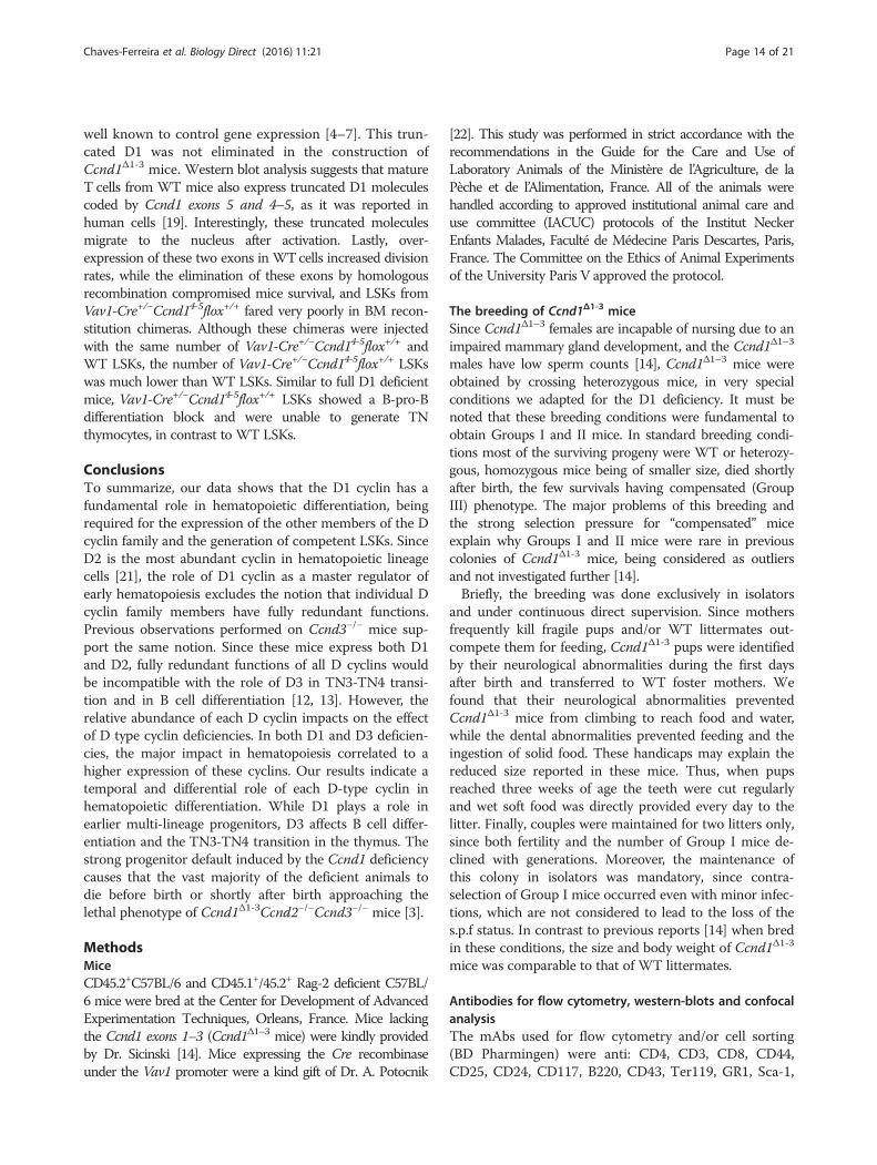
mice, hematopoietic differentiation was very heteroge-neous. Thymocyte numbers ranged from less than0.5×106 cells, to over 500x106 cells. Based on the thymussize we subdivided this colony into four Groups (Fig. 1a).Group I mice had a severe thymus atrophy. The thymihad very few CD4+CD8+ (double positive-DP) cells(Fig. 1b, upper graphs). The distribution of triple-negative (TN: CD4−CD8−CD3− Lineage− thymocytes)subpopulations was abnormal. In contrast to WT mice,where the more mature CD44−CD25+ (TN3) and CD44−
CD25− (TN4) populations are prevalent [16], TN3 andTN4 thymocytes were virtually absent (Fig. 1b, middlegraphs). Either the TN compartment was mostly consti-tuted of CD44+CD25− (TN1) and CD44+CD25+ (TN2)populations (Fig. 1b), or only had TN1 thymocytes (notshown). Since the TN1 thymocytes harbor different sub-populations, we further quantified the early thymocyteprogenitors (ETP), believed to be the progenitors of allthymocyte sets and characterized by the co-expressionof CD24 and c-kit (CD117) [16]. We found that inGroup I mice ETPs were reduced up to 50 fold whencompared to WT mice (Fig. 1b, lower graphs). There-fore, the Ccnd11-3 deficiency may block thymus differen-tiation at its earliest stage. The major reduction of theETPs further suggests that differentiation blocks mayprecede thymus seeding, i.e., may already be present inprogenitors located in the bone marrow (BM).Group II mice had moderate thymus atrophy. The
thymocyte populations were enriched in TN cells andhad fewer DP cells. The block on TN differentiation oc-curred at a later stage, i.e., in the TN3 to TN4 transition.Analysis of the TN1 population, however, showed a re-duction of the ETP compartment, although less import-ant than that found in Group I mice (Fig. 1b). Finally, inGroup III (Fig. 1b) and Group IV (not shown) thethymocyte sub-populations distribution appeared similarto that of WT mice. However, the analysis of the TN1compartment showed that these mice were also abnor-mal. In contrast to previous Groups where ETP popula-tions were reduced, these mice had more ETPs thantheir WT littermates (Fig. 1b, lower graphs).B cell differentiation in the BM progresses from pre-
pro B (B220low CD43+ CD24−) to pro B (B220low CD43+
CD24+) to pre-B (B220low CD43−) to naïve B cells(B220high IgM+) [17]. In Ccnd1Δ1-3 mice, B cell lineagedifferentiation blocks paralleled those found in the thymus(Fig. 2). Group I mice had a major pre-pro B differenti-ation block. Most B lineage cells were B220low CD43+
26 58
9.8 5
10 85
1.8 3
60 2
9 29
9 86
1.7 3
CD
4
CD8
Wild type Group I
Group II Group III
7.5
26 63
5.6 6.6
29 58
12 10
5 73
28 23
10 38
4.1Wild type Group I
Group II Group III
CD25
CD
44
CD117
CD
24
Wild type Group I
Group II Group III
Cel
l n
um
ber
/ th
ymi
(x10
-4)
Wild type
Group I
Group II
Group III
****
**
20
15
10
5
0
3,700
230
16,300
9,400
350280210
14070
10
5
0
Cel
l n
um
ber
/ th
ymi
(x10
-6)
******
**
WT Group I Group II Group III Group IV
1801401107035
20151050
Cel
l n
um
ber
/ th
ymi
(x10
-6)
******
****
**
+DP CD4 SP CD8+SP
Wild type
Group I
Group II
Group III
Cel
l n
um
ber
/ th
ymi
(x10
-6)
1.75
1.40
1.05
0.70
0.35
0TN1 TN2 TN3 TN4
**** *****
****
*****
Wild type
Group I
Group II
Group III
A
ETP
Fig. 1 (See legend on next page.)
Chaves-Ferreira et al. Biology Direct (2016) 11:21 Page 3 of 21
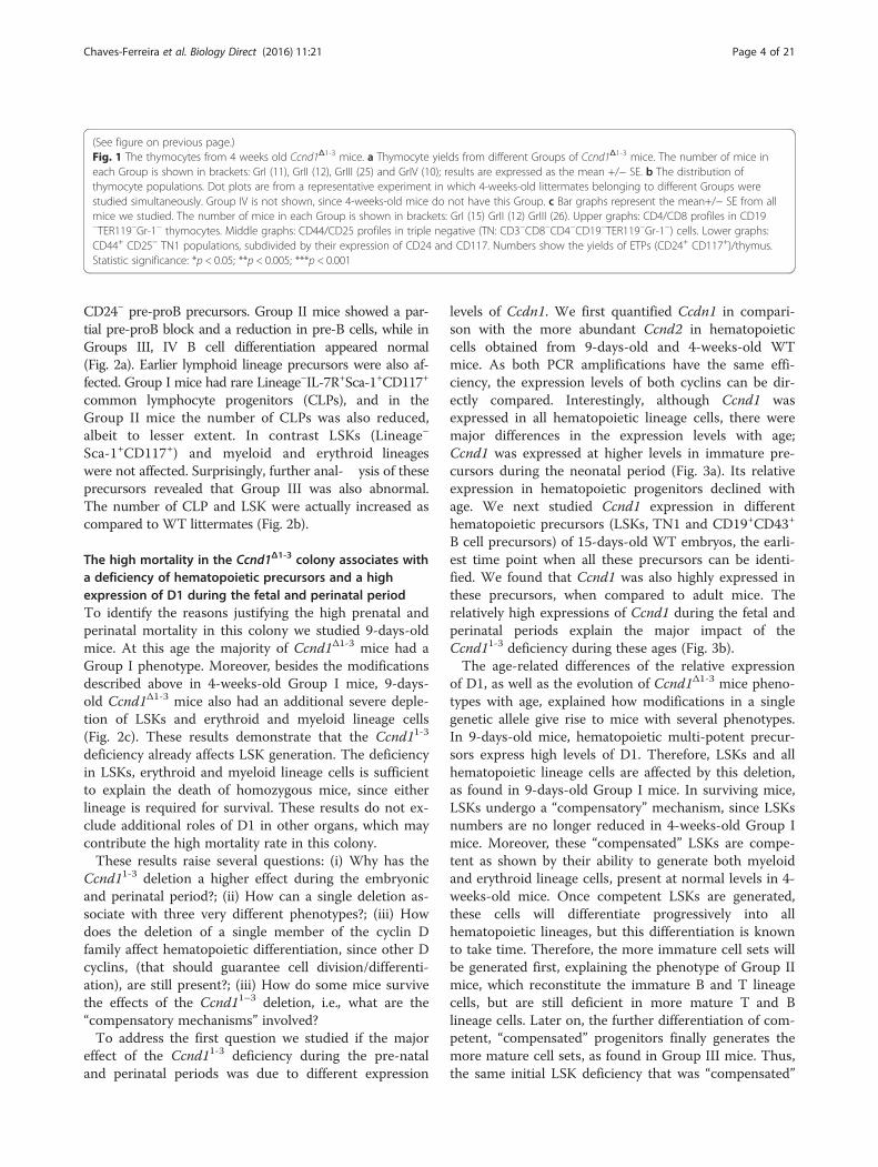
(See figure on previous page.)Fig. 1 The thymocytes from 4 weeks old Ccnd1Δ1-3 mice. a Thymocyte yields from different Groups of Ccnd1Δ1-3 mice. The number of mice ineach Group is shown in brackets: GrI (11), GrII (12), GrIII (25) and GrIV (10); results are expressed as the mean +/− SE. b The distribution ofthymocyte populations. Dot plots are from a representative experiment in which 4-weeks-old littermates belonging to different Groups werestudied simultaneously. Group IV is not shown, since 4-weeks-old mice do not have this Group. c Bar graphs represent the mean+/− SE from allmice we studied. The number of mice in each Group is shown in brackets: GrI (15) GrII (12) GrIII (26). Upper graphs: CD4/CD8 profiles in CD19−TER119−Gr-1− thymocytes. Middle graphs: CD44/CD25 profiles in triple negative (TN: CD3−CD8−CD4−CD19−TER119−Gr-1−) cells. Lower graphs:CD44+ CD25− TN1 populations, subdivided by their expression of CD24 and CD117. Numbers show the yields of ETPs (CD24+ CD117+)/thymus.Statistic significance: *p < 0.05; **p < 0.005; ***p < 0.001
Chaves-Ferreira et al. Biology Direct (2016) 11:21 Page 4 of 21
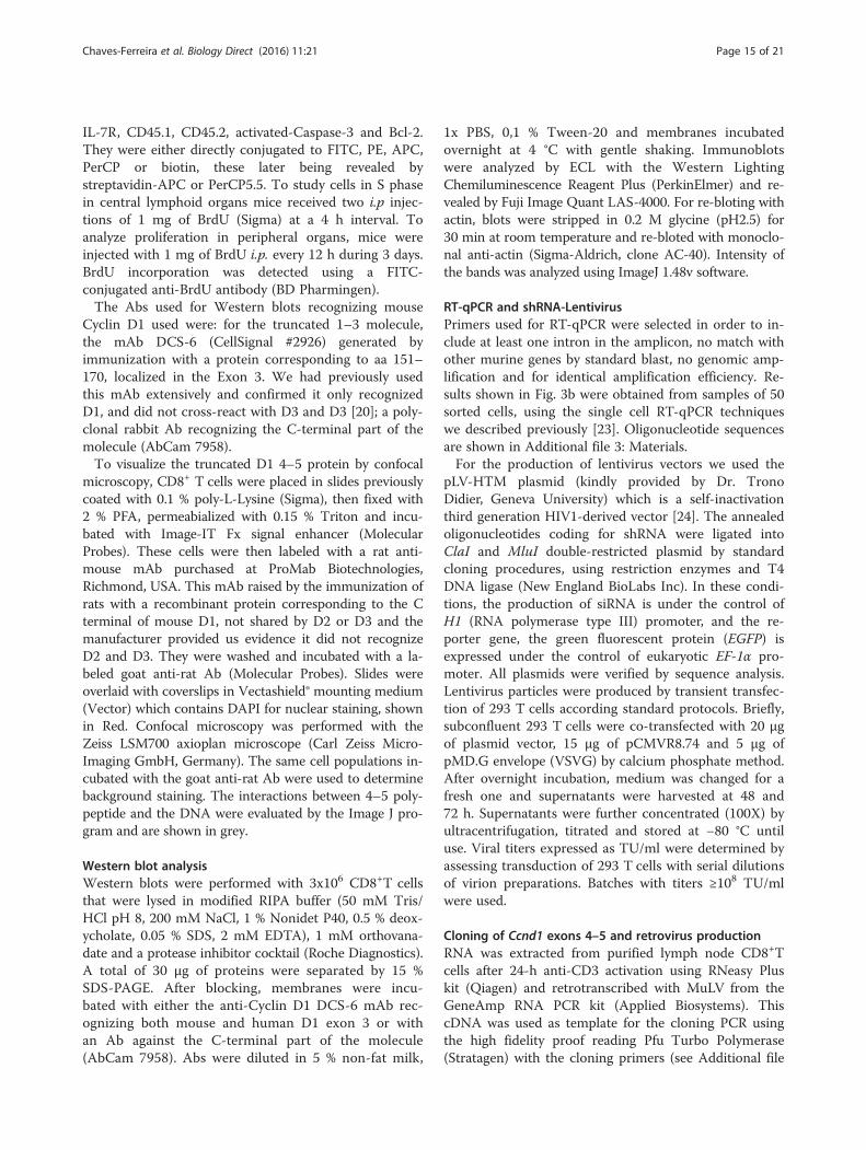
CD24− pre-proB precursors. Group II mice showed a par-tial pre-proB block and a reduction in pre-B cells, while inGroups III, IV B cell differentiation appeared normal(Fig. 2a). Earlier lymphoid lineage precursors were also af-fected. Group I mice had rare Lineage−IL-7R+Sca-1+CD117+
common lymphocyte progenitors (CLPs), and in theGroup II mice the number of CLPs was also reduced,albeit to lesser extent. In contrast LSKs (Lineage−
Sca-1+CD117+) and myeloid and erythroid lineageswere not affected. Surprisingly, further anal- ysis of theseprecursors revealed that Group III was also abnormal.The number of CLP and LSK were actually increased ascompared to WT littermates (Fig. 2b).
The high mortality in the Ccnd1Δ1-3 colony associates witha deficiency of hematopoietic precursors and a highexpression of D1 during the fetal and perinatal periodTo identify the reasons justifying the high prenatal andperinatal mortality in this colony we studied 9-days-oldmice. At this age the majority of Ccnd1Δ1-3 mice had aGroup I phenotype. Moreover, besides the modificationsdescribed above in 4-weeks-old Group I mice, 9-days-old Ccnd1Δ1-3 mice also had an additional severe deple-tion of LSKs and erythroid and myeloid lineage cells(Fig. 2c). These results demonstrate that the Ccnd11-3
deficiency already affects LSK generation. The deficiencyin LSKs, erythroid and myeloid lineage cells is sufficientto explain the death of homozygous mice, since eitherlineage is required for survival. These results do not ex-clude additional roles of D1 in other organs, which maycontribute the high mortality rate in this colony.These results raise several questions: (i) Why has the
Ccnd11-3 deletion a higher effect during the embryonicand perinatal period?; (ii) How can a single deletion as-sociate with three very different phenotypes?; (iii) Howdoes the deletion of a single member of the cyclin Dfamily affect hematopoietic differentiation, since other Dcyclins, (that should guarantee cell division/differenti-ation), are still present?; (iii) How do some mice survivethe effects of the Ccnd11–3 deletion, i.e., what are the“compensatory mechanisms” involved?To address the first question we studied if the major
effect of the Ccnd11-3 deficiency during the pre-nataland perinatal periods was due to different expression
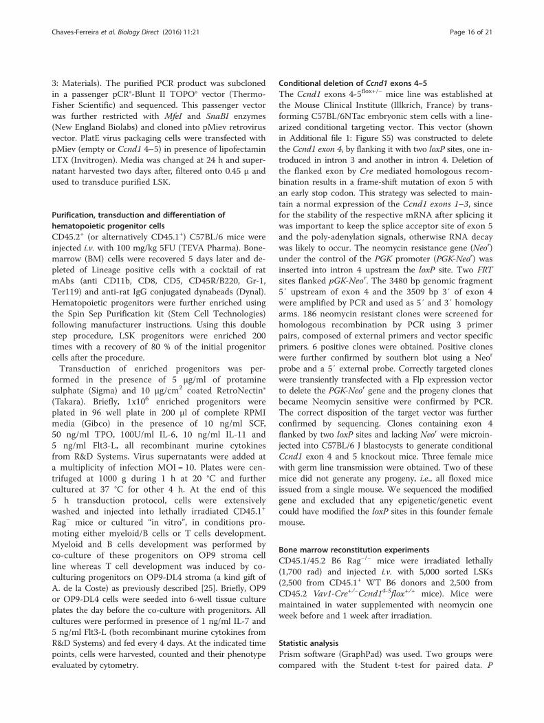
levels of Ccdn1. We first quantified Ccdn1 in compari-son with the more abundant Ccnd2 in hematopoieticcells obtained from 9-days-old and 4-weeks-old WTmice. As both PCR amplifications have the same effi-ciency, the expression levels of both cyclins can be dir-ectly compared. Interestingly, although Ccnd1 wasexpressed in all hematopoietic lineage cells, there weremajor differences in the expression levels with age;Ccnd1 was expressed at higher levels in immature pre-cursors during the neonatal period (Fig. 3a). Its relativeexpression in hematopoietic progenitors declined withage. We next studied Ccnd1 expression in differenthematopoietic precursors (LSKs, TN1 and CD19+CD43+
B cell precursors) of 15-days-old WT embryos, the earli-est time point when all these precursors can be identi-fied. We found that Ccnd1 was also highly expressed inthese precursors, when compared to adult mice. Therelatively high expressions of Ccnd1 during the fetal andperinatal periods explain the major impact of theCcnd11-3 deficiency during these ages (Fig. 3b).The age-related differences of the relative expression
of D1, as well as the evolution of Ccnd1Δ1-3 mice pheno-types with age, explained how modifications in a singlegenetic allele give rise to mice with several phenotypes.In 9-days-old mice, hematopoietic multi-potent precur-sors express high levels of D1. Therefore, LSKs and allhematopoietic lineage cells are affected by this deletion,as found in 9-days-old Group I mice. In surviving mice,LSKs undergo a “compensatory” mechanism, since LSKsnumbers are no longer reduced in 4-weeks-old Group Imice. Moreover, these “compensated” LSKs are compe-tent as shown by their ability to generate both myeloidand erythroid lineage cells, present at normal levels in 4-weeks-old mice. Once competent LSKs are generated,these cells will differentiate progressively into allhematopoietic lineages, but this differentiation is knownto take time. Therefore, the more immature cell sets willbe generated first, explaining the phenotype of Group IImice, which reconstitute the immature B and T lineagecells, but are still deficient in more mature T and Blineage cells. Later on, the further differentiation of com-petent, “compensated” progenitors finally generates themore mature cell sets, as found in Group III mice. Thus,the same initial LSK deficiency that was “compensated”
20
15
10
5
0
Cel
l n
um
ber
/ fe
mu
r(x
10-6
)
Wild type
Group I
Group II
Group III
**
*
B220+CD43+ B220+IgM+
Cel
l n
um
ber
/ fe
mu
r(x
10-6
)
TER119 Gr-1
80
60
40
20
0
**
**Wild type
Group I
A
B
C
100 101 102 103 104100 101 102 103 104
69%
100 101 102 103 104
9%
100 101 102 103 104
70% B220+CD43+
B220+CD43-
35
2038
54
1621 37
13
4475
75
1350
29
208
50
1227
40
1146
29
B220+
Wild type Group I Group II Group III
B22
0
CD43
B22
0
IgM
even
ts
CD24
Wild type Group I
CD
117
Sca-1
1,55623,220
Wild type Group I
TE
R11
9
Gr-1
6x10⁶
18x10659x106
30x106
100 101 102 103 104100 101 102 103 104 100 101 102 103 104100 101 102 103 104
Lin-IL-7R+
Wild type Group I Group II Group III
CD
117
Sca-1
1,700 567 773 2,340
CLP
4
3
2
1
0
Cel
l n
um
ber
/ fe
mu
r(x
10-3
)
***
*
Cel
l n
um
ber
/ fe
mu
r(x
10-3
)
HSC-LSK
30
20
10
0
***
10080
60
40
200
Cel
l n
um
ber
/ fe
mu
r(x
10- 3
)
HSC-LSK
**
CD
117
Sca-1
Lin-
Wild type Group I Group II Group III
79,00024,18030,08029,578
Wild type Group I Group II Group III
Fig. 2 (See legend on next page.)
Chaves-Ferreira et al. Biology Direct (2016) 11:21 Page 5 of 21
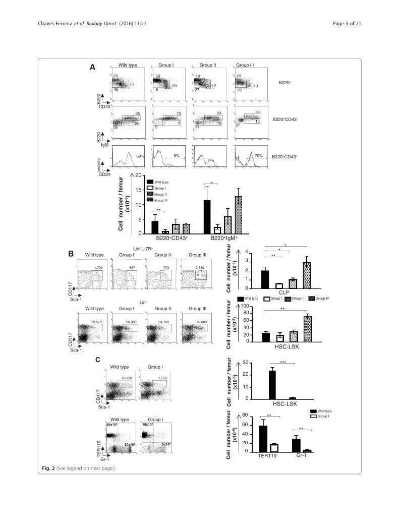
(See figure on previous page.)Fig. 2 The bone marrow cells from Ccnd1Δ1-3 mice. a, b Dot blots are from one representative experiment showing 4-weeks-old littermatesbelonging to different Groups studied simultaneously. Bar graphs represent the yield/femur of the different cell populations and are the mean+/− SE of all the mice we studied: GrI (15) GrII (12) GrIII (18). a B220+ B lineage cells. b Lineage negative (Lin−: CD3−CD4−CD8−CD19−TER119−Gr1−
Mac-1−NK1.1−) progenitors analyzed according to their expression of IL-7R, Sca-1 and CD117. Upper graphs: CLPs; Lower graphs: LSK/HSCs. Thenumbers in the dot-plots represent yields/femur. c Dot-plots are from one representative experiment showing 9-days-old WT and Ccnd1Δ1-3
littermates. Bar graphs are the mean+/SE of yields/femur all the 9-days-old mice we studied (n = 3). Upper graphs show LSK/HSC, lower graphserythroid and myeloid lineage cells. Statistic significance: *p < 0.05; **p < 0.005; ***p < 0.001
Chaves-Ferreira et al. Biology Direct (2016) 11:21 Page 6 of 21
can generate the different Ccnd1Δ1-3 mice phenotypes.Each of these phenotypes actually represents “snapshots”, taken at different time points after the occur-rence of the LSK “compensatory” event/s.
The expression of D cyclins in Ccnd1Δ1-3 miceTo investigate the mechanisms responsible for the modi-fications in hematopoietic differentiation in Ccnd1Δ1-3
mice, as well as the compensatory mechanisms ensuringthe survival of Group III mice, we studied how the vari-ous members of the D cyclin family were expressed inthe different Groups of mice. Besides mRNA coding forcyclin D2 (Ccnd2) and D3 (Ccnd3) we also evaluated theexpression of Ccnd1 exons 4–5, since these domains arenot deleted in Ccnd1Δ1-3 mice and were shown to havetranscription regulatory activities.Several mechanisms could explain the expression of a
truncated D1 molecule, coded by these 2 exons. It is wellknown that the introduction of the neomycin cassette asa strategy to generate deficient mice induces perturba-tions at and around the insertion locus [18]. Currentgene ablation strategies always delete the neomycin in-serts, but this was not done in Ccnd1Δ1-3 mice. The neo-mycin cassette has potential transcription initiating sites,which could lead to the transcription Ccnd1 exons 4–5.Moreover, Ccnd1 exon 4 is known to have transcription-initiating sites allowing transcription of exons 4–5. Inhumans (in which D1 has 85 % homology with mouseD1) these two exons, as well as exon 5 alone can betranscribed independently [19].We compared Ccnd expression in CD8+ T cells before
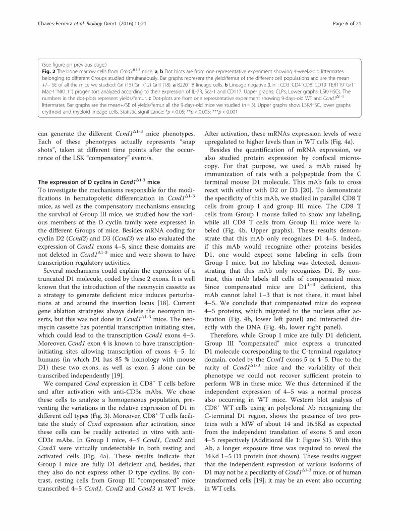
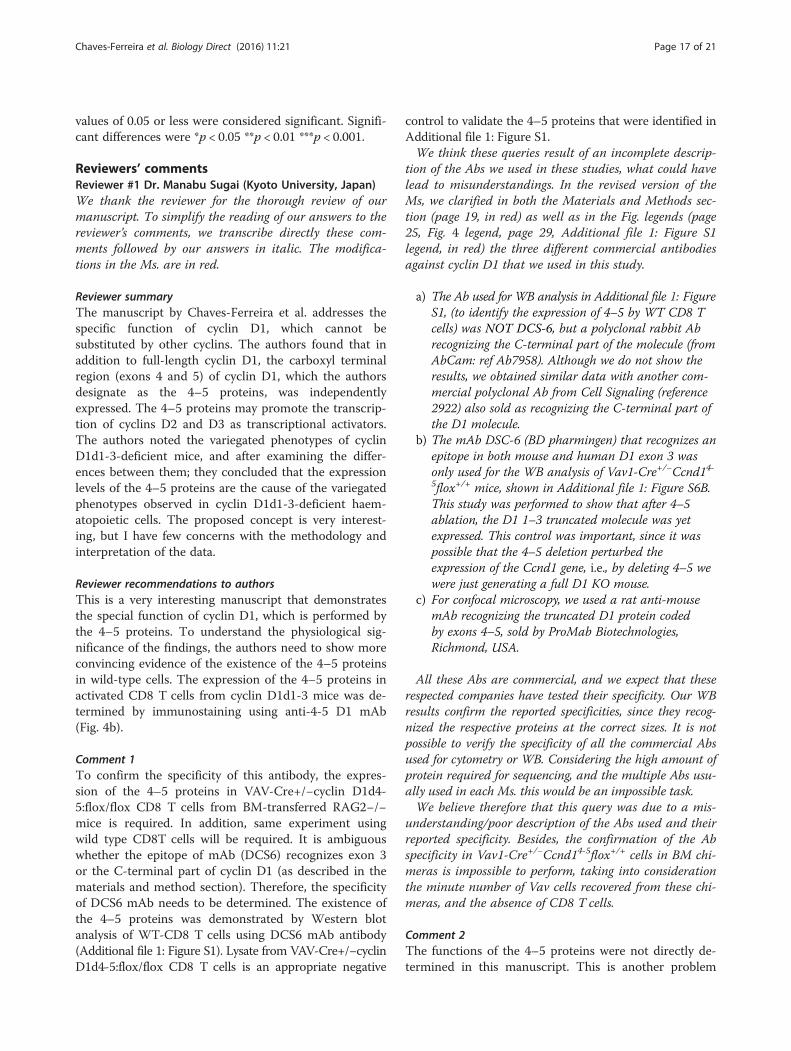
and after activation with anti-CD3ε mAbs. We chosethese cells to analyze a homogeneous population, pre-venting the variations in the relative expression of D1 indifferent cell types (Fig. 3). Moreover, CD8+ T cells facili-tate the study of Ccnd expression after activation, sincethese cells can be readily activated in vitro with anti-CD3ε mAbs. In Group I mice, 4–5 Ccnd1, Ccnd2 andCcnd3 were virtually undetectable in both resting andactivated cells (Fig. 4a). These results indicate thatGroup I mice are fully D1 deficient and, besides, thatthey also do not express other D type cyclins. By con-trast, resting cells from Group III “compensated” micetranscribed 4–5 Ccnd1, Ccnd2 and Ccnd3 at WT levels.
After activation, these mRNAs expression levels of wereupregulated to higher levels than in WT cells (Fig. 4a).Besides the quantification of mRNA expression, we
also studied protein expression by confocal micros-copy. For that purpose, we used a mAb raised byimmunization of rats with a polypeptide from the Cterminal mouse D1 molecule. This mAb fails to crossreact with either with D2 or D3 [20]. To demonstratethe specificity of this mAb, we studied in parallel CD8 Tcells from group I and group III mice. The CD8 Tcells from Group I mouse failed to show any labeling,while all CD8 T cells from Group III mice were la-beled (Fig. 4b, Upper graphs). These results demon-strate that this mAb only recognizes D1 4–5. Indeed,if this mAb would recognize other proteins besidesD1, one would expect some labeling in cells fromGroup I mice, but no labeling was detected, demon-strating that this mAb only recognizes D1. By con-trast, this mAb labels all cells of compensated mice.Since compensated mice are D11–3 deficient, thismAb cannot label 1–3 that is not there, it must label4–5. We conclude that compensated mice do express4–5 proteins, which migrated to the nucleus after ac-tivation (Fig. 4b, lower left panel) and interacted dir-ectly with the DNA (Fig. 4b, lower right panel).Therefore, while Group I mice are fully D1 deficient,
Group III “compensated” mice express a truncatedD1 molecule corresponding to the C-terminal regulatorydomain, coded by the Ccnd1 exons 5 or 4–5. Due to therarity of Ccnd1Δ1-3 mice and the variability of theirphenotype we could not recover sufficient protein toperform WB in these mice. We thus determined if theindependent expression of 4–5 was a normal processalso occurring in WT mice. Western blot analysis ofCD8+ WT cells using an polyclonal Ab recognizing theC-terminal D1 region, shows the presence of two pro-teins with a MW of about 14 and 16.5Kd as expectedfrom the independent translation of exons 5 and exon4–5 respectively (Additional file 1: Figure S1). With thisAb, a longer exposure time was required to reveal the34Kd 1–5 D1 protein (not shown). These results suggestthat the independent expression of various isoforms ofD1 may not be a peculiarity of Ccnd1Δ1-3 mice, or of humantransformed cells [19]; it may be an event also occurringin WTcells.

Ccnd1Ccnd2
Ccnd1Ccnd2
A
B
Fig. 3 The expression of Ccnd1 in mice of different ages. The individual subpopulations of hematopoietic lineage cells were sorted from WT micewith different ages (a). Quantification of Ccnd1 and Ccnd2 transcripts in different subpopulations of hematopoietic cells from 9-days and 4-weeks-old WT mice. Results show the mRNA expression of the Ccnd1 and Ccnd2, determined by RT-qPCR, as referred to the RPII house-keeping gene.They are the mean+/− SD of three independent samples. Please note: Since the different PCR amplifications had the same efficiency, theexpression levels of individual Ccnd can be directly compared. b Quantification of Ccnd1 in LSK, TN1 and CD19+IgM−CD43+ precursors of15-days-old fetus (F) and 4-weeks-old adult (A) mice, as referred to the GAPDH house-keeping gene. Results show the mean+/− SD of from threeindependent samples. SD deviations are too small to be visualized with the present scale. Statistic significance: *p < 0.05 (paired student t-test)
Chaves-Ferreira et al. Biology Direct (2016) 11:21 Page 7 of 21
These results provide a likely explanation for the dif-ferent phenotypes of Ccnd1Δ1-3 mice. Group I mice arefully D1 deficient, but also do not express Ccnd2 andCcnd3. These results indicate that the D1 cyclin controlsthe transcription of Ccnd2 and Ccnd3 in hematopoieticcells, as suggested by the highly significant binding of
D1 to the Ccnd2 and Ccnd3 promoters [9]. To furtherconfirm the absence of D cyclins in these mice, we stud-ied the capacity of cells from Group I mice to divide. Inthe absence of all D cyclins, hematopoietic cells shouldbe unable to divide. Indeed, this was the case. Group Imice showed a profound block in BrdU incorporation in
4-5Ccnd1
Ccnd2
Ccnd3
0.1
0.2
0.3
0.4
0.5
0.6
0.7
0.8Non activated
D c
yclin
mR
NA
exp
ress
ion
(rel
ativ
e to
RPII)
0.1
0.2
0.3
0.4
0.5
0.6
0.7
0.8
wild type Group I Group III
Activated
A
B
***
***
***
***
***
***
*
**
**
ns
ns
ns
Scale bar: upper micrographs = 30µm Lower micrographs = 10µm
Fig. 4 The expression of D cyclins in Ccnd1Δ1-3 mice. a Expression levels of Ccnd2, Ccnd3, and exons 4–5 of Ccnd1 relative to the RPIIhousekeeping gene. Results are from sorted naïve CD8+T cells before (upper graphs) or after 24 h stimulation with anti-CD3 mAbs (lower graphs).RPII housekeeping gene expression levels were similar in all mouse Groups. Results are the mean+/− SD of three independent experiments.Statistic significance: *p < 0.05; **p < 0.005; ***p < 0.001 (paired student t-test). In each experiment amplifications from different Groups wereperformed simultaneously. b Expression of truncated 4–5 D1 protein by resting CD8+ T cells from Group I (upper left panel) and Group III mice(upper right panel); by activated CD8+ T cells from group III mice (lower panels): lower right panel show 4–5 migration into the nucleus and lowerleft panel the interaction with the DNA (in grey) upon T cell activation. The 4–5 D1 fragment was identified with rat anti-mouse D1 4–5 fragmentmAb from ProMab Biotechnologies, Richmond, USA, revealed with a goat anti-rat Ab (Molecular Probes). The nucleus was labeled with DAPI andis shown in Red. Co-localization analysis was performed using the Image J program
Chaves-Ferreira et al. Biology Direct (2016) 11:21 Page 8 of 21
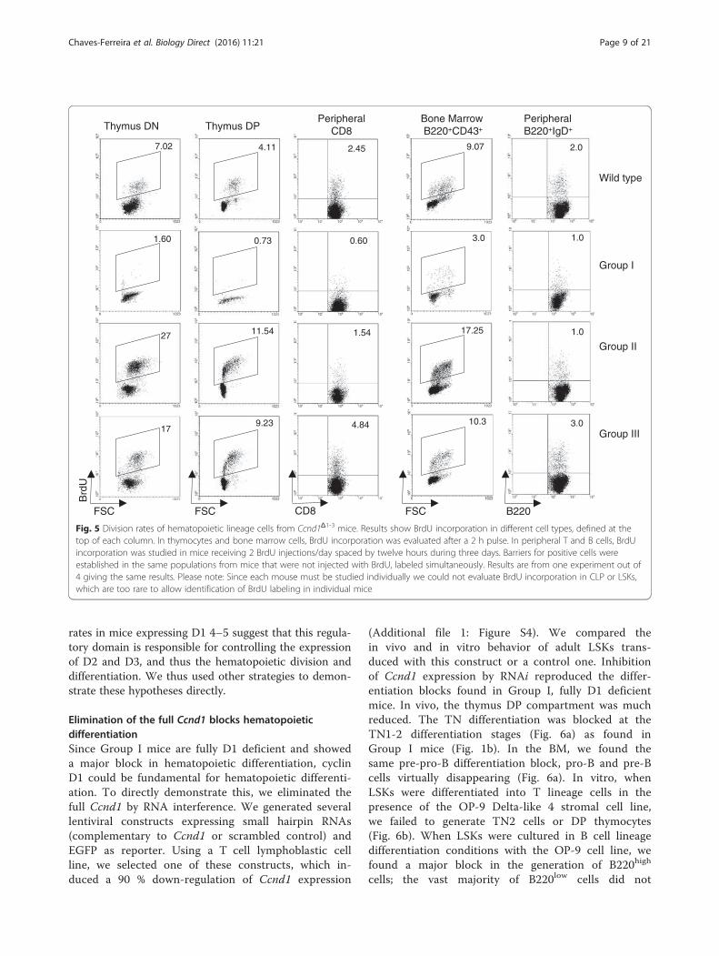
T cell precursors in the thymus and B cell precursors inthe BM, mature T and B cells yet showing reduced div-ision rates (Fig. 5). In contrast, the remaining Groupsexpress truncated 4–5 D1 molecules. In response to en-vironmental stimuli these cells show a major up-regulation of 4–5 Ccnd1 and other D cyclins, whichcould eventually lead to an increased division rate and
lymphoid hyperplasia. Indeed, we found an increasedBrdU incorporation in cells from Groups II and III mice(Fig. 5). By contrast we found no evidence of increasedcell death in any of these populations (Additional file 2:Note 1 and Additional file 1: Figures S2 & S3).These results indicate that the cyclin D1 is a master
regulator of hematopoiesis. Moreover, the high division
3.0
1.0
Wild type
Group I
Group II
Group III
9.07
3.0
17.25
10.3
1.0
B220FSC
4.84
1.54
0.60
2.45
CD8
1.60
7.02
27
17
FSC
Brd
U
9.23
11.54
4.11
0.73
FSC
Thymus DN Thymus DPPeripheral
CD8Bone MarrowB220+CD43+
2.0
Peripheral B220+IgD+
Fig. 5 Division rates of hematopoietic lineage cells from Ccnd1Δ1-3 mice. Results show BrdU incorporation in different cell types, defined at thetop of each column. In thymocytes and bone marrow cells, BrdU incorporation was evaluated after a 2 h pulse. In peripheral T and B cells, BrdUincorporation was studied in mice receiving 2 BrdU injections/day spaced by twelve hours during three days. Barriers for positive cells wereestablished in the same populations from mice that were not injected with BrdU, labeled simultaneously. Results are from one experiment out of4 giving the same results. Please note: Since each mouse must be studied individually we could not evaluate BrdU incorporation in CLP or LSKs,which are too rare to allow identification of BrdU labeling in individual mice
Chaves-Ferreira et al. Biology Direct (2016) 11:21 Page 9 of 21
rates in mice expressing D1 4–5 suggest that this regula-tory domain is responsible for controlling the expressionof D2 and D3, and thus the hematopoietic division anddifferentiation. We thus used other strategies to demon-strate these hypotheses directly.
Elimination of the full Ccnd1 blocks hematopoieticdifferentiationSince Group I mice are fully D1 deficient and showeda major block in hematopoietic differentiation, cyclinD1 could be fundamental for hematopoietic differenti-ation. To directly demonstrate this, we eliminated thefull Ccnd1 by RNA interference. We generated severallentiviral constructs expressing small hairpin RNAs(complementary to Ccnd1 or scrambled control) andEGFP as reporter. Using a T cell lymphoblastic cellline, we selected one of these constructs, which in-duced a 90 % down-regulation of Ccnd1 expression
(Additional file 1: Figure S4). We compared thein vivo and in vitro behavior of adult LSKs trans-duced with this construct or a control one. Inhibitionof Ccnd1 expression by RNAi reproduced the differ-entiation blocks found in Group I, fully D1 deficientmice. In vivo, the thymus DP compartment was muchreduced. The TN differentiation was blocked at theTN1-2 differentiation stages (Fig. 6a) as found inGroup I mice (Fig. 1b). In the BM, we found thesame pre-pro-B differentiation block, pro-B and pre-Bcells virtually disappearing (Fig. 6a). In vitro, whenLSKs were differentiated into T lineage cells in thepresence of the OP-9 Delta-like 4 stromal cell line,we failed to generate TN2 cells or DP thymocytes(Fig. 6b). When LSKs were cultured in B cell lineagedifferentiation conditions with the OP-9 cell line, wefound a major block in the generation of B220high
cells; the vast majority of B220low cells did not
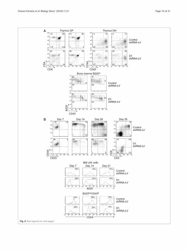
Fig. 6 (See legend on next page.)
Chaves-Ferreira et al. Biology Direct (2016) 11:21 Page 10 of 21
(See figure on previous page.)Fig. 6 Effects of cyclin D1 RNAi in LSK differentiation. Bone marrow LSK (Lin−Sca1+ ckithigh) progenitors were sorted from CD45.2+ mice BM, andwere transduced with either a cyclin D1 or a control shRNA-LV-EGFP. a They were directly injected into lethally irradiated (1,700 rad) CD45.1+Rag−
mice. Results show EGFP+ cells in four different individual mice two months after LSK transfer. Upper graphs: EGFP+ thymocytes; Lower graphs: Bcell lineage profiles in the BM, analyzed as described in Fig. 2. b They were cultured in in vitro conditions promoting T cell (upper graphs) or B/myeloid cell differentiation (lower graphs). Results show the phenotype of EGFP+ at different time points after culture
Table 1 Ccnd1 4–5 over-expression increases lymphocytedivision
BrdU+ CD8 + T cells (%)
Experimental Group Mouse # Mean SE p
1 2 3 4
EGFP+ MOCK 20.0 13.5 10.7 30.6 18.7 6.6 0.01
EGFP+ D1 4-5 32.8 16.3 18.8 41.7 27.4 9.9
LSKs were purified from 5FU-treated CD45.1 and CD45.2 wild type mice.CD45.1 LSK were transduced with a D1 4–5 coding retrovirus whereas CD45.2LSK were transduced with the same control (Mock) virus. Equal numbers ofeach transduced LSK were injected simultaneously into lethally irradiated Rag−
mice. 3 weeks after cell transfer, the proliferation rates of the EGFP+ CD8+Tsubset were evaluated after 3 i.p. pulses of BrdU. Table 1 shows the % of BrdU+ CD8+T cells generated from transduced LSKs (EGFP+) Mock (CD45.2) and D14–5 (CD45.1) in each individual mouse (n = 4). Differences in proliferationbetween Mock and D1 4–5 transduced lymphocytes were evaluated by thepaired Student t-test
Chaves-Ferreira et al. Biology Direct (2016) 11:21 Page 11 of 21
express CD24, i.e., the pre-pro-B differentiation blockfound in Group I mice was reproduced (Fig. 6b).These results formally demonstrate the master role ofthe D1 cyclin in hematopoietic differentiation.
Overexpression of Ccnd1 exons 4–5 in hematopoieticprecursors increases cell divisionTo demonstrate directly the role of the truncated D1 4–5molecule in hematopoiesis, one could express the 4–5truncated molecule in the hematopoietic precursors ofgroup I mice, and study if this overexpression rescuetheir phenotype. However, these experiments were im-possible to perform and, importantly, could not give reli-able results. We could not recover enough precursors toachieve efficient infection and reconstitution. Group Imice are quite young (what reduces the total number ofcells we can recover from the BM), virtually devoid ofprogenitors, and these progenitors do not divide. Be-sides, these mice are very rare excluding that we couldpool cells from several Group I mice to perform thesestudies. But more importantly, these experiments wereunlikely to give straightforward conclusions. Since wehad to wait several weeks before the analysis of the pro-geny of injected BM, and Group I rapidly compensatetheir phenotype, both transduced and non-transducedcells should show compensated phenotypes when tested(Additional file 2: Note 2). Therefore, to correlate 4–5expression to hematopoietic cells division we enforcedthe expression of 4–5 in LSKs from WT mice. LSKsoverexpressing 4–5 or infected with an empty vectorwere co-injected at the same number in lethally irradiatedRag− mice. In the 4 mice we studied in two independentexperiments, the proliferation rate of peripheral CD8+ Tcells generated from 4–5 enforced progenitors washigher (p = 0.01) than that of mock transduced cells(Table 1). These results show that overexpression of 4–5can modify division rates even in WTcells.
Conditional ablation of D1 4–5 prevents hematopoieticdifferentiationThe other possible alternative to evaluate the role of 4–5in hematopoietic cell division and differentiation, wasevaluating the effects of a deletion of 4–5. In these ex-periments it was fundamental to demonstrate that thedeletion of 4–5 did not perturb the overall expression ofthe D1 molecule, i.e., that by deleting 4–5 we were notjust generating a fully D1 deficient mice. Therefore, we
use an experimental strategy that should ensure the ex-pression of D1 1–3 in 4–5 deleted mice. We generatedconditional deficient mice by introducing two loxP sitesflanking the 4th exon (Additional file 1: Figure S5). Inthe selected strategy, after Cre recombination exon 4should be excised completely. The further splicing ofexon 3 to the splice acceptor site of exon 5 introduces a5′ stop codon, preventing the expression of the 5th exon.This strategy was designed to allow the stable expressionof exons 1–3, since neither the splice acceptor site northe poly-adenylation sites of exon 5 are modified, whichshould guarantee mRNA stability.We crossed these mice with mice expressing Cre
under the Vav1 promoter (which induces Cre expressionin LSKs and all hematopoietic lineage cells) but hadmajor problems in generating a viable progeny Vav-CreCcnd14-5flox+/+ progeny (Additional file 2: Note 3). Thevery rare mice that had this genotype, expressed Ccnd1 1–3but not Ccnd1 4–5 at early ages (4–7 days-old mice)(Additional file 1: Figure S6A). These results clearlydemonstrated that the deletion of 4–5 did not perturb theoverall expression of the D1 molecule, since exons 1–3were yet transcribed. Western blot analysis in older miceusing the DCS-6 mAb, that recognizes the D1 exon 3,confirmed the presence of a truncated molecule with aMW corresponding to the protein coded by Ccnd1 exons1–3. It must be noted that we used this mAb previously,and showed that it is fully D1 specific, i.e., it does notrecognize D2 or D3 or any other protein of the cell [20].However, besides this truncated molecule, a band of34Kd corresponding to the WT D1 protein was also

Chaves-Ferreira et al. Biology Direct (2016) 11:21 Page 12 of 21
present (Additional file 1: Figure S6B). To determine whythe WT protein was still expressed in these mice, westudied the kinetics of Cre expression by RT-qPCR.While Cre expression remained stable in Vav1-Cre mice,it rapidly declined in Vav1-Cre+/−Ccnd14-5flox+/+ mice(Additional file 1: Figure S6C). We conclude that thesemice also compensated their Ccnd1 exons 4–5 deficiencyby the selection of the very rare hematopoietic cells failingto express Cre.To study the role of Ccnd1 4–5, we thus performed
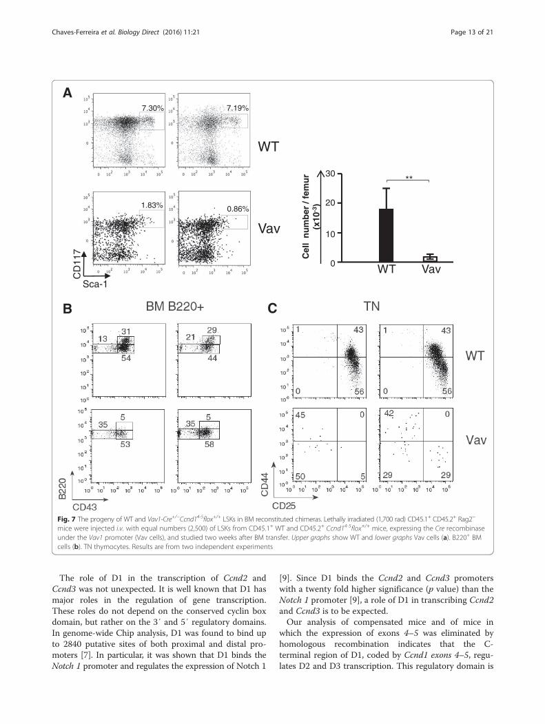
competitive BM reconstitution experiments. To preventthe emergency of compensatory events, these chimeraswere studied as early as at two weeks after BM transfer.When lethally irradiated CD45.1+/CD45.2+ Rag− micewere injected with identical numbers (2,500 cells) ofCD45.1+ WT and CD45.2+ LSKs from Vav1-Cre+/−Ccnd14-5flox+/+ mice (hereafter referred as Vav cells),only 7 % of the hematopoietic lineage cells were of Vavorigin. As expected, the frequency of CD45.2 Vav LSKswas much reduced when compared to that of CD45.1WT LSKs (Fig. 7a). WT CD45.1 cells generated B220high
B lineage cells while Vav precursors only generatedB220low precursors enriched in B220low CD43− precur-sors (Fig. 7b), as found in Group I mice. In the thymus,all WT thymocytes showed a coordinated differentiation,TN precursors being at the CD44+CD25+ (TN2) andCD44−CD25+ (TN3) differentiation stages (Fig. 7c). VavTN precursors in the thymus were virtually absent.Their CD44/CD25 phenotype did not reproduce that ofWT TN cells (Fig. 7c-left) or they showed a major accu-mulation of the CD44+ CD25− TN1 population, and amajor reduction of TN2 and TN3 cells (right), as foundin Group I mice (Fig. 1). These results confirm that theelimination of Ccnd1 exons 4–5 blocks hematopoieticdifferentiation, reproducing the phenotypes of fully D1deficient, Group I mice.
DiscussionThe present data emphasize the difficulty of evaluatingthe role of fundamental molecules since their ablationfrequently increases mortality or/and leads to the emer-gency of compensatory mechanisms, which may maskongoing major effects. Thus, the different phenotypes ofCcnd1Δ1-3 mice were initially very perplexing. It was notevident why the partial deletion of a single gene wouldgenerate mice with such different phenotypes as GroupI, II, III mice. The reasons behind this heterogeneity be-came clear, once we associated the mortality rates in thiscolony at different ages, with the variations in both phe-notypes and D1 expression levels. A substantial fractionof Ccnd1Δ1-3 embryos died, indicating that this defi-ciency can be embryonic lethal. Another large fractionof mice died shortly after birth. The comparison of 9-days and 4-weeks-old mice identified that the Ccnd11-3
deletion affected LSKs in 9-days-old mice, inducing se-vere reductions in erythroid and myeloid lineages. Thus,mice often died during this period, since either erythroidor myeloid lineages are required for survival. The higherpre-natal and perinatal death of these mice is also justi-fied by a much higher expression of Ccnd1 during theseperiods, when compared to adult mice. After this criticalpre- and peri-natal period, mice survived if they recon-stituted LSKs, erythroid and myeloid lineages. These re-sults indicated firstly that the Ccnd11-3 deletion alreadyaffected LSK generation, and secondly, that Ccnd1Δ1-3
mice developed one or several “compensatory” mecha-nisms overcoming the LSK deficiency. This compensa-tion not only increased LSK numbers, but alsogenerated competent LSKs as shown by their capacity toreconstitute the erythroid and myeloid lineages in 4-weeks-old mice. Moreover, “compensation” did notoccur simultaneously in all mice.The asynchronous generation of competent LSKs in
Ccnd1Δ1-3 mice was sufficient to fully explain the differ-ent phenotypes of individual mice from this colony.Once competent LSKs are generated, these cells differ-entiate progressively though this differentiation takestime. Therefore, they will first generate the more imma-ture hematopoietic cell sets. This explains the phenotypeof the Group II mice, which reconstitute the most im-mature, but are still deficient in more mature, T and Blineage cells. Later, the further differentiation of thesecompetent progenitors will lead to the generation ofmore mature cells, as found in “fully compensated”Group III mice, whose phenotype is largely equivalent tothat of normal mice. Thus, the same initial LSK deficiency,when “compensated” at different time points in differentmice, is expected to generate the three different phenotypes,which correspond to “snap-shots” taken at different timepoints after the occurrence of the LSK “compensation”.Our results also show why D1 is fundamental for
hematopoietic differentiation, and how “compensation”occurs. We demonstrated that the hematopoietic differ-entiation deficiency in Group I mice was indeed due tothe D1 deficiency, since we reproduced the samehematopoietic differentiation blockages by eliminatingthe full D1 molecule by RNAi. Concerning the mecha-nisms, we found that fully D1 deficient Group I mice didnot transcribe Ccnd2 and Ccnd3. The absence of allmembers of the D cyclins family in these mice is alsoconfirmed by a failure of their cells to divide, i.e.,to incorporate BrdU. Indeed, all subpopulations ofhematopoietic lineage cells from Group I mice showed avirtual absence of BrdU incorporation, while we foundno evidence that increased apoptosis contributed to theirreduced numbers, or to the hematopoietic differentiationblockage. These results indicate that D1 controls the ex-pression of D2 and D3, by regulating their transcription.
A
B C
7.30%
1.83%
7.19%
0.86%
WT
Vav
CD
117
Sca-1
Cel
l n
um
ber
/ fe
mu
r(x
10-3
)
30
20
10
0
**
WT Vav
Fig. 7 The progeny of WT and Vav1-Cre+/−Ccnd14-5flox+/+ LSKs in BM reconstituted chimeras. Lethally irradiated (1,700 rad) CD45.1+ CD45.2+ Rag2−
mice were injected i.v. with equal numbers (2,500) of LSKs from CD45.1+ WT and CD45.2+ Ccnd14-5flox+/+ mice, expressing the Cre recombinaseunder the Vav1 promoter (Vav cells), and studied two weeks after BM transfer. Upper graphs show WT and lower graphs Vav cells (a). B220+ BMcells (b). TN thymocytes. Results are from two independent experiments
Chaves-Ferreira et al. Biology Direct (2016) 11:21 Page 13 of 21
The role of D1 in the transcription of Ccnd2 andCcnd3 was not unexpected. It is well known that D1 hasmajor roles in the regulation of gene transcription.These roles do not depend on the conserved cyclin boxdomain, but rather on the 3′ and 5′ regulatory domains.In genome-wide Chip analysis, D1 was found to bind upto 2840 putative sites of both proximal and distal pro-moters [7]. In particular, it was shown that D1 binds theNotch 1 promoter and regulates the expression of Notch 1
[9]. Since D1 binds the Ccnd2 and Ccnd3 promoterswith a twenty fold higher significance (p value) than theNotch 1 promoter [9], a role of D1 in transcribing Ccnd2and Ccnd3 is to be expected.Our analysis of compensated mice and of mice in
which the expression of exons 4–5 was eliminated byhomologous recombination indicates that the C-terminal region of D1, coded by Ccnd1 exons 4–5, regu-lates D2 and D3 transcription. This regulatory domain is
Chaves-Ferreira et al. Biology Direct (2016) 11:21 Page 14 of 21
well known to control gene expression [4–7]. This trun-cated D1 was not eliminated in the construction ofCcnd1Δ1-3 mice. Western blot analysis suggests that matureT cells from WT mice also express truncated D1 moleculescoded by Ccnd1 exons 5 and 4–5, as it was reported inhuman cells [19]. Interestingly, these truncated moleculesmigrate to the nucleus after activation. Lastly, over-expression of these two exons in WTcells increased divisionrates, while the elimination of these exons by homologousrecombination compromised mice survival, and LSKs fromVav1-Cre+/−Ccnd14-5flox+/+ fared very poorly in BM recon-stitution chimeras. Although these chimeras were injectedwith the same number of Vav1-Cre+/−Ccnd14-5flox+/+ andWT LSKs, the number of Vav1-Cre+/−Ccnd14-5flox+/+ LSKswas much lower than WT LSKs. Similar to full D1 deficientmice, Vav1-Cre+/−Ccnd14-5flox+/+ LSKs showed a B-pro-Bdifferentiation block and were unable to generate TNthymocytes, in contrast to WT LSKs.
ConclusionsTo summarize, our data shows that the D1 cyclin has afundamental role in hematopoietic differentiation, beingrequired for the expression of the other members of the Dcyclin family and the generation of competent LSKs. SinceD2 is the most abundant cyclin in hematopoietic lineagecells [21], the role of D1 cyclin as a master regulator ofearly hematopoiesis excludes the notion that individual Dcyclin family members have fully redundant functions.Previous observations performed on Ccnd3−/− mice sup-port the same notion. Since these mice express both D1and D2, fully redundant functions of all D cyclins wouldbe incompatible with the role of D3 in TN3-TN4 transi-tion and in B cell differentiation [12, 13]. However, therelative abundance of each D cyclin impacts on the effectof D type cyclin deficiencies. In both D1 and D3 deficien-cies, the major impact in hematopoiesis correlated to ahigher expression of these cyclins. Our results indicate atemporal and differential role of each D-type cyclin inhematopoietic differentiation. While D1 plays a role inearlier multi-lineage progenitors, D3 affects B cell differ-entiation and the TN3-TN4 transition in the thymus. Thestrong progenitor default induced by the Ccnd1 deficiencycauses that the vast majority of the deficient animals todie before birth or shortly after birth approaching thelethal phenotype of Ccnd1Δ1-3Ccnd2−/−Ccnd3−/− mice [3].
MethodsMiceCD45.2+C57BL/6 and CD45.1+/45.2+ Rag-2 deficient C57BL/6 mice were bred at the Center for Development of AdvancedExperimentation Techniques, Orleans, France. Mice lackingthe Ccnd1 exons 1–3 (Ccnd1Δ1–3 mice) were kindly providedby Dr. Sicinski [14]. Mice expressing the Cre recombinaseunder the Vav1 promoter were a kind gift of Dr. A. Potocnik
[22]. This study was performed in strict accordance with therecommendations in the Guide for the Care and Use ofLaboratory Animals of the Ministère de l’Agriculture, de laPèche et de l’Alimentation, France. All of the animals werehandled according to approved institutional animal care anduse committee (IACUC) protocols of the Institut NeckerEnfants Malades, Faculté de Médecine Paris Descartes, Paris,France. The Committee on the Ethics of Animal Experimentsof the University Paris V approved the protocol.
The breeding of Ccnd1Δ1-3 miceSince Ccnd1Δ1–3 females are incapable of nursing due to animpaired mammary gland development, and the Ccnd1Δ1–3
males have low sperm counts [14], Ccnd1Δ1–3 mice wereobtained by crossing heterozygous mice, in very specialconditions we adapted for the D1 deficiency. It must benoted that these breeding conditions were fundamental toobtain Groups I and II mice. In standard breeding condi-tions most of the surviving progeny were WT or heterozy-gous, homozygous mice being of smaller size, died shortlyafter birth, the few survivals having compensated (GroupIII) phenotype. The major problems of this breeding andthe strong selection pressure for “compensated” miceexplain why Groups I and II mice were rare in previouscolonies of Ccnd1Δ1-3 mice, being considered as outliersand not investigated further [14].Briefly, the breeding was done exclusively in isolators
and under continuous direct supervision. Since mothersfrequently kill fragile pups and/or WT littermates out-compete them for feeding, Ccnd1Δ1-3 pups were identifiedby their neurological abnormalities during the first daysafter birth and transferred to WT foster mothers. Wefound that their neurological abnormalities preventedCcnd1Δ1-3 mice from climbing to reach food and water,while the dental abnormalities prevented feeding and theingestion of solid food. These handicaps may explain thereduced size reported in these mice. Thus, when pupsreached three weeks of age the teeth were cut regularlyand wet soft food was directly provided every day to thelitter. Finally, couples were maintained for two litters only,since both fertility and the number of Group I mice de-clined with generations. Moreover, the maintenance ofthis colony in isolators was mandatory, since contra-selection of Group I mice occurred even with minor infec-tions, which are not considered to lead to the loss of thes.p.f status. In contrast to previous reports [14] when bredin these conditions, the size and body weight of Ccnd1Δ1-3
mice was comparable to that of WT littermates.
Antibodies for flow cytometry, western-blots and confocalanalysisThe mAbs used for flow cytometry and/or cell sorting(BD Pharmingen) were anti: CD4, CD3, CD8, CD44,CD25, CD24, CD117, B220, CD43, Ter119, GR1, Sca-1,
Chaves-Ferreira et al. Biology Direct (2016) 11:21 Page 15 of 21
IL-7R, CD45.1, CD45.2, activated-Caspase-3 and Bcl-2.They were either directly conjugated to FITC, PE, APC,PerCP or biotin, these later being revealed bystreptavidin-APC or PerCP5.5. To study cells in S phasein central lymphoid organs mice received two i.p injec-tions of 1 mg of BrdU (Sigma) at a 4 h interval. Toanalyze proliferation in peripheral organs, mice wereinjected with 1 mg of BrdU i.p. every 12 h during 3 days.BrdU incorporation was detected using a FITC-conjugated anti-BrdU antibody (BD Pharmingen).The Abs used for Western blots recognizing mouse
Cyclin D1 used were: for the truncated 1–3 molecule,the mAb DCS-6 (CellSignal #2926) generated byimmunization with a protein corresponding to aa 151–170, localized in the Exon 3. We had previously usedthis mAb extensively and confirmed it only recognizedD1, and did not cross-react with D3 and D3 [20]; a poly-clonal rabbit Ab recognizing the C-terminal part of themolecule (AbCam 7958).To visualize the truncated D1 4–5 protein by confocal
microscopy, CD8+ T cells were placed in slides previouslycoated with 0.1 % poly-L-Lysine (Sigma), then fixed with2 % PFA, permeabialized with 0.15 % Triton and incu-bated with Image-IT Fx signal enhancer (MolecularProbes). These cells were then labeled with a rat anti-mouse mAb purchased at ProMab Biotechnologies,Richmond, USA. This mAb raised by the immunization ofrats with a recombinant protein corresponding to the Cterminal of mouse D1, not shared by D2 or D3 and themanufacturer provided us evidence it did not recognizeD2 and D3. They were washed and incubated with a la-beled goat anti-rat Ab (Molecular Probes). Slides wereoverlaid with coverslips in Vectashield® mounting medium(Vector) which contains DAPI for nuclear staining, shownin Red. Confocal microscopy was performed with theZeiss LSM700 axioplan microscope (Carl Zeiss Micro-Imaging GmbH, Germany). The same cell populations in-cubated with the goat anti-rat Ab were used to determinebackground staining. The interactions between 4–5 poly-peptide and the DNA were evaluated by the Image J pro-gram and are shown in grey.
Western blot analysisWestern blots were performed with 3x10⁶ CD8+T cellsthat were lysed in modified RIPA buffer (50 mM Tris/HCl pH 8, 200 mM NaCl, 1 % Nonidet P40, 0.5 % deox-ycholate, 0.05 % SDS, 2 mM EDTA), 1 mM orthovana-date and a protease inhibitor cocktail (Roche Diagnostics).A total of 30 μg of proteins were separated by 15 %SDS-PAGE. After blocking, membranes were incu-bated with either the anti-Cyclin D1 DCS-6 mAb rec-ognizing both mouse and human D1 exon 3 or withan Ab against the C-terminal part of the molecule(AbCam 7958). Abs were diluted in 5 % non-fat milk,
1x PBS, 0,1 % Tween-20 and membranes incubatedovernight at 4 °C with gentle shaking. Immunoblotswere analyzed by ECL with the Western LightingChemiluminescence Reagent Plus (PerkinElmer) and re-vealed by Fuji Image Quant LAS-4000. For re-bloting withactin, blots were stripped in 0.2 M glycine (pH2.5) for30 min at room temperature and re-bloted with monoclo-nal anti-actin (Sigma-Aldrich, clone AC-40). Intensity ofthe bands was analyzed using ImageJ 1.48v software.
RT-qPCR and shRNA-LentivirusPrimers used for RT-qPCR were selected in order to in-clude at least one intron in the amplicon, no match withother murine genes by standard blast, no genomic amp-lification and for identical amplification efficiency. Re-sults shown in Fig. 3b were obtained from samples of 50sorted cells, using the single cell RT-qPCR techniqueswe described previously [23]. Oligonucleotide sequencesare shown in Additional file 3: Materials.For the production of lentivirus vectors we used the
pLV-HTM plasmid (kindly provided by Dr. TronoDidier, Geneva University) which is a self-inactivationthird generation HIV1-derived vector [24]. The annealedoligonucleotides coding for shRNA were ligated intoClaI and MluI double-restricted plasmid by standardcloning procedures, using restriction enzymes and T4DNA ligase (New England BioLabs Inc). In these condi-tions, the production of siRNA is under the control ofH1 (RNA polymerase type III) promoter, and the re-porter gene, the green fluorescent protein (EGFP) isexpressed under the control of eukaryotic EF-1α pro-moter. All plasmids were verified by sequence analysis.Lentivirus particles were produced by transient transfec-tion of 293 T cells according standard protocols. Briefly,subconfluent 293 T cells were co-transfected with 20 μgof plasmid vector, 15 μg of pCMVR8.74 and 5 μg ofpMD.G envelope (VSVG) by calcium phosphate method.After overnight incubation, medium was changed for afresh one and supernatants were harvested at 48 and72 h. Supernatants were further concentrated (100X) byultracentrifugation, titrated and stored at −80 °C untiluse. Viral titers expressed as TU/ml were determined byassessing transduction of 293 T cells with serial dilutionsof virion preparations. Batches with titers ≥108 TU/mlwere used.
Cloning of Ccnd1 exons 4–5 and retrovirus productionRNA was extracted from purified lymph node CD8+Tcells after 24-h anti-CD3 activation using RNeasy Pluskit (Qiagen) and retrotranscribed with MuLV from theGeneAmp RNA PCR kit (Applied Biosystems). ThiscDNA was used as template for the cloning PCR usingthe high fidelity proof reading Pfu Turbo Polymerase(Stratagen) with the cloning primers (see Additional file
Chaves-Ferreira et al. Biology Direct (2016) 11:21 Page 16 of 21
3: Materials). The purified PCR product was subclonedin a passenger pCR®-Blunt II TOPO® vector (Thermo-Fisher Scientific) and sequenced. This passenger vectorwas further restricted with MfeI and SnaBI enzymes(New England Biolabs) and cloned into pMiev retrovirusvector. PlatE virus packaging cells were transfected withpMiev (empty or Ccnd1 4–5) in presence of lipofectaminLTX (Invitrogen). Media was changed at 24 h and super-natant harvested two days after, filtered onto 0.45 μ andused to transduce purified LSK.
Purification, transduction and differentiation ofhematopoietic progenitor cellsCD45.2+ (or alternatively CD45.1+) C57BL/6 mice wereinjected i.v. with 100 mg/kg 5FU (TEVA Pharma). Bone-marrow (BM) cells were recovered 5 days later and de-pleted of Lineage positive cells with a cocktail of ratmAbs (anti CD11b, CD8, CD5, CD45R/B220, Gr-1,Ter119) and anti-rat IgG conjugated dynabeads (Dynal).Hematopoietic progenitors were further enriched usingthe Spin Sep Purification kit (Stem Cell Technologies)following manufacturer instructions. Using this doublestep procedure, LSK progenitors were enriched 200times with a recovery of 80 % of the initial progenitorcells after the procedure.Transduction of enriched progenitors was per-
formed in the presence of 5 μg/ml of protaminesulphate (Sigma) and 10 μg/cm2 coated RetroNectin®(Takara). Briefly, 1x10⁶ enriched progenitors wereplated in 96 well plate in 200 μl of complete RPMImedia (Gibco) in the presence of 10 ng/ml SCF,50 ng/ml TPO, 100U/ml IL-6, 10 ng/ml IL-11 and5 ng/ml Flt3-L, all recombinant murine cytokinesfrom R&D Systems. Virus supernatants were added ata multiplicity of infection MOI = 10. Plates were cen-trifuged at 1000 g during 1 h at 20 °C and furthercultured at 37 °C for other 4 h. At the end of this5 h transduction protocol, cells were extensivelywashed and injected into lethally irradiated CD45.1+
Rag− mice or cultured “in vitro”, in conditions pro-moting either myeloid/B cells or T cells development.Myeloid and B cells development was performed byco-culture of these progenitors on OP9 stroma cellline whereas T cell development was induced by co-culturing progenitors on OP9-DL4 stroma (a kind gift ofA. de la Coste) as previously described [25]. Briefly, OP9or OP9-DL4 cells were seeded into 6-well tissue cultureplates the day before the co-culture with progenitors. Allcultures were performed in presence of 1 ng/ml IL-7 and5 ng/ml Flt3-L (both recombinant murine cytokines fromR&D Systems) and fed every 4 days. At the indicated timepoints, cells were harvested, counted and their phenotypeevaluated by cytometry.
Conditional deletion of Ccnd1 exons 4–5The Ccnd1 exons 4-5flox+/− mice line was established atthe Mouse Clinical Institute (Illkrich, France) by trans-forming C57BL/6NTac embryonic stem cells with a line-arized conditional targeting vector. This vector (shownin Additional file 1: Figure S5) was constructed to deletethe Ccnd1 exon 4, by flanking it with two loxP sites, one in-troduced in intron 3 and another in intron 4. Deletion ofthe flanked exon by Cre mediated homologous recom-bination results in a frame-shift mutation of exon 5 withan early stop codon. This strategy was selected to main-tain a normal expression of the Ccnd1 exons 1–3, sincefor the stability of the respective mRNA after splicing itwas important to keep the splice acceptor site of exon 5and the poly-adenylation signals, otherwise RNA decaywas likely to occur. The neomycin resistance gene (Neor)under the control of the PGK promoter (PGK-Neor) wasinserted into intron 4 upstream the loxP site. Two FRTsites flanked pGK-Neor. The 3480 bp genomic fragment5′ upstream of exon 4 and the 3509 bp 3′ of exon 4were amplified by PCR and used as 5′ and 3′ homologyarms. 186 neomycin resistant clones were screened forhomologous recombination by PCR using 3 primerpairs, composed of external primers and vector specificprimers. 6 positive clones were obtained. Positive cloneswere further confirmed by southern blot using a Neor
probe and a 5′ external probe. Correctly targeted cloneswere transiently transfected with a Flp expression vectorto delete the PGK-Neor gene and the progeny clones thatbecame Neomycin sensitive were confirmed by PCR.The correct disposition of the target vector was furtherconfirmed by sequencing. Clones containing exon 4flanked by two loxP sites and lacking Neor were microin-jected into C57BL/6 J blastocysts to generate conditionalCcnd1 exon 4 and 5 knockout mice. Three female micewith germ line transmission were obtained. Two of thesemice did not generate any progeny, i.e., all floxed miceissued from a single mouse. We sequenced the modifiedgene and excluded that any epigenetic/genetic eventcould have modified the loxP sites in this founder femalemouse.
Bone marrow reconstitution experimentsCD45.1/45.2 B6 Rag−/− mice were irradiated lethally(1,700 rad) and injected i.v. with 5,000 sorted LSKs(2,500 from CD45.1+ WT B6 donors and 2,500 fromCD45.2 Vav1-Cre+/−Ccnd14-5flox+/+ mice). Mice weremaintained in water supplemented with neomycin oneweek before and 1 week after irradiation.
Statistic analysisPrism software (GraphPad) was used. Two groups werecompared with the Student t-test for paired data. P
Chaves-Ferreira et al. Biology Direct (2016) 11:21 Page 17 of 21
values of 0.05 or less were considered significant. Signifi-cant differences were *p < 0.05 **p < 0.01 ***p < 0.001.
Reviewers’ commentsReviewer #1 Dr. Manabu Sugai (Kyoto University, Japan)We thank the reviewer for the thorough review of ourmanuscript. To simplify the reading of our answers to thereviewer’s comments, we transcribe directly these com-ments followed by our answers in italic. The modifica-tions in the Ms. are in red.
Reviewer summaryThe manuscript by Chaves-Ferreira et al. addresses thespecific function of cyclin D1, which cannot besubstituted by other cyclins. The authors found that inaddition to full-length cyclin D1, the carboxyl terminalregion (exons 4 and 5) of cyclin D1, which the authorsdesignate as the 4–5 proteins, was independentlyexpressed. The 4–5 proteins may promote the transcrip-tion of cyclins D2 and D3 as transcriptional activators.The authors noted the variegated phenotypes of cyclinD1d1-3-deficient mice, and after examining the differ-ences between them; they concluded that the expressionlevels of the 4–5 proteins are the cause of the variegatedphenotypes observed in cyclin D1d1-3-deficient haem-atopoietic cells. The proposed concept is very interest-ing, but I have few concerns with the methodology andinterpretation of the data.
Reviewer recommendations to authorsThis is a very interesting manuscript that demonstratesthe special function of cyclin D1, which is performed bythe 4–5 proteins. To understand the physiological sig-nificance of the findings, the authors need to show moreconvincing evidence of the existence of the 4–5 proteinsin wild-type cells. The expression of the 4–5 proteins inactivated CD8 T cells from cyclin D1d1-3 mice was de-termined by immunostaining using anti-4-5 D1 mAb(Fig. 4b).
Comment 1To confirm the specificity of this antibody, the expres-sion of the 4–5 proteins in VAV-Cre+/−cyclin D1d4-5:flox/flox CD8 T cells from BM-transferred RAG2−/−mice is required. In addition, same experiment usingwild type CD8T cells will be required. It is ambiguouswhether the epitope of mAb (DCS6) recognizes exon 3or the C-terminal part of cyclin D1 (as described in thematerials and method section). Therefore, the specificityof DCS6 mAb needs to be determined. The existence ofthe 4–5 proteins was demonstrated by Western blotanalysis of WT-CD8 T cells using DCS6 mAb antibody(Additional file 1: Figure S1). Lysate from VAV-Cre+/−cyclinD1d4-5:flox/flox CD8 T cells is an appropriate negative
control to validate the 4–5 proteins that were identified inAdditional file 1: Figure S1.We think these queries result of an incomplete descrip-
tion of the Abs we used in these studies, what could havelead to misunderstandings. In the revised version of theMs, we clarified in both the Materials and Methods sec-tion (page 19, in red) as well as in the Fig. legends (page25, Fig. 4 legend, page 29, Additional file 1: Figure S1legend, in red) the three different commercial antibodiesagainst cyclin D1 that we used in this study.
a) The Ab used for WB analysis in Additional file 1: FigureS1, (to identify the expression of 4–5 by WT CD8 Tcells) was NOT DCS-6, but a polyclonal rabbit Abrecognizing the C-terminal part of the molecule (fromAbCam: ref Ab7958). Although we do not show theresults, we obtained similar data with another com-mercial polyclonal Ab from Cell Signaling (reference2922) also sold as recognizing the C-terminal part ofthe D1 molecule.
b) The mAb DSC-6 (BD pharmingen) that recognizes anepitope in both mouse and human D1 exon 3 wasonly used for the WB analysis of Vav1-Cre+/−Ccnd14-5flox+/+ mice, shown in Additional file 1: Figure S6B.This study was performed to show that after 4–5ablation, the D1 1–3 truncated molecule was yetexpressed. This control was important, since it waspossible that the 4–5 deletion perturbed theexpression of the Ccnd1 gene, i.e., by deleting 4–5 wewere just generating a full D1 KO mouse.
c) For confocal microscopy, we used a rat anti-mousemAb recognizing the truncated D1 protein codedby exons 4–5, sold by ProMab Biotechnologies,Richmond, USA.
All these Abs are commercial, and we expect that theserespected companies have tested their specificity. Our WBresults confirm the reported specificities, since they recog-nized the respective proteins at the correct sizes. It is notpossible to verify the specificity of all the commercial Absused for cytometry or WB. Considering the high amount ofprotein required for sequencing, and the multiple Abs usu-ally used in each Ms. this would be an impossible task.We believe therefore that this query was due to a mis-
understanding/poor description of the Abs used and theirreported specificity. Besides, the confirmation of the Abspecificity in Vav1-Cre+/−Ccnd14-5flox+/+ cells in BM chi-meras is impossible to perform, taking into considerationthe minute number of Vav cells recovered from these chi-meras, and the absence of CD8 T cells.
Comment 2The functions of the 4–5 proteins were not directly de-termined in this manuscript. This is another problem
Chaves-Ferreira et al. Biology Direct (2016) 11:21 Page 18 of 21
that should be resolved before publication of this manu-script. As shown in Fig. 4a, the expression levels of the4–5 proteins were well correlated with those of cyclinsD2 and D3. However, the authors did not examinewhether the expression of cyclins D2 and D3 was due tothe expression of the 4–5 proteins. To reveal this, the4–5 proteins should be introduced into cyclin D1d1-3LSK cells via lentivirus to estimate the effects in tran-scription of cyclin D2 and D3 mRNAs. Please show thefunctional importance of the 4–5 proteins in cyclin D2and D3 expression.We now included in the text (page 12, in red) the direct
response to this comment: these experiments cannot pro-vide adequate information. We had already explainedthe serious limitations of these experiments in detail inAdditional file 2: Note 2- why these experiments cannotgive conclusive results; note that we transcribe bellow:Additional file 2: Note 2: To directly demonstrate that
the truncated D1 4–5 molecule has a role inhematopoiesis and in the expression of D2 and D3, twostrategies could have been used. One would be to inducethe expression of the truncated protein in thehematopoietic precursors from Group I mice and demon-strate that this over-expression would rescue their pheno-type, as well as D2 and D3 expression. However, thisapproach was virtually impossible to perform, and couldnot give reliable results. It was virtually impossible toperform because Group I mice are quite young (what re-duces the total number of cells we can recover from theBM), and virtually devoid of progenitors. Besides, thesemice are very rare excluding that we could pool severalGroup I mice to obtain the enough precursor cells toachieve efficient infection and reconstitution. But moreimportantly, these experiments could not give straightfor-ward conclusions, because both transduced and non-transduced cells should show compensated phenotypesand normal D2 and D3 expression when studied. Indeed,Injected BM progenitors take one month to generate thevarious thymocyte sub-populations and two months toreconstitute the peripheral pools. Therefore, injected micein these experiments should be studied two months afterthe transfer of progenitors, i.e. when these mice will bearound 10-weeks-old. However, as we state in the 2nd
chapter of results section, Group I mice compensate theirCcnd1 deficiency by 6 weeks of age. Therefore, non-transduced cells should have also have the compensatedphenotype when studied in 10-weeks-old mice, i.e. wouldbe identical to transduced cells. In transduced cells alsoshowing a compensated phenotype, it would be impos-sible to attribute this compensation to the expression ofCcnd1 4–5 we had induced or to any other non-identified compensatory mechanism that would be en-gaged in compensated, non-manipulated mice. Therefore,the only possible strategy to address the role of 4–5 was
to generate 4–5 deficient mice. Since this molecule waslikely important during embryogenesis, we generated con-ditional deficient mice.To further detail this point: We agree with reviewer
that this kind of experiments could be an importantfunctional clue. However, as explained in the Additionalfile 2: Note 2 and described in page 6–7, Ccnd1Δ1–3
Group I LSKs have to be recovered from very young mice,which have yet few cells in the BM and are practicallydevoid of LSK. Cell loss is considerable in sorting rarepopulations, so we could recover less than 103 LSKs aftersorting. This makes transduction and reconstitution verydifficult, since Group I mice are rare, so we cannot poolseveral Group I mice to increase the number of precur-sors available.But the major problem of this experiment is that read-
outs would not provide any reliable conclusions. SinceGroup I mice that survive compensate their deficienciesby rapidly expressing 4–5, it is likely that both trans-duced and non-transduced cells would express 4–5 andCcnd2 and Ccnd3 after the long time period required towait for BM reconstitution. It would be impossible to at-tribute these expressions to the 4–5 molecule we had in-troduced or to any other non-identified compensatorymechanism.
Reviewer #2 Dr. Wayne Hancock (University ofPennsylvania, USA)Reviewer summaryThis is an interesting and well-written report showingthat members of the cyclin D family of proteins (D1, D2and D3) can have distinct, non-redundant roles. This isa fine effort.
Reviewer recommendations to authorsNo recommendations for additional studies arenecessary.
Minor issuesNoneWe thank reviewer for the thorough review of our
manuscript.
Answers to re-revisionsPlease find enclosed the answers to the reviewer’s com-ments. To simplify the Editors and Reviewer’s tasks, wetranscribe each of the reviewer’s comments, followed byour answer to each comment. The modifications intro-duced in the text are shown with a yellow background.We thank the reviewer for his work, and hope the modi-fications introduced meet with his approval.
Chaves-Ferreira et al. Biology Direct (2016) 11:21 Page 19 of 21
Reviewer #1 Dr. Manabu Sugai (Kyoto University, Japan)First commentThank you for including the detailed information per-taining to antibodies used in each experiment. Iapologize for misunderstanding about the antibodies.However, one of the most important issues to beshown is the convincing evidence of the existence of4–5 proteins. Commercially available antibodies weretested for specificity by detecting full-length cyclinD1, but almost all the antibodies could also detectthe non-specific proteins. As shown in this manu-script, the 4–5 proteins were newly identified, but thebands that formed around 15 kDa using these anti-bodies were not previously verified by the antibodysuppliers. For this reason, the authors need to showthat the bands detected by the antibodies (Ab7958,2992) genuinely represent the 4–5 proteins. To sup-port this, mouse embryonic fibroblasts (MEF) derivedfrom cyclin D1d4-5:flox/flox mice would be useful.Cre-transduced cyclin D1d4-5:flox/flox MEF are anappropriate negative control for verifying the 4–5proteins. The authors need to verify the existence ofthe 4–5 proteins by immunoblotting assay using threeantibodies (Ab7958 from Abcam, 2992 from Cell Sig-naling Technology, and rat anti-mouse mAb fromProMab Biotechnologies) and compare the lysatesfrom Cre-transduced and non-transduced cyclinD1d4-5:flox/flox MEF. Confocal microscopic imagesof Cre-transduced and non-transduced cyclin D1d4-5:flox/flox MEF are also required.
1) To summarise comment #1: The reviewer objectsthat we did not demonstrate that wild type cells andthe cells from Ccnd11-3 compensated miceexpressed the protein coded by 4–5 exons of D1because we did not demonstrate the specificity ofthe Abs using an appropriate negative control. 2) Tosummarise this comment, the reviewer indicatedthat the truncated D1 molecule (cyclin D11-3),which is produced by an ablation of 4–5, might losesome functions of the full length cyclin D1.
We believe our Fig. 4 demonstrates clearly that “com-pensated” mice express a truncated D1 4–5 molecule. Inthis experiment we used a mAb raised against 4–5 tostudy 4–5 expression. The commercial firm that sold usthis Ab, send us data showing that this Ab does notcross-react with D2 and D3. It was however possible thatthe Ab would recognize any other protein expressed bythe cell, what would invalidate our results.Our controls for specificity were the cells from Group
I, fully D1 deficient mouse. If this mAb would recognizeother proteins besides D1, one would expect some label-ing in cells from Group I mice, but no labeling was
detected, demonstrating that this mAb only recognizesD1. By contrast, this mAb labels all cells of compensatedmice. Since compensated mice are D11–3 deficient, thismAb cannot label 1–3 that is not there, it must label 4–5.We conclude that compensated mice do express 4–5proteins, which migrate to the nucleus after T cell acti-vation and bind to the DNA. We now add this com-ment to the revised Ms, page 9 last paragraph andpage 10 first paragraph.Since in contrast to this mAb, the polyclonal Abs used
in WB recognize multiple cellular proteins, we tuneddown our conclusions concerning the expression of 4–5in WT cells. We now state that the WB suggests the in-dependent expression of 4–5 in WT cells (see page 10,second paragraph).The experiments suggested by the reviewer would re-
quire the back-cross of the Vav-Cre+ 4–5 flox/flox mice toWT mice to eliminate Vav Cre, followed by the inter-cross of 4–5 flox+/− mice to obtain flox/flox mice, the prep-aration of embryonic fibroblast lines, the transfection ofthese lines with Cre. These experiments required a vastbreeding program that would take months and could becompromised by the serious breeding problems of theVav-Cre+ 4–5 flox/flox line. We here show that they arenot required to demonstrate the important point thatcompensated mice express the 4–5 protein.
Second commentThe authors mentioned that the 4–5 proteins may pro-mote the transcription of cyclins D2 and D3 and rescuethe defect observed in cyclin D1d1–3-deficienthematopoietic cells. Although the idea is of great signifi-cance, the authors fail to describe whether the expres-sion of the 4–5 proteins is a cause or a consequence inrescuing this defect. As shown in Fig. 4a, the expressionlevels of the 4–5 proteins were well correlated withthose of cyclins D2 and D3. According to these data, theauthors can also conclude that the expression levels ofcyclins D2 and D3 are the cause of the rescued pheno-types observed in cyclin D1d1–3-deficient hematopoieticcells. Therefore, the authors need to demonstrate thatthe enforced expression of the 4–5 proteins rescue cyc-lin D1d1–3-deficient hematopoietic cells. These data areessential to get the manuscript published.
2) It appears this conclusion very unfair to thisextensive work. We have extensive datademonstrating directly the role of D1 inhematopoiesis including the block in hematopoiesisby eliminating D1 by RNAi. The review cannotignore all this data, and does not context it. Theonly point he objects is the role of 4–5. We hadpreviously explained why the enforced expression of4–5 in D1 deficient mice was impossible to perform
Chaves-Ferreira et al. Biology Direct (2016) 11:21 Page 20 of 21
and could not lead to conclusive results in anadditional note. We now also include a summary ofthese points in the text, page 12, last paragraph.
Second comment bisIn this regard, the generation of transgenic mice thatexpress the 4–5 proteins in hematopoietic cells isrequired.
Rather than generating a transgenic mouse, weenforced the expression of 4–5 in WT LSKs, whatshould give equivalent results, but was faster toperform. These cells were co-transferred togetherwith Mock transduced cells into lethally irradiatedRag- mice. We compared cell division by BrdU in-corporation in both populations of EFGP+ trans-duced cells. We show in the new Table 1 (page 45),that enforced expression of 4–5 significantly in-creases division rates (see also page 12, lastparagraph, page 13, first paragraph), thus correlatingdirectly the over-expression of 4–5 to cell division(see page 13, first paragraph).
Third commentThe authors examined the phenotype of VAV-Cre+/cyc-lin D1d4-5flox/flox mice to study the function of the 4–5proteins. This approach is not suitable to evaluate the4–5 proteins, because cyclin D1 function might be af-fected by the deletion of exon 4 and 5. Therefore, theycannot exclude the possible involvement of full-lengthcyclin D1 in the phenotypes observed in these mice.
3) To summarize this comment, the reviewer objectsthat our experiments of BM competition, (whereBM cells from Vav-Cre+ 4–5 flox/flox mice behaved asfull D1 deficient cells) are not valid, since the condi-tional ablation of 4–5 could have perturbed the ex-pression of the full D1 molecule. In this perspective,cells from Vav-Cre+ 4–5 flox/flox mice, rather than ex-pressing a truncated D1 molecule coded by exons1–3 could be fully D1 deficient.
We had demonstrated clearly that in creating the Vav-Cre Ccnd14-5flox+/+ mice we were not just generating afully D1 deficient mouse: a) the experimental strategy weused to generate these mice was designed to allow thestable expression of 1–3 (see page 13, second paragraph,in yellow); b) We clearly demonstrated that these cellswere not just full D1 deficient in Additional file 1: FigureS6B. The western blots shown in this Fig. clearly showthe presence of a 1–3 band, labeled with mAb DCS-6(see page 13, last paragraph and page 14 first paragraph).The reviewer could object that we do not know the
specificity of this Ab, and that the commercial firm that
sells it did not tested it. Two firms sell this Ab, B&Dand Cell Signaling. While B&D provides no information,Cellsignaling (reference #2926) describes the polypeptidelocated in exon 3 used for immunization, and the ab-sence of cross-reactivity with D2 and D3. IMPORT-ANTLY I have a long-term experience with this mAb.Prof. Sherr advised me to use it, due to its high quality.We used it extensively in one previous publication thatwe now quote (Veiga-fernandes & B. Rocha, Nat Immu-nol 2004). We demonstrated no cross-reactivity with D2and D3. Importantly, this mAb DOES NOT CROSS-REACT WITH ANY OTHER CELL PROTEIN. TheWB analysis of total cell proteins only shows the D1band. Therefore, our results demonstrate that the Floxedmice express the truncated D1 molecule correspondingto exons 1–3.
Reviewer #2 Dr. Wayne Hancock (University ofPennsylvania, USA)Reviewer summaryThis is an interesting and well-written report showingthat members of the cyclin D family of proteins (D1, D2and D3) can have distinct, non-redundant roles. This isa fine effort.
Reviewer recommendations to authorsNo recommendations for additional studies are necessary.
Minor issuesNoneWe thank both reviewers for the thorough review of our
manuscript.
Additional files
Additional file 1: Figure S1. Expression of C-terminal D1 polypeptidesin WT mice. Figure S2: Expression of activated-caspase-3 inhematopoietic lineage cells from Ccnd1Δ1–3 mice. Figure S3: Expressionof Bcl-2 in hematopoietic lineage cells from Ccnd1Δ1–3 mice. Figure S4:Efficiency and specificity of D1 RNA interference. Figure S5: The Target-ing vector used to generate Vav1-Cre+/−Ccnd14-5flox+/+ mice. Figure S6:Characterization of Vav1-Cre+/−Ccnd14-5flox+/+ mice. (PDF 1744 kb)
Additional file 2: Note 1: No alterations in cell death in Ccnd1D1‑3 mice.Note 2: Strategies that could be used to demonstrate that the truncated D14–5 molecule has a role in hematopoiesis and in the expression of D2 andD3. Note 3: The breeding of Vav1-Cre+/−Ccnd14-5flox+/+ mice generated evenless viable progeny than Ccnd1Δ1–3 breeding. (DOC 25 kb)
Additional file 3: Chaves-Ferreira et al, Additional Materials andMethods. (DOC 31 kb)
AbbreviationsBM: bone marrow; BrdU: Bromodeoxyuridine (5-bromo-2′-deoxyuridine);Ccnd1Δ1-3: deletion of exons 1–3 in cyclin D1 gene (Ccnd1); CDK: cyclindependent kinases; CLP: common lymphocyte progenitors (Lineage−IL-7R+
Sca-1+CD117+); EGFP: enhanced green fluorescent protein reporter;ETP: CD44+ CD25− TN1 early thymocyte progenitors, subdivided by theirexpression of CD24 and CD117; LSK: hematopoietic stem cells (Lineage−
Sca-1+CD117+); mAb: monoclonal antibody; OP-9: murine bone marrow
Chaves-Ferreira et al. Biology Direct (2016) 11:21 Page 21 of 21
stroma cell line; PFA: Paraformaldehyde; Rb: Retinoblastoma protein;RNAi: RNA interference; SDS-PAGE: sodium dodecyl sulfate polyacrylamidegel electrophoresis; TN: triple negative (CD4−CD8−CD3− Lineage−)thymocytes; WT: wild type mice.
Competing interestsThe authors declare that they have no competing interests.
Authors’ contributionsMC-F, GK, FV and PG performed substantial contributions to the acquisition,analysis and interpretation of data. FV also supervised the breeding ofCcnd1Δ1–3 mice. AB contributed to data acquisition, OA data analysis, S.P.and Z. Li contributed to data acquisition, A.C. with isolation and sorting offetal precursors. B.R. supervised the study and wrote the Ms., D.L. performedexperiments, oriented the study, and wrote the Ms. All authors read, revisedand approved the final manuscript.
AcknowledgementsWe thank P. Sicinski and A. Potocnik for mice, D. Trono for pLV-HTM plasmid,C. Regnaud, D. Compagno, S. Ezine J. Bustamante and Odile Burlen-Defranoux for advice, and R. Krenn for editing. This work was supported bythe A.N.R., France.
Author details1INSERM, U 1020, U1151 – CNRS, UMR 8253, Institut Necker Enfants Malades,Faculté de Médecine Paris Descartes, 25, Rue du Dr RouxCedex 15, Paris,France. 2Institut Pasteur, 25, Rue du Dr RouxCedex 15, Paris, France. 3Presentaddresses: Instituto de Medicina Molecular, Avenida Prof. Egas Moniz,1649-028 Lisbon, Portugal. 4IQUIBICEN-CONICET, Departamento de QuímicaBiológica, Facultad de Ciencias Exactas y Naturales, Universidad de BuenosAires, Intendente Guiraldes 2160, C1428EGA Buenos Aires, Argentina.
Received: 9 February 2016 Accepted: 7 April 2016
References1. Narasimha AM, Kaulich M, Shapiro GS, Choi YJ, Sicinski P, Dowdy SF. Cyclin
D activates the Rb tumor suppressor by mono-phosphorylation. Elife. 2014;3:1–21.
2. Sherr C, Roberts J. CDK inhibitors: positive and negative regulators of G1-phase progression. Genes Dev. 1999;13(12):1501–12.
3. Kozar K, Ciemerych MA, Rebel VI, Shigematsu H, Zagozdzon A, Sicinska E,Geng Y, Yu Q, Bhattacharya S, Bronson RT et al. Mouse development andcell proliferation in the absence of D-cyclins. Cell. 2004;118(4):477–91.
4. Coqueret O. Linking cyclins to transcriptional control. Gene. 2002;299(1–2):35–55.
5. Musgrove EA, Caldon CE, Barraclough J, Stone A, Sutherland RL. Cyclin D asa therapeutic target in cancer. Nat Rev Cancer. 2011;11(8):558–72.
6. Bienvenu F, Barre B, Giraud S, Avril S, Coqueret O. Transcriptional regulationby a DNA-associated form of cyclin D1. Mol Biol Cell. 2005;16(4):1850–8.
7. Casimiro MC, Crosariol M, Loro E, Ertel A, Yu Z, Dampier W, Saria EA,Papanikolaou A, Stanek TJ, Li Z et al. ChIP sequencing of cyclin D1 reveals atranscriptional role in chromosomal instability in mice. J Clin Invest. 2012;122(3):833–43.
8. Fu M, Wang C, Li Z, Sakamaki T, Pestell RG. Minireview: Cyclin D1: normaland abnormal functions. Endocrinology. 2004;145(12):5439–47.
9. Bienvenu F, Jirawatnotai S, Elias JE, Meyer CA, Mizeracka K, Marson A,Frampton GM, Cole MF, Odom DT, Odajima J et al. Transcriptional role ofcyclin D1 in development revealed by a genetic-proteomic screen. Nature.2010;463(7279):374–8.
10. Jirawatnotai S, Hu Y, Michowski W, Elias JE, Becks L, Bienvenu F, ZagozdzonA, Goswami T, Wang YE, Clark AB et al. A function for cyclin D1 in DNArepair uncovered by protein interactome analyses in human cancers.Nature. 2011;474(7350):230–4.
11. Rubio MF, Fernandez PN, Alvarado CV, Panelo LC, Grecco MR, Colo GP,Martinez-Noel GA, Micenmacher SM, Costas MA. Cyclin D1 is a NF-kappaBcorepressor. Biochim Biophys Acta. 2012;1823(6):1119–31.
12. Sicinska E, Aifantis I, Le Cam L, Swat W, Borowski C, Yu Q, Ferrando AA,Levin SD, Geng Y, von Boehmer H et al. Requirement for cyclin D3 inlymphocyte development and T cell leukemias. Cancer Cell.2003;4(6):451–61.
13. Cooper AB, Sawai CM, Sicinska E, Powers SE, Sicinski P, Clark MR, Aifantis I.A unique function for cyclin D3 in early B cell development. Nat Immunol.2006;7(5):489–97.
14. Sicinski P, Donaher JL, Parker SB, Li T, Fazeli A, Gardner H, Haslam SZ,Bronson RT, Elledge SJ, Weinberg RA. Cyclin D1 provides a link betweendevelopment and oncogenesis in the retina and breast. Cell.1995;82(4):621–30.
15. Sicinski P, Donaher JL, Geng Y, Parker SB, Gardner H, Park MY, Robker RL,Richards JS, McGinnis LK, Biggers JD, Eppig JJ, Bronson RT, Elledge SJ,Weinberg RA. Cyclin D2 is an FSH-responsive gene involved in gonadal cellproliferation and oncogenesis. Nature. 1996;384(6608):470–4.
16. Porritt HE, Rumfelt LL, Tabrizifard S, Schmitt TM, Zuniga-Pflucker JC, PetrieHT. Heterogeneity among DN1 prothymocytes reveals multiple progenitorswith different capacities to generate T cell and non-T cell lineages.Immunity. 2004;20(6):735–45.
17. Hardy RR, Carmack CE, Shinton SA, Kemp JD, Hayakawa K. Resolution andcharacterization of pro-B and pre-pro-B cell stages in normal mouse bonemarrow. J Exp Med. 1991;173(5):1213–25.
18. Pham CT, MacIvor DM, Hug BA, Heusel JW, Ley TJ. Long-range disruption ofgene expression by a selectable marker cassette. Proc Natl Acad Sci U S A.1996;93(23):13090–5.
19. Terrinoni A, Dell’Arciprete R, Fornaro M, Stella M, Alberti S. Cyclin D1 genecontains a cryptic promoter that is functional in human cancer cells. GenesChromosomes Cancer. 2001;31(3):209–20.
20. Veiga-Fernandes H, Rocha B. High expression of active CDK6 in the cytoplasmof CD8 memory cells favors rapid division. Nat Immunol. 2004;5(1):31–7.
21. Della Ragione F, Borriello A, Mastropietro S, Della Pietra V, Monno F, GabuttiV, et al. Expression of G1-phase cell cycle genes during hematopoieticlineage. Biochem Biophys Res Commun. 1997;231(1):73–6.
22. de Boer J, Williams A, Skavdis G, Harker N, Coles M, Tolaini M, Norton T,Williams K, Roderick K, Potocnik AJ et al. Transgenic mice withhematopoietic and lymphoid specific expression of Cre. Eur J Immunol.2003;33(2):314–25.
23. Peixoto A, Monteiro M, Rocha B, Veiga-Fernandes H. Quantification of multiplegene expression in individual cells. Genome Res. 2004;14(10A):1938–47.
24. Wiznerowicz M, Trono D. Conditional suppression of cellular genes:lentivirus vector-mediated drug-inducible RNA interference. J Virol. 2003;77(16):8957–61.
25. Schmitt TM, Zuniga-Pflucker JC. Induction of T cell development fromhematopoietic progenitor cells by delta-like-1 in vitro. Immunity.2002;17(6):749–56.
• We accept pre-submission inquiries
• Our selector tool helps you to find the most relevant journal
• We provide round the clock customer support
• Convenient online submission
• Thorough peer review
• Inclusion in PubMed and all major indexing services
• Maximum visibility for your research
Submit your manuscript atwww.biomedcentral.com/submit
Submit your next manuscript to BioMed Central and we will help you at every step: