The cuttlefish Sepia officinalis (Sepiidae, Cephalopoda ... · 1Departamento de Estratigrafía y...
4
The cuttlefish Sepia officinalis (Sepiidae, Cephalopoda) constructs cuttlebone from a liquid-crystal precursor Antonio G. Checa 1* , Julyan H.E. Cartwright 2 , Isabel Sánchez-Almazo 3 , José P. Andrade 4 , Francisco Ruiz-Raya 5 1 Departamento de Estratigrafía y Paleontología, Universidad de Granada, 18071 Granada, Spain. 2 Instituto Andaluz de Ciencias de la Tierra, CSIC-Universidad de Granada, 18071 Granada, Spain. 3 Centro de Instrumentación Científica, Universidad de Granada, 18071 Granada, Spain. 4 Centro de Ciências do Mar do Algarve, Universidade do Algarve, Faro, Portugal. 5 Departamento de Zoología, Universidad de Granada, 18071 Granada, Spain. *e-mail: [email protected]
Transcript of The cuttlefish Sepia officinalis (Sepiidae, Cephalopoda ... · 1Departamento de Estratigrafía y...

The cuttlefish Sepia officinalis (Sepiidae, Cephalopoda) constructs cuttlebone from a liquid-crystal precursor
Antonio G. Checa1*, Julyan H.E. Cartwright2, Isabel Sánchez-Almazo3, José P. Andrade4, Francisco Ruiz-Raya5
1Departamento de Estratigrafía y Paleontología, Universidad de Granada, 18071 Granada, Spain. 2Instituto Andaluz de Ciencias de la Tierra, CSIC-Universidad de Granada, 18071 Granada, Spain. 3Centro de Instrumentación Científica, Universidad de Granada, 18071 Granada, Spain. 4Centro de Ciências do Mar do Algarve, Universidade do Algarve, Faro, Portugal. 5Departamento de Zoología, Universidad de Granada, 18071 Granada, Spain.
*e-mail: [email protected]

Figure S1 | Raman analysis of the last formed chambers of an embedded juvenile cuttlebone.
When present, the most intense ν1 and ν4 bands of aragonite are indicated. According to the
spectra, the last chamber (a) is totally unmineralized, the penultimate septum (b) is slightly
mineralized, and the pillars of the penultimate chamber (c) and all the elements of the
antepenultimate chamber are mineralized with aragonite (d, e). The right image is a close up
of the framed area, where it can be appreciated that only the bases and some additional spots
of the pillars, as well as the penultimate septum, are mineralized. The SEM images have been
acquired in back scattered mode. ca= calcified areas, hm= horizontal membranes, s= septa.
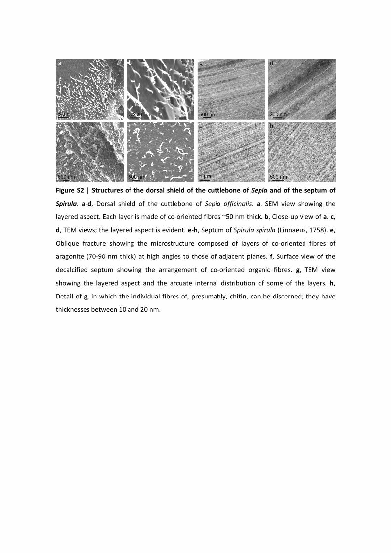
Figure S2 | Structures of the dorsal shield of the cuttlebone of Sepia and of the septum of
Spirula. a-d, Dorsal shield of the cuttlebone of Sepia officinalis. a, SEM view showing the
layered aspect. Each layer is made of co-oriented fibres ~50 nm thick. b, Close-up view of a. c,
d, TEM views; the layered aspect is evident. e-h, Septum of Spirula spirula (Linnaeus, 1758). e,
Oblique fracture showing the microstructure composed of layers of co-oriented fibres of
aragonite (70-90 nm thick) at high angles to those of adjacent planes. f, Surface view of the
decalcified septum showing the arrangement of co-oriented organic fibres. g, TEM view
showing the layered aspect and the arcuate internal distribution of some of the layers. h,
Detail of g, in which the individual fibres of, presumably, chitin, can be discerned; they have
thicknesses between 10 and 20 nm.
Supplementary Video S1 | Tomographic reconstruction of a fragment of the last three
chambers of a subadult specimen of Sepia officinalis. The cut out area of the last chamber is
the siphuncular area. Only the calcified elements are shown. The video is intended to show (1)
the lack of connection of the pillars of the last chamber with the chamber roof, due to
incomplete calcification, (2) the peculiar, antler-like morphology of pillars of the siphuncular
area and (3) the anteroposterior alignment of the pillars, which is particularly noticeable at
their dorsal ends. The venter is to the top.