The Croonian Lecture: Ionic Movements and Electrical...
39
The Croonian Lecture: Ionic Movements and Electrical Activity in Giant Nerve Fibres Author(s): A. L. Hodgkin Source: Proceedings of the Royal Society of London. Series B, Biological Sciences, Vol. 148, No. 930 (Jan. 1, 1958), pp. 1-37 Published by: The Royal Society Stable URL: http://www.jstor.org/stable/83088 . Accessed: 21/11/2014 15:31 Your use of the JSTOR archive indicates your acceptance of the Terms & Conditions of Use, available at . http://www.jstor.org/page/info/about/policies/terms.jsp . JSTOR is a not-for-profit service that helps scholars, researchers, and students discover, use, and build upon a wide range of content in a trusted digital archive. We use information technology and tools to increase productivity and facilitate new forms of scholarship. For more information about JSTOR, please contact [email protected]. . The Royal Society is collaborating with JSTOR to digitize, preserve and extend access to Proceedings of the Royal Society of London. Series B, Biological Sciences. http://www.jstor.org This content downloaded from 155.41.109.194 on Fri, 21 Nov 2014 15:31:27 PM All use subject to JSTOR Terms and Conditions
Transcript of The Croonian Lecture: Ionic Movements and Electrical...

The Croonian Lecture: Ionic Movements and Electrical Activity in Giant Nerve FibresAuthor(s): A. L. HodgkinSource: Proceedings of the Royal Society of London. Series B, Biological Sciences, Vol. 148, No.930 (Jan. 1, 1958), pp. 1-37Published by: The Royal SocietyStable URL: http://www.jstor.org/stable/83088 .
Accessed: 21/11/2014 15:31
Your use of the JSTOR archive indicates your acceptance of the Terms & Conditions of Use, available at .http://www.jstor.org/page/info/about/policies/terms.jsp
.JSTOR is a not-for-profit service that helps scholars, researchers, and students discover, use, and build upon a wide range ofcontent in a trusted digital archive. We use information technology and tools to increase productivity and facilitate new formsof scholarship. For more information about JSTOR, please contact [email protected].
.
The Royal Society is collaborating with JSTOR to digitize, preserve and extend access to Proceedings of theRoyal Society of London. Series B, Biological Sciences.
http://www.jstor.org
This content downloaded from 155.41.109.194 on Fri, 21 Nov 2014 15:31:27 PMAll use subject to JSTOR Terms and Conditions
The Croonian Lecture
Ionic movements and electrical activity in
giant nerve fibres
By A. L. Hodgkin, F.R.S.
Physiological Laboratory, University of Cambridge and the Laboratory
of the Marine Biological Association, Plymouth
{Delivered 16 May 1957?Received 16 July 1957)
[Plate 1]
During the course of an investigation into the central nervous systems of squids and cuttlefish, Professor J. Z. Young (1936a, b) noticed certain transparent tubular
structures in the peripheral nerves. These must have seemed too large to be nerve
fibres, and in a subsequent article Young (1944) remarked that he first took them
to be blood vessels. However, on examining them more closely, he was able to
prove that the tubes were, in fact, nerve fibres of exceptional size. Like many
important discoveries, this was not an entirely new observation. It had been
known since the time of Remak (1843) that Crustacea possessed giant nerve fibres, but with one exception the still larger fibres in cephalopods seem not to have been
recognized as such. As Young pointed out, the exception was L. W. Williams who
wrote a monograph on the squid which was published in 1909. In this monograph, Williams referred briefly to the large fibres in the nervous system. Williams did not
commit himself as to the size of the fibres, but it is clear that he must have seen
them. Thus he remarked that ' The very size of the nerve processes has prevented their discovery, since it is
well nigh impossible to believe that such a large structure can be a nerve fibre.'
Williams did not follow up this observation and it seems to have passed un?
noticed until Young began his investigations in 1933.
In a full grown specimen of the squid, Loligo forbesi, the giant fibres may be as
much as 1 mm in diameter and are therefore about fifty times bigger than the
largest fibres in man. When isolated from the animal, the fibres continue to conduct
electrical impulses which are essentially similar to those found in other nerves.
Young realized at once that such a preparation was likely to be of great value to
biologists, and it was not long before several physiologists were working with the
preparation at Woods Hole in Massachusetts and at Plymouth in England. The
main object of this work has naturally been to explain the conduction of nervous
impulses in physical terms. At first, electrical methods were the most profitable,
but in the years since the war it has been possible to use radioactive tracers to
follow ionic movements more directly. The upshot is that we now have a fairly definite picture of the way in which a nerve conducts impulses. This picture is not
accepted by everyone and it probably does not apply to all excitable tissues.
1 [11 Vol. 148. B.
This content downloaded from 155.41.109.194 on Fri, 21 Nov 2014 15:31:27 PMAll use subject to JSTOR Terms and Conditions

2 A. L. Hodgkin
Nevertheless, the evidence that most nerves work in essentially the same manner
seems sufficiently good to justify the large amount of effort which has been devoted
to squid fibres.
Some general properties of giant nerve fibres
When viewed under a low-power objective a squid giant fibre appears as a
transparent cylinder of protoplasm surrounded by a thin sheath of connective
tissue. In the fresh state the protoplasm of cephalopod axons appears to be a gel and when a glass capillary, which has been inserted into the protoplasm, is with?
drawn, it leaves a fluid-filled space down which particles can be seen to fall under
the influence of gravity.
Figure 1, plate 1, shows an isolated fibre under dark ground illumination. In this
case a glass capillary, 100/x in diameter, has been inserted down the axis of the
fibre for a distance of about 30 mm; the capillary can be used either for injecting substances or for recording electrical changes. Provided that the capillary does not
touch the surface it does little harm and a large fibre will continue to conduct
impulses for many hours after being impaled in this manner.
Another advantage of the giant axon is that samples of protoplasm, or axoplasm as it is called, can be obtained by squeezing out the contents of the axon, in much
the same way that one would squeeze out a tube of toothpaste. Since axoplasm obtained in this way is only very slightly contaminated with extracellular fluid, it
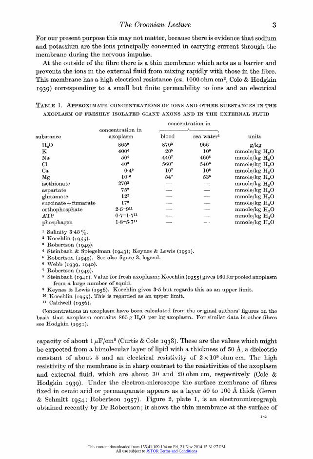
provides excellent material for chemical analyses. Table 1 summarizes the results
of some of these measurements. It will be seen that the axoplasm contains a
high concentration of potassium and a low concentration of sodium and chloride.
This is the reverse of the situation in the animal's blood, or in sea water, where
sodium and chloride are the dominant ions and potassium is relatively dilute.
Potassium ions are probably in the free state inside the fibre and do not seem to be
bound to proteins or other large molecules. Thus the mobility and diffusion co?
efficient of labelled potassium are about the same inside the axon as in free
solution (Hodgkin & Keynes 1953), and the membrane potential at which the
passive fluxes of potassium are equal, agrees with that calculated from the Nernst
equation on the basis that the activity coefficient of potassium ions inside the fibre
is approximately equal to that in the external solution (Curtis & Cole 1942;
Hodgkin & Keynes 19556; for evidence in muscle see Adrian 1956; Conway 1957). An interesting point which has come to light recently is that the concentration of
calcium in axoplasm is only about 1/30 of that in the external solution (Keynes & Lewis 1956). This figure refers to total calcium and there is evidence from
mobility measurements (Hodgkin & Keynes 1957) that most of this calcium must
be bound and that the concentration of ionized calcium may be only about 1/1000 of that outside.
The excess of potassium inside the fibre is balanced by organic anions of which, in squid fibres, isethionic acid appears to be the most important (Koechlin 1955). Other organic compounds which help to balance the cations are aspartic acid and
phosphate esters such as adenosine triphosphate, ATP. Isethionic acid is probably not present in vertebrate nerve and here the nature of the anions is still uncertain.
This content downloaded from 155.41.109.194 on Fri, 21 Nov 2014 15:31:27 PMAll use subject to JSTOR Terms and Conditions

Hodgkin Proc. Roy. Soc. B, volume 148, plate 1
Figure 1. Isolated giant axon with lOOjx glass capillary inside it, dark-ground illumination; from an experiment of Hodgkin & Keynes (1956).
Figure 2. Electronmicrograph of a thin section of two small unmyelinated fibres from the
frog's sciatic nerve, from Robertson (1957). The section was fixed with permanganate. A, axon; S, Schwann cell; M, mitochondrion. Note the membrane at the surface of the axon and Schwann cell, and the space between the axon and Schwann cell. This photo? graph was kindly provided by Dr Robertson at a time when his paper was in press.
(Facing p. 2)
This content downloaded from 155.41.109.194 on Fri, 21 Nov 2014 15:31:27 PMAll use subject to JSTOR Terms and Conditions
The Croonian Lecture 3
For our present purpose this may not matter, because there is evidence that sodium
and potassium are the ions principally concerned in carrying current through the
membrane during the nervous impulse. At the outside of the fibre there is a thin membrane which acts as a barrier and
prevents the ions in the external fluid from mixing rapidly with those in the fibre.
This membrane has a high electrical resistance (ca. 1000 ohm cm2, Cole & Hodgkin
1939) corresponding to a small but finite permeability to ions and an electrical
Table 1. Approximate concentrations of ions and other substances in the
axoplasm of freshly isolated giant axons and in the external fluid
concentration in
substance
H20 K Na Cl Ca
Mg isethionate
aspartate glutamate succinate + fumarate
orthophosphate ATP
phosphagen
concentration in
axoplasm 8652 4004
504 408
0-49 1010
2702 752 122 172
2-5-911 0-7-1-711 1-8-5-711
blood
8703 205
4407 5607
107 547
sea water1
966 106
4606 5406
106 536
units
g/kg mmole/kg H20 mmole/kg H20 mmole/kg H20 mmole/kg H20 mmole/kg H20 mmole/kg H20 mmole/kg H20 mmole/kg H20 mmole/kg H20 mmole/kg H20 mmole/kg H20 mmole/kg HsO
1 Salinity 3-45 %. 2 Koechlin (1955). 3 Robertson (1949). 4 Steinbach & Spiegelman (1943); Keynes & Lewis (1951). 5 Robertson (1949). See also figure 3, legend. 6 Webb (1939, 1940). 7 Robertson (1949). 8 Steinbach (1941). Value for fresh axoplasm; Koechlin (1955) gives 160 for pooled axoplasm
from a large number of squid. 9 Keynes & Lewis (1956). Koechlin gives 3-5 but regards this as an upper limit. 10 Koechlin (1955). This is regarded as an upper limit. 11 Caldwell (1956). Concentrations in axoplasm have been calculated from the original authors' figures on the
basis that axoplasm contains 865 g H20 per kg axoplasm. For similar data in other fibres see Hodgkin (1951).
capacity of about 1 /xF/cm2 (Curtis & Cole 1938). These are the values which might be expected from a bimolecular layer of lipid with a thickness of 50 A, a dielectric
constant of about 5 and an electrical resistivity of 2 x 10⁹ ohm cm. The high
resistivity of the membrane is in sharp contrast to the resistivities of the axoplasm and external fluid, which are about 30 and 20 ohm cm, respectively (Cole &
Hodgkin 1939). Under the electron-microscope the surface membrane of fibres
fixed in osmic acid or permanganate appears as a layer 50 to 100 A thick (Geren & Schmitt 1954; Robertson 1957). Figure 2, plate 1, is an electronmicrograph obtained recently by Dr Robertson; it shows the thin membrane at the surface of
This content downloaded from 155.41.109.194 on Fri, 21 Nov 2014 15:31:27 PMAll use subject to JSTOR Terms and Conditions
4 A. L. Hodgkin
two small unmyelinated fibres from the frog, and the Schwann cells which surround
but do not completely enclose the fibres. In the squid axon, the structure of the
Schwann cell layer is more complicated, but indirect evidence suggests that this
layer may be only a relatively weak barrier to ionic movement, and that the main
restriction occurs at the 50 to 100 A membrane at the surface of the axon (see Frankenhaeuser & Hodgkin 1956).
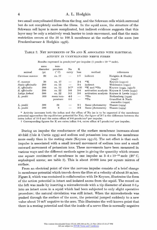
Table 2. Net movements of Na and K associated with electrical
ACTIVITY IN UNMYELINATED NERVE FIBRES
Results expressed in pmole/cm2 per impulse (1 pmole = 10-12 mole).
axon diameter
animal (jn) Carcinus maenas 30
C. maenas Sepia officinalis S. officinalis S. officinalis Loligo forbesi L.
L. L.
30 200 200 200 500 500
500 500
tem? perature
(?C) ca. 17
ca. 17 ca. 15 ca. 15 ca. 22 ca. 22
room tem? perature
24 6
Na entry
3-7* 3-8 3-5 4-5 4-4
K loss
1-7
2-4 3-4 4-3f 3-6 3-0
3-1 9-3
method
indirect
42K indirect 42K and 24Na activation analysis activation analysis 24Na 24Na
flame photometry flame photometry
references
Hodgkin & Huxley (1947)
Keynes (1951a) Weidmann (1951) Keynes (1949, 1951&) Keynes & Lewis (1951) Keynes & Lewis (1951) Rothenberg (1950) Grundfest & Nach- mansohn (1950)
Shanes (1954) Shanes (1954)
* Activity increases both the influx and the efflux of Na (as is to be expected if the membrane potential approaches the equilibrium potential for Na); the figure of 3*7 is the difference between the extra influx of 10-3 and the extra efflux of 6*6 pmole/cm2 per impulse.
?)? Corresponding figures for K are extra influx 0*4, extra efflux 4*7 pmole/cm2 per impulse.
During an impulse the conductance of the surface membrane increases about
40-fold (Cole & Curtis 1939) and sodium and potassium ions cross the membrane
more easily than in the resting state (Keynes 19516). The net effect is that each
impulse is associated with a small inward movement of sodium ions and a small
outward movement of potassium ions. These movements have been measured in
various ways and the different methods agree in giving the quantity which crosses
one square centimetre of membrane in one impulse as 3-4 x 10-12 mole (20? C,
cephalopod axons; see table 2). This is about 20000 ions per square micron of
surface.
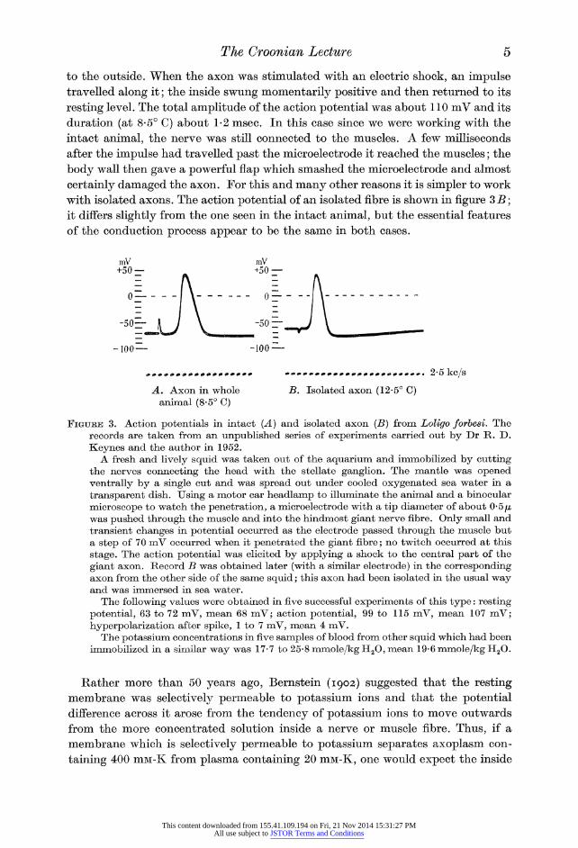
From an electrical point of view the nervous impulse consists of a brief change in membrane potential which travels down the fibre at a velocity of about 20 m/sec.
Figure 3, which was contained in collaboration with Dr Keynes, illustrates the form
of the action potential in intact and isolated axons from the squid. The record on
the left was made by inserting a microelectrode with a tip diameter of about 0-5 fi into an intact axon in a squid which had been subjected to only slight operative
procedure; the natural circulation was still intact. When the microelectrode was
pushed through the surface of the axon, the potential jumped suddenly to a new
value about 70 mV negative to the zero. This illustrates the well-known point that
there is a resting potential and that the inside of a nerve fibre is normally negative
This content downloaded from 155.41.109.194 on Fri, 21 Nov 2014 15:31:27 PMAll use subject to JSTOR Terms and Conditions
The Croonian Lecture 5
to the outside. When the axon was stimulated with an electric shock, an impulse travelled along it; the inside swung momentarily positive and then returned to its
resting level. The total amplitude of the action potential was about 110 mV and its
duration (at 8-5? C) about 1-2 msec. In this case since we were working with the
intact animal, the nerve was still connected to the muscles. A few milliseconds
after the impulse had travelled past the microelectrode it reached the muscles; the
body wall then gave a powerful flap which smashed the microelectrode and almost
certainly damaged the axon. For this and many other reasons it is simpler to work
with isolated axons. The action potential of an isolated fibre is shown in figure 3 B; it differs slightly from the one seen in the intact animal, but the essential features
of the conduction process appear to be the same in both cases.
-100? -100 ?
??.*. .?.*.2-5kc/s
A. Axon in whole B. Isolated axon (12-5? C) animal (8-5? C)
Figube 3. Action potentials in intact (A) and isolated axon (B) from Loligo forbesi. The records are taken from an unpublished series of experiments carried out by Dr R. D.
Keynes and the author in 1952. A fresh and lively squid was taken out of the aquarium and immobilized by cutting
the nerves connecting the head with the stellate ganglion. The mantle was opened ventrally by a single cut and was spread out under cooled oxygenated sea water in a
transparent dish. Using a motor car headlamp to illuminate the animal and a binocular
microscope to watch the penetration, a microelectrode with a tip diameter of about 0*5^, was pushed through the muscle and into the hindmost giant nerve fibre. Only small and transient changes in potential occurred as the electrode passed through the muscle but a step of 70 mV occurred when it penetrated the giant fibre; no twitch occurred at this
stage. The action potential was elicited by applying a shock to the central part of the
giant axon. Record B was obtained later (with a similar electrode) in the corresponding axon from the other side of the same squid; this axon had been isolated in the usual way and was immersed in sea water.
The following values were obtained in Ove successful experiments of this type: resting potential, 63 to 72 mV, mean 68 mV; action potential, 99 to 115 mV, mean 107 mV; hyperpolarization after spike, 1 to 7 mV, mean 4 mV.
The potassium concentrations in five samples of blood from other squid which had been immobilized in a similar way was 17-7 to 25-8 mmole/kg H20, mean 19-6 mmole/kg H20.
Rather more than 50 years ago, Bernstein (1902) suggested that the resting membrane was selectively permeable to potassium ions and that the potential difference across it arose from the tendency of potassium ions to move outwards
from the more concentrated solution inside a nerve or muscle fibre. Thus, if a
membrane which is selectively permeable to potassium separates axoplasm con?
taining 400 niM-K from plasma containing 20 niM-K, one would expect the inside
This content downloaded from 155.41.109.194 on Fri, 21 Nov 2014 15:31:27 PMAll use subject to JSTOR Terms and Conditions

6 A. L. Hodgkin
of the membrane to be 75 mV negative to the external solution. This value is
obtained from the Nernst relation
K.-^lnS (1)
where VK is the equilibrium potential of the potassium ion defined in the sense,
internal potential minus external potential, [K]0 and [K]^ are the potassium concentrations (or strictly activities) outside and inside the fibre and R, T and F
have their usual significance. In an undissected squid axon, with natural circula?
tion, resting potentials of about 70 mV have been observed by Moore & Cole (1955) and by the author and Dr Keynes (figure 3). In muscle, where equation (1) predicts a value of 102 mV for the potassium equilibrium potential, the resting potential is 90 to 95 mV (Adrian 1956).
The resting potential of an isolated squid axon in sea water containing 10 mM-K
is only 50 to 60 mV, whereas the potassium equilibrium potential should be about
90 mV. The reason for the low resting potential in isolated axons is not fully understood but possible factors may be (1) that small branches have to be cut
through in isolating the axon, (2) the ends of the fibre are cut, (3) the fluid used to
bathe the isolated fibres is different from that in which they are naturally immersed.
The large difference between the potassium equilibrium potential and the resting
potential in isolated fibres probably accounts for the large underswing, or transient
phase of hyperpolarization, which follows the main part of the spike in isolated
fibres. During the later stages of the spike, the potassium permeability rises and
the state of increased potassium permeability persists for several milliseconds after
the spike. The effect of this is to make the membrane potential approach more
closely to the potassium equilibrium potential, with the result that the fibre
undergoes a transient phase of hyperpolarization. Evidence for this suggestion is
provided by the observation that the membrane potential during the underswing is affected by potassium concentrations which have relatively little effect on the
resting membrane potential (Hodgkin & Katz 1949; Hodgkin & Keynes 19556; Erankenhaeuser & Hodgkin 1956). In other words, during the underswing
equation (1) holds down to lower potassium concentrations than it does in the
resting state (Hodgkin & Keynes 1955). In support of Bernstein's idea it is found that the resting potential disappears
if the external potassium concentration is made equal to the internal concentration
and that, except at low external concentrations of potassium, the potential varies
in the manner predicted by equation (1) (see Curtis & Cole 1942; Adrian 1956;
Hodgkin & Keynes 19556). However, these experiments also show that at low
concentrations potassium is not the only ion which carries current through the
membrane, for the variation with external potassium concentration is less steep in
the physiological region than one would expect from (1). At present we do not know
what other ions besides potassium contribute to the resting potential in nerve.
Since chloride ions are more concentrated outside the fibre they would tend to make
the inside of the fibre negative to the outside. In muscle, where the distribution
of chloride and potassium ions closely resembles that in a Donnan system (Boyle & Con way 1941), there is evidence that both ions contribute to the potential and
This content downloaded from 155.41.109.194 on Fri, 21 Nov 2014 15:31:27 PMAll use subject to JSTOR Terms and Conditions

The Croonian Lecture 7
that, for very small displacements of potential, the transport numbers are approxi?
mately 0-3 for K and 0-6 for Cl (unpublished experiments with Dr P. Horowicz).
During the nervous impulse the internal potential swings momentarily from its
resting value of ? 70 mV to one of about + 40 mV (figure 3 A). Activity therefore
involves a reversal of potential difference and so cannot depend on a breakdown
of the membrane as Bernstein supposed. A simple way of explaining the reversal
of membrane potential is to assume that when the membrane is activated by an
electric current it momentarily becomes selectively permeable to sodium ions. On
this basis the potential difference at the crest of activity arises from the tendency of the sodium ions to move into the fibre from the more concentrated solution
outside. In support of this idea it is found that the action potential of many but
not all excitable tissues disappears in the absence of external sodium or lithium
ions, and that the reversed potential difference across the active membrane varies
with external sodium concentration in the same manner as a sodium electrode. The
state of increased sodium permeability wears off after about a millisecond and the
potential therefore tends to return to its original level. The process of repolarization is greatly accelerated by the fact that depolarization causes a delayed rise in the
permeability to potassium ions. This speeds up the exit of potassium and keeps the
whole action potential reasonably short.
The effect of sodium concentration on the action potential
The importance of sodium in maintaining excitability was first recognized by Overton (1902) who suggested that impulse conduction was accompanied by an
exchange between intracellular potassium ions and extracellular sodium ions.
References to papers dealing with the effect of sodium on the action potentials of
different excitable tissues are given in table 3.
Deviations from the typical sodium electrode type of behaviour have been seen
in turtle ventricle, and in frog muscles treated with tetraethylammonium ions. In
both cases the fibres fail in the complete absence of sodium, but at intermediate
concentrations the overshoot changes much less than would be expected from the
simple theory. Crab muscle fibres, previously treated with tetrabutylammonium
ions, give a large overshoot in the absence of sodium or any other monovalent
cation so that the sodium theory clearly does not apply in this case. However, in
the majority of excitable fibres, the effects of sodium-deficient solutions agree well
with the hypothesis that the action potential depends on an increase in sodium
permeability. The relation between sodium concentration and overshoot has been investigated
with solutions containing extra sodium chloride as well as those in which choline
replaces sodium, or sucrose replaces sodium chloride. Since sodium-rich solutions
increase the spike, it cannot be argued that their action is due to some kind of
reversible damage or narcosis of the membrane. An extreme example of the action
of added NaCI is afforded by an experiment mentioned by Stampfli (1956) in which
a sudden four-fold increase of NaCl raised the overshoot of a node of Ranvier by about 38 mV. In carrying out experiments with solutions containing extra sodium
it is important either to measure the action potential very soon after changing the
This content downloaded from 155.41.109.194 on Fri, 21 Nov 2014 15:31:27 PMAll use subject to JSTOR Terms and Conditions

8 A. L. Hodgkin
solution, or to make the comparison between isosmotic solutions, for example, between one solution containing extra sodium chloride and another containing an
osmotically equivalent amount of sucrose or choline chloride. If this is not done,
the effect of the high external sodium will be neutralized by the rise in internal
sodium concentration which results from the removal of water by the hypertonic
Table 3. Effect of sodium ions on the action potentials of
different tissues-summary and references
active mem? brane behaves like a sodium
electrode, i.e. slope of
58 mV ? ca. 5 mV for 10-fold
change
substance used to replace
Na or NaCl
choline, dextrose
choline choline, dextrose,
sucrose choline, and other
quartenary ammonium salts
choline*, dextrose
rapid reversible block in Na-free solution
yes
preparation
squid giant axon choline, dextrose yes yes
Sepia giant axon crab (Carcinus) choline, dextrose, yes
nerve fibre crab muscle
references
Cole Hodgkin & Katz (1949), (1955)
unpublished Katz (1947), Hodgkin & Katz
(1949) Fatt & Katz (1953)
frog single mye- choline^, dextrose yes yes linated nerve fibre*
frog sartorius choline, sucrose yes yes muscle
frog sartorius muscle
dog Purkinje sucrose yes yes fibres
frog ventricle choline, sucrose yes yes turtle ventricle choline after atropine yes no guinea-pig choline yes no
ventricle
For the action of guanidine and other quarternary ammonium compounds in frog nerve see Larramendi, Lorente de No & Vidal (1956), Lorente de No, Vidal & Larramendi (1957) and Lorente de No (1949).
* Miiller (1956) reports prolonged action potentials in Na-free choline in a node which had pre? viously been depolarized with a large outward current.
tetraethylammonium
Huxley & Stampfli (1949, I95i)
Overton (1902), Nastuk & Hodgkin (1950), Hagiwara & Watanabe (1955)
Hagiwara & Watanabe (1955)
Draper & Weidmann (1951)
Brady & Woodbury (1957) S. Weidmann (unpublished) Coraboeuf & Otsuka (1956)
solution. This point seems to have been overlooked by Shaw, Simon, Johnstone &
Holman (1956) and may help to account for the difference between their conclu?
sions and those of other workers. Thus Nastuk & Hodgkin (1950, table 4) found
overshoots of 33, 25 and 33 mV in frog muscle fibres immersed in Ringer's fluid,
Ringer plus 100 mM-sucrose and Ringer plus 50 niM-NaCl, respectively. Shaw et al.
(1956) found about the same overshoot in Ringer's fluid or Ringer's fluid plus 26 to 60 mM-NaCl, but they apparently did not carry out any control experiments with Ringer's fluid plus sucrose or choline chloride.
Evidence about the relation between the internal sodium concentration and
overshoot is somewhat conflicting. Desmedt (1953) found that the active mem?
brane behaved like a sodium electrode except when the internal sodium concen-
This content downloaded from 155.41.109.194 on Fri, 21 Nov 2014 15:31:27 PMAll use subject to JSTOR Terms and Conditions

The Croonian Lecture 9
tration was low, in which case the contribution of other ions might no longer be
small compared with that of the sodium ion. On the other hand Shaw, Simon &
Johnstone (1956) and Shaw et al. (1956) consider that there is no correlation between
the overshoot of toad muscle and the internal sodium concentration calculated on
the assumption that the extracellular space occupies 15% of the muscle volume.
In the writer's opinion the difference between the two conclusions may result from
the very great difficulty of making accurate measurements of internal sodium
concentration in a tissue in which much of the total sodium may be in the extra?
cellular space. In the squid giant axon, raising the internal sodium by micro-
injection reduced the overshoot by approximately the amount expected from the
Nernst equation (Hodgkin & Keynes 1956).
The local circuit theory
While there is still much argument about the nature of the action potential,
everyone is agreed that conduction of the nervous impulse is essentially an electrical
process and that propagation is brought about by current flowing in a local circuit
at the front of the action potential. In explaining how the impulse propagates, it
? 4- +_?_- + + + -f +i_?.JziL - - -t- -t- -hi- ?_ ->- 4- 4- ++"%-
""
+? -h H~ =F - ---+ ++.-H-++. + 4.
Figttbe 4. Diagrams illustrating the local circuit theory; the upper sketch represents an unmyelinated nerve fibre, the lower a myelinated nerve fibre. For evidence about salta? tory conduction in myelinated fibres see numerous papers by Tasaki and colleagues (reviewed in Tasaki 1953); Huxley & Stampfli (1949); Stampfli (1952).
is convenient to adopt the view outlined in the first part of this article, and to
assume that when the membrane potential is reduced beyond a certain level the
membrane changes from a stage of moderate potassium permeability to one of high sodium permeability. This assumption is made for the sake of definiteness; it is not
essential to the local circuit theory, which can be formulated without specifying how the action potential arises.
The upper part of figure 4 illustrates the flow of current in an unmyelinated axon, of which the squid giant fibre is an example. Suppose that point A is active and
that point B is resting. A is sodium permeable so the inside of the fibre is positive; B is potassium permeable so the inside is negative. Current therefore flows in a
local circuit between resting and active nerve. It will be seen that current flows in
the same direction as the impulse in the axis cylinder and returns in the opposite direction in the external fluid. This current reduces the membrane potential just ahead of the active region by drawing charge out of the membrane capacity. When
This content downloaded from 155.41.109.194 on Fri, 21 Nov 2014 15:31:27 PMAll use subject to JSTOR Terms and Conditions

10 A. L. Hodgkin
the potential difference has been reduced by about 20 mV the permeability to
sodium rises and the inside of the fibre becomes positive. Point B is now active
and can stimulate the next region in precisely the same manner. In this way, a
wave of increased sodium permeability propagates along the fibre.
This is a convenient point at which to draw attention to an important difference
between our own nerves and those of the squid. In most invertebrates, the nerve
fibres are continuous unmyelinated structures and the impulse spreads smoothly from one point to the next. Owing to the large capacity of the surface membrane
and the resistance of the axis cylinder, this type of conduction only gives a high
velocity if the fibre is large. In the myelinated fibres, which form the bulk of our
own nerves, a neater method of getting a high velocity has been evolved. These
fibres are coated over most of their length with a layer of myelin which acts as an
insulator. The excitable membrane is exposed only at certain points known as
nodes of Ranvier. When one node becomes active, current flows in a local circuit
through the next node in the manner shown in figure 4. Some current is also
wasted in charging up the myelin, but since the myelin sheath is relatively thick
its capacity is much less than that of an excitable membrane. The effect of this
type of conduction, which is known as saltatory conduction, is that in myelinated fibres the impulse is conducted both at greater velocity and with greater economy than in unmyelinated fibres of comparable size.
Current-voltage relations of the membrane ; the voltage-clamp method*
The electrical properties of the membrane have been studied quantitatively by a technique, known as the voltage-clamp method, in which the membrane potential is suddenly displaced from its resting value and held at a fixed potential by a feedback amplifier. The current which flows through a definite area of membrane
under the influence of the impressed voltage is measured with a separate amplifier
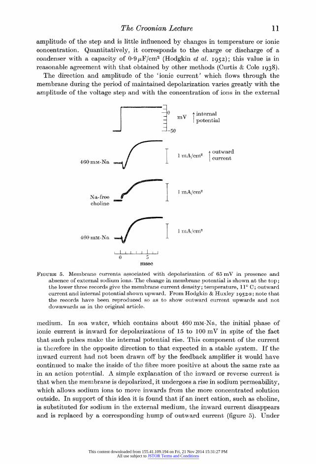
{see Cole 1949; Hodgkin, Huxley & Katz 1952). In the experiment of figure 5, the internal potential of the fibre was raised
suddenly by 65 mV by applying a rectangular pulse to the feedback amplifier which
controlled the membrane potential. Since the inside of the fibre was normally at
about ? 50 mV the effect of the pulse was to swing the membrane potential from ? 50 to +15 mV. The lower records show the current which flowed through the
membrane in the presence and absence of external sodium ions.
When the inside of the fibre is made more positive the potential difference and
charge on the membrane is reduced, or reversed if the pulse is large enough. The
immediate effect of the sudden change is that a large surge of capacity current
flows through the membrane. This surge is complete in about 20/xsec and is too
rapid to show on records such as those in figure 5. The surge is proportional to the
* Convention as to signs. In this article membrane potentials are given in the sense internal potential minus external potential; outward currents are therefore taken as positive and are shown as upward deflexions. Although this convention seems the more logical and is widely used, I have previously hesitated to adopt it because it conflicts with the accepted use of words such as negative after-potential. However, since many workers now use internal electrodes and since the action potential is almost invariably shown as an upward deflexion, it seems right to make a change.
This content downloaded from 155.41.109.194 on Fri, 21 Nov 2014 15:31:27 PMAll use subject to JSTOR Terms and Conditions
The Croonian Lecture 11
amplitude of the step and is little influenced by changes in temperature or ionic
concentration. Quantitatively, it corresponds to the charge or discharge of a
condenser with a capacity of 0-9^F/cm2 (Hodgkin et al. 1952); this value is in
reasonable agreement with that obtained by other methods (Curtis & Cole 1938). The direction and amplitude of the 'ionic current5 which flows through the
membrane during the period of maintained depolarization varies greatly with the
amplitude of the voltage step and with the concentration of ions in the external
mV I internal I potential
-J-50
460 mM-Na
Na-free choline
I outward
I current
1 mA/cm2
460 mM-Na 1 mA/cm2
I 1 i j 1_1 1 J
JFigxjub 5. Membrane currents associated with depolarization of 65 mV in presence and absence of external sodium ions. The change in membrane potential is shown at the top; the lower three records give the membrane current density; temperature, 11? C; outward current and internal potential shown upward. From Hodgkin & Huxley 1952 a; note that the records have been reproduced so as to show outward current upwards and not downwards as in the original article.
medium. In sea water, which contains about 460 mM-Na, the initial phase of
ionic current is inward for depolarizations of 15 to 100 mV in spite of the fact
that such pulses make the internal potential rise. This component of the current
is therefore in the opposite direction to that expected in a stable system. If the
inward current had not been drawn off by the feedback amplifier it would have
continued to make the inside of the fibre more positive at about the same rate as
in an action potential. A simple explanation of the inward or reverse current is
that when the membrane is depolarized, it undergoes a rise in sodium permeability, which allows sodium ions to move inwards from the more concentrated solution
outside. In support of this idea it is found that if an inert cation, such as choline, is substituted for sodium in the external medium, the inward current disappears and is replaced by a corresponding hump of outward current (figure 5). Under
This content downloaded from 155.41.109.194 on Fri, 21 Nov 2014 15:31:27 PMAll use subject to JSTOR Terms and Conditions
12 A. L. Hodgkin
these conditions there are no sodium ions in the external solution and the outward
current arises from the tendency of the sodium ions in the axoplasm to move to
the sodium-free solution outside.
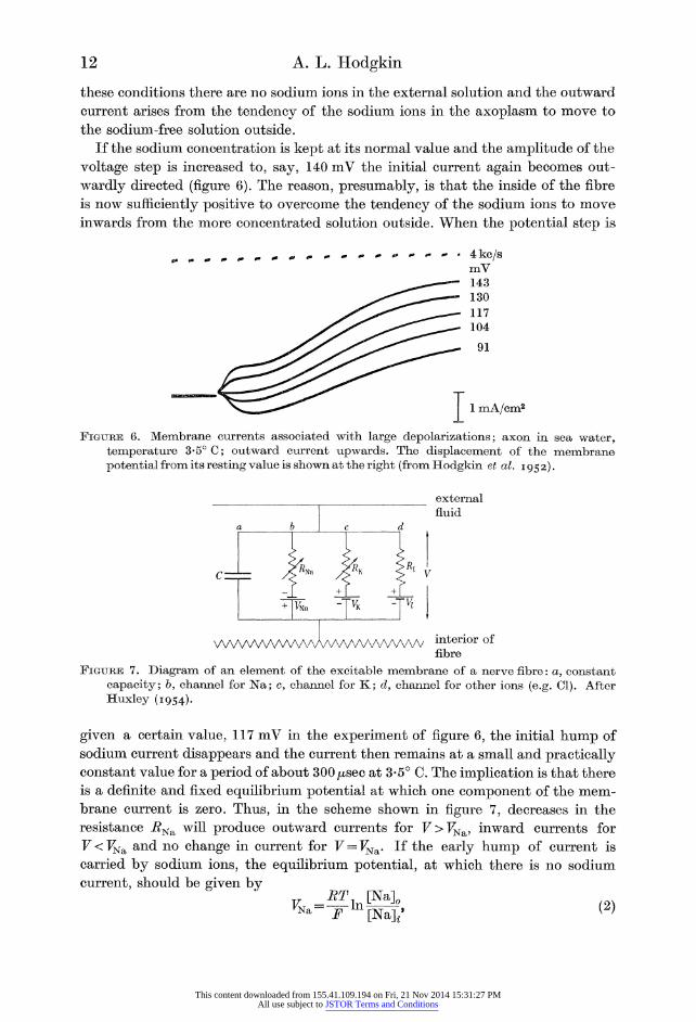
If the sodium concentration is kept at its normal value and the amplitude of the
voltage step is increased to, say, 140 mV the initial current again becomes out?
wardly directed (figure 6). The reason, presumably, is that the inside of the fibre
is now sufficiently positive to overcome the tendency of the sodium ions to move
inwards from the more concentrated solution outside. When the potential step is
1 mA/cm2
Figure 6. Membrane currents associated with large depolarizations; axon in sea water, temperature 3-5? C; outward current upwards. The displacement of the membrane potential from its resting value is shown at the right (from Hodgkin et al. 1952).
external fluid
WVWWVWA WWWV interior of fibre
Figure 7. Diagram of an element of the excitable membrane of a nerve fibre: a, constant capacity; b, channel for Na; c, channel for K; d, channel for other ions (e.g. Cl). After Huxley (1954).
given a certain value, 117 mV in the experiment of figure 6, the initial hump of
sodium current disappears and the current then remains at a small and practically constant value for a period of about 300/xsec at 3-5? C. The implication is that there
is a definite and fixed equilibrium potential at which one component of the mem?
brane current is zero. Thus, in the scheme shown in figure 7, decreases in the
resistance i?Na will produce outward currents for F>K^a, inward currents for
T^^Na and no change in current for V = VNa>. If the early hump of current is
carried by sodium ions, the equilibrium potential, at which there is no sodium
current, should be given by
Kn?- JP m[NaV (2)
This content downloaded from 155.41.109.194 on Fri, 21 Nov 2014 15:31:27 PMAll use subject to JSTOR Terms and Conditions

The Croonian Lecture 13
where J^Ta is the equilibrium potential of the sodium ion, defined as an internal
potential, [Na]0 and [Na]^ are the sodium concentrations (or strictly activities)
outside and inside the fibre. The validity of (2) was tested by measuring the relation
between [Na]0 and T^a, using choline as a substitute for sodium; the results
(Hodgkin & Huxley 1952a, table 1) showed that the equation holds to within about
1 mV over a ten-fold range of [Na]0. After a short time, about 500/xsec in the experiment of figure 6, the initial
current (which we may now call sodium current) declines, and is replaced by a
second component which is always outwardly directed for voltage steps in which
the inside of the fibre becomes more positive. The second component can be seen
uncomplicated by sodium current if the membrane potential during the voltage
step is made equal to VNsb (117 mV below the resting potential in the experiment of figure 6). Here the current changes little at first and then increases along an
S-shaped curve to an approximately steady value of about 3 mA/cm2. This com?
ponent of the current is not much altered if sodium ions are replaced by choline
and is also present in chloride-deficient solutions. Since potassium ions are known
to move outwards during the spike it seemed likely that the second, or delayed,
component of the current, was carried by potassium ions. In support of this
idea, subsequent experiments with tracers showed that depolarization caused a
large increase in the efflux of potassium from Sepia axons and that the quantity of
potassium ions crossing a given area of membrane in a given time was equivalent to the total electric charge passing through the same area of membrane in the same
time (Hodgkin & Huxley 1953; see also figure 8 of this article). Further evidence
is that the equilibrium potential of the second component varies with the external
potassium concentration, though not as steeply as predicted by the Nernst equation. Later work (Frankenhaeuser & Hodgkin 1956) suggests that the discrepancy is at
least partly explained by the fact that potassium ions tend to accumulate in a space outside the membrane, and that the effective concentration immediately outside
the potassium-sensitive layer may not be the same as that in the external solution.
Having established that the early current was probably carried by sodium ions,
and the delayed current by potassium ions, the next step was to separate the total
current into its two components. The method is illustrated by figure 9. Here curve A
shows the ionic current when an axon immersed in sea water was depolarized by
56 mV; under these conditions both sodium and potassium ions contribute to the
current. In principle, sodium current can be eliminated by reducing the sodium
concentration until the sodium potential is at 56 mV. In practice it would be
difficult to hit off exactly the right sodium concentration so the procedure was to
interpolate between curves in high and low sodium. Curve B in figure 9, which was
in fact very close to the record in 46 mM-Na, was obtained in this way and is taken
as the potassium current. If it is assumed (and this may not be quite correct) that
the potassium current is independent of the sodium and choline concentrations,
then the sodium current in record A can be obtained by subtracting curve B from
curve A. This method works well if the sodium current is reasonably large compared
to the potassium current, but, as might be expected, it gives errors and anomalies
when the former is small compared with the latter.
This content downloaded from 155.41.109.194 on Fri, 21 Nov 2014 15:31:27 PMAll use subject to JSTOR Terms and Conditions

14 A. L. Hodgkin
A convenient way of expressing the permeability of the membrane is to calculate
sodium or potassium conductances, #Na or #K, by means of the relations
or
M
.a
#Na: LNa
F-R Na
LK F-I; K
outward current density (jh coulomb cm-2 sec-1)
Figure 8. Relation between membrane current density and potassium efflux when a Sepia axon is depolarized (from Hodgkin & Huxley 1953). The axon was depolarized by an applied current for periods of 60 to 600 sec. Vertical lines show ? 2 x s.e. ; the horizontal line is drawn at a level corresponding to complete suppression of the average resting efflux.
Subsequent experiments (Hodgkin & Keynes 1955 b) indicate that the influx of K remains at a low value when the fibre is depolarized by applied currents; hence the increase in efflux shown above should be very nearly equal to the total outward movement ofK.
These definitions would be valid whatever the relations between /Na and F -
or between 7K and F - "^Na?
FK. However, the usefulness of the definitions is increased
by the observation that for sudden changes in potential, the instantaneous value
of the current, 7Na or JTK, is directly proportional to the driving force, V ? T^a
or V-VK. The continuous curves in figure 10 illustrate the way in which the conductances
change when the fibre is depolarized by 56 mV. The sodium conductance rises
rapidly to a peak and then declines, in spite of the fact that the fibre is held in the
This content downloaded from 155.41.109.194 on Fri, 21 Nov 2014 15:31:27 PMAll use subject to JSTOR Terms and Conditions

The Groonian Lecture 15
internal potential
B. IK (from current with reduced Na) A. INa + IK (current with 460 mM-Na)
1 mA/cm2
C7.lv
time (msec)
Figure 9. Separation of current into components carried by Na and K, from Hodgkin & Huxley (1952 a, figure 5). A depolarization of 56 mV was applied at t = 0; the temperature was 8-5?C Outward current is shown upwards.
internal
potential
sodium conductance
potassium conductance
time (msec)
Figure 10. Changes in sodium and potassium conductance associated with depolarization of 56 mV, applied at t=0; temperature 8-5? C. The continuous curves, which were obtained from the experiment of figure 9, show the changes in conductance when the depolarization was maintained; the broken curves, which were drawn from data obtained in other experiments, give the effect of repolarizing the fibre after 0-6 or 6-3 msec. From Hodgkin & Huxley (1952a, b, d).
This content downloaded from 155.41.109.194 on Fri, 21 Nov 2014 15:31:27 PMAll use subject to JSTOR Terms and Conditions

16 A. L. Hodgkin
depolarized state. The potassium conductance changes little at first and then rises,
along an S-shaped curve to a level which is maintained as long as the fibre is kept
depolarized. The broken lines in figure 10 give the effect of repolarizing the mem?
brane at different times and show that the changes in conductance are reversible
In contrast to the S-shaped rise in conductance associated with depolarization, both sodium and potassium conductances return to their resting values along
sodium conductance potassium conductance
0 2 time (msec)
Figure 11. Changes in sodium and potassium conductance associated with different depolari? zations at 6? C. The numbers attached to the curves give the depolarizations used. The circles are experimental estimates and the smooth curves are solutions of the equations used to describe the changes in conductance. In a similar figure, reproduced in Hodgkin & Huxley (1952 e,f) the potassium curves were drawn 25 % too large on the vertical scale. The results were plotted correctly in the original article (Hodgkin & Huxley 1952d) and the error has been corrected in the figure shown above.
approximately exponential curves when the membrane is repolarized. For sodium,
the time constant is 0-1 to 0-2 msec at 6?C; for potassium it is 5 to 10 msec. The
inactivation process which leads to a decline in sodium conductance during a
maintained depolarization is also reversed by repolarizing the membrane and has
a time constant of about 10 msec at 6? C. The rate constants governing the turning- on and turning-off of the sodium or potassium conductance have been shown to
depend on temperature, membrane potential and calcium concentration, but are
little affected by changes in sodium concentration, or by the direction of the ionic
current (Hodgkin & Huxley 1952a ? d; Frankenhaeuser & Hodgkin 1957, for the
effect of Ca).
This content downloaded from 155.41.109.194 on Fri, 21 Nov 2014 15:31:27 PMAll use subject to JSTOR Terms and Conditions
The Croonian Lecture 17
The changes in sodium and potassium conductances at various depolarizations are given in figure 11. The amplitude and time course of the two conductances
vary greatly with membrane potential, but there is no sudden break or dis?
continuity in the relation between conductance and potential.
Quantitative description of the changes in membrane conductance
The results described in the preceding paragraphs suggest that reducing the
membrane potential leads to a transient increase in sodium conductance and a
delayed increase in potassium conductance. At first sight these changes appear too
simple to account for the varied and complicated reactions of a nerve fibre to
electrical stimuli. In order to examine this point, Huxley and I developed a series
of equations which described the changes in conductance with reasonable accuracy
(Hodgkin & Huxley 1952 c?). The main object of the equations was to obtain an
empirical description of the way in which the permeability depends on time and
membrane potential. However, it is perhaps worth mentioning the general type of physical picture which we had in mind in formulating the equations.
To account for the change in potassium conductance we assumed that a path for potassium was formed when four charged particles had moved to a certain
region of the membrane under the influence of the electric field. If n is the
probability that a single particle is in the right place, then gK = ^K n^, where ^K is
the maximum potassium conductance. The value of n is given by
Jt = ocn(l--n)-finn, (3)
where otn and j$n are rate constants which, at a fixed temperature and calcium
concentration, depend only on the membrane potential, V. ocn increases and /3n decreases as the inside of the fibre becomes more positive.
For the sodium channel we assumed that three simultaneous events each of
probability m, opened the channel to Na and that a single event of probability
(1?h) blocked it. These events need not be specified, but may be thought of as the
movement of three activating particles and of one blocking particle to a certain
region of the membrane. The probability that there will be three activating
particles and no blocking particle is therefore mzh. Hence #Na = gNam3h, where <7Na is the maximum sodium conductance. The values of m and h are given by relations
similar to (3), i.e. .,
-^t=ujl-m)-pmm, (4)
dh
Trah(l-h)-j3hh. (5)
The a's and /?'s in these equations depend on temperature, calcium concentration
and membrane potential. The effect of making the inside of the fibre more positive is to increase <xm and j3h and to decrease j3m and oth.
It is relatively simple to apply equations (3), (4) and (5) to the voltage-clamp data. At a fixed voltage the a's and /?'s are constant so equations (3), (4) and (5)
2 Vol. 148. B.
This content downloaded from 155.41.109.194 on Fri, 21 Nov 2014 15:31:27 PMAll use subject to JSTOR Terms and Conditions

18 A. L. Hodgkin
lead to exponential expressions for n, m and h; conductances are then calculated
from the relations #K = g^n* &nd #Na = <7Nam%. With values of oc and /? which change with membrane potential in a consistent manner, a fair though not a perfect fit is
obtained. This is illustrated by figure 11 in which the smooth curves have been
calculated and the circles are experimental estimates of the sodium and potassium conductance. The effects of repolarizing the membrane in shutting-off the con?
ductances were taken into account in formulating the equations, and are described
by them without any further assumption. The complete expression for the membrane current density, I, is
I = c d~ + (V - FK) g^ + (V - FNa) gNam% + (V - VL) gL. (6)
The first term on the right-hand side is the capacity current, c being the membrane
capacity per unit area. The second and third terms give the potassium and sodium
currents while the last term, which is relatively unimportant, gives the current
carried by ions other than sodium and potassium through a leak conductance gL. If an action potential is elicited over a length of nerve, by applying a short shock
to a long metal wire in the axoplasm, there is no longitudinal current and no radial
current at any time after the shock. Under these conditions (6) can be simplified
by putting 1 = 0 for t > 0, the boundary condition being the initial displacement of F. Solutions of this kind have been worked out numerically, or with an auto?
matic computer; the results agree reasonably with the behaviour of a real nerve.
The case in which 1 = constant has been calculated by Cole (1954) and shows that, above a certain strength of current, the theoretical membrane gives a train of
spikes, which again are not unlike those in a real nerve.
To calculate the form and velocity of the propagated action potential equa? tions (3) to (6) are used in conjunction with the well-known relation for the
current density in a continuous nerve fibre surrounded by a large volume of external
fluid, that is
where a is the radius of the axoplasm and R is its resistivity; x is distance along the nerve. In the case of a fibre propagating at constant velocity (6), x may be
replaced by ? di.
Hence
a d2F dF
2W^ =
CW + (F-F^^ (8)
In this equation, the conduction velocity, d, is constant, but its value is unknown
at the beginning of the computation. The procedure is to guess a value for 6 and
start a trial solution. It is found that F goes to + 00 according to whether 6 has
been chosen too high or too low. The correct value of d, which corresponds to the
natural velocity of propagation, brings the potential back to its resting value at
the end of the run. A numerical solution along these lines was worked out by Mr Huxley and was found to agree reasonably with the behaviour of a real nerve
This content downloaded from 155.41.109.194 on Fri, 21 Nov 2014 15:31:27 PMAll use subject to JSTOR Terms and Conditions
The Croonian Lecture 19
in the following respects: (1) the form, amplitude and velocity of the action
potential, (2) the time course and amplitude of the conductance change, (3) the
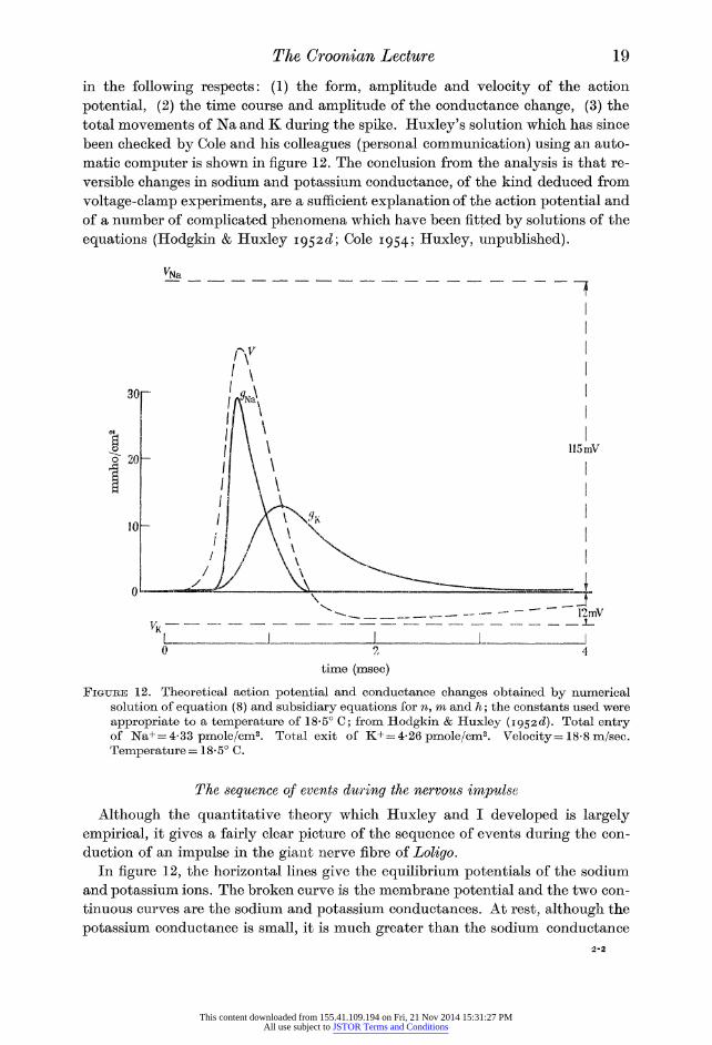
total movements of Na and K during the spike. Huxley's solution which has since
been checked by Cole and his colleagues (personal communication) using an auto?
matic computer is shown in figure 12. The conclusion from the analysis is that re?
versible changes in sodium and potassium conductance, of the kind deduced from
voltage-clamp experiments, are a sufficient explanation of the action potential and
of a number of complicated phenomena which have been fitted by solutions of the
equations (Hodgkin & Huxley 1952^; Cole 1954; Huxley, unpublished).
time (msec)
Figure 12. Theoretical action potential and conductance changes obtained by numerical solution of equation (8) and subsidiary equations for n, m and h; the constants used were appropriate to a temperature of 18-5? C; from Hodgkin & Huxley (1952c?). Total entry of JSTa+ = 4-33 pmole/cm2. Total exit of K+ = 4-26 pmole/cm2. Velocity = 18-8 m/sec. Temperature = 18-5? C.
The sequence of events during the nervous impulse
Although the quantitative theory which Huxley and I developed is largely
empirical, it gives a fairly clear picture of the sequence of events during the con?
duction of an impulse in the giant nerve fibre of Loligo. In figure 12, the horizontal lines give the equilibrium potentials of the sodium
and potassium ions. The broken curve is the membrane potential and the two con?
tinuous curves are the sodium and potassium conductances. At rest, although the
potassium conductance is small, it is much greater than the sodium conductance
This content downloaded from 155.41.109.194 on Fri, 21 Nov 2014 15:31:27 PMAll use subject to JSTOR Terms and Conditions
20 A. L. Hodgkin
so the membrane potential is fairly close to the equilibrium potential of the
potassium ion. As the impulse advances, the membrane just ahead of the active
region becomes depolarized by electric currents flowing in a local circuit through the axoplasm and external fluid. Under the influence of the change in membrane
potential the sodium conductance rises and sodium ions enter the fibre; this
inward current makes the inside of the fibre positive (by carrying charge through the dielectric) and provides the current required to depolarize the resting mem?
brane ahead of the active region. At the crest of the spike the slow changes which
result from depolarization begin to take effect. The sodium conductance declines
and the potassium conductance rises so that the rate at which potassium ions leave
the fibre exceeds the rate at which sodium ions enter; this makes the potential
swing towards the equilibrium potential of the potassium ion. As the potential
approaches the resting level, any sodium conductance which has not been in?
activated is cut off and this may accelerate the rate of repolarization. This effect
is conspicuous in myelinated fibres (Tasaki 1956) and although it is not obvious in
calculations appropriate to a squid fibre at 18? C, those for 6? C show it quite
clearly (Hodgkin & Huxley 19526Z, figure 12). The slow effects of depolarization,
namely raised potassium conductance and inactivation of the sodium carrying
system, persist for a few milliseconds after the spike and give rise to the refractory
period. Under some, but not all, conditions the potential and conductance may return to their resting value in an oscillatory manner. About 10 msec after a spike the
membrane is back in its original state and can conduct another impulse. The fibre
has gained a small quantity of Na and has lost a similar quantity of K. These
movements provide the immediate source of energy for the conduction of impulses and are reversed later by a slow process which requires metabolic energy.
Possible application to other tissues
At present it is uncertain how far the mechanism worked out on the giant axons
of cephalopods applies to other excitable tissues, or whether it always applies in these fibres. Hagiwara & Tasaki (1957) have shown that when tetraethyl- ammonium ions are injected into a squid axon, the action potential becomes greatly
prolonged and resembles that found in the Purkinje fibres of the heart (Draper &
Weidmann 1951). These prolonged action potentials are abolished by removing sodium ions from the external medium, but the ionic properties of membranes
altered by injection of tetraethylammonium ions remain to be worked out. From
the voltage-clamp records obtained by Hagiwara & Tasaki it would appear that
after treatment with tetraethylammonium the rise in potassium conductance, which normally follows depolarization, must either be absent or very greatly
delayed. Since the action potentials and resting potentials of a number of tissues vary
with sodium and potassium concentration in approximately the same manner as
in the squid fibre, it would seem likely that movements of these ions are involved
in many cells. However, there must be some fairly radical differences; thus the
action potential of heart muscle lasts about 300 times longer than that in the squid
giant axon, and in striated muscle the membrane rectifies direct current in the
This content downloaded from 155.41.109.194 on Fri, 21 Nov 2014 15:31:27 PMAll use subject to JSTOR Terms and Conditions

The Croonian Lecture 21
opposite direction to that found in nerve (anomalous rectification, Katz 1949). At present it is uncertain whether these differences arise from peculiarities of the
systems controlling sodium and potassium permeability, or whether other ions or
charged particles are involved. The clearest evidence of propagation in the absence
of sodium or of a substitute such as lithium is provided by the experiments of
Fatt & Katz (1953) on crustacean muscle fibres. Here it was found that muscles
which had previously been treated with tetrabutylammonium ions gave a pro?
longed heart-like action potential in the absence of all monovalent cations in the
external medium. As Fatt & Katz suggested, it would seem that the membrane
of a crustacean muscle fibre must either become permeable to one of the internal
anions, or perhaps, to calcium and magnesium ions.
Lorente de No (1949) showed that tetraethylammonium ions restore excitability to the B and C fibres of frog nerves after they have previously been made
inexcitable by sodium-deficient solutions. Guanidinium and certain other quater?
nary ammonium compounds will restore excitability to the A fibres of frog nerves
which have been blocked with sodium-deficient solutions (Larramendi, Lorente
de No & Vidal 1956; Lorente de No, Vidal & Larramendi 1957). One way of
explaining these results is to suppose that certain quaternary ammonium ions
substitute directly for sodium ions. However, in view of the results of Fatt & Katz
it is conceivable that treatment with quaternary compounds may change the
mechanism in such a way that neither sodium nor the quaternary ions is directly involved in carrying current through the membrane. Another puzzling result, described by Muller (1956) is that, after the passage of a large outward current a
node of Ranvier will apparently give a prolonged action potential in the complete absence of sodium or potassium from the external medium.
The nature of the permeability changes
Selectivity
Although little is known about the molecular organization of the membrane
there are certain clues as to the nature of the mechanism which controls the
permeability to the sodium and potassium ions. In the first place there is evidence
that the membrane, under appropriate conditions, is capable of showing a high
degree of selectivity towards either sodium or potassium. An example of potassium
selectivity is afforded by the variation of membrane potential with potassium concentration during the period of increased conductance which follows the action
potential in cephalopod axons. With potassium concentrations above 20 ira, the
membrane potential agrees closely with that calculated for a potassium electrode, mean values of F being ?44 or ? 65 mV with 52 or 21 mM-K in the external
solution and a measured internal concentration of 270 mmole-K per litre axoplasm
(Hodgkin & Keynes 19556). With 10 or 3-5 mM-K in the external solution (perhaps
slightly more near the membrane) the membrane potential is ? 76 or ?93 mV,
respectively. Since the external solution contained a high concentration of sodium
([Na]o + [K]o==490 him), it is evident that the selectivity of the membrane for
potassium is very great. This conclusion can be made more quantitative by
This content downloaded from 155.41.109.194 on Fri, 21 Nov 2014 15:31:27 PMAll use subject to JSTOR Terms and Conditions

22 A. L. Hodgkin
calculating the relative permeability to sodium and potassium by the following
equation (cf. Goldman 1943; Sollner, Dray, Grim & Neihof 1955):
RT ]n[K]^+6[Na]0
Fl [K], + &[NaV {V)
With the data given above, the constant b, which expresses the apparent
permeability of Na relative to that of K, is found to be about 0-01. Data obtained
recently by Horowicz and myself on single muscle fibres immersed in chloride-free
sulphate media also give a value for b of about 0-01. The ability of the membrane
to discriminate between different alkali metals has not yet been fully worked out, but it seems clear that the membrane is always less permeable to caesium than to
potassium. In frog muscle rubidium depolarizes less than potassium (see Keynes & Adrian 1956), but in crab nerve it has a slightly greater effect on both membrane
potential and membrane conductance (Wilbrandt 1937; Hodgkin 1947); in Sepia
axons, R. H. Adrian found that rubidium depolarized slightly more than potassium. The ability of different tissues to concentrate cations shows a similar variation
between K and Rb (Krogh 1946).
During the period of high sodium permeability the discrimination appears to be
almost equally great, but the membrane now favours sodium and lithium rather
than potassium and rubidium. From the fact that the membrane obeys the
equation for a sodium electrode to within 1 mV when the choline concentration is
ten times greater than the sodium concentration (Hodgkin & Huxley 1952 a, table 1) it follows that the permeability of the active membrane to sodium ions must be at
least 200 times greater than that to choline. It is not easy to give a figure for the
relative permeability of the active membrane to sodium and potassium, but the
voltage-clamp data suggest that the ratio of sodium to potassium conductance
reaches at least 30 (Hodgkin & Huxley 1952d). Higher values would probably be
obtained if the resting potassium conductance were depressed by hyperpolarizing the fibre before applying the cathodal pulse (Frankenhaeuser & Hodgkin 1957). As was first shown by Overton (1902) the mechanism which produces an action
potential does not discriminate between sodium and lithium. Thus, the size of the
action potential does not alter to any appreciable extent when sodium ions are
replaced with lithium ions (Hodgkin & Katz 1949; Huxley & Stampfli 1951).
Variation of conductance with membrane potential
One interesting point about the system which controls the sodium and potassium conductance of the membrane is that the variation of conductance with membrane
potential is extremely steep. Thus an e-fold increase of sodium conductance can be
brought about by a change in membrane potential of the order of 5 mV (Hodgkin & Huxley 1952a). In a physical device, such as a vacuum tube or a crystal rectifier, an e-fold change in conductance is usually associated with a potential change of the
order of JcTje. At room temperature kTje is 25 mV, and one would expect that
a similar quantity would apply to any system in which the conductance was
controlled by the movement of particles bearing a single electronic charge. Since
the conductance of the membrane changes e-fold in about 5 mV, it would seem that
This content downloaded from 155.41.109.194 on Fri, 21 Nov 2014 15:31:27 PMAll use subject to JSTOR Terms and Conditions

The Croonian Lecture 23
alterations in permeability must be brought about by the simultaneous movement
of a number of charges. These charges might all be located on one particle, or, as
mentioned on p. 17, several particles with a smaller charge might be involved at
each site where ions go through the membrane.
The quantitative expressions which Huxley and I used to describe the changes in conductance represent a compromise between the ideas of several singly charged
particles and one multiply charged particle. Thus the assumption that the potas? sium conductance is proportional to n^ (p. 17) implies that four particles are
involved at each site, while the expressions used for the relation of the rate
constants ocn and j3n to membrane potential are roughly consistent with each
particle being divalent. The evidence (Hodgkin & Keynes 19556) that potassium ions appear to move in single file, possibly along a chain of sites, may be relevant
to the suggestion that several particles are involved at each site where ions go
through the membrane.
The influence of calcium
Another line of attack comes from studies of the action of calcium. It has been
known for a long time that nerves often become spontaneously active if the
concentration of calcium in the external medium is reduced. Recent experiments
suggests that the increase of excitability in low calcium depends on an increase of
sodium conductance, the curve relating sodium conductance to membrane potential
being shifted 6 to 9 mV along the voltage axis for an e-fold increase of calcium
concentration (Weidmann 1955; Frankenhaeuser & Hodgkin 1957; Franken-
haeuser 1957). These experiments also suggest that in squid fibres the curves
relating potassium conductance and inactivation are shifted in the same direction,
though, in the latter case, possibly by a smaller amount. One explanation of the
result is that calcium ions are adsorbed on the membrane and that this alters the
local electric field inside the membrane without changing the overall potential difference between external and internal solutions. In this case, calcium ions would
be important in so far as their concentration influenced the permeability and
excitability of the membrane, but they could not be regarded as taking any very direct part in the conduction of impulses. This is perhaps the safest way of
interpreting the results, although there are certain facts which point to calcium
ions being involved in a more direct way. One piece of evidence is that a small quantity of calcium enters a squid fibre
when it conducts an impulse (Fltickiger & Keynes 1955; Hodgkin & Keynes 1957). A resting fibre is only very sparingly permeable to calcium, but the entry can be
speeded up about 20-fold by stimulation at 100/sec. The quantity of calcium which
normally enters the fibres in one impulse is about 0-006 pmole/cm2; this is only
1/700 of the total sodium which enters the fibre in one impulse and is too small to
carry much current through the membrane. The interesting point about the results
is that movements of calcium may have something to do with the change in
permeability. Since there appear to be substances in the axoplasm with a high
affinity for calcium, it is conceivable that depolarization allows calcium ions to be
handed on from the membrane to the axoplasm and that this reaction liberates
carriers which transport sodium through the membrane.
This content downloaded from 155.41.109.194 on Fri, 21 Nov 2014 15:31:27 PMAll use subject to JSTOR Terms and Conditions

24 A. L. Hodgkin
Movements of ions against concentration differences
Although much more might be said about the action of calcium and the nature
of the permeability changes, it is time to consider the fate of the sodium and
potassium ions which move across the membrane during the impulse. In giant nerve fibres, the effects of a single impulse are very small; from the figures in table 1
and 2 it can be seen that a 500 /x axon loses only about one-millionth of its potas? sium in one impulse. Nevertheless, if a nerve fibre is to be of any use to the animal, it must be equipped with a mechanism for extruding sodium and reabsorbing
potassium ions. In contrast to the initial ionic movements during the impulse, the
recovery process is slow and, in a large fibre, it may take several hours to wipe out
the effects of a short burst of electrical activity.
Sodium extrusion during recovery
The forced ionic movements which occur during recovery may conveniently be
studied with radioactive tracers. For sodium, one method (Hodgkin & Keynes
1955 a) is to load the fibres with 24Na by stimulation in a solution containing this
isotope. The fibre is then washed with a steady flow of artificial sea water made up with ordinary, inactive sodium. The fluid which flows past the central part of the
fibre is collected and its radioactivity measured at intervals. In this way one can
study the fate of the sodium ions which enter during activity and are subsequently
ejected by the action of a metabolic pump.
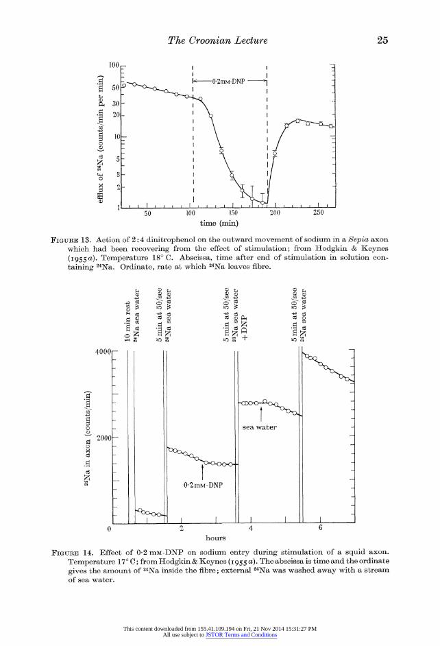
Figure 13 illustrates the effect of 0-2 mM-dinitrophenol on the rate at which a
Sepia axon eliminates labelled sodium during the period of recovery which follows
a burst of electrical activity. At this concentration, dinitrophenol probably does
not reduce respiration but it prevents metabolism from doing useful work, possibly
by interfering with oxidative phosphorylation. It inhibits movement of ions against concentration gradients in gastric mucosa (Davies 1951), frog skin (Fuhrman 1952), chicken erythrocytes (Maizels 1954) and in certain plant cells (Robertson, Wilkins
& Weeks 1951; Scott & Hayward 1954). It does not have any obvious effect on
sodium transport in frog muscle (Keynes & Maisel 1954) or mammalian erythro?
cytes (Maizels 1951), possibly because glycolysis is an important source of energy in these tissues. Applied to a Sepia nerve fibre, it reduces the sodium efflux to
about 1/20 of its normal value; the inhibitory effect takes place with a time
constant of 15 to 30 min and is largely, but often not completely, reversed by
washing the dinitrophenol away. Cyanide (1 to 10 nn) and azide (3 nn) have
essentially the same action as dinitrophenol on the outflow of sodium.
Although agents like dinitrophenol or cyanide impair the recovery mechanism,
they do not have any rapid effect on the action potential of giant axons, and fibres
which have been poisoned with these substances continue to conduct impulses for
many hours. The experiment of figure 14 shows that when a squid fibre is treated
with dinitrophenol, sodium ions still move into the fibre during activity, but that
they accumulate inside the fibre and are not pumped out until the dinitrophenol is removed. The conclusion from experiments of this kind is that the metabolic
reactions inhibited by cyanide or dinitrophenol are not of immediate importance
This content downloaded from 155.41.109.194 on Fri, 21 Nov 2014 15:31:27 PMAll use subject to JSTOR Terms and Conditions
The Croonian Lecture 25
100 150 time (min)
Figure 13. Action of 2:4 dinitrophenol on the outward movement of sodium in a Sepia axon which had been recovering from the effect of stimulation; from Hodgkin & Keynes (1955 a). Temperature 18? C. Abscissa, time after end of stimulation in solution con?
taining 24Na. Ordinate, rate at which 24Na leaves fibre.
4000i
co
2000
co a!
r^HxJ
t
I +
0-2mM-DNP
sea water
o ?
13
hours
Figure 14. Effect of 0-2 niM-DNP on sodium entry during stimulation of a squid axon.
Temperature 17? C; from Hodgkin & Keynes (1955a). The abscissa is time and the ordinate
gives the amount of 24Na inside the fibre; external 24Na was washed away with a stream of sea water.
This content downloaded from 155.41.109.194 on Fri, 21 Nov 2014 15:31:27 PMAll use subject to JSTOR Terms and Conditions
26 A. L. Hodgkin
in generating the spike but are essential for recovery. This is very reasonable.
During the spike ions move downhill and this does not require metabolic energy?
although of course it may need a complicated and tricky mechanism to change the
permeability at the right time and in the right way. During recovery, a consider?
able amount of work has to be done, and it is therefore not surprising that metabolic
inhibitors are effective.
Although it may be some time before much is known about chemical events in
the membrane, information about the nature of the biochemical reactions which
drive the ion transport system can be obtained by the methods used by Caldwell
and Keynes. Caldwell (1956) examined the effect of agents which inhibit the
sodium pump on the phosphate esters of squid nerve. In a normal fibre, the three
main forms of phosphate are inorganic phosphate, arginine phosphate and ATP.
When a substance like cyanide or dinitrophenol is applied, at a concentration
which reduces sodium efflux, the concentrations of both arginine phosphate and
ATP decrease to about one-tenth of their normal value. At the same time there
is an increase in inorganic phosphate. The concentrations of ATP and arginine
phosphate recover to a considerable extent when the inhibitor is removed and, in
general, there is a satisfactory correlation between the concentrations of ATP or
arginine phosphate and the rate at which sodium is extruded from the nerve.
Caldwell (1957) also showed that if 0-2 mM-dinitrophenol is applied at an alkaline
pH, when it has no effect on the sodium pump, it leaves ATP at its normal level
but reduces the concentration of arginine phosphate. At a neutral or slightly acid pH, where dinitrophenol reaches a higher concentration in the axoplasm, both
ATP and arginine phosphate are reduced, as is the efflux of sodium from the
axoplasm. The implication of these results is that dephosphorylation of ATP may be the immediate source of energy for the pump and that arginine phosphate acts
by replenishing the ATP. The conclusion is perhaps what might be expected from
the analogy of vertebrate muscle, but it is satisfactory to have experimental evidence of a connexion between ATP concentration and sodium transport.
Caldwell & Keynes (1957) have now followed up these results by injecting ATP
and arginine phosphate into nerves poisoned with cyanide. They find that injection of ATP partially restores the efflux of sodium, whereas injections of the same
solution hydrolyzed by boiling have no effect (figure 15). Arginine phosphate acts
in much the same way as ATP, as one would expect from the fact that it probably
generates ATP. The effect of both substances is transient, presumably because they are used up in driving the ion transport system, and perhaps in side reactions as
well. The quantity of Na extruded was found to be roughly proportional to the
amount of ATP injected. On the assumption that all the ATP is used in driving the sodium pump, Caldwell & Keynes (1957) calculate that about four phosphate bonds are broken for each sodium ion expelled. The efficiency calculated from this
result, about 10%, seems rather low, but the measurements on which it depends were made in the presence of cyanide and a normal fibre may utilize ATP more
effectively. In connexion with these results it should be mentioned that, although an injection such as that used in the experiment of figure 15 raised the ATP
concentration in the axoplasm to about the same level as in a fresh fibre, the sodium
This content downloaded from 155.41.109.194 on Fri, 21 Nov 2014 15:31:27 PMAll use subject to JSTOR Terms and Conditions

The Croonian Lecture 27
efflux was not fully restored. The incompleteness of the restoration might be ex?
plained by supposing that ATP is normally produced near the membrane at a
higher concentration, or in more general terms, that microinjection of ATP is not
a perfect imitation of the normal effect of metabolism. Nevertheless, in spite of
uncertainties of the kind usually raised by new results, these experiments support
very strongly the idea that high-energy phosphates, such as ATP, are necessary for
the process which extrudes sodium from the axon.
in2mM-CN
f % N_
??$^ -i-
5 hours
broken-down ATP ATP injected
injected
Figure 15. Effect of injecting ATP on the rate at which labelled sodium leaves a squid axon which has been poisoned with cyanide, from Caldwell & Keynes (1957).
A 780ju, squid axon was loaded by injecting 6700 counts/min of 22Na over 12 mm. A solution of ATP, which was hydrolyzed by boiling in the first injection but not in the second, was introduced over the same 12 mm length of axon. The solution contained 16 x 10~9 mole ATP and should have raised the ATP concentration by about 3 mM.
Reabsorption of potassium during recovery
The next question to consider is whether a metabolic system is also required to
absorb potassium ions. Here then are two useful kinds of evidence. In the first
place, it is found that agents which inhibit or reduce sodium extrusion have a
corresponding effect on the uptake of potassium during recovery (Hodgkin &
Keynes 1955a). This inhibition takes place without any appreciable change in
membrane potential so that it is unlikely to be an electrical consequence of the
reduction in sodium transfer. The second point is that when potassium ions are
removed from the external fluid the sodium efflux drops suddenly to about one-
fourth of its former value and is restored at once when potassium ions are replaced
(figure 16 of this article and figure 11 of Hodgkin & Keynes 1955a). Increasing the external potassium concentration five-fold raises the sodium efflux, but the
effect is small compared to that of removing potassium.
Perhaps the most obvious explanation of the effect of K-free solution on the
outflow of sodium is that the rise in resting potential, associated with removal of
potassium, slows up sodium extrusion by increasing the electrical gradient against which these ions have to be forced. This possibility was tested by varying the
This content downloaded from 155.41.109.194 on Fri, 21 Nov 2014 15:31:27 PMAll use subject to JSTOR Terms and Conditions

28 A. L. Hodgkin
membrane potential with an applied current and seeing whether such variations
had any effect on sodium efflux (Hodgkin & Keynes 1954). The answer was that
large increases in membrane potential?up to 40 mV?had no effect on sodium
outflow. Since the rise in membrane potential produced by removing potassium ions from the external fluid was only 5 to 10 mV, it is clear that the effect of K-free
solutions on sodium efflux cannot be caused by the change in membrane potential. The most likely possibility is that both the outflow of sodium and the uptake of
potassium depend on metabolism, and that the two movements are coupled in a
cyclical mechanism of the type shown in figure 17 (p. 32). With such a mechanism
one would expect that interfering with metabolism would reduce the uptake of
potassium and that removal of external potassium ions might reduce the outflow
of sodium. 10 mM-K K-free 10 mM-K 50 mM-K 10 mM-K
100 time (min)
Figure 16. Effect of changing the external potassium concentration on the rate at which sodium ions leave a Sepia axon, from an experiment of Hodgkin & Keynes (1955 a). The 24Na was introduced by stimulating the fibre in a radioactive solution at 50/sec for 9 min. The axon diameter was 193^, and the temperature 18? C; vertical lines show ? Is.e.
General nature of the system which moves Na and K against concentration gradients
The tentative conclusion from the experiments mentioned in the previous section
is that the same secretory system acts on both sodium and potassium ions and that
sodium extrusion may be coupled to potassium influx. Evidence for a coupling between sodium efflux and potassium influx has been obtained in muscle (Stein? bach 1952; Keynes 1954), red cells (Harris & Maizels 1951; Shaw 1954; Glynn 1956) and frog skin (Ussing 1954; Koefoed Johnsen & Ussing 1956). An attractive
possibility is that there may be a tight coupling between Na and K movements
and that one sodium ion is extruded for each potassium ion absorbed. This does
not mean that the sodium efflux must be equal to the potassium influx or that
the former should fall to zero in the absence of external potassium; for there may be an exchange of sodium ions across the membrane under conditions in which
active transport has ceased (see Ussing 1949; Glynn 1956; Swan & Keynes 1956).
However, if the idea of a tight coupling is correct it follows that there should be
no net movement of sodium against a concentration difference in the absence of
external potassium, and that the sodium efflux should fall to zero when both
sodium and potassium ions are removed from the external medium. The first
prediction is supported by Glynn's (1956) studies with red cells, but has not been
This content downloaded from 155.41.109.194 on Fri, 21 Nov 2014 15:31:27 PMAll use subject to JSTOR Terms and Conditions

The Croonian Lecture 29
adequately tested in nerve and muscle. The second appears to be at variance with
the observation that an efflux of sodium (which is inhibited by dinitrophenol) can take
place from a Sepia axon into an isotonic sugar solution from which all the potassium and sodium have been removed (Hodgkin & Keynes 1955 a). However, in such an
experiment it is difficult to be sure that the potassium concentration immediately outside the membrane has not been raised by leakage of KC1 to an extent which
allows a coupled movement of Na and K to continue. Further experiments along these lines are clearly needed; for the time being the position may be summarized
by saying that although there is evidence for a loose or partial coupling it has not
been proved that the pump operates by ejecting one sodium ion for each potassium ion absorbed.
A physical basis for a coupled system is provided by a hypothesis of the kind
discussed by Harris (1954), Shaw (1954), Hodgkin & Keynes (1954) and Glynn
(1956). Suppose that potassium enters the cell in association with a carrier (X) and
that on the inside of the membrane this carrier is changed chemically into a
second molecule (Y) which is selective for sodium and diffuses outward with
sodium. On the outside of the membrane the carrier reverts to the potassium form
and is once more available for taking potassium into the cell. This system could be
interrupted either by removing external potassium ions or by withdrawing the
source of energy which is assumed to convert the potassium carrier, X, into the
sodium carrier, Y. If the undissociated compounds KX and NaF were able to
cross the membrane much more freely than the dissociated carriers, X and Y, the
system would act as an electrically neutral pump which extruded one sodium
ion for each potassium ion absorbed.
At present there is no evidence as to the chemical nature of the carriers postu? lated to explain movements of substances against concentration differences. Even
if such carriers can be shown to exist, the problem of chemical identification will
not be easy since there are indications that the density of carriers in the membrane
must be very low. A few years ago Schatzmann (1953) showed that one of the
cardiac glycosides, strophanthin, inhibited the active transport of sodium and
potassium in human red cells. Other cardiac glycosides have since been found to
act in a similar manner (Joyce & Weatherall 1955; Kahn & Acheson 1955;
Solomon, Gill & Gold 1956; Glynn 1957) and these compounds have been shown
to inhibit ion transport in a number of tissues, including nruscle (Johnson, 1956; Edwards & Harris 1957) and squid giant axons (Caldwell & Keynes, unpublished). Schatzmann showed that concentrations of strophanthin which inhibit ion trans?
port do not affect the oxygen consumption or glycolysis of red cells, and it seems
that these substances act directly on the membrane rather than on the energy
supply to the pump (Glynn 1955, 1957; Whittam 1957). Some of the cardiac
glycosides inhibit ion transport at remarkably low concentrations and, from an
analysis of the effect of small quantities on red cells, Glynn (1957) concluded that
the number of sites at which active transport occurs must be less than 1000 to 2000
per cell. This suggests that the substances concerned with transporting ions
through the membrane against concentration differences must be present in very low concentrations indeed. In connexion with Glynn's results it is interesting
This content downloaded from 155.41.109.194 on Fri, 21 Nov 2014 15:31:27 PMAll use subject to JSTOR Terms and Conditions

30 A. L. Hodgkin
that electron micrographs of the inside of the red cell envelope show small par? ticles about 250 A in diameter spaced at distances which correspond very roughly to 2000 per cell (Hillier & Hoffman 1953, figure 34, and Hoffman, personal
communication). One interesting point about the active transport mechanism is that it operates
with very little time lag. If 24Na is suddenly injected into an axon, these ions start
appearing in the external fluid within about 10 sec (Hodgkin & Keynes 1956). Most of this delay is caused by diffusion in the axoplasm and extracellular fluid; there is little time left to allow sodium ions to be collected in, say, a contractile
vacuole and to be ejected in the manner in which water is thrown out of an amoeba.
Although lithium and sodium ions appear to be equally effective in generating an action potential, there is evidence that the secretory mechanism may dis?
criminate between the two ions. Thus, lithium appears to be extruded more slowly than sodium from red blood-cells (Flynn & Maizels 1950) and muscle fibres (Swan & Keynes 1956). In the light of subsequent work (Koefoed Johnsen & Ussing
1956), it would seem that the lithium current in frog skin may be due to a passive accumulation of lithium in the skin (Zerahn 1954), and that this ion is pumped more slowly than sodium. Ritchie & Straub's (1957) observations on the after-
potentials of mammalian C fibres are also consistent with the idea that lithium ions
enter a nerve during activity but are pumped out more slowly than sodium.
Maintenance of the steady state*
If a nerve or muscle fibre is to remain in a steady state there must be a balance
between the rates at which Na and K leak through the membrane under the
influence of electrochemical gradients, and the rate at which these ions are pumped
by the metabolic transport system. Although little is known about the way in
which the internal concentrations of ions are regulated, it is, nevertheless, reason?
able to ask whether a neutral pump, which absorbed one potassium ion for each
sodium ion ejected, could maintain a cell in a steady state with differences in
concentration and electrical potential of the kind observed experimentally. If the
pump is strictly neutral it can transfer no charge across the membrane. This means
that in the steady state the rate at which sodium ions leak into the fibre must be
equal to the rate at which potassium ions leak out. The membrane potential at
which this condition is satisfied will depend on the relative permeabilities to Na
and K and with certain reservations can be calculated by means of the equation
F [K], + 6[Na], v ;
Taking b as 0-01 and using the data in table 1 as a basis for calculation it is found
that the resting potential will be about 5 mV less than the equilibrium potential for the potassium ion, i.e. ? 70 mV instead of ? 75 mV. This appears to be
consistent with the observations mentioned on p. 6 which indicate that, in a living
squid, the resting potential is within a few millivolts of the potassium equilibrium
* The theoretical arguments in this section were developed in conjunction with Dr P. Horowicz and Dr R. H. Adrian.
This content downloaded from 155.41.109.194 on Fri, 21 Nov 2014 15:31:27 PMAll use subject to JSTOR Terms and Conditions
The Croonian Lecture 31
potential; the larger difference in isolated axons may occur because branches are
cut through in isolating the axon. Experiments with frog's muscle suggest that the
resting potential is about 10 mV less than that expected from the potassium concentration ratio, i.e. ? 92 mV instead of ? 102 mV (Adrian 1956).
At present it is considered that while the movements of sodium and to a lesser
extent potassium are influenced by the activity of the metabolic transport system, chloride ions distribute themselves passively across the membrane (Conway 1957). In this case the ratio of chloride concentration should be given by the equation
BT a F [Cl]0
K '
If this equation is combined with (9) we obtain
(11) [K]0 + 6[Na]0_[Cl],
[K], + 6[Na], [Ciy
Since b 4=0-01 in muscle this equation approximates to the Donnan equation
(12) except when [K]0 is small compared to [Na]0:
[KL = [C1],
[K], [Cl]0- l '
As is well known from the work of Boyle & Conway (1941), and others, the
ratios of the potassium and chloride concentrations in muscle agree satisfactorily with equation (12) provided [K]0 is greater than about 10 im. It seems possible that the deviations which occur at lower potassium concentrations are explained
satisfactorily by an equation such as (11) which takes account of the imperfect
selectivity of the resting membrane towards potassium. In the theory of Boyle & Conway (1941) the concentrations of K+ and Cl~ inside
the muscle, and the cell volume were calculated by combining equation (12) with
the relations imposed by the conditions of electroneutrality and osmotic balance.
A similar argument applies if Na+ is regarded as a permeable ion which is main?
tained at a fixed internal concentration [Na]; by a neutral pump. In this case, the
internal concentrations of K and Cl, and the cell volume, v, are determined by
equation (11) used in conjunction with
[K]* + [Na],. + [Cl]* + ? = C (osmotic balance), (13)
zA [K], + [Na]; - [GIL + ? = 0 (electro-neutrality), (14)
where C is the total concentration of all particles in the external solution, A is the
total quantity of indiffusible particles inside the cell and z is the average valency of these particles. It can be shown that if b = 0-01 the solutions of equations (11),
(13) and (14), which are similar to those obtained from the simpler theory of Boyle & Conway, agree satisfactorily with data obtained on muscle. The conclusion is
that, if the permeability to sodium is small compared with that of potassium, a neutral
pump, which extruded one Na ion for each K absorbed, could keep a cell in a
balanced state with concentrations similar to those in muscle or nerve.
This content downloaded from 155.41.109.194 on Fri, 21 Nov 2014 15:31:27 PMAll use subject to JSTOR Terms and Conditions

32 A. L. Hodgkin
As a by-product, it is interesting to consider what the steady-state concentra?
tions would be in a cell in which the membrane was not selectively permeable to
potassium ions. If the permeabilities to Na and K are assumed to be in the same
ratio as the mobilities in aqueous solution, the constant b would be 0-67. In this
case, if A is taken as a monovalent anion (z = ? 1), solution of equations (11), (13) and (14) gives a result which is similar to that in the human red blood corpuscle. Thus with 5 mM-K, 145 mM-Na and 150 mM-Cl in the external fluid, the internal
concentrations calculated on the assumption that [Na]; is maintained at 15 niM are
[Cl];== 106 mM and [K]^= 135 mM. Such a system resembles the human red blood
corpuscle in being able to maintain a steady state with a [K] [Cl] product inside
the cell about twenty times greater than that in the external fluid.
Conclusion
The implication of the experiments considered in this article is that there are
two quite distinct systems in a nerve fibre. On the one hand, there is a secretory mechanism which builds up concentration differences by absorbing potassium and
K+
outside
high [Na] low [K]
membrane
Na+
A
impi ulse!
metabolism Na+ inside low [Na] high [K]
K+
Figure 17. Diagram illustrating movements of ions through the nerve membrane (from Hodgkin & Keynes 1955 a). The downhill movements which occur during the impulse are shown on the right; uphill movements during recovery are shown on the left. The broken line represents the component of the sodium efflux which is not abolished by removing external potassium ions.
ejecting sodium. In parallel with this system are the special channels which allow
sodium and potassium ions to move at varying rates down their concentration
gradients. There are a number of differences between the two systems. Metabolic
inhibitors affect the secretory system but, at all events in giant nerve fibres, seem
to have little effect on the system controlling permeability. Changes in membrane
potential or changes in external calcium concentration have a large effect on
permeability, but do not seem to have much effect on the secretory system. There
are also indications of difference in the ability of the two systems to discriminate
between the sodium and lithium ions. For these reasons the two systems have been
drawn in figure 17 as physically separate. This conclusion may have to be revised
when more direct information is forthcoming; on present evidence it seems to be
the most satisfactory inference from experiment.
This content downloaded from 155.41.109.194 on Fri, 21 Nov 2014 15:31:27 PMAll use subject to JSTOR Terms and Conditions

The Croonian Lecture 33
Figure 17 provides a convenient opportunity for summarizing the role of sodium
and potassium ions in the conduction of nervous impulses. To begin with, the fibre
is in its resting condition with a large potential difference across the membrane.
The sodium permeability is very low so that only a trickle of ions enters the fibre; this trickle is dealt with by the pump which keeps the fibre in a steady state. When
the nerve conducts an impulse the channels open up, allowing first sodium ions and
then potassium ions to move down their concentration gradients. These movements
generate the action potential and the fibre pays by accumulating sodium and losing
potassium. After activity, the original state is restored by the cyclical process which uses metabolic energy to eject sodium and absorb potassium.
The whole mechanism seems admirably designed for a system in which infor?
mation is conveyed as a series of brief impulses spaced at widely varying intervals.
The ionic concentration differences allow electrical energy to be dissipated at a high rate. If the nerve has to handle a large number of impulses in a short time it can
run into debt by accumulating sodium and losing potassium. However, sooner or
later the debt must be paid off, and this can be done during the periods of rest
which alternate with those of more intense activity.
In addition [to those mentioned in the text, my thanks are due to Mr A. F.
Huxley & Dr R. D. Keynes for reading the manuscript and for much helpful discussion. Many of the ideas mentioned in this article originated in the course
of conversation with Mr A. F. Huxley, to whom I wish to express my particular indebtedness.
References
Adrian, R. H. 1956 The effect of internal and external potassium concentration on the membrane potential of frog muscle. J. Physiol. 133, 631.
Bernstein, J. 1902 Untersuchungen zur Thermodynamik der bioelektrischen Strome. Erster Theil. Pfliig. Arch. ges. Physiol. 92, 521.
Boyle, P. J. & Conway, E. J. 1941 Potassium accumulation in muscle and associated changes. J. Physiol. 100, 1.
Brady, A. J. & Woodbury, J. W. 1957 Effects of sodium and potassium on repolariza- tion in frog ventricular fibres. Ann. N.Y. Acad. Sci. 65, 687.
Caldwell, P. C. 1956 The effects of certain metabolic inhibitors on the phosphate esters of the squid giant axon. J. Physiol. 132, 35 P.
Caldwell, P. C. 1957 The sensitivity to pH of the inhibitory effects of dinitrophenol on squid giant axons. Biochem. J. 67, 1p.
Caldwell, P. C. & Keynes, R. D. 1957 The utilization of phosphate bond energy for sodium extrusion from giant axons. J. Physiol. 137, 12P.
Cole, K. S. 1949 Dynamic electrical characteristics of the squid axon membrane. Arch. Sci.
physiol. 3, 253. Cole, K. S. 1954 Electro-ionics of nerve action. U.S. Naval Medical Research Institute.
Lecture and Review Series. 54~6, 213. Cole, K. S. 1955 Ions, potentials and the nerve impulse. In Electrochemistry in biology and
medicine, p. 121. New York: Wiley. Cole, K. S. & Curtis, H. J. 1939 Electric impedance of the squid giant axon during activity.
J. Gen. Physiol. 22, 649. Cole, K. S. & Hodgkin, A. L. 1939 Membrane and protoplasm resistance in the squid giant
axon. J. Gen. Physiol. 22, 671.
Conway, E. J. 1957 Nature and significance of concentration relations of potassium and sodium ions in skeletal muscle. Physiol. Rev. 37, 84.
Vol. 148. B.
This content downloaded from 155.41.109.194 on Fri, 21 Nov 2014 15:31:27 PMAll use subject to JSTOR Terms and Conditions

34 A. L. Hodgkin
Coraboeuf, E. & Otsuka, M. 1956 L'action des solutions hyposodique sur les potentiels cellulaire de tissu cardiaque de Mammiferes, C.R. Soc. Biol., Paris, 243, 441.
Curtis, H. J. & Cole, K. S. 1938 Transverse electric impedance of the squid giant axon. J. Gen. Physiol. 21, 757.
Curtis, H. J. & Cole, K. S. 1942 Membrane resting and action potentials from the squid giant axon. J. Cell. Comp. Physiol. 19, 135.
Davies, R. E. 1951 The mechanism of hydrochloric acid production by the stomach. Biol. Rev. 26, 87.
Desmedt, J. E. 1953 Electrical activity and intracellular sodium concentration in frog muscle. J. Physiol. 121, 191.
Draper, M. H. & Weidmann, S. 1951 Cardiac resting and action potentials recorded with an intracellular electrode. J. Physiol. 115, 74.
Edwards, C. & Harris, E. J. 1957 Factors influencing the Na movement in frog muscle with a discussion of the mechanism of Na movement. J. Physiol. 135, 567.
Fatt, P. & Katz, B. 1953 Electrical properties of crustacean muscle. J. Physiol. 120, 171.
Fluckiger, E. & Keynes, R. D. 1955 The Ca permeability of Loligo axons. J. Physiol. 128, 41 P.
Flynn, F. & Maizels, M. 1950 Cation control in human erythrocytes. J. Physiol. 110, 301. Frankenhaeuser, B. 1957 The effect of calcium on the myelinated nerve fibre. J. Physiol.
137, 245. Frankenhaeuser, B. & Hodgkin, A. L. 1956 The after-effects of impulses in the giant nerve
fibres of Loligo. J. Physiol. 131, 341. Frankenhaeuser, B. & Hodgkin, A. L. 1957 The action of calcium on the electrical properties
of squid axons. J. Physiol. 137, 218.
Fuhrman, F. A. 1952 Inhibition of active sodium transport in the isolated frog skin. Amer. J. Physiol. 171, 266.
Geren, B. B. & Schmitt, F. O. 1954 The structure of the Schwann cell and its relation to the axon in certain invertebrate nerve fibres. Proc. Nat. Acad. Sci., Wash., 40, 863.
Glynn, I. M. 1955 Action of cardiac glycosides on red cells. J. Physiol. 128, 56P.
Glynn, I. M. 1956 Sodium and potassium movements in human red cells. J. Physiol. 134, 278.
Glynn, I. M. 1957 The action of cardiac glycosides on sodium and potassium movements in human red cells. J. Physiol. 136, 148.
Goldman, D. E. 1943 Potential, impedance and rectification in membranes. J. Gen. Physiol. 27, 37.
Grundfest, H. & Nachmansohn, D. 1950 Increased sodium entry into squid giant axons at
high frequencies and during reversible inactivation of cholinesterase. Fed. Proc. 9, 53.
Hagiwara, S. & Tasaki, I. 1957 Demonstration of two stable potential states in the squid giant axon under tetraethylammonium chloride. J. Gen. Physiol. 40, 859.
Hagiwara, S. & Watanabe, A. 1955 The effect of tetraethylammonium chloride on the muscle membrane examined with an intracellular electrode. J. Physiol. 129, 513.
Harris, E. J. 1954 Linkage of sodium- and potassium-active transport in human erythrocytes. Symp. Soc. Exp. Biol. 8, 228.
Harris, E. J. & Maizels, M. 1951 The permeability of human erythrocytes to sodium. J. Physiol. 113, 506.
Hillier, J. & Hoffman, J. F. 1953 On the ultrastructure of the plasma membrane as deter? mined by the electron microscope. J. Cell. Comp. Physiol. 42, 203.
Hodgkin, A. L. 1947 The effect of potassium on the surface membrane of an isolated axon. J. Physiol. 106, 319.
Hodgkin, A. L. 1951 The ionic basis of electrical activity in nerve and muscle. Biol. Rev. 26, 339.
Hodgkin, A. L. & Huxley, A. F. 1947 Potassium leakage from an active nerve fibre. J. Physiol. 106, 341.
Hodgkin, A. L. & Huxley, A. F. 1952 a. Currents carried by sodium and potassium ions
through the membrane of the giant axon of Loligo. J. Physiol. 116, 449.
Hodgkin, A. L. & Huxley, A. F. 1953& The components of membrane conductance in the
giant axon of Loligo. J. Physiol. 116, 473.
Hodgkin, A. L. & Huxley, A. F. 1952c The dual effect of membrane potential on sodium conductance in the giant axon of Loligo. J. Physiol. 116, 497.
Hodgkin, A. L. & Huxley, A. F. 1952 c? A quantitative description of membrane current and its application to conduction and excitation in nerve. J. Physiol. Ill, 500.
This content downloaded from 155.41.109.194 on Fri, 21 Nov 2014 15:31:27 PMAll use subject to JSTOR Terms and Conditions

The Croonian Lecture 35
Hodgkin, A. L. & Huxley, A. F. 1952 e Propagation of electric signals along giant nerve fibres. Proc. Roy. Soc. B, 140, 177.
Hodgkin, A. L. & Huxley, A. F. 1952/ Movements of sodium and potassium ions during nervous activity. Cold Spr. Harb. Symp. Quant. Biol. 17, 43.
Hodgkin, A. L. & Huxley, A. F. 1953 Movement of radioactive potassium and membrane current in a giant axon. J. Physiol. 121, 403.
Hodgkin, A. L., Huxley, A. F. & Katz, B. 1952 Measurements of current-voltage relations in the membrane of the giant axon of Loligo. J. Physiol. 116, 424.
Hodgkin, A. L. & Katz, B. 1949 The effect of sodium ions on the electrical activity of the giant axon of the squid. J. Physiol. 108, 37.
Hodgkin, A. L. & Keynes, R. D. 1953 The mobility and diffusion coefficient of potassium in
giant axons from Sepia. J. Physiol. 119, 513.
Hodgkin, A. L. & Keynes, R. D. 1954 Movements of cations during recovery in nerve. Symp. Soc. Exp. Biol. 8, 423.
Hodgkin, A. L. & Keynes, R. D. 1955 a Active transport of cations in giant axons from Sepia and Loligo. J. Physiol. 128, 28.
Hodgkin, A. L. & Keynes, R. D. 19556 The potassium permeability of a giant nerve fibre. J. Physiol. 128, 61.
Hodgkin, A. L. & Keynes, R. D. 1956 Experiments on the injection of substances into squid giant axons by means of a microsyringe. J. Physiol. 131, 592.
Hodgkin, A. L. & Keynes, R. D. 1957 Movements of labelled calcium in giant axons. J. Physiol. 138, 253.
Huxley, A. F. 1954 Electrical processes in nerve conduction. In Ion transport across mem? branes, p. 23. New York: Academic Press.
Huxley, A. F. & Stampfli, R. 1949 Evidence for saltatory conduction in peripheral myelinated nerve fibres. J. Physiol. 108, 315.
Huxley, A. F. & Stampfli, R. 1951 Effect of potassium and sodium on resting and action potentials of single myelinated nerve fibres. J. Physiol. 112, 496.
Johnson, J. A. 1956 Influence of ouabain, strophanthidin and dihydrostrophanthidin on sodium and potassium transport in frog sartorii. Amer. J. Physiol. 187, 328.
Joyce, C. R. B. & Weatherall, M. 1955 Cardiac glycosides and the potassium exchange of human erythrocytes. J. Physiol. 127', 33 P.
Kahn, J. B. & Acheson, G. H. 1955 Effect of cardiac glycosides and other lactones, and of certain other compounds on cation transfer in human erythrocytes. J. Pharmacol. 115, 305.
Katz, B. 1947 The effect of electrolyte deficiency on the rate of conduction in a single nerve fibre. J. Physiol. 106, 411.
Katz, B. 1949 Les constantes electriques de la membrane du muscle. Arch. Sci. physiol. 3, 285.
Keynes, R. D. 1949 The movements of radioactive ions in resting and stimulated nerve. Arch. Sci. physiol. 3, 165.
Keynes, R. D. 1951 a The leakage of radioactive potassium from stimulated nerve. J. Physiol. 113, 99.
Keynes, R. D. 19516 The ionic movements during nervous activity. J. Physiol. 114, 119. Keynes, R. D. 1954 The ionic fluxes in frog muscle. Proc. Roy. Soc. B, 142, 359. Keynes, R. D. & Adrian, R. H. 1956 The ionic selectivity of nerve and muscle membranes.
Disc. Faraday Soc. 21, 265.
Keynes, R. D. & Lewis, P. R. 1951 The sodium and potassium content of cephalopod nerve fibres. J. Physiol. 114, 151.
Keynes, R. D. & Lewis, P. R. 1956 The intracellular calcium concentration of some invertebrate nerves. J. Physiol. 134, 399.
Keynes, R. D. & Maisel, G. W. 1954 The energy requirements for sodium extrusion from a frog muscle. Proc. Roy. Soc. B, 142, 383.
Koechlin, B. A. 1955 On the chemical composition of the axoplasm of squid giant nerve fibres with particular reference to its ion pattern. J. biophys. biochem. Cytol. 1, 511.
Koefoed Johnsen, V. & Ussing, H. H. 1956 Nature of the frog skin potential. Abstr. Comm. XXth int. Physiol. Gongr. p. 511.
Krogh, A. 1946 The active and passive exchanges of inorganic ions through the surfaces of living cells and through living membranes generally. Proc. Roy. Soc. B, 133, 140.
3-2
This content downloaded from 155.41.109.194 on Fri, 21 Nov 2014 15:31:27 PMAll use subject to JSTOR Terms and Conditions
36 A. L. Hodgkin
Larramendi, L. M. H., Lorente de No, R. & Vidal, F. 1956 Restoration of sodium deficient nerve fibres by an isotonic solution of guanidinium chloride. Nature, Lond. 178, 316.
Lorente de No, R. 1949 On the effect of certain quaternary ammonium ions upon frog nerve. J. Cell. Comp. Physiol. 33, Supplement.
Lorente de No, R., Vidal, F. & Larramendi, L. M. H. 1957 Restoration of sodium-deficient
frog nerve fibres by onium ions. Nature, Lond. 179, 737. Maizels, M. 1951 Factors in the active transport of cations. J. Physiol. 112, 59. Maizels, M. 1954 Active cation transport in erythrocytes. Symp. Soc. Exp. Biol. 8, 202. Moore, J. W. & Cole, K. S. 1955 Membrane potentials of the squid giant axon in vivo. Fed.
Proc. 14, 103. Miiller, P. 1956 fiber verlangerte Aktionspotentiale in Na-haltigen und Na-freien Aussen-
medium. Internationales Symposium uber den Mechanismus der Erregung. p. 63. Berlin: Deutsche Verlag der Wissenschaften.
Nastuk, W. L. & Hodgkin, A. L. 1950 The electrical activity of single muscle fibres. J. Cell.
Comp. Physiol. 35, 39.
Overton, E. 1902 Beitrage zur allgemeinen Muskel- und Nervenphysiologie. Pfliig. Arch.
ges. Physiol. 92, 346. Remak, R. 1843 Ueber den Inhalt der Nervenprimitivrohrchen. Arch. Anat. Physiol. Wiss.
Med., p. 197. Ritchie, J. M. & Straub, R. W. 1957 The hyperpolarization which follows activity in mam?
malian non-medullated fibres. J. Physiol. 136, 80.
Robertson, J. D. 1949 Ionic regulation in some marine invertebrates. J. Exp. Biol. 26, 182.
Robertson, J. D. 1957 New observations on the ultrastructure of the membrane of frog peripheral nerve fibres. J. biophys. biochem. Cytol. (in the Press).
Robertson, R. N., Wilkins, M. J. & Weeks, D. C. 1951 Studies in the metabolism of plant cells. IX. The effects of 2, 4-dinitrophenol on salt accumulation and salt respiration. Aust. J. Sci. Res. 4, 248.
Rothenberg, M. A. 1950 Studies on permeability in relation to nerve function. II. Ionic movements across axonal membranes. Biochim. biophys. Acta, 4, 96.
Schatzmann, H. J. 1953 Herzglykoside als Hemmstoffe fur den aktiven Kalium und Natrium
Transport durch die Erythrocytenmembran. Helv. physiol. Acta, 11, 346.
Scott, G. T. & Hayward, H. R. 1954 Evidence for the presence of separate mechanisms
regulating potassium and sodium distribution in Ulva lactuca. J. Gen. Physiol. 37, 601.
Shanes, A. M. 1954 Effect of temperature on potassium liberation during nerve activity. Amer. J. Physiol. Ill, 377.
Shaw, T. I. 1954 Sodium and potassium movements in red cells. Ph.D. Thesis. Cambridge University.
Shaw, F. H., Simon, Shirley E. & Johnstone, B. M. 1956 The non-correlation of bioelectric
potentials with ionic gradients. J. Gen. Physiol. 40, 1.
Shaw, F. H., Simon, Shirley E., Johnstone, B. M. & Holman, Mollie, E. 1956 The effect of
changes of environment on the electrical and ionic pattern of muscle. J. Gen. Physiol. 40, 263.
Sollner, K., Dray, S., Grim, E. & Neihof, R. 1955 Membranes of high electrochemical activity in studies of biological interest. In Electrochemistry in biology and medicine, p. 65. New York: Wiley.
Solomon, A. K., Gill, T. J. & Gold, G. L. 1956 The kinetics of glycoside inhibition of potassium transport in human erythrocytes. J. Gen. Physiol. 40, 327.
Stampfli, R. 1952 Bau und Funktion isolierter markhaltiger Nervenfasern. Ergebn. Physiol. 47, 70.
Stampfli, R. 1956 Nouvelle methode pour enregistrer le potentiel d'action d'un seul etrangle- ment de Ranvier et sa modification par un brusque changement de la concentration du milieu exterieur. J. Physiol. Paris. 48, 710.
Steinbach, H. B. 1941 Chloride in the giant axons of the squid. J. cell. comp. Physiol. 17, 57.
Steinbach, H. B. 1952 On the sodium and potassium balance of isolated frog muscles. Proc. Nat. Acad. Sci., Wash., 38, 451.
Steinbach, H. B. & Spiegelman, S. 1943 The sodium and potassium balance in squid nerve
axoplasm. J. Cell. Comp. Physiol. 22, 187.
Swan, R. C. & Keynes, R. D. 1956 Sodium efflux from amphibian muscle. Abstr. Gomm. XXth int. Physiol. Congr., p. 869.
This content downloaded from 155.41.109.194 on Fri, 21 Nov 2014 15:31:27 PMAll use subject to JSTOR Terms and Conditions

The Croonian Lecture 37
Tasaki, I. 1953 Nervous transmission. Springfield: Thomas.
Tasaki, I. 1956 Initation and abolition of the action potential of a single node of Ranvier. J. Gen. Physiol. 39, 377.
Ussing, H. H. 1949 Transport of ions across cellular membranes. Physiol. Rev. 29, 127.
Ussing, H. H. 1954 Ion transport across biological membranes. In Ion transport across membranes. New York: Academic Press.
Webb, D. A. 1939 The sodium and potassium content of sea water. J. Exp. Biol. 116, 178, Webb, D. A. 1940 Ionic regulation in Carcinus maenas. Proc. Roy. Soc. B, 129, 107.
Weidmann, S. 1951 Electrical characteristics of Sepia axons. J. Physiol. 114, 372.
Weidmann, S. 1955 Effects of calcium ions and local anaesthetics on electrical properties of
Purkinje fibres. J. Physiol. 129, 568. Whittam, R. 1957 Potassium movements and phosphate metabolism in red cells. J. Physiol.
137, 13P. Wilbrandt, W. 1937 The effect of organic ions on the membrane potential of nerves. J. Gen.
Physiol. 20, 519. Williams, L. W. 1909 The Anatomy of the common squid, Loligo pealii, Lesueur. Leiden:
E. J. Brill.
Young, J. Z. 1936 a The giant nerve fibres and epistellar body of cephalopods. Quart. J. Micr. Sci. 78, 367.
Young, J. Z. 19366 Structure of nerve fibres and synapses in some invertebrates. Cold Spr. Harb. Symp. Quant. Biol. 4, 1.
Young, J. Z. 1944 Giant nerve fibres. Endeavour, 3, 108. Zerahn, K. 1954 Studies on the active transport of lithium in the isolated frog skin. Ada
physiol. scand. 33, 347.
This content downloaded from 155.41.109.194 on Fri, 21 Nov 2014 15:31:27 PMAll use subject to JSTOR Terms and Conditions


![BDI Final Managing AEs part 3 - REV 10-21-10 [Read-Only] · Omniscan Differences in Chelates Non-Ionic Ionic Gadovist Non-Ionic Slide - 16 MultiHance Eovist Linear Non-Ionic Ionic](https://static.fdocuments.us/doc/165x107/5e1e62bb04363471c63c7ff4/bdi-final-managing-aes-part-3-rev-10-21-10-read-only-omniscan-differences-in.jpg)