
The Contribution of Acinetobacter baumannii A424 resistance island ... · ii Abstract The...
192
The Contribution of Acinetobacter baumannii A424 resistance island TnAbaR23 on fitness and virulence associated phenotypes Thesis submitted for the degree of Doctor of Philosophy at the University of Leicester by Nutan Prasai Sapkota MSc Department of Infection, Immunity and Inflammation University of Leicester January 2016
Transcript of The Contribution of Acinetobacter baumannii A424 resistance island ... · ii Abstract The...
The Contribution of Acinetobacter baumannii A424 resistance
island TnAbaR23 on fitness and virulence associated
phenotypes
Thesis submitted for the degree of
Doctor of Philosophy
at the University of Leicester
by
Nutan Prasai Sapkota MSc
Department of Infection, Immunity and Inflammation
University of Leicester
January 2016
ii
Abstract
The Contribution of Acinetobacter baumannii A424 resistance island
TnAbaR23 on fitness and virulence associated phenotypes
Nutan Prasai Sapkota
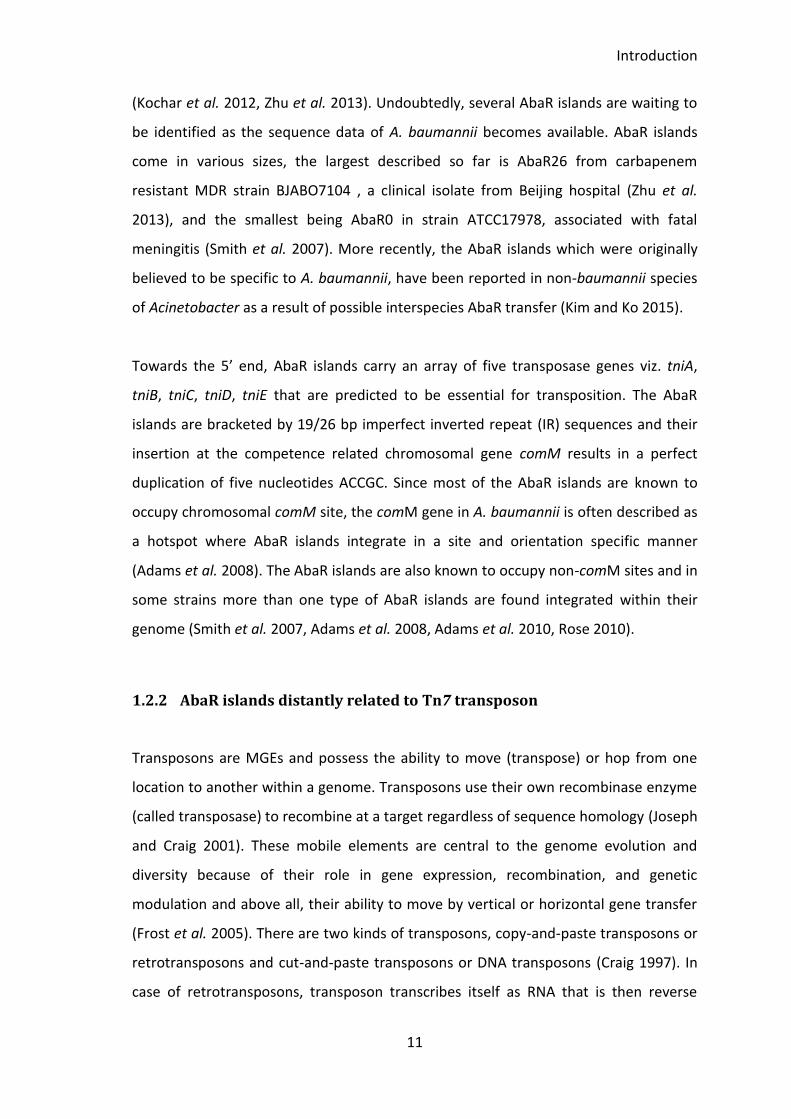
The Acinetobacter baumannii specific resistance islands called AbaR islands are foreign acquired resistance islands and are widely represented in the genome of multi-drug resistance [MDR] A. baumannii strains. The resistance genes and determinants within AbaR are found to contribute minimally towards the overall MDR phenotype of the host. Therefore, the maintenance of AbaR islands purely for the enhancement and development of antimicrobial resistance phenotype appears to be an inadequate explanation. This study investigates the contribution of AbaR island called TnAbaR23, in a MDR A. baumannii strain A424, towards the resistance and virulence phenotypes of the host. The TnAbaR23 deleted mutants were created by allelic exchange and the fitness and virulence of mutants was compared with their parent in a head-to-head competition assay and Galleria mellonella killing assay. During this study, spontaneous deletion of internal region of TnAbaR23 was observed in wild type A424 sub-population. In this study, the role of transposase gene tnpA in the spontaneous deletion within TnAbaR23 was also investigated. The spontaneous mutants of A424 were isolated and analysed for their antimicrobial resistance, fitness and virulence phenotypes. Although complete or partial deletion of TnAbaR23 had no effect on the growth of bacteria, the wild type appeared less fit in a head-to-head growth competition with TnAbaR23 deleted and spontaneous mutants. Except from the anticipated antibiotic susceptibility upon the deletion of TnAbaR23, the overall resistance phenotype in mutants remained unchanged. Intriguingly, the TnAbaR23 deleted and spontaneous mutants exhibited reduced virulence compared to their parent in a G. mellonella killing assay. Despite the associated cost, the maintenance of TnAbaR23 appears to be vital to exhibit enhanced virulence or pathogenesis in A. baumannii strain A424. It is therefore possible that A. baumannii AbaR islands positively contribute towards the development of traits that are vital for the survival of these bacteria.

iii
Acknowledgements
I firstly would like to thank my supervisor Dr Kumar Rajakumar for giving me the
opportunity to undertake this PhD, and for the support, wisdom and encouragement
all the way through my PhD.
My sincere thanks also go to Dr Hasan Yesilkaya, Dr Mike Barer and Dr Marialuisa
Crosatti providing me crucial feedback on my thesis chapters. I also thank my progress
review panel supervisors Dr Christopher Bayliss and Prof Martha Clokie for providing
me with critical feedback and comments throughout my studies.
I especially would like to thank Dr Marialuisa Crosatti for helping me master the basics
of molecular biology in the early stages of my PhD. I’m also very grateful to lab 212
members Toyosi Obasanjo, David Ngmenterebo, Robeena Farzand, Dr Zaaima Al Jabri,
Ros Abdul Aziz, Dr Eva Horvath-Papp and Dr Mohammad Al Madadha for their advice
and assistance throughout my PhD and for being there during our emotional ups and
downs right from the beginning of our studies.
This PhD would not have been possible without the constant love, support and
encouragement of my husband Umesh and I am glad that we shared the joy and
despair of my PhD. Most importantly, I thank my beloved son Abhinav for giving me all
the delight of the world; you were the reason I remained positive in the face of
hardship! Huge thanks to my Mummy, Dada, Muma and Buwa for remaining
inspirational and encouraging in my life; without you all, this journey would not have
been possible.
I dedicate this work to my Mummy (Shobha Devi Sapkota) who has been a source of
inspiration throughout my life!
iv
Table of Contents Abstract ............................................................................................................................. ii
Acknowledgements ......................................................................................................... iii
List of Figures ....................................................................................................................ix
List of Tables .....................................................................................................................xi
Abbreviations ................................................................................................................... xii
Presentations .................................................................................................................. xiii
1 Introduction .............................................................................................................. 1
1.1 Acinetobacter ..................................................................................................... 2
1.1.1 Taxonomy of genus Acinetobacter ............................................................. 3
1.1.2 Cell Structure and metabolism of Acinetobacter ....................................... 5
1.1.3 Ecology and epidemiology of Acinetobacter .............................................. 5
1.1.4 Resistance and virulence determinants in A. baumannii ........................... 7
1.2 Mobile Genetic Elements (MGEs) in A. baumannii ............................................ 9
1.2.1 Acinetobacter baumannii Resistance (AbaR) island ................................. 10
1.2.2 AbaR islands distantly related to Tn7 transposon .................................... 11
1.2.3 Features of AbaR island backbone ........................................................... 13
1.2.4 AbaR and Tn7 transposon similarity ......................................................... 15
1.3 Costs associated with development of resistance and virulence phenotypes in
bacteria ....................................................................................................................... 15
1.4 A. baumannii strain A424 and its TnAbaR23 resistance island ........................ 17
2 Aims of the study .................................................................................................... 20
3 Materials and methods ........................................................................................... 22
3.1 Media, reagents and solutions ......................................................................... 23
v
3.2 Growth conditions for bacteria ........................................................................ 24
3.3 Strains and plasmids used ................................................................................ 24
3.4 Genomic DNA extraction.................................................................................. 30
3.5 DNA quantification ........................................................................................... 30
3.6 Restriction enzymes and restriction digestions ............................................... 30
3.7 Ligation reactions ............................................................................................. 30
3.8 DNA sequencing ............................................................................................... 31
3.9 PCR primer design and synthesis ..................................................................... 31
3.10 In silico PCR ................................................................................................... 32
3.11 Polymerase chain reaction (PCR) ................................................................. 35
3.12 Colony PCR .................................................................................................... 35
3.13 Splicing overlap extension (SOE) PCR ........................................................... 35
3.14 Agarose gel electrophoresis ......................................................................... 37
3.15 Pulse Field Gel Electrophoresis (PFGE) ......................................................... 37
3.15.1 Plug preparation ....................................................................................... 37
3.15.2 Restriction digestion of plugs ................................................................... 38
3.15.3 Running condition for PFGE ...................................................................... 38
3.16 Preparation and transformation of chemically competent E. coli and A.
baumannii cells for heat shock transformation.......................................................... 39
3.17 Preparation and transformation of electro-competent E. coli and A.
baumannii cells ........................................................................................................... 40
3.18 Lambda Red Recombination ........................................................................ 40
3.19 Conjugations ................................................................................................. 42
3.20 Growth Curves .............................................................................................. 43
3.21 Bioinformatics tools ...................................................................................... 43
3.22 Plasmids maps and in silico construction of mutant genomes .................... 44
vi
3.23 Biofilm Assay for A. baumannii .................................................................... 44
3.24 Determination of minimum inhibitory concentration, MIC, by broth
microdilution ............................................................................................................... 45
3.25 Antibiotic susceptibility by Etest and disc diffusion method ....................... 45
3.26 Sucrose Counter-Selection Method ............................................................. 46
3.27 Head-to-head in vitro competition between wild type and mutants .......... 46
3.28 Galleria mellonella killing assay .................................................................... 47
3.29 Estimating the proportion of A424 cells with spontaneous deletion within
TnAbaR23 .................................................................................................................... 48
3.30 AbaR-islands survey ...................................................................................... 48
Blastn parameters ................................................................................................... 49
Blastp parameters ................................................................................................... 49
3.31 Statistical Analysis ........................................................................................ 50
4 Survey of AbaR, the A. baumannii resistance islandBackground ........................... 51
4.1 Aims and objectives ......................................................................................... 53
4.2 Result ................................................................................................................ 53
4.2.1 Survey of AbaR-like islands in A. baumannii ............................................. 53
4.2.2 PCR mapping of GI_comM_A92 ................................................................ 61
4.2.3 Phylogenetic relationship between AbaR islands ..................................... 63
4.3 Discussion ......................................................................................................... 71
4.3.1 Survey of AbaR islands .............................................................................. 71
4.3.2 Lineage of AbaR islands ............................................................................ 72
5 Plasticity of AbaR islands ........................................................................................ 75
5.1 Chapter overview ............................................................................................. 76
5.2 Aim ................................................................................................................... 76
5.3 Results .............................................................................................................. 77
vii
5.3.1 PCR detection of spontaneous deletion within TnAbaR23 ...................... 77
5.4 Detection of double stranded circular element excised from Tn6018-MARR-
Tn6018 ........................................................................................................................ 78
5.5 Isolation of the Tn6018-MARR deleted spontaneous mutants ....................... 79
5.5.1 Determining the proportion of A424 derived cells that have lost Tn6018-
MARR 79
5.5.2 Verification of Tn6018-MARR deleted spontaneous mutants ................. 80
5.6 Determining the proportion of cells carrying circular Tn6018-MARR ............. 82
5.7 Determining the proportion of cells harboring Tn6018-MARR deletion within
TnAbaR23 .................................................................................................................... 83
5.8 Role of tnpA in spontaneous deletion of Tn6018-MARR ................................. 85
5.9 Determining the proportion of tnpA knock-out mutants exhibiting
spontaneous deletion of MARR .................................................................................. 88
5.10 Determining the proportion of tnpA knock-out mutant cells carrying circular
Tn6018-MARR element ............................................................................................... 89
5.11 Discussion and conclusion ............................................................................ 90
5.11.1 Plasticity of AbaR islands .......................................................................... 90
5.11.2 Role of tnpA in spontaneous deletion of Tn6018-MARR from TnAbaR23 92
6 Contribution of TnAbaR23 island on the phenotype of Acinetobacter baumannii
strain A424 ...................................................................................................................... 95
6.1 Background ...................................................................................................... 96
6.2 Aims and objectives ....................................................................................... 100
6.3 Results ............................................................................................................ 101
6.3.1 Modified protocol for en bloc deletion of TnAbaR23 ............................. 101
6.3.2 Creation of new independent TnAbaR23 deleted mutants ................... 104
6.3.3 Verification of new independent TnAbaR23 deleted mutants .............. 105
6.3.4 PFGE of new TnAbaR23 deleted mutants ............................................... 106
viii
6.3.5 Rendering the TnAbaR23 deleted DCO mutant markerless ................... 107
6.3.6 Complementation of blocks of Tn6018-MARR region in spontaneous
mutant ΔMARR-3 .................................................................................................. 109
6.3.7 Effect of TnAbaR23 on the phenotype of A424 ...................................... 111
6.3.8 Loss of TnAbaR23 does not result in elevated ciprofloxacin resistance 115
6.3.9 No effect of TnAbaR23 on growth dynamics .......................................... 115
6.3.10 No effect of TnAbaR23 on biofilm production ....................................... 117
6.3.11 Effect of TnAbaR23 on in vitro growth fitness ........................................ 118
6.3.12 Effect of TnAbaR23 on virulence ............................................................ 120
6.3.13 DCO174 harbour mutation in AdeABC regulator gene adeS .................. 123
6.3.14 Replacement of adeSWT with adeSG178A caused enhanced ciprofloxacin
resistance .............................................................................................................. 124
6.3.15 Fitness and virulence phenotypes of DCO174 ........................................ 128
6.3.16 Growth dynamics in adeSG178A mutants ................................................. 131
6.3.17 Biofilm formation in adeSG178A mutant ................................................... 132
6.3.18 In vitro competitive fitness in adeSG178A mutants ................................... 132
6.4 Discussion and conclusion.............................................................................. 133
6.4.1 New TnAbaR23 deleted mutants with stable genetic configuration
created 133
6.4.2 Markerless TnAbaR23 deleted mutants created using pFLP2-derived
plasmid 133
6.4.3 Effect of TnAbaR23 on the phenotypes of the strain A424 .................... 134
6.4.4 DCO174 an unusual TnAbaR23 deleted mutant of A424 ....................... 136
7 Final discussion and conclusion ............................................................................ 138
8 Future recommendations ..................................................................................... 144
9 Appendices ............................................................................................................ 147
References .................................................................................................................... 168
ix
List of Figures
Figure 1.1 Common transposons forming backbone of AbaR islands. ........................... 13
Figure 1.2 Structure of TnAbaR23 in A. baumannii strain A424. .................................... 17
Figure 4.1 Schematic representation of various backbone transposons in AbaR islands.
........................................................................................................................................ 60
Figure 4.2 PCR mapping of GI_comM_A92 and GI_comM_IS116. ................................. 62
Figure 4.3 Phylogenetic tree constructed using tniA nucleotide sequence from AbaR
islands. ............................................................................................................................ 63
Figure 4.4 Phylogenetic tree constructed using tniA amino acid sequence from AbaR
islands. ............................................................................................................................ 65
Figure 4.5 Phylogenetic tree constructed using tniB nucleotide sequence from AbaR
islands ............................................................................................................................. 66
Figure 4.6 Phylogenetic tree constructed using tniB amino acid sequence from AbaR
islands ............................................................................................................................. 67
Figure 4.7 Phylogenetic tree constructed using concatenated tniABCDE nucleotide
sequence from AbaR islands. .......................................................................................... 68
Figure 4.8 Phylogenetic tree constructed using concatenated TniABCDE amino acid
sequence from AbaR islands. .......................................................................................... 70
Figure 5.1 Detection of Tn6018-MARR deletion in AYE and A424 ................................. 77
Figure 5.2 Detection of circular Tn6018-MARR element in A424 .................................. 79
Figure 5.3 PFGE of Tn6018-MARR deleted spontaneous mutants ................................. 81
Figure 5.4 Circular junction detection sensitivity assay ................................................. 82
Figure 5.5 Deletion detection sensitivity assay .............................................................. 84
Figure 5.6 Deletion detection test in A424 ..................................................................... 84
Figure 5.7 Deletion of tnpA from left and right Tn6018 in A424 .................................... 85
Figure 5.8 Schematics of various circular elements and the calculated size of circular
junction ........................................................................................................................... 87
Figure 5.9 Deletion detection assay in various tnpA deleted mutants .......................... 88
Figure 5.10 Circular junction detection assay ................................................................. 90
x
Figure 6.1 Schematics of suicide vector pJTAG ............................................................ 101
Figure 6.2 Verification of TnAbaR23 deleted mutants ................................................. 106
Figure 6.3 Verification of new independent DCOs by PFGE ......................................... 107
Figure 6.4 Schematics of plasmids pFLP2_sul1 and pKOBEG_Apra ............................. 108
Figure 6.5 Verification of markerless mutants derived from DCOA14 ......................... 109
Figure 6.6 Plasmid complementation of ΔMARR-3 ...................................................... 110
Figure 6.7 Growth curve of the A424WT and its various mutants ............................... 116
Figure 6.8 Biofilm formed by A424WT and its various TnAbaR23 deleted mutants .... 117
Figure 6.9 Head-to-head in vitro competition between A424 wild type and its mutants
...................................................................................................................................... 119
Figure 6.10 Determination of inoculum size for Galleria killing assay ......................... 121
Figure 6.11 Effect of TnAbaR23 on virulence ............................................................... 122
Figure 6.12 A non-synonymous SNP in DCO174 ........................................................... 124
Figure 6.13 In vitro competitive fitness of DCO174 ...................................................... 129
Figure 6.14 Virulence of DCO174 .................................................................................. 130
Figure 6.15 Growth curve of A424 wild type and its mutants carrying adeSG178A ........ 131
Figure 6.16 Biofilm formation by adeSG178A and their parent strains ........................... 132
Figure 6.17 In vitro competitive fitness of A424 wild type with various mutants carrying
adeSG178A ....................................................................................................................... 133
xi
List of Tables
Table 1.1 Acinetobacter species isolated from various sources ....................................... 3
Table 3.1 Bacterial strains and plasmid used and created in this study ........................ 25
Table 3.2 Primers used in the PCR mapping of GI_comM_A92 ..................................... 31
Table 3.3 PCR amplicon sizes obtained during the mapping of GI_comM_A92 ............ 32
Table 3.4 Primers used in this study ............................................................................... 33
Table 3.5 SOE PCR reaction setup ................................................................................... 36
Table 3.6 SOE PCR thermocycling condition ................................................................... 36
Table 3.7 Query sequences for AbaR survey. ................................................................. 49
Table 4.1 Distribution of AbaR islands in the A. baumannii strains ............................... 55
Table 4.2 Representation of backbone transposons in various AbaR islands ................ 59
Table 6.1 Antimicrobial susceptibility profile of MDR A. baumannii strains .................. 97
Table 6.2 Conjugative delivery of pJTAG into A424 and selection of SCO mutants ..... 103
Table 6.3 Sucrose counter-selection for creation of DCO mutant ............................... 105
Table 6.4 Antibiotic susceptibility of A424WT and its various mutants ....................... 112
Table 6.5 Antibiotic susceptibility profile of strains carrying adeSWT and adeSG178A .... 127
xii
Abbreviations
Abbreviation Explanation
% Per cent
λ lambda
µg microgram
µl microliter
°C degree Celsius
Blast Basic local alignment search tool
bp base pairs
DNA deoxyribose nucleic acid
dNTP dinucleotide triphosphate
Etest Epsilometer test
h hours
kb kilobase pair(s)
Mb Megabase pairs
MDR Multi-drug resistant
min Minutes
mM milli molar
mm Millimetre
ng nanograms
nm nanometer
OD Optical Density
PDR Pan-drug resistant
rpm Revolutions per minute
s seconds
sp. Species
spp. Plural of species
UV Ultra violet
WT Wild type
w/v Weight/volume
XDR Extensive-drug resistant
xiii
Presentations
Fifth Annual Postgraduate Conference
University of Leicester, Department of Infection, Immunity and Inflammation, April
2013
Oral presentation entitled: “Contribution of TnAbaR23 on the phenotype of
Acinetobacter baumannii”
Microbiology and infectious disease seminar
University Hospitals of Leicester NHS Trust, November 2014
Oral presentation entitled: “Contribution of TnAbaR23 island on the phenotype of
Acinetobacter baumannii strain A424”
ELTU Research Festival
University of Leicester, 18th February 2015
Poster presentation entitled: “Antibiotic resistance and virulence in pathogenic
bacteria Acinetobacter baumannii”
Society for General Microbiology
Annual Conference 2015 (30 March - 2 April), ICC, Birmingham, UK
Poster presentation entitled: “Fitness and virulence-associated phenotypes of the
plastic TnAbaR23 Acinetobacter baumannii A424 resistance transposon”
2nd London Postgraduate Research Symposium on Bacterial Pathogenesis and Host
Response 2015
Birkbeck, University of London, Friday 27th November 2015
Oral presentation entitled: “Acinetobacter baumannii resistance island and its
contribution on the fitness and virulence associated phenotypes”

1
1 Introduction
Introduction
2
1.1 Acinetobacter
The word Acinetobacter is originally derived from Greek word akinetos meaning
unable to move. The genus Acinetobacter belongs to non-motile, free-living, non-
fermentative, oxidase-negative, strictly aerobic gram negative bacteria (Brisou and
Prevot 1954). These bacteria can utilize a wide range of carbon sources and can be
easily grown on nutrient agar in the laboratory at 20°C -30°C (Bergogne-Berezin and
Towner 1996). These bacteria are also known to be intrinsically resistant to many
antimicrobial agents. In recent years, Acinetobacter baumannii has been frequently
associated with serious nosocomial and community associated infections. Pathogenic
strains of A. baumannii are considered opportunistic as they often cause serious and
life threatening infections in immunosuppressed patients (Falagas et al. 2006). In
recent decade, there has been a global increase in the reports of isolation of multi
drug-resistant (MDR), pan drug-resistant (PDR) and extensively drug-resistant (XDR) A.
baumannii strains from various clinical settings (Gottig et al. 2014, Teo et al. 2015). In
the absence of new drug as a treatment alternative, the option for managing A.
baumannii infections is becoming highly problematic and restricted.
The following sections will describe the taxonomy, ecology and clinical importance of
A. baumannii. The factors associated with drug resistance in A. baumannii will also be
briefly discussed in the sections below paying particular attention to the A. baumannii
specific genomic resistance islands called Acinetobacter baumannii Resistance island
or AbaR in short followed by sections providing a synopsis of evolution and divergence
of AbaR together with the discussion on the AbaR associated resistance phenotype in
A. baumannii. The final section will discuss in detail the aims and objectives of the
study.
Introduction
3
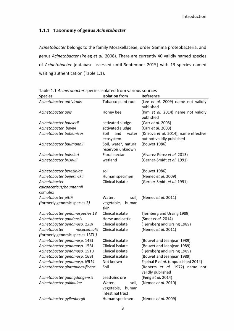
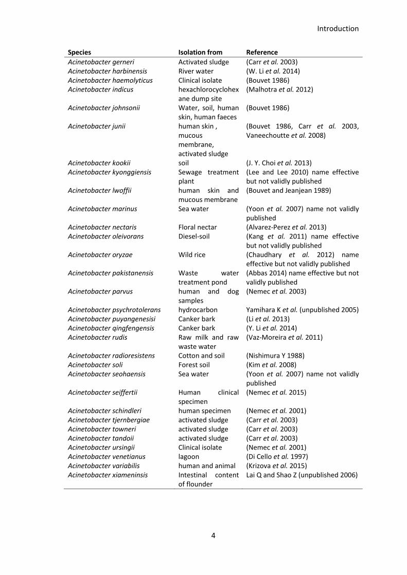
1.1.1 Taxonomy of genus Acinetobacter
Acinetobacter belongs to the family Moraxellaceae, order Gamma proteobacteria, and
genus Acinetobacter (Peleg et al. 2008). There are currently 40 validly named species
of Acinetobacter [database assessed until September 2015] with 13 species named
waiting authentication (Table 1.1).
Table 1.1 Acinetobacter species isolated from various sources Species Isolation from Reference
Acinetobacter antiviralis Tobacco plant root (Lee et al. 2009) name not validly published
Acinetobacter apis Honey bee (Kim et al. 2014) name not validly published
Acinetobacter bouvetii activated sludge (Carr et al. 2003) Acinetobacter. baylyi activated sludge (Carr et al. 2003) Acinetobacter bohemicus Soil and water
ecosystem (Krizova et al. 2014), name effective but not validly published
Acinetobacter baumannii Soil, water, natural reservoir unknown
(Bouvet 1986)
Acinetobacter boissieri Floral nectar (Alvarez-Perez et al. 2013) Acinetobacter brisouii wetland (Gerner-Smidt et al. 1991)
Acinetobacter bereziniae soil (Bouvet 1986) Acinetobacter beijerinckii Human specimen (Nemec et al. 2009) Acinetobacter calcoaceticus/baumannii complex
Clinical isolate (Gerner-Smidt et al. 1991)
Acinetobacter pittii (formerly genomic species 3)
Water, soil, vegetable, human skin
(Nemec et al. 2011)
Acinetobacter genomospecies 13 Clinical isolate Tjernberg and Ursing 1989) Acinetobacter gandensis Horse and cattle (Smet et al. 2014) Acinetobacter genomosp. 13BJ Clinical isolate (Tjernberg and Ursing 1989) Acinetobacter nosocomialis (formerly genomic species 13TU)
Clinical isolate (Nemec et al. 2011)
Acinetobacter genomosp. 14BJ Clinical isolate (Bouvet and Jeanjean 1989) Acinetobacter genomosp. 15BJ Clinical isolate (Bouvet and Jeanjean 1989) Acinetobacter genomosp. 15TU Clinical isolate (Tjernberg and Ursing 1989) Acinetobacter genomosp. 16BJ Clinical isolate (Bouvet and Jeanjean 1989) Acinetobacter genomosp. NB14 Not known Espinal P et al. (unpublished 2014) Acinetobacter glutaminasificans Soil (Roberts et al. 1972) name not
validly published Acinetobacter guangdongensis Lead-zinc ore (Feng et al. 2014) Acinetobacter guillouiae Water, soil,
vegetable, human intestinal tract
(Nemec et al. 2010)
Acinetobacter gyllenbergii Human specimen (Nemec et al. 2009)
Introduction
4
Species Isolation from Reference
Acinetobacter gerneri Activated sludge (Carr et al. 2003) Acinetobacter harbinensis River water (W. Li et al. 2014) Acinetobacter haemolyticus Clinical isolate (Bouvet 1986) Acinetobacter indicus hexachlorocyclohex
ane dump site (Malhotra et al. 2012)
Acinetobacter johnsonii Water, soil, human skin, human faeces
(Bouvet 1986)
Acinetobacter junii human skin , mucous membrane, activated sludge
(Bouvet 1986, Carr et al. 2003, Vaneechoutte et al. 2008)
Acinetobacter kookii soil (J. Y. Choi et al. 2013) Acinetobacter kyonggiensis Sewage treatment
plant (Lee and Lee 2010) name effective but not validly published
Acinetobacter lwoffii human skin and mucous membrane
(Bouvet and Jeanjean 1989)
Acinetobacter marinus Sea water (Yoon et al. 2007) name not validly published
Acinetobacter nectaris Floral nectar (Alvarez-Perez et al. 2013) Acinetobacter oleivorans Diesel-soil (Kang et al. 2011) name effective
but not validly published Acinetobacter oryzae Wild rice (Chaudhary et al. 2012) name
effective but not validly published Acinetobacter pakistanensis Waste water
treatment pond (Abbas 2014) name effective but not validly published
Acinetobacter parvus human and dog samples
(Nemec et al. 2003)
Acinetobacter psychrotolerans hydrocarbon Yamihara K et al. (unpublished 2005) Acinetobacter puyangenesisi Canker bark (Li et al. 2013) Acinetobacter qingfengensis Canker bark (Y. Li et al. 2014) Acinetobacter rudis Raw milk and raw
waste water (Vaz-Moreira et al. 2011)
Acinetobacter radioresistens Cotton and soil (Nishimura Y 1988) Acinetobacter soli Forest soil (Kim et al. 2008) Acinetobacter seohaensis Sea water (Yoon et al. 2007) name not validly
published Acinetobacter seiffertii Human clinical
specimen (Nemec et al. 2015)
Acinetobacter schindleri human specimen (Nemec et al. 2001) Acinetobacter tjernbergiae activated sludge (Carr et al. 2003) Acinetobacter towneri activated sludge (Carr et al. 2003) Acinetobacter tandoii activated sludge (Carr et al. 2003) Acinetobacter ursingii Clinical isolate (Nemec et al. 2001) Acinetobacter venetianus lagoon (Di Cello et al. 1997) Acinetobacter variabilis human and animal (Krizova et al. 2015) Acinetobacter xiameninsis Intestinal content
of flounder Lai Q and Shao Z (unpublished 2006)
Introduction
5
1.1.2 Cell Structure and metabolism of Acinetobacter
Acinetobacter are rod shaped bacteria that often occur in pairs and tend to become
coccoid towards the stationary growth phase. These bacteria range from 1.0 - 2.5 µm
and form a pale yellow colony on nutrient agar media. These bacteria can utilize a wide
range of carbon sources except glucose (Baumann 1968). Acinetobacter can be
isolated from the clinical samples on the commercially available selective medium such
as Herellea agar or Difco (Charles 1968) or CHROMagarTM where the colonies of
Acinetobacter sp. appear red. Specific antibiotics can be supplemented in the selective
medium to suppress the non-specific bacteria. Other selective media such as Leeds
Acinetobacter medium, minimum medium supplemented with acetate and 0.2 %
sodium acetate agar in a mineral salt base has been used to isolate Acinetobacter sp.
from various samples (Berlau et al. 1999). Several molecular methods have been
developed to distinguish Acinetobacter at species level. The common molecular
methods frequently used to distinguish Acinetobacter sp. include 16S rRNA gene
restriction analysis (ARDRA), amplified fragment length polymorphism (AFLP),
ribotyping, tRNA spacer fingerprinting, restriction analysis of 16S-23S rRNA, sequence
analysis of rpoB and flanking region, detection of blaOXA-51 , and multi-locus sequence
typing (MLST). Despite its inconvenience, the DNA-DNA hybridization technique
remains a reference method to identify Acinetobacter at species level (Peleg et al.
2008).
1.1.3 Ecology and epidemiology of Acinetobacter
Species of Acinetobacter are ubiquitous and these bacteria have been isolated from a
wide variety of surfaces including food, vegetables, environmental samples like soil
and hospital surfaces including bed rails and pillows (Table 1.1). Acinetobacter often
show minimum nutrient requirement because of which they can survive on various
clinical surfaces and can endure harsh environmental conditions for a prolonged
period of time (Baumann et al. 1968, Costerton 1999). Despite being considered as a
Introduction
6
common skin commensal, these bacteria have been isolated as deadly pathogens from
combat victims and victims of natural disasters (CDC 2004, Lockhart et al. 2007).
Various species of Acinetobacter are usually non-pathogenic to healthy individuals but
A. baumannii colonization can cause potentially lethal infections especially in patients
with compromised immune system (Maragakis and Perl 2008). In the early 1960s,
species of Acinetobacter were considered as low-grade pathogens and they were
usually ignored when isolated from clinical samples (Kempf and Rolain 2012). In the
last few decades, and especially after the increased incidence of A. baumannii
colonization associated with combat victims returning from the Iraq conflict, this
bacteria has gained reputation as a notorious opportunistic pathogen among
microbiologists and clinicians (Lockhart et al. 2007).
Among various species of Acinetobacter, A. baumannii are clinically important because
they belong to an epidemic clonal group EC I and EC II and are frequently associated
with hospital associated and community-acquired infections (Post et al. 2010). The
reports of A. baumannii infections in trauma patients and patients from natural
disasters in the community are increasing rapidly. In the clinical settings, colonization
of A. baumannii strains in a wide range of surfaces and devices and can cause various
infections like ventilator associated pneumonia, skin and wound infections,
bacteraemia leading to sepsis and septic shock, urinary tract infections, meningitis,
surgical site infections, abdominal infections, central nervous system infections
(Gaynes and Edwards 2005). Studies have indicated that presence of invasive medical
devices, serious underlying diseases, previous history of receiving broad-spectrum
antibiotics, major surgery, burns, immune-suppression, chronic alcoholism, cancer,
bronchopulmonary disease etc. can be a predisposition factors for A. baumannii
infections (Kempf and Rolain 2012).
Successful treatment of the infection relies on the resistance status of the causative
agent. Despite the best efforts to contain and combat the A. baumannii associated
infections, the report of MDR, PDR and XDR A. baumannii isolates colonizing various
community and healthcare settings around the globe is increasing in an alarming rate
Introduction
7
(Jung and Park 2015). Due to this rapid emergence and dissemination of antibiotic
resistant in A. baumannii and no new drugs on the development pipeline, the
antibiotic options for the treatment of A. baumannii infections is limited (Ozdem et al.
2011) and the treatment of infections has become highly problematic and restricted.
The arsenal of antibiotics currently in use in the frontline of A. baumannii infection
includes colistins, sulbactams co-formulated with ampicillins, tigecycline, minocycline,
carbapenems, piperacillin/tazobactam, cefepime, doxycycline, aminoglycosides and
quinolones (Dijkshoorn et al. 2007, Viehman et al. 2014). Due to the sub-optimal
pharmacokinetics of the drugs and rapid emergence of resistance, combination drug
therapy for infection treatment is attracting frequent attention (Batirel et al. 2014).
1.1.4 Resistance and virulence determinants in A. baumannii
Bacterial are also well known for their ability to adopt various resistance mechanisms
in order to resist the antimicrobial agents present in their environment. Enzymatic
inactivation of the antimicrobial agent and extruding the toxic agents by efflux are
widespread in bacteria as a mechanism of antimicrobial resistance. Bacteria can also
reduce the entry of antimicrobial agents into the cells by reducing the number of
antimicrobial targets or protecting the antimicrobial targets (like porin) on the cell
surface. Replacement of the susceptible antimicrobial targets and acquisition of novel
pumps to extrude antimicrobial agents from cell are also common resistance
mechanisms in bacteria (Andersson and Hughes 2010).
In a complex natural ecosystem, bacteria evolve and thrive either by mutating their
intrinsic or acquired genes or by acquiring foreign DNA that carry beneficial genes (van
Hoek et al. 2011). Mobile Genetic Elements (MGE) like plasmids, transposons, insertion
sequences and integrative conjugative elements that carry beneficial genes are
favourably taken up by bacteria, by means of transformation, conjugation or
transduction, to enhance the resistance and virulence phenotypes (Andersson and
Hughes 2010, Domingues et al. 2012). The successful acquisition and integration of
MGEs and foreign genetic elements that carries advantageous traits is believed to an
Introduction
8
important factor in the evolution of bacterial genome (Roberts and Kreth 2014,
Mullany et al. 2015).
A. baumannii displays a combination of features like survival under harsh condition,
efficient host adherence and invasion, antibiotic resistance and ability to acquire
foreign genetic elements that has enabled this bacterium to emerge, spread and
persist as a successful pathogen (Frost et al. 2005, Smith et al. 2007).
The core genome of A. baumannii contains a range of genes and operons that confers
innate resistance against a wide variety of antimicrobial agents. A. baumannii show
high level of intrinsic antimicrobial resistance by means of regulation of antibiotic
resistance genes for the production of Aminoglycoside Modification Enzymes (AMEs),
Acinetobacter-derived cephalosporinases (AmpC β-lactamase), other β-lactamases
coded by blaOXA-51-like genes, extended-spectrum β-lactamases (ESBLs) and metallo-β-
lactamases (MBLs). These bacteria exhibit sulbactam resistance by reducing the
expression of penicillin binding protein 2, a drug target for sulbactams, on the surface
of their cells (Viehman et al. 2014). Modification of the target protein for colistin and
fluoroquinolones is also a well-known mechanism of resistance in these bacteria.
These bacteria also limit the entry of antimicrobial agents into the cells by down
regulating the expression of porin channels and mutating the recognition sites for
antimicrobial agents like outer membrane protein (Hamidian et al. 2013).
In A. baumannii, various efflux pumps are actively involved in extruding antimicrobial
agents like tetracycline, rifampicin, fluoroquinolones that had gained access into the
cell. Efflux pumps belonging to the Major Facilitator Super-family (MFS) like
tetracycline repressor protein class A and class B (TetA/B), chloramphenicol efflux
protein (CmlA), the Resistance-Nodulation-Division super-family (RND) like AdeABC
and the Multidrug And Toxic compound Extrusion family, MATE, like AbeM have been
well studied for their function and substrate in various A. baumannii strains (Vila et al.
2007).

Introduction
9
Sequence analysis of A. baumannii have also shown the presence of various inherent
and acquired genes and determinants that are likely to be associated with the
virulence of this bacteria. Some of the determinants associated with the virulence of A.
baumannii include CsuA/BABCDE chaperone-usher pili assembly system, siderophores,
type I pili, hemin utilization proteins, outer membrane protein OmpA, AbaI auto
inducer synthase, biofilm associated protein Bap, two-component regulatory system
BfmRS, penicillin binding protein, PNAG-constituted biofilm, capsule,
lipopolysaccharide, phospholipase D and phospholipase C (Cerqueira and Peleg 2011).
A swift genetic response to the changing environmental condition by means of
regulation of native genes and uptake of beneficial genes from the environment is
crucial for the survival and persistence of the bacteria (Frost et al. 2005, Poirel and
Nordmann 2006, Yoon et al. 2013). Genome analysis shows that A. baumannii has
acquired a number of resistance determinants including transposons, plasmids,
genomic islands (GIs), insertion sequences (IS), and integrons from its environment by
horizontal gene transfer (Diancourt et al. 2010, Imperi et al. 2011). Various species of
Acinetobacter possess the ability of naturally acquiring assimilating and disseminating
mobile genetic elements by means of horizontal gene transfer (Metzgar et al. 2004);
however, there is no report on natural transformation exhibited by A. baumannii
strains. This thesis report A. baumannii specific resistance island AbaR and its role in
various phenotypes of the host strain. The following sections provide a synopsis on the
MGEs focussing particularly on the A. baumannii specific genomic island.
1.2 Mobile Genetic Elements (MGEs) in A. baumannii
The average size of A. baumannii genome is about 4 MB. The pan genome of A.
baumannii, which corresponds to a sum of all core (present in all strains of a species)
and dispensable genome (absent in at least one strain), contains 8818 genes of which
less than 17 % of the genes are considered as core-conserved genome and ~ 83 % as
dispensable genome (Field et al. 2006, Imperi et al. 2011). Of this vast pool of
dispensable genome, 25 % of the genes are again found to be unique and strain
Introduction
10
specific (Imperi et al. 2011). A major portion of this so called dispensable genome
comprises of genomic islands (GIs). Some A. baumannii GIs are strain specific while
others are completely or partially conserved in more than one strain (Di Nocera et al.
2011). The nomenclature of these GIs is based on the nature of genes they carry, like
resistance islands (carry resistance determinants), metabolic islands (carry genes for
specific metabolisms) and phase islands (carry genes coding phase products).
The genome sequence analysis shows that almost all of the resistance islands in A.
baumannii are a composite of several mobile genetic elements (MGEs) like
transposons and insertions sequences. The MGEs are DNA that are able to move
between the cells during horizontal gene transfer via plasmids, bacteriophages,
Insertion Sequence (IS) elements, Integrative and Conjugative Elements (ICEs),
transposons (Tns) and miniature inverted repeat transposable elements (MITEs).
Interestingly, the origin of A. baumannii resistance island-associated MGEs has been
traced to other bacterial species including Pseudomonas, Salmonella and E. coli.
(Fournier et al. 2006). In the following sections, A. baumannii specific resistance islands
called AbaR will be discussed and the similarities of AbaR with bacterial transposon
Tn7 will also be highlighted.
1.2.1 Acinetobacter baumannii Resistance (AbaR) island
During a comparative genome study of MDR A. baumannii strain AYE and susceptible
strain SDF (Fournier et al. 2006), a cluster of around 45 antimicrobial resistance
determinants were found as a resistance island in 86 kb island on the chromosome of
AYE. The comparative genome analysis also showed the presence of similar island
inserted in the identical position in the chromosome of strain SDF, however, this island
in the SDF strain was much smaller and it was empty, in a sense, the island was lacking
determinants for antimicrobial resistance. Since the AbaR in AYE was the first island of
this nature to be identified in A. baumannii, it was named Acinetobacter baumannii
resistance island 1 or AbaR1. To-date, various AbaR-like islands have been reported
from numerous A. baumannii strains and they are numbered from AbaR0 to AbaR27
Introduction
11
(Kochar et al. 2012, Zhu et al. 2013). Undoubtedly, several AbaR islands are waiting to
be identified as the sequence data of A. baumannii becomes available. AbaR islands
come in various sizes, the largest described so far is AbaR26 from carbapenem
resistant MDR strain BJABO7104 , a clinical isolate from Beijing hospital (Zhu et al.
2013), and the smallest being AbaR0 in strain ATCC17978, associated with fatal
meningitis (Smith et al. 2007). More recently, the AbaR islands which were originally
believed to be specific to A. baumannii, have been reported in non-baumannii species
of Acinetobacter as a result of possible interspecies AbaR transfer (Kim and Ko 2015).
Towards the 5’ end, AbaR islands carry an array of five transposase genes viz. tniA,
tniB, tniC, tniD, tniE that are predicted to be essential for transposition. The AbaR
islands are bracketed by 19/26 bp imperfect inverted repeat (IR) sequences and their
insertion at the competence related chromosomal gene comM results in a perfect
duplication of five nucleotides ACCGC. Since most of the AbaR islands are known to
occupy chromosomal comM site, the comM gene in A. baumannii is often described as
a hotspot where AbaR islands integrate in a site and orientation specific manner
(Adams et al. 2008). The AbaR islands are also known to occupy non-comM sites and in
some strains more than one type of AbaR islands are found integrated within their
genome (Smith et al. 2007, Adams et al. 2008, Adams et al. 2010, Rose 2010).
1.2.2 AbaR islands distantly related to Tn7 transposon
Transposons are MGEs and possess the ability to move (transpose) or hop from one
location to another within a genome. Transposons use their own recombinase enzyme
(called transposase) to recombine at a target regardless of sequence homology (Joseph
and Craig 2001). These mobile elements are central to the genome evolution and
diversity because of their role in gene expression, recombination, and genetic
modulation and above all, their ability to move by vertical or horizontal gene transfer
(Frost et al. 2005). There are two kinds of transposons, copy-and-paste transposons or
retrotransposons and cut-and-paste transposons or DNA transposons (Craig 1997). In
case of retrotransposons, transposon transcribes itself as RNA that is then reverse
Introduction
12
transcribed as a new copy of DNA for transposition. The DNA transposons on the other
hand, does not form RNA intermediate and jumps from its original site in its entirety to
integrate elsewhere in the genome. Bacterial transposon Tn7 is a cut-and-paste
transposon and five “transposition genes” towards its 5’ end tightly regulate the
mechanism of transposition. The genes and determinants involved in antimicrobial
resistance, DNA repair, DNA modification, DNA restriction, several other transposase
including many other genes of unknown functions are present towards the 3’ end of
Tn7.
The five Tn7 transposition genes code for transposases proteins viz. TnsA, TnsB, TnsC,
TnsD, TnsE, and they show a highly conserved synteny. A stretch of 90 bp and 150 bp
non-identical segments necessary for the transposition lie at the extreme left and right
ends of the Tn7 and a short 19/26 bp imperfect IR sequence brackets Tn7 transposon.
During the Tn7 transposition, transposases TnsD and TnsE select the target site attTn7.
Once TnsD/E binds at the target, attTn7, TnsAB excise the DNA from the native site as
a double stranded DNA. TnsC forms a DNA complex with the excised transposon and
TnsD/E at the target site (Waddell and Craig 1989). The 3’ ends attach covalently with
the target site but a five bp gap flanks the 5’ end. The process of target-site duplication
then repairs this staggered cut. This results in five bp (ACCGC) target site duplication,
also called direct repeat sequence or DR is a characteristic feature of Tn7
transpositional recombination (Joseph and Craig 2001). In several bacteria, many Tn7-
like elements have been identified based on sequence homology with Tn7
transposition genes.
The AbaR islands are often considered as composite transposons that are built on the
“backbone” of other transposons like Tn6019, Tn6021 and Tn6022 (Post and Hall 2009,
Post et al. 2010, Seputiene et al. 2012). These three common AbaR backbone
transposons, Tn6019, Tn6021 and Tn6022, are closely related to each other in a sense
that they have same gene cluster towards their 5’ end and 3’ end and their mechanism
of transposition is identical (Figure 1.1). The complexity and mosaic internal
architecture of AbaR islands is believed to have arisen due to integration of several
transposons and IS elements from various bacteria like E. coli, Pseudomonas spp. and
Introduction
13
Salmonella spp. (Fournier et al. 2006). In this section, the basic structure of AbaR
islands will be explained aiming to provide a synopsis on the diversity of AbaR islands
in A. baumannii genome.
1.2.3 Features of AbaR island backbone
A majority of AbaR islands are borne on the backbone of an arsenic resistance
transposon Tn6019 (Post et al. 2010). In the transposon Tn6019, towards the 5’ end
immediately after the 19 - 26 bp left IR sequence an array of five gene cluster tniA,
tniB, tniC, orf2 and orf3 (Figure 1.1) is present which is believed to be essential for
transposition. This cluster of genes is therefore considered as “core transposition
genes” or “transposition machinery” (Post and Hall 2009).
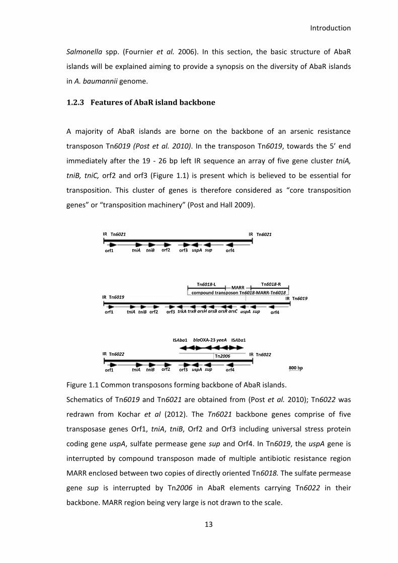
Figure 1.1 Common transposons forming backbone of AbaR islands.
Schematics of Tn6019 and Tn6021 are obtained from (Post et al. 2010); Tn6022 was
redrawn from Kochar et al (2012). The Tn6021 backbone genes comprise of five
transposase genes Orf1, tniA, tniB, Orf2 and Orf3 including universal stress protein
coding gene uspA, sulfate permease gene sup and Orf4. In Tn6019, the uspA gene is
interrupted by compound transposon made of multiple antibiotic resistance region
MARR enclosed between two copies of directly oriented Tn6018. The sulfate permease
gene sup is interrupted by Tn2006 in AbaR elements carrying Tn6022 in their
backbone. MARR region being very large is not drawn to the scale.
Introduction
14
Towards the 3’ end of this transposon are three genes, universal stress protein uspA,
orf4 and sulfate permease sup. The uspA gene, which belongs to the universal stress
protein family and whose expression is enhanced under stressful conditions, is usually
interrupted by a “compound transposon” Tn6018-MARR-Tn6018.
The Tn6018 is a heavy metal transposon and two directly oriented copies of this
transposon enclose a Multiple-antibiotic resistance region (MARR) to make Tn6018-
MARR-Tn6018 compound transposon (Post and Hall 2009). The interruption of uspA
gene Tn6018-MARR-Tn6018 is considered as the basic structure of AbaR islands,
however, the MARR region in different AbaR islands varies considerably in terms of
both size and type of cargo genes. Nonetheless, a common feature of all AbaR
associated MARR is the presence of a wide variety of resistant determinants including
genes associated with mobility, genes for antibiotics, antiseptics, heavy metal
resistance genes and efflux associated genes (Kochar et al. 2012).
The second common transposon that forms the backbone of AbaR islands is Tn6021.
The Tn6021 transposon also has a cluster of five core transposition genes towards its
5’ end and three genes uspA, sup and orf4 towards it 3’ end. The uspA in Tn6021 is also
a target for Tn6018-MARR-Tn6018. The third transposon that is also commonly found
to form the backbone of AbaR islands is Tn6022, which is essentially a Tn6021
transposon where sup gene is interrupted by ISAbaR1 flanked, blaOxa23 carrying
transposon Tn2006 (Figure 1.1).
As described in above sections, the AbaR islands in various MDR strains of A.
baumannii differ from each other in terms of length, gene content, and gene sequence
due to various events of DNA integration, deletion and rearrangements (Post and Hall
2009). Single or multiple intramolecular recombination events between the identical
copies of sequences have been reported in various AbaR islands (Post et al. 2010).
Various events of gene deletion, gene replacement and gene acquisition mediated by
the homologous recombination between copies of IS26, copies of Tn6022 and copies
Introduction
15
of CR2 segments are described as the heart of diversification of AbaR islands (Krizova
et al. 2011, Harmer et al. 2014).
Although the above described three transposons are commonly found in AbaR islands,
other complex transposons like Tn6166, Tn6167 and Tn6168 are frequently being
reported to constitute the backbone of AbaR islands (Nigro et al. 2011, Nigro and Hall
2012b, Saule et al. 2013). Various transposons forming the backbone of AbaR islands
and their phylogenetic relationship will be analysed and discussed in chapter 3.
1.2.4 AbaR and Tn7 transposon similarity
The cut-and-paste bacterial transposon Tn7 is well studied in many bacterial species
(Parks and Peters 2009, K. Y. Choi et al. 2013) and its mechanism of target site
selection and transposition is well established. Like Tn7 transposon, the AbaR islands
are bounded by 19-26 bp imperfect IR sequences and their integration results in DR
sequence at the target. The integration of AbaR islands at comM disrupts this gene and
forms ACCGC as a DR sequence at the integration site (Adams et al. 2010). Towards
their 5’ end, the AbaR islands also carry a cluster of five genes, tniA, tniB, tniC, tniD,
tniE, that are predicted to be “transposition genes” (Post et al. 2010). Bioinformatics
analysis showed a striking similarity between the five transposition genes of Tn7 with
the core transposition genes of AbaR1 (Rose 2010). The comparison of nucleotide and
protein sequence of these five transposition genes from AbaR1 revealed a distant
relation of AbaR islands with a promiscuous transposon Tn7 (Rose 2010). Therefore,
Kochar et al. (2012) named the AbaR island in MDR A. baumannii strain A424 as
TnAbaR23 where Tn was used to indicate the relationship with Tn7 transposon.
1.3 Costs associated with development of resistance and virulence
phenotypes in bacteria
It is generally considered that the acquisition or development of antimicrobial
resistance is physiologically and energetically costly for the cell. However, studies have
Introduction
16
shown that the cost associated with the acquisition of resistance can be, over time,
mitigated during growth or by means of other beneficial associations stress free
environment and association with host genome (Starikova et al. 2013). Enhanced
antimicrobial resistance has been found to reduce the growth rate in various bacteria
including E. coli and Salmonella sp. (Smani et al. 2012). A study done in a clinical isolate
of A. baumannii showed that despite no growth cost, the enhanced fluoroquinolone
resistance due to overexpression of efflux genes resulted in decreased virulence of the
bacteria (Smani et al. 2012). A bigger cost of acquiring colistin resistance was observed
in A. baumannii strain 19606 where the colistin resistant mutants displayed reduced
growth rate, reduced in vitro/in vivo fitness and attenuated virulence as compared to
their parent wild type (Beceiro et al. 2014). It is important to note that, development
of resistance can be cost free and various factors like epistasis, environmental
conditions and compensatory mutations can affect the cost associated with it
(Andersson and Hughes 2010).
A majority of AbaR islands described so far in MDR A. baumannii strains carry genes
and determinants for antimicrobial resistance and genome mobilization. The
antimicrobial resistance phenotype associated with these AbaR islands is largely
predicted based on their genetic composition. A limited number of experimental
studies have reported the role of AbaR islands in the development of drug resistance
phenotype. The deletion of AbaR-like island in A. baumannii strain A424 was assumed
to be associated with enhanced resistance towards ciprofloxacin (Kochar et al. 2012)
while other reports show no contribution of AbaR islands towards the overall
resistance phenotype of the host strain (Krizova et al. 2011). Recently, there has been
a report on the development of antimicrobial resistance in a susceptible strain when
AbaR was conjugally delivered into its genome (Hamidian et al. 2014a). In this study, I
will be investigating the AbaR-like island in strain A424 aiming to assess its contribution
on the phenotypes of the host strain.
Introduction
17
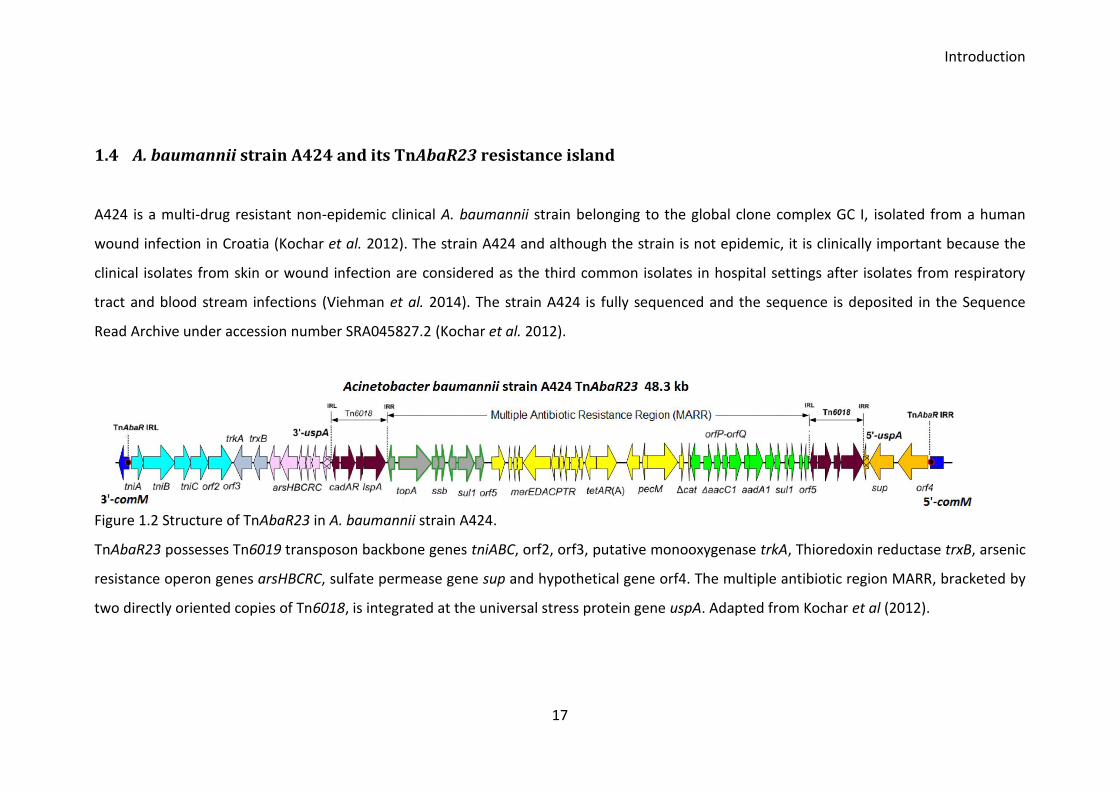
1.4 A. baumannii strain A424 and its TnAbaR23 resistance island
A424 is a multi-drug resistant non-epidemic clinical A. baumannii strain belonging to the global clone complex GC I, isolated from a human
wound infection in Croatia (Kochar et al. 2012). The strain A424 and although the strain is not epidemic, it is clinically important because the
clinical isolates from skin or wound infection are considered as the third common isolates in hospital settings after isolates from respiratory
tract and blood stream infections (Viehman et al. 2014). The strain A424 is fully sequenced and the sequence is deposited in the Sequence
Read Archive under accession number SRA045827.2 (Kochar et al. 2012).
Figure 1.2 Structure of TnAbaR23 in A. baumannii strain A424.
TnAbaR23 possesses Tn6019 transposon backbone genes tniABC, orf2, orf3, putative monooxygenase trkA, Thioredoxin reductase trxB, arsenic
resistance operon genes arsHBCRC, sulfate permease gene sup and hypothetical gene orf4. The multiple antibiotic region MARR, bracketed by
two directly oriented copies of Tn6018, is integrated at the universal stress protein gene uspA. Adapted from Kochar et al (2012).
Introduction
18
AbaR island called TnAbaR23 is inserted at the chromosomal comM gene (Figure 1.2).
The strain is found resistant to various antibiotics like tetracycline, carbenicillin,
ampicillin, trimethoprim, sulfamethoxazole, and chloramphenicol (Kochar et al. 2012).
Besides possessing various efflux-associated genes, TnAbaR23 island possess a cluster
of genes predicted to confer resistance to a wide range of antimicrobial agents.
In 2012, contribution of TnAbaR23 to the antibiotic resistance phenotype of host strain
A424 was attempted to examine experimentally for the first time (Kochar et al. 2012).
The 48.3 kb TnAbaR23 was deleted en bloc and the antibiotic resistance phenotype of
the TnAbaR23 deleted mutants DCO174 and DCO163 were compared with the parent
wild type. Surprisingly and unexpectedly, DCO163 indicated a possible genetic deletion
or re-arrangement elsewhere in the genome in a pulsed field gel electrophoresis
(PFGE) profile. The second mutant DCO174 on the other hand displayed unusual
elevated resistance towards ciprofloxacin. The authors hinted that the mutant DCO174
might have an additional cryptic mutation in the efflux genes that would have resulted
in the unexpected ciprofloxacin resistance. Since the TnAbaR23 deleted mutant
created by Kochar et al. (2012) displayed additional mutation/s elsewhere in their
genome, the antimicrobial resistance phenotype reported in TnAbaR23 deleted
mutants by the authors can be considered inconclusive. In the light of additional
mutations in both of the mutants DCO163 and DCO174, the antibiotic resistance
phenotype reported by the authors cannot be attributed to the carriage of TnAbaR23.
The first attempt to examine the contribution of TnAbaR23 on the antibiotic resistance
phenotype of the strain A424 can be considered as inconclusive since the authors were
unsuccessful in creating a true TnAbaR23 deleted mutants for antibiotic resistance
phenotype comparison with the parent wild type.
The main aim of this thesis will be to explore and understand the role of TnAbaR23 on
the phenotype of the host strain A424. Alongside, I will be investigating the features of
various AbaR islands reported in A. baumannii strains aiming to understand the
diversity of AbaR islands. I will be presenting the data on the survey of various AbaR
islands in A. baumannii genome and attempt to establish a phylogenetic relationship
between them. As described in earlier sections, deletion, integration, rearrangement

Introduction
19
and gene shuffling are commonly observed phenomenon within the internal region of
AbaR elements. In my subsequent result chapter, I will be investigating the plasticity of
TnAbaR23. The final result chapter is central to this research. In this chapter, I will be
providing data to show how TnAbaR23 affects various phenotypes of the host A424. I
will be deleting the TnAbaR23 en bloc as described in Kochar et al. (2012) aiming to
create mutants that do not harbour unintended mutations. I will then use the mutants
in various in vitro and in vivo assays to compare their phenotypes with parent A424
wild type.

20
2 Aims of the study
21
The key question I will be addressing is how TnAbaR23 contributes on the resistance,
fitness and virulence phenotypes of the host strain A424. I will be carrying out a range
of experiments to answer this question. Also, I will be surveying various AbaR islands
reported in A. baumannii and attempt to establish a phylogenetic relationship
between them. I will also be examining the stability of the internal region of TnAbaR23
in A424 strain
Survey of AbaR islands to establish the phylogenetic relationship between the
common AbaR islands and novel AbaR-like islands
Investigate the plasticity of TnAbaR23 and study the contribution of Tn6018-
associated transposase gene tnpA in the stability of internal region of
TnAbaR23
Construction of A. baumannii mutants lacking TnAbaR23 transposon
Compare the antibiotic resistance phenotype of mutant and parent wild type
Compare the fitness phenotype in in vitro head-to-head competition, growth
and biofilm assays
Compare the virulence phenotype using Galleria mellonella as an infection
model

22
3 Materials and methods
Materials and methods
23
3.1 Media, reagents and solutions
Brain Heart Infusion (BHI) broth plus 30 % glycerol
USE: Storage of bacterial stocks at -20 and -80°C
47 g of brain heart infusion broth powder (Oxoid) was dissolved in a final volume of 1 l
of distilled water containing 30 % (v/v) glycerol, autoclaved before use.
Lysogeny Broth (LB) and Agar (LA)
USE: Standard liquid (LB) and solid growth (LA) medium for bacterial cultures
Lysogeny Broth (LB) was prepared by dissolving 4 g of tryptone, 2 g of yeast extract
and 2 g of NaCl in distilled water to a final volume of 400 ml. LB agar (LA) was prepared
as for LB with 1.5 % (w/v) of agar, both media types autoclaved before use.
Simmon’s Citrate Agar (SCA)
USE: Selection for A. baumannii during conjugation experiments
9.2 g of Simmon’s citrate agar powder (Oxoid) was dissolved in a final volume of 400
ml distilled water, media autoclaved before use.
Super Optimal broth with Catabolite repression (SOC)
USE: Broth for non-selective outgrowth of bacteria post-transformation
SOC was prepared by dissolving 5 g of tryptone, 2.5 g of yeast extract and 5 g of NaCl
into 200 ml of distilled water. After autoclaving, 50 μl of 2 M MgCl2 (filter sterilized)
and 200 μl of 1M glucose (filter sterilized) were added to 1 ml of medium.
Tris-EDTA (TE) buffer
10 mM Tris-HCl, pH 8.0
1 mM EDTA, pH 8.0
Tris-acetate-EDTA (TAE) buffer
2 M Tris-HCl
2 M Acetic acid
50 mM EDTA
Materials and methods
24
1 M MgSO4
24.6 g MgSO4·7H2O
Distilled water to 100 ml
0.9% NaCl (saline)
9 g NaCl (154 mM final; 0.9% w/v)
Distilled water to 1 litre
Filter sterilise
3.2 Growth conditions for bacteria
Bacterial strains were routinely grown at 37°C on Lysogeny Agar (LA) or Lysogeny Broth
(LB) unless otherwise stated. Routine growth in liquid culture was carried out as
overnight culture in 2 ml of LB at 37°C and 200 rpm for 16 - 18 h in a 20 ml universal
tube unless otherwise stated. The frozen stock of bacteria was maintained at - 20°C
and - 80°C on brain heart infusion broth supplemented with 30 % glycerol.
3.3 Strains and plasmids used
A collection of Acinetobacter baumannii clinical isolates were thankfully received from
Dr Kevin Towner collected at the Queen’s medical Centre, Nottingham. Various wild
type and mutant strains along with E. coli strains and plasmids used are listed in Table
3.1.
Materials and methods
25
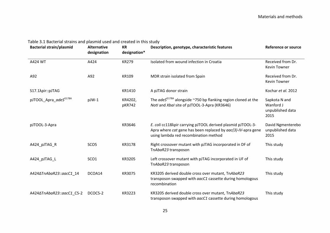
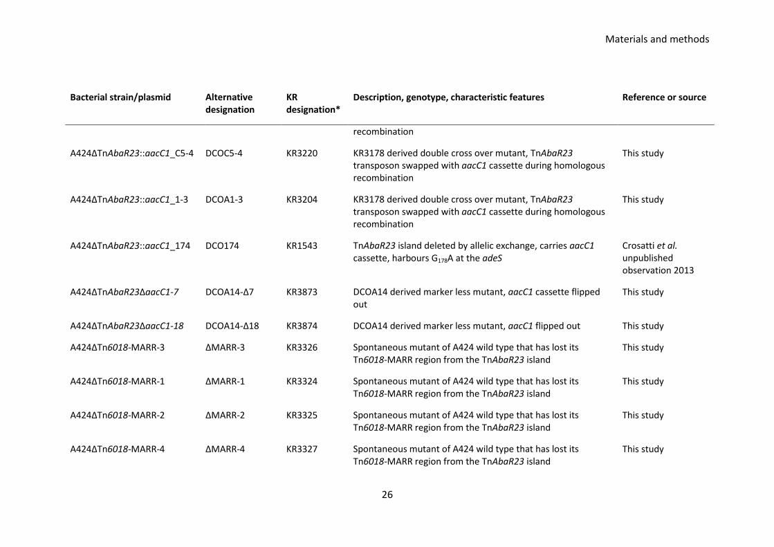
Table 3.1 Bacterial strains and plasmid used and created in this study Bacterial strain/plasmid Alternative
designation KR designation*
Description, genotype, characteristic features Reference or source
A424 WT A424 KR279 Isolated from wound infection in Croatia Received from Dr. Kevin Towner
A92 A92 KR109 MDR strain isolated from Spain Received from Dr. Kevin Towner
S17.1λpir::pJTAG KR1410 A pJTAG donor strain Kochar et al. 2012
pJTOOL_Apra_adeSG178A pJW-1 KR4202, pKR742
The adeSG178A alongside ~750 bp flanking region cloned at the NotI and XbaI site of pJTOOL-3-Apra (KR3646)
Sapkota N and Wanford J unpublished data 2015
pJTOOL-3-Apra KR3646 E. coli cc118λpir carrying pJTOOL derived plasmid pJTOOL-3-Apra where cat gene has been replaced by aac(3)-IV-apra gene using lambda red recombination method
David Ngmenterebo unpublished data 2015
A424_pJTAG_R SCO5 KR3178 Right crossover mutant with pJTAG incorporated in DF of TnAbaR23 transposon
This study
A424_pJTAG_L SCO1 KR3205 Left crossover mutant with pJTAG incorporated in UF of TnAbaR23 transposon
This study
A424ΔTnAbaR23::aacC1_14 DCOA14 KR3075 KR3205 derived double cross over mutant, TnAbaR23 transposon swapped with aacC1 cassette during homologous recombination
This study
A424ΔTnAbaR23::aacC1_C5-2 DCOC5-2 KR3223 KR3205 derived double cross over mutant, TnAbaR23 transposon swapped with aacC1 cassette during homologous
This study
Materials and methods
26
Bacterial strain/plasmid Alternative designation
KR designation*
Description, genotype, characteristic features Reference or source
recombination
A424ΔTnAbaR23::aacC1_C5-4 DCOC5-4 KR3220 KR3178 derived double cross over mutant, TnAbaR23 transposon swapped with aacC1 cassette during homologous recombination
This study
A424ΔTnAbaR23::aacC1_1-3 DCOA1-3 KR3204 KR3178 derived double cross over mutant, TnAbaR23 transposon swapped with aacC1 cassette during homologous recombination
This study
A424ΔTnAbaR23::aacC1_174 DCO174 KR1543 TnAbaR23 island deleted by allelic exchange, carries aacC1 cassette, harbours G178A at the adeS
Crosatti et al. unpublished observation 2013
A424ΔTnAbaR23ΔaacC1-7 DCOA14-Δ7 KR3873 DCOA14 derived marker less mutant, aacC1 cassette flipped out
This study
A424ΔTnAbaR23ΔaacC1-18 DCOA14-Δ18 KR3874 DCOA14 derived marker less mutant, aacC1 flipped out This study
A424ΔTn6018-MARR-3 ΔMARR-3 KR3326 Spontaneous mutant of A424 wild type that has lost its Tn6018-MARR region from the TnAbaR23 island
This study
A424ΔTn6018-MARR-1 ΔMARR-1 KR3324 Spontaneous mutant of A424 wild type that has lost its Tn6018-MARR region from the TnAbaR23 island
This study
A424ΔTn6018-MARR-2 ΔMARR-2 KR3325 Spontaneous mutant of A424 wild type that has lost its Tn6018-MARR region from the TnAbaR23 island
This study
A424ΔTn6018-MARR-4 ΔMARR-4 KR3327 Spontaneous mutant of A424 wild type that has lost its Tn6018-MARR region from the TnAbaR23 island
This study
Materials and methods
27
Bacterial strain/plasmid Alternative designation
KR designation*
Description, genotype, characteristic features Reference or source
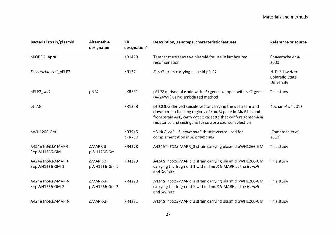
pKOBEG_Apra KR1479 Temperature sensitive plasmid for use in lambda red recombination
Chaveroche et al. 2000
Escherichia coli_pFLP2 KR137 E. coli strain carrying plasmid pFLP2 H. P. Schweizer Colorado State University
pFLP2_sul1 pNS4 pKR631 pFLP2 derived plasmid with bla gene swapped with sul1 gene (A424WT) using lambda red method
This study
pJTAG KR1358 pJTOOL-3 derived suicide vector carrying the upstream and downstream flanking regions of comM gene in AbaR1 island from strain AYE, carry aacC1 cassette that confers gentamicin resistance and sacB gene for sucrose counter selection
Kochar et al. 2012
pWH1266-Gm KR3945, pKR710
~8 kb E. coli - A. baumannii shuttle vector used for complementation in A. baumannii
(Camarena et al. 2010)
A424ΔTn6018-MARR-3::pWH1266-GM
ΔMARR-3-pWH1266-Gm
KR4278 A424ΔTn6018-MARR_3 strain carrying plasmid pWH1266-GM This study
A424ΔTn6018-MARR-3::pWH1266-GM-1
ΔMARR-3-pWH1266-Gm-1
KR4279 A424ΔTn6018-MARR_3 strain carrying plasmid pWH1266-GM carrying the fragment 1 within Tn6018-MARR at the BamHI and SalI site
This study
A424ΔTn6018-MARR-3::pWH1266-GM-2
ΔMARR-3-pWH1266-Gm-2
KR4280 A424ΔTn6018-MARR_3 strain carrying plasmid pWH1266-GM carrying the fragment 2 within Tn6018-MARR at the BamHI and SalI site
This study
A424ΔTn6018-MARR- ΔMARR-3- KR4281 A424ΔTn6018-MARR_3 strain carrying plasmid pWH1266-GM This study
Materials and methods
28
Bacterial strain/plasmid Alternative designation
KR designation*
Description, genotype, characteristic features Reference or source
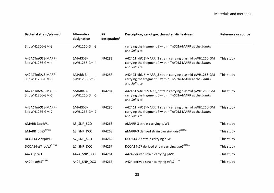
3::pWH1266-GM-3 pWH1266-Gm-3 carrying the fragment 3 within Tn6018-MARR at the BamHI and SalI site
A424ΔTn6018-MARR-3::pWH1266-GM-4
ΔMARR-3-pWH1266-Gm-4
KR4282 A424ΔTn6018-MARR_3 strain carrying plasmid pWH1266-GM carrying the fragment 4 within Tn6018-MARR at the BamHI and SalI site
This study
A424ΔTn6018-MARR-3::pWH1266-GM-5
ΔMARR-3-pWH1266-Gm-5
KR4283 A424ΔTn6018-MARR_3 strain carrying plasmid pWH1266-GM carrying the fragment 5 within Tn6018-MARR at the BamHI and SalI site
This study
A424ΔTn6018-MARR-3::pWH1266-GM-6
ΔMARR-3-pWH1266-Gm-6
KR4284 A424ΔTn6018-MARR_3 strain carrying plasmid pWH1266-GM carrying the fragment 6 within Tn6018-MARR at the BamHI and SalI site
This study
A424ΔTn6018-MARR-3::pWH1266-GM-7
ΔMARR-3-pWH1266-Gm-7
KR4285 A424ΔTn6018-MARR_3 strain carrying plasmid pWH1266-GM carrying the fragment 7 within Tn6018-MARR at the BamHI and SalI site
This study
ΔMARR-3::pJW1 Δ3_SNP_SCO KR4263 ΔMARR-3 strain carrying pJW1 This study
ΔMARR_adeSG178A Δ3_SNP_DCO KR4268 ΔMARR-3 derived strain carrying adeSG178A This study
DCOA14-Δ7::pJW1 Δ7_SNP_SCO KR4262 DCOA14-Δ7 strain carrying pJW1 This study
DCOA14-Δ7_adeSG178A Δ7_SNP_DCO KR4267 DCOA14-Δ7 derived strain carrying adeSG178A This study
A424::pJW1 A424_SNP_SCO KR4261 A424 derived strain carrying pJW1 This study
A424:: adeSG178A A424_SNP_DCO KR4266 A424 derived strain carrying adeSG178A This study
Materials and methods
29
Bacterial strain/plasmid Alternative designation
KR designation*
Description, genotype, characteristic features Reference or source
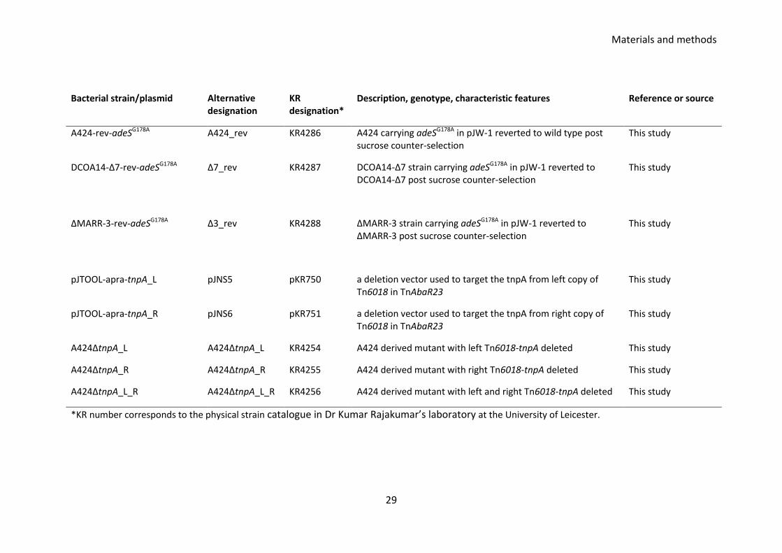
A424-rev-adeSG178A A424_rev KR4286 A424 carrying adeSG178A in pJW-1 reverted to wild type post sucrose counter-selection
This study
DCOA14-Δ7-rev-adeSG178A Δ7_rev KR4287 DCOA14-Δ7 strain carrying adeSG178A in pJW-1 reverted to DCOA14-Δ7 post sucrose counter-selection
This study
ΔMARR-3-rev-adeSG178A Δ3_rev KR4288 ΔMARR-3 strain carrying adeSG178A in pJW-1 reverted to ΔMARR-3 post sucrose counter-selection
This study
pJTOOL-apra-tnpA_L pJNS5 pKR750 a deletion vector used to target the tnpA from left copy of Tn6018 in TnAbaR23
This study
pJTOOL-apra-tnpA_R pJNS6 pKR751 a deletion vector used to target the tnpA from right copy of Tn6018 in TnAbaR23
This study
A424ΔtnpA_L A424ΔtnpA_L KR4254 A424 derived mutant with left Tn6018-tnpA deleted This study
A424ΔtnpA_R A424ΔtnpA_R KR4255 A424 derived mutant with right Tn6018-tnpA deleted This study
A424ΔtnpA_L_R A424ΔtnpA_L_R KR4256 A424 derived mutant with left and right Tn6018-tnpA deleted This study
*KR number corresponds to the physical strain catalogue in Dr Kumar Rajakumar’s laboratory at the University of Leicester.
Materials and methods
30
3.4 Genomic DNA extraction
Genomic DNA was extracted by lysing the cells in 500 µl of overnight culture
containing ~2×10⁹ colony forming units (CFU) using the 5Prime ArchivePure DNA
Purification Kit (VWR). Plasmid DNA was extracted by lysing the cells in 500 µl of
overnight bacterial broth using GenElute Plasmid Miniprep Kit (Sigma-Aldrich). The
plasmids that were to be used for downstream cloning were eluted in 50 µl of PCR
grade water. The DNA was stored at – 20°C until used.
3.5 DNA quantification
DNA was quantified using Nanodrop 2000 using DNA elution buffer or PCR grade water
as blank where appropriate during quantification.
3.6 Restriction enzymes and restriction digestions
Restriction enzymes were purchased from New England Biolabs (www.neb.uk.com),
Roche (www.roche.co.uk), Promega (www.promega.co.uk), or ThermoFisher Scientific
(http://www.thermofisher.com). The routine volume of 20 - 50 µl digestion mixture
was set up for digestion using appropriate buffers at 1 x concentration and when
required 1 x bovine serum albumin (BSA) concentration unless otherwise stated.
Following incubation at the appropriate time and temperature, restriction digests were
inactivated by heat treatment. Digests were cleaned up to wash buffers and enzymes
using PCR DNA purification kit Geneflow and pure DNA were eluted in 20 - 50 µl of PCR
grade water.
3.7 Ligation reactions
Ligation reactions were carried using 3U of T4 DNA ligase (Promega) and 10 × T4 DNA
ligase buffers. The volume of ligation reaction was in general 10 - 20 µl with insert to
vector ratio of at least 2:1. Ligation was done either at 22°C for 3 h or at 4°C for ~16 h.
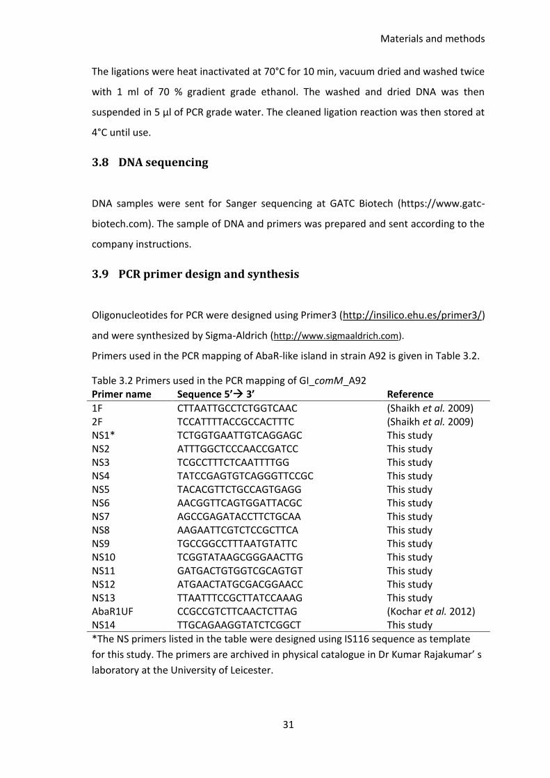
Materials and methods
31
The ligations were heat inactivated at 70°C for 10 min, vacuum dried and washed twice
with 1 ml of 70 % gradient grade ethanol. The washed and dried DNA was then
suspended in 5 μl of PCR grade water. The cleaned ligation reaction was then stored at
4°C until use.
3.8 DNA sequencing
DNA samples were sent for Sanger sequencing at GATC Biotech (https://www.gatc-
biotech.com). The sample of DNA and primers was prepared and sent according to the
company instructions.
3.9 PCR primer design and synthesis
Oligonucleotides for PCR were designed using Primer3 (http://insilico.ehu.es/primer3/)
and were synthesized by Sigma-Aldrich (http://www.sigmaaldrich.com).
Primers used in the PCR mapping of AbaR-like island in strain A92 is given in Table 3.2.
Table 3.2 Primers used in the PCR mapping of GI_comM_A92 Primer name Sequence 5’ 3’ Reference
1F CTTAATTGCCTCTGGTCAAC (Shaikh et al. 2009) 2F TCCATTTTACCGCCACTTTC (Shaikh et al. 2009) NS1* TCTGGTGAATTGTCAGGAGC This study NS2 ATTTGGCTCCCAACCGATCC This study NS3 TCGCCTTTCTCAATTTTGG This study NS4 TATCCGAGTGTCAGGGTTCCGC This study NS5 TACACGTTCTGCCAGTGAGG This study NS6 AACGGTTCAGTGGATTACGC This study NS7 AGCCGAGATACCTTCTGCAA This study NS8 AAGAATTCGTCTCCGCTTCA This study NS9 TGCCGGCCTTTAATGTATTC This study NS10 TCGGTATAAGCGGGAACTTG This study NS11 GATGACTGTGGTCGCAGTGT This study NS12 ATGAACTATGCGACGGAACC This study NS13 TTAATTTCCGCTTATCCAAAG This study AbaR1UF CCGCCGTCTTCAACTCTTAG (Kochar et al. 2012) NS14 TTGCAGAAGGTATCTCGGCT This study
*The NS primers listed in the table were designed using IS116 sequence as template
for this study. The primers are archived in physical catalogue in Dr Kumar Rajakumar’ s
laboratory at the University of Leicester.
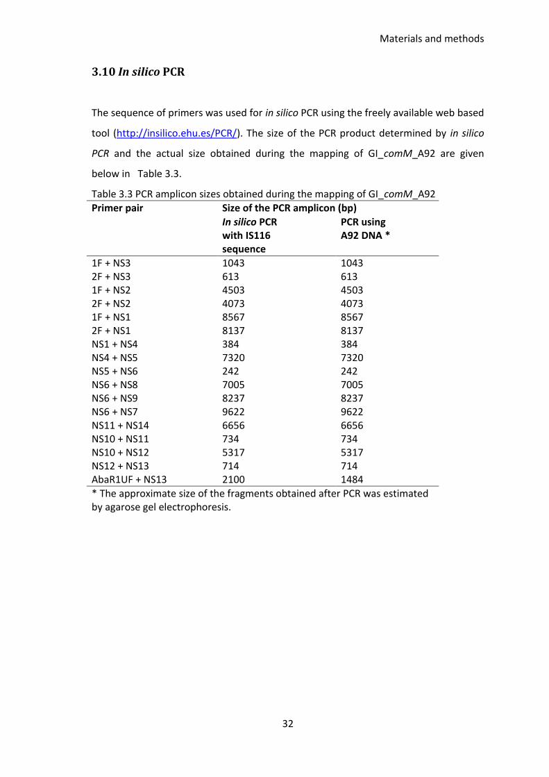
Materials and methods
32
3.10 In silico PCR
The sequence of primers was used for in silico PCR using the freely available web based
tool (http://insilico.ehu.es/PCR/). The size of the PCR product determined by in silico
PCR and the actual size obtained during the mapping of GI_comM_A92 are given
below in Table 3.3.
Table 3.3 PCR amplicon sizes obtained during the mapping of GI_comM_A92
Primer pair Size of the PCR amplicon (bp)
In silico PCR with IS116 sequence
PCR using A92 DNA *
1F + NS3 1043 1043 2F + NS3 613 613 1F + NS2 4503 4503 2F + NS2 4073 4073 1F + NS1 8567 8567 2F + NS1 8137 8137 NS1 + NS4 384 384 NS4 + NS5 7320 7320 NS5 + NS6 242 242 NS6 + NS8 7005 7005 NS6 + NS9 8237 8237 NS6 + NS7 9622 9622 NS11 + NS14 6656 6656 NS10 + NS11 734 734 NS10 + NS12 5317 5317 NS12 + NS13 714 714 AbaR1UF + NS13 2100 1484
* The approximate size of the fragments obtained after PCR was estimated by agarose gel electrophoresis.
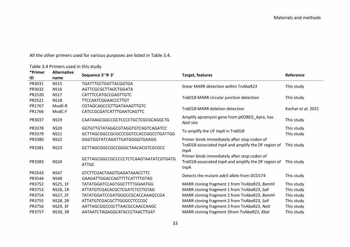
Materials and methods
33
All the other primers used for various purposes are listed in Table 3.4.
Table 3.4 Primers used in this study *Primer ID
Alternative name
Sequence 5’ 3’ Target, features Reference
PR3031 NS15 TGATTTGCTGGTTACGGTGA linear MARR detection within TnAbaR23 This study
PR3032 NS16 AGTTCGCGCTTAGCTGGATA PR2520 NS17 CATTTCCATGCCGAGTTGTC
Tn6018-MARR circular junction detection This study PR2521 NS18 TTCCAATCGGAACCCTTGT PR1767 ModK-R CGTAGCAGCCGTTGATAAAGTTGTC
Tn6018-MARR deletion detection Kochar et al. 2021 PR1766 ModC-F CATCCGCGATCATTTGAATCAGTTC
PR3037 NS19 CAATAAGCGGCCGCTCCCCTGCTCGCGCAGGCTG Amplify apramycin gene from pKOBEG_Apra, has NotI site
This study
PR3378 NS20 GGTGTTGTATAGGCGTAGGTGTCAGTCAGATCC To amplify the UF tnpA in Tn6018
This study This study PR3379 NS21 GCTTAGCGGCCGCGCCCGGTCCACCGGCCTGATTGG
PR3380 NS22 GGGTGGTATCAGATTGATGGGGTGAAGG Primer binds immediately after stop codon of Tn6018-associated tnpA and amplify the DF region of tnpA
This study PR3381 NS23 GCTTAGCGGCCGCCGGGCTAACACGTCGCGCC
PR3383 NS24 GCTTAGCGGCCGCCCCCTCTCAAGTAATATCGTGATGATTGC
Primer binds immediately after stop codon of Tn6018-associated tnpA and amplify the DF region of tnpA
This study
PR3543 NS47 GTCTTCGACTAAGTGAGATAAACCTTC Detects the mutant adeS allele from DCO174 This study
PR3544 NS48 GAAGATTGGACCAGTTTTCATTTTGTAG PR3752 NS25, 1F TATATGGATCCAGTGGCTTTTGGAATGG MARR cloning fragment 1 from TnAbaR23, BamHI This study PR3753 NS26, 1R ATTATGTCGACACGCTCGATCTCCTGTAG MARR cloning fragment 1 from TnAbaR23, SalI This study PR3754 NS27, 2F TATATGGATCCGATGGGCCGCACCAAAGCCGA MARR cloning fragment 2 from TnAbaR23, BamHI This study PR3755 NS28, 2R ATTATGTCGACGCTTGGGCCTCCCGC MARR cloning fragment 2 from TnAbaR23, SalI This study PR3756 NS29, 3F AATTAGCGGCCGCTTAACGCCAACCAAGC MARR cloning fragment 3 from TnAbaR23, NotI This study PR3757 NS30, 3R AATAATCTAGAGGCATACCCTAACTTGAT MARR cloning fragment 3from TnAbaR23, XbaI This study

Materials and methods
34
*Primer ID
Alternative name
Sequence 5’ 3’ Target, features Reference
PR3758 NS31, 4F TATATGGATCCTCGGCGCAGAGCGA MARR cloning fragment 4 from TnAbaR23, BamHI This study PR3759 NS32, 4R ATTATGTCGACATCGCTTGACTCCGTACAT MARR cloning fragment 4 from TnAbaR23, SalI This study PR3760 NS33, 5F AATTAGCGGCCGCGATGAACTCCTGTGA MARR cloning fragment 5 from TnAbaR23, NotI This study PR3761 NS34, 5R AATAATCTAGATTTTCGGGCCTCGCAT MARR cloning fragment 5 from TnAbaR23, XbaI This study PR3762 NS35, 6F AATTAGCGGCCGCGCGATTACAAGACCTCCG MARR cloning fragment 6 from TnAbaR23, NotI This study PR3763 NS36, 6R AATAATCTAGAATCGGTTTTCTTGTTGCC MARR cloning fragment 6 from TnAbaR23, XbaI This study PR3764 NS37, 7F AATTAGCGGCCGCTTTGCAACAGTGCCAA MARR cloning fragment 7 from TnAbaR23, NotI This study PR3765 NS38, 7R AATAATCTAGAAACCATTATTGGCGC MARR cloning fragment 7 from TnAbaR23, XbaI This study PR2514 NS39 CATACTCTTCCTTTTTCAATATTATTGAAGC To amplify the upstream region of bla gene from
pFLP2 This study PR2515 NS40
GACCGAAGCGAGACCTTTTTATAGGTTAATGTCATGATAATAATGG
PR2822 NS41 CAATAATATTGAAAAAGGAAGAGTATGAAAGGCGTGACGTGGAAGTCGCC sul1 from TnAbaR23 in A. baumannii strain A424 This study
PR2823 NS42 GAGTAAACTTGGTCTGACAGCTAGGCATGATCTAACC PR2824 NS43 GCAGGAAGCGAGTAATCAGG Universal primer to amplify downstream of bla gene
from pFLP2 This study
PR2825 NS44 ACTCTTCCTTTTTCAATATTATTG PR2826 NS45 CTGTCAGACCAAGTTTACTC Universal primer to amplify upstream of bla gene
from pFLP2 This study
PR2827 NS46 AGTTCGGTGTAGGTCGTTCG
*The PR numbers in primers corresponds to the physical catalogue in Dr Kumar Rajakumar’s laboratory at the University of Leicester.
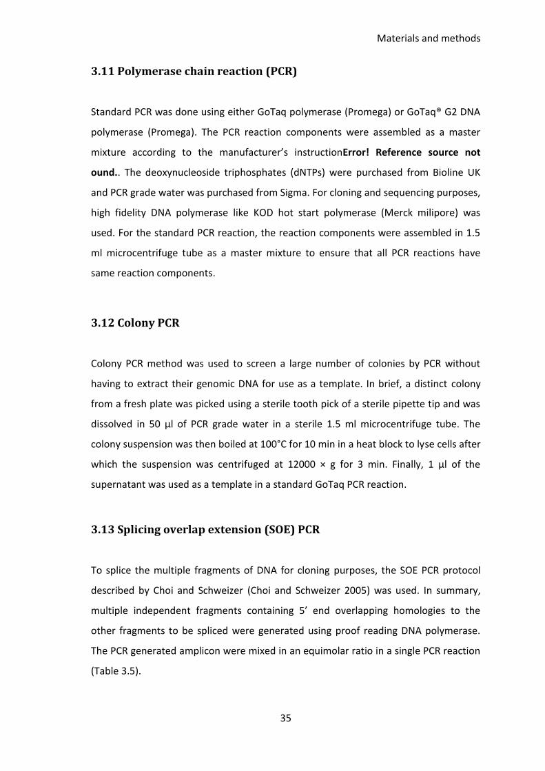
Materials and methods
35
3.11 Polymerase chain reaction (PCR)
Standard PCR was done using either GoTaq polymerase (Promega) or GoTaq® G2 DNA
polymerase (Promega). The PCR reaction components were assembled as a master
mixture according to the manufacturer’s instructionError! Reference source not
ound.. The deoxynucleoside triphosphates (dNTPs) were purchased from Bioline UK
and PCR grade water was purchased from Sigma. For cloning and sequencing purposes,
high fidelity DNA polymerase like KOD hot start polymerase (Merck milipore) was
used. For the standard PCR reaction, the reaction components were assembled in 1.5
ml microcentrifuge tube as a master mixture to ensure that all PCR reactions have
same reaction components.
3.12 Colony PCR
Colony PCR method was used to screen a large number of colonies by PCR without
having to extract their genomic DNA for use as a template. In brief, a distinct colony
from a fresh plate was picked using a sterile tooth pick of a sterile pipette tip and was
dissolved in 50 µl of PCR grade water in a sterile 1.5 ml microcentrifuge tube. The
colony suspension was then boiled at 100°C for 10 min in a heat block to lyse cells after
which the suspension was centrifuged at 12000 × g for 3 min. Finally, 1 µl of the
supernatant was used as a template in a standard GoTaq PCR reaction.
3.13 Splicing overlap extension (SOE) PCR
To splice the multiple fragments of DNA for cloning purposes, the SOE PCR protocol
described by Choi and Schweizer (Choi and Schweizer 2005) was used. In summary,
multiple independent fragments containing 5’ end overlapping homologies to the
other fragments to be spliced were generated using proof reading DNA polymerase.
The PCR generated amplicon were mixed in an equimolar ratio in a single PCR reaction
(Table 3.5).
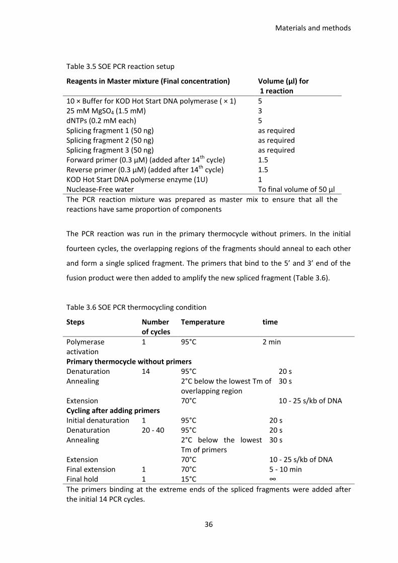
Materials and methods
36
Table 3.5 SOE PCR reaction setup
Reagents in Master mixture (Final concentration) Volume (µl) for 1 reaction
10 × Buffer for KOD Hot Start DNA polymerase ( × 1) 25 mM MgSO4 (1.5 mM) dNTPs (0.2 mM each) Splicing fragment 1 (50 ng) Splicing fragment 2 (50 ng) Splicing fragment 3 (50 ng) Forward primer (0.3 µM) (added after 14th cycle) Reverse primer (0.3 µM) (added after 14th cycle) KOD Hot Start DNA polymerse enzyme (1U) Nuclease-Free water
5 3 5 as required as required as required 1.5 1.5 1 To final volume of 50 µl
The PCR reaction mixture was prepared as master mix to ensure that all the reactions have same proportion of components
The PCR reaction was run in the primary thermocycle without primers. In the initial
fourteen cycles, the overlapping regions of the fragments should anneal to each other
and form a single spliced fragment. The primers that bind to the 5’ and 3’ end of the
fusion product were then added to amplify the new spliced fragment (Table 3.6).
Table 3.6 SOE PCR thermocycling condition
Steps Number of cycles
Temperature time
Polymerase activation
1 95°C 2 min
Primary thermocycle without primers Denaturation 14 95°C 20 s Annealing 2°C below the lowest Tm of
overlapping region 30 s
Extension 70°C 10 - 25 s/kb of DNA Cycling after adding primers Initial denaturation 1 95°C 20 s Denaturation 20 - 40 95°C 20 s Annealing 2°C below the lowest
Tm of primers 30 s
Extension 70°C 10 - 25 s/kb of DNA Final extension 1 70°C 5 - 10 min Final hold 1 15°C ∞
The primers binding at the extreme ends of the spliced fragments were added after the initial 14 PCR cycles.
Materials and methods
37
3.14 Agarose gel electrophoresis
DNA was visualized under the ultraviolet light by gel electrophoresis on 0.8 %
molecular biology grade agarose prepared in Tris-Acetate-EDTA or TAE buffer
supplemented with 10 µg/ml ethidium bromide. Loading dye was added in the DNA
sample to the final concentration of 1 × prior to loading the sample on the gel. DNA
markers like λ-HindIII, Generuler DNA ladder, and 2 - log DNA ladder were run
alongside the samples for size estimation.
3.15 Pulse Field Gel Electrophoresis (PFGE)
The PFGE protocol described here is adapted from that described with Pseudomonas
aeruginosa (MacKenzie et al. 2005).
3.15.1 Plug preparation
A 1:100 dilution of the overnight broth of the bacteria was inoculated in to 2 ml of LB
which was incubated for further 4 - 5 h. The culture was harvested by centrifuging at
3000 x g for 5 min. The pellet was re-suspended in 2 ml sterile Tris-EDTA or TE buffer.
Absorbance of the suspension was measured at OD540 nm and was adjusted to 3.0 in a
final volume of 1 ml TE. Equal volumes of bacterial suspension and 2 % molten (kept at
56°C) low-melt agarose were gently mixed to make a final volume of 500 μl and this
was transferred immediately into the plug cast. The plugs were kept at 4°C for 20 min
to solidify then recovered and transferred into 0.5 M EDTA–1 % N-lauroyl sarcosine
buffer or ES buffer containing Proteinase K to a final concentration of 1 mg/ml. The
plugs were digested in ES + Proteinase K buffer for 4 h in a shaking water bath at 55°C
then recovered and washed five times in a shaking water bath with 20 ml TE to remove
Proteinase K. After this, the plugs were stored in TE at 4°C and discarded after 2
months if unused.
Materials and methods
38
3.15.2 Restriction digestion of plugs
Using a clean scalpel, a 2 - 3 mm wide plug was cut for each strain and was transferred
to 1.5 ml microcentrifuge tube for digestion with enzyme. The plugs were washed
twice with 200 µL of restriction digestion buffer (without enzyme) prior to incubation
with enzyme. Each plug was digested with 10U of the restriction enzyme at
recommended temperature for 9 h in 200 µl of restriction digestion mix. After
digestion, the plugs were recovered and incubated on ice for 30 min with 1 ml of 0.5 ×
Tris/borate/EDTA) or TBE buffer to wash off the restriction enzyme. The washed plugs
and Saccharomyces cereviseae DNA marker were sealed on the tooth of the pulsed
field gel electrophoresis comb using 1.2 % pulsed field certified agarose gel prepared in
0.5 × TBE buffer. After assembling the casting tray, ~150 ml of 1.2 % agarose gel was
poured in the casting tray with comb containing sealed plugs. Once the gel solidified,
the comb was removed and the wells were sealed with molten 1.2 % agarose. The gel
containing plugs was then transferred inside the CHEF-DR II tank containing 2 L of 0.5 ×
TBE buffer at 12°C.
3.15.3 Running condition for PFGE
Run time= 28 h
Final pulse = 100
Initial pulse = 50
Volt/cm = 6
Running temperature = 12°C
After completion of the run, the gel was removed from the tray and was stained on
rocking stand in 400 ml nanopure water containing 200 µl of ethidium bromide for 2 h.
The bands were visualized under UV light and images taken. Where appropriate, the
gel was stained overnight and was de-stained in 400 ml nanopure water prior to
visualization.
Materials and methods
39
3.16 Preparation and transformation of chemically competent E. coli
and A. baumannii cells for heat shock transformation
The procedure of preparation and transformation of chemically competent E. coli and
A. baumannii cells described here is adapted from the protocol described in Methods
in Enzymology (Hanahan et al. 1991).
The overnight bacterial culture was re-inoculated in to 100 ml fresh LB at a ratio of
1:100 and incubated as described above for further 4 - 5 h or until absorbance of the
culture at 600 nm reached 0.4 - 0.5. The culture was then removed and was kept on ice
for 20 min. The cells were harvested by centrifuging at 3000 × g for 10 min at 4˚C. The
remainder of the procedure was carried out while working on ice or in cold room at
4°C. The bacterial pellet was first washed with 50 ml of ice cold MgCl2 (100 mM) then
with 50 ml of ice cold CaCl2 (100 mM). During each wash, the culture was incubated on
ice for 10 min then centrifuged at 3000 × g for 10 min at 4˚C. The final re-suspension
was done in 1 ml of ice cold sterile CaCl2 (100 mM) in 15 % glycerol w/v. The batch of
competent cells was dispensed as 50 µl aliquots in pre-chilled 1.5 ml microcentrifuge
tubes for immediate use or storage at - 80˚C until used.
For transformation, the competent cells were thawed on ice for 10 min. The plasmid or
DNA to be used for transformation was also placed on ice for 10 min. To the 50 µl
competent cells, 1 - 10 µl of DNA was added and the mixture was incubated on ice for
another 30 min. During heat shock transformation, the tube was directly placed in a
heat block at 42˚C for 45 s after which the tube was removed and placed on ice for 2
min for recovery. To the transformation mixture, 950 µl of Super Optimal broth with
Catabolite repression or SOC broth at room temperature was added and the cells
incubated for 1 h. The transformed cells were then plated on appropriate differential
media and were left to grow overnight.
Materials and methods
40
3.17 Preparation and transformation of electro-competent E. coli and A.
baumannii cells
The procedure of preparation and transformation of electro-competent E. coli and A.
baumannii cells described here is adapted from the protocol described by William J
Dower (Dower et al. 1988).
A 1:100 dilution of the overnight culture of the bacteria was grown in 100 ml LB for 4 -
5 h or until the absorbance at 600nm reached 0.4 - 0.5. The culture was kept on ice for
20 min before harvesting by centrifugation at 3000 × g for 10 min at 4˚C. The
supernatant was discarded and the pellet washed with 100 ml of ice cold 10 % glycerol
followed by centrifugation. The washing step was performed further 4 times using 50
ml, 25 ml, 10 ml and 5 ml of ice cold 10 % glycerol and the final pellet re-suspended in
1 ml of ice cold 10 % glycerol. The batch of competent cells was dispensed as 50 µl
aliquots in pre-chilled microcentrifuge tubes for immediate use and storage at - 80˚C.
For electroporation, 100 µl of competent cells were transferred to a pre-chilled 2 mm
gap-width electroporation cuvette (Geneflow, UK) and the cells were electroporated in
a Genepulser II system (Biorad, UK) with capacitance at 25 mF, resistance at 200 Ω and
voltage at 2.5 kV. Post electroporation, 900 µl of SOC at room temperature was added
to the cuvette and the cells were transferred to 1.5 ml microcentrifuge tube for
recovery at 37 ˚C and 200 rpm for 1 - 2 h. The recovered cells were then plated on the
relevant selective agar media.
3.18 Lambda Red Recombination
The Lambda Red Recombinase System employs three key genes from the phage λ red
locus bet (β), exo, and gam (γ) (Chaveroche et al. 2000). The Exo protein is a 5’ 3’
exonuclease and degrades the 5’ end of linear DNA molecule generating a single
stranded 3’ end of the linear DNA. The Bet protein is a single stranded DNA binding
protein and binds to the 3’ end of the linear DNA generated by Exo. This binding
Materials and methods
41
promotes the annealing of linear DNA to the complementary DNA. The Gam protein
binds to the host RecBCD exonuclease complex and inhibits its exonuclease activity.
This ensures that the linear DNA introduced into the host is protected from the host
exonuclease activity and that the Bet and Exo proteins can perform their function to
assist the homologous recombination. These three genes are under the control of the
arabinose-inducible promoter PBAD (Guzman et al. 1995) in the apramycin resistance
conferring plasmid pKOBEG_Apra. The plasmid pKOBEG_Apra contains araC which is
under the control of promoter PC. The product AraC is a regulatory protein which
binds to L-arabinose and facilitates the initiation of transcription from the PBAD
promoter. Once transcription is initiated, the recombinase genes bet, gam and exo are
expressed. The presence of L-arabinose is essential for the transcription of PBAD and
the subsequent expression of the Lambda Red Recombinase genes which promote
homologous recombination. The expression vector pKOBEG_Apra is a temperature
sensitive, low copy number plasmid and is lost above 30°C.
This method was used to swap the gene/gene cassette or DNA in the plasmid with the
desirable insert in a homologous recombination employing the Gam, Bet and Exo
proteins from the plasmid pKOBEG_Apra. The plasmid pKOBEG_Apra was
electroporated into the E. coli strain carrying the plasmid of interest to be
mutagenized. The electroporated E. coli cells were recovered in 950 µl of SOC at 30°C
for 1.5 h in an incubator shaking at 200 rpm. After the recovery, the culture was plated
on agarose plate containing 30 µg/ml apramycin. The plates were incubated at 30°C
for 48 h. The E. coli cells were then verified for the presence of two plasmids.
For the induction of lambda red genes in pKOBEG_Apra, the overnight culture of E. coli
carrying both plasmids was re-inoculated into 100 ml LB containing 30 µg/ml
apramycin and the appropriate second antibiotic. The culture was allowed to grow
until OD600 0.2 then induced with arabinose at the final concentration of 0.2 % (w/v) to
express gam, bet and exo genes then the culture was allowed to grow until the
absorbance at 600 nm (OD600) reaches 1 after which washed several times with 10 %
glycerol as described above to make them electro-competent.
Materials and methods
42
The insert to be cloned was constructed by amplifying 450 - 550 bp regions
immediately upstream and downstream of the cloning site in the vector of interest and
sowed with an appropriate resistance gene in a SOE PCR. The arabinose induced E. coli
carrying two plasmids was then electroporated with 20 ng of insert using the
Genepulser II system (Biorad, UK) under the conditions described earlier. After
electroporation, the cells were recovered in SOC as described above. The recovered
cells were plated on LA supplemented with appropriate antibiotic and were incubated
overnight. The transformed cells were verified for the presence of insert. The
pKOBEG_Apra plasmid is lost on incubation at 37°C.
3.19 Conjugations
For the conjugative delivery of plasmids into Acinetobacter baumannii strains, an
adapted version of the mating method described by Dennis and Zylstra (Dennis and
Zylstra 1998) was used. In brief, single colonies from the donor and the recipient
strains were picked from overnight plates and inoculated into 2 ml LB (100 μg/ml
gentamicin supplemented for the E. coli donor) then grown overnight. A 1:100 dilution
of both cultures of donor and recipient were then re-inoculated in 2 ml fresh LB for 6 h
after which, the bacteria were harvested by centrifugation at room temperature for 10
min. The pellets were washed by re-suspending in 1 ml/tube pre-warmed LB. Bacteria
were washed twice to remove the excess antibiotics. After the second wash, bacterial
pellets were re-suspended in 500 µl of pre-warmed LB. The donor and recipient were
mixed in 1:1 ratio and the mixture was centrifuged at 3000 x g for 10 min. The pellet
was then re-suspended in 30 µl of pre-warmed sterile 10 % glycerol.
The mixed bacterial suspension was then spotted on the centre of pre-warmed LA
plate. The spot was allowed to dry and the conjugation plate was incubated overnight.
Donor-donor and recipient-recipient conjugation was set up as a conjugation control.
The next day, the conjugation spot was collected in 600 µl of sterile 10 % glycerol.
Serial dilution of the collected culture was made up to 1:100. The transconjugants
were selected on Simmon’s citrate agar [SCA] plate supplemented with appropriate
Materials and methods
43
concentration of gentamicin and were verified by PCR for being the expected
transconjugant.
3.20 Growth Curves
The overnight broth culture of the strains was diluted 1:100 in broth medium. In 96
well polystyrene flat bottom plates (NUNC, Roskilde, Denmark), a 200 µl aliquot of the
prepared suspension was added per well and the plate incubated in the shaking
Varioskan® flash spectral scanning multimode reader with continuous shaking at 37°C
for 24 h measuring the absorbance at 600 nm every 10 min. The size of the initial
inoculum was determined by plating the appropriate dilution of the bacterial
suspension on agar media and counting the CFU the following day.
3.21 Bioinformatics tools
Following bioinformatics tools were used in this study
Blastn: To search DNA database for similar DNA sequences
(http://blast.ncbi.nlm.nih.gov/Blast.cgi)
ApE, A plasmid Editor v2.0.47: To edit DNA sequence in Genbank format files, virtual
digests and sequence annotation
(http://biologylabs.utah.edu/jorgensen/wayned/ape/)
Finch TV: To view sequence trace file (http://finchtv.software.informer.com/1.4/)
ClustalW: To visualize multiple sequence (nucleotide and protein) alignment
(http://www.ebi.ac.uk/Tools/msa/clustalo/)
In silico PCR: Online program to perform in silico PCR (http://insilico.ehu.es/)
Oligo Calc: for Oligonucleotide analysis (http://simgene.com/OligoCalc)
Graphpad Prism version 6.00 for windows: For the analysis of data and graph
construction (www.graphpad.com)
Phylogenetic analysis was done using MEGA version 6 (Tamura et al. 2013).
Materials and methods
44
3.22 Plasmids maps and in silico construction of mutant genomes
Plasmid maps and genomes were visualized and constructed using Snapgene
(www.snapgene.com), pDRAW32 (http://www.acaclone.com) and geneious
(www.geneious.com) softwares.
3.23 Biofilm Assay for A. baumannii
The biofilm assay for Acinetobacter was adapted from Malloy et al. (Malloy et al.
2005). From the overnight bacterial plate, a single colony was inoculated into a broth
media which was further grown overnight. The following day, 1:100 dilution of
overnight culture was inoculated into 2 ml LB. The culture was grown to OD600 2.0 then
diluted 1:200 in sterile LB. In the wells of flat bottom 96-well polystyrene plate (NUNC,
Roskilde, Denmark); 200 µl aliquots of the suspension were dispensed per well with
three replicates per strain. In order to control the quality of assay, CFU of the bacterial
suspension at OD600 0.2 was determined by plating the appropriate dilution on LA.
The plates were sealed to avoid evaporation and incubated at 37°C. The plates were
harvested after 24 h, 48 h and 120 h to estimate the biofilm formed. The medium was
discarded and the plates were washed three times by gentle immersion in phosphate
buffer saline or PBS. After the third wash, the plates were air dried for 30 min. The
plates were then stained using 200 µl per well of 1 % w/v Crystal Violet. The staining
plates were left at room temperature for 15 min and were washed with PBS until the
wash was clear. The plates were air dried at room temperature for 30 min in an
inverted position. The biofilm stain was then dissolved in 95 % ethanol (200 µl per
well) and the absorbance of the dissolved stain was measured at OD 595 nm using the
enzyme-linked immunosorbent assay or ELISA plate reader. The experiment was
repeated independently three times.
Materials and methods
45
3.24 Determination of minimum inhibitory concentration, MIC, by
broth microdilution
The protocol for the antimicrobial MIC by broth microdilution described here is in
accordance to the Clinical and Laboratory Standards Institute (CLSI 2009). In brief, from
an overnight plate, 2 - 3 isolated colonies the bacteria were aseptically suspended in 3
ml of sterile saline (0.9 % NaCl). The turbidity of the suspension was then matched to a
McFarland standard 0.5 (~1 - 2 x 10⁸ CFU/ml).The McFarland adjusted suspension was
diluted 1:100 in saline and added in 100 µl aliquots to a 96 well flat bottom NUNC
plate (with lid) together with antibiotic at varying concentrations. The plate was sealed
and incubated at 37°C overnight. At least three replicate dilutions were examined for
each strain tested. The purity of the inoculum was also confirmed. The 96 well plates
were examined the following day and each well carefully checked for turbidity to
determine the MIC of the antibiotic used in the test.
3.25 Antibiotic susceptibility by Etest and disc diffusion method
From the overnight plate, 2 - 4 colonies were picked and were suspended in sterile 0.9
% NaCl. The turbidity of the suspension was adjusted to match 0.5 McFarland
standards and used within 15 min of preparation. After a further 1:100 dilution in
sterile 0.9 % NaCl the suspension was streaked on iso-sensitest agar to create a semi-
confluent growth using a sterile cotton swab. The inoculum was allowed to dry for 10
min before applying the antibiotic discs or Etest strips. The discs and Etest strips were
then applied taking care to avoid bubbles between the agar and disc and the plates
incubated at 37°C for 20 h. The plates with uneven growth were discarded. The zone of
growth inhibition around the discs was measured using a ruler. For Etest, the MIC
(µg/ml) was read from the scale on the Etest strip where the symmetrical inhibition
ellipse edge intercepts the strip. BSAC version 2013 guideline (Wootton 2013) was
referred to interpret the MIC and zone diameter breakpoints for Acinetobacter.
Materials and methods
46
Antibiotic abbreviation and the concentration is shown in parenthesis: Tetracycline
[TET (10 µg/ml)], Imipenem [IMP (10 µg/ml)], Carbenicillin [CAR (100 µg/ml)],
Ampicillin [AMP (25 µg/ml)], Cefotaxime [CTX (30 µg/ml)], Streptomycin [STR (10
µg/ml)], Tobramycin [TOB(10 µg/ml)], Trimethoprim [TMP (2.5 µg/ml)],
Chloramphenicol [CHL (10 µg/ml)], Rifampicin [RIF (2 µg/ml)],
Sulfamethoxazole/Trimethoprim [SXT (25 µg/ml)], Ciprofloxacin [CIP (5 µg/ml)],
Amikacin [AMK (30 µg/ml)], Gentamicin [GEN (10 µg/ml)], Sulfamethoxazole [SUL (25
µg/ml)].
3.26 Sucrose Counter-Selection Method
A single well-grown colony of the bacteria containing counter-selectable vector
(integrated into the genome or existing as a free plasmid) was aseptically inoculated in
2 ml LB to grow overnight. The following day, a 1:100 dilution of overnight culture was
re-inoculated into fresh 2 ml LB which was further incubated for 4 h. The culture was
harvested and then diluted to OD600 0.2 in 1 ml volume. This suspension was serially
diluted up to 10-5 and the 10-1 and 10-2dilutions plated on LA supplemented with 6 %
sucrose. The lower dilutions (10-4 and 10-5) were plated on LA to estimate the CFU
count of the undiluted cell suspension. Plates were then incubated overnight. The
colonies growing on sucrose were tested for loss of the sacB gene carrying vector from
their genome.
3.27 Head-to-head in vitro competition between wild type and mutants
The protocol for in vitro head-to-head competition assay was adapted from Fernandez
et al. (Fernandez et al. 2012). In short, frozen stocks of the competing strain were
recovered and streaked twice before commencing the experiment. A single, fully
grown colony was picked and inoculated in 2 ml LB to grow overnight. 1:100 dilution of
the overnight culture was re-inoculated in 2 ml LB and was left to grow for 4-6 h. The
culture was harvested then re-suspended in 2 ml of 10 mM MgSO4 to adjusted the
OD600 0.2. The suspensions of competing strains at OD600 0.2 were then serially diluted
Materials and methods
47
up to 10-5. A 100 µl aliquot of competing strains at 10-2 dilution were mixed and then
inoculated into 2 ml LB in a 20 ml plastic universal tube. The competition culture was
left to grow overnight and 1:100 dilution was sub-cultured in 2 ml LB the following day.
Control cultures were maintained by growing the competing strains alone in broth
under identical growth conditions with the same sub-culturing protocol. The cultures
were sampled at the specified time points to determine the population of competing
strains by counting CFU on LA and LA supplemented with the appropriate antibiotic.
3.28 Galleria mellonella killing assay
The G. mellonella killing assay described here is modified from Peleg (2009).
Inoculum preparation
A suspension of bacteria at OD600 0.2 was prepared following the protocol as per
described in head-to-head in vitro competition assay. Appropriate dilutions of the OD
adjusted suspension were prepared as an inoculum and kept on ice until required. The
bacterial load in the inoculum was determined by CFU count.
Galleria preparation
Galleria larvae at the sixth instar stage were used for the experiment. Larvae were
ordered from LiveFood UK Limited (LiveFood) and kept in the dark at room
temperature and were used within 2 weeks of the shipment. On the day of inoculation,
larvae weighing ~250 mg were recovered and grouped (15 per group) in sterile petri
dishes. These groups were held at room temperature in dark for 2 h before
inoculation.
Galleria inoculation
Individual larvae were held head down in the 96-well flat bottom NUNC plates during
the process of inoculation. The rear left pro-leg of the larva was disinfected using a
cotton swab dipped in 70 % ethanol. Using a BD micro-fine 1 ml syringe affixed on a
repetitive pipette from StepperTM series, 20 µl of the inoculum (containing roughly 104
Materials and methods
48
bacteria) was injected into the haemocoel via the rear left pro-leg. After injection,
larvae were incubated at 37°C, in darkness. Two control groups of larvae were
maintained; first group contained larvae inoculated with 20 µl of 10 mM MgSO4 and
second group included untouched larvae.
Observation
Mortality of the larvae was recorded over a period of 5 days. They were considered
dead if no movement was elicited upon touch. The experiment was discarded if there
were 2 deaths in any of the control groups (untouched and 10 mM MgSO4 injected
group).
3.29 Estimating the proportion of A424 cells with spontaneous
deletion within TnAbaR23
A semi-quantitative PCR based method was used to estimate the proportion of cells
where the deletion and excision of region within the chromosome of strain A424 had
occurred.
A stock solution of 2 µg of A424 gDNA per 5 µL of PCR grade water was prepared. A
series of 1/10 dilutions of the gDNA stock solution was set up to obtain dilutions with
200 ng/5 µL, 20 ng/5 µL , 2 ng/5 µL, 0.2 ng/5 µL, 0.02 ng/5 µL, and 2 pg/5 µL water.
PCR was set up using 5 µl of the gDNA template at various dilutions in each reaction.
Detailed calculation is given in the relevant result section. The detection of PCR
amplicon in diluted gDNA was used to determine the proportion of cells where
deletion or excision of Tn6018-MARR had occurred.
3.30 AbaR-islands survey
To perform the survey of AbaR islands, the nucleotide and amino acid sequences of
TnAbaR23-associated genes were used to query NCBI database (Table 3.7) were also
used as query sequence in the survey. Both putative and characterized AbaR islands
Materials and methods
49
were located in the genome of various A. baumannii strains and their boundaries were
delineated. Following parameters were used for blastn and blastp searches.
Table 3.7 Query sequences for AbaR survey.
Gene/region within TnAbaR23
TnAbaR23 loci Length of sequence used
tniA 3056..3766 710 bp tniB 3767..5677 1910 bp tniC 5682..6602 920 bp tniD 6632..7747 1115 bp tniE 7725..9188 1463 bp IRL TnAbaR23 to tniE 2901..9188 6287 bp sup to IRR TnAbaR23 47568..51246 3678 bp
Blastn parameters
The query sequence was analysed by blastn with following parameters:
Database: The nucleotide collection (nr/nt) database was selected for search. This
database includes Genbank, EMBL, DDBJ, PDB, RefSeq sequences but excludes EST,
STS, GSS, WGS, TSA, patent sequences as well as phase 0, 1, and 2 HTGS sequences.
Organism: The database was limited to search within A. baumannii (taxid: 470)
Blastn algorithm: Following algorithm parameters were set for blastn: max target
sequences 100, short queries tick automatically adjust parameters for short input
sequences, expect threshold 10, word size 28, max matches in a query range 0, scoring
parameters match/mismatch scores 1-2, gap costs linear, filter tick the low complexity
regions, mask tick mask for lookup table only
Blastp parameters
The amino acid sequence of TniA, TniB, TniC, TniD, TniE, Orf4 and Sup from TnAbaR23
and the sequence of concatenated proteins TniABCDE and Sup-Orf4 were used as a
query sequence in the blastp analysis. Following algorithm and parameters were set
for the analysis.
Database: All non-redundant Genbank CDS translations, PDB, SwissProt, PIR, PRF
excluding environmental samples from WGS projects.
Materials and methods
50
Blastp algorithm: maximum number of aligned sequences to display 100,
automatically adjust word size and other parameters, expect threshold 10, word size 3,
maximum matches in a query range 0, scoring matrix BLOSUM62, gap costs existence
11 extension 1 and conditional compositional score matrix adjustment.
3.31 Statistical Analysis
The statistical analysis in growth curve and biofilm assays was performed by either
one-way-ANOVA (for comparison of more than two groups) and by Two-way-ANOVA
(for comparison of two groups). The survival curve in the Galleria killing assay was
analyzed by Mantel-cox test. The p values less than 0.05 were considered as
statistically significant. In in vitro head-to-head competition, at each time point (T), the
percentage of competing strains was calculated by counting CFU of the competing
strains on selective and non-selective plates. The competitive index [CI] at any time
point T was then calculated by dividing the ratio of A424 wild type by the ratio of
mutant bacteria in the culture.
CI = Ratio of wild type at time T
Ratio of mutant at time T

51
4 Survey of AbaR, the A. baumannii resistance
island
Chapter 4
52
4.1 Background
Recent advancement in the field of DNA sequencing has facilitated a rapid and
comprehensive study of several A. baumannii genomes that led to the identification of
several AbaR islands in these bacteria. As described earlier in introduction section, the
complex structure of AbaR island is built on the backbone transposons like Tn6019,
Tn6021 and Tn6022. The Tn6019 backbone is found in AbaR3-like islands whereas
Tn6021 and Tn6022 backbone is found in AbaR4-like islands. In recent years, complex
and divergent AbaR islands are being reported in MDR A. baumannii strains that
appear novel and distinctive from commonly known AbaR3 or AbaR4 like islands.
Shaikh et al. (2009) reported the presence of comM associated AbaR-like DNA
sequence in A. baumannii strains A92 and A25 that appeared distinct from known
AbaR-like islands. The sequence of comM-associated DNA in strain A25 shared only
88% identity with the terminal sequence of AbaR1 island whereas the sequence of
comM-associated DNA in strain A92 showed “no match” with the genome database at
nucleotide and protein level. Therefore, the authors hypothesized that the comM-
associated divergent DNA in strains A25 and A92 could be two distinct lineages of
AbaR family. Based on the basic architectural difference between AbaR3 and AbaR4
islands, Hamidian and Hall (2011) also proposed AbaR3 and AbaR4 as two different
lineages of AbaR islands. Recently identified island AbaR27 in MDR A. baumannii strain
BJABO715 was also reported as a distinct AbaR4-like island that lack the key AbaR4
genes uspA and sup (Zhu et al. 2013).
Despite the ongoing debate on lineages of AbaR-like islands, a phylogenetic study that
encompasses all the known diverse AbaR islands is lacking. This chapter sets out with a
specific aim to establish a phylogenetic relationship between AbaR-islands known to-
date. The internal architecture of AbaR islands is very diverse, but as described
previously, they are built on the backbone of other transposons. In this study, I will be
analysing the backbone transposons genes of AbaR-like islands reported in various A.
baumannii strains to study their relatedness. I will then select transposon backbones
for phylogenetic studies ensuring an unbiased representation of all known AbaR
Chapter 4
53
islands. I will subsequently establish a phylogenetic relationship between the
representative AbaR-backbone transposons and, by extension, AbaR islands based on
the sequence similarity of their transposition genes tniA, tniB, tniC, tniD and tniE.
4.2 Aims and objectives
The published literatures and Genbank sequence will be used as a source of data for
this survey and various bioinformatics tools will be used for the nucleotide and amino
acid sequence analysis. In order to establish a phylogenetic relationship between AbaR
islands, I will be carrying out following set of experiments and exercise
Search the published literature and compile known AbaR-like islands in A.
baumannii
Search the Genbank database to identify putative AbaR-like islands in A.
baumannii using the nucleotide and amino acid sequences of tniA, tniB, tniC,
tniD, tniE, sup and orf4 genes from TnAbaR23 island as a query sequence
Search published literatures and Genbank sequences to identify the transposon
that constitutes backbone of AbaR islands compiled during the survey
Compare the sequences of transposition genes in the representative backbone
transposons to build a phylogenetic relationship
4.3 Result
4.3.1 Survey of AbaR-like islands in A. baumannii
The Genbank database and published literatures were searched from August 2014 to
April 2015 to identify the AbaR islands reported in A. baumannii. The AbaR-like islands
reported after this period, are not included in this study.
A total of 58 AbaR-like islands that were unique to each other (in terms of their
integration site, genetic composition and host strain) were found represented in 56 A.
baumannii strains (Table 4.1). Rose (2010) has reported the presence two AbaR
Chapter 4
54
islands, both at non-comM site, in the strain A473 but those AbaR islands are not
included in this study due to the unavailability of sequence data. Three strains A85
GC1, AB0057 and BJABO715 were also reported to carry the AbaR island integrated at
a non-comM loci. A novel AbaR island in strain MDR-TJ has been identified in this study
that has not integrated within the comM gene of the strain. A MDR strains AB0057 was
reported to carry AbaR3 and AbaR4 on its chromosome while another MDR strain A85
clone GC1 strain was found to possess a copy of AbaR3 on its chromosome and a copy
of AbaR4 on its plasmid pA85-3. The smallest AbaR island was reported in A9380 (9.1
kb) while AbaR26 in strain BJABO868 was the largest island identified so far (120 kb).
During this survey, previously unreported putative AbaR-like islands were identified in
7 different A. baumannii strains. The boundaries of these putative AbaR-like islands
were delineated based (whenever applicable) on the presence of AbaR terminal
inverted repeat (IR) sequences and the putative AbaR islands were given interim name
GI _strain-name. Where applicable, the backbone transposons of the putative AbaR-
islands were also identified based on the sequence comparison with other AbaR
backbones. The comM integrated resistance islands in strains A92 and A25 reported by
Shaikh et al. (2009) were also assigned interim names as GI_comM_A92 and
GI_comM_A25 respectively.
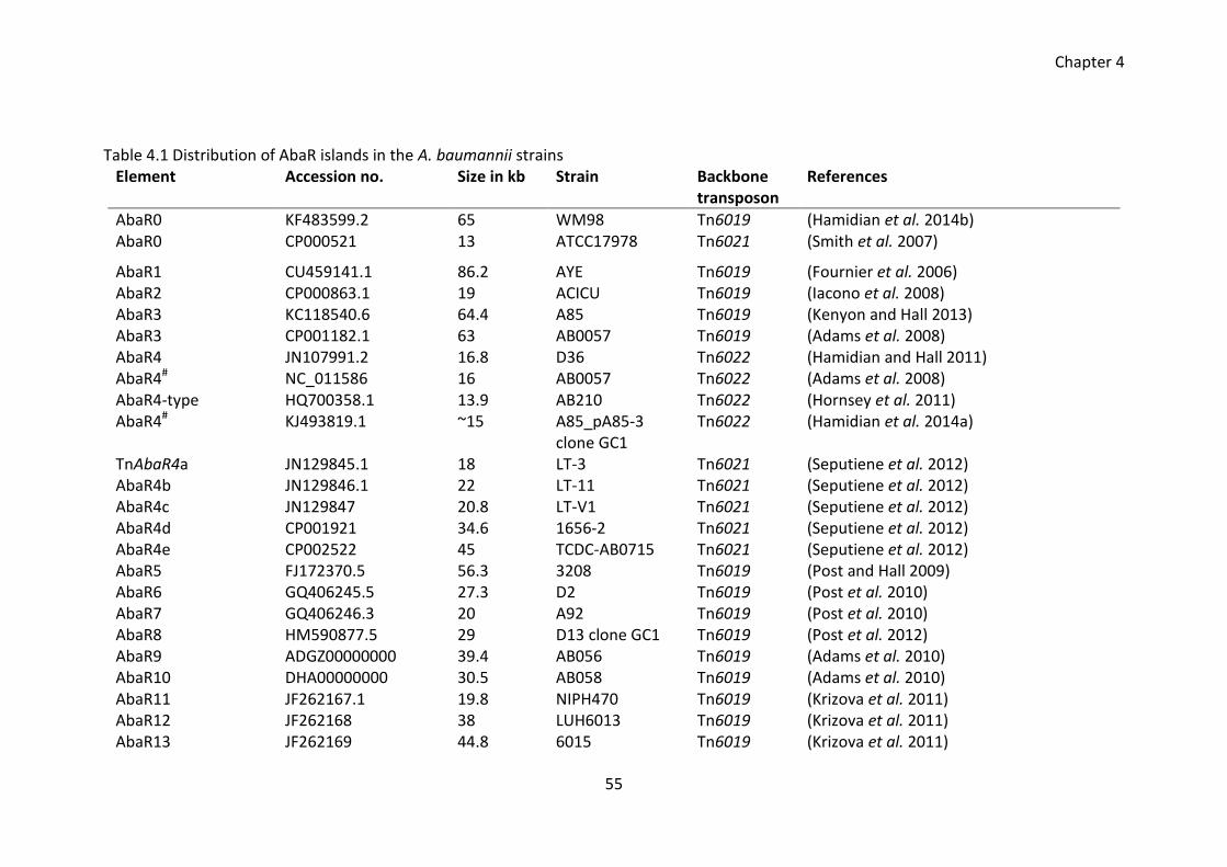
Chapter 4
55
Table 4.1 Distribution of AbaR islands in the A. baumannii strains Element Accession no. Size in kb Strain Backbone
transposon References
AbaR0 KF483599.2 65 WM98 Tn6019 (Hamidian et al. 2014b) AbaR0 CP000521 13 ATCC17978 Tn6021 (Smith et al. 2007)
AbaR1 CU459141.1 86.2 AYE Tn6019 (Fournier et al. 2006) AbaR2 CP000863.1 19 ACICU Tn6019 (Iacono et al. 2008) AbaR3 KC118540.6 64.4 A85 Tn6019 (Kenyon and Hall 2013) AbaR3 CP001182.1 63 AB0057 Tn6019 (Adams et al. 2008) AbaR4 JN107991.2 16.8 D36 Tn6022 (Hamidian and Hall 2011) AbaR4# NC_011586 16 AB0057 Tn6022 (Adams et al. 2008) AbaR4-type HQ700358.1 13.9 AB210 Tn6022 (Hornsey et al. 2011) AbaR4# KJ493819.1 ~15 A85_pA85-3
clone GC1 Tn6022 (Hamidian et al. 2014a)
TnAbaR4a JN129845.1 18 LT-3 Tn6021 (Seputiene et al. 2012) AbaR4b JN129846.1 22 LT-11 Tn6021 (Seputiene et al. 2012) AbaR4c JN129847 20.8 LT-V1 Tn6021 (Seputiene et al. 2012) AbaR4d CP001921 34.6 1656-2 Tn6021 (Seputiene et al. 2012) AbaR4e CP002522 45 TCDC-AB0715 Tn6021 (Seputiene et al. 2012) AbaR5 FJ172370.5 56.3 3208 Tn6019 (Post and Hall 2009) AbaR6 GQ406245.5 27.3 D2 Tn6019 (Post et al. 2010) AbaR7 GQ406246.3 20 A92 Tn6019 (Post et al. 2010) AbaR8 HM590877.5 29 D13 clone GC1 Tn6019 (Post et al. 2012) AbaR9 ADGZ00000000 39.4 AB056 Tn6019 (Adams et al. 2010) AbaR10 DHA00000000 30.5 AB058 Tn6019 (Adams et al. 2010) AbaR11 JF262167.1 19.8 NIPH470 Tn6019 (Krizova et al. 2011) AbaR12 JF262168 38 LUH6013 Tn6019 (Krizova et al. 2011) AbaR13 JF262169 44.8 6015 Tn6019 (Krizova et al. 2011)
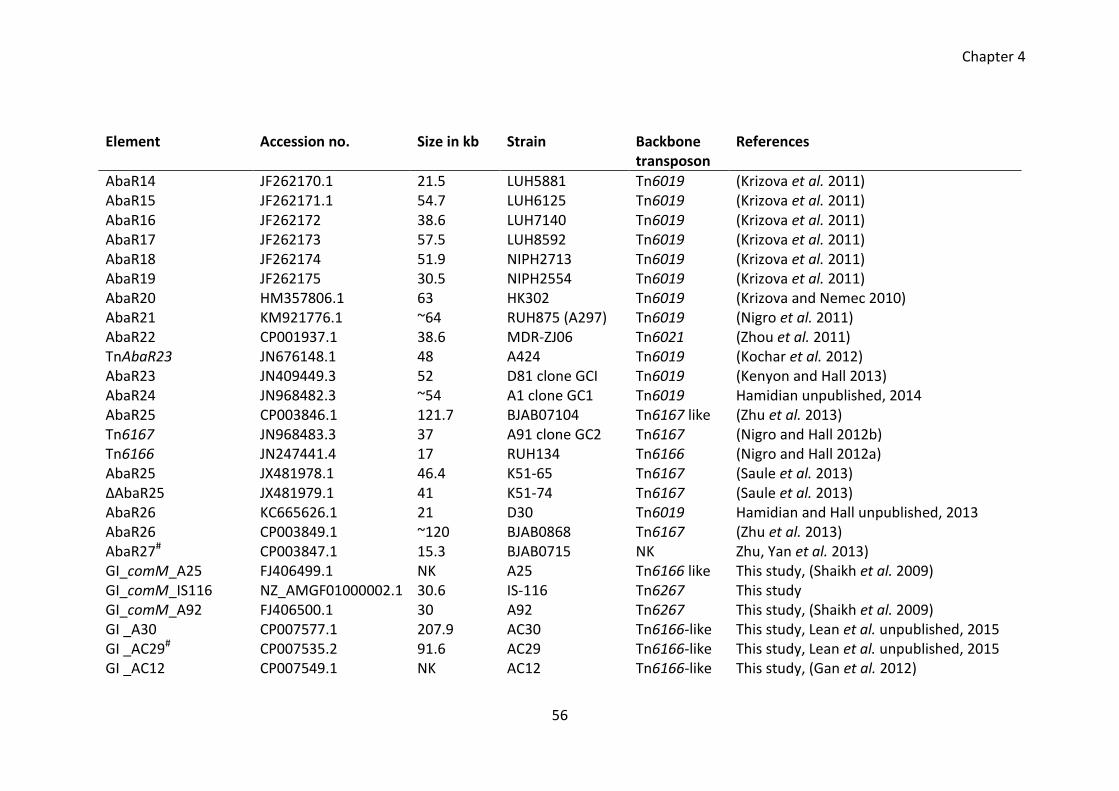
Chapter 4
56
Element Accession no. Size in kb Strain Backbone transposon
References
AbaR14 JF262170.1 21.5 LUH5881 Tn6019 (Krizova et al. 2011) AbaR15 JF262171.1 54.7 LUH6125 Tn6019 (Krizova et al. 2011) AbaR16 JF262172 38.6 LUH7140 Tn6019 (Krizova et al. 2011) AbaR17 JF262173 57.5 LUH8592 Tn6019 (Krizova et al. 2011) AbaR18 JF262174 51.9 NIPH2713 Tn6019 (Krizova et al. 2011) AbaR19 JF262175 30.5 NIPH2554 Tn6019 (Krizova et al. 2011) AbaR20 HM357806.1 63 HK302 Tn6019 (Krizova and Nemec 2010) AbaR21 KM921776.1 ~64 RUH875 (A297) Tn6019 (Nigro et al. 2011) AbaR22 CP001937.1 38.6 MDR-ZJ06 Tn6021 (Zhou et al. 2011) TnAbaR23 JN676148.1 48 A424 Tn6019 (Kochar et al. 2012) AbaR23 JN409449.3 52 D81 clone GCI Tn6019 (Kenyon and Hall 2013) AbaR24 JN968482.3 ~54 A1 clone GC1 Tn6019 Hamidian unpublished, 2014 AbaR25 CP003846.1 121.7 BJAB07104 Tn6167 like (Zhu et al. 2013) Tn6167 JN968483.3 37 A91 clone GC2 Tn6167 (Nigro and Hall 2012b) Tn6166 JN247441.4 17 RUH134 Tn6166 (Nigro and Hall 2012a) AbaR25 JX481978.1 46.4 K51-65 Tn6167 (Saule et al. 2013) ΔAbaR25 JX481979.1 41 K51-74 Tn6167 (Saule et al. 2013) AbaR26 KC665626.1 21 D30 Tn6019 Hamidian and Hall unpublished, 2013 AbaR26 CP003849.1 ~120 BJAB0868 Tn6167 (Zhu et al. 2013) AbaR27# CP003847.1 15.3 BJAB0715 NK Zhu, Yan et al. 2013) GI_comM_A25 FJ406499.1 NK A25 Tn6166 like This study, (Shaikh et al. 2009) GI_comM_IS116 NZ_AMGF01000002.1 30.6 IS-116 Tn6267 This study GI_comM_A92 FJ406500.1 30 A92 Tn6267 This study, (Shaikh et al. 2009) GI _A30 CP007577.1 207.9 AC30 Tn6166-like This study, Lean et al. unpublished, 2015 GI _AC29# CP007535.2 91.6 AC29 Tn6166-like This study, Lean et al. unpublished, 2015 GI _AC12 CP007549.1 NK AC12 Tn6166-like This study, (Gan et al. 2012)
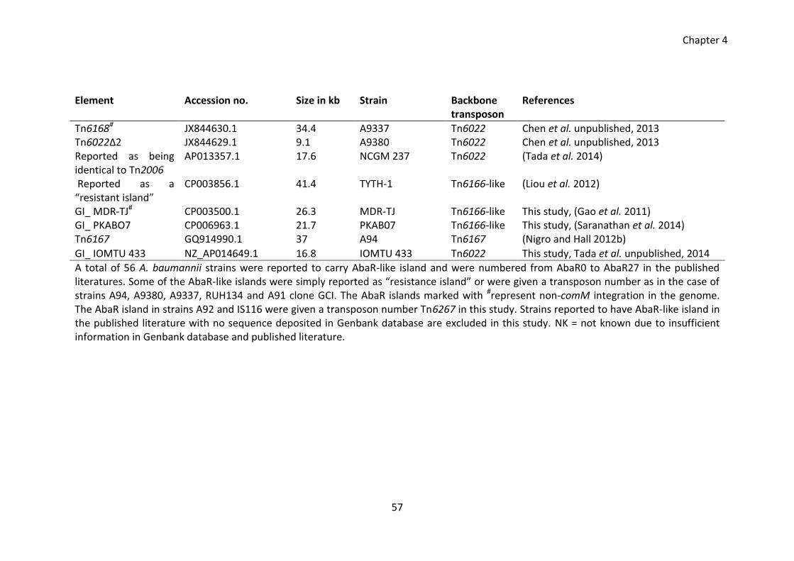
Chapter 4
57
Element Accession no. Size in kb Strain Backbone transposon
References
Tn6168# JX844630.1 34.4 A9337 Tn6022 Chen et al. unpublished, 2013 Tn6022Δ2 JX844629.1 9.1 A9380 Tn6022 Chen et al. unpublished, 2013 Reported as being identical to Tn2006
AP013357.1 17.6 NCGM 237 Tn6022 (Tada et al. 2014)
Reported as a “resistant island”
CP003856.1 41.4 TYTH-1 Tn6166-like (Liou et al. 2012)
GI_ MDR-TJ# CP003500.1 26.3 MDR-TJ Tn6166-like This study, (Gao et al. 2011) GI_ PKABO7 CP006963.1 21.7 PKAB07 Tn6166-like This study, (Saranathan et al. 2014) Tn6167 GQ914990.1 37 A94 Tn6167 (Nigro and Hall 2012b)
GI_ IOMTU 433 NZ_AP014649.1 16.8 IOMTU 433 Tn6022 This study, Tada et al. unpublished, 2014
A total of 56 A. baumannii strains were reported to carry AbaR-like island and were numbered from AbaR0 to AbaR27 in the published literatures. Some of the AbaR-like islands were simply reported as “resistance island” or were given a transposon number as in the case of strains A94, A9380, A9337, RUH134 and A91 clone GCI. The AbaR islands marked with #represent non-comM integration in the genome. The AbaR island in strains A92 and IS116 were given a transposon number Tn6267 in this study. Strains reported to have AbaR-like island in the published literature with no sequence deposited in Genbank database are excluded in this study. NK = not known due to insufficient information in Genbank database and published literature.
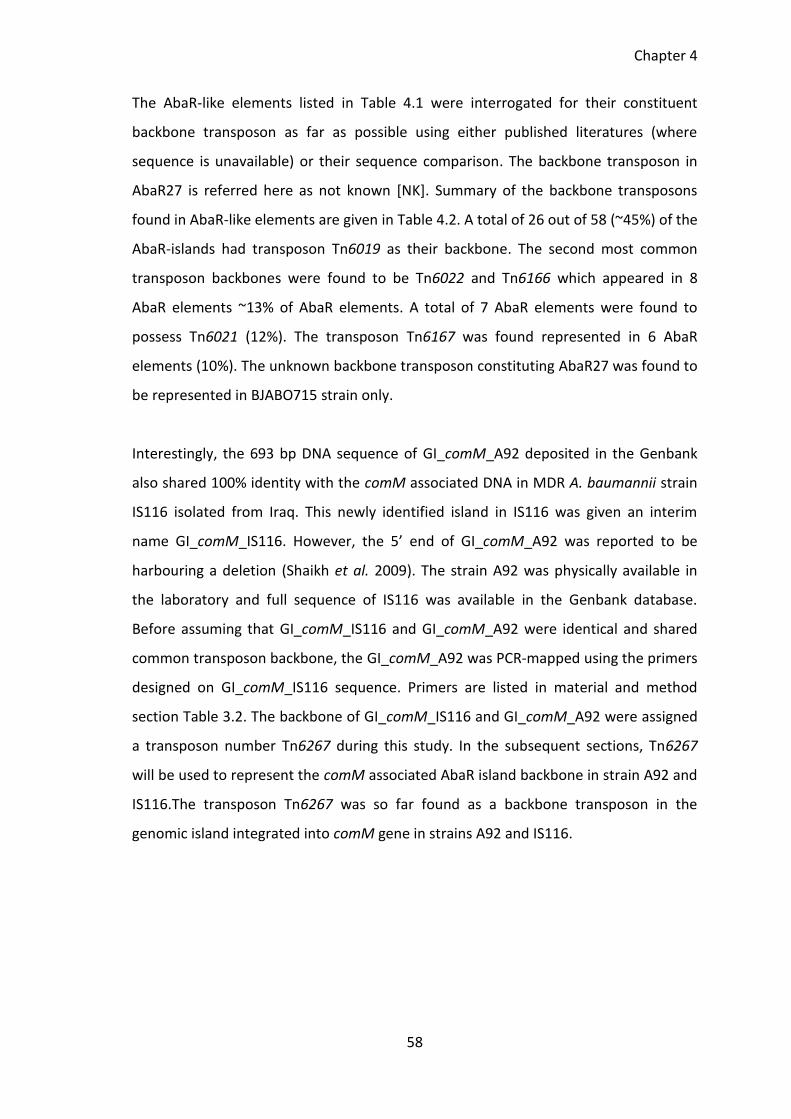
Chapter 4
58
The AbaR-like elements listed in Table 4.1 were interrogated for their constituent
backbone transposon as far as possible using either published literatures (where
sequence is unavailable) or their sequence comparison. The backbone transposon in
AbaR27 is referred here as not known [NK]. Summary of the backbone transposons
found in AbaR-like elements are given in Table 4.2. A total of 26 out of 58 (~45%) of the
AbaR-islands had transposon Tn6019 as their backbone. The second most common
transposon backbones were found to be Tn6022 and Tn6166 which appeared in 8
AbaR elements ~13% of AbaR elements. A total of 7 AbaR elements were found to
possess Tn6021 (12%). The transposon Tn6167 was found represented in 6 AbaR
elements (10%). The unknown backbone transposon constituting AbaR27 was found to
be represented in BJABO715 strain only.
Interestingly, the 693 bp DNA sequence of GI_comM_A92 deposited in the Genbank
also shared 100% identity with the comM associated DNA in MDR A. baumannii strain
IS116 isolated from Iraq. This newly identified island in IS116 was given an interim
name GI_comM_IS116. However, the 5’ end of GI_comM_A92 was reported to be
harbouring a deletion (Shaikh et al. 2009). The strain A92 was physically available in
the laboratory and full sequence of IS116 was available in the Genbank database.
Before assuming that GI_comM_IS116 and GI_comM_A92 were identical and shared
common transposon backbone, the GI_comM_A92 was PCR-mapped using the primers
designed on GI_comM_IS116 sequence. Primers are listed in material and method
section Table 3.2. The backbone of GI_comM_IS116 and GI_comM_A92 were assigned
a transposon number Tn6267 during this study. In the subsequent sections, Tn6267
will be used to represent the comM associated AbaR island backbone in strain A92 and
IS116.The transposon Tn6267 was so far found as a backbone transposon in the
genomic island integrated into comM gene in strains A92 and IS116.
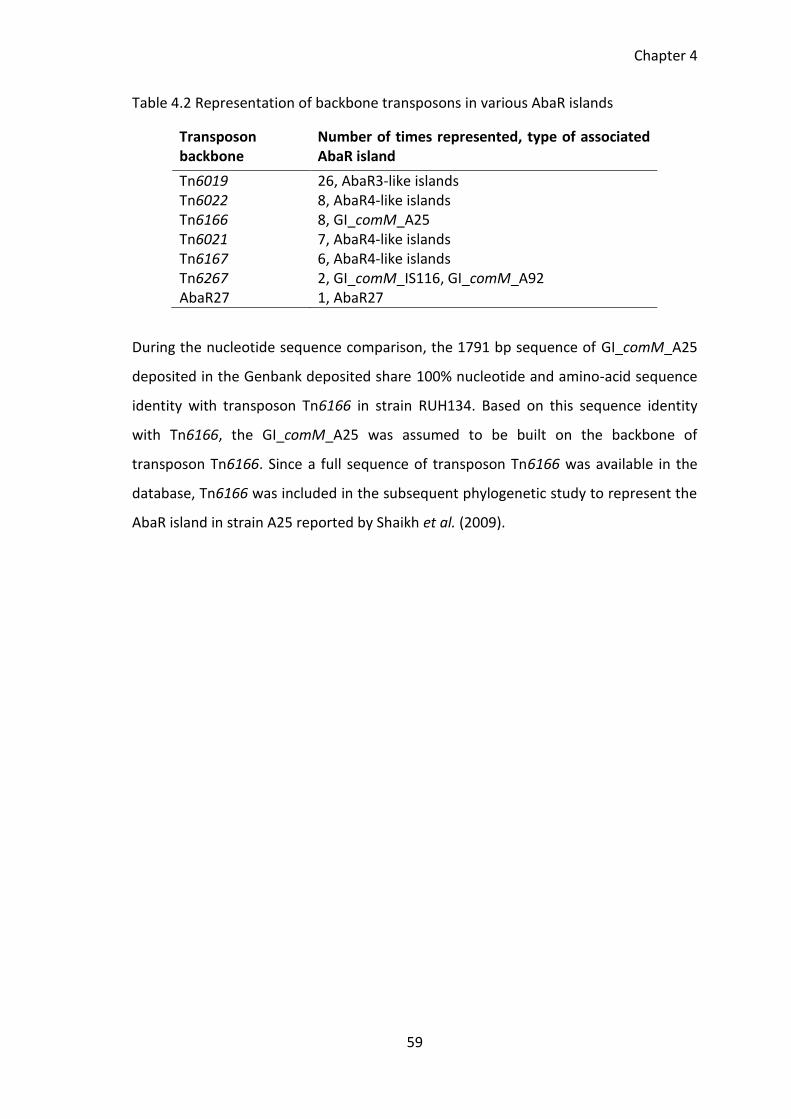
Chapter 4
59
Table 4.2 Representation of backbone transposons in various AbaR islands
Transposon backbone
Number of times represented, type of associated AbaR island
Tn6019 26, AbaR3-like islands Tn6022 8, AbaR4-like islands Tn6166 8, GI_comM_A25 Tn6021 7, AbaR4-like islands Tn6167 6, AbaR4-like islands Tn6267 2, GI_comM_IS116, GI_comM_A92 AbaR27 1, AbaR27
During the nucleotide sequence comparison, the 1791 bp sequence of GI_comM_A25
deposited in the Genbank deposited share 100% nucleotide and amino-acid sequence
identity with transposon Tn6166 in strain RUH134. Based on this sequence identity
with Tn6166, the GI_comM_A25 was assumed to be built on the backbone of
transposon Tn6166. Since a full sequence of transposon Tn6166 was available in the
database, Tn6166 was included in the subsequent phylogenetic study to represent the
AbaR island in strain A25 reported by Shaikh et al. (2009).
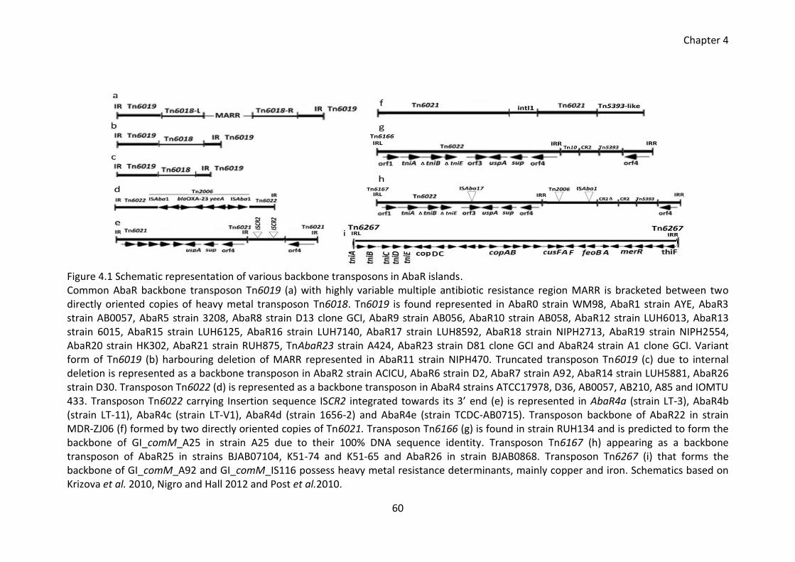
Chapter 4
60
Figure 4.1 Schematic representation of various backbone transposons in AbaR islands. Common AbaR backbone transposon Tn6019 (a) with highly variable multiple antibiotic resistance region MARR is bracketed between two directly oriented copies of heavy metal transposon Tn6018. Tn6019 is found represented in AbaR0 strain WM98, AbaR1 strain AYE, AbaR3 strain AB0057, AbaR5 strain 3208, AbaR8 strain D13 clone GCI, AbaR9 strain AB056, AbaR10 strain AB058, AbaR12 strain LUH6013, AbaR13 strain 6015, AbaR15 strain LUH6125, AbaR16 strain LUH7140, AbaR17 strain LUH8592, AbaR18 strain NIPH2713, AbaR19 strain NIPH2554, AbaR20 strain HK302, AbaR21 strain RUH875, TnAbaR23 strain A424, AbaR23 strain D81 clone GCI and AbaR24 strain A1 clone GCI. Variant form of Tn6019 (b) harbouring deletion of MARR represented in AbaR11 strain NIPH470. Truncated transposon Tn6019 (c) due to internal deletion is represented as a backbone transposon in AbaR2 strain ACICU, AbaR6 strain D2, AbaR7 strain A92, AbaR14 strain LUH5881, AbaR26 strain D30. Transposon Tn6022 (d) is represented as a backbone transposon in AbaR4 strains ATCC17978, D36, AB0057, AB210, A85 and IOMTU 433. Transposon Tn6022 carrying Insertion sequence ISCR2 integrated towards its 3’ end (e) is represented in AbaR4a (strain LT-3), AbaR4b (strain LT-11), AbaR4c (strain LT-V1), AbaR4d (strain 1656-2) and AbaR4e (strain TCDC-AB0715). Transposon backbone of AbaR22 in strain MDR-ZJ06 (f) formed by two directly oriented copies of Tn6021. Transposon Tn6166 (g) is found in strain RUH134 and is predicted to form the backbone of GI_comM_A25 in strain A25 due to their 100% DNA sequence identity. Transposon Tn6167 (h) appearing as a backbone transposon of AbaR25 in strains BJAB07104, K51-74 and K51-65 and AbaR26 in strain BJAB0868. Transposon Tn6267 (i) that forms the backbone of GI_comM_A92 and GI_comM_IS116 possess heavy metal resistance determinants, mainly copper and iron. Schematics based on Krizova et al. 2010, Nigro and Hall 2012 and Post et al.2010.
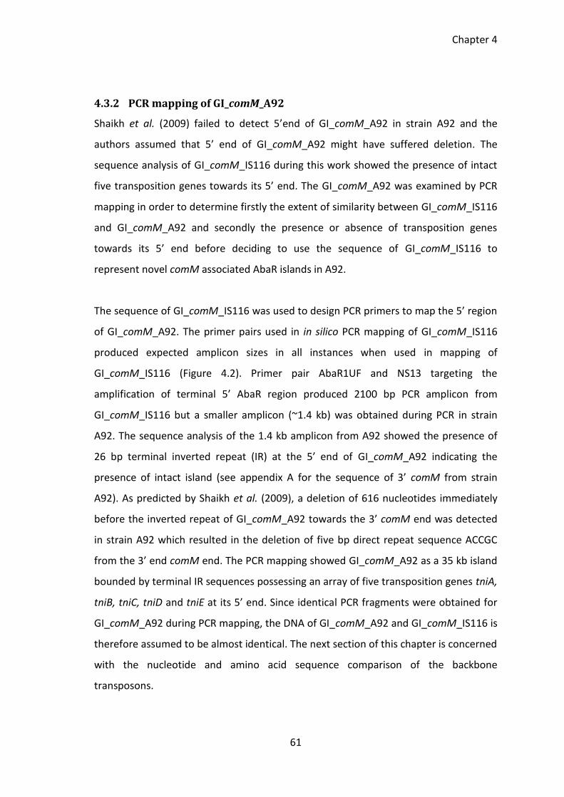
Chapter 4
61
4.3.2 PCR mapping of GI_comM_A92
Shaikh et al. (2009) failed to detect 5’end of GI_comM_A92 in strain A92 and the
authors assumed that 5’ end of GI_comM_A92 might have suffered deletion. The
sequence analysis of GI_comM_IS116 during this work showed the presence of intact
five transposition genes towards its 5’ end. The GI_comM_A92 was examined by PCR
mapping in order to determine firstly the extent of similarity between GI_comM_IS116
and GI_comM_A92 and secondly the presence or absence of transposition genes
towards its 5’ end before deciding to use the sequence of GI_comM_IS116 to
represent novel comM associated AbaR islands in A92.
The sequence of GI_comM_IS116 was used to design PCR primers to map the 5’ region
of GI_comM_A92. The primer pairs used in in silico PCR mapping of GI_comM_IS116
produced expected amplicon sizes in all instances when used in mapping of
GI_comM_IS116 (Figure 4.2). Primer pair AbaR1UF and NS13 targeting the
amplification of terminal 5’ AbaR region produced 2100 bp PCR amplicon from
GI_comM_IS116 but a smaller amplicon (~1.4 kb) was obtained during PCR in strain
A92. The sequence analysis of the 1.4 kb amplicon from A92 showed the presence of
26 bp terminal inverted repeat (IR) at the 5’ end of GI_comM_A92 indicating the
presence of intact island (see appendix A for the sequence of 3’ comM from strain
A92). As predicted by Shaikh et al. (2009), a deletion of 616 nucleotides immediately
before the inverted repeat of GI_comM_A92 towards the 3’ comM end was detected
in strain A92 which resulted in the deletion of five bp direct repeat sequence ACCGC
from the 3’ end comM end. The PCR mapping showed GI_comM_A92 as a 35 kb island
bounded by terminal IR sequences possessing an array of five transposition genes tniA,
tniB, tniC, tniD and tniE at its 5’ end. Since identical PCR fragments were obtained for
GI_comM_A92 during PCR mapping, the DNA of GI_comM_A92 and GI_comM_IS116 is
therefore assumed to be almost identical. The next section of this chapter is concerned
with the nucleotide and amino acid sequence comparison of the backbone
transposons.
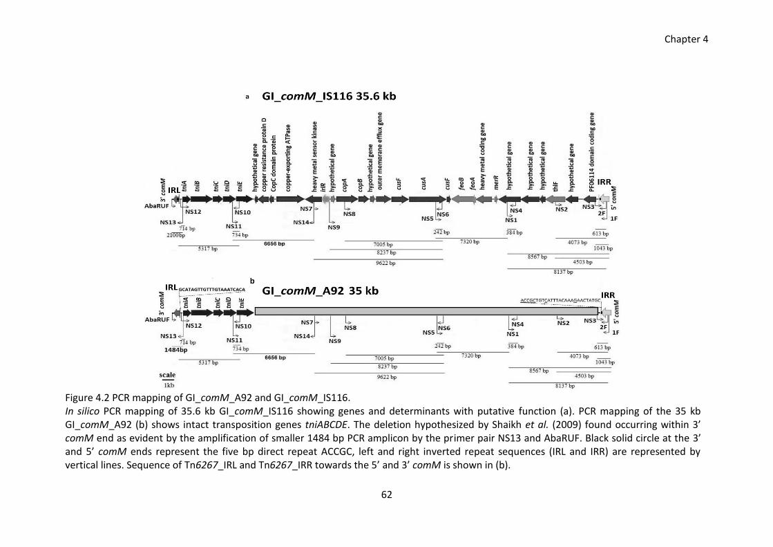
Chapter 4
62
Figure 4.2 PCR mapping of GI_comM_A92 and GI_comM_IS116. In silico PCR mapping of 35.6 kb GI_comM_IS116 showing genes and determinants with putative function (a). PCR mapping of the 35 kb GI_comM_A92 (b) shows intact transposition genes tniABCDE. The deletion hypothesized by Shaikh et al. (2009) found occurring within 3’ comM end as evident by the amplification of smaller 1484 bp PCR amplicon by the primer pair NS13 and AbaRUF. Black solid circle at the 3’ and 5’ comM ends represent the five bp direct repeat ACCGC, left and right inverted repeat sequences (IRL and IRR) are represented by vertical lines. Sequence of Tn6267_IRL and Tn6267_IRR towards the 5’ and 3’ comM is shown in (b).
Chapter 4
63
4.3.3 Phylogenetic relationship between AbaR islands
Nucleotide and amino acid sequences of the transposition genes tniA, tniB, tniC, tniD
and tniE were extracted from all the AbaR elements listed in the above Table 4.1. The
sequences were aligned (whenever available) by clustalW with cut off value of 30%
and 500 bootstrap replicates were used for analysis using MEGA6. Strains carrying
truncated or interrupted genes were excluded from the trees of genes.
Figure 4.3 Phylogenetic tree constructed using tniA nucleotide sequence from AbaR islands. The nucleotide sequence of tniA gene was used to construct the tree. The tree was inferred using the Neighbour-joining method. The evolutionary distances were computed using the maximum composite likelihood method and are in the units of the number of base substitutions per site.
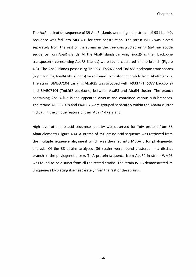
Chapter 4
64
The tniA nucleotide sequence of 39 AbaR islands were aligned a stretch of 931 bp tniA
sequence was fed into MEGA 6 for tree construction. The strain IS116 was placed
separately from the rest of the strains in the tree constructed using tniA nucleotide
sequence from AbaR islands. All the AbaR islands carrying Tn6019 as their backbone
transposon (representing AbaR3 islands) were found clustered in one branch (Figure
4.3). The AbaR islands possessing Tn6021, Tn6022 and Tn6166 backbone transposons
(representing AbaR4-like islands) were found to cluster separately from AbaR3 group.
The strain BJABO7104 carrying AbaR25 was grouped with A9337 (Tn6022 backbone)
and BJAB07104 (Tn6167 backbone) between AbaR3 and AbaR4 cluster. The branch
containing AbaR4-like island appeared diverse and contained various sub-branches.
The strains ATCC17978 and PKAB07 were grouped separately within the AbaR4 cluster
indicating the unique feature of their AbaR4-like island.
High level of amino acid sequence identity was observed for TniA protein from 38
AbaR elements (Figure 4.4). A stretch of 290 amino acid sequence was retrieved from
the multiple sequence alignment which was then fed into MEGA 6 for phylogenetic
analysis. Of the 38 strains analysed, 36 strains were found clustered in a distinct
branch in the phylogenetic tree. TniA protein sequence from AbaR0 in strain WM98
was found to be distinct from all the tested strains. The strain IS116 demonstrated its
uniqueness by placing itself separately from the rest of the strains.
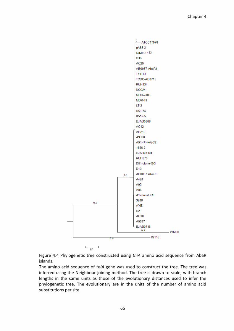
Chapter 4
65
Figure 4.4 Phylogenetic tree constructed using tniA amino acid sequence from AbaR islands. The amino acid sequence of tniA gene was used to construct the tree. The tree was inferred using the Neighbour-joining method. The tree is drawn to scale, with branch lengths in the same units as those of the evolutionary distances used to infer the phylogenetic tree. The evolutionary are in the units of the number of amino acid substitutions per site.
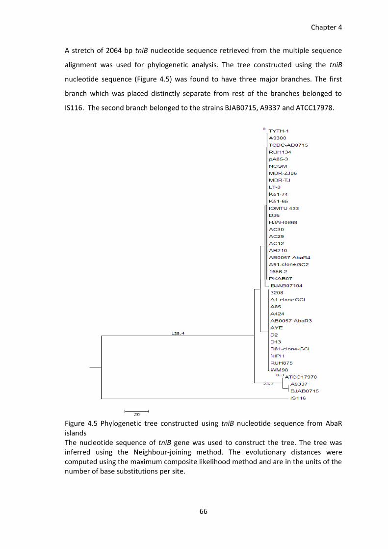
Chapter 4
66
A stretch of 2064 bp tniB nucleotide sequence retrieved from the multiple sequence
alignment was used for phylogenetic analysis. The tree constructed using the tniB
nucleotide sequence (Figure 4.5) was found to have three major branches. The first
branch which was placed distinctly separate from rest of the branches belonged to
IS116. The second branch belonged to the strains BJAB0715, A9337 and ATCC17978.
Figure 4.5 Phylogenetic tree constructed using tniB nucleotide sequence from AbaR islands The nucleotide sequence of tniB gene was used to construct the tree. The tree was inferred using the Neighbour-joining method. The evolutionary distances were computed using the maximum composite likelihood method and are in the units of the number of base substitutions per site.
Chapter 4
67
The tniB nucleotide sequence of these three strains was very similar but distinctly
different from the other tested strains. The third large branch collectively possessed
strains carrying AbaR3 and AbaR4-like islands with a clear sub-branching leading to
AbaR3 and AbaR4 carrying strains. The TniB amino sequences of the 38 strains were
aligned and a stretch of 653 amino acid long TniB protein sequence was obtained for
use in phylogenetic analysis. Except from the strain IS116, the TniB amino acid
sequence of all the tested strains was found highly similar. The TniB amino acid tree in
Figure 4.6 can be seen divided into two major branches. One branch carries only one
strain IS116 and the other branch carries all the tested 37 strains.
Figure 4.6 Phylogenetic tree constructed using tniB amino acid sequence from AbaR islands The amino acid sequence of TniB was used to construct the tree. The tree was inferred using the Neighbour-joining method and is drawn to scale, with branch lengths in the same units as those of the evolutionary distances used to infer the phylogenetic tree. The evolutionary distances are in the units of the number of amino acid substitutions per site. The transposition genes of some of the AbaR elements were found truncated due to
integration, deletion and genetic rearrangement within their AbaR host. In order to
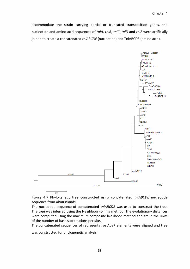
Chapter 4
68
accommodate the strain carrying partial or truncated transposition genes, the
nucleotide and amino acid sequences of tniA, tniB, tniC, tniD and tniE were artificially
joined to create a concatenated tniABCDE (nucleotide) and TniABCDE (amino acid).
Figure 4.7 Phylogenetic tree constructed using concatenated tniABCDE nucleotide sequence from AbaR islands. The nucleotide sequence of concatenated tniABCDE was used to construct the tree. The tree was inferred using the Neighbour-joining method. The evolutionary distances were computed using the maximum composite likelihood method and are in the units of the number of base substitutions per site. The concatenated sequences of representative AbaR elements were aligned and tree
was constructed for phylogenetic analysis.
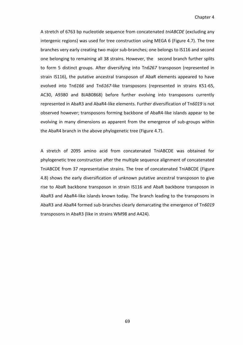
Chapter 4
69
A stretch of 6763 bp nucleotide sequence from concatenated tniABCDE (excluding any
intergenic regions) was used for tree construction using MEGA 6 (Figure 4.7). The tree
branches very early creating two major sub-branches; one belongs to IS116 and second
one belonging to remaining all 38 strains. However, the second branch further splits
to form 5 distinct groups. After diversifying into Tn6267 transposon (represented in
strain IS116), the putative ancestral transposon of AbaR elements appeared to have
evolved into Tn6166 and Tn6167-like transposons (represented in strains K51-65,
AC30, A9380 and BJAB0868) before further evolving into transposons currently
represented in AbaR3 and AbaR4-like elements. Further diversification of Tn6019 is not
observed however; transposons forming backbone of AbaR4-like islands appear to be
evolving in many dimensions as apparent from the emergence of sub-groups within
the AbaR4 branch in the above phylogenetic tree (Figure 4.7).
A stretch of 2095 amino acid from concatenated TniABCDE was obtained for
phylogenetic tree construction after the multiple sequence alignment of concatenated
TniABCDE from 37 representative strains. The tree of concatenated TniABCDE (Figure
4.8) shows the early diversification of unknown putative ancestral transposon to give
rise to AbaR backbone transposon in strain IS116 and AbaR backbone transposon in
AbaR3 and AbaR4-like islands known today. The branch leading to the transposons in
AbaR3 and AbaR4 formed sub-branches clearly demarcating the emergence of Tn6019
transposons in AbaR3 (like in strains WM98 and A424).
Chapter 4
70
Figure 4.8 Phylogenetic tree constructed using concatenated TniABCDE amino acid sequence from AbaR islands. The amino acid sequence of TniA, TniB, TniC, TniD and TniE was artificially joined to create a concatenated TniABCDE protein. The tree was inferred using the Neighbour-joining method and is drawn to scale, with branch lengths in the same units as those of the evolutionary distances used to infer the phylogenetic tree. The evolutionary distances are in the units of the number of amino acid substitutions per site. The tree was constructed in MEGA 6. Various AbaR4-like islands carry variant forms of backbone transposons like Tn6021,
Tn6022, Tn6166 and Tn6167. The sub-branching of the cluster of strains carrying
AbaR4-like islands clearly shows the diversification of their backbone transposons
giving rise to novel backbone transposons like the one observed in AbaR25 in strain
BJAB0715. Compared to AbaR4 forming backbone transposons, very little
diversification was observed among the AbaR3 backbone forming transposon Tn6019.
Chapter 4
71
4.4 Discussion
4.4.1 Survey of AbaR islands
This study was set out to establish the phylogenetic relationship between diverse
appearing AbaR islands and aiming to establish the lineages of these elements in A.
baumannii. Genbank database was searched using TnAbaR23 sequence from strain
A424 as a query sequence. A total of 51 AbaR islands were reported in the published
literature with sequence available in the Genbank database. The AbaR-islands or
strains whose sequence is not available in the Genbank database are excluded from
this study. In this study, by comparative sequence analysis, seven previously
unreported AbaR elements have been identified. Using comparative sequence analysis,
the constituent backbone transposon was identified in all 57 AbaR-like islands. For
AbaR27 in strain BJAB0715, Zhu et al. (2013) have not identified the backbone
transposon, therefore, in this study the AbaR27 island backbone has been considered
as not known [NK].
The comM-associated sequence in strain A92 (Shaikh et al. 2008) was found almost
identical (based on PCR mapping) to the comM associated sequence in strain IS116.
The backbone transposon of AbaR island in strains A92 and IS116 (this study) was
subsequently assigned a transposon number Tn6267. The comparative sequence
analysis showed AbaR island in strain A25 (Shaikh et al 2008) to have a backbone
transposon Tn6166; the AbaR island in strain A25 was subsequently given an interim
name GI_comM_A25. During the survey, previously unreported seven AbaR islands
were identified in strains IS116, MDR-TJ, PKAB07, IOMTU 433, A30, AC29 and AC12
and the AbaR islands in these strains were given interim name GI_comM_IS116,
GI_MDR-TJ, GI_PKAB07, GI_IOMTU 433, GI_A30, GI_AC29 and GI_AC12 respectively.
The in silico mapping of GI_comM_IS116 and PCR mapping of GI_comM_A92 revealed
the presence 35.6 kb and 35 kb heavy metal resistance determinants (mainly copper
resistance determinants and genes associated with iron acquisition) carrying AbaR-like
islands build on the backbone of novel transposon Tn6267.
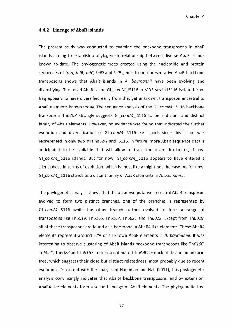
Chapter 4
72
4.4.2 Lineage of AbaR islands
The present study was conducted to examine the backbone transposons in AbaR
islands aiming to establish a phylogenetic relationship between diverse AbaR islands
known to-date. The phylogenetic trees created using the nucleotide and protein
sequences of tniA, tniB, tniC, tniD and tniE genes from representative AbaR backbone
transposons shows that AbaR islands in A. baumannii have been evolving and
diversifying. The novel AbaR island GI_comM_IS116 in MDR strain IS116 isolated from
Iraq appears to have diversified early from the, yet unknown, transposon ancestral to
AbaR elements known today. The sequence analysis of the GI_comM_IS116 backbone
transposon Tn6267 strongly suggests GI_comM_IS116 to be a distant and distinct
family of AbaR elements. However, no evidence was found that indicated the further
evolution and diversification of GI_comM_IS116-like islands since this island was
represented in only two strains A92 and IS116. In future, more AbaR sequence data is
anticipated to be available that will allow to trace the diversification of, if any,
GI_comM_IS116 islands. But for now, GI_comM_IS116 appears to have entered a
silent phase in terms of evolution, which is most likely might not the case. As for now,
GI_comM_IS116 stands as a distant family of AbaR elements in A. baumannii.
The phylogenetic analysis shows that the unknown putative ancestral AbaR transposon
evolved to form two distinct branches, one of the branches is represented by
GI_comM_IS116 while the other branch further evolved to form a range of
transposons like Tn6019, Tn6166, Tn6167, Tn6021 and Tn6022. Except from Tn6019,
all of these transposons are found as a backbone in AbaR4-like elements. These AbaR4
elements represent around 52% of all known AbaR elements in A. baumannii. It was
interesting to observe clustering of AbaR islands backbone transposons like Tn6166,
Tn6021, Tn6022 and Tn6167 in the concatenated TniABCDE nucleotide and amino acid
tree, which suggests their close but distinct relatedness, most probably due to recent
evolution. Consistent with the analysis of Hamidian and Hall (2011), this phylogenetic
analysis convincingly indicates that AbaR4 backbone transposons, and by extension,
AbaR4-like elements form a second lineage of AbaR elements. The phylogenetic tree
Chapter 4
73
also shows an extensive branching and sub-branching within AbaR4 elements
exhibiting their evolution on-the-go.
Undoubtedly, AbaR4-like elements are evolving and diversifying creating a wide range
of diverse AbaR elements like AbaR islands represented in strains A9337 (Tn6022-like),
BJAB07104 (Tn6167-like), ATCC17978 (Tn6021-like), and PKAB07 (Tn6166-like) and last
but not least strain BJAB07150 (AbaR27). Along with their capabilities of rapid
evolution, it is possible that the AbaR4-like elements are capable of disseminating their
cargo resistance determinants across the species. The recent identification of novel
AbaR4-like island in MDR A. baumannii from China (Zhu et al. 2013) and non-
baumannii Acinetobacter species from South Korea (Kim and Ko 2015) strengthens the
idea that diverse AbaR4 elements are highly mobile. This makes AbaR4 elements one
of the highly concerning mobile elements in terms of emergence and dissemination of
antimicrobial resistance.
Except from AbaR4, the first AbaR elements in A. baumannii were identified in MDR
clinical A. baumannii strains. All these AbaR elements were found to be built on
Tn6019 backbone. The work of Krizova et al. (2011) elegantly shows the evolution and
diversification of AbaR3-like elements, however, during the phylogenetic analysis it
was interestingly noted that unlike AbaR4-like elements, AbaR3-like elements were
showing less diversification and therefore less evolution. It is however important to
recall that much of diversity among AbaR3-like elements is observed within their
multiple antibiotic resistance region (MARR) and that multiple events of intermolecular
recombination have been reported within this MARR region of AbaR3-like elements.
Although, compared to AbaR4-like elements, AbaR3-like elements appear quiescent in-
terms of evolution; it is likely that AbaR3-like elements are actively involved in
disseminating the antimicrobial resistance across the species. Consistent with the
observation of Hamidian and Hall (2011), this phylogenetic analysis have established
Tn6019 and, by extension, AbaR3-like islands as a discrete family of AbaR islands.
This study was based on the nucleotide and amino acid sequence comparison of only
five transposition genes from AbaR backbone transposons. Therefore these results
Chapter 4
74
need to be interpreted with caution. As more and more sequence data of A. baumannii
becomes available, a clear picture of the families of AbaR islands with discrete lineages
is expected to emerge. However, this study provides some dependable conclusions
that A. baumannii AbaR islands might have diverged from a common yet unknown
ancestral transposon.
Based on the phylogenetic analysis of transposon backbone gene, it is hypothesized
that AbaR islands have diversified to form three major lineages. The first lineage is
represented by AbaR3 islands built on backbone of Tn6019 and around 45% of MDR A.
baumannii possess variant forms of this island. The second lineage is represented by
AbaR4-like islands built on the backbone of diverse transposons like Tn6022, Tn6166,
Tn6167 and Tn6021. The AbaR4-like islands are the most abundant islands and are
found represented in 50% of MDR A. baumannii genome. The novel GI_comM_IS116-
like island represented in strains IS116 and A92 represents another distant lineage of
AbaR-elements. Since AbaR4-like elements were found to be diversifying to form their
variant versions, in agreement with Zhu et al. (2013), I have described AbaR27 as a
variant form of AbaR4-like islands and but I have not considered it as a major lineage
of AbaR islands. Since AbaR4-like islands appear to be diversifying rapidly, it will be
interesting to watch the evolutionary journey of AbaR4-like elements and how they
drive the emergence and dissemination of antimicrobial resistance in A. baumannii.

75
5 Plasticity of AbaR islands
Chapter 5
76
5.1 Chapter overview
The survey of Acinetobacter baumannii resistance islands (AbaR) has shown that the
genome of MDR A. baumannii strains possesses at least one type of these AbaR
islands. Multiple events of genetic rearrangements have been reported in diverse AbaR
islands making them unstable features in the genome of A. baumannii (Krizova et al.
2011). In the introduction chapter, I have briefly described the presence of 48.3 kb
AbaR island called TnAbaR23 in A424 which was found inserted in a site and
orientation-specific manner within the chromosomal comM gene (Kochar et al. 2012).
Deletion and intramolecular genetic rearrangements mediated by identical copies of
insertion sequences and transposons within several AbaR islands have been frequently
reported and this phenomenon is described as “central” to the evolution and
diversification of AbaR islands (Krizova et al. 2011, Saule et al. 2013, Harmer et al.
2014). During the PCR analysis of the TnAbaR23, a possible deletion of a large region
internal to TnAbaR23 was noticed. The deletion was found occurring at multiple-
antibiotic resistance region or MARR. This MARR is a region internal to TnAbaR23 and
is bracketed by two identical copies of heavy metal transposon Tn6018.
5.2 Aim
In this chapter, I will be investigating the stability of TnAbaR23 island in in vitro growth
conditions. Using PCR based semi-quantitative assays I will be studying the rate of
deletion and loss of the region/s internal to MARR in a population of A424 strain.
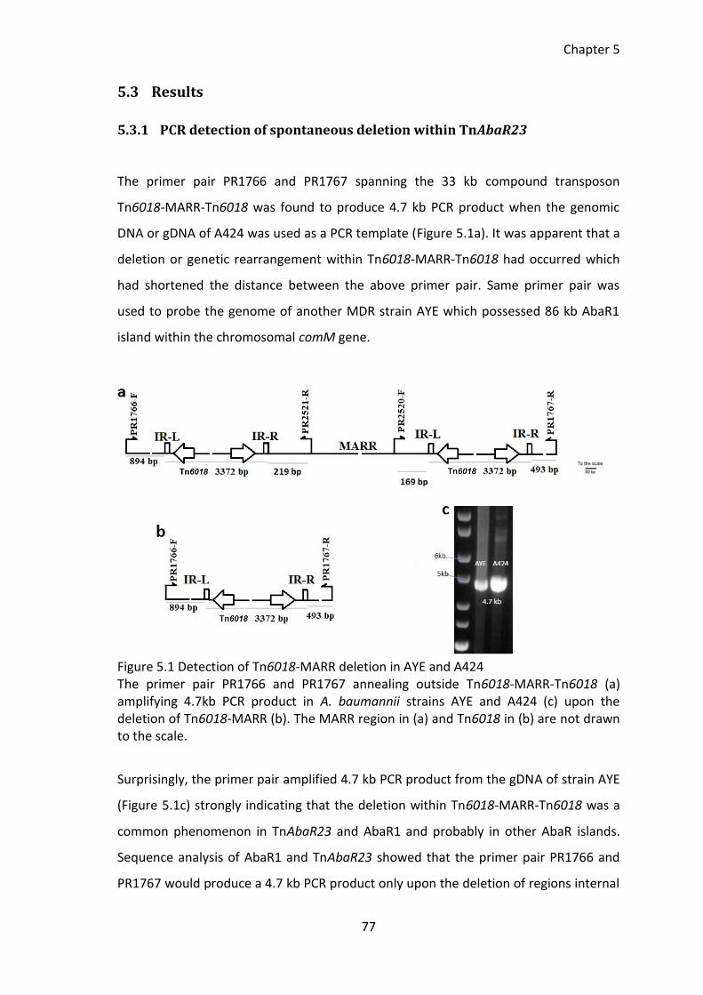
Chapter 5
77
5.3 Results
5.3.1 PCR detection of spontaneous deletion within TnAbaR23
The primer pair PR1766 and PR1767 spanning the 33 kb compound transposon
Tn6018-MARR-Tn6018 was found to produce 4.7 kb PCR product when the genomic
DNA or gDNA of A424 was used as a PCR template (Figure 5.1a). It was apparent that a
deletion or genetic rearrangement within Tn6018-MARR-Tn6018 had occurred which
had shortened the distance between the above primer pair. Same primer pair was
used to probe the genome of another MDR strain AYE which possessed 86 kb AbaR1
island within the chromosomal comM gene.
Figure 5.1 Detection of Tn6018-MARR deletion in AYE and A424 The primer pair PR1766 and PR1767 annealing outside Tn6018-MARR-Tn6018 (a) amplifying 4.7kb PCR product in A. baumannii strains AYE and A424 (c) upon the deletion of Tn6018-MARR (b). The MARR region in (a) and Tn6018 in (b) are not drawn to the scale.
Surprisingly, the primer pair amplified 4.7 kb PCR product from the gDNA of strain AYE
(Figure 5.1c) strongly indicating that the deletion within Tn6018-MARR-Tn6018 was a
common phenomenon in TnAbaR23 and AbaR1 and probably in other AbaR islands.
Sequence analysis of AbaR1 and TnAbaR23 showed that the primer pair PR1766 and
PR1767 would produce a 4.7 kb PCR product only upon the deletion of regions internal
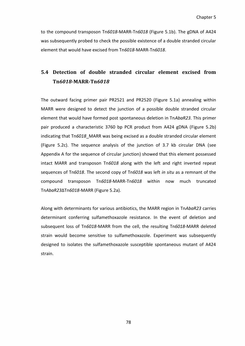
Chapter 5
78
to the compound transposon Tn6018-MARR-Tn6018 (Figure 5.1b). The gDNA of A424
was subsequently probed to check the possible existence of a double stranded circular
element that would have excised from Tn6018-MARR-Tn6018.
5.4 Detection of double stranded circular element excised from
Tn6018-MARR-Tn6018
The outward facing primer pair PR2521 and PR2520 (Figure 5.1a) annealing within
MARR were designed to detect the junction of a possible double stranded circular
element that would have formed post spontaneous deletion in TnAbaR23. This primer
pair produced a characteristic 3760 bp PCR product from A424 gDNA (Figure 5.2b)
indicating that Tn6018_MARR was being excised as a double stranded circular element
(Figure 5.2c). The sequence analysis of the junction of 3.7 kb circular DNA (see
Appendix A for the sequence of circular junction) showed that this element possessed
intact MARR and transposon Tn6018 along with the left and right inverted repeat
sequences of Tn6018. The second copy of Tn6018 was left in situ as a remnant of the
compound transposon Tn6018-MARR-Tn6018 within now much truncated
TnAbaR23ΔTn6018-MARR (Figure 5.2a).
Along with determinants for various antibiotics, the MARR region in TnAbaR23 carries
determinant conferring sulfamethoxazole resistance. In the event of deletion and
subsequent loss of Tn6018-MARR from the cell, the resulting Tn6018-MARR deleted
strain would become sensitive to sulfamethoxazole. Experiment was subsequently
designed to isolates the sulfamethoxazole susceptible spontaneous mutant of A424
strain.
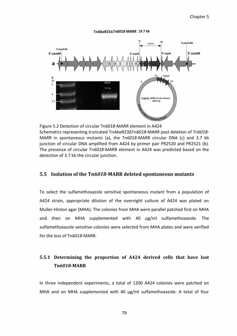
Chapter 5
79
Figure 5.2 Detection of circular Tn6018-MARR element in A424 Schematics representing truncated TnAbaR23ΔTn6018-MARR post deletion of Tn6018-MARR in spontaneous mutants (a), the Tn6018-MARR circular DNA (c) and 3.7 kb junction of circular DNA amplified from A424 by primer pair PR2520 and PR2521 (b). The presence of circular Tn6018-MARR element in A424 was predicted based on the detection of 3.7 kb the circular junction.
5.5 Isolation of the Tn6018-MARR deleted spontaneous mutants
To select the sulfamethoxazole sensitive spontaneous mutant from a population of
A424 strain, appropriate dilution of the overnight culture of A424 was plated on
Muller-Hinton agar (MHA). The colonies from MHA were parallel patched first on MHA
and then on MHA supplemented with 40 µg/ml sulfamethoxazole. The
sulfamethoxazole sensitive colonies were selected from MHA plates and were verified
for the loss of Tn6018-MARR.
5.5.1 Determining the proportion of A424 derived cells that have lost
Tn6018-MARR
In three independent experiments, a total of 1200 A424 colonies were patched on
MHA and on MHA supplemented with 40 µg/ml sulfamethoxazole. A total of four
Chapter 5
80
sulfamethoxazole susceptible colonies were obtained. The proportion of cells lacking
Tn6018-MARR element was found to be around 1 in 300 cells.
5.5.2 Verification of Tn6018-MARR deleted spontaneous mutants
The loss of Tn6018-MARR in sulfamethoxazole sensitive colonies was verified by
amplifying the empty region previously occupied by Tn6018-MARR. The parent strain
A424 was used as a positive control and the strain DCOA14 that lack TnAbaR23 was
used as a negative control.
The PCR reaction for the positive and negative controls contained 20 ng of gDNA
whereas the test samples contained 10 ng gDNA of putative spontaneous mutants
(Figure 5.3b). The faint 4.7 kb band observed in A424 indicates that only a small
proportion of cells in the entire population might harbour Tn6018-MARR deletion
within their TnAbaR23. To check whether the excised Tn6018-MARR has integrated
elsewhere in the genome, the sulfamethoxazole sensitive colonies were tested for the
presence of MARR associated aadA1 gene (Figure 5.3c). The gDNA of A424 and AbaR1
carrying MDR strain AYE was used as a positive control. The negative aadA1 PCR in all
tested putative mutants confirmed that upon the excision from its native site, the
Tn6018-MARR element did not re-integrated elsewhere in the genome of the
spontaneous mutants and was lost entirely from the cell.
The Pulsed Field Gel Electrophoresis or PFGE profile of spontaneous mutants (Figure
5.3a) was also consistent with the deletion of Tn6018-MARR. The strain DCOA14 which
lack TnAbaR23 was used in the first lanes of gels containing NotI and SfiI digested high
molecular gDNA. The spontaneous mutant specific bands that appeared in NotI and SfiI
digestion are indicated by white arrows. All four independent spontaneous mutants
exhibited indistinguishable PFGE profile and band discrepancy was not observed in all
four tested mutants.
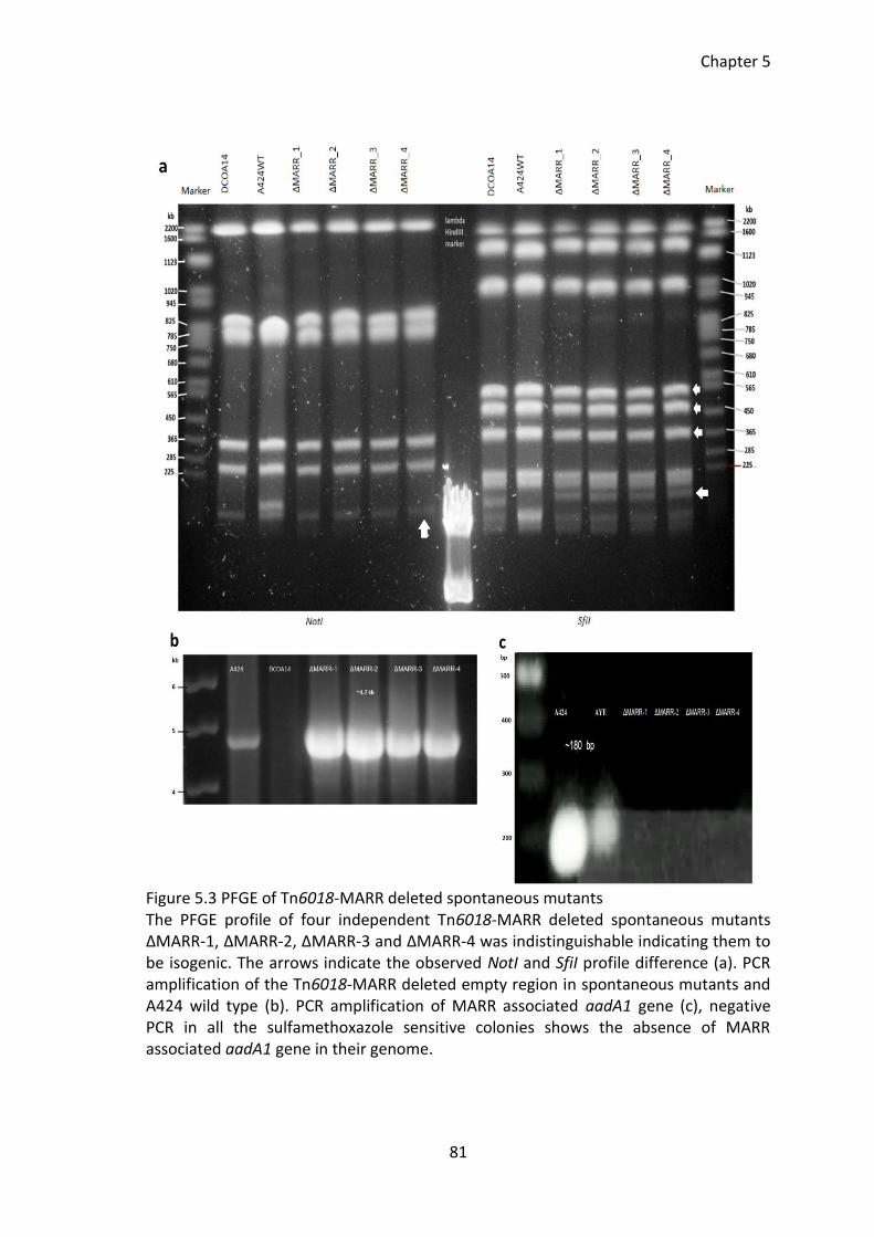
Chapter 5
81
Figure 5.3 PFGE of Tn6018-MARR deleted spontaneous mutants The PFGE profile of four independent Tn6018-MARR deleted spontaneous mutants ΔMARR-1, ΔMARR-2, ΔMARR-3 and ΔMARR-4 was indistinguishable indicating them to be isogenic. The arrows indicate the observed NotI and SfiI profile difference (a). PCR amplification of the Tn6018-MARR deleted empty region in spontaneous mutants and A424 wild type (b). PCR amplification of MARR associated aadA1 gene (c), negative PCR in all the sulfamethoxazole sensitive colonies shows the absence of MARR associated aadA1 gene in their genome.
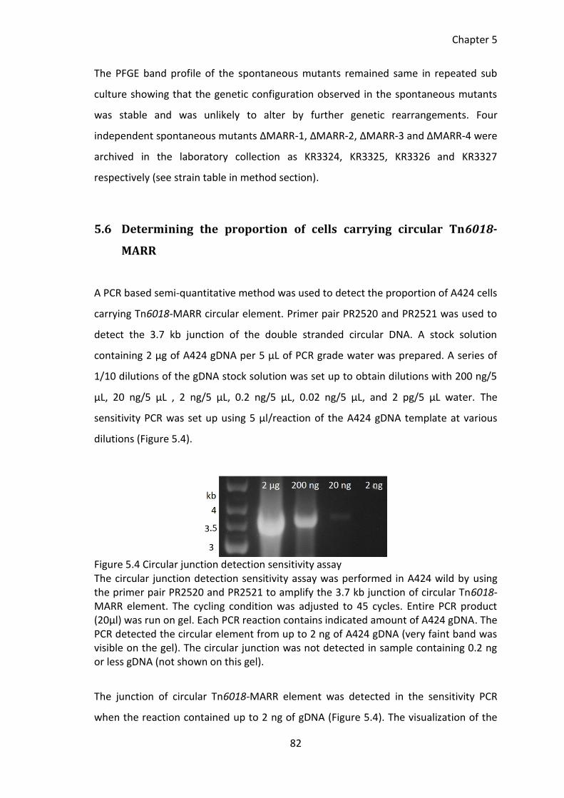
Chapter 5
82
The PFGE band profile of the spontaneous mutants remained same in repeated sub
culture showing that the genetic configuration observed in the spontaneous mutants
was stable and was unlikely to alter by further genetic rearrangements. Four
independent spontaneous mutants ΔMARR-1, ΔMARR-2, ΔMARR-3 and ΔMARR-4 were
archived in the laboratory collection as KR3324, KR3325, KR3326 and KR3327
respectively (see strain table in method section).
5.6 Determining the proportion of cells carrying circular Tn6018-
MARR
A PCR based semi-quantitative method was used to detect the proportion of A424 cells
carrying Tn6018-MARR circular element. Primer pair PR2520 and PR2521 was used to
detect the 3.7 kb junction of the double stranded circular DNA. A stock solution
containing 2 µg of A424 gDNA per 5 µL of PCR grade water was prepared. A series of
1/10 dilutions of the gDNA stock solution was set up to obtain dilutions with 200 ng/5
µL, 20 ng/5 µL , 2 ng/5 µL, 0.2 ng/5 µL, 0.02 ng/5 µL, and 2 pg/5 µL water. The
sensitivity PCR was set up using 5 µl/reaction of the A424 gDNA template at various
dilutions (Figure 5.4).
Figure 5.4 Circular junction detection sensitivity assay The circular junction detection sensitivity assay was performed in A424 wild by using the primer pair PR2520 and PR2521 to amplify the 3.7 kb junction of circular Tn6018-MARR element. The cycling condition was adjusted to 45 cycles. Entire PCR product (20µl) was run on gel. Each PCR reaction contains indicated amount of A424 gDNA. The PCR detected the circular element from up to 2 ng of A424 gDNA (very faint band was visible on the gel). The circular junction was not detected in sample containing 0.2 ng or less gDNA (not shown on this gel).
The junction of circular Tn6018-MARR element was detected in the sensitivity PCR
when the reaction contained up to 2 ng of gDNA (Figure 5.4). The visualization of the
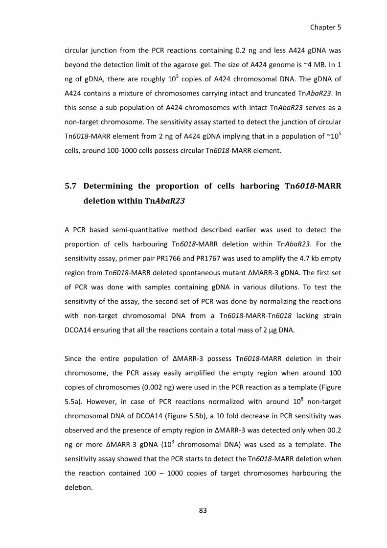
Chapter 5
83
circular junction from the PCR reactions containing 0.2 ng and less A424 gDNA was
beyond the detection limit of the agarose gel. The size of A424 genome is ~4 MB. In 1
ng of gDNA, there are roughly 105 copies of A424 chromosomal DNA. The gDNA of
A424 contains a mixture of chromosomes carrying intact and truncated TnAbaR23. In
this sense a sub population of A424 chromosomes with intact TnAbaR23 serves as a
non-target chromosome. The sensitivity assay started to detect the junction of circular
Tn6018-MARR element from 2 ng of A424 gDNA implying that in a population of ~105
cells, around 100-1000 cells possess circular Tn6018-MARR element.
5.7 Determining the proportion of cells harboring Tn6018-MARR
deletion within TnAbaR23
A PCR based semi-quantitative method described earlier was used to detect the
proportion of cells harbouring Tn6018-MARR deletion within TnAbaR23. For the
sensitivity assay, primer pair PR1766 and PR1767 was used to amplify the 4.7 kb empty
region from Tn6018-MARR deleted spontaneous mutant ΔMARR-3 gDNA. The first set
of PCR was done with samples containing gDNA in various dilutions. To test the
sensitivity of the assay, the second set of PCR was done by normalizing the reactions
with non-target chromosomal DNA from a Tn6018-MARR-Tn6018 lacking strain
DCOA14 ensuring that all the reactions contain a total mass of 2 µg DNA.
Since the entire population of ΔMARR-3 possess Tn6018-MARR deletion in their
chromosome, the PCR assay easily amplified the empty region when around 100
copies of chromosomes (0.002 ng) were used in the PCR reaction as a template (Figure
5.5a). However, in case of PCR reactions normalized with around 108 non-target
chromosomal DNA of DCOA14 (Figure 5.5b), a 10 fold decrease in PCR sensitivity was
observed and the presence of empty region in ΔMARR-3 was detected only when 00.2
ng or more ΔMARR-3 gDNA (103 chromosomal DNA) was used as a template. The
sensitivity assay showed that the PCR starts to detect the Tn6018-MARR deletion when
the reaction contained 100 – 1000 copies of target chromosomes harbouring the
deletion.
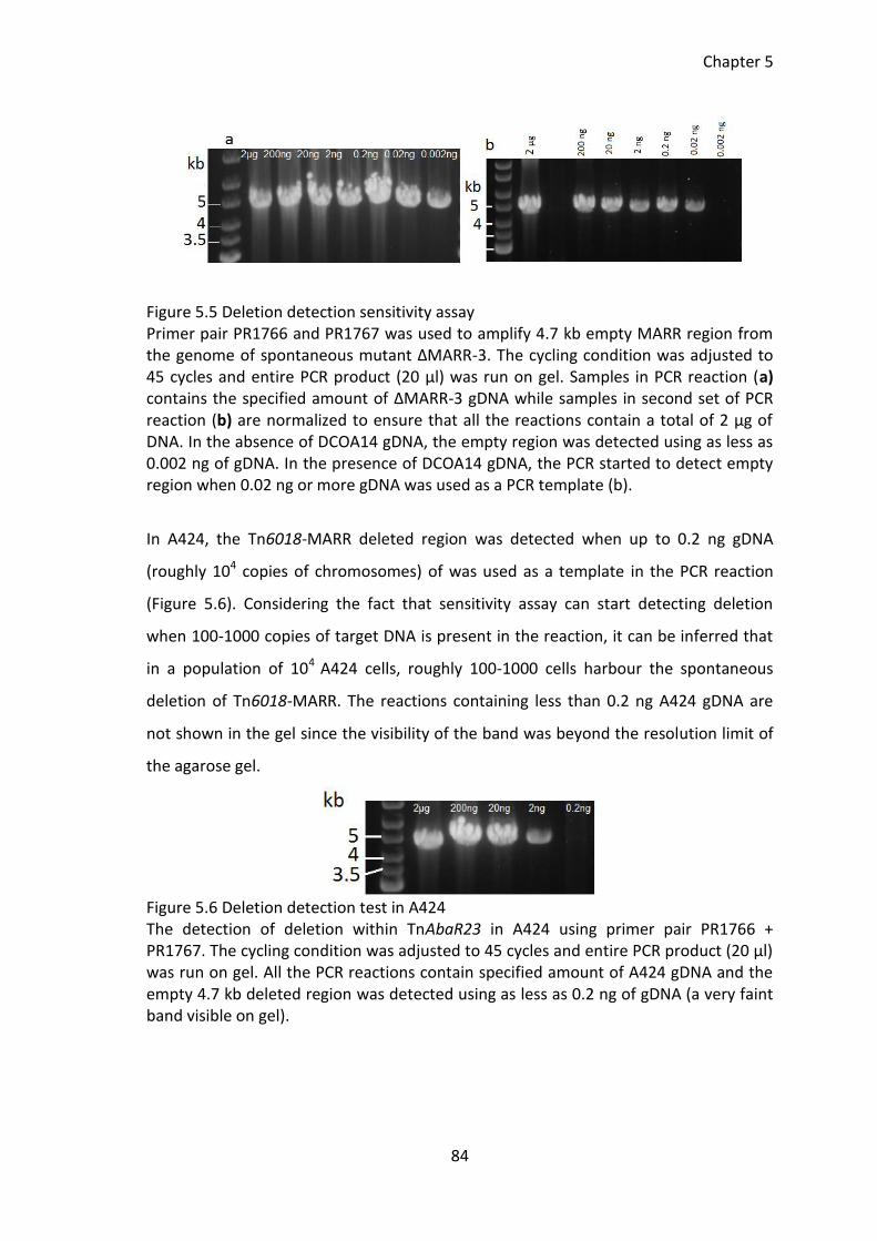
Chapter 5
84
Figure 5.5 Deletion detection sensitivity assay Primer pair PR1766 and PR1767 was used to amplify 4.7 kb empty MARR region from the genome of spontaneous mutant ΔMARR-3. The cycling condition was adjusted to 45 cycles and entire PCR product (20 µl) was run on gel. Samples in PCR reaction (a) contains the specified amount of ΔMARR-3 gDNA while samples in second set of PCR reaction (b) are normalized to ensure that all the reactions contain a total of 2 µg of DNA. In the absence of DCOA14 gDNA, the empty region was detected using as less as 0.002 ng of gDNA. In the presence of DCOA14 gDNA, the PCR started to detect empty region when 0.02 ng or more gDNA was used as a PCR template (b).
In A424, the Tn6018-MARR deleted region was detected when up to 0.2 ng gDNA
(roughly 104 copies of chromosomes) of was used as a template in the PCR reaction
(Figure 5.6). Considering the fact that sensitivity assay can start detecting deletion
when 100-1000 copies of target DNA is present in the reaction, it can be inferred that
in a population of 104 A424 cells, roughly 100-1000 cells harbour the spontaneous
deletion of Tn6018-MARR. The reactions containing less than 0.2 ng A424 gDNA are
not shown in the gel since the visibility of the band was beyond the resolution limit of
the agarose gel.
Figure 5.6 Deletion detection test in A424 The detection of deletion within TnAbaR23 in A424 using primer pair PR1766 + PR1767. The cycling condition was adjusted to 45 cycles and entire PCR product (20 µl) was run on gel. All the PCR reactions contain specified amount of A424 gDNA and the empty 4.7 kb deleted region was detected using as less as 0.2 ng of gDNA (a very faint band visible on gel).
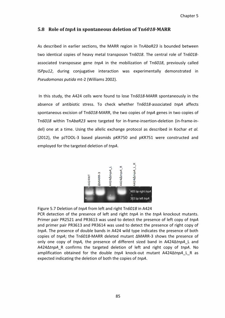
Chapter 5
85
5.8 Role of tnpA in spontaneous deletion of Tn6018-MARR
As described in earlier sections, the MARR region in TnAbaR23 is bounded between
two identical copies of heavy metal transposon Tn6018. The central role of Tn6018-
associated transposase gene tnpA in the mobilization of Tn6018, previously called
ISPpu12, during conjugative interaction was experimentally demonstrated in
Pseudomonas putida mt-2 (Williams 2002).
In this study, the A424 cells were found to lose Tn6018-MARR spontaneously in the
absence of antibiotic stress. To check whether Tn6018-associated tnpA affects
spontaneous excision of Tn6018-MARR, the two copies of tnpA genes in two copies of
Tn6018 within TnAbaR23 were targeted for in-frame-insertion-deletion (in-frame-in-
del) one at a time. Using the allelic exchange protocol as described in Kochar et al.
(2012), the pJTOOL-3 based plasmids pKR750 and pKR751 were constructed and
employed for the targeted deletion of tnpA.
Figure 5.7 Deletion of tnpA from left and right Tn6018 in A424 PCR detection of the presence of left and right tnpA in the tnpA knockout mutants. Primer pair PR2521 and PR3613 was used to detect the presence of left copy of tnpA and primer pair PR3613 and PR3614 was used to detect the presence of right copy of tnpA. The presence of double bands in A424 wild type indicates the presence of both copies of tnpA; the Tn6018-MARR deleted mutant ΔMARR-3 shows the presence of only one copy of tnpA, the presence of different sized band in A424ΔtnpA_L and A424ΔtnpA_R confirms the targeted deletion of left and right copy of tnpA. No amplification obtained for the double tnpA knock-out mutant A424ΔtnpA_L_R as expected indicating the deletion of both the copies of tnpA.
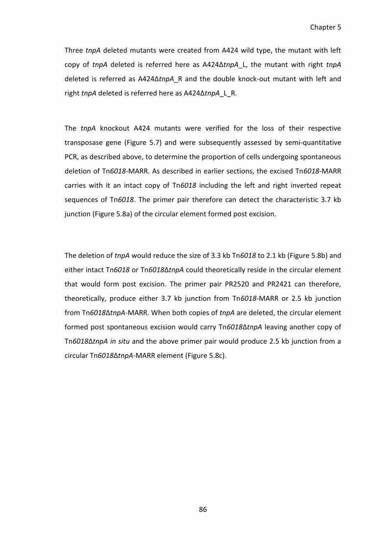
Chapter 5
86
Three tnpA deleted mutants were created from A424 wild type, the mutant with left
copy of tnpA deleted is referred here as A424ΔtnpA_L, the mutant with right tnpA
deleted is referred as A424ΔtnpA_R and the double knock-out mutant with left and
right tnpA deleted is referred here as A424ΔtnpA_L_R.
The tnpA knockout A424 mutants were verified for the loss of their respective
transposase gene (Figure 5.7) and were subsequently assessed by semi-quantitative
PCR, as described above, to determine the proportion of cells undergoing spontaneous
deletion of Tn6018-MARR. As described in earlier sections, the excised Tn6018-MARR
carries with it an intact copy of Tn6018 including the left and right inverted repeat
sequences of Tn6018. The primer pair therefore can detect the characteristic 3.7 kb
junction (Figure 5.8a) of the circular element formed post excision.
The deletion of tnpA would reduce the size of 3.3 kb Tn6018 to 2.1 kb (Figure 5.8b) and
either intact Tn6018 or Tn6018ΔtnpA could theoretically reside in the circular element
that would form post excision. The primer pair PR2520 and PR2421 can therefore,
theoretically, produce either 3.7 kb junction from Tn6018-MARR or 2.5 kb junction
from Tn6018ΔtnpA-MARR. When both copies of tnpA are deleted, the circular element
formed post spontaneous excision would carry Tn6018ΔtnpA leaving another copy of
Tn6018ΔtnpA in situ and the above primer pair would produce 2.5 kb junction from a
circular Tn6018ΔtnpA-MARR element (Figure 5.8c).
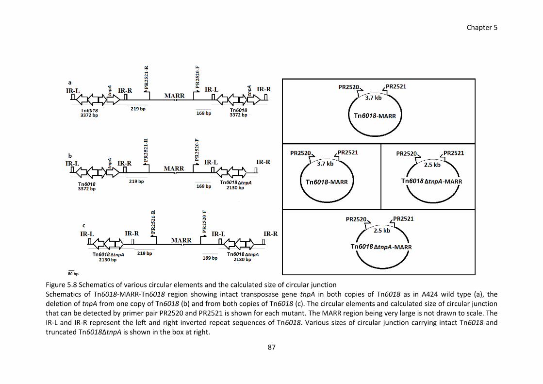
Chapter 5
87
Figure 5.8 Schematics of various circular elements and the calculated size of circular junction Schematics of Tn6018-MARR-Tn6018 region showing intact transposase gene tnpA in both copies of Tn6018 as in A424 wild type (a), the deletion of tnpA from one copy of Tn6018 (b) and from both copies of Tn6018 (c). The circular elements and calculated size of circular junction that can be detected by primer pair PR2520 and PR2521 is shown for each mutant. The MARR region being very large is not drawn to scale. The IR-L and IR-R represent the left and right inverted repeat sequences of Tn6018. Various sizes of circular junction carrying intact Tn6018 and truncated Tn6018ΔtnpA is shown in the box at right.
Chapter 5
88
5.9 Determining the proportion of tnpA knock-out mutants exhibiting
spontaneous deletion of MARR
A semi-quantitative PCR described as above was carried out to determine the
proportion of A424 mutants displaying spontaneous deletion of MARR upon the
deletion of left copy, right copy or both copies of tnpA.
In a population of 104 A424 wild type cells, it was found that around 100 - 1000 cells,
more than half of the population, were undergoing the spontaneous deletion of
Tn6018-MARR. Upon the deletion of either left or right copy of tnpA, the deletion
detection assay started detecting the presence of empty region when 107 or more
chromosomes were present in the reaction (Figure 5.9 a, b).
Figure 5.9 Deletion detection assay in various tnpA deleted mutants The cycling condition was adjusted to 45 cycles and entire PCR product (20 µl) was run on gel. The Tn6018-MARR deletion in A424ΔtnpA_L (a) and A424ΔtnpA_R (b) was detected when up to 200 ng of gDNA was used as a PCR template. PCR failed to amplify the deleted region from the double tnpA knock-out mutant A424ΔtnpA_L_R even when up to 2 µg of gDNA was used as a PCR template (c). In reaction c, A424 gDNA was used as control to show that the assay was functional.
It can therefore be inferred that in a population of 107 single tnpA knock-out mutant
cells, around 100 - 1000 cells exhibited Tn6018-MARR deletion which, a 1000 folds
decrease compared to A424 wild type carrying two intact copies of tnpA. When both
the copies of tnpA were deleted in the mutant A424ΔtnpA_L_R, a visible PCR product
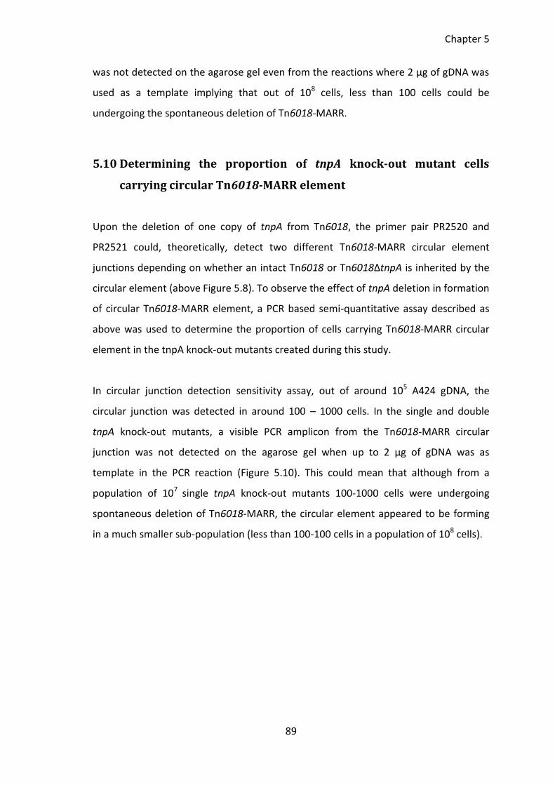
Chapter 5
89
was not detected on the agarose gel even from the reactions where 2 µg of gDNA was
used as a template implying that out of 108 cells, less than 100 cells could be
undergoing the spontaneous deletion of Tn6018-MARR.
5.10 Determining the proportion of tnpA knock-out mutant cells
carrying circular Tn6018-MARR element
Upon the deletion of one copy of tnpA from Tn6018, the primer pair PR2520 and
PR2521 could, theoretically, detect two different Tn6018-MARR circular element
junctions depending on whether an intact Tn6018 or Tn6018ΔtnpA is inherited by the
circular element (above Figure 5.8). To observe the effect of tnpA deletion in formation
of circular Tn6018-MARR element, a PCR based semi-quantitative assay described as
above was used to determine the proportion of cells carrying Tn6018-MARR circular
element in the tnpA knock-out mutants created during this study.
In circular junction detection sensitivity assay, out of around 105 A424 gDNA, the
circular junction was detected in around 100 – 1000 cells. In the single and double
tnpA knock-out mutants, a visible PCR amplicon from the Tn6018-MARR circular
junction was not detected on the agarose gel when up to 2 µg of gDNA was as
template in the PCR reaction (Figure 5.10). This could mean that although from a
population of 107 single tnpA knock-out mutants 100-1000 cells were undergoing
spontaneous deletion of Tn6018-MARR, the circular element appeared to be forming
in a much smaller sub-population (less than 100-100 cells in a population of 108 cells).
Chapter 5
90
Figure 5.10 Circular junction detection assay The junction of Tn6018-MARR circular element in A424ΔtnpA_L, A424ΔtnpA_R and A424ΔtnpA_L_R mutants was detected using the primer PR2520 and PR2521. The cycling condition was adjusted to 45 cycles and entire PCR product (20 µl) was run on gel. Indicated amount of gDNA was used in each reaction tube. A424 wild type gDNA was used as a control to show that the assay was functional. Faint band observed for A424 WT 20 ng gDNA.
5.11 Discussion and conclusion
5.11.1 Plasticity of AbaR islands
The AbaR elements in A. baumannii are considered to be acquired from various
bacteria like Pseudomonas spp., E. coli and Salmonella spp. by means of horizontal
gene transfer (Fournier et al. 2006). These elements subsequently evolved and
diversified during multiple events of insertion, deletion and recombination mostly
mediated by insertion sequence elements and transposons (Krizova et al. 2011). The
AbaR islands seem to be stably integrated in the chromosome of their host and
spontaneous deletion or transposition of the intact islands has not been reported yet
in any A. baumannii strains. In this study, for the first time, the spontaneous deletion
of a large region internal to AbaR1 and TnAbaR23 in MDR A. baumannii strains AYE and
A424 is reported. In this study, a sub-population of AYE and A424 were found to
spontaneously lose Tn6018-MARR region from their AbaR islands under the laboratory
in vitro growth conditions and in the absence of antibiotic stress. The spontaneously
excised double stranded (ds) Tn6018-MARR element was found to attain a circular
form and was subsequently lost in its entirety from the cell giving rise to a sub-
population of A424 carrying truncated TnAbaR23. Although the circular Tn6018-MARR
Chapter 5
91
was not visualized or physically detected, its presence in a sub-population of A424 cells
is inferred from the circular element junction PCR amplicon.
A total of four independent Tn6018-MARR deleted spontaneously arising mutants
were isolated from a population of A424 wild type by parallel-plate patching based on
the susceptibility of spontaneous mutants towards sulphametoxazole 40 µg/ml. The
Tn6018-MARR deleted strain screening was executed using a protocol that ensured no
prior exposure of the selected Tn6018-MARR deleted mutants to antibiotics. The
deletion of Tn6018-MARR in spontaneous mutants was verified by PCR. The PFGE band
pattern of all four spontaneous mutants appeared indistinguishable and the PFGE
profile was consistent with the deletion of Tn6018-MARR.
In each of the four Tn6018-MARR deleted mutants studied, the MARR was inferred
being excised as a double stranded circular structure that carried a single copy of
Tn6018 transposon, with the second copy of this element remaining in situ within the
now markedly truncated TnAbaR23. Importantly, PCR analysis verified that following
excision from its native site that the MARR segment did not re-integrate elsewhere in
the genomes of the four Tn6018-MARR deleted mutants investigated, but that it was
lost in entirety from these strains.
In a population of 104 A424 wild type cells, around 100 - 1000 cells were found to be
losing Tn6018-MARR spontaneously as detected by the semi-quantitative PCR assay.
However, the circular Tn6018-MARR entity was possessed by around 100 -1000 cells in
a population of 105 A424 wild type cells as detected by the semi-quantitative PCR
assay. The sub-population of A424 cells carrying circular Tn6018-MARR appears to be
10 folds less that the sub-population undergoing spontaneous deletion of Tn6018-
MARR. It is possible that, post excision, a small proportion of Tn6018-MARR
reintegrated at their native site instead of forming a circular element. A very high
proportion of A424 cells (around 4 in 1200) were found to lose Tn6018-MARR from
their TnAbaR23 as determined by the colony patching method. However, caution must
be applied before accepting that 4 in 1200 A424 cells were spontaneously losing
Chapter 5
92
Tn6018-MARR since a small population of A424 cells were screened for their
sulfamethoxazole sensitivity.
As discussed earlier, the spontaneous deletion of Tn6018-MARR was also observed in
AbaR1 island from MDR strain AYE. Given the fact that large proportion of A424 cells
exhibited spontaneous deletion of Tn6018-MARR to give rise to a sub-population of
sulfamethoxazole sensitive mutants carrying truncated TnAbaR23, it can be
hypothesized that the spontaneous deletion and loss of Tn6018-MARR observed in this
study could be a common phenomenon that might be very frequently happening in
several AbaR islands, at least in in vitro growth conditions.
5.11.2 Role of tnpA in spontaneous deletion of Tn6018-MARR from TnAbaR23
In the strain A424, the tnpA from left and right Tn6018 were deleted individually
creating a left tnpA knock-out mutant A424ΔtnpA_L, a right tnpA knock-out mutant
A424ΔtnpA_R and a double tnpA knock-out mutant A424ΔtnpA_L_R. A 1000 fold
decrease in the rate of spontaneous deletion of Tn6018-MARR was observed when
tnpA was deleted from either left or right copy of Tn6018 as evident by the semi-
quantitative PCR where upon the deletion of only one copy of tnpA, 100 – 1000 cells in
every 107 cells were found to undergo a spontaneous deletion of Tn6018-MARR
region. Thus, the pre-requisite for the high rate of spontaneous deletion of this nature
appears to be the presence of at least one copy of tnpA gene in either of the two
copies of Tn6018 from the compound transposon Tn6018-MARR-Tn6018. It is also
likely that one copy of tnpA could provide a threshold amount of Transposase to drive
the spontaneous deletion of Tn6018-MARR as observed in A424ΔtnpA_L and
A424ΔtnpA_R mutants. When tnpA was deleted from both copies of Tn6018 in A424,
the semi-quantitative PCR assay failed to detect the spontaneous deletion of Tn6018-
MARR. Intriguingly, it appears that for the excised Tn6018-MARR element to exist as a
circular entity, presence of intact copies of two tnpA gene appears to be essential.
Theoretically, the excised Tn6018-MARR could form two different sizes of ds circular
Tn6018-MARR in single tnpA knock-out mutants. Remarkably, the junction of Tn6018-
Chapter 5
93
MARR circular element was not detected in all three tnpA deleted mutants. Failure to
detect the circular junction A424ΔtnpA_L and A424ΔtnpA_R shows that despite the
occurrence of spontaneous deletion within TnAbaR23, the circular Tn6018-MARR
element could not form in these single tnpA knock-out mutants. It is likely that in the
absence of two copies of functional tnpA, the Tn6018-MARR failed to attain a circular
state post excision and re-integrated into its native site. Equally, it is possible that the
region available for the homologous recombination were shorter in single tnpA and
double tnpA knock-out mutants which results in the reduction of spontaneous excision
of Tn6018-MARR. This could explain why the spontaneous deletion was detected but
the circular element was not detected in single tnpA knockout mutants. It is also likely
that two copies of intact tnpA were prerequisite for the Tn6018-MARR to exist as a
circular entity. The presence of Tn6018-MARR as a circular entity was inferred on the
basis of detection of circular element junction by PCR and, in this instance, there is no
physical evidence of the presence of different sized Tn6018-MARR circular element in
tnpA knock-out mutants.
Study on the movement of the antibiotic resistance genes flanked by two directly
oriented copes of IS26 (Harmer et al. 2014), in a recA negative strain of E. coli showed
that one copy of IS26 together with the antibiotic resistance genes forms a mobile
transposable unit (the authors called it “translocatable unit”). The authors went on
explaining that this translocatable unit recognizes IS26 as a target and a high frequency
of recombination between translocatable units at its target occurs when both of the
IS26 possess active transposase Tnp26. During this study, the exact mechanism of
Tn6018-MARR excision, its target site selection and its mode of transposition (if there
is any) could not be investigated due to time constraints but it is possible that Tn6018-
MARR entity can behaves like the IS26-like translocatable unit in-terms of target site
selection and mode of transposition. In this study, a 1000 fold decrease in spontaneous
excision of Tn6018-MARR was observed when one copy of Tn6018 transposase tnpA
was knocked-out and, intriguingly, the spontaneous excision was not detected when
both the copies of tnpA were knocked-out. In consistent with Harmer et al. (2014)
findings, the importance of active transposase for the Tn6018-MARR excision has been
clearly demonstrated in this study. Importantly, the circularization of Tn6018-MARR
Chapter 5
94
entity was not detected by PCR despite lower rate of Tn6018-MARR excision in
mutants with one copy of tnpA knocked-out. It is likely that the length of region
available for homologous recombination is also important for the high rate of excision
and circularization of Tn6018-MARR and that a functional Tn6018 might not be
required for the excision of Tn6018-MARR.
This study demonstrates the precise deletion and circularization of Tn6018-MARR from
TnAbaR23 in strain A424. The circularized Tn6018-MARR was not obtained as a
physical entity in this study but it would be fascinating to study its transposition ability
and efficiency in order to better understand the contribution of these elements in the
development of traits and characteristics that are vital for the survival of the strain in
the environment. In the next chapter, the Tn6018-MARR deleted spontaneous mutants
will be further analysed for their contribution on the phenotype of A424 strain.
95
6 Contribution of TnAbaR23 island on the phenotype of Acinetobacter baumannii strain
A424
Chapter 6
96
6.1 Background
In the introduction chapter, I have briefly described the study conducted by Kochar et
al. (2012) and their conclusions regarding the contribution of TnAbaR23 on the
antibiotic resistance phenotype of A424. The author reported that despite having the
wild type gyrA and parC genes the TnAbaR23 deleted mutant DCO174 was found to
show an enhanced resistance to ciprofloxacin with MIC 8 fold higher than its parent
A424. Importantly, the author hypothesized that the unusual ciprofloxacin resistance
exhibited by DCO174 could be due to either the deletion of TnAbaR23 or a cryptic
secondary mutation in the efflux gene.
Importantly, AbaR islands were considered to contribute minimally towards the overall
antibiotic resistance phenotype of the host strain (Krizova et al. 2011). To validate this
claim, the published literatures were searched to compile data (Table 6.1) on the
antibiotic resistance conferring genes and determinants on AbaR island and the
antibiotic resistance phenotype of the associated host strain.
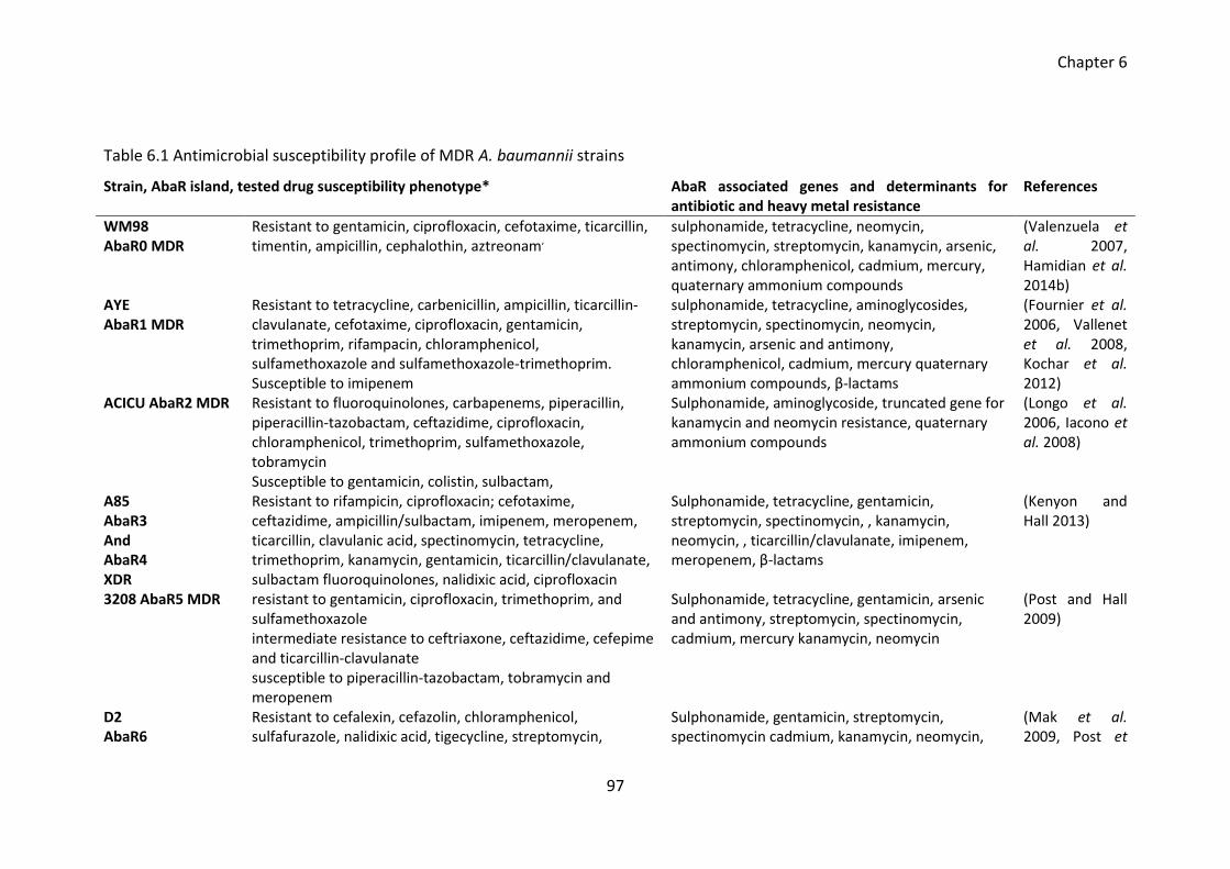
Chapter 6
97
Table 6.1 Antimicrobial susceptibility profile of MDR A. baumannii strains
Strain, AbaR island, tested drug susceptibility phenotype* AbaR associated genes and determinants for antibiotic and heavy metal resistance
References
WM98 AbaR0 MDR
Resistant to gentamicin, ciprofloxacin, cefotaxime, ticarcillin, timentin, ampicillin, cephalothin, aztreonam,
sulphonamide, tetracycline, neomycin, spectinomycin, streptomycin, kanamycin, arsenic, antimony, chloramphenicol, cadmium, mercury, quaternary ammonium compounds
(Valenzuela et al. 2007, Hamidian et al. 2014b)
AYE AbaR1 MDR
Resistant to tetracycline, carbenicillin, ampicillin, ticarcillin-clavulanate, cefotaxime, ciprofloxacin, gentamicin, trimethoprim, rifampacin, chloramphenicol, sulfamethoxazole and sulfamethoxazole-trimethoprim. Susceptible to imipenem
sulphonamide, tetracycline, aminoglycosides, streptomycin, spectinomycin, neomycin, kanamycin, arsenic and antimony, chloramphenicol, cadmium, mercury quaternary ammonium compounds, β-lactams
(Fournier et al. 2006, Vallenet et al. 2008, Kochar et al. 2012)
ACICU AbaR2 MDR Resistant to fluoroquinolones, carbapenems, piperacillin, piperacillin-tazobactam, ceftazidime, ciprofloxacin, chloramphenicol, trimethoprim, sulfamethoxazole, tobramycin Susceptible to gentamicin, colistin, sulbactam,
Sulphonamide, aminoglycoside, truncated gene for kanamycin and neomycin resistance, quaternary ammonium compounds
(Longo et al. 2006, Iacono et al. 2008)
A85 AbaR3 And AbaR4 XDR
Resistant to rifampicin, ciprofloxacin; cefotaxime, ceftazidime, ampicillin/sulbactam, imipenem, meropenem, ticarcillin, clavulanic acid, spectinomycin, tetracycline, trimethoprim, kanamycin, gentamicin, ticarcillin/clavulanate, sulbactam fluoroquinolones, nalidixic acid, ciprofloxacin
Sulphonamide, tetracycline, gentamicin, streptomycin, spectinomycin, , kanamycin, neomycin, , ticarcillin/clavulanate, imipenem, meropenem, β-lactams
(Kenyon and Hall 2013)
3208 AbaR5 MDR resistant to gentamicin, ciprofloxacin, trimethoprim, and sulfamethoxazole intermediate resistance to ceftriaxone, ceftazidime, cefepime and ticarcillin-clavulanate susceptible to piperacillin-tazobactam, tobramycin and meropenem
Sulphonamide, tetracycline, gentamicin, arsenic and antimony, streptomycin, spectinomycin, cadmium, mercury kanamycin, neomycin
(Post and Hall 2009)
D2 AbaR6
Resistant to cefalexin, cefazolin, chloramphenicol, sulfafurazole, nalidixic acid, tigecycline, streptomycin,
Sulphonamide, gentamicin, streptomycin, spectinomycin cadmium, kanamycin, neomycin,
(Mak et al. 2009, Post et
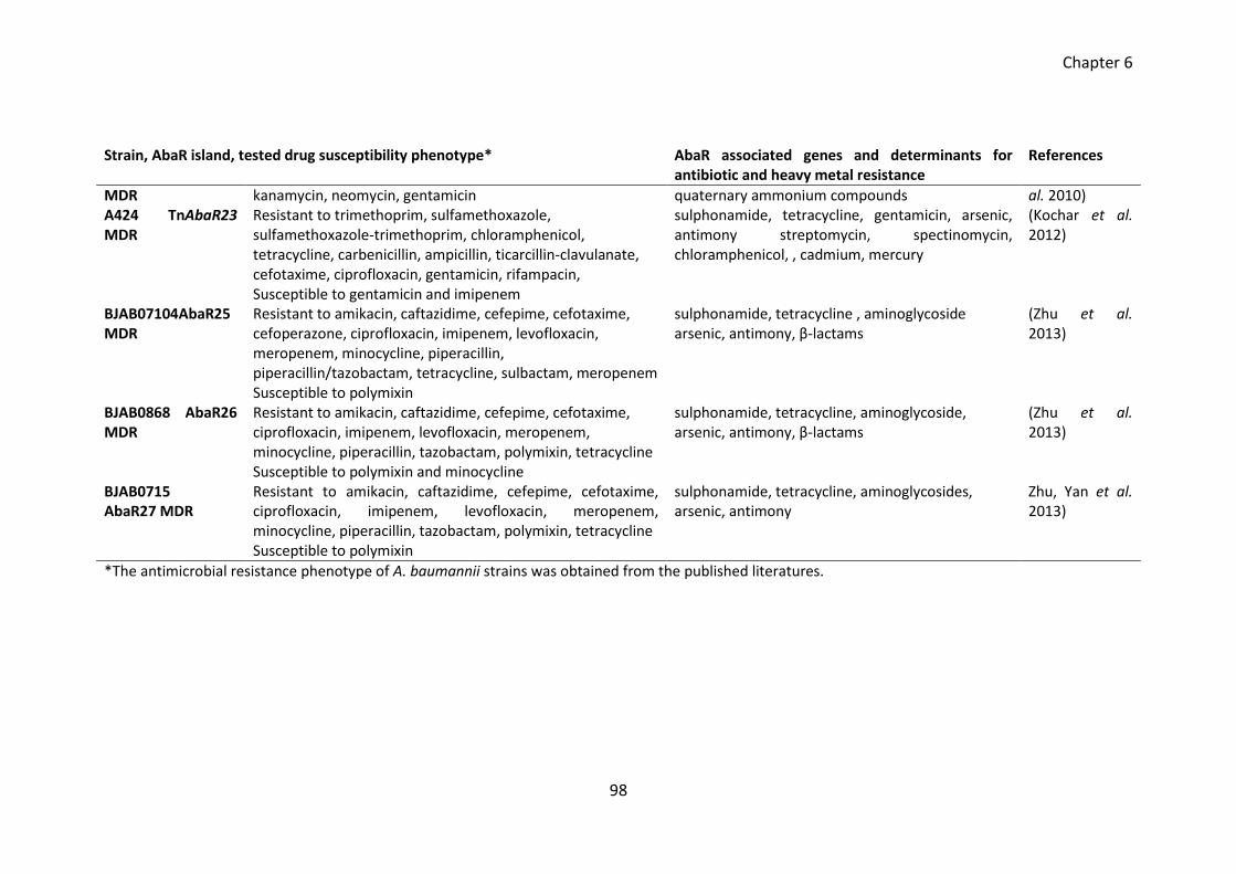
Chapter 6
98
Strain, AbaR island, tested drug susceptibility phenotype* AbaR associated genes and determinants for antibiotic and heavy metal resistance
References
MDR kanamycin, neomycin, gentamicin quaternary ammonium compounds al. 2010) A424 TnAbaR23 MDR
Resistant to trimethoprim, sulfamethoxazole, sulfamethoxazole-trimethoprim, chloramphenicol, tetracycline, carbenicillin, ampicillin, ticarcillin-clavulanate, cefotaxime, ciprofloxacin, gentamicin, rifampacin, Susceptible to gentamicin and imipenem
sulphonamide, tetracycline, gentamicin, arsenic, antimony streptomycin, spectinomycin, chloramphenicol, , cadmium, mercury
(Kochar et al. 2012)
BJAB07104AbaR25 MDR
Resistant to amikacin, caftazidime, cefepime, cefotaxime, cefoperazone, ciprofloxacin, imipenem, levofloxacin, meropenem, minocycline, piperacillin, piperacillin/tazobactam, tetracycline, sulbactam, meropenem Susceptible to polymixin
sulphonamide, tetracycline , aminoglycoside arsenic, antimony, β-lactams
(Zhu et al. 2013)
BJAB0868 AbaR26 MDR
Resistant to amikacin, caftazidime, cefepime, cefotaxime, ciprofloxacin, imipenem, levofloxacin, meropenem, minocycline, piperacillin, tazobactam, polymixin, tetracycline Susceptible to polymixin and minocycline
sulphonamide, tetracycline, aminoglycoside, arsenic, antimony, β-lactams
(Zhu et al. 2013)
BJAB0715 AbaR27 MDR
Resistant to amikacin, caftazidime, cefepime, cefotaxime, ciprofloxacin, imipenem, levofloxacin, meropenem, minocycline, piperacillin, tazobactam, polymixin, tetracycline Susceptible to polymixin
sulphonamide, tetracycline, aminoglycosides, arsenic, antimony
Zhu, Yan et al. 2013)
*The antimicrobial resistance phenotype of A. baumannii strains was obtained from the published literatures.
Chapter 6
99
The maintenance and preservation of AbaR islands solely aiming to enhance the
resistance towards few antimicrobial agents that are not currently used in treatment
seems to be an inadequate explanation given the fact that AbaR associated resistance
determinants played a minimum role towards the overall multi-drug-resistance
phenotype of the host strain (Table 6.1). Therefore it is possible that along with drug
resistance phenotypes, AbaR islands in MDR A. baumannii strains are associated with
other traits that are vital for the propagation of A. baumannii.
In this chapter, I am intending to investigate the phenotypes associated with
TnAbaR23 in strain A424. Both of the TnAbaR23 deleted mutants DCO163 and DCO174
reported by Kochar et al. (2012) were unsuitable to use in this phenotypic comparison
study due to their unintended genomic rearrangements (DCO163) and unexpected
resistance phenotype (DCO174). Therefore I will be creating new independent
TnAbaR23 deletion mutants with expected genotype and compare the fitness and
virulence phenotypes of A424 wild type and TnAbaR23 deleted mutants using various
in vitro and in vivo assays.
Here, I will also investigate the association of TnAbaR23 deletion on enhanced
ciprofloxacin resistance in new independent TnAbaR23 deleted mutants. The
comparative sequence analysis of A424 and DCO174 (Crosatti et al. unpublished data
2013) revealed a substitution of nucleotide G to A at 2601753 position on the
chromosome of DCO174. This single nucleotide polymorphism (SNP) was reported to
lie within the adeS gene, regulator of AdeABC efflux pump, and it appeared to be solely
responsible for the elevated ciprofloxacin resistance in DCO174 (Crosatti et al.
unpublished data 2013). In this study, the impact of this adeS-associated SNP (here
referred as adeSG178A) on the ciprofloxacin resistance of the A424 strain will be
investigated. Experiments will be designed to swap the ades allele (here referred as
adeSWT) present in various A424 strain background with mutated adeSG178A. The
phenotypes of the strains carrying mutated adeSG178A and wild type adeSWT will be
subsequently compared in various in vitro assays.
Chapter 6
100
6.2 Aims and objectives
Experiments were designed to create the TnAbaR23 deleted mutants for phenotypic
comparison with parent A424 wild type. The phenotypes of Tn6018-MARR deleted
spontaneous mutants lacking Tn6018-MARR (reported in chapter 4) was also studied in
various in vivo and in vitro assays. The experiments were designed and conducted in
order to-
delete TnAbaR23 island en bloc and create markerless mutant
complement the TnAbaR23 deleted mutants with 4 – 6 kb blocks of Tn6018-
MARR fragments
assess the antibiotic resistance profile of A424 wild type and its various
mutants
compare the fitness of A424 wild type and its various mutants in an in vitro
head-to-head competition assay
compare the virulence of wild type and its various mutants in Galleria
mellonella killing assay
replace the adeSWT with adeSG178A in various A424 strain background
assess the antibiotic resistance profile of the strains carrying mutant adeSG178A
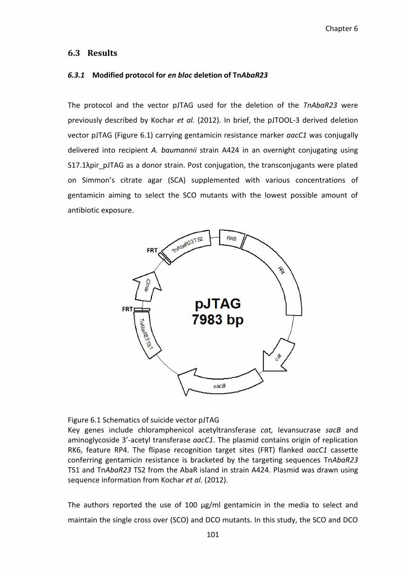
Chapter 6
101
6.3 Results
6.3.1 Modified protocol for en bloc deletion of TnAbaR23
The protocol and the vector pJTAG used for the deletion of the TnAbaR23 were
previously described by Kochar et al. (2012). In brief, the pJTOOL-3 derived deletion
vector pJTAG (Figure 6.1) carrying gentamicin resistance marker aacC1 was conjugally
delivered into recipient A. baumannii strain A424 in an overnight conjugating using
S17.1λpir_pJTAG as a donor strain. Post conjugation, the transconjugants were plated
on Simmon’s citrate agar (SCA) supplemented with various concentrations of
gentamicin aiming to select the SCO mutants with the lowest possible amount of
antibiotic exposure.
Figure 6.1 Schematics of suicide vector pJTAG Key genes include chloramphenicol acetyltransferase cat, levansucrase sacB and aminoglycoside 3’-acetyl transferase aacC1. The plasmid contains origin of replication RK6, feature RP4. The flipase recognition target sites (FRT) flanked aacC1 cassette conferring gentamicin resistance is bracketed by the targeting sequences TnAbaR23 TS1 and TnAbaR23 TS2 from the AbaR island in strain A424. Plasmid was drawn using sequence information from Kochar et al. (2012).
The authors reported the use of 100 µg/ml gentamicin in the media to select and
maintain the single cross over (SCO) and DCO mutants. In this study, the SCO and DCO
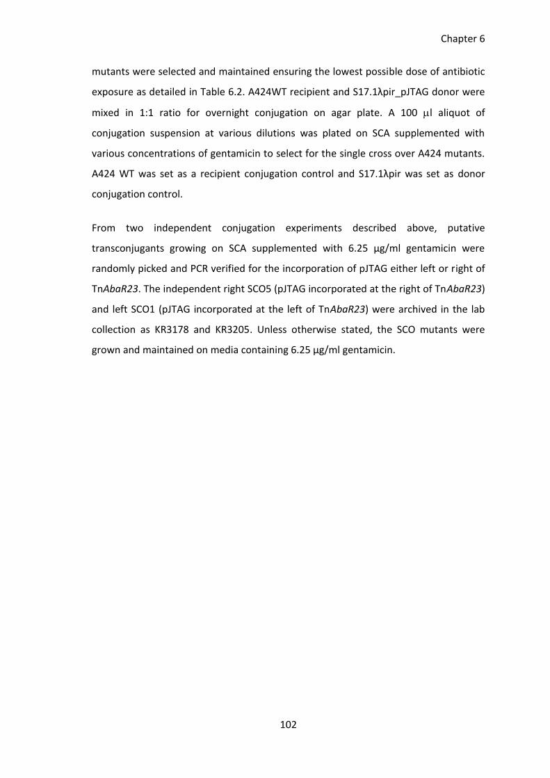
Chapter 6
102
mutants were selected and maintained ensuring the lowest possible dose of antibiotic
exposure as detailed in Table 6.2. A424WT recipient and S17.1λpir_pJTAG donor were
mixed in 1:1 ratio for overnight conjugation on agar plate. A 100 l aliquot of
conjugation suspension at various dilutions was plated on SCA supplemented with
various concentrations of gentamicin to select for the single cross over A424 mutants.
A424 WT was set as a recipient conjugation control and S17.1λpir was set as donor
conjugation control.
From two independent conjugation experiments described above, putative
transconjugants growing on SCA supplemented with 6.25 µg/ml gentamicin were
randomly picked and PCR verified for the incorporation of pJTAG either left or right of
TnAbaR23. The independent right SCO5 (pJTAG incorporated at the right of TnAbaR23)
and left SCO1 (pJTAG incorporated at the left of TnAbaR23) were archived in the lab
collection as KR3178 and KR3205. Unless otherwise stated, the SCO mutants were
grown and maintained on media containing 6.25 µg/ml gentamicin.
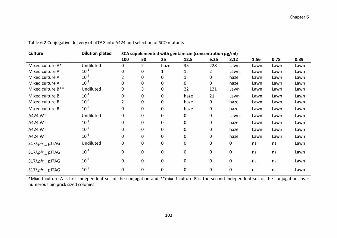
Chapter 6
103
Table 6.2 Conjugative delivery of pJTAG into A424 and selection of SCO mutants
Culture Dilution plated SCA supplemented with gentamicin (concentration g/ml) 100 50 25 12.5 6.25 3.12 1.56 0.78 0.39
Mixed culture A* Undiluted 0 2 haze 35 228 Lawn Lawn Lawn Lawn Mixed culture A 10-1 0 0 1 1 2 Lawn Lawn Lawn Lawn Mixed culture A 10-2 2 0 0 1 0 haze Lawn Lawn Lawn Mixed culture A 10-3 0 0 0 0 0 haze Lawn Lawn Lawn Mixed culture B** Undiluted 0 3 0 22 121 Lawn Lawn Lawn Lawn
Mixed culture B 10-1 0 0 0 haze 21 Lawn Lawn Lawn Lawn Mixed culture B 10-2 2 0 0 haze 0 haze Lawn Lawn Lawn
Mixed culture B 10-3 0 0 0 haze 0 haze Lawn Lawn Lawn
A424 WT Undiluted 0 0 0 0 0 Lawn Lawn Lawn Lawn
A424 WT 10-1 0 0 0 0 0 haze Lawn Lawn Lawn
A424 WT 10-2 0 0 0 0 0 haze Lawn Lawn Lawn
A424 WT 10-3 0 0 0 0 0 haze Lawn Lawn Lawn
S17pir _ pJTAG Undiluted 0 0 0 0 0 0 ns ns Lawn
S17pir _ pJTAG 10-1 0 0 0 0 0 0 ns ns Lawn
S17pir _ pJTAG 10-2 0 0 0 0 0 0 ns ns Lawn
S17pir _ pJTAG 10-3 0 0 0 0 0 0 ns ns Lawn
*Mixed culture A is first independent set of the conjugation and **mixed culture B is the second independent set of the conjugation. ns = numerous pin prick sized colonies
Chapter 6
104
6.3.2 Creation of new independent TnAbaR23 deleted mutants
The independent SCO mutants KR3178 and KR3205 were used to create independent
TnAbaR23 deleted mutants during sucrose counter-selection. In brief, a suspension of
SCO mutants grown on antibiotic free media were plated on LA plate supplemented
with 6 % sucrose. The suicide vector pJTAG carries on its backbone a sucrase gene
called sacB. In the presence of sucrose, an intermediate product called levansucrase is
formed which is toxic to the cells. In order to survive in the media containing sucrose,
the single cross-over mutant must get rid of pJTAG integrated on its chromosome. This
provokes a second homologous recombination event between the two copies of
TnAbaR23 targeting sequence 1 [TS1] or TnAbaR23 targeting sequence 2 [TS2].
During the second homologous recombination event, the plasmid pJTAG would be lost
from the chromosome and depending on where the homologous recombination has
occurred [TS1 or TS2], the colonies growing on sucrose plate could be either the
mutants reverted to wild type [only pJTAG lost from chromosome] or the TnAbaR23
island deleted mutants [pJTAG and TnAbaR23 lost from the cell]. The sucrose counter-
selection was carried out using two independent SCO mutants. A 1000 fold decrease in
the number of colonies on sucrose plates was observed during counter-selection
(Table 6.3). The gentamicin resistant putative DCO mutants were validated for the loss
of TnAbaR23. Independent TnAbaR23 deleted mutants were archived in the laboratory
strain collection.
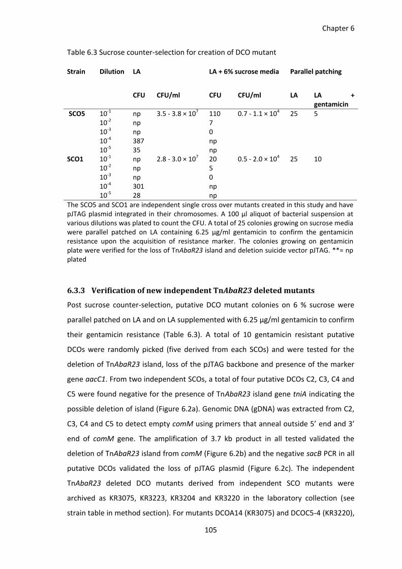
Chapter 6
105
Table 6.3 Sucrose counter-selection for creation of DCO mutant
6.3.3 Verification of new independent TnAbaR23 deleted mutants
Post sucrose counter-selection, putative DCO mutant colonies on 6 % sucrose were
parallel patched on LA and on LA supplemented with 6.25 μg/ml gentamicin to confirm
their gentamicin resistance (Table 6.3). A total of 10 gentamicin resistant putative
DCOs were randomly picked (five derived from each SCOs) and were tested for the
deletion of TnAbaR23 island, loss of the pJTAG backbone and presence of the marker
gene aacC1. From two independent SCOs, a total of four putative DCOs C2, C3, C4 and
C5 were found negative for the presence of TnAbaR23 island gene tniA indicating the
possible deletion of island (Figure 6.2a). Genomic DNA (gDNA) was extracted from C2,
C3, C4 and C5 to detect empty comM using primers that anneal outside 5’ end and 3’
end of comM gene. The amplification of 3.7 kb product in all tested validated the
deletion of TnAbaR23 island from comM (Figure 6.2b) and the negative sacB PCR in all
putative DCOs validated the loss of pJTAG plasmid (Figure 6.2c). The independent
TnAbaR23 deleted DCO mutants derived from independent SCO mutants were
archived as KR3075, KR3223, KR3204 and KR3220 in the laboratory collection (see
strain table in method section). For mutants DCOA14 (KR3075) and DCOC5-4 (KR3220),
Strain Dilution LA LA + 6% sucrose media Parallel patching
CFU CFU/ml CFU CFU/ml LA LA + gentamicin
SCO5 10-1 np 3.5 - 3.8 × 10⁷ 110 0.7 - 1.1 × 10⁴ 25 5 10-2 np 7 10-3 np 0 10-4 387 np 10-5 35 np
SCO1 10-1 np 2.8 - 3.0 × 10⁷ 20 0.5 - 2.0 × 10⁴ 25 10 10-2 np 5
10-3 np 0 10-4 301 np 10-5 28 np
The SCO5 and SCO1 are independent single cross over mutants created in this study and have pJTAG plasmid integrated in their chromosomes. A 100 µl aliquot of bacterial suspension at various dilutions was plated to count the CFU. A total of 25 colonies growing on sucrose media were parallel patched on LA containing 6.25 µg/ml gentamicin to confirm the gentamicin resistance upon the acquisition of resistance marker. The colonies growing on gentamicin plate were verified for the loss of TnAbaR23 island and deletion suicide vector pJTAG. **= np plated
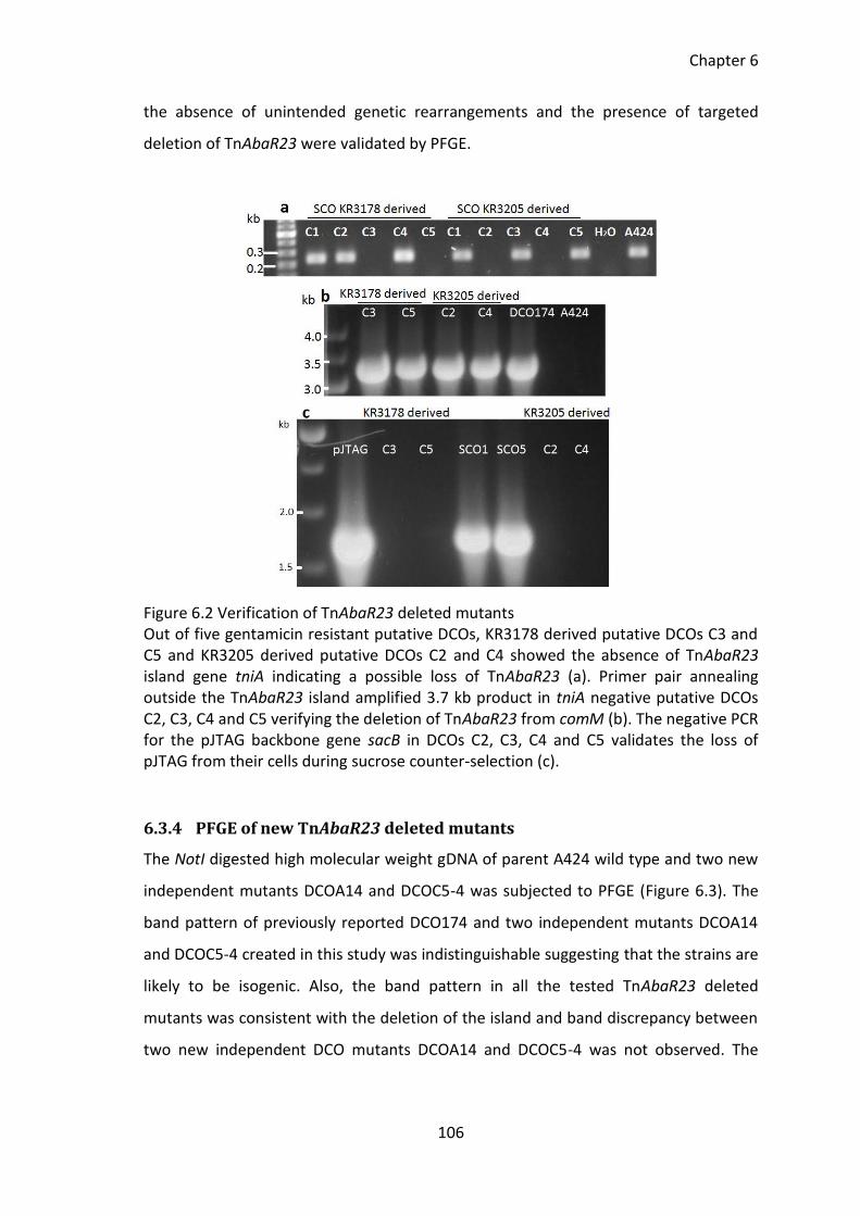
Chapter 6
106
the absence of unintended genetic rearrangements and the presence of targeted
deletion of TnAbaR23 were validated by PFGE.
Figure 6.2 Verification of TnAbaR23 deleted mutants Out of five gentamicin resistant putative DCOs, KR3178 derived putative DCOs C3 and C5 and KR3205 derived putative DCOs C2 and C4 showed the absence of TnAbaR23 island gene tniA indicating a possible loss of TnAbaR23 (a). Primer pair annealing outside the TnAbaR23 island amplified 3.7 kb product in tniA negative putative DCOs C2, C3, C4 and C5 verifying the deletion of TnAbaR23 from comM (b). The negative PCR for the pJTAG backbone gene sacB in DCOs C2, C3, C4 and C5 validates the loss of pJTAG from their cells during sucrose counter-selection (c).
6.3.4 PFGE of new TnAbaR23 deleted mutants
The NotI digested high molecular weight gDNA of parent A424 wild type and two new
independent mutants DCOA14 and DCOC5-4 was subjected to PFGE (Figure 6.3). The
band pattern of previously reported DCO174 and two independent mutants DCOA14
and DCOC5-4 created in this study was indistinguishable suggesting that the strains are
likely to be isogenic. Also, the band pattern in all the tested TnAbaR23 deleted
mutants was consistent with the deletion of the island and band discrepancy between
two new independent DCO mutants DCOA14 and DCOC5-4 was not observed. The
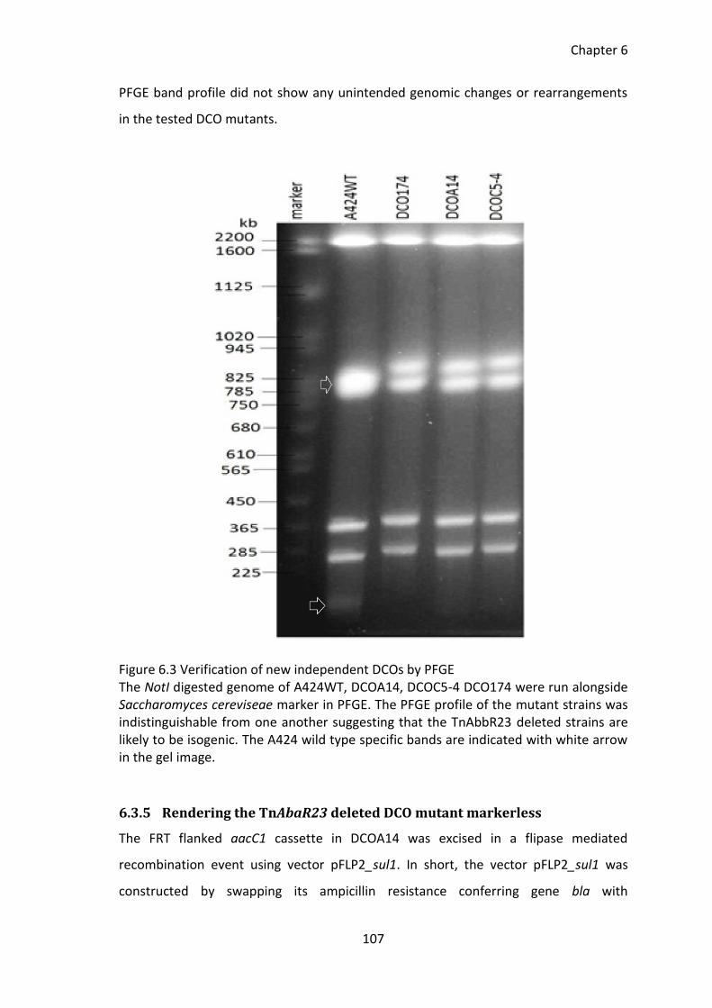
Chapter 6
107
PFGE band profile did not show any unintended genomic changes or rearrangements
in the tested DCO mutants.
Figure 6.3 Verification of new independent DCOs by PFGE The NotI digested genome of A424WT, DCOA14, DCOC5-4 DCO174 were run alongside Saccharomyces cereviseae marker in PFGE. The PFGE profile of the mutant strains was indistinguishable from one another suggesting that the TnAbbR23 deleted strains are likely to be isogenic. The A424 wild type specific bands are indicated with white arrow in the gel image.
6.3.5 Rendering the TnAbaR23 deleted DCO mutant markerless
The FRT flanked aacC1 cassette in DCOA14 was excised in a flipase mediated
recombination event using vector pFLP2_sul1. In short, the vector pFLP2_sul1 was
constructed by swapping its ampicillin resistance conferring gene bla with
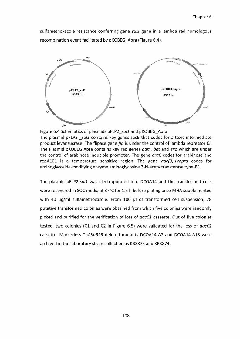
Chapter 6
108
sulfamethoxazole resistance conferring gene sul1 gene in a lambda red homologous
recombination event facilitated by pKOBEG_Apra (Figure 6.4).
Figure 6.4 Schematics of plasmids pFLP2_sul1 and pKOBEG_Apra The plasmid pFLP2 _sul1 contains key genes sacB that codes for a toxic intermediate product levansucrase. The flipase gene flp is under the control of lambda repressor CI. The Plasmid pKOBEG Apra contains key red genes gam, bet and exo which are under the control of arabinose inducible promoter. The gene araC codes for arabinose and repA101 is a temperature sensitive region. The gene aac(3)-IVapra codes for aminoglycoside-modifying enzyme aminoglycoside 3-N-acetyltransferase type-IV.
The plasmid pFLP2-sul1 was electroporated into DCOA14 and the transformed cells
were recovered in SOC media at 37°C for 1.5 h before plating onto MHA supplemented
with 40 µg/ml sulfamethoxazole. From 100 µl of transformed cell suspension, 78
putative transformed colonies were obtained from which five colonies were randomly
picked and purified for the verification of loss of aacC1 cassette. Out of five colonies
tested, two colonies (C1 and C2 in Figure 6.5) were validated for the loss of aacC1
cassette. Markerless TnAbaR23 deleted mutants DCOA14-Δ7 and DCOA14-Δ18 were
archived in the laboratory strain collection as KR3873 and KR3874.
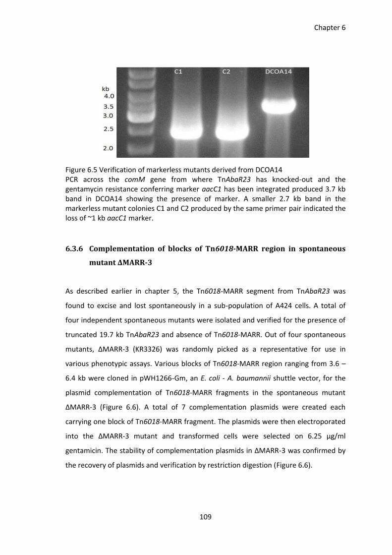
Chapter 6
109
Figure 6.5 Verification of markerless mutants derived from DCOA14 PCR across the comM gene from where TnAbaR23 has knocked-out and the gentamycin resistance conferring marker aacC1 has been integrated produced 3.7 kb band in DCOA14 showing the presence of marker. A smaller 2.7 kb band in the markerless mutant colonies C1 and C2 produced by the same primer pair indicated the loss of ~1 kb aacC1 marker.
6.3.6 Complementation of blocks of Tn6018-MARR region in spontaneous
mutant ΔMARR-3
As described earlier in chapter 5, the Tn6018-MARR segment from TnAbaR23 was
found to excise and lost spontaneously in a sub-population of A424 cells. A total of
four independent spontaneous mutants were isolated and verified for the presence of
truncated 19.7 kb TnAbaR23 and absence of Tn6018-MARR. Out of four spontaneous
mutants, ΔMARR-3 (KR3326) was randomly picked as a representative for use in
various phenotypic assays. Various blocks of Tn6018-MARR region ranging from 3.6 –
6.4 kb were cloned in pWH1266-Gm, an E. coli - A. baumannii shuttle vector, for the
plasmid complementation of Tn6018-MARR fragments in the spontaneous mutant
ΔMARR-3 (Figure 6.6). A total of 7 complementation plasmids were created each
carrying one block of Tn6018-MARR fragment. The plasmids were then electroporated
into the ΔMARR-3 mutant and transformed cells were selected on 6.25 µg/ml
gentamicin. The stability of complementation plasmids in ΔMARR-3 was confirmed by
the recovery of plasmids and verification by restriction digestion (Figure 6.6).
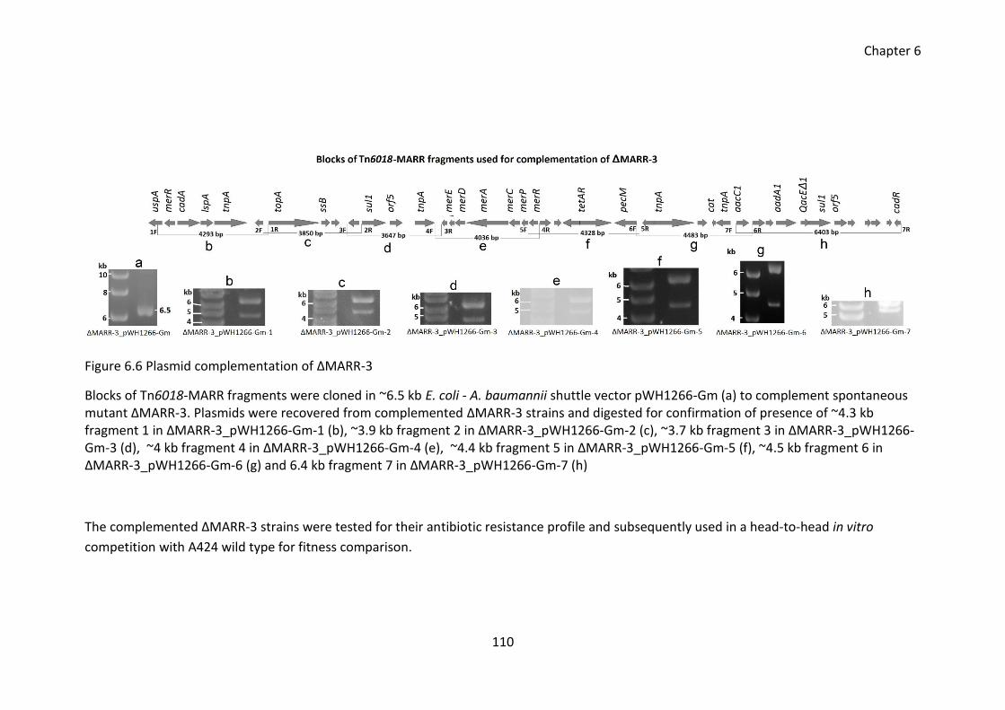
Chapter 6
110
Figure 6.6 Plasmid complementation of ΔMARR-3
Blocks of Tn6018-MARR fragments were cloned in ~6.5 kb E. coli - A. baumannii shuttle vector pWH1266-Gm (a) to complement spontaneous mutant ΔMARR-3. Plasmids were recovered from complemented ΔMARR-3 strains and digested for confirmation of presence of ~4.3 kb fragment 1 in ΔMARR-3_pWH1266-Gm-1 (b), ~3.9 kb fragment 2 in ΔMARR-3_pWH1266-Gm-2 (c), ~3.7 kb fragment 3 in ΔMARR-3_pWH1266-Gm-3 (d), ~4 kb fragment 4 in ΔMARR-3_pWH1266-Gm-4 (e), ~4.4 kb fragment 5 in ΔMARR-3_pWH1266-Gm-5 (f), ~4.5 kb fragment 6 in ΔMARR-3_pWH1266-Gm-6 (g) and 6.4 kb fragment 7 in ΔMARR-3_pWH1266-Gm-7 (h)
The complemented ΔMARR-3 strains were tested for their antibiotic resistance profile and subsequently used in a head-to-head in vitro
competition with A424 wild type for fitness comparison.
Chapter 6
111
6.3.7 Effect of TnAbaR23 on the phenotype of A424
Antibiotic susceptibility profile of A424 wild type, TnAbaR23 deleted mutants and
Tn6018-MARR complemented strains was assessed for 15 different antibiotics (Table
6.4) using antibiotic discs and e-test strips according to the British Society for
Antimicrobial Chemotherapy (BSAC) methods for antimicrobial susceptibility testing
guideline version 12,May 2013 (Wootton 2013). For the disc test, the diameter of zone
of clearance was measured to the nearest 0.5 mm, values in bold represent the MIC
(shown in µg/ml, tested by using Etest strips). The experiment was repeated three
times independently. Reference strain NCTC12241 was used as BSAC reference control
(Wootton 2013). BSAC version 2013 guideline on zone diameter breakpoints are as
follows: for IMP, resistant (R) ≤ 13, intermediate (I) 14 - 24 and susceptible (S) ≥ 25; for
AMK, resistant (R) ≤ 18, intermediate (I) 19-20 and susceptible (S) ≥ 21; for gentamicin,
resistant (R) ≤ 19 and susceptible (S) ≥ 20; for ciprofloxacin, resistant (R) ≤ 20 and
susceptible (S) ≥ 21. BSAC guideline on MIC breakpoint for Acinetobacter species: for
gentamicin, resistant (R) > 4 and susceptible (S) ≤ 4; for amikacin, resistant (R) > 16,
intermediate (I) 16 and susceptible (S) ≤ 8; for imipenem, resistant (R) > 8,
intermediate (I) 4-8 and susceptible (S) ≤ 2 and for ciprofloxacin, resistant (R) > 1 and
susceptible (S) ≤ 1.
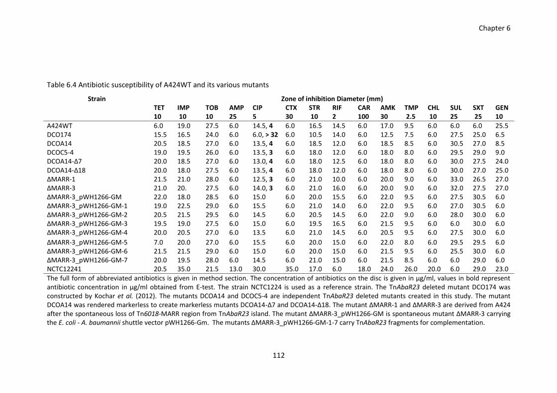
Chapter 6
112
Table 6.4 Antibiotic susceptibility of A424WT and its various mutants
Strain Zone of inhibition Diameter (mm) TET 10
IMP 10
TOB 10
AMP 25
CIP 5
CTX 30
STR 10
RIF 2
CAR 100
AMK 30
TMP 2.5
CHL 10
SUL 25
SXT 25
GEN 10
A424WT 6.0 19.0 27.5 6.0 14.5, 4 6.0 16.5 14.5 6.0 17.0 9.5 6.0 6.0 6.0 25.5 DCO174 15.5 16.5 24.0 6.0 6.0, > 32 6.0 10.5 14.0 6.0 12.5 7.5 6.0 27.5 25.0 6.5 DCOA14 20.5 18.5 27.0 6.0 13.5, 4 6.0 18.5 12.0 6.0 18.5 8.5 6.0 30.5 27.0 8.5 DCOC5-4 19.0 19.5 26.0 6.0 13.5, 3 6.0 18.0 12.0 6.0 18.0 8.0 6.0 29.5 29.0 9.0 DCOA14-Δ7 20.0 18.5 27.0 6.0 13.0, 4 6.0 18.0 12.5 6.0 18.0 8.0 6.0 30.0 27.5 24.0 DCOA14-Δ18 20.0 18.0 27.5 6.0 13.5, 4 6.0 18.0 12.0 6.0 18.0 8.0 6.0 30.0 27.0 25.0 ΔMARR-1 21.5 21.0 28.0 6.0 12.5, 3 6.0 21.0 10.0 6.0 20.0 9.0 6.0 33.0 26.5 27.0 ΔMARR-3 21.0 20. 27.5 6.0 14.0, 3 6.0 21.0 16.0 6.0 20.0 9.0 6.0 32.0 27.5 27.0 ΔMARR-3_pWH1266-GM 22.0 18.0 28.5 6.0 15.0 6.0 20.0 15.5 6.0 22.0 9.5 6.0 27.5 30.5 6.0 ΔMARR-3_pWH1266-GM-1 19.0 22.5 29.0 6.0 15.5 6.0 21.0 14.0 6.0 22.0 9.5 6.0 27.0 30.5 6.0 ΔMARR-3_pWH1266-GM-2 20.5 21.5 29.5 6.0 14.5 6.0 20.5 14.5 6.0 22.0 9.0 6.0 28.0 30.0 6.0 ΔMARR-3_pWH1266-GM-3 19.5 19.0 27.5 6.0 15.0 6.0 19.5 16.5 6.0 21.5 9.5 6.0 6.0 30.0 6.0 ΔMARR-3_pWH1266-GM-4 20.0 20.5 27.0 6.0 13.5 6.0 21.0 14.5 6.0 20.5 9.5 6.0 27.5 30.0 6.0
ΔMARR-3_pWH1266-GM-5 7.0 20.0 27.0 6.0 15.5 6.0 20.0 15.0 6.0 22.0 8.0 6.0 29.5 29.5 6.0 ΔMARR-3_pWH1266-GM-6 21.5 21.5 29.0 6.0 15.0 6.0 20.0 15.0 6.0 21.5 9.5 6.0 25.5 30.0 6.0 ΔMARR-3_pWH1266-GM-7 20.0 19.5 28.0 6.0 14.5 6.0 21.0 15.0 6.0 21.5 8.5 6.0 6.0 29.0 6.0 NCTC12241 20.5 35.0 21.5 13.0 30.0 35.0 17.0 6.0 18.0 24.0 26.0 20.0 6.0 29.0 23.0
The full form of abbreviated antibiotics is given in method section. The concentration of antibiotics on the disc is given in µg/ml, values in bold represent antibiotic concentration in µg/ml obtained from E-test. The strain NCTC1224 is used as a reference strain. The TnAbaR23 deleted mutant DCO174 was constructed by Kochar et al. (2012). The mutants DCOA14 and DCOC5-4 are independent TnAbaR23 deleted mutants created in this study. The mutant DCOA14 was rendered markerless to create markerless mutants DCOA14-Δ7 and DCOA14-Δ18. The mutant ΔMARR-1 and ΔMARR-3 are derived from A424 after the spontaneous loss of Tn6018-MARR region from TnAbaR23 island. The mutant ΔMARR-3_pWH1266-GM is spontaneous mutant ΔMARR-3 carrying the E. coli - A. baumannii shuttle vector pWH1266-Gm. The mutants ΔMARR-3_pWH1266-GM-1-7 carry TnAbaR23 fragments for complementation.
Chapter 6
113
The antibiotic sensitivity of two new independent TnAbaR23 deleted mutants DCOA14
and DCOC5-4 was highly comparable with each other. The expected increased
susceptibility towards tetracycline and sulfamethoxazole was observed for TnAbaR23
deleted mutant DCO174 reported by Kochar et al. (2012) and new independent
TnAbaR23 deleted mutants DCOA14 and DCOC5-4. This increased susceptibility
towards tetracycline and sulfamethoxazole in TnAbaR23 deleted mutants could almost
certainly be due to the loss of two copies of sulfamethoxazole resistance conferring
sul1 genes and tetracycline resistance conferring determinants tetA and tetR within
TnAbaR23 island. The TnAbaR23 deleted mutants DCO174, DCOA14 and DCOC5-4 also
showed expected increase in gentamicin resistance due to the presence of aacC1
marker in their chromosomes. Unlike DCO174, none of the two newly generated
TnAbaR23 deleted mutants DCOA14 and DCOC5-4 show increased resistance towards
ciprofloxacin and their diameter of zone of clearance around ciprofloxacin disc was
comparable to their parent A424WT. Apart from DCO174, none of the tested mutants
exhibited any unusual and unexpected susceptibility towards the above tested 15
different antibiotics.
The markerless TnAbaR23 deleted mutants were created by flipping out aacC1
cassette from TnAbaR23 deleted mutant DCOA14. The loss of the aacC1 cassette in
two markerless mutants DCOA14-Δ7 and DCOA14-Δ18 was concurrent with the
reversion of gentamicin sensitivity. In effect, the gentamicin susceptibility of
markerless mutants DCOA14-Δ7 and DCOA14-Δ18 was comparable to that of their
parent DCOA14 and A424 wild type. The tetracycline and sulfamethoxazole sensitivity
of markerless TnAbaR23 deleted mutants DCOA14-Δ7 and DCOA14-Δ18 was also
comparable to their parent DCOA14. The markerless TnAbaR23 deleted mutants
DCOA14-Δ7 and DCOA14-Δ18 did not show any unusual antibiotic resistance towards
the above tested antibiotics.
As discussed earlier in chapter 5, four independent spontaneous mutant viz. ΔMARR-1,
ΔMARR-2, ΔMARR-3 and ΔMARR-4 were isolated from A424 wild type. Two
representative spontaneous mutant ΔMARR-1 and ΔMARR-3 were tested for their
Chapter 6
114
antimicrobial susceptibility. The spontaneous mutants showed expected increased
sensitivity towards sulfamethoxazole and tetracycline almost certainly due to the loss
of two copies of sulfamethoxazole resistance conferring sul1 genes and tetracycline
resistance conferring determinants tetA and tetR from Tn6018-MARR region within
TnAbaR23. The antibiotic sensitivity of two representative spontaneous mutant
ΔMARR-1 and ΔMARR-3 towards rest of the 13 antibiotics tested was highly
comparable to their parent A424 wild type.
Antibiotic susceptibility profile of plasmid complemented ΔMARR-3 strain was also
highly comparable to their parent ΔMARR-3 except for the expected increased
gentamycin resistance due to the presence of gentamicin resistance marker in
pWH1266-GM. The TnAbaR23 complementation fragment 3 and fragment 7 contains
one copy of sul1 gene. Therefore, sulfamethoxazole sensitive spontaneous mutant
ΔMARR-3 when fragments 3 and 7 were complemented in spontaneous mutant strains
ΔMARR-3; the sulfamethoxazole resistance was restored as evident from the increased
sulfamethoxazole resistance in plasmid complemented strains pWH1266-GM-3 and
ΔMARR-3_pWH1266-GM-7. Also, the tetracycline resistance was restored when the
spontaneous mutant was complemented with TnAbaR23 fragment carrying tetA/R
genes (fragment 5). The presence of pWH1266-GM plasmids in the ΔMARR-3 strain did
not result in unusual and altered antibiotic susceptibility towards the tested
antibiotics.
Other than the expected increased susceptibility towards the antibiotics whose
determinants were present in the deleted region, the overall antibiotic resistance
profile of A424 wild type and various A424 derived mutants created in this study
remained almost same. Upon the deletion and complementation of TnAbaR23-
associated regions, the overall susceptibility profile of the mutants was found
unchanged for the 15 antibiotics tested in this study.
Chapter 6
115
6.3.8 Loss of TnAbaR23 does not result in elevated ciprofloxacin resistance
As described earlier in introduction section, the deletion of TnAbaR23 was assumed to
be associated with the unexpected increase in ciprofloxacin MIC in DCO174 (Kochar et
al. 2012). The wild type strain A424 and various TnAbaR23 deleted mutants created in
this study were investigated for their susceptibility towards 15 different antibiotics
including ciprofloxacin. It is apparent from Table 6.4 that none of the TnAbaR23
deleted mutants created in this study exhibited unexpected resistance or susceptibility
towards any of the 15 antibiotics tested. Therefore, no evidence was found to support
the possible association of TnAbaR23 deletion on the enhanced ciprofloxacin
resistance in A424 strain.
6.3.9 No effect of TnAbaR23 on growth dynamics
The growth of A424 and its various TnAbaR23 deleted mutants in the antibiotic free
broth was assessed over a period of 24 h. The growth curve of A424WT and its various
TnAbaR23 deleted (a) and complemented mutants (b) appeared indistinguishable from
each other with no apparent defect in growth dynamics due to mutagenesis (Figure
6.7).
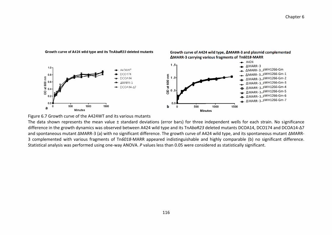
Chapter 6
116
Figure 6.7 Growth curve of the A424WT and its various mutants The data shown represents the mean value ± standard deviations (error bars) for three independent wells for each strain. No significance difference in the growth dynamics was observed between A424 wild type and its TnAbaR23 deleted mutants DCOA14, DCO174 and DCOA14-Δ7 and spontaneous mutant ΔMARR-3 (a) with no significant difference. The growth curve of A424 wild type, and its spontaneous mutant ΔMARR-3 complemented with various fragments of Tn6018-MARR appeared indistinguishable and highly comparable (b) no significant difference. Statistical analysis was performed using one-way ANOVA. P values less than 0.05 were considered as statistically significant.
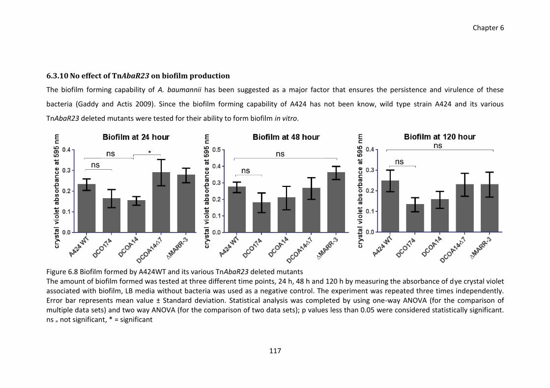
Chapter 6
117
6.3.10 No effect of TnAbaR23 on biofilm production
The biofilm forming capability of A. baumannii has been suggested as a major factor that ensures the persistence and virulence of these
bacteria (Gaddy and Actis 2009). Since the biofilm forming capability of A424 has not been know, wild type strain A424 and its various
TnAbaR23 deleted mutants were tested for their ability to form biofilm in vitro.
Figure 6.8 Biofilm formed by A424WT and its various TnAbaR23 deleted mutants The amount of biofilm formed was tested at three different time points, 24 h, 48 h and 120 h by measuring the absorbance of dye crystal violet associated with biofilm, LB media without bacteria was used as a negative control. The experiment was repeated three times independently. Error bar represents mean value ± Standard deviation. Statistical analysis was completed by using one-way ANOVA (for the comparison of multiple data sets) and two way ANOVA (for the comparison of two data sets); p values less than 0.05 were considered statistically significant. ns ₌ not significant, * = significant
Chapter 6
118
The markerless mutant DCOA14-Δ7 derived from TnAbaR23 deleted aacC1 marker
carrying mutant DCOA14 showed increased biofilm forming ability (p = 0.03) in 24 hour
(Figure 6.8). However, no significant difference was observed in the biofilm forming
ability of the all the tested strains in 24 h, 48 h and 120 h. Overall, the biofilm forming
ability of the strain was not affected upon the deletion of TnAbaR23.
6.3.11 Effect of TnAbaR23 on in vitro growth fitness
The fitness of A424 wild type and its various TnAbaR23 deleted mutants was assessed
in an in vitro head-to-head competition assay. The population of parent wild type and
the mutant was estimated at the beginning of the competition and at specific time
points by sampling the culture and plating the appropriate dilution on selective and
non-selective plates. The five day head-to-head competition was repeated three times
independently. The CFU obtained on both selective and non-selective plates was used
to calculate the percentage of wild type and mutant at each time point (T).
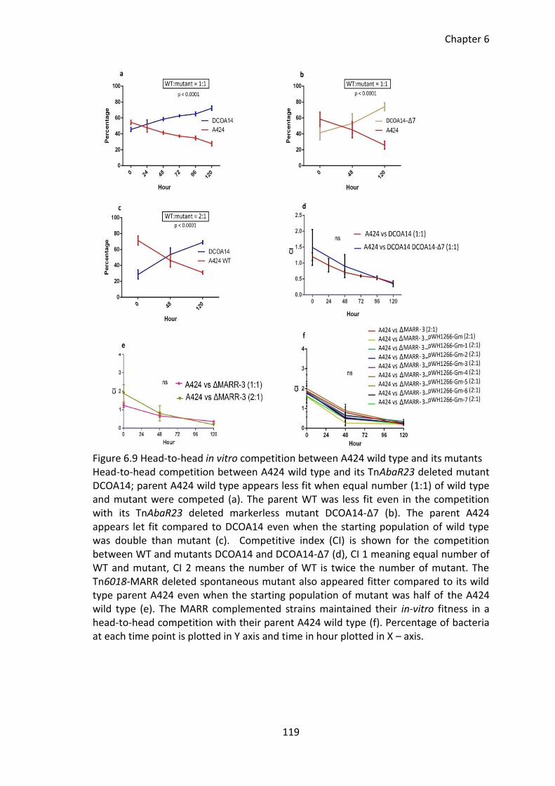
Chapter 6
119
Figure 6.9 Head-to-head in vitro competition between A424 wild type and its mutants Head-to-head competition between A424 wild type and its TnAbaR23 deleted mutant DCOA14; parent A424 wild type appears less fit when equal number (1:1) of wild type and mutant were competed (a). The parent WT was less fit even in the competition with its TnAbaR23 deleted markerless mutant DCOA14-Δ7 (b). The parent A424 appears let fit compared to DCOA14 even when the starting population of wild type was double than mutant (c). Competitive index (CI) is shown for the competition between WT and mutants DCOA14 and DCOA14-Δ7 (d), CI 1 meaning equal number of WT and mutant, CI 2 means the number of WT is twice the number of mutant. The Tn6018-MARR deleted spontaneous mutant also appeared fitter compared to its wild type parent A424 even when the starting population of mutant was half of the A424 wild type (e). The MARR complemented strains maintained their in-vitro fitness in a head-to-head competition with their parent A424 wild type (f). Percentage of bacteria at each time point is plotted in Y axis and time in hour plotted in X – axis.
Chapter 6
120
The Figure 6.9 shows the in vitro competitive fitness of A424 wild type and its various
mutants. The parent A424 was out-competed by TnAbaR23 deleted mutant DCOA14
(a), TnAbaR23 deleted markerless DCOA14-Δ7 (b) and Tn6018-MARR deleted
spontaneous mutant ΔMARR-3 (e) when equal number of wild type and mutants were
present at the beginning of competition. The mutants DCOA14 and ΔMARR-3 exhibited
enhanced competitive fitness in a head-to-head competition with A424 wild type even
when their starting number was half the number of wild type (c and e). The presence
of aacC1 marker did not seem to affect the overall competitive fitness since no
significant difference in CI was observed for DCOA14 and DCOA14-Δ7 in their head-to-
head competition with A424 (d). Importantly, the presence of Tn6018-MARR
fragments in plasmid complemented ΔMARR-3 mutants did not alter the fitness of
complemented strains and no significant difference in CI was observed between empty
ΔMARR-3 strains and plasmid complemented strains (f). These data suggests that the
possession of intact TnAbaR23 results in decreased in vitro growth fitness, however, it
was not possible to pin point the region within TnAbaR23 that would impose the
observed physiological cost in A424.
6.3.12 Effect of TnAbaR23 on virulence
The virulence of A424 wild type and its various TnAbaR23 deleted mutants was
compared in a killing assay using the sixth instar larva of G. mellonella. Each larva was
injected with a 20 µl of bacterial suspension and the mortality of the larva was
monitored over a period of 5 days. At the end of the experiment, the surviving larvae
were disposed. Before commencing the killing assay, the appropriate dose of bacterial
inoculum was determined for A424 wild type strain.
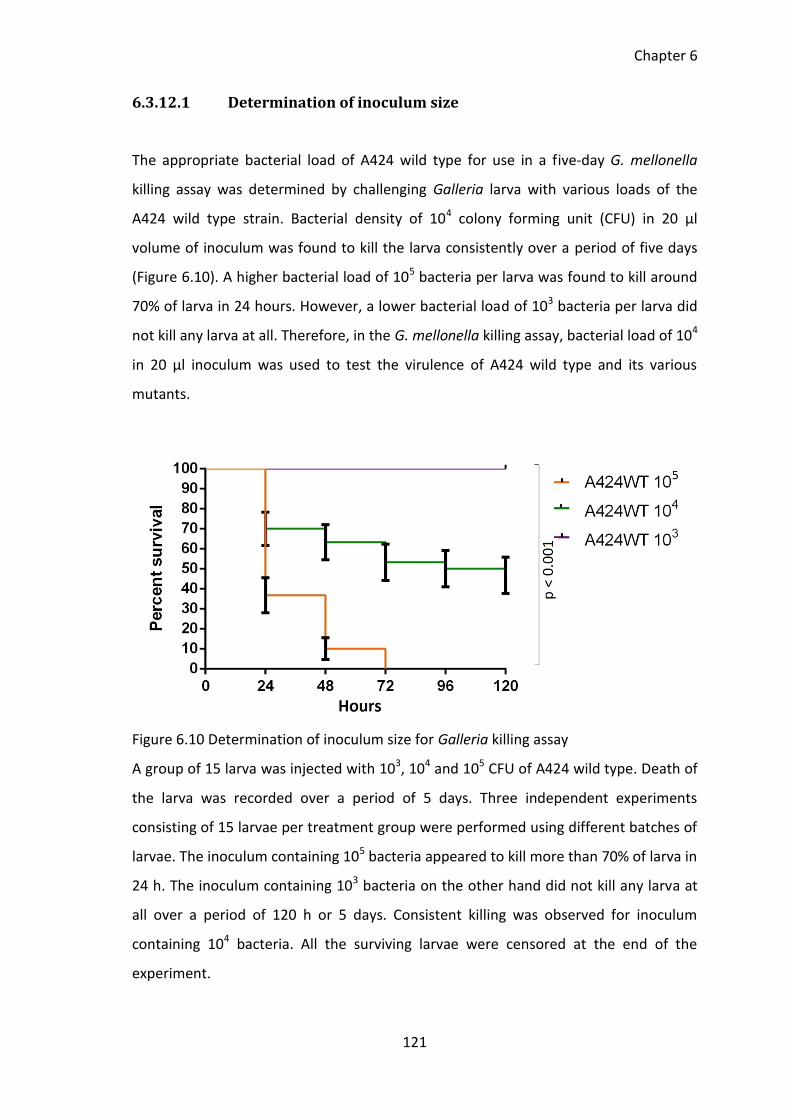
Chapter 6
121
6.3.12.1 Determination of inoculum size
The appropriate bacterial load of A424 wild type for use in a five-day G. mellonella
killing assay was determined by challenging Galleria larva with various loads of the
A424 wild type strain. Bacterial density of 104 colony forming unit (CFU) in 20 µl
volume of inoculum was found to kill the larva consistently over a period of five days
(Figure 6.10). A higher bacterial load of 105 bacteria per larva was found to kill around
70% of larva in 24 hours. However, a lower bacterial load of 103 bacteria per larva did
not kill any larva at all. Therefore, in the G. mellonella killing assay, bacterial load of 104
in 20 µl inoculum was used to test the virulence of A424 wild type and its various
mutants.
Figure 6.10 Determination of inoculum size for Galleria killing assay
A group of 15 larva was injected with 103, 104 and 105 CFU of A424 wild type. Death of
the larva was recorded over a period of 5 days. Three independent experiments
consisting of 15 larvae per treatment group were performed using different batches of
larvae. The inoculum containing 105 bacteria appeared to kill more than 70% of larva in
24 h. The inoculum containing 103 bacteria on the other hand did not kill any larva at
all over a period of 120 h or 5 days. Consistent killing was observed for inoculum
containing 104 bacteria. All the surviving larvae were censored at the end of the
experiment.
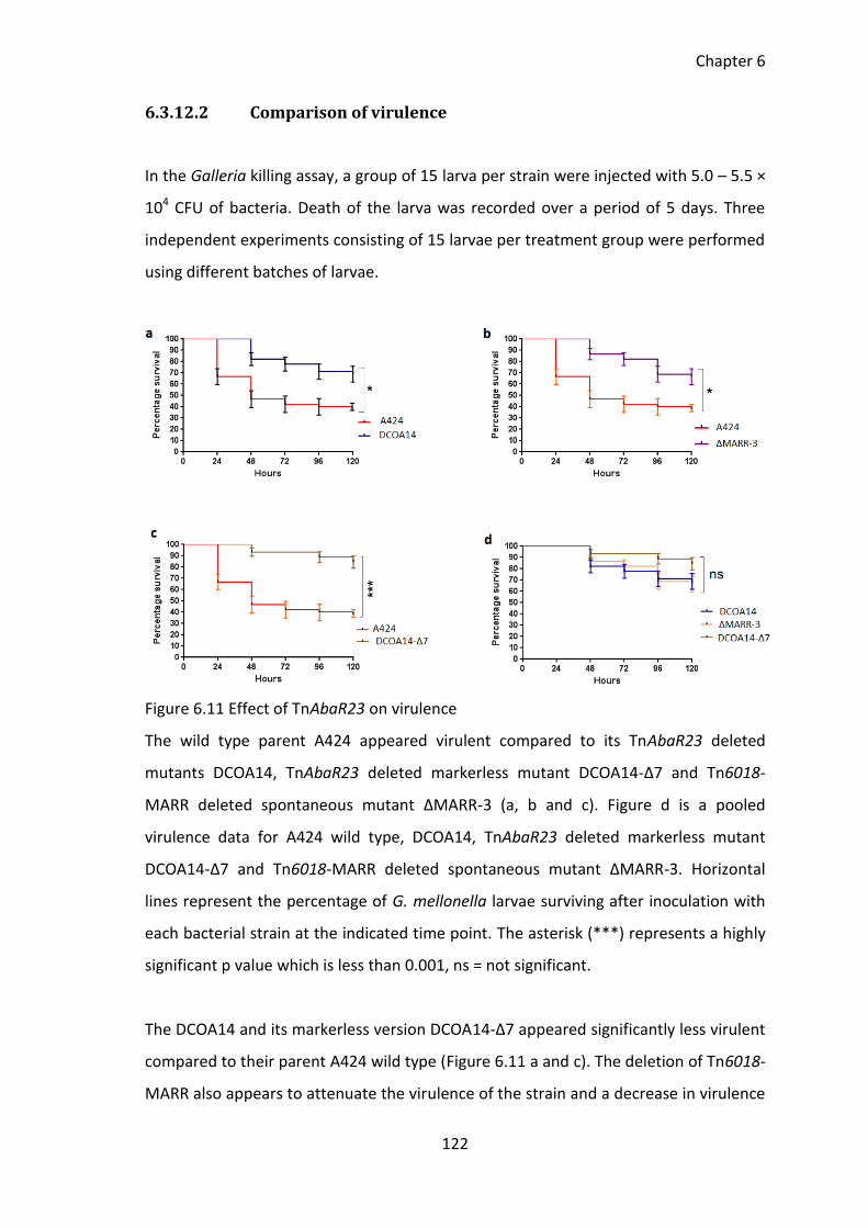
Chapter 6
122
6.3.12.2 Comparison of virulence
In the Galleria killing assay, a group of 15 larva per strain were injected with 5.0 – 5.5 ×
104 CFU of bacteria. Death of the larva was recorded over a period of 5 days. Three
independent experiments consisting of 15 larvae per treatment group were performed
using different batches of larvae.
Figure 6.11 Effect of TnAbaR23 on virulence
The wild type parent A424 appeared virulent compared to its TnAbaR23 deleted
mutants DCOA14, TnAbaR23 deleted markerless mutant DCOA14-Δ7 and Tn6018-
MARR deleted spontaneous mutant ΔMARR-3 (a, b and c). Figure d is a pooled
virulence data for A424 wild type, DCOA14, TnAbaR23 deleted markerless mutant
DCOA14-Δ7 and Tn6018-MARR deleted spontaneous mutant ΔMARR-3. Horizontal
lines represent the percentage of G. mellonella larvae surviving after inoculation with
each bacterial strain at the indicated time point. The asterisk (***) represents a highly
significant p value which is less than 0.001, ns = not significant.
The DCOA14 and its markerless version DCOA14-Δ7 appeared significantly less virulent
compared to their parent A424 wild type (Figure 6.11 a and c). The deletion of Tn6018-
MARR also appears to attenuate the virulence of the strain and a decrease in virulence
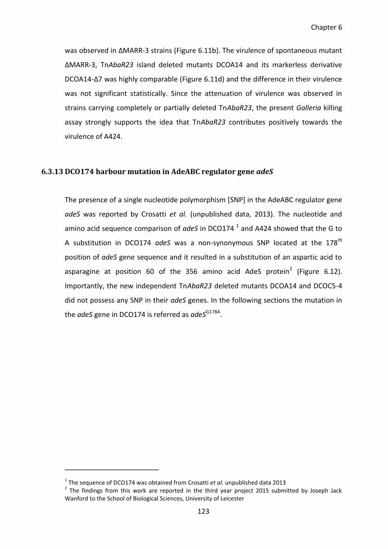
Chapter 6
123
was observed in ΔMARR-3 strains (Figure 6.11b). The virulence of spontaneous mutant
ΔMARR-3, TnAbaR23 island deleted mutants DCOA14 and its markerless derivative
DCOA14-Δ7 was highly comparable (Figure 6.11d) and the difference in their virulence
was not significant statistically. Since the attenuation of virulence was observed in
strains carrying completely or partially deleted TnAbaR23, the present Galleria killing
assay strongly supports the idea that TnAbaR23 contributes positively towards the
virulence of A424.
6.3.13 DCO174 harbour mutation in AdeABC regulator gene adeS
The presence of a single nucleotide polymorphism [SNP] in the AdeABC regulator gene
adeS was reported by Crosatti et al. (unpublished data, 2013). The nucleotide and
amino acid sequence comparison of adeS in DCO174 1 and A424 showed that the G to
A substitution in DCO174 adeS was a non-synonymous SNP located at the 178th
position of adeS gene sequence and it resulted in a substitution of an aspartic acid to
asparagine at position 60 of the 356 amino acid AdeS protein2 (Figure 6.12).
Importantly, the new independent TnAbaR23 deleted mutants DCOA14 and DCOC5-4
did not possess any SNP in their adeS genes. In the following sections the mutation in
the adeS gene in DCO174 is referred as adeSG178A.
1 The sequence of DCO174 was obtained from Crosatti et al. unpublished data 2013
2 The findings from this work are reported in the third year project 2015 submitted by Joseph Jack
Wanford to the School of Biological Sciences, University of Leicester
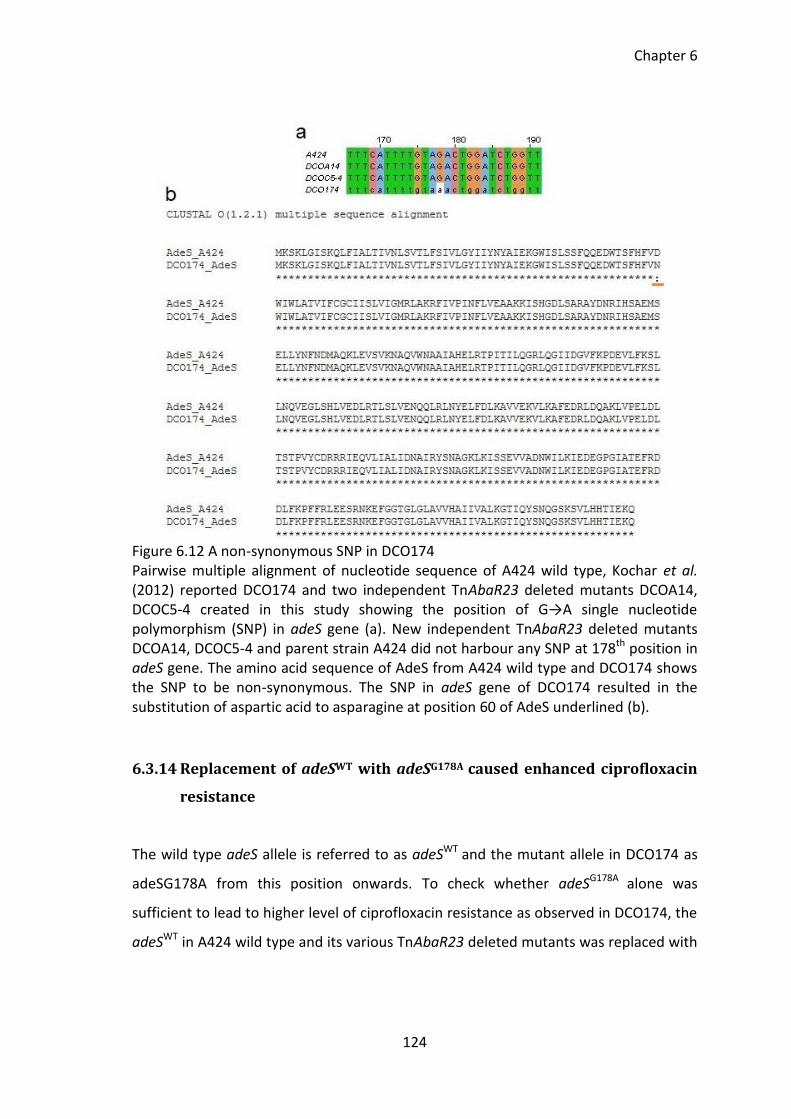
Chapter 6
124
Figure 6.12 A non-synonymous SNP in DCO174 Pairwise multiple alignment of nucleotide sequence of A424 wild type, Kochar et al. (2012) reported DCO174 and two independent TnAbaR23 deleted mutants DCOA14, DCOC5-4 created in this study showing the position of G→A single nucleotide polymorphism (SNP) in adeS gene (a). New independent TnAbaR23 deleted mutants DCOA14, DCOC5-4 and parent strain A424 did not harbour any SNP at 178th position in adeS gene. The amino acid sequence of AdeS from A424 wild type and DCO174 shows the SNP to be non-synonymous. The SNP in adeS gene of DCO174 resulted in the substitution of aspartic acid to asparagine at position 60 of AdeS underlined (b).
6.3.14 Replacement of adeSWT with adeSG178A caused enhanced ciprofloxacin
resistance
The wild type adeS allele is referred to as adeSWT and the mutant allele in DCO174 as
adeSG178A from this position onwards. To check whether adeSG178A alone was
sufficient to lead to higher level of ciprofloxacin resistance as observed in DCO174, the
adeSWT in A424 wild type and its various TnAbaR23 deleted mutants was replaced with
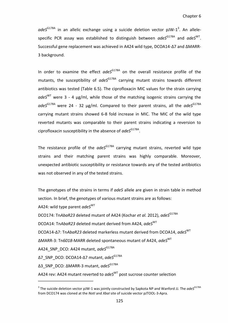
Chapter 6
125
adeSG178A in an allelic exchange using a suicide deletion vector pJW-13. An allele-
specific PCR assay was established to distinguish between adeSG178A and adeSWT.
Successful gene replacement was achieved in A424 wild type, DCOA14-Δ7 and ΔMARR-
3 background.
In order to examine the effect adeSG178A on the overall resistance profile of the
mutants, the susceptibility of adeSG178A carrying mutant strains towards different
antibiotics was tested (Table 6.5). The ciprofloxacin MIC values for the strain carrying
adeSWT were 3 - 4 µg/ml, while those of the matching isogenic strains carrying the
adeSG178A were 24 - 32 µg/ml. Compared to their parent strains, all the adeSG178A
carrying mutant strains showed 6-8 fold increase in MIC. The MIC of the wild type
reverted mutants was comparable to their parent strains indicating a reversion to
ciprofloxacin susceptibility in the absence of adeSG178A.
The resistance profile of the adeSG178A carrying mutant strains, reverted wild type
strains and their matching parent strains was highly comparable. Moreover,
unexpected antibiotic susceptibility or resistance towards any of the tested antibiotics
was not observed in any of the tested strains.
The genotypes of the strains in terms if adeS allele are given in strain table in method
section. In brief, the genotypes of various mutant strains are as follows:
A424: wild type parent adeSWT
DCO174: TnAbaR23 deleted mutant of A424 (Kochar et al. 2012), adeSG178A
DCOA14: TnAbaR23 deleted mutant derived from A424, adeSWT
DCOA14-Δ7: TnAbaR23 deleted markerless mutant derived from DCOA14, adeSWT
ΔMARR-3: Tn6018-MARR deleted spontaneous mutant of A424, adeSWT
A424_SNP_DCO: A424 mutant, adeSG178A
Δ7_SNP_DCO: DCOA14-Δ7 mutant, adeSG178A
Δ3_SNP_DCO: ΔMARR-3 mutant, adeSG178A
A424 rev: A424 mutant reverted to adeSWT post sucrose counter selection
3 The suicide deletion vector pJW-1 was jointly constructed by Sapkota NP and Wanford JJ. The adeS
G178A
from DCO174 was cloned at the NotI and XbaI site of suicide vector pJTOOL-3-Apra.
Chapter 6
126
Δ7_rev: DCOA14-Δ7 mutant reverted to adeSWT post sucrose counter selection
Δ3_rev: ΔMARR-3 mutant reverted to adeSWT post sucrose counter selection
For the disc test, the diameter of zone of clearance was measured to the nearest 0.5
mm, values in bold represent the MIC (shown in µg/ml, tested by using Etest strips).
The experiment was repeated three times independently. Reference strain NCTC12241
was used as BSAC reference control (Wootton 2013). The full form of antibiotics
abbreviated below is given in method section. BSAC version 2013 guideline on zone
diameter breakpoints are as follows: for IMP, resistant (R) ≤ 13, intermediate (I) 14 - 24
and susceptible (S) ≥ 25; for AMK, resistant (R) ≤ 18, intermediate (I) 19-20 and
susceptible (S) ≥ 21; for gentamicin, resistant (R) ≤ 19 and susceptible (S) ≥ 20; for
ciprofloxacin, resistant (R) ≤ 20 and susceptible (S) ≥ 21. BSAC guideline on MIC
breakpoint for Acinetobacter species: for gentamicin, resistant (R) > 4 and susceptible
(S) ≤ 4; for amikacin, resistant (R) > 16, intermediate (I) 16 and susceptible (S) ≤ 8; for
imipenem, resistant (R) > 8, intermediate (I) 4-8 and susceptible (S) ≤ 2 and for
ciprofloxacin, resistant (R) > 1 and susceptible (S) ≤ 1.
Chapter 6
127
Table 6.5 Antibiotic susceptibility profile of strains carrying adeSWT and adeSG178A
Strain Zone of inhibition Diameter (mm) TET 10
IMP 10
TOB 10
CIP 5
STR 10
RIF 2
AMK 30
TMP 2.5
SUL 25
SXT 25
GEN 10
A424WT 6.0 19.0 27.5 14.5, 4 16.5 14.5 17.0 9.5 6.0 6.0 25.5 DCO174 15.5 16.5 24.0 6.0, > 32 10.5 14.0 12.5 7.5 27.5 25.0 6.5 DCOA14 20.5 18.5 27.0 13.5, 4 18.5 12.0 18.5 8.5 30.5 27.0 8.5 DCOA14-Δ7 20.0 18.5 27.0 13.0, 4 18.0 12.5 18.0 8.0 30.0 27.5 24.0 ΔMARR-3 21.0 20. 27.5 14.0, 3 21.0 16.0 20.0 9.0 32.0 27.5 27.0 A424_SNP_DCO 6.0 19.5 26.5 24 17.0 16.0 16.5 10.5 6.0 6.0 28.5 Δ7_SNP_DCO 23.0 20.5 28.0 24 19.0 14.5 17.0 9.0 33.0 29.5 24.0 Δ3_SNP_DCO 22.5 20.5 29.5 > 32 23.0 17.5 21.0 9.5 32.0 27.0 27.0 A424_rev 6.5 18.0 26.5 2 16.0 14.0 17.5 9.0 6.0 6.0 25.0 Δ7_rev 21.0 17.0 25.5 3 18.5 12.0 19.0 8.5 32.0 27.0 25.5
Δ3_rev 21.0 22.0 27.0 3 23.5 15.0 21.5 9.0 33.0 27.0 28.5 NCTC12241 20.5 35.0 21.5 30.0 17.0 6.0 24.0 26.0 6.0 29.0 23.0
The full form of abbreviated antibiotics is given in method section. The concentration of antibiotics on the disc is given in µg/ml, values in bold represent antibiotic concentration in µg/ml obtained from E-test. The strain NCTC1224 is used as a reference strain. The parent A424 wild type carrying mutant adeSG178A adeS allele (A424_SNP_DCO) shows 6 folds increase in ciprofloxacin resistance. A 6 – 8 folds increases in ciprofloxacin resistance observed in strains Δ7 and Δ3 where the wild type adeSWT allele has been replaced by mutant adeSG178A allele.
Chapter 6
128
6.3.15 Fitness and virulence phenotypes of DCO174
The competitive growth fitness and virulence phenotype of DCO174 was studied in an
in vitro head-to-head competition assay and Galleria killing assay as described above.
The deletion of TnAbaR23 adeSG178A in DCO174 did not altered its biofilm forming
ability (Figure 6.8) and growth dynamics (Figure 6.7). However, the competitive fitness
of DCO174 was found markedly reduced in an in vitro head-to-head competition with
its parent A424. The mutant DCO174 was outcompeted by A424 wild type even when
population of DCO174 was nine times more than that of A424 at the beginning of
competition (Figure 6.13b). The Two-way ANOVA showed no significant difference in
the CI when the starting population of DCO174 was equal to or 9 times greater than
A424 wild type (Figure 6.13d).
The TnAbaR23 deleted isogenic mutants DCOA14 and DCOA14-Δ7, on the other hand,
exhibited significantly enhanced fitness in a head-to-head competition with their
parent A424 wild type (Figure 6.13c). Compared to their parent A424 wild type, the
TnAbaR23 deleted mutants DCOA14, DCOA14-Δ7 and TnAbaR23 truncated mutant
ΔMARR-3 exhibited significantly (p < 0.001) attenuated virulence in Galleria killing
assay (Figure 6.11). Surprisingly, DCO174 did not exhibit attenuated virulence in
Galleria killing assay. Moreover, the virulence of A424 wild type and DCO174 appeared
highly comparable in the Galleria killing assay (Figure 6.14).
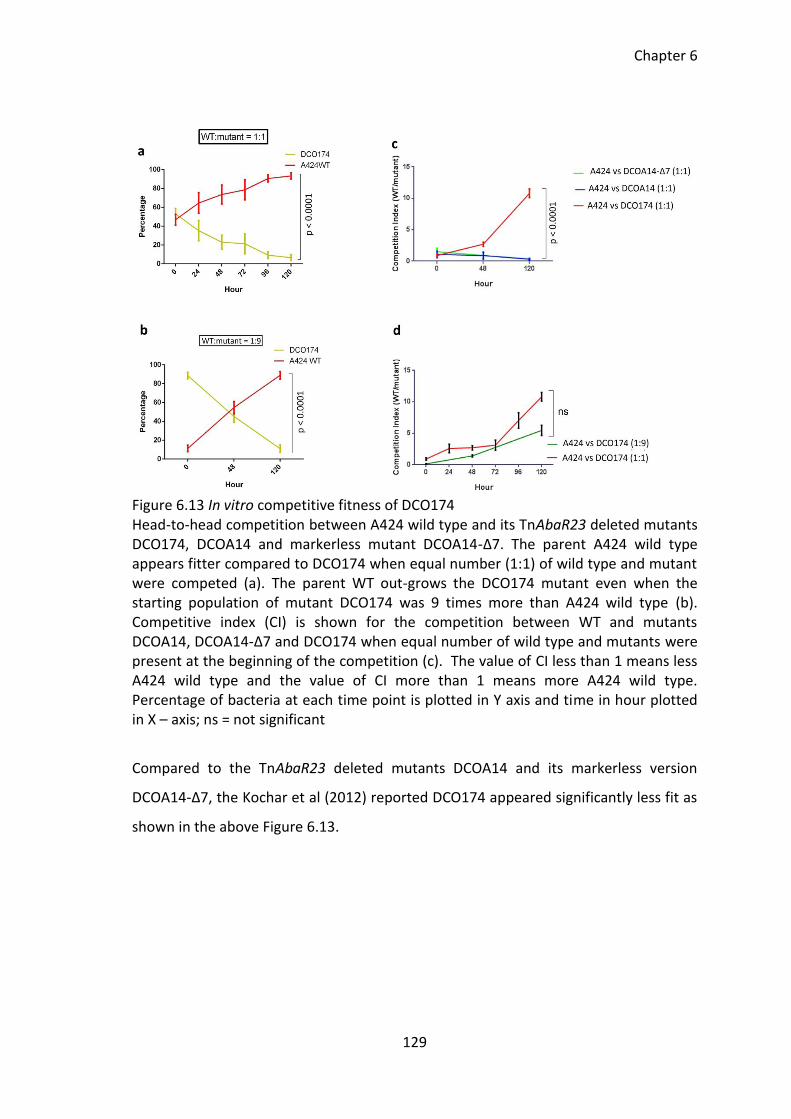
Chapter 6
129
Figure 6.13 In vitro competitive fitness of DCO174 Head-to-head competition between A424 wild type and its TnAbaR23 deleted mutants DCO174, DCOA14 and markerless mutant DCOA14-Δ7. The parent A424 wild type appears fitter compared to DCO174 when equal number (1:1) of wild type and mutant were competed (a). The parent WT out-grows the DCO174 mutant even when the starting population of mutant DCO174 was 9 times more than A424 wild type (b). Competitive index (CI) is shown for the competition between WT and mutants DCOA14, DCOA14-Δ7 and DCO174 when equal number of wild type and mutants were present at the beginning of the competition (c). The value of CI less than 1 means less A424 wild type and the value of CI more than 1 means more A424 wild type. Percentage of bacteria at each time point is plotted in Y axis and time in hour plotted in X – axis; ns = not significant
Compared to the TnAbaR23 deleted mutants DCOA14 and its markerless version
DCOA14-Δ7, the Kochar et al (2012) reported DCO174 appeared significantly less fit as
shown in the above Figure 6.13.
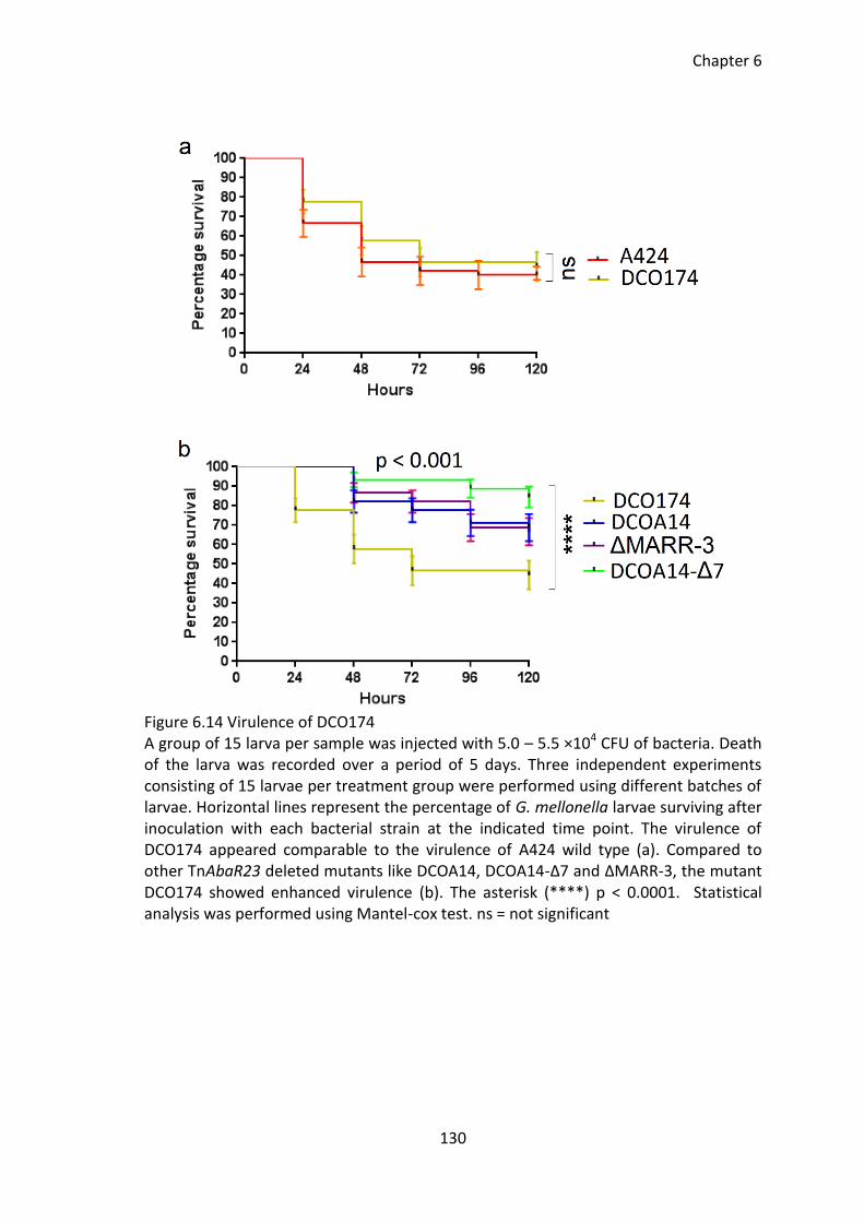
Chapter 6
130
Figure 6.14 Virulence of DCO174 A group of 15 larva per sample was injected with 5.0 – 5.5 ×10⁴ CFU of bacteria. Death of the larva was recorded over a period of 5 days. Three independent experiments consisting of 15 larvae per treatment group were performed using different batches of larvae. Horizontal lines represent the percentage of G. mellonella larvae surviving after inoculation with each bacterial strain at the indicated time point. The virulence of DCO174 appeared comparable to the virulence of A424 wild type (a). Compared to other TnAbaR23 deleted mutants like DCOA14, DCOA14-Δ7 and ΔMARR-3, the mutant DCO174 showed enhanced virulence (b). The asterisk (****) p < 0.0001. Statistical analysis was performed using Mantel-cox test. ns = not significant
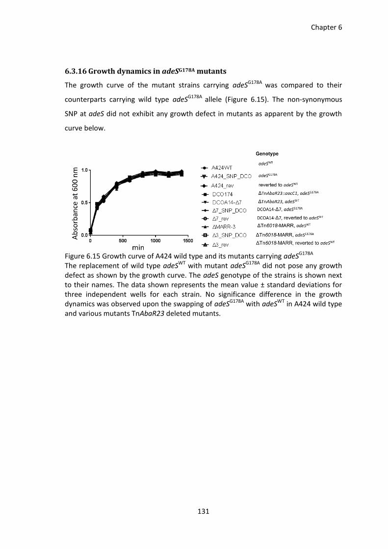
Chapter 6
131
6.3.16 Growth dynamics in adeSG178A mutants
The growth curve of the mutant strains carrying adeSG178A was compared to their
counterparts carrying wild type adeSG178A allele (Figure 6.15). The non-synonymous
SNP at adeS did not exhibit any growth defect in mutants as apparent by the growth
curve below.
Figure 6.15 Growth curve of A424 wild type and its mutants carrying adeSG178A
The replacement of wild type adeSWT with mutant adeSG178A did not pose any growth defect as shown by the growth curve. The adeS genotype of the strains is shown next to their names. The data shown represents the mean value ± standard deviations for three independent wells for each strain. No significance difference in the growth dynamics was observed upon the swapping of adeSG178A with adeSWT in A424 wild type and various mutants TnAbaR23 deleted mutants.
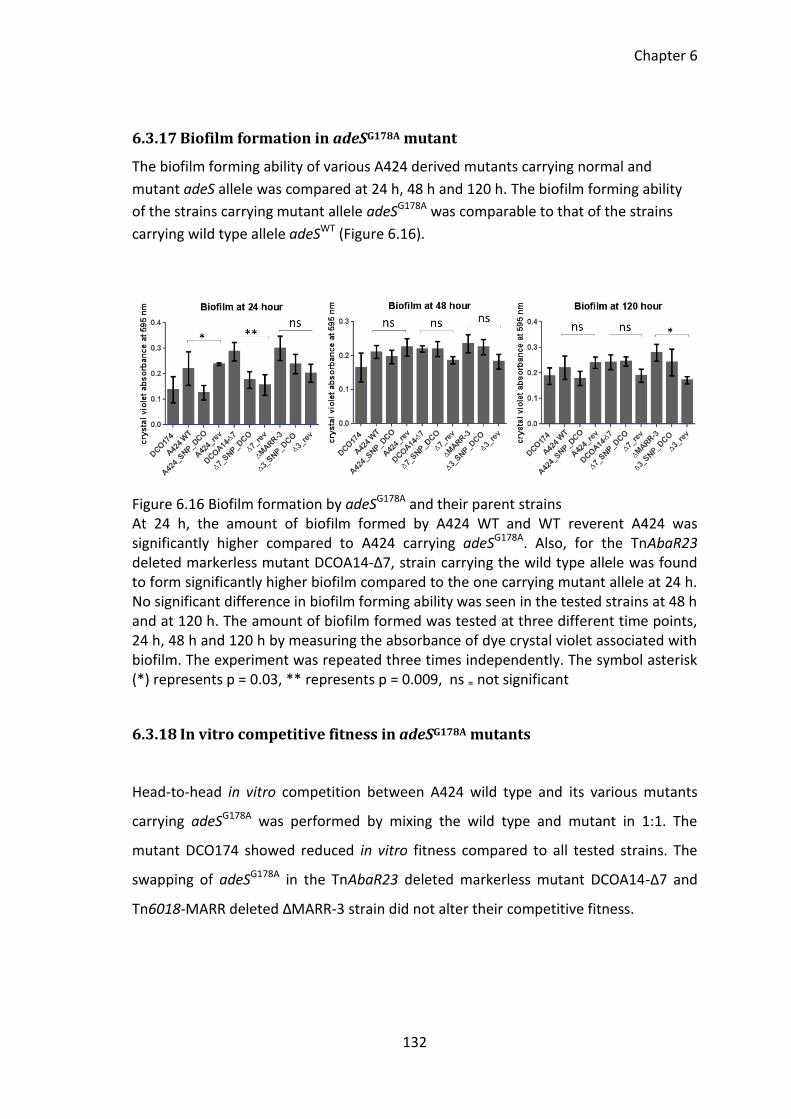
Chapter 6
132
6.3.17 Biofilm formation in adeSG178A mutant
The biofilm forming ability of various A424 derived mutants carrying normal and
mutant adeS allele was compared at 24 h, 48 h and 120 h. The biofilm forming ability
of the strains carrying mutant allele adeSG178A was comparable to that of the strains
carrying wild type allele adeSWT (Figure 6.16).
Figure 6.16 Biofilm formation by adeSG178A and their parent strains At 24 h, the amount of biofilm formed by A424 WT and WT reverent A424 was significantly higher compared to A424 carrying adeSG178A. Also, for the TnAbaR23 deleted markerless mutant DCOA14-Δ7, strain carrying the wild type allele was found to form significantly higher biofilm compared to the one carrying mutant allele at 24 h. No significant difference in biofilm forming ability was seen in the tested strains at 48 h and at 120 h. The amount of biofilm formed was tested at three different time points, 24 h, 48 h and 120 h by measuring the absorbance of dye crystal violet associated with biofilm. The experiment was repeated three times independently. The symbol asterisk (*) represents p = 0.03, ** represents p = 0.009, ns ₌ not significant
6.3.18 In vitro competitive fitness in adeSG178A mutants
Head-to-head in vitro competition between A424 wild type and its various mutants
carrying adeSG178A was performed by mixing the wild type and mutant in 1:1. The
mutant DCO174 showed reduced in vitro fitness compared to all tested strains. The
swapping of adeSG178A in the TnAbaR23 deleted markerless mutant DCOA14-Δ7 and
Tn6018-MARR deleted ΔMARR-3 strain did not alter their competitive fitness.
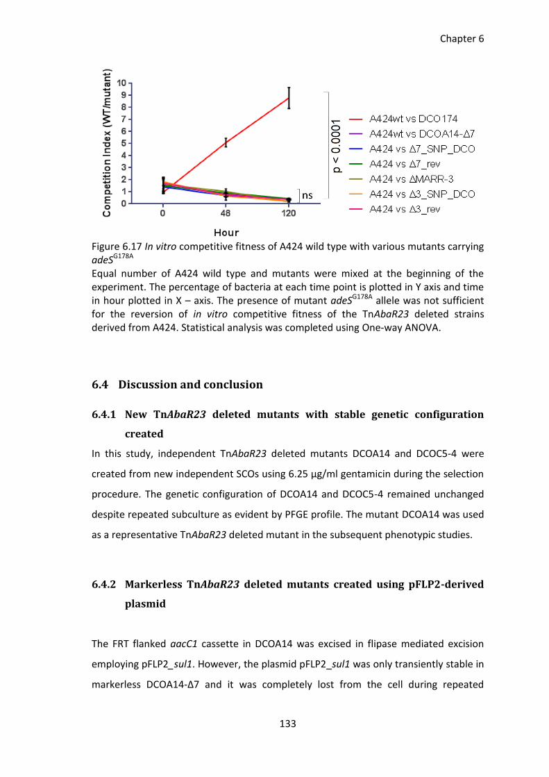
Chapter 6
133
Figure 6.17 In vitro competitive fitness of A424 wild type with various mutants carrying adeSG178A Equal number of A424 wild type and mutants were mixed at the beginning of the experiment. The percentage of bacteria at each time point is plotted in Y axis and time in hour plotted in X – axis. The presence of mutant adeSG178A allele was not sufficient for the reversion of in vitro competitive fitness of the TnAbaR23 deleted strains derived from A424. Statistical analysis was completed using One-way ANOVA.
6.4 Discussion and conclusion
6.4.1 New TnAbaR23 deleted mutants with stable genetic configuration
created
In this study, independent TnAbaR23 deleted mutants DCOA14 and DCOC5-4 were
created from new independent SCOs using 6.25 µg/ml gentamicin during the selection
procedure. The genetic configuration of DCOA14 and DCOC5-4 remained unchanged
despite repeated subculture as evident by PFGE profile. The mutant DCOA14 was used
as a representative TnAbaR23 deleted mutant in the subsequent phenotypic studies.
6.4.2 Markerless TnAbaR23 deleted mutants created using pFLP2-derived
plasmid
The FRT flanked aacC1 cassette in DCOA14 was excised in flipase mediated excision
employing pFLP2_sul1. However, the plasmid pFLP2_sul1 was only transiently stable in
markerless DCOA14-Δ7 and it was completely lost from the cell during repeated
Chapter 6
134
growth. This observation was consistent with previous report regarding the instability
of pFLP2 plasmid in A. baumannii ATCC17978 (Isberg et al. 2012). The use of pFLP2-
sul1 was reproducible and highly successful in the excision of FRT-flanked cassette in
DCOA14. Therefore, despite its apparent instability in A. baumannii, the protocol
developed in this study for the flipase-mediated excision of FRT flanked cassette in A.
baumannii is highly recommended because it is a one-step, easy to use technique that
does not require the downstream curing of plasmid.
6.4.3 Effect of TnAbaR23 on the phenotypes of the strain A424
The only significant impact on resistance phenotype following loss of TnAbaR23 or
Tn6018-MARR was observed as a reversion to sulphamethoxazole and tetracycline
susceptibility. Kochar (2012) suggested that the loss of TnAbaR23 could result in
elevated ciprofloxacin resistance. This does not appear to be the case as unusual
antibiotic resistance or susceptibility was not observed in any of the TnAbaR23 deleted
mutants created in this study.
The phenotype of various TnAbaR23 deleted mutants created/isolated during this
work was assessed using various in vitro and in vivo assays. The head-to-head in vitro
growth competition assay clearly and reproducibly demonstrated that the DCOA14,
DCOA14-Δ7 and ΔMARR-3 mutants were significantly 'fitter' (p < 0.001) than their
parent A424 wild type. Since enhanced in vitro competitive fitness was observed in
A424 mutants upon the loss of TnAbaR23 and Tn6018-MARR, it is possible that the
Tn6018-MARR region within TnAbaR23 is exerting the cost that leads to decreased in
vitro competitive fitness observed in A424 wild type. This finding is in agreement with
those reported by (Lopez-Rojas et al. 2011, Guo et al. 2012) where acquisition of the
resistance determinants was found associated with fitness cost. Interestingly the,
complementation of ΔMARR-3 with blocks of MARR region was not sufficient to
diminish the in vitro fitness of ΔMARR-3. The reason for this is not clear but it is
possible that the physiological cost that ultimately leads to a reduced fitness is exerted
by an intact Tn6018-MARR region rather than its individual genes and determinants. A
Chapter 6
135
further research involving the capture of circular Tn6018-MARR for complementation
of ΔMARR-3 mutants could help pin point the determinants posing energetic or
physiological costs and understand the molecular mechanism that leads to reduced
competitive growth fitness in A424 strain.
Attenuated virulence was observed in TnAbaR23 and Tn6018-MARR deleted mutant in
the G. mellonella killing assay as determined by the survival plots and percentage
survival over a period of five days. In reviewing the literature, no data was found that
on the association of AbaR islands with virulence in A. baumannii. However, prior
studies have noted the attenuation of virulence upon the deletion of pathogenicity
islands in Pseudomonas aeruginosa strains (Miyata et al. 2003, Harrison et al. 2010)
Although the possession of the TnAbaR23 appears to be energetically and/or
physiologically costly for A424 as evident in competitive fitness comparison, the
preservation of this island in its intact form seems to be important for the strain in
order to maintain its virulence-associated phenotypes. Further research should be
taken to understand the underlying mechanism by which Tn6018-MARR and Tn6018-
MARR borne determinants enhances the virulence of A424 strain.
The review on A. baumannii biofilm (Gaddy and Actis 2009) suggests that the ability to
form biofilm could enhance the virulence in pathogenic A. baumannii strains. Despite
showing reduced virulence upon the deletion of TnAbaR23, no significant difference
was observed regarding the biofilm forming capability in A424 wild type and isogenic
TnAbaR23 deleted mutants. Based on the findings of this work, while preliminary, it is
possible to hypothesize that AbaR islands specifically contribute positively to antibiotic
resistance, virulence and/or other environmental survival traits despite the associated
physiological costs.
Chapter 6
136
6.4.4 DCO174 an unusual TnAbaR23 deleted mutant of A424
As speculated by Kochar et al. (2012), the genome analysis of DCO174 showed a non-
synonymous SNP in the regulatory gene of efflux pump AdeABC where a substitution
of nucleotide G to A at 178th position of adeS resulted in substitution of aspartic acid to
asparagine in a region of AdeS protein devoid of identified functional domains. In this
study, the adeSWT allele was replaced with mutant adeSG178A allele in A424 wild type
and various TnAbaR23 deleted strains using an allelic exchange protocol. The mutant
strains carrying adeSG178A showed 6-8 folds increase in ciprofloxacin MIC. The reversion
of ciprofloxacin MIC in the DCOs which reverted to wild type post sucrose-counter
selection suggests that the observed heightened ciprofloxacin MIC in adeSG178A
mutants created in this study was not due to the genetic stress, antibiotic exposure or
any alternative mechanisms. It can thus be suggested that the presence of adeSG178A
on its own was sufficient to lead to elevated level of ciprofloxacin resistance.
The TnAbaR23 deleted mutant DCO174 exhibited markedly reduced fitness in a head-
to-head competition with its parent A424 wild type. Various TnAbaR23 deleted
mutants created in this study showed significant attenuation of virulence in the G.
mellonella killing assay. Interestingly, the virulence of DCO174 and A424 wild type was
highly comparable. The reason for the observed virulence in DCO174 is not clear but
adeSG178A could be a factor contributing towards the maintenance of virulence in
DCO174. In A424 derived mutant strains carrying the mutant allele adeSG178A, the
growth dynamics and biofilm forming ability was comparable to their counter-parts
carrying wild type allele adeSWT. Interestingly, the possession of adeSG178A did not seem
to be enough to reverse the in vitro competitive fitness associated with A424 derived
strains carrying wild type allele adeSWT as observed in the in vitro competitive fitness
assay. It is therefore possible that the mutant DCO174 possess other, yet unknown,
secondary mutations elsewhere in the genome which has severely affected the in vitro
competitive fitness of this strain. For now, it can be said that enhanced antimicrobial
resistance and virulence coupled with other secondary mutations in the genome
contributed towards the highly reduced competitive fitness observed in this mutant. In
future investigations, it might be possible to adopt the allele replacement approach

Chapter 6
137
described this study in order to investigate whether or not the mutated efflux systems
are associated with virulence in A. baumannii. In the next chapter I will summarize and
discuss the main findings of this thesis and will provide recommendations for future
work.

138
7 Final discussion and conclusion
Final discussion and conclusion
139
The comparative sequence analysis of A. baumannii genome has revealed that a large
proportion of the genome was occupied by the genetic materials that were acquired
from various bacteria by means of horizontal gene transfer (Fournier et al. 2006). In
2006, Fournier et al. identified and described A. baumannii specific resistance island
AbaR1 in MDR French epidemic strain AYE and a similar but empty (no resistance
determinants) AbaR-like island was identified in the susceptible strains SDF. To date,
several AbaR islands, numbered from AbaR0 to AbaR27, have been described in MDR
A. baumannii strains, all of these islands have been described in MDR A. baumannii.
The AbaR islands are composite transposons build on the backbone of simple
transposons like Tn6019, Tn6021 and Tn6022. Towards their 5’ end, these islands carry
five genes tniA, tniB, tniC, tniD and tniE that are believed to be essential for the
transposition activity. The comparative sequence analysis of the five transposition
genes indicated a distant relationship of AbaR-like islands to a highly promiscuous
transposon Tn7 (Rose 2010).
The genome database was accessed from August 2014 to April 2015 in order to survey
the characterized and putative AbaR islands in A. baumannii genome. A total of 58
unique (in terms of their host and integration site) AbaR islands were found
represented in 56 A. baumannii strains. Most of the MDR strains carry only one copy of
AbaR island in their genome, however, strains AB0057 and A85 were reported to carry
two copies of AbaR-like islands in their genome.
A majority of the AbaR transposons are reported to have a backbone of transposon
Tn6019 (as in AbaR3), Tn6021 or Tn6022 (as in AbaR4) transposons. Few AbaR-like
islands were reported to possess multiple copies of Tn6021 and Tn6022 making a
complex looking backbone like Tn6166, Tn6167 and Tn6168. The AbaR-like
transposons that have Tn6167, Tn6168 or Tn6166 on their backbone were considered
distinct from AbaR3 derived transposons and were therefore assumed to represent a
separate lineage (Shaikh et al. 2009, Nigro and Hall 2012a). The diversification of
AbaR3 islands resulting in a creation of wide range of AbaR3 variant has been
extensively described Krizova et al. (2011). In this study, transposon backbones
Final discussion and conclusion
140
represented in currently known AbaR islands were analysed for their nucleotide and
amino acid similarity. The backbone transposon in GI_comM_A92 was named Tn6267.
The sequence comparison of core transposition genes of the representative backbone
transposons clearly showed three distinct lineages of AbaR islands. The first lineage
comprises of AbaR4-like islands. The members of this family are most abundant. They
carry Tn6022, Tn6021, Tn6166 and Tn6167 in their backbone and usually have the sup
gene interrupted by Tn2006. The novel AbaR element found in strains A92 and IS116
forms the second lineage of AbaR elements. The backbone transposon of these novel
AbaR islands has been named Tn6267 in this study. The comM associated AbaR islands
in strains A92 and IS116 were given interim name GI_comM_A92 and GI_comM_IS116
respectively. The GI_comM_IS116-like islands formed a second distinct family of AbaR
islands and at the present time, it appears to be only distantly related to other known
AbaR elements. It is like that islands like GI_comM_A92 and other known AbaR
elements have evolved from a common, yet unknown, ancestral transposon. The third
family consists of AbaR3-like islands. The members of this family carry Tn6019 on their
backbone and usually have the uspA gene interrupted by large compound transposon
Tn6018-MARR-Tn6018. While the AbaR3 and AbaR4 families appeared to have
diverted only recently, on the other hand, the islands like GI_comM_IS116 might have
diverted very early during their evolution from a common, yet unknown, transposon
ancestor.
The AbaR-like transposons are reported as unstable features in the genome of A.
baumannii. There are several reports of IS mediated insertion, deletion and
rearrangements within these transposons (Harmer et al. 2014). Also, intramolecular
recombination between ΔTn6022b and corresponding region of Tn6022a/Tn2006 was
reported in the AbaR25 transposon (Saule et al. 2013). The ability of Tn6018 to
transpose on its own was described in recent years (Williams et al. 2002). In this study,
for the first time, I am reporting the spontaneous excision and subsequent deletion of
28.6 kb Tn6018-MARR segment from TnAbaR23 in a MDR strain A424. Following
excision from its native site, the Tn6018-MARR segment did not re-integrate elsewhere
in the genome and was lost in entirety from the cell. In the strain A424, this
Final discussion and conclusion
141
spontaneous transposition was found to be occurring in in vitro culture condition and
in the absence of antibiotic stress. In every 104 A424 wild type cells, around 100- 1000
cells were found to harbor the deletion of Tn6018-MARR region. Also, out of every 105
cells, around 100-1000 cells were found to possess the excised Tn6018-MARR as a
double stranded circular entity. From A424 wild type, spontaneous mutants that have
completely lost their Tn6018-MARR region were isolated using a protocol that ensures
no prior exposure to any antibiotics. The absence of Tn6018-MARR region in the
genome of spontaneous mutants shows that post excision, the Tn6018-MARR circular
element does not re-integrate elsewhere in the genome and is lost in its entirety from
the cell. The spontaneous deletion of Tn6018-MARR was also observed within AbaR1
transposon in strain AYE during this study. In both the strains A424 and AYE, the
spontaneous deletion of this nature was found occurring under the laboratory culture
conditions in the absence of antibiotic stress and it is therefore very likely that the
spontaneous deletion of Tn6018-MARR is a common phenomenon which is happening
across many other AbaR islands at least in in vitro growth conditions.
While examining the nature of spontaneous deletion within TnAbaR23, it was noticed
that the deletion of one copy of Tn6018 associated transposase gene tnpA resulted in
the reduction of deletion of Tn6018-MARR by 1000 folds. Interestingly, the circular
Tn6018-MARR was not detected when only one copy of tnpA was knocked out.
Observations from this study further supports previous research on the role of tnpA in
mediating transposition (Williams et al. 2002). Although the transposition ability and
target of excised Tn6018-MARR entity is not known at this stage, it can be
hypothesized that these circular entities specifically recognize Tn6018 and integrate
via homologous recombination at the target site much like the “translocatable units”
reported by Harmer et al. (2014). Future studies involving the experimental
demonstration of transposition of Tn6018-MARR entity, it mode of integration and
target selection would be highly commended for future studies. The spontaneous
excision of Tn6018-MARR was observed in in vitro settings in this study. It would be
fascinating to know whether the spontaneous deletion of this nature occurs in AbaR
islands in the in vivo and/or environmental settings since it could be a very significant
Final discussion and conclusion
142
event in terms of dissemination and acquisition of a large cargo of antimicrobial
resistance determinants.
In almost all MDR strains, the MARR associated resistance determinants have been
found to contribute minimally to antibiotic resistance of the host. Therefore,
preservation of AbaR islands as a resistance determinant seems to be an inadequate
explanation in the face of apparent instability of MARR segments within AbaR-like
islands. The contribution of TnAbaR23, an AbaR3-like island in strain A424, on the
fitness and virulence phenotypes of the host was examined in this study by deleting
TnAbaR23 en bloc. In a head-to-head growth competition assay, the parent wild type
appeared less fit compared to its TnAbaR23 deleted mutants and it appeared that the
possession of TnAbaR23 was physiologically costly for the wild type strain A424 when
grown in a mixed culture. However, in the Galleria killing assay, the TnAbaR23 deleted
mutants displayed attenuated virulence compared to their parent wild type. Although
TnAbaR23 did not contributed substantially towards the overall resistance phenotype
of A424, preservation of this island appeared to be important for the A424 strain in
order to preserve and enhance virulence-associated phenotypes.
In this study the role of AbaR-like resistance island TnAbaR23 was studied, for the first
time, for its contribution on the fitness and virulence phenotypes of the host strain
A424. The TnAbaR23 element and corresponding Tn6018-MARR segments were found
to contribute positively to antibiotic resistance and virulence phenotype of the host by
a yet unknown molecular mechanism. Therefore, despite being energetically costly,
the preservation of TnAbaR23 island in its intact form appeared important for the
strain in order to maintain its drug resistance and particularly virulence-associated
phenotypes.
Understanding of the overall role of widespread AbaR islands could help shed light in
the understanding of emergence, dissemination and persistence of highly virulent A.
baumannii strains, therefore, the phenotypic traits associated with AbaR islands and
other various genomic resistance islands in MDR A. baumannii strains warrants more
studies. Most importantly, this work has changed the perception of “resistance

Final discussion and conclusion
143
islands” solely as resistance determinants. This study therefore suggests investigating
the overall phenotype associated with the foreign acquired genomic islands in order to
fully appreciate their role in the persistence, virulence and survival of the bacteria.

144
8 Future recommendations
Future recommendations
145
Further work is required to prove the possible cross talk between the TnAbaR23 island
and virulence phenotype of the host strain. The work can be extended to other MDR A.
baumannii strains in order to get a better picture of the link between AbaR-like island
and virulence/pathogenesis of the host strain.
The instability within TnAbaR23 and AbaR1 has been demonstrated in this study. But
there are several questions that have remained unanswered. For example:
1. Does homologous recombination between two segments of Tn6018 play a role
in the observed spontaneous deletion within the AbaR transposons?
2. What is the phenotype associated with Tn6018-MARR?
3. Is the observed spontaneous deletion one of mechanisms by which A.
baumannii disseminates a cargo of genes in the environment?
4. Is it possible to capture Tn6018-MARR in its circular form and assess its
transposition ability and virulence phenotype?
5. If the spontaneous deletion of this kind is occurring in the natural environment
of A. baumannii, what could be the fate of the circular Tn6018-MARR, can this
element naturally transform into other bacteria?
In order to fully understand the role played by transposase gene tnpA in the
spontaneous deletion of Tn6018-MARR, the study similar to the present work can be
done in the recA minus strains of A. baumannii. To rule out the requirement of intact
length of Tn6018 for homologous recombination for the spontaneous excision event in
tnpA deleted mutant strains, rate of spontaneous excision of Tn6018-MARR can be
studied in new mutant strains carrying functionally disrupted tnpA gene (by insertional
mutagenesis) and unaltered length of Tn6018 transposon. The complementation of
Tn6018-MARR blocks did not help pin point the region within Tn6018-MARR
associated with enhanced fitness.
Future work involving sequencing of spontaneous mutants and identification of
virulence associated genes including the single gene knock-out/complementation in
spontaneous mutant background can help better understand the mechanism involved
in the enhanced virulence associated with TnAbaR23 and other AbaR-like islands. Also,

Future recommendations
146
island deletion and phenotypic assays carried out in this study can be applied to study
the contribution of large genomic islands in the virulence of pathogenic bacteria.

147
9 Appendices

Appendices
148
Index
Appendix A ------------------------------Nucleotide and amino acid sequences
Appendix B ------------------------------ Pairwise alignment matrix for AbaR sequences
Appendix c ------------------------------Posters presented in various events
Appendices
149
Appendix A Nucleotide and amino acid sequence analysis
Artificial concatenated TniABCDE
>Acinetobacter baumannii strain 1656-2
MKKLSDEYLPVRKAQTVYGSISGNYAFRGEKTIWFESTLERDFILKQEFNNNVIDVIGQPVVIPYITELGNQSTYTPD
FLVQFSSSNCDDFEDFPVPMLIEIKPKKKLVEDWDKLKPKFRAAHRFAADKGWKFKIISETRLYDQYWENINFLKKFR
RSHVGAVDQQLLLDTLQELGQSTINQLPAHIFKDKTNVLKGIHQVWALVAKRMIACDLYCPLTAETVIWVNQNDAFAR
NIMIESNAVNIDRSRIILKPNVVVGYEGQPYKIVNVLNANDIVISSLDSVRSLQVNARSLTIFNGGNTLTERLNKGDQ
DISNEAWQIALQRYEIIKPLIEYSTTELVENRANEYDVNSTLWKWLKDYRENNSLMALIPKKRGWTTEKSRLSPQVSN
IIKQAINDEYLNAKKPSISKTIEIVKAECSRLLEAPHENSIRRRIEALNDYQVTKARLGSKAAIDKFKAAAGSFPNAD
YPLAYVQIDHTPLDIEIVDDEFREAIGKPHLTLAIDVFSRMIVGYYLSLEAPSTTSVAMCIASCILSKKRKLIELDID
AEWQVEGIMDSVHTDNGPDFRTNHISKACLKYGIHWEYRPIGGARFGGHIERMLGIVNLEMHVLDGTTFSNVQQRGTY
DSAKQACMTLKELEYYIVYWITKVYHQKKHSALGTSPIVKWEEGVWGTKTTAGTGLKERVSDEDTLFIDFLPEFEATI
QRTGVQKDNLFYFADCLRQWVNSIDPEDNNRKRKRKFLFKRDPRDISQIWFYEPYSNTYFKVPTAKREIPPISLFEYK
QVQNYLKSERQDVQNQDEIYKAILHLRDQLNQARSLTRKQRRSNQRKKENAKAITQLSEQNQSKKAVVSESLQTSDDL
WSTPLTAFDDLRMEKYEHLAEHVLEIMQLSDSERIETLFTDRWIGYKKAITIMNTLTDILNRPRKLRPECLLIVGDSN
MGKTTIIHEFARQYYTKTVSDADMDLLSVTKPVLPILAPAKANVKELYINILNHFFVPFRASDPEAKLRNQAVHLMRK
YETKMLIIDEIHNCLTGSAKLLPRNGLRYFHQLPYIDALKIKTEMITEIEDGFTVVLNGQYYWLPKTQ
>Acinetobacter baumannii strain 3208
MQNYINRHECLNGEFFMKKLSDEYSPVRKAQTVYGSISGNYAFRGEKTIWFESTLERDFILKQEFNNNVIDVVGQPVV
IPYITELGNQSTYTPDFLVQFSSSNCDDFEDFPVPMLIEIKPKKKLVEDWDKLKPKFRAAHRFAADKGWKFKIISETR
LYDQYWENINFLKKFRRSHVDAVDQQLLLDTLQKLGQSTINQLPAHIFKDKTNVLKGIHQVWALVAKRMVTCDLYCPL
TAETVIWVNQNDAFARNIMIESNAVNIDRSRIILKPNVVVGYEGQPYKIVNVLNANDIVISSLDSVRSLQVNARSLTV
FNGENTLTESLNKGDQDISNEAWQIALQRYEIIKPLIEYSTTELVENRANEYDVNRSTLWKWLKDYRENNSLMALIPK
KRGWTTEKSRLSPQVSNIIKQAINDEYLNAKKPSISKTIEIVKAECSRLQLEAPHENSIRRRIEALNDYQVTKARLGS
KAAIDKFKAAAGSFPNADYPLAYVQIDHTPLDIEIVDDEFREAIGKPHLTLAIDVFSRMIVGYYLSLEAPSTTSVAMC
IASCILSKKRKLIELDIDAEWQVEGIMDSVHTDNGPDFRTNHISKACLKYGIHWEYRPIGGARFGGHIERMLGIVNLE
MHVLDGTTFSNVQQRGTYDSAKQACMTLKELEYYIVYWITKVYHQKKHSALGTSPIVKWEEGVWGTKTTAGTGLKERV
SDEDTLFIDFLPEFEATIQRTGVQKDNLFYFADCLRQWVNSIDPEDNNRKRKRKFLFKRDPRDISQIWFYEPFSNTYF
KVPTAKREIPPISLFEYKQVQNYLKSERQDVQNQDEIYMAILHLREQLNQARSLTRKQRRSNQRKKENEKAITQLSEQ
NQSKKAVVSESLQTSDDLWNTPLTAFDDLRMEKYEHLAEHVLEIMQLSDSERIETLFTDRWIGYKKAITIMNTLTDIL
NRPRKLRPECLLIVGDSNMGKTTIIHEFARQYYTKTVSDADMDLLSVTKPVLPILAPAKANVKELYINILNHFFVPFR
ASDPEAKLRNQAVHLMRKYETKMLIIDEIHNCLTGSAKLLPEVMNTLKNLSNELSLNIVGVGTREAITILHTDPQYAS
RFDVVNLPKWELNQDFLRLLVSYVRLLPLKKQSNLASKEIATLIFEVSGGNLGDLNRLLVECAKEAILQGDEEITYDI
VHKFKWLKPTEGLRNIRNINLSLSMRNHYRELEMSQPEQCWYIRTPIQQGEVFSSWLIRSALDVGCSPMVLIEALWGK
WRALTIDLDKGVDAERFDALLSHSMESKQNIQQSMLSSVVSEIHTNYDPKQNTPWVLSLGTRNRSNTSGRQVCVECLK
SRENPPYLRLMWRMGWHCSCVEHQVSLIDHCPECGVTIQPFKADMEHGCLAICTTCGFDLRHCEENKIINLNALDFQN
KAEQVLKQKFGFYNQSPVTSQVWFEIARAWLSEIRFLVNTPNKNVIQLFESFDVNLHLPNPITPLAFEYLNTQERIVL
LSILDQIMDIPCDLLVQRSNEYGVSRANFWDKRKKLPVQLQQMKDLMIKPTRRYPVSRVAITVTKPKSKATVQRQWLN
LLRRSNNSGARHIDMERGILTENVIEHDCHGCNQSVSFIKKRYKGKKYCSTCYARIFKKRLCPSCGEFARLPRDDEQA
ICNECIKKQPCIRCNQTNKPIGKLTEYGVVCNSCSVYFRPIEPCERCGTPSQKLTRISRFNDDLRVCPKCATRDYETC
PSCQKHRLLESDISGQRTCKKCRDKPQKSCKACHCMIAAGCADLCDDCYWHQNLWNKFDQNQKVFESSYLKQQYENYT
GWLEKKVGSHKAALYINKHTHFFMKTEIDWNQSVPTPKQLLVRLRSSGLRKFELVMQWLEEVHDIRIDMDNKKSCSER
DQMEKLVQRILQPSFAYDVVLEYKNKLEEKIKRGDTSIRSARLAIKPAVALMLSIGEESDQLPNLEHVKAYLADYSGQ
AAALTGFINFLNENYGISIDYLKLKKSSFLKTKQKKKLEMELVALTQTDLSDNELILSWVRNGLRYFHQLPYIDALKI
KTEMITEIEDGYDVRFNGHSYWLPKPIELQKNS
>Acinetobacter baumannii strain A1-clone-GCI
MQNYINRHECLNGEFFMKKLSDEYSPVRKAQTVYGSISGNYAFRGEKTIWFESTLERDFILKQEFNNNVIDVVGQPVV
IPYITELGNQSTYTPDFLVQFSSSNCDDFEDFPVPMLIEIKPKKKLVEDWDKLKPKFRAAHRFAADKGWKFKIISETR
LYDQYWENINFLKKFRRSHVDAVDQQLLLDTLQKLGQSTINQLPAHIFKDKTNVLKGIHQVWALVAKRMVTCDLYCPL
TAETVIWVNQNDAFARNIMIESNAVNIDRSRIILKPNVVVGYEGQPYKIVNVLNANDIVISSLDSVRSLQVNARSLTV
FNGENTLTESLNKGDQDISNEAWQIALQRYEIIKPLIEYSTTELVENRANEYDVNRSTLWKWLKDYRENNSLMALIPK
KRGWTTEKSRLSPQVSNIIKQAINDEYLNAKKPSISKTIEIVKAECSRLQLEAPHENSIRRRIEALNDYQVTKARLGS
KAAIDKFKAAAGSFPNADYPLAYVQIDHTPLDIEIVDDEFREAIGKPHLTLAIDVFSRMIVGYYLSLEAPSTTSVAMC
IASCILSKKRKLIELDIDAEWQVEGIMDSVHTDNGPDFRTNHISKACLKYGIHWEYRPIGGARFGGHIERMLGIVNLE
Appendices
150
MHVLDGTTFSNVQQRGTYDSAKQACMTLKELEYYIVYWITKVYHQKKHSALGTSPIVKWEEGVWGTKTTAGTGLKERV
SDEDTLFIDFLPEFEATIQRTGVQKDNLFYFADCLRQWVNSIDPEDNNRKRKRKFLFKRDPRDISQIWFYEPFSNTYF
KVPTAKREIPPISLFEYKQVQNYLKSERQDVQNQDEIYMAILHLREQLNQARSLTRKQRRSNQRKKENEKAITQLSEQ
NQSKKAVVSESLQTSDDLWNTPLTAFDDLRMEKYEHLAEHVLEIMQLSDSERIETLFTDRWIGYKKAITIMNTLTDIL
NRPRKLRPECLLIVGDSNMGKTTIIHEFARQYYTKTVSDADMDLLSVTKPVLPILAPAKANVKELYINILNHFFVPFR
ASDPEAKLRNQAVHLMRKYETKMLIIDEIHNCLTGSAKLLPEVMNTLKNLSNELSLNIVGVGTREAITILHTDPQYAS
RFDVVNLPKWELNQDFLRLLVSYVRLLPLKKQSNLASKEIATLIFEVSGGNLGDLNRLLVECAKEAILQGDEEITYDI
VHKFKWLKPTEGLRNIRNINLSLSMRNHYRELEMSQPEQCWYIRTPIQQGEVFSSWLIRSALDVGCSPMVLIEALWGK
WRALTIDLDKGVDAERFDALLSHSMESKQNIQQSMLSSVVSEIHTNYDPKQNTPWVLSLGTRNRSNTSGRQVCVECLK
SRENPPYLRLMWRMGWHCSCVEHQVSLIDHCPECGVTIQPFKADMEHGCLAICTTCGFDLRHCEENKIINLNALDFQN
KAEQVLKQKFGFYNQSPVTSQVWFEIARAWLSEIRFLVNTPNKNVIQLFESFDVNLHLPNPITPLAFEYLNTQERIVL
LSILDQIMDIPCDLLVQRSNEYGVSRANFWDKRKKLPVQLQQMKDLMIKPTRRYPVSRVAITVTKPKSKATVQRQWLN
LLRRSNNSGARHIDMERGILTENVIEHDCHGCNQSVSFIKKRYKGKKYCSTCYARIFKKRLCPSCGEFARLPRDDEQA
ICNECIKKQPCIRCNQTNKPIGKLTEYGVVCNSCSVYFRPIEPCERCGTPSQKLTRISRFNDDLRVCPKCATRDYETC
PSCQKHRLLESDISGQRTCKKCRDKPQKSCKACHCMIAAGCADLCDDCYWHQNLWNKFDQNQKVFESSYLKQQYENYT
GWLEKKVGSHKAALYINKHTHFFMKTEIDWNQSVPTPKQLLVRLRSSGLRKFELVMQWLEEVHDIRIDMDNKKSCSER
DQMEKLVQRILQPSFAYDVVLEYKNKLEEKIKRGDTSIRSARLAIKPAVALMLSIGEESDQLPNLEHVKAYLADYSGQ
AAALTGFINFLNENYGISIDYLKLKKSSFLKTKQKKKLEMELVALTQTDLSDNELILSWVRNGLRYFHQLPYIDALKI
KTEMITEIEDGYDVRFNGHSYWLPKPIELQKNS
>Acinetobacter baumannii strain A85
MKKLSDEYSPVRKAQTVYGSISGNYAFRGEKTIWFESTLERDFILKQEFNNNVIDVVGQPVVIPYITELGNQSTYTPD
FLVQFSSSNCDDFEDFPVPMLIEIKPKKKLVEDWDKLKPKFRAAHRFAADKGWKFKIISETRLYDQYWENINFLKKFR
RSHVDAVDQQLLLDTLQKLGQSTINQLPAHIFKDKTNVLKGIHQVWALVAKRMVTCDLYCPLTAETVIWVNQNDAFAR
NIMIESNAVNIDRSRIILKPNVVVGYEGQPYKIVNVLNANDIVISSLDSVRSLQVNARSLTVFNGENTLTESLNKGDQ
DISNEAWQIALQRYEIIKPLIEYSTTELVENRANEYDVNRSTLWKWLKDYRENNSLMALIPKKRGWTTEKSRLSPQVS
NIIKQAINDEYLNAKKPSISKTIEIVKAECSRLQLEAPHENSIRRRIEALNDYQVTKARLGSKAAIDKFKAAAGSFPN
ADYPLAYVQIDHTPLDIEIVDDEFREAIGKPHLTLAIDVFSRMIVGYYLSLEAPSTTSVAMCIASCILSKKRKLIELD
IDAEWQVEGIMDSVHTDNGPDFRTNHISKACLKYGIHWEYRPIGGARFGGHIERMLGIVNLEMHVLDGTTFSNVQQRG
TYDSAKQACMTLKELEYYIVYWITKVYHQKKHSALGTSPIVKWEEGVWGTKTTAGTGLKERVSDEDTLFIDFLPEFEA
TIQRTGVQKDNLFYFADCLRQWVNSIDPEDNNRKRKRKFLFKRDPRDISQIWFYEPFSNTYFKVPTAKREIPPISLFE
YKQVQNYLKSERQDVQNQDEIYMAILHLREQLNQARSLTRKQRRSNQRKKENEKAITQLSEQNQSKKAVVSESLQTSD
DLWNTPLTAFDDLRMEKYEHLAEHVLEIMQLSDSERIETLFTDRWIGYKKAITIMNTLTDILNRPRKLRPECLLIVGD
SNMGKTTIIHEFARQYYTKTVSDADMDLLSVTKPVLPILAPAKANVKELYINILNHFFVPFRASDPEAKLRNQAVHLM
RKYETKMLIIDEIHNCLTGSAKLLPEVMNTLKNLSNELSLNIVGVGTREAITILHTDPQYASRFDVVNLPKWELNQDF
LRLLVSYVRLLPLKKQSNLASKEIATLIFEVSGGNLGDLNRLLVECAKEAILQGDEEITYDIVHKFKWLKPTEGLRNI
RNINLSLSMRNHYRELEMSQPEQCWYIRTPIQQGEVFSSWLIRSALDVGCSPMVLIEALWGKWRALTIDLDKGVDAER
FDALLSHSMESKQNIQQSMLSSVVSEIHTNYDPKQNTPWVLSLGTRNRSNTSGRQVCVECLKSRENPPYLRLMWRMGW
HCSCVEHQVSLIDHCPECGVTIQPFKADMEHGCLAICTTCGFDLRHCEENKIINLNALDFQNKAEQVLKQKFGFYNQS
PVTSQVWFEIARAWLSEIRFLVNTPNKNVIQLFESFDVNLHLPNPITPLAFEYLNTQERIVLLSILDQIMDIPCDLLV
QRSNEYGVSRANFWDKRKKLPVQLQQMKDLMIKPTRRYPVSRAAITVTKPKSKATVQRQWLNLLRRSNNSGARHIDMI
AAGCADLCDDCYWHQNLWNKFDQNQKVFESSYLKQQYENYTGWLEKKVGSHKAALYINKHTHFFMKTEIDWNQSVPTP
KQLLVRLRSSGLRKFELVMQWLEEVHDIRIDMDNKKSCSERDQMEKLVQRILQPSFAYDVVLEYKNKLEEKIKRGDTS
IRSARLAIKPAVALMLSIGEESDQLPNLEHVKAYLADYSGQAAALTGFINFLNENYGISIDYLKLKKSSFLKTKQKKK
LEMELVALTQTDLSDNELILSWVRNGLRYFHQLPYIDALKIKTEMITEIEDGYDVRFNGHSYWLPKPIELQKNS
>Acinetobacter baumannii strain A91-clone-GC2
MKKLSDEYLPVRKAQTVYGSISGNYAFRGEKTIWFESTLERDFILKQEFNNNVIDVIGQPVVIPYITELGNQSTYTPD
FLVQFSSSNCDDFEDFPVPMLIEIKPKKKLVEDWDKLKPKFRAAHRFAADKGWKFKIISETRLYDQYWENINFLKKFR
RSHVGAVDQQLLLDTLQELGQSTINQLPAHIFKDKTNVLKGIHQVWALVAKRMIACDLYCPLTAETVIWVNQNDAFAR
NIMIESNAVNIDRSRIILKPNVVVGYEGQPYKIVNVLNANDIVISSLDSVRSLQVNARSLTIFNGGNTLTERLNKGDQ
DISNEAWQIALQRYEIIKPLIEYSTTELVENRANEYDVNRSTLWKWLKDYRENNSLMALIPKKRGWTTEKSRLSPQVS
NIIKQAINDEYLNAKKPSISKTIEIVKAECSRLQLEAPHENSIRRRIEALNDYQVTKARLGSKAAIDKFKAAAGSFPN
ADYPLAYVQIDHTPLDIEIVDDEFREAIGKPHLTLAIDVFSRMIVGYYLSLEAPSTTSVAMCIASCILSKKRKLIELD
IDAEWQVEGIMDSVHTDNGPDFRTNHISKACLKYGIHWEYRPIGGARFGGHIERMLGIVNLEMHVLDGTTFSNVQQRG
TYDSAKQACMTLKELEYYIVYWITKVYHQKKHSALGTSPIVKWEEGVWGTKTTAGTGLKERVSDEDTLFIDFLPEFEA
TIQRTGVQKDNLFYFADCLRQWVNSIDPEDNNRKRKRKFLFKRDPRDISQIWFYEPYSNTYFKVPTAKREIPPISLFE
YKQVQNYLKSERQDVQNQDEIYKAILHLRDQLNQARSLTRKQRRSNQRKKENAKAITQLSEQNQSKKAVVSESLQTSD
DLWSTPLTAFDDLR
>Acinetobacter baumannii strain A424
MKKLSDEYSPVRKAQTVYGSISGNYAFRGEKTIWFESTLERDFILKQEFNNNVIDVVGQPVVIPYITELGNQSTYTPD
FLVQFSSSNCDDFEDFPVPMLIEIKPKKKLVEDWDKLKPKFRAAHRFAADKGWKFKIISETRLYDQYWENINFLKKFR
RSHVDAVDQQLLLDTLQKLGQSTINQLPAHIFKDKTNVLKGIHQVWALVAKRMVTCDLYCPLTAETVIWVNQNDAFAR
NIMIESNAVNIDRSRIILKPNVVVGYEGQPYKIVNVLNANDIVISSLDSVRSLQVNARSLTVFNGENTLTESLNKGDQ
Appendices
151
DISNEAWQIALQRYEIIKPLIEYSTTELVENRANEYDVNRSTLWKWLKDYRENNSLMALIPKKRGWTTEKSRLSPQVS
NIIKQAINDEYLNAKKPSISKTIEIVKAECSRLQLEAPHENSIRRRIEALNDYQVTKARLGSKAAIDKFKAAAGSFPN
ADYPLAYVQIDHTPLDIEIVDDEFREAIGKPHLTLAIDVFSRMIVGYYLSLEAPSTTSVAMCIASCILSKKRKLIELD
IDAEWQVEGIMDSVHTDNGPDFRTNHISKACLKYGIHWEYRPIGGARFGGHIERMLGIVNLEMHVLDGTTFSNVQQRG
TYDSAKQACMTLKELEYYIVYWITKVYHQKKHSALGTSPIVKWEEGVWGTKTTAGTGLKERVSDEDTLFIDFLPEFEA
TIQRTGVQKDNLFYFADCLRQWVNSIDPEDNNRKRKRKFLFKRDPRDISQIWFYEPFSNTYFKVPTAKREIPPISLFE
YKQVQNYLKSERQDVQNQDEIYMAILHLREQLNQARSLTRKQRRSNQRKKENEKAITQLSEQNQSKKAVVSESLQTSD
DLWNTPLTAFDDLRMEKYEHLAEHVLEIMQLSDSERIETLFTDRWIGYKKAITIMNTLTDILNRPRKLRPECLLIVGD
SNMGKTTIIHEFARQYYTKTVSDADMDLLSVTKPVLPILAPAKANVKELYINILNHFFVPFRASDPEAKLRNQAVHLM
RKYETKMLIIDEIHNCLTGSAKLLPEVMNTLKNLSNELSLNIVGVGTREAITILHTDPQYASRFDVVNLPKWELNQDF
LRLLVSYVRLLPLKKQSNLASKEIATLIFEVSGGNLGDLNRLLVECAKEAILQGDEEITYDIVHKFKWLKPTEGLRNI
RNINLSLSMSQPEQCWYIRTPIQQGEVFSSWLIRSALDVGCSPMVLIEALWGKWRALTIDLDKGVDAERFDALLSHSM
ESKQNIQQSMLSSVVSEIHTNYDPKQNTPWVLSLGTRNRSNTSGRQVCVECLKSRENPPYLRLMWRMGWHCSCVEHQV
SLIDHCPECGVTIQPFKADMEHGCLAICTTCGFDLRHCEENKIINLNALDFQNKAEQVLKQKFGFYNQSPVTSQVWFE
IARAWLSEIRFLVNTPNKNVIQLFESFDVNLHLPNPITPLAFEYLNTQERIVLLSILDQIMDIPCDLLVQRSNEYGVS
RANFWDKRKKLPVQLQQMKDLMIKPTRRYPVSRVAITVTKPKSKATVQRQWLNLLRRSNNSGARHIDMERGILTENVI
EHDCHGCNQSVSFIKKRYKGKKYCSTCYARIFKKRLCPSCGEFARLPRDDEQAICNECIKKQPCIRCNQTNKPIGKLT
EYGVVCNSCSVYFRPIEPCERCGTPSQKLTRISRFNDDLRVCPKCATRDYETCPSCQKHRLLESDISGQRTCKKCRDK
PQKSCKACHCMIAAGCADLCDDCYWHQNLWNKFDQNQKVFESSYLKQQYENYTGWLEKKVGSHKAALYINKHTHFFMK
TEIDWNQSVPTPKQLLVRLRSSGLRKFELVMQWLEEVHDIRIDMDNKKSCSERDQMEKLVQRILQPSFAYDVVLEYKN
KLEEKIKRGDTSIRSARLAIKPAVALMLSIGEESDQLPNLEHVKAYLADYSGQAAALTGFINFLNENYGISIDYLKLK
KSSFLKTKQKKKLEMELVALTQTDLSDNELILSWVRNGLRYFHQLPYIDALKIKTEMITEIEDGYDVRFNGHSYWLPK
PIELQKNS
>Acinetobacter baumannii strain A9337
MKKLSDEYLPVRKAQTVYGSISGNYAFRGEKTIWFESTLERDFILKQEFNNNVIDVIGQPVVIPYITELGNQSNYTPD
FLVQFSSSNCDDFEDFPVPMLIEIKPKKKLAEDWDKLKPKFRAAHRFAADKGWKFKIISETRLYDQYWENINFLKKFR
RSHIDAVDQQLLLDTLQKLGQSTINQLPAHLFKDKTNVLKGIHQVWALVAKRMIACDLYCPLTAETVIWVNQNDAFVR
NIMIESNAVSIDRSRITLKPNVVVGYEGNPYKIVNVLNANDIVISSLDSVRSLQVNSKSLTVFEGENTLTENLNKGDK
DISNEAWQIALQRYEIIKPLIEYSTTELVENRANEYDVNRSTLWKWLRDYRENNSLMTLIPKKRGWTTEKSRLSPQVS
NIIKQAINDEYLNAKKPSISKTIEIVKAECSRLQLEAPHENSIRRRIEALNNYQVTKARLGSKAAIDKFKAAAGSFPN
ADYPLAYVQIDHTPLDIEIVDDEFREAIGKPHLTLAIDVFSRMIVGYYLSLEAPSTTSVAMCIASCILSKKRKLIELN
IDAEWQVEGIMDSVHTDNGPDFRTNHISKACLKYGIHWEYRPIGGARFGGHIERMLGIVNLEMHVLDGTTFSNVQQRG
TYDSAKQACMTLKELEYYIVYWITKVYHQKKHSALGTSPIVKWEEGVWGTKTTAGTGLKERVSDEDTLFIDFLPEFEA
TIQRTGVQKDNLFYFADCLRQWVNSIDPDDNNRKRKRKFLFKRDPRDISQIWFYEPFSNTYFKVPTAKREIPPISLFE
YRQVQNYLKNGRQDVQNQDEIYKAILHLREQLNQARSLTRKQRRSNQRKKENEKAITQLSEQNQSKKAVVSESLQTSD
DLWSTPLTAFDDLRMEKYEHLAEHVLEIMQLSDSERIETLFTDRWIGYKKAITIMNTLTDILNRPRKLRPECLLIVGD
SNMGKTTIIHEFARQYYTKTVSDADMDLLSVTKPVLPILAPAKANVKELYINILNHFFVPFRASDPEAKLRNQAVHLM
RKYETKMLIIDEIHNCLTGSAKLLPEVMNTLKNLSNELSLNIVGVGTREAITILHTDPQYASRFDVVNLPKWELNQDF
LRLLVSYVRLLPLKKQSNLASKEIATLIFEVSGGNLGDLNRLLVECAKEAILQGDEEITYDIVHKFKWLKPTEGLRNI
RNINLSLSMRNHYREVEMSQPEQCWYIRTPIQQGEIFSSWLIRSALDVGCSPMVLIEALWGKWRALTIDLDKGVDAER
FDALLSHSMESKQKIQQSMLSSVVSQIQPNYDSNQNIPWVLSLGTRNRSNTSGRQVCVECLKSHENPPYLRLMWRIGW
HCSCVEHQLSLIDHCPECGVTIQPFKADMEHGCLAICTTCGFDLRRCEESKNINLNALNFQNKAEQVLKQKIGFYNQS
PVTTQVWFEIARSWLSEIRFLVNTPNKNVIQLFESFDVNLHLSHPVTPLAFEYLSTQERIVLLSMLDQIMDIPCDLLV
QRSKEYGVSRANFWDKRKKLPVQLQQMKDLMIKPTRRYPVSRAAITVTKPKSKATVQRQWLNLLRRSNNSGAMHID
>Acinetobacter baumannii strain A9380
MKKLSDEYLPVRKAQTVYGSISGNYAFRGEKTIWFESTLERDFILKQEFNNNVIDVIGQPVVIPYITELGNQSTYTPD
FLVQFSSSNCDDFEDFPVPMLIEIKPKKKLVEDWDKLKPKFRAAHRFAADKGWKFKIISETRLYDQYWENINFLKKFR
RSHVGAVDQQLLLDTLQELGQSTINQLPAHIFKDKTNVLKGIHQVWALVAKRMIACDLYCPLTAETVIWVNQNDAFAR
NIMIESNAVNIDRSRIILKPNVVVGYEGQPYKIVNVLNANDIVISSLDSVRSLQVNARSLTIFNGGNTLTERLNKGDQ
DISNEAWQIALQRYEIIKPLIEYSTTELVENRANEYDVNRSTLWKWLKDYRENNSLMALIPKKRGWTTEKSRLSPQVS
NIIKQAINDEYLNAKKPSISKTIEIVKAECSRLQLEAPHENSIRRRIEALNDYQVTKARLGSKAAIDKFKAAAGSFPN
ADYPLAYVQIDHTPLDIEIVDDEFREAIGKPHLTLAIDVFSRMIVGYYLSLEAPSTTSVAMCIASCILSKKRKLIELD
IDAEWQVEGIMDSVHTDNGPDFRTNHISKACLKYGIHWEYRPIGGARFGGHIERMLGIVNLEMHVLDGTTFSNVQQRG
TYDSAKQACMTLKELEYYIVYWITKVYHQKKHSALGTSPIVKWEEGVWGTKTTAGTGLKERVSDEDTLFIDFLPEFEA
TIQRTGVQKDNLFYFADCLRQWVNSIDPEDNNRKRKRKFLFKRDPRDISQIWFYEPYSNTYFKVPTAKREIPPISLFE
YKQVQNYLKSERQDVQNQDEIYKAILHLRDQLNQARSLTRKQRRSNQRKKENAKAITQLSEQNQSKKAVVSESLQTSD
DLWSTPLTAFDDLR
>Acinetobacter baumannii strain AB0057_AbaR3
MKKLSDEYSPVRKAQTVYGSISGNYAFRGEKTIWFESTLERDFILKQEFNNNVIDVVGQPVVIPYITELGNQSTYTPD
FLVQFSSSNCDDFEDFPVPMLIEIKPKKKLVEDWDKLKPKFRAAHRFAADKGWKFKIISETRLYDQYWENINFLKKFR
RSHVDAVDQQLLLDTLQKLGQSTINQLPAHIFKDKTNVLKGIHQVWALVAKRMVTCDLYCPLTAETVIWVNQNDAFAR
NIMIESNAVNIDRSRIILKPNVVVGYEGQPYKIVNVLNANDIVISSLDSVRSLQVNARSLTVFNGENTLTESLNKGDQ
Appendices
152
DISNEAWQIALQRYEIIKPLIEYSTTELVENRANEYDVNRSTLWKWLKDYRENNSLMALIPKKRGWTTEKSRLSPQVS
NIIKQAINDEYLNAKKPSISKTIEIVKAECSRLQLEAPHENSIRRRIEALNDYQVTKARLGSKAAIDKFKAAAGSFPN
ADYPLAYVQIDHTPLDIEIVDDEFREAIGKPHLTLAIDVFSRMIVGYYLSLEAPSTTSVAMCIASCILSKKRKLIELD
IDAEWQVEGIMDSVHTDNGPDFRTNHISKACLKYGIHWEYRPIGGARFGGHIERMLGIVNLEMHVLDGTTFSNVQQRG
TYDSAKQACMTLKELEYYIVYWITKVYHQKKHSALGTSPIVKWEEGVWGTKTTAGTGLKERVSDEDTLFIDFLPEFEA
TIQRTGVQKDNLFYFADCLRQWVNSIDPEDNNRKRKRKFLFKRDPRDISQIWFYEPFSNTYFKVPTAKREIPPISLFE
YKQVQNYLKSERQDVQNQDEIYMAILHLREQLNQARSLTRKQRRSNQRKKENEKAITQLSEQNQSKKAVVSESLQTSD
DLWNTPLTAFDDLRMEKYEHLAEHVLEIMQLSDSERIETLFTDRWIGYKKAITIMNTLTDILNRPRKLRPECLLIVGD
SNMGKTTIIHEFARQYYTKTVSDADMDLLSVTKPVLPILAPAKANVKELYINILNHFFVPFRASDPEAKLRNQAVHLM
RKYETKMLIIDEIHNCLTGSAKLLPEVMNTLKNLSNELSLNIVGVGTREAITILHTDPQYASRFDVVNLPKWELNQDF
LRLLVSYVRLLPLKKQSNLASKEIATLIFEVSGGNLGDLNRLLVECAKEAILQGDEEITYDIVHKFKWLKPTEGLRNI
RNINLSLSMSQPEQCWYIRTPIQQGEVFSSWLIRSALDVGCSPMVLIEALWGKWRALTIDLDKGVDAERFDALLSHSM
ESKQNIQQSMLSSVVSEIHTNYDPKQNTPWVLSLGTRNRSNTSGRQVCVECLKSRENPPYLRLMWRMGWHCSCVEHQL
SLIDHCPECGVTIQPFKADMEHGCLAICTTCGFDLRHCEENKIINLNALDFQNKAEQVLKQKFGFYNQSPVTSQVWFE
IARAWLSEIRFLVNTPNKNVIQLFESFDVNLHLSHPVTPLAFEYLSTQERIVLLSILDQIMDIPCDLLVQRSNEYGVS
RANFWDKRKKLPVQLQQMKDLMIKPTRRYPVSRVAITVTKPKSKATVQRQWLNLLRRSNNSGARHIDMIAAGCADLCD
DCYWHQNLWNKFDQNQKVFESSYLKQQYENYTGWLEKKVGSHKAALYINKHTHFFMKTEIDWNQSVPTPKQLLVRLRS
SGLRKFELVMQWLEEVHDIRIDMDNKKSCSERDQMEKLVQRILQPSFAYDVVLEYKNKLEEKIKRGDTSIRSARLAIK
PAVALMLSIGEESDQLPNLEHVKAYLADYSGQAAALTGFINFLNENYGISIDYLKLKKSSFLKTKQKKKLEMELVALT
QTDLSDNELILSWVRNGLRYFHQLPYIDALKIKTEMITEIEDGYDVRFNGHSYWLPKPIELQKNS
>Acinetobacter baumannii strain AB0057_AbaR4
MKKLSDEYLPVRKAQTVYGSISGNYAFRGEKTIWFESTLERDFILKQEFNNNVIDVIGQPVVIPYITELGNQSTYTPD
FLVQFSSSNCDDFEDFPVPMLIEIKPKKKLVEDWDKLKPKFRAAHRFAADKGWKFKIISETRLYDQYWENINFLKKFR
RSHVGAVDQQLLLDTLQKLGQSTINQLPAHIFKDKTNVLKGIHQVWALVAKRMIACDLYCPLTAETVIWVNQNDAFAR
NIMIESNAVNIDRSRIILKPNVVVGYEGQPYKIVNVLNANDIVISSLDSVRSLQVNARSLTIFNGGNTLTERLNKGDQ
DISNEAWQIALQRYEIIKPLIEYSTTELVENRANEYDVNRSTLWKWLKDYRENNSLMALIPKKRGWTTEKSRLSPQVS
NIIKQAINDEYLNAKKPSISKTIEIVKAECSRLQLEAPHENSIRRRIEALNDYQVTKARLGSKAAIDKFKAAAGSFPN
ADYPLAYVQIDHTPLDIEIVDDEFREAIGKPHLTLAIDVFSRMIVGYYLSLEAPSTTSVAMCIASCILSKKRKLIELD
IDAEWQVEGIMDSVHTDNGPDFRTNHISKACLKYGIHWEYRPIGGARFGGHIERMLGIVNLEMHVLDGTTFSNVQQRG
TYDSAKQACMTLKELEYYIVYWITKVYHQKKHSALGTSPIVKWEEGVWGTKTTAGTGLKERVSDEDTLFIDFLPEFEA
TIQRTGVQKDNLFYFADCLRQWVNSIDPEDNNRKRKRKFLFKRDPRDISQIWFYEPYSNTYFKVPTAKREIPPISLFE
YKQVQNYLKSERQDVQNQDEIYKAILHLRDQLNQARSLTRKQRRSNQRKKENAKAITQLSEQNQSKKAVVSESLQTSD
DLWSTPLTAFDDLRMEKYEHLAEHVLEIMQLSDSERIETLFTDRWIGYKKAITIMNTLTDILNRPRKLRPECLLIVGD
SNMGKTTIIHEFARQYYTKTVSDADMDLLSVTKPVLPILAPAKANVKELYINILNHFFVPFRASDPEAKLRNQAVHLM
RKYETKMLIIDEIHNCLTGSAKLLPEVMNTLKNLSNELSLNIVGVGTREAITILHTDPQYASRFDVVNLPKWELNQDF
LRLLVSYVRLLPLKKQSNLASKEIATLIFEVSGGNLGDLNRLLVECAKEAILQGDEEITYDIVHKFKWLKPTEGLRNI
RNINLSLSMSQPEQCWYIRTPIQQGEIFSSWLIRSALDVGCSPMVLIEALWGKWRALTIDLDKGVDAERFDALLSHSM
ESKQKIQQSMLSSVVSQIQPNYDSNQNIPWVLSLGTRNRSNTSGRQVCVECLKSHENPPYLRLMWRIGWHCSCVEHQL
SLIDHCPECGVTIQPFKADMEHGCLAICTTCGFDLRRCEESKNINLNALNFQNKAEQVLKQKIGFYNQSPVTTQVWFE
IARSWLSEIRFLVNTPNKNVIQLFESFDVNLHLSHPVTPLAFEYLSTQERIVLLSMLDQIMDIPCDLLVQRSKEYGVS
RANFWDKRKKLPVQLQQMKDLMIKPTRRYPVSRAAITVTKPKSKATVQRQWLNLLRRSNNSGAMHIDMTENVIEHDCH
GCNQSVSFIKKRYKGKKYCSTCYARIFKKRLCPSCGDFARLPRDDEQAICNECIKKQPCIRCNQTNKPIGKLTEYGVV
CNSCSVYFRPIEPCERCGTPSQKLTRISRFNDDLRVCPKCATRDYETCPSCQKHRLLESDVSGQRTCKKCRDKPQKSC
KACHCMIAAGCADLCDDCYWHQNLWNKFDQNQKVFESSDLKQQYENYIGWLEKKVGSHKAALYINKHTHFFIKTEIDW
NQSVPTPKQLLVRLRSSGLRKFELVMQWLEEVHDIRIDMDNKKSCSERDQMEKLVQRILQPSLAYDVVLEYKNKLEEK
IKRGETSIRSARLAVKPAVALMLSMEGESAQLPNLEHVKAYLAEYSGQAAALTGFINFLNENYGASIDLFKIKKSDFL
KTKQKKKLEMELIALTQTDLNDSELILSWVRNGLRYFHQLPYIDALKIKTEMITEIEDGFTVVLNGQYYWLPKTQ
>Acinetobacter baumannii strain AB210
MKKLSDEYLPVRKAQTVYGSISGNYAFRGEKTIWFESTLERDFILKQEFNNNVIDVIGQPVVIPYITELGNQSTYTPD
FLVQFSSSNCDDFEDFPVPMLIEIKPKKKLVEDWDKLKPKFRAAHRFAADKGWKFKIISETRLYDQYWENINFLKKFR
RSHVGAVDQQLLLDTLQELGQSTINQLPAHIFKDKTNVLKGIHQVWALVAKRMIACDLYCPLTAETVIWVNQNDAFAR
NIMIESNAVNIDRSRIILKPNVVVGYEGQPYKIVNVLNANDIVISSLDSVRSLQVNARSLTIFNGGNTLTERLNKGDQ
DISNEAWQIALQRYEIIKPLIEYSTTELVENRANEYDVNRSTLWKWLKDYRENNSLMALIPKKRGWTTEKSRLSPQVS
NIIKQAINDEYLNAKKPSISKTIEIVKAECSRLQLEAPHENSIRRRIEALNDYQVTKARLGSKAAIDKFKAAAGSFPN
ADYPLAYVQIDHTPLDIEIVDDEFREAIGKPHLTLAIDVFSRMIVGYYLSLEAPSTTSVAMCIASCILSKKRKLIELD
IDAEWQVEGIMDSVHTDNGPDFRTNHISKACLKYGIHWEYRPIGGARFGGHIERMLGIVNLEMHVLDGTTFSNVQQRG
TYDSAKQACMTLKELEYYIVYWITKVYHQKKHSALGTSPIVKWEEGVWGTKTTAGTGLKERVSDEDTLFIDFLPEFEA
TIQRTGVQKDNLFYFADCLRQWVNSIDPEDNNRKRKRKFLFKRDPRDISQIWFYEPYSNTYFKVPTAKREIPPISLFE
YKQVQNYLKSERQDVQNQDEIYKAILHLRDQLNQARSLTRKQRRSNQRKKENAKAITQLSEQNQSKKAVVSESLQTSD
DLWSTPLTAFDDLR
>Acinetobacter baumannii strain AC12
Appendices
153
MKKLSDEYLPVRKAQTVYGSISGNYAFRGEKTIWFESTLERDFILKQEFNNNVIDVIGQPVVIPYITELGNQSTYTPD
FLVQFSSSNCDDFEDFPVPMLIEIKPKKKLVEDWDKLKPKFRAAHRFAADKGWKFKIISETRLYDQYWENINFLKKFR
RSHVGAVDQQLLLDTLQELGQSTINQLPAHIFKDKTNVLKGIHQVWALVAKRMIACDLYCPLTAETVIWVNQNDAFAR
NIMIESNAVNIDRSRIILKPNVVVGYEGQPYKIVNVLNANDIVISSLDSVRSLQVNARSLTIFNGGNTLTERLNKGDQ
DISNEAWQIALQRYEIIKPLIEYSTTELVENRANEYDVNRSTLWKWLKDYRENNSLMALIPKKRGWTTEKSRLSPQVS
NIIKQAINDEYLNAKKPSISKTIEIVKAECSRLQLEAPHENSIRRRIEALNDYQVTKARLGSKAAIDKFKAAAGSFPN
ADYPLAYVQIDHTPLDIEIVDDEFREAIGKPHLTLAIDVFSRMIVGYYLSLEAPSTTSVAMCIASCILSKKRKLIELD
IDAEWQVEGIMDSVHTDNGPDFRTNHISKACLKYGIHWEYRPIGGARFGGHIERMLGIVNLEMHVLDGTTFSNVQQRG
TYDSAKQACMTLKELEYYIVYWITKVYHQKKHSALGTSPIVKWEEGVWGTKTTAGTGLKERVSDEDTLFIDFLPEFEA
TIQRTGVQKDNLFYFADCLRQWVNSIDPEDNNRKRKRKFLFKRDPRDISQIWFYEPYSNTYFKVPTAKREIPPISLFE
YKQVQNYLKSERQDVQNQDEIYKAILHLRDQLNQARSLTRKQRRSNQRKKENAKAITQLSEQNQSKKAVVSESLQTSD
DLWSTPLTAFDDLRMEKYEHLAEHVLEIMQLSDSERIETLFTDRWIGYKKAITIMNTLTDILNRPRKLRPECLLIVGD
SNMGKTTIIHEFARQYYTKTVSDADMDLLSVTKPVLPILAPAKANVKELYINILNHFFVPFRASDPEAKLRNQAVHLM
RKYETKMLIIDEIHNCLTGSAKLLPRNGLRYFHQLPYIDALKIKTEMITEIEDGFTVVLNGQYYWLPKTQ
>Acinetobacter baumannii strain AC29
MKKLSDEYLPVRKAQTVYGSISGNYAFRGEKTIWFESTLERDFILKQEFNNNVIDVIGQPVVIPYITELGNQSTYTPD
FLVQFSSSNCDDFEDFPVPMLIEIKPKKKLVEDWDKLKPKFRAAHRFAADKGWKFKIISETRLYDQYWENINFLKKFR
RSHVGAVDQQLLLDTLQKLGQSTINQLPAHIFKDKTNVLKGIHQVWALVAKRMIACDLYCPLTAETVIWVNQNDAFAR
NIMIESNAVNIDRSRIILKPNVVVGYEGQPYKIVNVLNANDIVISSLDSVRSLQVNARSLTIFNGGNTLTERLNKGDQ
DISNEAWQIALQRYEIIKPLIEYSTTELVENRANEYDVNRSTLWKWLKDYRENNSLMALIPKKRGWTTEKSRLSPQVS
NIIKQAINDEYLNAKKPSISKTIEIVKAECSRLQLEAPHENSIRRRIEALNDYQVTKARLGSKAAIDKFKAAAGSFPN
ADYPLAYVQIDHTPLDIEIVDDEFREAIGKPHLTLAIDVFSRMIVGYYLSLEAPSTTSVAMCIASCILSKKRKLIELD
IDAEWQVEGIMDSVHTDNGPDFRTNHISKACLKYGIHWEYRPIGGARFGGHIERMLGIVNLEMHVLDGTTFSNVQQRG
TYDSAKQACMTLKELEYYIVYWITKVYHQKKHSALGTSPIVKWEEGVWGTKTTAGTGLKERVSDEDTLFIDFLPEFEA
TIQRTGVQKDNLFYFADCLRQWVNSIDPEDNNRKRKRKFLFKRDPRDISQIWFYEPYSNTYFKVPTAKREIPPISLFE
YKQVQNYLKSERQDVQNQDEIYKAILHLRDQLNQARSLTRKQRRSNQRKKENAKAITQLSEQNQSKKAVVSESLQTSD
DLWSTPLTAFDDLR
>Acinetobacter baumannii strain AC30
MKKLSDEYLPVRKAQTVYGSISGNYAFRGEKTIWFESTLERDFILKQEFNNNVIDVIGQPVVIPYITELGNQSTYTPD
FLVQFSSSNCDDFEDFPVPMLIEIKPKKKLVEDWDKLKPKFRAAHRFAADKGWKFKIISETRLYDQYWENINFLKKFR
RSHVGAVDQQLLLDTLQKLGQSTINQLPAHIFKDKTNVLKGIHQVWALVAKRMIACDLYCPLTAETVIWVNQNDAFAR
NIMIESNAVNIDRSRIILKPNVVVGYEGQPYKIVNVLNANDIVISSLDSVRSLQVNARSLTIFNGGNTLTERLNKGDQ
DISNEAWQIALQRYEIIKPLIEYSTTELVENRANEYDVNRSTLWKWLKDYRENNSLMALIPKKRGWTTEKSRLSPQVS
NIIKQAINDEYLNAKKPSISKTIEIVKAECSRLQLEAPHENSIRRRIEALNDYQVTKARLGSKAAIDKFKAAAGSFPN
ADYPLAYVQIDHTPLDIEIVDDEFREAIGKPHLTLAIDVFSRMIVGYYLSLEAPSTTSVAMCIASCILSKKRKLIELD
IDAEWQVEGIMDSVHTDNGPDFRTNHISKACLKYGIHWEYRPIGGARFGGHIERMLGIVNLEMHVLDGTTFSNVQQRG
TYDSAKQACMTLKELEYYIVYWITKVYHQKKHSALGTSPIVKWEEGVWGTKTTAGTGLKERVSDEDTLFIDFLPEFEA
TIQRTGVQKDNLFYFADCLRQWVNSIDPEDNNRKRKRKFLFKRDPRDISQIWFYEPYSNTYFKVPTAKREIPPISLFE
YKQVQNYLKSERQDVQNQDEIYKAILHLRDQLNQARSLTRKQRRSNQRKKENAKAITQLSEQNQSKKAVVSESLQTSD
DLWSTPLTAFDDLRMEKYEHLAEHVLEIMQLSDSERIETLFTDRWIGYKKAITIMNTLTDILNRPRKLRPECLLIVGD
SNMGKTTIIHEFARQYYTKTVSDADMDLLSVTKPVLPILAPAKANVKELYINILNHFFVPFRASDPEAKLRNQAVHLM
RKYETKMLIIDEIHNCLTGSAKLLP
>Acinetobacter baumannii strain ATCC17978
MKKLSDEYLPVRKAQTVYGSISGNYAFRGEKTIWFESTLERDFILKQEFNNNVIDVIGQPVVIPYITELGNQSTYTPD
FLVQFSSSNCDDFEDFPVPMLIEIKPKKKLVEDWDKLKPKFRAAHRFAADKGWKFKIISETRLYVGLTHYHLMVVGYE
GNPYKIVNVLNANDIVISSLDSVRSLQVNSKSLTVFEGENTLTENLNKGDKDISNEAWQIALQRYEIIKPLIEYSTTE
LVENRANEYDVNRSTLWKWLRDYRENNSLMTLIPKKRGWTTEKSRLSPQVSNIIKQAINDEYLNAKKPSISKTIEIVK
AECSRLQLEAPHENSIRRRIEALNNYQVTKARLGSKAAIDKFKAAAGSFPNADYPLAYVQIDHTPLDIESWMTSLEKR
LENLIMIVGYYLSLEAPSTTSVAMCIASCILSKKRKLIELDIDAEWQVEGIMDSVHTDNGPDFRTNHISKACLKYGIH
WEYRPIGGARFGGHIERMLGIVNLEMHVLDGTTFSNVQQRGTYDSAKQACMTLKELEYYIVYWITKVYHQKKHSALGT
SPIVKWEEGVWGTKTTAGTGLKERVSDEDTLFIDFLPEFEATIQRTGVQKDNLFYFADCLRQWVNSIDPEDNNRKRKR
KFLFKRDPRDISQIWFYEPYSNTYFKVPTAKREIPPISLFEYKQVQNYLKSERQDVQNQDEIYKAILHLRDQLNQARS
LTRKQRRSNQRKKENAKAITQLSEQNQSKKAVVSESLQTSDDLWSTPLTAFDDLRMEKYEHLAEHVLEIMQLSDSERI
ETLFTDRWIGYKKAITIMNTLTDILNRPRKLRPECLLIVGDSNMGKTTIIHEFARQYYTKTVSDADMDLLSVTKPVLP
ILAPAKANVKELYINILNHFFVPFRASDPEAKLRNQAVHLMRKYETKMLIIDEIHNCLTGSAKLLPEVMNTLKNLSNE
LSLNIVGVGTREAITILHTDPQYASRFDVVNLPKWELNQDFLRLLVSYVRLLPLKKQSNLASKEIATLIFEVSGGNLG
DLNRLLVECAKEAILQGDEEITYDIVHKFKWLKPTEGLRNIRNINLSLSMSQPEQCWYIRTPIQQGEIFSSWLIRSAL
DVGCSPMVLIEALWGKWRALTIDLDKGVDAERFDALLSHSMESKQKIQQSMLSSVVSQIQPNYDSNQNIPWVLSLGTR
NRSNTSGRQVCVECLKSHENPPYLRLMWRIGWHCSCVEHQLSLIDHCPECGVTIQPFKADMEHGCLAICTTCGFDLRR
CEESKNINLNALNFQNKAEQVLKQKIGFYNQSPVTTQVWFEIARSWLSEIRFLVNTPNKNVIQLFESFDVNLHLSHPV
TPLAFEYLSTQERIVLLSMLDQIMDIPCDLLVQRSKEYGVSRANFWDKRKKLPVQLQQMKDLMIKPTRRYPVSRAAIT
VTKPKSKATVQRQWLNLLRRSNNSGAMHIDMIAAGCADLCDDCYWHQNLWNKFDQNQKVFESSDLKQQYENYIGWLEK
Appendices
154
KVGSHKAALYINKHTHFFIKTEIDWNQSVPTPKQLLVRLRSSGLRKFELVMQWLEEVHDIRIDMDNKKSCSERDQMEK
LVQRILQPSLAYDVVLEYKNKLEEKIKRGETSIRSARLAVKPAVALMLSMEGESAQLPNLEHVKAYLAEYSGQAAALT
GFINFLNENYGASIDYLKLKKSDFLKTKQKKKLEMELIALTQTDLNDSELILSWVRNGLRYFHQLPYIDALKIKTEMI
TEIEDGFTVVLNGQYYWLPKTQ
>Acinetobacter baumannii strain AYE
MQNYINRHECLNGEFFMKKLSDEYSPVRKAQTVYGSISGNYAFRGEKTIWFESTLERDFILKQEFNNNVIDVVGQPVV
IPYITELGNQSTYTPDFLVQFSSSNCDDFEDFPVPMLIEIKPKKKLVEDWDKLKPKFRAAHRFAADKGWKFKIISETR
LYDQYWENINFLKKFRRSHVDAVDQQLLLDTLQKLGQSTINQLPAHIFKDKTNVLKGIHQVWALVAKRMVTCDLYCPL
TAETVIWVNQNDAFARNIMIESNAVNIDRSRIILKPNVVVGYEGQPYKIVNVLNANDIVISSLDSVRSLQVNARSLTV
FNGENTLTESLNKGDQDISNEAWQIALQRYEIIKPLIEYSTTELVENRANEYDVNRSTLWKWLKDYRENNSLMALIPK
KRGWTTEKSRLSPQVSNIIKQAINDEYLNAKKPSISKTIEIVKAECSRLQLEAPHENSIRRRIEALNDYQVTKARLGS
KAAIDKFKAAAGSFPNADYPLAYVQIDHTPLDIEIVDDEFREAIGKPHLTLAIDVFSRMIVGYYLSLEAPSTTSVAMC
IASCILSKKRKLIELDIDAEWQVEGIMDSVHTDNGPDFRTNHISKACLKYGIHWEYRPIGGARFGGHIERMLGIVNLE
MHVLDGTTFSNVQQRGTYDSAKQACMTLKELEYYIVYWITKVYHQKKHSALGTSPIVKWEEGVWGTKTTAGTGLKERV
SDEDTLFIDFLPEFEATIQRTGVQKDNLFYFADCLRQWVNSIDPEDNNRKRKRKFLFKRDPRDISQIWFYEPFSNTYF
KVPTAKREIPPISLFEYKQVQNYLKSERQDVQNQDEIYMAILHLREQLNQARSLTRKQRRSNQRKKENEKAITQLSEQ
NQSKKAVVSESLQTSDDLWNTPLTAFDDLRMMEKYEHLAEHVLEIMQLSDSERIETLFTDRWIGYKKAITIMNTLTDI
LNRPRKLRPECLLIVGDSNMGKTTIIHEFARQYYTKTVSDADMDLLSVTKPVLPILAPAKANVKELYINILNHFFVPF
RASDPEAKLRNQAVHLMRKYETKMLIIDEIHNCLTGSAKLLPEVMNTLKNLSNELSLNIVGVGTREAITILHTDPQYA
SRFDVVNLPKWELNQDFLRLLVSYVRLLPLKKQSNLASKEIATLIFEVSGGNLGDLNRLLVECAKEAILQGDEEITYD
IVHKFKWLKPTEGLRNIRNINLSLSMSQPEQCWYIRTPIQQGEVFSSWLIRSALDVGCSPMVLIEALWGKWRALTIDL
DKGVDAERFDALLSHSMESKQNIQQSMLSSVVSEIHTNYDPKQNTPWVLSLGTRNRSNTSGRQVCVECLKSRENPPYL
RLMWRMGWHCSCVEHQVSLIDHCPECGVTIQPFKADMEHGCLAICTTCGFDLRHCEENKIINLNALDFQNKAEQVLKQ
KFGFYNQSPVTSQVWFEIARAWLSEIRFLVNTPNKNVIQLFESFDVNLHLPNPITPLAFEYLNTQERIVLLSILDQIM
DIPCDLLVQRSNEYGVSRANFWDKRKKLPVQLQQMKDLMIKPTRRYPVSRVAITVTKPKSKATVQRQWLNLLRRSNNS
GARHIDMERGILTENVIEHDCHGCNQSVSFIKKRYKGKKYCSTCYARIFKKRLCPSCGEFARLPRDDEQAICNECIKK
QPCIRCNQTNKPIGKLTEYGVVCNSCSVYFRPIEPCERCGTPSQKLTRISRFNDDLRVCPKCATRDYETCPSCQKHRL
LESDISGQRTCKKCRDKPQKSCKACHCMIAAGCADLCDDCYWHQNLWNKFDQNQKVFESSYLKQQYENYTGWLEKKVG
SHKAALYINKHTHFFMKTEIDWNQSVPTPKQLLVRLRSSGLRKFELVMQWLEEVHDIRIDMDNKKSCSERDQMEKLVQ
RILQPSFAYDVVLEYKNKLEEKIKRGDTSIRSARLAIKPAVALMLSIGEESDQLPNLEHVKAYLADYSGQAAALTGFI
NFLNENYGISIDYLKLKKSSFLKTKQKKKLEMELVALTQTDLSDNELILSWVRNGLRYFHQLPYIDALKIKTEMITEI
EDGYDVRFNGHSYWLPKPIELQKNS
>Acinetobacter baumannii strain BJAB0868
MKKLSDEYLPVRKAQTVYGSISGNYAFRGEKTIWFESTLERDFILKQEFNNNVIDVIGQPVVIPYITELGNQSTYTPD
FLVQFSSSNCDDFEDFPVPMLIEIKPKKKLVEDWDKLKPKFRAAHRFAADKGWKFKIISETRLYDQYWENINFLKKFR
RSHVGAVDQQLLLDTLQELGQSTINQLPAHIFKDKTNVLKGIHQVWALVAKRMIACDLYCPLTAETVIWVNQNDAFAR
NIMIESNAVNIDRSRIILKPNVVVGYEGQPYKIVNVLNANDIVISSLDSVRSLQVNARSLTIFNGGNTLTERLNKGDQ
DISNEAWQIALQRYEIIKPLIEYSTTELVENRANEYDVNRSTLWKWLKDYRENNSLMALIPKKRGWTTEKSRLSPQVS
NIIKQAINDEYLNAKKPSISKTIEIVKAECSRLQLEAPHENSIRRRIEALNDYQVTKARLGSKAAIDKFKAAAGSFPN
ADYPLAYVQIDHTPLDIEIVDDEFREAIGKPHLTLAIDVFSRMIVGYYLSLEAPSTTSVAMCIASCILSKKRKLIELD
IDAEWQVEGIMDSVHTDNGPDFRTNHISKACLKYGIHWEYRPIGGARFGGHIERMLGIVNLEMHVLDGTTFSNVQQRG
TYDSAKQACMTLKELEYYIVYWITKVYHQKKHSALGTSPIVKWEEGVWGTKTTAGTGLKERVSDEDTLFIDFLPEFEA
TIQRTGVQKDNLFYFADCLRQWVNSIDPEDNNRKRKRKFLFKRDPRDISQIWFYEPYSNTYFKVPTAKREIPPISLFE
YKQVQNYLKSERQDVQNQDEIYKAILHLRDQLNQARSLTRKQRRSNQRKKENAKAITQLSEQNQSKKAVVSESLQTSD
DLWSTPLTAFDDLRMEKYEYLAEHVLEIMQLSDSERIETLFTDRWIGYKKAHSIVNKTFLSFIFNGLFSC
>Acinetobacter baumannii strain BJAB07104
MKKLSDEYLPVRKAQTVYGSISGNYAFRGEKTIWFESTLERDFILKQEFNNNVIDVIGQPVVIPYITELGNQSTYTPD
FLVQFSSSNCDDFEDFPVPMLIEIKPKKKLVEDWDKLKPKFRAAHRFAADKGWKFKIISETRLYDQYWENINFLKKFR
RSHVGAVDQQLLLDTLQELGQSTINQLPAHLFKDKTNVLKGIHQVWALVAKRMIACDLYCPLTAETVIWVNQNDAFVR
NIMIESNAVSIDRSRITLKPNVVVGYEGNPYKIVNVLNANDIVISSLDSVRSLQVNARSLTIFNGGNTLTERLNKGDQ
DISNEAWQIALQRYEIIKPLIEYSTTELVENRANEYDVNRSTLWKWLKDYRENNSLMALIPKKRGWTTEKSRLSPQVS
NIIKQAINDEYLNAKKPSISKTIEIVKAECSRLQLEAPHENSIRRRIEALNDYQVTKARLGSKAAIDKFKAAAGSFPN
ADYPLAYVQIDHTPLDIEIVDDEFREAIGKPHLTLAIDVFSRMIVGYYLSLEAPSTTSVAMCIASCILSKKRKLIELD
IDAEWQVEGIMDSVHTDNGPDFRTNHISKACLKYGIHWEYRPIGGARFGGHIERMLGIVNLEMHVLDGTTFSNVQQRG
TYDSAKQACMTLKELEYYIVYWITKVYHQKKHSALGTSPIVKWEEGVWGTKTTAGTGLKERVSDEDTLFIDFLPEFEA
TIQRTGVQKDNLFYFADCLRQWVNSIDPEDNNRKRKRKFLFKRDPRDISQIWFYEPYSNTYFKVPTAKREIPPISLFE
YKQVQNYLKSERQDVQNQDEIYKAILHLRDQLNQARSLTRKQRRSNQRKKENAKAITQLSEQNQSKKAVVSESLQTSD
DLWSTPLTAFDDLRMEKYEHLAEHVLEIMQLSDSERIETLFTDRWIGYKKAITIMNTLTDILNRPRKLRPECLLIVGD
SNMGKTTIIHEFARQYYTKTVSDADMDLLSVTKPVLPILAPAKANVKELYINILNHFFVPFRASDPEAKLRNQAVHLM
RKYETKMLIIDEIHNCLTGSAKLLPEVMNTLKNLSNELSLNIVGVGTREAITILHTDPQYASRFDVVNLPKWELNQDF
LRLLVSYVRLLPLKKQSNLASKEIATLIFEVSGGNLGDLNRLLVECAKEAILQGDEEITYDIVHKFKWLKPTEGLRNI
RNINLSLSMSQPEQCWYIRTPIQQGEIFSSWLIRSALDVGCSPMVLIEALWGKWRALTIDLDKGVDAERFDALLSHSM
Appendices
155
ESKQKIQQSMLSSVVSQIQPNYDSNQNIPWVLSLGTRNRSNTSGRQVCVECLKSHENPPYLRLMWRIGWHCSCVEHQL
SLIDHCPECGVTIQPFKADMEHGCLAICTTCGFDLRRCEESKNINLNALNFQNKAEQVLKQKIGFYNQSPVTTQVWFE
IARSWLSEIRFLVNTPNKNVIQLFESFDVNLHLSHPVTPLAFEYLSTQERIVLLSMLDQIMDIPCDLLVQRSKEYGVS
RANFWDKRKKLPVQLQQMKDLMIKPTRRYPVSRAAITVTKPKSKATVQRQWLNLLRRSNNSGAMHIDMTENVIEHDCH
GCNQSVSFIKKRYKGKKYCSTCYARIFKKRLCPSCGDFARLPRDDEQAICNECIKKQPCIRCNQTNKPIGKLTEYGVV
CNSCSVYFRPIEPCERCGTPSQKLTRISRFNDDLRVCPKCATRDYETCPSCQKHRLLESDVSGQRTCKKCRDKPQKSC
KACHCMIAAGCADLCDDCYWHQNLWNKFDQNQKVFESSDLKQQYENYIGWLEKKVGSHKAALYINKHTHFFIKTEIDW
NQSVPTPKQLLVRLRSSGLRKFELVMQWLEEVHDIRIDMDNKKSCSERDQMEKLVQRILQPSLAYDVVLEYKNKLEEK
IKRGETSIRSARLAVKPAVALMLSMEGESAQLPNLEHVKAYLAEYSGQAAALTGFINFLNENYGASIDYLKLKKSALY
TTNFTEPLSYQGSAFLKLPKFP
>Acinetobacter baumannii strain BJAB0715
MKKLSDEYLPVRKAQTVYGSISGNYAFRGEKTIWFESTLERDFILKQEFNNNVIDVIGQPVVIPYITELGNQSNYTPD
FLVQFSSSNCDDFEDFPVPMLIEIKPKKKLAEDWDKLKPKFRAAHRFAADKGWKFKIISETRLYDQYWENINFLKKFR
RSHIDAVDQQLLLDTLQKLGQSTINQLPAHLFKDKTNVLKGIHQVWALVAKRMIACDLYCPLTAETVIWVNQNDAFVR
NIMIESNAVSIDRSRITLKPNVVVGYEGNPYKIVNVLNANDIVISSLDSVRSLQVNSKSLTVFEGENTLTENLNKGDK
DISNEAWQIALQRYEIIKPLIEYSTTELVENRANEYDVNRSTLWKWLRDYRENNSLMTLIPKKRGWTTEKSRLSPQVS
NIIKQAINDEYLNAKKPSISKTIEIVKAECSRLQLEAPHENSIRRRIEALNNYQVTKARLGSKAAIDKFKAAAGSFPN
ADYPLAYVQIDHTPLDIEIVDDEFREAIGKPHLTLAIDVFSRMIVGYYLSLEAPSTTSVAMCIASCILSKKRKLIELN
IDAEWQVEGIMDSVHTDNGPDFRTNHISKACLKYGIHWEYRPIGGARFGGHIERMLGIVNLEMHVLDGTTFSNVQQRG
TYDSAKQACMTLKELEYYIVYWITKVYHQKKHSALGTSPIVKWEEGVWGTKTTAGTGLKERVSDEDTLFIDFLPEFEA
TIQRTGVQKDNLFYFADCLRQWVNSIDPDDNNRKRKRKFLFKRDPRDISQIWFYEPFSNTYFKVPTAKREIPPISLFE
YRQVQNYLKNGRQDVQNQDEIYKAILHLREQLNQARSLTRKQRRSNQRKKENEKAITQLSEQNQSKKAVVSESLQTSD
DLWSTPLTAFDDLRMEKYEYLAEHVLEIMQLSDSERIETLFTDRWIGYKKAHSIVNKTFLSFIFNGLFSC
>Acinetobacter baumannii strain D2
MQNYINRHECLNGEFFMKKLSDEYSPVRKAQTVYGSISGNYAFRGEKTIWFESTLERDFILKQEFNNNVIDVVGQPVV
IPYITELGNQSTYTPDFLVQFSSSNCDDFEDFPVPMLIEIKPKKKLVEDWDKLKPKFRAAHRFAADKGWKFKIISETR
LYDQYWENIYFLKKFRRSHVDAVDQQLLLDTLQKLGQSTINQLPAHIFKDKTNVLKGIHQVWALVAKRMVTCDLYCPL
TAETVIWVNQNDAFARNIMIESNAVNIDRSRIILKPNVVVGYEGQPYKIVNVLNANDIVISSLDSVRSLQVNARSLTV
FNGENTLTESLNKGDQDISNEAWQIALQRYEIIKPLIEYSTTELVENRANEYDVNRSTLWKWLKDYRENNSLMALIPK
KRGWTTEKSRLSPQVSNIIKQAINDEYLNAKKPSISKTIEIVKAECSRLQLEAPHENSIRRRIEALNDYQVTKARLGS
KAAIDKFKAAAGSFPNADYPLAYVQIDHTPLDIEIVDDEFREAIGKPHLTLAIDVFSRMIVGYYLSLEAPSTTSVAMC
IASCILSKKRKLIELDIDAEWQVEGIMDSVHTDNGPDFRTNHISKACLKYGIHWEYRPIGGARFGGHIERMLGIVNLE
MHVLDGTTFSNVQQRGTYDSAKQACMTLKELEYYIVYWITKVYHQKKHSALGTSPIVKWEEGVWGTKTTAGTGLKERV
SDEDTLFIDFLPEFEATIQRTGVQKDNLFYFADCLRQWVNSIDPEDNNRKRKRKFLFKRDPRDISQIWFYEPFSNTYF
KVPTAKREIPPISLFEYKQVQNYLKSERQDVQNQDEIYMAILHLREQLNQARSLTRKQRRSNQRKKENEKAITQLSEQ
NQSKKAVVSESLQTSDDLWNTPLTAFDDLRMEKYEHLAEHVLEIMQLSDSERIETLFTDRWIGYKKAITIMNTLTDIL
NRPRKLRPECLLIVGDSNMGKTTIIHEFARQYYTKTVSDADMDLLSVTKPVLPILAPAKANVKELYINILNHFFVPFR
ASDPEAKLRNQAVHLMRKYETKMLIIDEIHNCLTGSAKLLPEVMNTLKNLSNELSLNIVGVGTREAITILHTDPQYAS
RFDVVNLPKWELNQDFLRLLVSYVRLLPLKKQSNLASKEIATLIFEVSGGNLGDLNRLLVECAKEAILQGDEEITYDI
VHKFKWLKPTEGLRNIRNINLSLSMRNHYRELEMSQPEQCWYIRTPIQQGEVFSSWLIRSALDVGCSPMVLIEALWGK
WRALTIDLDKGVDAERFDALLSHSMESKQNIQQSMLSSVVSEIHTNYDPKQNTPWVLSLGTRNRSNTSGRQVCVECLK
SRENPPYLRLMWRMGWHCSCVEHQVSLIDHCPECGVTIQPFKADMEHGCLAICTTCGFDLRHCEENKIINLNALDFQN
KAEQVLKQKFGFYNQSPVTSQVWFEIARAWLSEIRFLVNTPNKNVIQLFESFDVNLHLPNPITPLAFEYLNTQERIVL
LSILDQIMDIPCDLLVQRSNEYGVSRANFWDKRKKLPVQLQQMKDLMIKPTRRYPVSRVAITVTKPKSKATVQRQWLN
LLRRSNNSGARHIDMERGILTENVIEHDCHGCNQSVSFIKKRYKGKKYCSTCYARIFKKRLCPSCGEFARLPRDDEQA
ICNECIKKQPCIRCNQTNKPIGKLTEYGVVCNSCSVYFRPIEPCERCGTPSQKLTRISRFNDDLRVCPKCATRDYETC
PSCQKHRLLESDISGQRTCKKCRDKPQKSCKACHCMIAAGCADLCDDCYWHQNLWNKFDQNQKVFESSYLKQQYENYT
GWLEKKVGSHKAALYINKHTHFFMKTEIDWNQSVPTPKQLLVRLRSSGLRKFELVMQWLEEVHDIRIDMDNKKSCSER
DQMEKLVQRILQPSFAYDVVLEYKNKLEEKIKRGDTSIRSARLAIKPAVALMLSIGEESDQLPNLEHVKAYLADYSGQ
AAALTGFINFLNENYGISIDYLKLKKSSFLKTKQKKKLEMELVALTQTDLSDNELILSWVRNGLRYFHQLPYIDALKI
KTEMITEIEDGYDVRFNGHSYWLPKPIELQKNS
>Acinetobacter baumannii strain D13
MQNYINRHECLNGEFFMKKLSDEYSPVRKAQTVYGSISGNYAFRGEKTIWFESTLERDFILKQEFNNNVIDVVGQPVV
IPYITELGNQSTYTPDFLVQFSSSNCDDFEDFPVPMLIEIKPKKKLVEDWDKLKPKFRAAHRFAADKGWKFKIISETR
LYDQYWENINFLKKFRRSHVDAVDQQLLLDTLQKLGQSTINQLPAHIFKDKTNVLKGIHQVWALVAKRMVTCDLYCPL
TAETVIWVNQNDAFARNIMIESNAVNIDRSRIILKPNVVVGYEGQPYKIVNVLNANDIVISSLDSVRSLQVNARSLTV
FNGENTLTESLNKGDQDISNEAWQIALQRYEIIKPLIEYSTTELVENRANEYDVNRSTLWKWLKDYRENNSLMALIPK
KRGWTTEKSRLSPQVSNIIKQAINDEYLNAKKPSISKTIEIVKAECSRLQLEAPHENSIRRRIEALNDYQVTKARLGS
KAAIDKFKAAAGSFPNADYPLAYVQIDHTPLDIEIVDDEFREAIGKPHLTLAIDVFSRMIVGYYLSLEAPSTTSVAMC
IASCILSKKRKLIELDIDAEWQVEGIMDSVHTDNGPDFRTNHISKACLKYGIHWEYRPIGGARFGGHIERMLGIVNLE
MHVLDGTTFSNVQQRGTYDSAKQACMTLKELEYYIVYWITKVYHQKKHSALGTSPIVKWEEGVWGTKTTAGTGLKERV
SDEDTLFIDFLPEFEATIQRTGVQKDNLFYFADCLRQWVNSIDPEDNNRKRKRKFLFKRDPRDISQIWFYEPFSNTYF
Appendices
156
KVPTAKREIPPISLFEYKQVQNYLKSERQDVQNQDEIYMAILHLREQLNQARSLTRKQRRSNQRKKENEKAITQLSEQ
NQSKKAVVSESLQTSDDLWNTPLTAFDDLRMEKYEHLAEHVLEIMQLSDSERIETLFTDRWIGYKKAITIMNTLTDIL
NRPRKLRPECLLIVGDSNMGKTTIIHEFARQYYTKTVSDADMDLLSVTKPVLPILAPAKANVKELYINILNHFFVPFR
ASDPEAKLRNQAVHLMRKYETKMLIIDEIHNCLTGSAKLLPEVMNTLKNLSNELSLNIVGVGTREAITILHTDPQYAS
RFDVVNLPKWELNQDFLRLLVSYVRLLPLKKQSNLASKEIATLIFEVSGGNLGDLNRLLVECAKEAILQGDEEITYDI
VHKFKWLKPTEGLRNIRNINLSLSMRNHYRELEMSQPEQCWYIRTPIQQGEVFSSWLIRSALDVGCSPMVLIEALWGK
WRALTIDLDKGVDAERFDALLSHSMESKQNIQQSMLSSVVSEIHTNYDPKQNTPWVLSLGTRNRSNTSGRQVCVECLK
SRENPPYLRLMWRMGWHCSCVEHQVSLIDHCPECGVTIQPFKADMEHGCLAICTTCGFDLRHCEENKIINLNALDFQN
KAEQVLKQKFGFYNQSPVTSQVWFEIARAWLSEIRFLVNTPNKNVIQLFESFDVNLHLPNPITPLAFEYLNTQERIVL
LSILDQIMDIPCDLLVQRSNEYGVSRANFWDKRKKLPVQLQQMKDLMIKPTRRYPVSRVAITVTKPKSKATVQRQWLN
LLRRSNNSGARHIDMERGILTENVIEHDCHGCNQSVSFIKKRYKGKKYCSTCYARIFKKRLCPSCGEFARLPRDDEQA
ICNECIKKQPCIRCNQTNKPIGKLTEYGVVCNSCSVYFRPIEPCERCGTPSQKLTRISRFNDDLRVCPKCATRDYETC
PSCQKHRLLESDISGQRTCKKCRDKPQKSCKACHCMIAAGCADLCDDCYWHQNLWNKFDQNQKVFESSYLKQQYENYT
GWLEKKVGSHKAALYINKHTHFFMKTEIDWNQSVPTPKQLLVRLRSSGLRKFELVMQWLEEVHDIRIDMDNKKSCSER
DQMEKLVQRILQPSFAYDVVLEYKNKLEEKIKRGDTSIRSARLAIKPAVALMLSIGEESDQLPNLEHVKAYLADYSGQ
AAALTGFINFLNENYGISIDYLKLKKSSFLKTKQKKKLEMELVALTQTDLSDNELILSWVRNGLRYFHQLPYIDALKI
KTEMITEIEDGYDVRFNGHSYWLPKPIELQKNS
>Acinetobacter baumannii strain D36
MQNYINRHECLNGDFFMKKLSDEYLPVRKAQTVYGSISGNYAFRGEKTIWFESTLERDFILKQEFNNNVIDVIGQPVV
IPYITELGNQSTYTPDFLVQFSSSNCDDFEDFPVPMLIEIKPKKKLVEDWDKLKPKFRAAHRFAADKGWKFKIISETR
LYDQYWENINFLKKFRRSHVGAVDQQLLLDTLQKLGQSTINQLPAHIFKDKTNVLKGIHQVWALVAKRMIACDLYCPL
TAETVIWVNQNDAFARNIMIESNAVNIDRSRIILKPNVVVGYEGQPYKIVNVLNANDIVISSLDSVRSLQVNARSLTI
FNGGNTLTERLNKGDQDISNEAWQIALQRYEIIKPLIEYSTTELVENRANEYDVNRSTLWKWLKDYRENNSLMALIPK
KRGWTTEKSRLSPQVSNIIKQAINDEYLNAKKPSISKTIEIVKAECSRLQLEAPHENSIRRRIEALNDYQVTKARLGS
KAAIDKFKAAAGSFPNADYPLAYVQIDHTPLDIEIVDDEFREAIGKPHLTLAIDVFSRMIVGYYLSLEAPSTTSVAMC
IASCILSKKRKLIELDIDAEWQVEGIMDSVHTDNGPDFRTNHISKACLKYGIHWEYRPIGGARFGGHIERMLGIVNLE
MHVLDGTTFSNVQQRGTYDSAKQACMTLKELEYYIVYWITKVYHQKKHSALGTSPIVKWEEGVWGTKTTAGTGLKERV
SDEDTLFIDFLPEFEATIQRTGVQKDNLFYFADCLRQWVNSIDPEDNNRKRKRKFLFKRDPRDISQIWFYEPYSNTYF
KVPTAKREIPPISLFEYKQVQNYLKSERQDVQNQDEIYKAILHLRDQLNQARSLTRKQRRSNQRKKENAKAITQLSEQ
NQSKKAVVSESLQTSDDLWSTPLTAFDDLRMEKYEHLAEHVLEIMQLSDSERIETLFTDRWIGYKKAITIMNTLTDIL
NRPRKLRPECLLIVGDSNMGKTTIIHEFARQYYTKTVSDADMDLLSVTKPVLPILAPAKANVKELYINILNHFFVPFR
ASDPEAKLRNQAVHLMRKYETKMLIIDEIHNCLTGSAKLLPEVMNTLKNLSNELSLNIVGVGTREAITILHTDPQYAS
RFDVVNLPKWELNQDFLRLLVSYVRLLPLKKQSNLASKEIATLIFEVSGGNLGDLNRLLVECAKEAILQGDEEITYDI
VHKFKWLKPTEGLRNIRNINLSLSMRNHYREVEMSQPEQCWYIRTPIQQGEIFSSWLIRSALDVGCSPMVLIEALWGK
WRALTIDLDKGVDAERFDALLSHSMESKQKIQQSMLSSVVSQIQPNYDSNQNIPWVLSLGTRNRSNTSGRQVCVECLK
SHENPPYLRLMWRIGWHCSCVEHQLSLIDHCPECGVTIQPFKADMEHGCLAICTTCGFDLRRCEESKNINLNALNFQN
KAEQVLKQKIGFYNQSPVTTQVWFEIARSWLSEIRFLVNTPNKNVIQLFESFDVNLHLSHPVTPLAFEYLSTQERIVL
LSMLDQIMDIPCDLLVQRSKEYGVSRANFWDKRKKLPVQLQQMKDLMIKPTRRYPVSRAAITVTKPKSKATVQRQWLN
LLRRSNNSGAMHIDMERCILTENVIEHDCHGCNQSVSFIKKRYKGKKYCSTCYARIFKKRLCPSCGDFARLPRDDEQA
ICNECIKKQPCIRCNQTNKPIGKLTEYGVVCNSCSVYFRPIEPCERCGTPSQKLTRISRFNDDLRVCPKCATRDYETC
PSCQKHRLLESDVSGQRTCKKCRDKPQKSCKACHCMIAAGCADLCDDCYWHQNLWNKFDQNQKVFESSDLKQQYENYI
GWLEKKVGSHKAALYINKHTHFFIKTEIDWNQSVPTPKQLLVRLRSSGLRKFELVMQWLEEVHDIRIDMDNKKSCSER
DQMEKLVQRILQPSLAYDVVLEYKNKLEEKIKRGETSIRSARLAVKPAVALMLSMEGESAQLPNLEHVKAYLAEYSGQ
AAALTGFINFLNENYGASIDYLKLKKSDFLKTKQKKKLEMELIALTQTDLNDSELILSWVRNGLRYFHQLPYIDALKI
KTEMITEIEDGFTVVLNGQYYWLPKTQ
>Acinetobacter baumannii strain D81-clone-GC1
MQNYINRHECLNGEFFMKKLSDEYSPVRKAQTVYGSISGNYAFRGEKTIWFESTLERDFILKQEFNNNVIDVVGQPVV
IPYITELGNQSTYTPDFLVQFSSSNCDDFEDFPVPMLIEIKPKKKLVEDWDKLKPKFRAAHRFAADKGWKFKIISETR
LYDQYWENINFLKKFRRSHVDAVDQQLLLDTLQKLGQSTINQLPAHIFKDKTNVLKGIHQVWALVAKRMVTCDLYCPL
TAETVIWVNQNDAFARNIMIESNAVNIDRSRIILKPNVVVGYEGQPYKIVNVLNANDIVISSLDSVRSLQVNARSLTV
FNGENTLTESLNKGDQDISNEAWQIALQRYEIIKPLIEYSTTELVENRANEYDVNRSTLWKWLKDYRENNSLMALIPK
KRGWTTEKSRLSPQVSNIIKQAINDEYLNAKKPSISKTIEIVKAECSRLQLEAPHENSIRRRIEALNDYQVTKARLGS
KAAIDKFKAAAGSFPNADYPLAYVQIDHTPLDIEIVDDEFREAIGKPHLTLAIDVFSRMIVGYYLSLEAPSTTSVAMC
IASCILSKKRKLIELDIDAEWQVEGIMDSVHTDNGPDFRTNHISKACLKYGIHWEYRPIGGARFGGHIERMLGIVNLE
MHVLDGTTFSNVQQRGTYDSAKQACMTLKELEYYIVYWITKVYHQKKHSALGTSPIVKWEEGVWGTKTTAGTGLKERV
SDEDTLFIDFLPEFEATIQRTGVQKDNLFYFADCLRQWVNSIDPEDNNRKRKRKFLFKRDPRDISQIWFYEPFSNTYF
KVPTAKREIPPISLFEYKQVQNYLKSERQDVQNQDEIYMAILHLREQLNQARSLTRKQRRSNQRKKENEKAITQLSEQ
NQSKKAVVSESLQTSDDLWNTPLTAFDDLRMEKYEHLAEHVLEIMQLSDSERIETLFTDRWIGYKKAITIMNTLTDIL
NRPRKLRPECLLIVGDSNMGKTTIIHEFARQYYTKTVSDADMDLLSVTKPVLPILAPAKANVKELYINILNHFFVPFR
ASDPEAKLRNQAVHLMRKYETKMLIIDEIHNCLTGSAKLLPEVMNTLKNLSNELSLNIVGVGTREAITILHTDPQYAS
RFDVVNLPKWELNQDFLRLLVSYVRLLPLKKQSNLASKEIATLIFEVSGGNLGDLNRLLVECAKEAILQGDEEITYDI
VHKFKWLKPTEGLRNIRNINLSLSMRNHYRELEMSQPEQCWYIRTPIQQGEVFSSWLIRSALDVGCSPMVLIEALWGK
WRALTIDLDKGVDAERFDALLSHSMESKQNIQQSMLSSVVSEIHTNYDPKQNTPWVLSLGTRNRSNTSGRQVCVECLK
SRENPPYLRLMWRMGWHCSCVEHQVSLIDHCPECGVTIQPFKADMEHGCLAICTTCGFDLRHCEENKIINLNALDFQN
Appendices
157
KAEQVLKQKFGFYNQSPVTSQVWFEIARAWLSEIRFLVNTPNKNVIQLFESFDVNLHLPNPITPLAFEYLNTQERIVL
LSILDQIMDIPCDLLVQRSNEYGVSRANFWDKRKKLPVQLQQMKDLMIKPTRRYPVSRVAITVTKPKSKATVQRQWLN
LLRRSNNSGARHIDMERGILTENVIEHDCHGCNQSVSFIKKRYKGKKYCSTCYARIFKKRLCPSCGEFARLPRDDEQA
ICNECIKKQPCIRCNQTNKPIGKLTEYGVVCNSCSVYFRPIEPCERCGTPSQKLTRISRFNDDLRVCPKCATRDYETC
PSCQKHRLLESDISGQRTCKKCRDKPQKSCKACHCMIAAGCADLCDDCYWHQNLWNKFDQNQKVFESSYLKQQYENYT
GWLEKKVGSHKAALYINKHTHFFMKTEIDWNQSVPTPKQLLVRLRSSGLRKFELVMQWLEEVHDIRIDMDNKKSCSER
DQMEKLVQRILQPSFAYDVVLEYKNKLEEKIKRGDTSIRSARLAIKPAVALMLSIGEESDQLPNLEHVKAYLADYSGQ
AAALTGFINFLNENYGISIDYLKLKKSSFLKTKQKKKLEMELVALTQTDLSDNELILSWVRNGLRYFHQLPYIDALKI
KTEMITEIEDGYDVRFNGHSYWLPKPIELQKNS
>Acinetobacter baumannii strain IOMTU 433
MKKLSDEYLPVRKAQTVYGSISGNYAFRGEKTIWFESTLERDFILKQEFNNNVIDVIGQPVVIPYITELGNQSTYTPD
FLVQFSSSNCDDFEDFPVPMLIEIKPKKKLVEDWDKLKPKFRAAHRFAADKGWKFKIISETRLYDQYWENINFLKKFR
RSHVGAVDQQLLLDTLQKLGQSTINQLPAHIFKDKTNVLKGIHQVWALVAKRMIACDLYCPLTAETVIWVNQNDAFAR
NIMIESNAVNIDRSRIILKPNVVVGYEGQPYKIVNVLNANDIVISSLDSVRSLQVNARSLTIFNGGNTLTERLNKGDQ
DISNEAWQIALQRYEIIKPLIEYSTTELVENRANEYDVNRSTLWKWLKDYRENNSLMALIPKKRGWTTEKSRLSPQVS
NIIKQAINDEYLNAKKPSISKTIEIVKAECSRLQLEAPHENSIRRRIEALNDYQVTKARLGSKAAIDKFKAAAGSFPN
ADYPLAYVQIDHTPLDIEIVDDEFREAIGKPHLTLAIDVFSRMIVGYYLSLEAPSTTSVAMCIASCILSKKRKLIELD
IDAEWQVEGIMDSVHTDNGPDFRTNHISKACLKYGIHWEYRPIGGARFGGHIERMLGIVNLEMHVLDGTTFSNVQQRG
TYDSAKQACMTLKELEYYIVYWITKVYHQKKHSALGTSPIVKWEEGVWGTKTTAGTGLKERVSDEDTLFIDFLPEFEA
TIQRTGVQKDNLFYFADCLRQWVNSIDPEDNNRKRKRKFLFKRDPRDISQIWFYEPYSNTYFKVPTAKREIPPISLFE
YKQVQNYLKSERQDVQNQDEIYKAILHLRDQLNQARSLTRKQRRSNQRKKENAKAITQLSEQNQSKKAVVSESLQTSD
DLWSTPLTAFDDLRMEKYEHLAEHVLEIMQLSDSERIETLFTDRWIGYKKAITIMNTLTDILNRPRKLRPECLLIVGD
SNMGKTTIIHEFARQYYTKTVSDADMDLLSVTKPVLPILAPAKANVKELYINILNHFFVPFRASDPEAKLRNQAVHLM
RKYETKMLIIDEIHNCLTGSAKLLPEVMNTLKNLSNELSLNIVGVGTREAITILHTDPQYASRFDVVNLPKWELNQDF
LRLLVSYVRLLPLKKQSNLASKEIATLIFEVSGGNLGDLNRLLVECAKEAILQGDEEITYDIVHKFKWLKPTEGLRNI
RNINLSLSMSQPEQCWYIRTPIQQGEIFSSWLIRSALDVGCSPMVLIEALWGKWRALTIDLDKGVDAERFDALLSHSM
ESKQKIQQSMLSSVVSQIQPNYDSNQNIPWVLSLGTRNRSNTSGRQVCVECLKSHENPPYLRLMWRIGWHCSCVEHQL
SLIDHCPECGVTIQPFKADMEHGCLAICTTCGFDLRRCEESKNINLNALNFQNKAEQVLKQKIGFYNQSPVTTQVWFE
IARSWLSEIRFLVNTPNKNVIQLFESFDVNLHLSHPVTPLAFEYLSTQERIVLLSMLDQIMDIPCDLLVQRSKEYGVS
RANFWDKRKKLPVQLQQMKDLMIKPTRRYPVSRAAITVTKPKSKATVQRQWLNLLRRSNNSGAMHIDMIAAGCADLCD
DCYWHQNLWNKFDQNQKVFESSDLKQQYENYIGWLEKKVGSHKAALYINKHTHFFIKTEIDWNQSVPTPKQLLVRLRS
SGLRKFELVMQWLEEVHDIRIDMDNKKSCSERDQMEKLVQRILQPSLAYDVVLEYKNKLEEKIKRGETSIRSARLAVK
PAVALMLSMEGESAQLPNLEHVKAYLAEYSGQAAALTGFINFLNENYGASIDYLKLKKSDFLKTKQKKKLEMELIALT
QTDLNDSELILSWVRNGLRYFHQLPYIDALKIKTEMITEIEDGFTVVLNGQYYWLPKTQ
>Acinetobacter baumannii strain IS116
MYNQLCDTVKIVPARRKVGNTRISVSGLYPFKKDSAVAFESTLERDFLIRLEIDPNVLAVESQPFTIEYIDNGKKRVY
TPDFLVTYKHDSYLPFIPPKLVEVKPAAELEQNLNLWKTRYRAAMIICKEEGWKFHIAHEGRIRDRLWLNAMFLQKYR
KMNFSFADSKHLIDYVRDREVVTFDTLLARHFSGKWDRANGISQIWYLVAHGYLVCDLSLPLQPSTELWISGNMNDRR
INIKIAAGESVQVKGVPHTILEIIDLKSVFVENKNTGRKGIVAISELQPALLTEKYEDIDLIPDKDWAEAERRFNIIR
PFIEDGVSGRKEVELRAKQHDINPATLYRWLKIYNAYGTIDGLRPKKEGLPQGQKMINKHAEAIIEKAIKDLYLTNQR
ISVQKVVLEVKRLCHEYHILDVPHANTVRNRIAAISQREVLRKRGHKEKANNRFTPAAGKFPHADYPLAVVQIDHTPV
DLILVDDFNRQPIGRPYLTLAIDVFSRVVVGYYLSLDAPSVMSIAMCVSQAVLPKEKWLALRKIQAEWPVWGIMNTIH
VDNGPEFHSETFRNACMAHDIRLEYRPVKKPRFGGHIERLMGTFATDIHAVPGTTFSNIFQRKDYDSDTQAVFTFTEF
EDWLIDFICNVYHKRKHSALGTSPLHAFELGIFGDKNTLGSGAARLPANDHNFFLDFLPSFERTIQTTGVTIDGLAYY
ADVLRSWINSIDPDNHKEKRKFIFRRDPRNISEIWFFDPEIKNYFKVPLADQSIPPFSIWEHKKIQELKKETGEYLKD
HELGQALAKLRERVQEASIKTRRARRAYQRSTQHQQSINPASIQDKKSQQESTVMEQQLENLFGLLPLDELENDEDIS
MSNYPHIFQEFRPIVDQSNEDRLYFLEEDRWIGYPAANDLLDQLKGMLTLPKRSRMPNLLVLSKPNNGKTSIINQFFK
LYGEGYINELNNAVKPVIVVQAPVSPDEKALYMAILDRFWEPFRERDPVAKLRYQVVHCLKLYEVKLLIIDEMNSLLC
GTPIRQRTVMNAIKYLCNETHIPVVGFGTEEAVRVLYTDPQHVSRFRVVNLPLWQLDKEFQALVNKFEKVLPLKQPSR
LAEPATVKYLHDITEGNLGNLRSLLREAAKKAIISGQEYIDHHLLEDVRMAPTRETYYWVTRTPLLQDEALSSWLIRV
ALGCGCDPLSLTGVIWPKWRVWTIDIDKGLTQEHLEILARKTAVSQDQLKNATFKKLFLQNSAINLEPWILALGTRNR
KHKSGWQYCPKCLESDPVAYFRLNWRYVLHVGCVKHNQRLLDQCPHCQKAIQPRLLEAPDQTLSCCALCKEKLFDVKA
DVIDHNALALQKDFDLFLKQGYAIYNDTLIPISEWLSVIQLFNQFIRKVLRSSLNSKGWAFLNALDISVPHPQLSSTG
LVLSQISVLEREKIFACISQLLKVSQKKFIETAHSLSMNRASFWDKRCKIPEILIPLEKQLDKVSRGYILKGNVSSTP
RPTCKKAILRKMWRLKRKILMPQCDDCGRSVEKIHKNYKSTKFCHTCYVRVFKKRACSSCGKLARLYKYDNSAICQKC
ENNRPCIRCQRVDYSIGKITKYGPVCCSCSVYFKEFQACERCGCFSQKLSRISRFSDNLRVCPKCATRDYRTCPSCRR
YRLLEEDVKSGQMYCKKCLNSPPHYCLICKLKIPAGRGNYCESCSWHQILERRVGKLANNLVDTHLRKHFKNYIKWLE
QRVGSHKAALFTAKHIKFFEETEDLWIEQVPAYTELLGRLRTSGLRKFVLPMQWLTQVHHLQVDIQAKEFCSELDQLN
RLKNICLEPTFPAQILQKYFDTLMNRVSDGTTTIRSARLAMKPASALMFLVSNSRFNLPRIWHVKHYLSHHPGQAAEL
IGFIIFLNRNYDTNLNFSFIKNSNFIKAIKNQKLEKEIIKLSKLTKNRFELLLWVRLCLMYFHKFEITHSKQIELNMI
NEIEDGLEISFRNEIFWIPKINNFFDISQEST
>Acinetobacter baumannii strain K51-65
Appendices
158
MKKLSDEYLPVRKAQTVYGSISGNYAFRGEKTIWFESTLERDFILKQEFNNNVIDVIGQPVVIPYITELGNQSTYTPD
FLVQFSSSNCDDFEDFPVPMLIEIKPKKKLVEDWDKLKPKFRAAHRFAADKGWKFKIISETRLYDQYWENINFLKKFR
RSHVGAVDQQLLLDTLQELGQSTINQLPAHIFKDKTNVLKGIHQVWALVAKRMIACDLYCPLTAETVIWVNQNDAFAR
NIMIESNAVNIDRSRIILKPNVVVGYEGQPYKIVNVLNANDIVISSLDSVRSLQVNARSLTIFNGGNTLTERLNKGDQ
DISNEAWQIALQRYEIIKPLIEYSTTELVENRANEYDVNRSTLWKWLKDYRENNSLMALIPKKRGWTTEKSRLSPQVS
NIIKQAINDEYLNAKKPSISKTIEIVKAECSRLQLEAPHENSIRRRIEALNDYQVTKARLGSKAAIDKFKAAAGSFPN
ADYPLAYVQIDHTPLDIEIVDDEFREAIGKPHLTLAIDVFSRMIVGYYLSLEAPSTTSVAMCIASCILSKKRKLIELD
IDAEWQVEGIMDSVHTDNGPDFRTNHISKACLKYGIHWEYRPIGGARFGGHIERMLGIVNLEMHVLDGTTFSNVQQRG
TYDSAKQACMTLKELEYYIVYWITKVYHQKKHSALGTSPIVKWEEGVWGTKTTAGTGLKERVSDEDTLFIDFLPEFEA
TIQRTGVQKDNLFYFADCLRQWVNSIDPEDNNRKRKRKFLFKRDPRDISQIWFYEPYSNTYFKVPTAKREIPPISLFE
YKQVQNYLKSERQDVQNQDEIYKAILHLRDQLNQARSLTRKQRRSNQRKKENAKAITQLSEQNQSKKAVVSESLQTSD
DLWSTPLTAFDDLR
>Acinetobacter baumannii strain K51-74
MKKLSDEYLPVRKAQTVYGSISGNYAFRGEKTIWFESTLERDFILKQEFNNNVIDVIGQPVVIPYITELGNQSTYTPD
FLVQFSSSNCDDFEDFPVPMLIEIKPKKKLVEDWDKLKPKFRAAHRFAADKGWKFKIISETRLYDQYWENINFLKKFR
RSHVGAVDQQLLLDTLQELGQSTINQLPAHIFKDKTNVLKGIHQVWALVAKRMIACDLYCPLTAETVIWVNQNDAFAR
NIMIESNAVNIDRSRIILKPNVVVGYEGQPYKIVNVLNANDIVISSLDSVRSLQVNARSLTIFNGGNTLTERLNKGDQ
DISNEAWQIALQRYEIIKPLIEYSTTELVENRANEYDVNRSTLWKWLKDYRENNSLMALIPKKRGWTTEKSRLSPQVS
NIIKQAINDEYLNAKKPSISKTIEIVKAECSRLQLEAPHENSIRRRIEALNDYQVTKARLGSKAAIDKFKAAAGSFPN
ADYPLAYVQIDHTPLDIEIVDDEFREAIGKPHLTLAIDVFSRMIVGYYLSLEAPSTTSVAMCIASCILSKKRKLIELD
IDAEWQVEGIMDSVHTDNGPDFRTNHISKACLKYGIHWEYRPIGGARFGGHIERMLGIVNLEMHVLDGTTFSNVQQRG
TYDSAKQACMTLKELEYYIVYWITKVYHQKKHSALGTSPIVKWEEGVWGTKTTAGTGLKERVSDEDTLFIDFLPEFEA
TIQRTGVQKDNLFYFADCLRQWVNSIDPEDNNRKRKRKFLFKRDPRDISQIWFYEPYSNTYFKVPTAKREIPPISLFE
YKQVQNYLKSERQDVQNQDEIYKAILHLRDQLNQARSLTRKQRRSNQRKKENAKAITQLSEQNQSKKAVVSESLQTSD
DLWSTPLTAFDDLR
>Acinetobacter baumannii strain LT-3
MKKLSDEYLPVRKAQTVYGSISGNYAFRGEKTIWFESTLERDFILKQEFNNNVIDVIGQPVVIPYITELGNQSTYTPD
FLVQFSSSNCDDFEDFPVPMLIEIKPKKKLVEDWDKLKPKFRAAHRFAADKGWKFKIISETRLYDQYWENINFLKKFR
RSHVGAVDQQLLLDTLQELGQSTINQLPAHIFKDKTNVLKGIHQVWALVAKRMIACDLYCPLTAETVIWVNQNDAFAR
NIMIESNAVNIDRSRIILKPNVVVGYEGQPYKIVNVLNANDIVISSLDSVRSLQVNARSLTIFNGGNTLTERLNKGDQ
DISNEAWQIALQRYEIIKPLIEYSTTELVENRANEYDVNRSTLWKWLKDYRENNSLMALIPKKRGWTTEKSRLSPQVS
NIIKQAINDEYLNAKKPSISKTIEIVKAECSRLQLEAPHENSIRRRIEALNDYQVTKARLGSKAAIDKFKAAAGSFPN
ADYPLAYVQIDHTPLDIEIVDDEFREAIGKPHLTLAIDVFSRMIVGYYLSLEAPSTTSVAMCIASCILSKKRKLIELD
IDAEWQVEGIMDSVHTDNGPDFRTNHISKACLKYGIHWEYRPIGGARFGGHIERMLGIVNLEMHVLDGTTFSNVQQRG
TYDSAKQACMTLKELEYYIVYWITKVYHQKKHSALGTSPIVKWEEGVWGTKTTAGTGLKERVSDEDTLFIDFLPEFEA
TIQRTGVQKDNLFYFADCLRQWVNSIDPEDNNRKRKRKFLFKRDPRDISQIWFYEPYSNTYFKVPTAKREIPPISLFE
YKQVQNYLKSERQDVQNQDEIYKAILHLRDQLNQARSLTRKQRRSNQRKKENAKAITQLSEQNQSKKAVVSESLQTSD
DLWSTPLTAFDDLR
>Acinetobacter baumannii strain LT-11
MTHLNELYLILNKSLKWNKSHLKCFALIMLVIILKQTCNLSSASKALPIKCLPQSFYRRMQRFFAGQYFDYRQISQLI
FNMFSFDQVQLTLDRTNWKWGKRNINILMLAIVYRGIAIPILWTLLNKRGNSDTKERIALIQRFIAIFGKDRIVNVFA
DREFIGEQWFTWLIEQDINFCIRVKKTSLSPIIMVGRVKLYISALQLENGELLLVVSPQFNANAIQDYALRWEIETLF
SCLKGRGFNLGNTRLTDPRRVKKLIAVLAISFCWCYLTGEWQHDQKKAIKIKKHGRLSMSLFRYGLDYVQMAIQRLIG
FGKKEEFKEILAILRRQNPDRIRVL
>Acinetobacter baumannii strain LT-V1
TQLSEQNQSKKAVVSESLQTSDDLWSTPLTAFDDLRMEKYEHLAEHVLEIMQLSDSERIETLFTDRWIGYKKAITIMN
TLTDILNRPRKLRPECLLIVGDSNMGKTTIIHEFARQYYTKTVSDADMDLLSVTKPVLPILAPAKANVKELYINILNH
FFVPFRASDPEAKLRNQAVHLMRKYETKMLIIDEIHNCLTGSAKLLPEVMNTLKNLSNELSLNIVGVGTREAITILHT
DPQYASRFDVVNLPKWELNQDFLRLLVSYVRLLPLKKQSNLASKEIATLIFEVSGGNLGDLNRLLVECAKEAILQGDE
EITYDIVHKFKWLKPTEGLRNIRNINLSLSMRNHYREVEMSQPEQCWYIRTPIQQGEIFSSWLIRSALDVGCSPMVLI
EALWGKWRALTIDLDKGVDAERFDALLSHSMESKQKIQQSMLSSVVSQIQPNYDSNQNIPWVLSLGTRNRSNTSGRQV
CVECLKSHENPPYLRLMWRIGWHCSCVEHQLSLIDHCPECGVTIQPFKADMEHGCLAICTTCGFDLRRCEESKNINLN
ALNFQNKAEQVLKQKIGFYNQSPVTTQVWFEIARSWLSEIRFLVNTPNKNVIQLFESFDVNLHLSHPVTPLAFEYLST
QERIVLLSMLDQIMDIPCDLLVQRSKEYGVSRANFWDKRKKLPVQLQQMKDLMIKPTRRYPVSRAAITVTKPKSKATV
QRQWLNLLRRSNNSGAMHIDMIAAGCADLCDDCYWHQNLWNKFDQNQKVFESSDLKQQYENYIGWLEKKVGSHKAALY
INKHTHFFIKTEIDWNQSVPTPKQLLVRLRSSGLRKFELVMQWLEEVHDIRIDMDNKKSCSERDQMEKLVQRILQPSL
AYDVVLEYKNKLEEKIKRGETSIRSARLAVKPAVALMLSMEGESAQLPNLEHVKAYLAEYSGQAAALTGFINFLNENY
GASIDLFKIKKSDFLKTKQKKKLEMELIALTQTDLNDSELILSWVRNGLRYFHQLPYIDALKIKTEMITEIEDGFTVV
LNGQYYWLPKTQ
Appendices
159
>Acinetobacter baumannii strain MDR-TJ
MKKLSDEYLPVRKAQTVYGSISGNYAFRGEKTIWFESTLERDFILKQEFNNNVIDVIGQPVVIPYITELGNQSTYTPD
FLVQFSSSNCDDFEDFPVPMLIEIKPKKKLVEDWDKLKPKFRAAHRFAADKGWKFKIISETRLYDQYWENINFLKKFR
RSHVGAVDQQLLLDTLQELGQSTINQLPAHIFKDKTNVLKGIHQVWALVAKRMIACDLYCPLTAETVIWVNQNDAFAR
NIMIESNAVNIDRSRIILKPNVVVGYEGQPYKIVNVLNANDIVISSLDSVRSLQVNARSLTIFNGGNTLTERLNKGDQ
DISNEAWQIALQRYEIIKPLIEYSTTELVENRANEYDVNRSTLWKWLKDYRENNSLMALIPKKRGWTTEKSRLSPQVS
NIIKQAINDEYLNAKKPSISKTIEIVKAECSRLQLEAPHENSIRRRIEALNDYQVTKARLGSKAAIDKFKAAAGSFPN
ADYPLAYVQIDHTPLDIEIVDDEFREAIGKPHLTLAIDVFSRMIVGYYLSLEAPSTTSVAMCIASCILSKKRKLIELD
IDAEWQVEGIMDSVHTDNGPDFRTNHISKACLKYGIHWEYRPIGGARFGGHIERMLGIVNLEMHVLDGTTFSNVQQRG
TYDSAKQACMTLKELEYYIVYWITKVYHQKKHSALGTSPIVKWEEGVWGTKTTAGTGLKERVSDEDTLFIDFLPEFEA
TIQRTGVQKDNLFYFADCLRQWVNSIDPEDNNRKRKRKFLFKRDPRDISQIWFYEPYSNTYFKVPTAKREIPPISLFE
YKQVQNYLKSERQDVQNQDEIYKAILHLRDQLNQARSLTRKQRRSNQRKKENAKAITQLSEQNQSKKAVVSESLQTSD
DLWSTPLTAFDDLRMEKYEHLAEHVLEIMQLSDSERIETLFTDRWIGYKKAITIMNTLTDILNRPRKLRPECLLIVGD
SNMGKTTIIHEFARQYYTKTVSDADMDLLSVTKPVLPILAPAKANVKELYINILNHFFVPFRASDPEAKLRNQAVHLM
RKYETKMLIIDEIHNCLTGSAKLLPEVMNTLKNLSNELSLNIVGVGTREAITILHTDPQYASRFDVVNLPKWELNQDF
LRLLVSYVRLLPLKKQSNLASKEIATLIFEVSGGNLGDLNRLLVECAKEAILQGDEEITYDIVHKFKWLKPTEGLRNI
RNINLSLSMSQPEQCWYIRTPIQQGEIFSSWLIRSALDVGCSPMVLIEALWGKWRALTIDLDKGVDAERFDALLSHSM
ESKQKIQQSMLSSVVSQIQPNYDSNQNIPWVLSLGTRNRSNTSGRQVCVECLKSHENPPYLRLMWRIGWHCSCVEHQL
SLIDHCPECGVTIQPFKADMEHGCLAICTTCGFDLRRCEESKNINLNALNFQNKAEQVLKQKIGFYNQSPVTTQVWFE
IARSWLSEIRFLVNTPNKNVIQLFESFDVNLHLSHPVTPLAFEYLSTQERIVLLSMLDQIMDIPCDLLVQRSKEYGVS
RANFWDKRKKLPVQLQQMKDLMIKPTRRYPVSRAAITVTKPKSKATVQRQWLNLLRRSNNSGAMHIDMTENVIEHDCH
GCNQSVSFIKKRYKGKKYCSTCYARIFKKRLCPSCGDFARLPRDDEQAICNECIKKQPCIRCNQTNKPIGKLTEYGVV
CNSCSVYFRPIEPCERCGTPSQKLTRISRFNDDLRVCPKCATRDYETCPSCQKHRLLESDVSGQRTCKKCRDKPQKSC
KACHCMIAAGCADLCDDCYWHQNLWNKFDQNQKVFESSDLKQQYENYIGWLEKKVGSHKAALYINKHTHFFIKTEIDW
NQSVPTPKQLLVRLRSSGLRKFELVMQWLEEVHDIRIDMDNKKSCSERDQMEKLVQRILQPSLAYDVVLEYKNKLEEK
IKRGETSIRSARLAVKPAVALMLSMEGESAQLPNLEHVKAYLAEYSGQAAALTGFINFLNENYGASIDYLKLKKSDFL
KTKQKKKLEMELIALTQTDLNDSELILSWVRNGLRYFHQLPYIDALKIKTEMITEIEDGFTVVLNGQYYWLPKTQ
>Acinetobacter baumannii strain MDR-ZJ06
MKKLSDEYLPVRKAQTVYGSISGNYAFRGEKTIWFESTLERDFILKQEFNNNVIDVIGQPVVIPYITELGNQSTYTPD
FLVQFSSSNCDDFEDFPVPMLIEIKPKKKLVEDWDKLKPKFRAAHRFAADKGWKFKIISETRLYDQYWENINFLKKFR
RSHVGAVDQQLLLDTLQELGQSTINQLPAHIFKDKTNVLKGIHQVWALVAKRMIACDLYCPLTAETVIWVNQNDAFAR
NIMIESNAVNIDRSRIILKPNVVVGYEGQPYKIVNVLNANDIVISSLDSVRSLQVNARSLTIFNGGNTLTERLNKGDQ
DISNEAWQIALQRYEIIKPLIEYSTTELVENRANEYDVNRSTLWKWLKDYRENNSLMALIPKKRGWTTEKSRLSPQVS
NIIKQAINDEYLNAKKPSISKTIEIVKAECSRLQLEAPHENSIRRRIEALNDYQVTKARLGSKAAIDKFKAAAGSFPN
ADYPLAYVQIDHTPLDIEIVDDEFREAIGKPHLTLAIDVFSRMIVGYYLSLEAPSTTSVAMCIASCILSKKRKLIELD
IDAEWQVEGIMDSVHTDNGPDFRTNHISKACLKYGIHWEYRPIGGARFGGHIERMLGIVNLEMHVLDGTTFSNVQQRG
TYDSAKQACMTLKELEYYIVYWITKVYHQKKHSALGTSPIVKWEEGVWGTKTTAGTGLKERVSDEDTLFIDFLPEFEA
TIQRTGVQKDNLFYFADCLRQWVNSIDPEDNNRKRKRKFLFKRDPRDISQIWFYEPYSNTYFKVPTAKREIPPISLFE
YKQVQNYLKSERQDVQNQDEIYKAILHLRDQLNQARSLTRKQRRSNQRKKENAKAITQLSEQNQSKKAVVSESLQTSD
DLWSTPLTAFDDLRMEKYEHLAEHVLEIMQLSDSERIETLFTDRWIGYKKAITIMNTLTDILNRPRKLRPECLLIVGD
SNMGKTTIIHEFARQYYTKTVSDADMDLLSVTKPVLPILAPAKANVKELYINILNHFFVPFRASDPEAKLRNQAVHLM
RKYETKMLIIDEIHNCLTGSAKLLPEVMNTLKNLSNELSLNIVGVGTREAITILHTDPQYASRFDVVNLPKWELNQDF
LRLLVSYVRLLPLKKQSNLASKEIATLIFEVSGGNLGDLNRLLVECAKEAILQGDEEITYDIVHKFKWLKPTEGLRNI
RNINLSLSMSQPEQCWYIRTPIQQGEIFSSWLIRSALDVGCSPMVLIEALWGKWRALTIDLDKGVDAERFDALLSHSM
ESKQKIQQSMLSSVVSQIQPNYDSNQNIPWVLSLGTRNRSNTSGRQVCVECLKSHENPPYLRLMWRIGWHCSCVEHQL
SLIDHCPECGVTIQPFKADMEHGCLAICTTCGFDLRRCEESKNINLNALNFQNKAEQVLKQKIGFYNQSPVTTQVWFE
IARSWLSEIRFLVNTPNKNVIQLFESFDVNLHLSHPVTPLAFEYLSTQERIVLLSMLDQIMDIPCDLLVQRSKEYGVS
RANFWDKRKKLPVQLQQMKDLMIKPTRRYPVSRAAITVTKPKSKATVQRQWLNLLRRSNNSGAMHIDMTENVIEHDCH
GCNQSVSFIKKRYKGKKYCSTCYARIFKKRLCPSCGDFARLPRDDEQAICNECIKKQPCIRCNQTNKPIGKLTEYGVV
CNSCSVYFRPIEPCERCGTPSQKLTRISRFNDDLRVCPKCATRDYETCPSCQKHRLLESDVSGQRTCKKCRDKPQKSC
KACHCMIAAGCADLCDDCYWHQNLWNKFDQNQKVFESSDLKQQYENYIGWLEKKVGSHKAALYINKHTHFFIKTEIDW
NQSVPTPKQLLVRLRSSGLRKFELVMQWLEEVHDIRIDMDNKKSCSERDQMEKLVQRILQPSLAYDVVLEYKNKLEEK
IKRGETSIRSARLAVKPAVALMLSMEGESAQLPNLEHVKAYLAEYSGQAAALTGFINFLNENYGASIDYLKLKKSDFL
KTKQKKKLEMELIALTQTDLNDSELILSWVRNGLRYFHQLPYIDALKIKTEMITEIEDGFTVVLNGQYYWLPKTQ
>Acinetobacter baumannii strain NCGM 237
MQNYINRHECLNGDFFMKKLSDEYLPVRKAQTVYGSISGNYAFRGEKTIWFESTLERDFILKQEFNNNVIDVIGQPVV
IPYITELGNQSTYTPDFLVQFSSSNCDDFEDFPVPMLIEIKPKKKLVEDWDKLKPKFRAAHRFAADKGWKFKIISETR
LYDQYWENINFLKKFRRSHVGAVDQQLLLDTLQELGQSTINQLPAHIFKDKTNVLKGIHQVWALVAKRMIACDLYCPL
TAETVIWVNQNDAFARNIMIESNAVNIDRSRIILKPNVVVGYEGQPYKIVNVLNANDIVISSLDSVRSLQVNARSLTI
FNGGNTLTERLNKGDQDISNEAWQIALQRYEIIKPLIEYSTTELVENRANEYDVNRSTLWKWLKDYRENNSLMALIPK
KRGWTTEKSRLSPQVSNIIKQAINDEYLNAKKPSISKTIEIVKAECSRLQLEAPHENSIRRRIEALNDYQVTKARLGS
KAAIDKFKAAAGSFPNADYPLAYVQIDHTPLDIEIVDDEFREAIGKPHLTLAIDVFSRMIVGYYLSLEAPSTTSVAMC
IASCILSKKRKLIELDIDAEWQVEGIMDSVHTDNGPDFRTNHISKACLKYGIHWEYRPIGGARFGGHIERMLGIVNLE
Appendices
160
MHVLDGTTFSNVQQRGTYDSAKQACMTLKELEYYIVYWITKVYHQKKHSALGTSPIVKWEEGVWGTKTTAGTGLKERV
SDEDTLFIDFLPEFEATIQRTGVQKDNLFYFADCLRQWVNSIDPEDNNRKRKRKFLFKRDPRDISQIWFYEPYSNTYF
KVPTAKREIPPISLFEYKQVQNYLKSERQDVQNQDEIYKAILHLRDQLNQARSLTRKQRRSNQRKKENAKAITQLSEQ
NQSKKAVVSESLQTSDDLWSTPLTAFDDLRMEKYEHLAEHVLEIMQLSDSERIETLFTDRWIGYKKAITIMNTLTDIL
NRPRKLRPECLLIVGDSNMGKTTIIHEFARQYYTKTVSDADMDLLSVTKPVLPILAPAKANVKELYINILNHFFVPFR
ASDPEAKLRNQAVHLMRKYETKMLIIDEIHNCLTGSAKLLPRNGLRYFHQLPYIDALKIKTEMITEIEDGFTVVLNGQ
YYWLPKTQ
>Acinetobacter baumannii strain pA85-3
MKKLSDEYLPVRKAQTVYGSISGNYAFRGEKTIWFESTLERDFILKQEFNNNVIDVIGQPVVIPYITELGNQSTYTPD
FLVQFSSSNCDDFEDFPVPMLIEIKPKKKLVEDWDKLKPKFRAAHRFAADKGWKFKIISETRLYDQYWENINFLKKFR
RSHVGAVDQQLLLDTLQKLGQSTINQLPAHIFKDKTNVLKGIHQVWALVAKRMIACDLYCPLTAETVIWVNQNDAFAR
NIMIESNAVNIDRSRIILKPNVVVGYEGQPYKIVNVLNANDIVISSLDSVRSLQVNARSLTIFNGGNTLTERLNKGDQ
DISNEAWQIALQRYEIIKPLIEYSTTELVENRANEYDVNRSTLWKWLKDYRENNSLMALIPKKRGWTTEKSRLSPQVS
NIIKQAINDEYLNAKKPSISKTIEIVKAECSRLQLEAPHENSIRRRIEALNDYQVTKARLGSKAAIDKFKAAAGSFPN
ADYPLAYVQIDHTPLDIEIVDDEFREAIGKPHLTLAIDVFSRMIVGYYLSLEAPSTTSVAMCIASCILSKKRKLIELD
IDAEWQVEGIMDSVHTDNGPDFRTNHISKACLKYGIHWEYRPIGGARFGGHIERMLGIVNLEMHVLDGTTFSNVQQRG
TYDSAKQACMTLKELEYYIVYWITKVYHQKKHSALGTSPIVKWEEGVWGTKTTAGTGLKERVSDEDTLFIDFLPEFEA
TIQRTGVQKDNLFYFADCLRQWVNSIDPEDNNRKRKRKFLFKRDPRDISQIWFYEPYSNTYFKVPTAKREIPPISLFE
YKQVQNYLKSERQDVQNQDEIYKAILHLRDQLNQARSLTRKQRRSNQRKKENAKAITQLSEQNQSKKAVVSESLQTSD
DLWSTPLTAFDDLRMEKYEHLAEHVLEIMQLSDSERIETLFTDRWIGYKKAITIMNTLTDILNRPRKLRPECLLIVGD
SNMGKTTIIHEFARQYYTKTVSDADMDLLSVTKPVLPILAPAKANVKELYINILNHFFVPFRASDPEAKLRNQAVHLM
RKYETKMLIIDEIHNCLTGSAKLLPEVMNTLKNLSNELSLNIVGVGTREAITILHTDPQYASRFDVVNLPKWELNQDF
LRLLVSYVRLLPLKKQSNLASKEIATLIFEVSGGNLGDLNRLLVECAKEAILQGDEEITYDIVHKFKWLKPTEGLRNI
RNINLSLSMRNHYREVEMSQPEQCWYIRTPIQQGEIFSSWLIRSALDVGCSPMVLIEALWGKWRALTIDLDKGVDAER
FDALLSHSMESKQKIQQSMLSSVVSQIQPNYDSNQNIPWVLSLGTRNRSNTSGRQVCVECLKSHENPPYLRLMWRIGW
HCSCVEHQLSLIDHCPECGVTIQPFKADMEHGCLAICTTCGFDLRRCEESKNINLNALNFQNKAEQVLKQKIGFYNQS
PVTTQVWFEIARSWLSEIRFLVNTPNKNVIQLFESFDVNLHLSHPVTPLAFEYLSTQERIVLLSILDQIMDIPCDLLV
QRSKEYGVSRANFWDKRKKLPVQLQQMKDLMIKPTRRYPVSRAAITVTKPKSKATVQRQWLNLLRRSNNSGAMHIDME
RCILTENVIEHDCHGCNQSVSFIKKRYKGKKYCSTCYARIFKKRLCPSCGDFARLPRDDEQAICNECIKKQPCIRCNQ
TNKPIGKLTEYGVVCNSCSVYFRPIEPCERCGTPSQKLTRISPFNDDLRVCPKCATRDYETCPSCQKHRLLESDVSGQ
RTCKKCRDKPQKSCKACHCMIAAGCADLCDDCYWHQNLWNKFDQNQKVFESSDLKQQYENYIGWLEKKVGSHKAALYI
NKHTHFFIKTEIDWNQSVPTPKQLLVRLRSSGLRKFELVMQWLEEVHDIRIDMDNKKSCSERDQMEKLVQRILQPSLA
YDVVLEYKNKLEEKIKRGETSIRSARLAVKPAVALMLSMEGESAQLPNLEHVKAYLAEYSGQAAALTGFINFLNENYG
ASIDYLKLKKSDFLKTKQKKKLEMELIALTQTDLNDSELILSWVRNGLRYFHQLPYIDALKIKTEMITEIEDGFTVVL
NGQYYWLPKTQ
>Acinetobacter baumannii strain RUH134
MKKLSDEYLPVRKAQTVYGSISGNYAFRGEKTIWFESTLERDFILKQEFNNNVIDVIGQPVVIPYITELGNQSTYTPD
FLVQFSSSNCDDFEDFPVPMLIEIKPKKKLVEDWDKLKPKFRAAHRFAADKGWKFKIISETRLYDQYWENINFLKKFR
RSHVGAVDQQLLLDTLQELGQSTINQLPAHIFKDKTNVLKGIHQVWALVAKRMIACDLYCPLTAETVIWVNQNDAFAR
NIMIESNAVNIDRSRIILKPNVVVGYEGQPYKIVNVLNANDIVISSLDSVRSLQVNARSLTIFNGGNTLTERLNKGDQ
DISNEAWQIALQRYEIIKPLIEYSTTELVENRANEYDVNRSTLWKWLKDYRENNSLMALIPKKRGWTTEKSRLSPQVS
NIIKQAINDEYLNAKKPSISKTIEIVKAECSRLQLEAPHENSIRRRIEALNDYQVTKARLGSKAAIDKFKAAAGSFPN
ADYPLAYVQIDHTPLDIEIVDDEFREAIGKPHLTLAIDVFSRMIVGYYLSLEAPSTTSVAMCIASCILSKKRKLIELD
IDAEWQVEGIMDSVHTDNGPDFRTNHISKACLKYGIHWEYRPIGGARFGGHIERMLGIVNLEMHVLDGTTFSNVQQRG
TYDSAKQACMTLKELEYYIVYWITKVYHQKKHSALGTSPIVKWEEGVWGTKTTAGTGLKERVSDEDTLFIDFLPEFEA
TIQRTGVQKDNLFYFADCLRQWVNSIDPEDNNRKRKRKFLFKRDPRDISQIWFYEPYSNTYFKVPTAKREIPPISLFE
YKQVQNYLKSERQDVQNQDEIYKAILHLRDQLNQARSLTRKQRRSNQRKKENAKAITQLSEQNQSKKAVVSESLQTSD
DLWSTPLTAFDDLR
>Acinetobacter baumannii strain RUH875
MQNYINRHECLNGEFFMKKLSDEYSPVRKAQTVYGSISGNYAFRGEKTIWFESTLERDFILKQEFNNNVIDVVGQPVV
IPYITELGNQSTYTPDFLVQFSSSNCDDFEDFPVPMLIEIKPKKKLVEDWDKLKPKFRAAHRFAADKGWKFKIISETR
LYDQYWENINFLKKFRRSHVDAVDQQLLLDTLQKLGQSTINQLPAHIFKDKTNVLKGIHQVWALVAKRMVTCDLYCPL
TAETVIWVNQNDAFARNIMIESNAVNIDRSRIILKPNVVVGYEGQPYKIVNVLNANDIVISSLDSVRSLQVNARSLTV
FNGENTLTESLNKGDQDISNEAWQIALQRYEIIKPLIEYSTTELVENRANEYDVNRSTLWKWLKDYRENNSLMALIPK
KRGWTTEKSRLSPQVSNIIKQAINDEYLNAKKPSISKTIEIVKAECSRLQLEAPHENSIRRRIEALNDYQVTKARLGS
KAAIDKFKAAAGSFPNADYPLAYVQIDHTPLDIEIVDDEFREAIGKPHLTLAIDVFSRMIVGYYLSLEAPSTTSVAMC
IASCILSKKRKLIELDIDAEWQVEGIMDSVHTDNGPDFRTNHISKACLKYGIHWEYRPIGGARFGGHIERMLGIVNLE
MHVLDGTTFSNVQQRGTYDSAKQACMTLKELEYYIVYWITKVYHQKKHSALGTSPIVKWEEGVWGTKTTAGTGLKERV
SDEDTLFIDFLPEFEATIQRTGVQKDNLFYFADCLRQWVNSIDPEDNNRKRKRKFLFKRDPRDISQIWFYEPFSNTYF
KVPTAKREIPPISLFEYKQVQNYLKSERQDVQNQDEIYMAILHLREQLNQARSLTRKQRRSNQRKKENEKAITQLSEQ
NQSKKAVVSESLQTSDDLWNTPLTAFDDLRMEKYEHLAEHVLEIMQLSDSERIETLFTDRWIGYKKAITIMNTLTDIL
NRPRKLRPECLLIVGDSNMGKTTIIHEFARQYYTKTVSDADMDLLSVTKPVLPILAPAKANVKELYINILNHFFVPFR
Appendices
161
ASDPEAKLRNQAVHLMRKYETKMLIIDEIHNCLTGSAKLLPEVMNTLKNLSNELSLNIVGVGTREAITILHTDPQYAS
RFDVVNLPKWELNQDFLRLLVSYVRLLPLKKQSNLASKEIATLIFEVSGGNLGDLNRLLVECAKEAILQGDEEITYDI
VHKFKWLKPTEGLRNIRNINLSLSMRNHYRELEMSQPEQCWYIRTPIQQGEVFSSWLIRSALDVGCSPMVLIEALWGK
WRALTIDLDKGVDAERFDALLSHSMESKQNIQQSMLSSVVSEIHTNYDPKQNTPWVLSLGTRNRSNTSGRQVCVECLK
SRENPPYLRLMWRMGWHCSCVEHQVSLIDHCPECGVTIQPFKADMEHGCLAICTTCGFDLRHCEENKIINLNALDFQN
KAEQVLKQKFGFYNQSPVTSQVWFEIARAWLSEIRFLVNTPNKNVIQLFESFDVNLHLPNPITPLAFEYLNTQERIVL
LSILDQIMDIPCDLLVQRSNEYGVSRANFWDKRKKLPVQLQQMKDLMIKPTRRYPVSRVAITVTKPKSKATVQRQWLN
LLRRSNNSGARHIDMERGILTENVIEHDCHGCNQSVSFIKKRYKGKKYCSTCYARIFKKRLCPSCGEFARLPRDDEQA
ICNECIKKQPCIRCNQTNKPIGKLTEYGVVCNSCSVYFRPIEPCERCGTPSQKLTRISRFNDDLRVCPKCATRDYETC
PSCQKHRLLESDISGQRTCKKCRDKPQKSCKACHCMIAAGCADLCDDCYWHQNLWNKFDQNQKVFESSYLKQQYENYT
GWLEKKVGSHKAALYINKHTHFFMKTEIDWNQSVPTPKQLLVRLRSSGLRKFELVMQWLEEVHDIRIDMDNKKSCSER
DQMEKLVQRILQPSFAYDVVLEYKNKLEEKIKRGDTSIRSARLAIKPAVALMLSIGEESDQLPNLEHVKAYLADYSGQ
AAALTGFINFLNENYGISIDYLKLKKSSFLKTKQKKKLEMELVALTQTDLSDNELILSWVRNGLRYFHQLPYIDALKI
KTEMITEIEDGYDVRFNGHSYWLPKPIELQKNS
>Acinetobacter baumannii strain TCDC-AB0715
MKKLSDEYLPVRKAQTVYGSISGNYAFRGEKTIWFESTLERDFILKQEFNNNVIDVIGQPVVIPYITELGNQSTYTPD
FLVQFSSSNCDDFEDFPVPMLIEIKPKKKLVEDWDKLKPKFRAAHRFAADKGWKFKIISETRLYDQYWENINFLKKFR
RSHVGAVDQQLLLDTLQELGQSTINQLPAHIFKDKTNVLKGIHQVWALVAKRMIACDLYCPLTAETVIWVNQNDAFAR
NIMIESNAVNIDRSRIILKPNVVVGYEGQPYKIVNVLNANDIVISSLDSVRSLQVNARSLTIFNGGNTLTERLNKGDQ
DISNEAWQIALQRYEIIKPLIEYSTTELVENRANEYDVNRSTLWKWLKDYRENNSLMALIPKKRGWTTEKSRLSPQVS
NIIKQAINDEYLNAKKPSISKTIEIVKAECSRLQLEAPHENSIRRRIEALNDYQVTKARLGSKAAIDKFKAAAGSFPN
ADYPLAYVQIDHTPLDIEIVDDEFREAIGKPHLTLAIDVFSRMIVGYYLSLEAPSTTSVAMCIASCILSKKRKLIELD
IDAEWQVEGIMDSVHTDNGPDFRTNHISKACLKYGIHWEYRPIGGARFGGHIERMLGIVNLEMHVLDGTTFSNVQQRG
TYDSAKQACMTLKELEYYIVYWITKVYHQKKHSALGTSPIVKWEEGVWGTKTTAGTGLKERVSDEDTLFIDFLPEFEA
TIQRTGVQKDNLFYFADCLRQWVNSIDPEDNNRKRKRKFLFKRDPRDISQIWFYEPYSNTYFKVPTAKREIPPISLFE
YKQVQNYLKSERQDVQNQDEIYKAILHLRDQLNQARSLTRKQRRSNQRKKENAKAITQLSEQNQSKKAVVSESLQTSD
DLWSTPLTAFDDLRMEKYEHLAEHVLEIMQLSDSERIETLFTDRWIGYKKAITIMNTLTDILNRPRKLRPECLLIVGD
SNMGKTTIIHEFARQYYTKTVSDADMDLLSVTKPVLPILAPAKANVKELYINILNHFFVPFRASDPEAKLRNQAVHLM
RKYETKMLIIDEIHNCLTGSAKLLPQKWA
>Acinetobacter baumannii strain TYTH-1
MKKLSDEYLPVRKAQTVYGSISGNYAFRGEKTIWFESTLERDFILKQEFNNNVIDVIGQPVVIPYITELGNQSTYTPD
FLVQFSSSNCDDFEDFPVPMLIEIKPKKKLVEDWDKLKPKFRAAHRFAADKGWKFKIISETRLYDQYWENINFLKKFR
RSHVGAVDQQLLLDTLQELGQSTINQLPAHIFKDKTNVLKGIHQVWALVAKRMIACDLYCPLTAETVIWVNQNDAFAR
NIMIESNAVNIDRSRIILKPNVVVGYEGQPYKIVNVLNANDIVISSLDSVRSLQVNARSLTIFNGGNTLTERLNKGDQ
DISNEAWQIALQRYEIIKPLIEYSTTELVENRANEYDVNRSTLWKWLKDYRENNSLMALIPKKRGWTTEKSRLSPQVS
NIIKQAINDEYLNAKKPSISKTIEIVKAECSRLQLEAPHENSIRRRIEALNDYQVTKARLGSKAAIDKFKAAAGSFPN
ADYPLAYVQIDHTPLDIEIVDDEFREAIGKPHLTLAIDVFSRMIVGYYLSLEAPSTTSVAMCIASCILSKKRKLIELD
IDAEWQVEGIMDSVHTDNGPDFRTNHISKACLKYGIHWEYRPIGGARFGGHIERMLGIVNLEMHVLDGTTFSNVQQRG
TYDSAKQACMTLKELEYYIVYWITKVYHQKKHSALGTSPIVKWEEGVWGTKTTAGTGLKERVSDEDTLFIDFLPEFEA
TIQRTGVQKDNLFYFADCLRQWVNSIDPEDNNRKRKRKFLFKRDPRDISQIWFYEPYSNTYFKVPTAKREIPPISLFE
YKQVQNYLKSERQDVQNQDEIYKAILHLRDQLNQARSLTRKQRRSNQRKKENAKAITQLSEQNQSKKAVVSESLQTSD
DLWSTPLTAFDDLRMEKYEHLAEHVLEIMQLSDSERIETLFTDRWIGYKKAITIMNTLTDILNRPRKLRPECLLIVGD
SNMGKTTIIHEFARQYYTKTVSDADMDLLSVTKPVLPILAPAKANVKELYINILNHFFVPFRASDPEAKLRNQAVHLM
RKYETKMLIIDEIHNCLTGSAKLLPEVMNTLKNLSNELSLNIVGVGTREAITILHTDPQYASRFDVVNLPKWELNQDF
LRLLVSYVRLLPLKKQSNLASKEIATLIFEVSGGNLGDLNRLLVECAKEAILQGDEEITYDIVHKFKWLKPTEGLRNI
RNINLSLSMSQPEQCWYIRTPIQQGEIFSSWLIRSALDVGCSPMVLIEALWGKWRALTIDLDKGVDAERFDALLSHSM
ESKQKIQQSMLSSVVSQIQPNYDSNQNIPWVLSLGTRNRSNTSGRQVCVECLKSHENPPYLRLMWRIGWHCSCVEHQL
SLIDHCPECGVTIQPFKADMEHGCLAICTTCGFDLRRCEESKNINLNALNFQNKAEQVLKQKIGFYNQSPVTTQVWFE
IARSWLSEIRFLVNTPNKNVIQLFESFDVNLHLSHPVTPLAFEYLSTQERIVLLSMLDQIMDIPCDLLVQRSKEYGVS
RANFWDKRKKLPVQLQQMKDLMIKPTRRYPVSRAAITVTKPKSKATVQRQWLNLLRRSNNSGAMHIDMTENVIEHDCH
GCNQSVSFIKKRYKGKKYCSTCYARIFKKRLCPSCGDFARLPRDDEQAICNECIKKQPCIRCNQTNKPIGKLTEYGVV
CNSCSVYFRPIEPCERCGTPSQKLTRISRFNDDLRVCPKCATRDYETCPSCQKHRLLESDVSGQRTCKKCRDKPQKSC
KACHCMIAAGCADLCDDCYWHQNLWNKFDQNQKVFESSDLKQQYENYIGWLEKKVGSHKAALYINKHTHFFIKTEIDW
NQSVPTPKQLLVRLRSSGLRKFELVMQWLEEVHDIRIDMDNKKSCSERDQMEKLVQRILQPSLAYDVVLEYKNKLEEK
IKRGETSIRSARLAVKPAVALMLSMEGESAQLPNLEHVKAYLAEYSGQAAALTGFINFLNENYGASIDYLKLKKSDFL
KTKQKKKLEMELIALTQTDLNDSELILSWVRNGLRYFHQLPYIDALKIKTEMITEIEDGFTVVLNGQYYWLPKTQ
>Acinetobacter baumannii strain WM98
MNYNRHCNGMKKSDYSVRKATVYGSSGNYARGKTWSTRDKNNNVDVVGVVYTGNSTYTDVSSSNCDDDVMKKKKVDWD
KKKRAAHRAADKGWKKSTRYDYWNNKKRRSHVDAVDDTKGSTNAHKDKTNVKGHVWAVAKRMVTCDYCTATVWVNNDA
ARNMIESNAVNIDRSRIILKPNVVVGYEGQPYKIVNVLNANDIVISSLDSVRSLQVNARSLTVFNGENTLTESLNKGD
QDISNEAWQIALQRYEIIKPLIEYSTTELVENRANEYDVNRSTLWKWLKDYRENNSLMALIPKKRGWTTEKSRLSPQV
SNIIKQAINDEYLNAKKPSISKTIEIVKAECSRLQLEAPHENSIRRRIEALNDYQVTKARLGSKAAIDKFKAAAGSFP
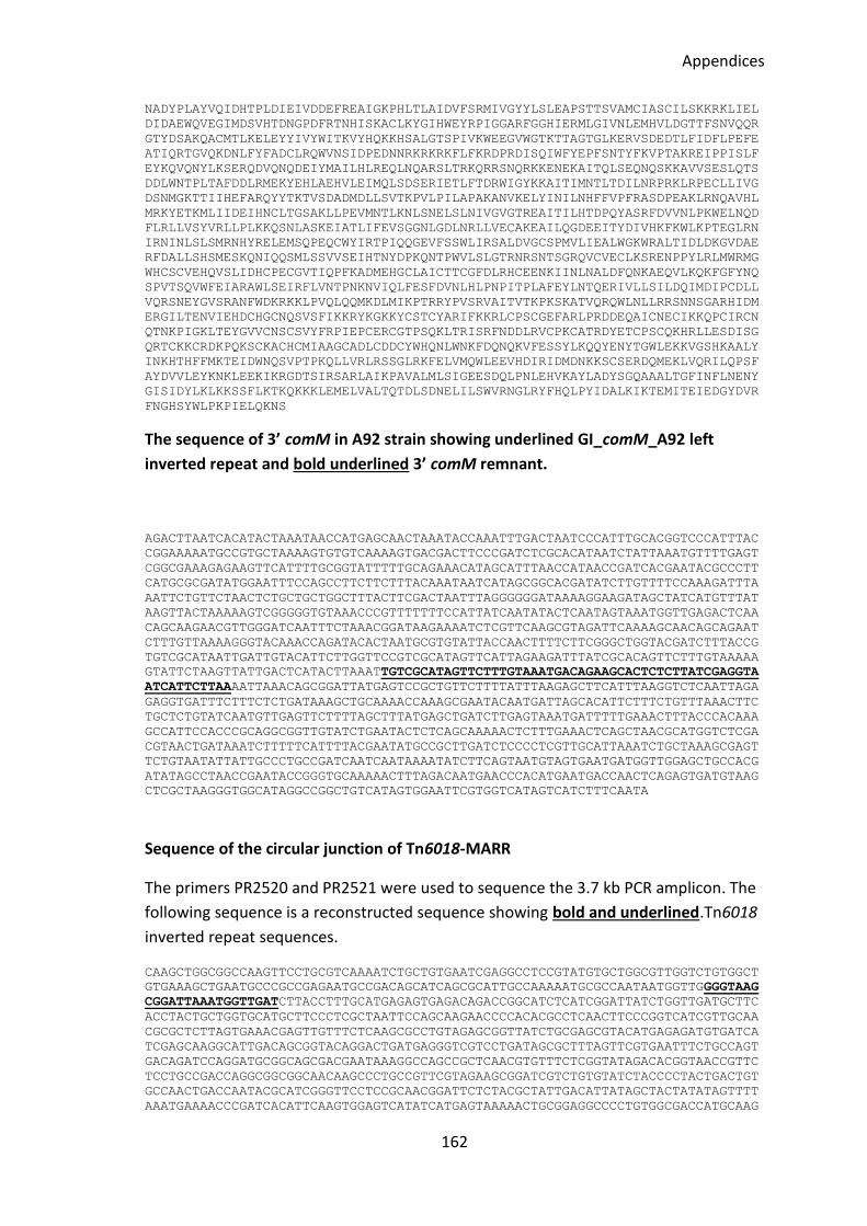
Appendices
162
NADYPLAYVQIDHTPLDIEIVDDEFREAIGKPHLTLAIDVFSRMIVGYYLSLEAPSTTSVAMCIASCILSKKRKLIEL
DIDAEWQVEGIMDSVHTDNGPDFRTNHISKACLKYGIHWEYRPIGGARFGGHIERMLGIVNLEMHVLDGTTFSNVQQR
GTYDSAKQACMTLKELEYYIVYWITKVYHQKKHSALGTSPIVKWEEGVWGTKTTAGTGLKERVSDEDTLFIDFLPEFE
ATIQRTGVQKDNLFYFADCLRQWVNSIDPEDNNRKRKRKFLFKRDPRDISQIWFYEPFSNTYFKVPTAKREIPPISLF
EYKQVQNYLKSERQDVQNQDEIYMAILHLREQLNQARSLTRKQRRSNQRKKENEKAITQLSEQNQSKKAVVSESLQTS
DDLWNTPLTAFDDLRMEKYEHLAEHVLEIMQLSDSERIETLFTDRWIGYKKAITIMNTLTDILNRPRKLRPECLLIVG
DSNMGKTTIIHEFARQYYTKTVSDADMDLLSVTKPVLPILAPAKANVKELYINILNHFFVPFRASDPEAKLRNQAVHL
MRKYETKMLIIDEIHNCLTGSAKLLPEVMNTLKNLSNELSLNIVGVGTREAITILHTDPQYASRFDVVNLPKWELNQD
FLRLLVSYVRLLPLKKQSNLASKEIATLIFEVSGGNLGDLNRLLVECAKEAILQGDEEITYDIVHKFKWLKPTEGLRN
IRNINLSLSMRNHYRELEMSQPEQCWYIRTPIQQGEVFSSWLIRSALDVGCSPMVLIEALWGKWRALTIDLDKGVDAE
RFDALLSHSMESKQNIQQSMLSSVVSEIHTNYDPKQNTPWVLSLGTRNRSNTSGRQVCVECLKSRENPPYLRLMWRMG
WHCSCVEHQVSLIDHCPECGVTIQPFKADMEHGCLAICTTCGFDLRHCEENKIINLNALDFQNKAEQVLKQKFGFYNQ
SPVTSQVWFEIARAWLSEIRFLVNTPNKNVIQLFESFDVNLHLPNPITPLAFEYLNTQERIVLLSILDQIMDIPCDLL
VQRSNEYGVSRANFWDKRKKLPVQLQQMKDLMIKPTRRYPVSRVAITVTKPKSKATVQRQWLNLLRRSNNSGARHIDM
ERGILTENVIEHDCHGCNQSVSFIKKRYKGKKYCSTCYARIFKKRLCPSCGEFARLPRDDEQAICNECIKKQPCIRCN
QTNKPIGKLTEYGVVCNSCSVYFRPIEPCERCGTPSQKLTRISRFNDDLRVCPKCATRDYETCPSCQKHRLLESDISG
QRTCKKCRDKPQKSCKACHCMIAAGCADLCDDCYWHQNLWNKFDQNQKVFESSYLKQQYENYTGWLEKKVGSHKAALY
INKHTHFFMKTEIDWNQSVPTPKQLLVRLRSSGLRKFELVMQWLEEVHDIRIDMDNKKSCSERDQMEKLVQRILQPSF
AYDVVLEYKNKLEEKIKRGDTSIRSARLAIKPAVALMLSIGEESDQLPNLEHVKAYLADYSGQAAALTGFINFLNENY
GISIDYLKLKKSSFLKTKQKKKLEMELVALTQTDLSDNELILSWVRNGLRYFHQLPYIDALKIKTEMITEIEDGYDVR
FNGHSYWLPKPIELQKNS
The sequence of 3’ comM in A92 strain showing underlined GI_comM_A92 left
inverted repeat and bold underlined 3’ comM remnant.
AGACTTAATCACATACTAAATAACCATGAGCAACTAAATACCAAATTTGACTAATCCCATTTGCACGGTCCCATTTAC
CGGAAAAATGCCGTGCTAAAAGTGTGTCAAAAGTGACGACTTCCCGATCTCGCACATAATCTATTAAATGTTTTGAGT
CGGCGAAAGAGAAGTTCATTTTGCGGTATTTTTGCAGAAACATAGCATTTAACCATAACCGATCACGAATACGCCCTT
CATGCGCGATATGGAATTTCCAGCCTTCTTCTTTACAAATAATCATAGCGGCACGATATCTTGTTTTCCAAAGATTTA
AATTCTGTTCTAACTCTGCTGCTGGCTTTACTTCGACTAATTTAGGGGGGATAAAAGGAAGATAGCTATCATGTTTAT
AAGTTACTAAAAAGTCGGGGGTGTAAACCCGTTTTTTTCCATTATCAATATACTCAATAGTAAATGGTTGAGACTCAA
CAGCAAGAACGTTGGGATCAATTTCTAAACGGATAAGAAAATCTCGTTCAAGCGTAGATTCAAAAGCAACAGCAGAAT
CTTTGTTAAAAGGGTACAAACCAGATACACTAATGCGTGTATTACCAACTTTTCTTCGGGCTGGTACGATCTTTACCG
TGTCGCATAATTGATTGTACATTCTTGGTTCCGTCGCATAGTTCATTAGAAGATTTATCGCACAGTTCTTTGTAAAAA
GTATTCTAAGTTATTGACTCATACTTAAATTGTCGCATAGTTCTTTGTAAATGACAGAAGCACTCTCTTATCGAGGTA
ATCATTCTTAAAATTAAACAGCGGATTATGAGTCCGCTGTTCTTTTATTTAAGAGCTTCATTTAAGGTCTCAATTAGA
GAGGTGATTTCTTTCTCTGATAAAGCTGCAAAACCAAAGCGAATACAATGATTAGCACATTCTTTCTGTTTAAACTTC
TGCTCTGTATCAATGTTGAGTTCTTTTAGCTTTATGAGCTGATCTTGAGTAAATGATTTTTGAAACTTTACCCACAAA
GCCATTCCACCCGCAGGCGGTTGTATCTGAATACTCTCAGCAAAAACTCTTTGAAACTCAGCTAACGCATGGTCTCGA
CGTAACTGATAAATCTTTTTCATTTTACGAATATGCCGCTTGATCTCCCCTCGTTGCATTAAATCTGCTAAAGCGAGT
TCTGTAATATTATTGCCCTGCCGATCAATCAATAAAATATCTTCAGTAATGTAGTGAATGATGGTTGGAGCTGCCACG
ATATAGCCTAACCGAATACCGGGTGCAAAAACTTTAGACAATGAACCCACATGAATGACCAACTCAGAGTGATGTAAG
CTCGCTAAGGGTGGCATAGGCCGGCTGTCATAGTGGAATTCGTGGTCATAGTCATCTTTCAATA
Sequence of the circular junction of Tn6018-MARR
The primers PR2520 and PR2521 were used to sequence the 3.7 kb PCR amplicon. The
following sequence is a reconstructed sequence showing bold and underlined.Tn6018
inverted repeat sequences.
CAAGCTGGCGGCCAAGTTCCTGCGTCAAAATCTGCTGTGAATCGAGGCCTCCGTATGTGCTGGCGTTGGTCTGTGGCT
GTGAAAGCTGAATGCCCGCCGAGAATGCCGACAGCATCAGCGCATTGCCAAAAATGCGCCAATAATGGTTGGGGTAAG
CGGATTAAATGGTTGATCTTACCTTTGCATGAGAGTGAGACAGACCGGCATCTCATCGGATTATCTGGTTGATGCTTC
ACCTACTGCTGGTGCATGCTTCCCTCGCTAATTCCAGCAAGAACCCCACACGCCTCAACTTCCCGGTCATCGTTGCAA
CGCGCTCTTAGTGAAACGAGTTGTTTCTCAAGCGCCTGTAGAGCGGTTATCTGCGAGCGTACATGAGAGATGTGATCA
TCGAGCAAGGCATTGACAGCGGTACAGGACTGATGAGGGTCGTCCTGATAGCGCTTTAGTTCGTGAATTTCTGCCAGT
GACAGATCCAGGATGCGGCAGCGACGAATAAAGGCCAGCCGCTCAACGTGTTTCTCGGTATAGACACGGTAACCGTTC
TCCTGCCGACCAGGCGGCGGCAACAAGCCCTGCCGTTCGTAGAAGCGGATCGTCTGTGTATCTACCCCTACTGACTGT
GCCAACTGACCAATACGCATCGGGTTCCTCCGCAACGGATTCTCTACGCTATTGACATTATAGCTACTATATAGTTTT
AAATGAAAACCCGATCACATTCAAGTGGAGTCATATCATGAGTAAAAACTGCGGAGGCCCCTGTGGCGACCATGCAAG
Appendices
163
GCCTGCGGCGGATACCGATATGCAGGCCTCCTCCGATGCGTCGGGGGAATGGGTCAGTGTTTATGCCGTGCCGAAGAT
GGACTGTCCATCAGAAGAGCGCATGATTCGCCTGGCCCTGAACGGCGTTGAGGGGATTCGGGTACTGTCCTTCGACTT
GTCGAACCGCCGGTTGAAGGTCGTGCATGACGGTGAGGTCGAGCCCGTCACCTCGAAACTGAAGACCTTGGGGCTAGG
CGCCTCGCTTCAGGAAACCGTCGCTGCAAATCCGGAGACCATCAAGGCCGCCGAGTTTTCGGCAGCTTCTGCTAAGCA
AGAATCCGGGACCCTGCGCTGGTTGCTCGGCATCAATGCACTTCTGTTCGTGGTGGAAATGACTGCCGGTCTGATCGC
CCGGTCCACCGGCCTGATTGGAGAATCCCTGGACAATTTTGCCGATGCGGCGGTGTACGGGCTTGCCCTTTATGCGGT
TGGACATAGCGTGAAAATGCAGGTACGTGCCGCGCATCTTGCTGGTGTACTGCAACTGATCTTGGCTGTGGGCGTACT
CATAGAGGTGGTGAGACGCTTTGTATTCGGTAGTGAGCCTGAATCGCTGGTGATGATGGCTATCGCATTCGTCGCATT
GATTGCCAATACCAGTTGTCTGCTGCTCATATCCAAACATCGGGAGGGCGGGGCGCACATGAAGGCAAGCTGGATATT
CTCGGCCAACGACGTGGTGATCAACCTGGGGGTCATCACCGCCGGCGCTCTGGTCGCGTGGACCGGGTCCAATTATCC
GGATCTGATTATCGGCACCATCGCGGGGGTAATTGTACTTAACGGTGCTAGACGCATTCTGGCGCTCAAGGATTAAGC
AATGTCCATTTTTGGCAAACATCTGTCACCGTACGCTCTGTTGTCCATATCGGGCCTGCTGGCAGCATCTGATCAGGC
CATAAAATGGCTGGTGCAACAATCAATGGCATATGGCGAGTCTATTTCAGTGACCTCATTCTTTAACTGGGTACACGT
ATGGAATACCGGTGCCGCATTCAGTCTTTTTGCGAATGGTGGAGGCTGGCAGCGCTACTTTTTTATCGGAATCGCGGT
AGTGGTCTCGATTTTTCTGATCAAGCTGATCCTTGAAAATCGTCATAAAGGAGAAGCCATCGCTTACAGTCTTATCCT
CGGTGGCGCCATGGGCAACCTGATTGACCGGGTCTTTCGCGGCTATGTTGTGGATTCCTTTGATTTCTATTGGCGAGA
CTGGCATTGGCCGGCCTTCAACCTGGCTGATATTGCAATTGTCCTCGGTGCCTTACTTTTCGTTTCCAGCAGCTTGTT
GGGTAAAAAAGCAAACACCAATGCCGAGTCGGATGGATCTGACTGACACCTACGCCTATACAACACCATGACCGAACT
TCCCGACAACATCCTTCACCTGCCGCAATACCAAGTACTGGGCTGCAAATCAACCGACGACGAAATGCACTTCCAGGT
GGACGTGCCCGATCCCATCGCCTGCGAGGAATGCGGCGTGCAGGGTGAGTTCGTACGGTTCGGCAAGCGTGACGTTCC
CTATCGTGATCTGCCCATCCACGGCAAGCGGGTCACTCTCTGGGTGGTCCGCCGCCGATACACCTGCCGGGCCTGCAA
GACAACATTCAGGCCCCAGCTACCGGAGATGGTGGACGGATTCCGTATGACACTGCGGCTGCATGAGTACGTGGAGAA
GGAATCCTTCAACCACCCCTACACCTTTGTGGCGGCACAGACCGGCCTGGACGAGAAGACGGTGCGCGACATCTTCAA
CGCCCGCGCCGAGTTCCTGGGGCGCTGGCACCGCTTCGAGACGCCCCGCATCCTGGGCATTGACGAGCTATACCTGAA
CAAGCGCTACCGCTGCATTCTGACCAACATTGAGGAGCGAACCCTGCTCGACCTGCTGGCCACCCGCCGACAGGACGT
GGTGACCAACTACCTGATGAAGCTGAAAGACCGGCAGAAGGTCGAGATCGTCAGCATGGACATGTGGAACCCCTACCG
GGCAGCGGTCAAGGCTGTGCTGCCCCAGGCCCGTATCGTGGTCGATAAGTTCCATGTGGTGCGCATGGCCAACGATGC
CCTAGAGAGAGTGCGCAAGGACCTCAGAAAGGAGCTGAAACCGTCCCAGAGCCGGACTCTCAAGGGAGACCGGAAAAT
CCTGCTGAAACGCGCTCACGAAGTCTCAGACCGGGAGCGCCTCATCATGGAGACCTGGACAGGCGCGTTCCCGCAACT
GCTGGCCGCCTACGAGCACAAGGAGCGCTTCTACGGCATCTGGGACGCCACCACACGGCTCCAGGCAGAAGCCGCCCT
GGACGAGTGGATAGCCACCATCCCGAAGGGCCAAAAGGAAGTCTGGAGCGATCTGGTCAGGGCAGTGGGAAACTGGCG
CGAAGAGACCATGACCTACTTCGAGACGGACATGCCCGTCACCAACGCTTACACAGAGTCCATCAACCGACTGGCCAA
GGACAAGAACCGTGAAGGGCGCGGTTACTCCTTCGAGGTGATGCGGGCACGAATGCTCTACACCACGAAGCACAAGAA
GAAGGCACCGACTGCGAAGGTCTCTCCTTTCTACAAGAAAACCATCGGTTACGGACTGCCGGACTTCGCAGAGGAACT
CAACTACGGAGTCGATCTATCAACCATCTGAGGGTGGTATCAGATTGATGGGGTGAAGGTGCCCCATCAACCATTAAA
TCCGTATACCCCAATTTGGATGCGGTCGCGCGCGGCTTGCGCGAACGCTGGCCGGATCGGTTGCTATTGATTGCAGCG
GACAATGACCACCGGGCCGAGAAGGAGCTACGGCCAGACGGCAAGCCCCGCGAAAATGTTGGCTTGAAGAAGGCACAA
GAGGCGGCGGCCAACCATAAGGGTGTGGCACTGGTGCCGCCCTTCGCCGATGGGG
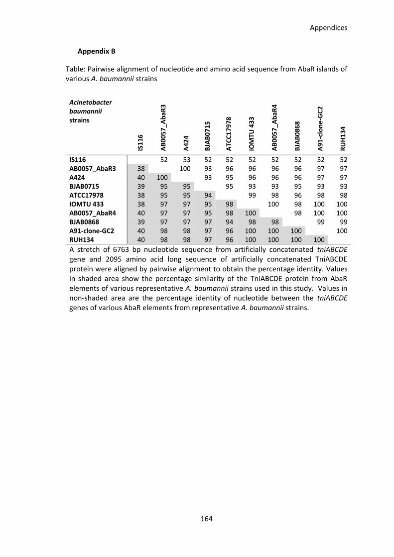
Appendices
164
Appendix B
Table: Pairwise alignment of nucleotide and amino acid sequence from AbaR islands of various A. baumannii strains
Acinetobacter baumannii strains
IS1
16
AB
00
57
_Ab
aR3
A4
24
BJA
B0
71
5
ATC
C1
79
78
IOM
TU 4
33
AB
00
57
_Ab
aR4
BJA
B0
86
8
A9
1-c
lon
e-G
C2
RU
H1
34
IS116 52 53 52 52 52 52 52 52 52 AB0057_AbaR3 38 100 93 96 96 96 96 97 97 A424 40 100 93 95 96 96 96 97 97 BJAB0715 39 95 95 95 93 93 95 93 93 ATCC17978 38 95 95 94 99 98 96 98 98 IOMTU 433 38 97 97 95 98 100 98 100 100 AB0057_AbaR4 40 97 97 95 98 100 98 100 100 BJAB0868 39 97 97 97 94 98 98 99 99 A91-clone-GC2 40 98 98 97 96 100 100 100 100 RUH134 40 98 98 97 96 100 100 100 100
A stretch of 6763 bp nucleotide sequence from artificially concatenated tniABCDE gene and 2095 amino acid long sequence of artificially concatenated TniABCDE protein were aligned by pairwise alignment to obtain the percentage identity. Values in shaded area show the percentage similarity of the TniABCDE protein from AbaR elements of various representative A. baumannii strains used in this study. Values in non-shaded area are the percentage identity of nucleotide between the tniABCDE genes of various AbaR elements from representative A. baumannii strains.
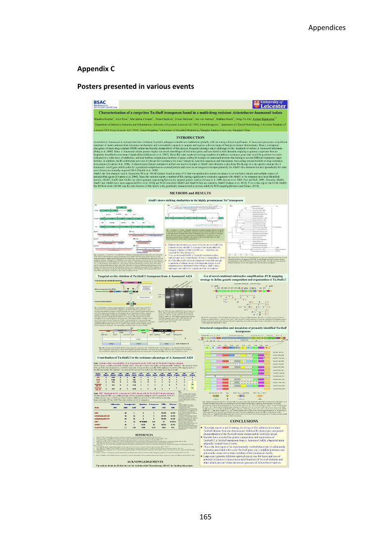
Appendices
165
Appendix C
Posters presented in various events
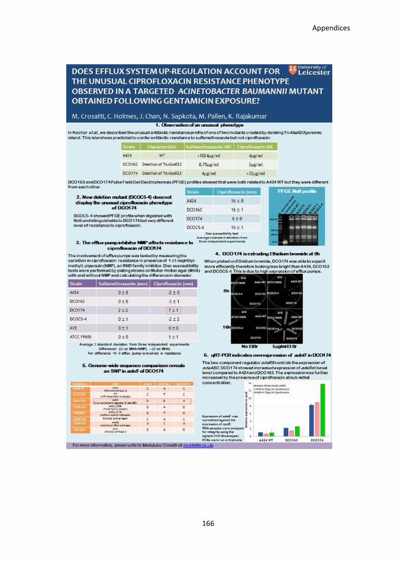
Appendices
166
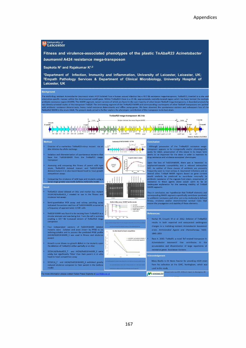
Appendices
167

168
References

169
Abbas, S., Ahmed, I., Kudo, T., Iida, T., Ali, G.M., Fujiwara, T., and Ohkuma (2014) 'Heavy metal-tolerant and psychrotolerant bacterium Acinetobacter pakistanensis sp. nov. isolated from a textile dyeing wastewater treatment pond', Pak. J. Agri. Sci, 51, 595-608.
Adams, M. D., Chan, E. R., Molyneaux, N. D. and Bonomo, R. A. (2010) 'Genomewide analysis
of divergence of antibiotic resistance determinants in closely related isolates of Acinetobacter baumannii', Antimicrob Agents Chemother, 54(9), 3569-77.
Adams, M. D., Goglin, K., Molyneaux, N., Hujer, K. M., Lavender, H., Jamison, J. J., MacDonald,
I. J., Martin, K. M., Russo, T., Campagnari, A. A., Hujer, A. M., Bonomo, R. A. and Gill, S. R. (2008) 'Comparative genome sequence analysis of multidrug-resistant Acinetobacter baumannii', J Bacteriol, 190(24), 8053-64.
Alvarez-Perez, S., Lievens, B., Jacquemyn, H. and Herrera, C. M. (2013) 'Acinetobacter nectaris
sp. nov. and Acinetobacter boissieri sp. nov., isolated from floral nectar of wild Mediterranean insect-pollinated plants', Int J Syst Evol Microbiol, 63(Pt 4), 1532-9.
Andersson, D. I. and Hughes, D. (2010) 'Antibiotic resistance and its cost: is it possible to
reverse resistance?', Nat Rev Microbiol, 8(4), 260-71. Batirel, A., Balkan, II, Karabay, O., Agalar, C., Akalin, S., Alici, O., Alp, E., Altay, F. A., Altin, N.,
Arslan, F., Aslan, T., Bekiroglu, N., Cesur, S., Celik, A. D., Dogan, M., Durdu, B., Duygu, F., Engin, A., Engin, D. O., Gonen, I., Guclu, E., Guven, T., Hatipoglu, C. A., Hosoglu, S., Karahocagil, M. K., Kilic, A. U., Ormen, B., Ozdemir, D., Ozer, S., Oztoprak, N., Sezak, N., Turhan, V., Turker, N. and Yilmaz, H. (2014) 'Comparison of colistin-carbapenem, colistin-sulbactam, and colistin plus other antibacterial agents for the treatment of extremely drug-resistant Acinetobacter baumannii bloodstream infections', Eur J Clin Microbiol Infect Dis, 33(8), 1311-22.
Baumann, P. (1968) 'Isolation of Acinetobacter from soil and water', J Bacteriol, 96(1), 39-42. Baumann, P., Doudoroff, M. and Stanier, R. Y. (1968) 'A study of the Moraxella group. II.
Oxidative-negative species (genus Acinetobacter)', J Bacteriol, 95(5), 1520-41. Beceiro, A., Moreno, A., Fernandez, N., Vallejo, J. A., Aranda, J., Adler, B., Harper, M., Boyce, J.
D. and Bou, G. (2014) 'Biological cost of different mechanisms of colistin resistance and their impact on virulence in Acinetobacter baumannii', Antimicrob Agents Chemother, 58(1), 518-26.
Bergogne-Berezin, E. and Towner, K. J. (1996) 'Acinetobacter spp. as nosocomial pathogens:
microbiological, clinical, and epidemiological features', Clin Microbiol Rev, 9(2), 148-65. Berlau, J., Aucken, H., Malnick, H. and Pitt, T. (1999) 'Distribution of Acinetobacter species on
skin of healthy humans', Eur J Clin Microbiol Infect Dis, 18(3), 179-83. Bouvet, P. J. and Jeanjean, S. (1989) 'Delineation of new proteolytic genomic species in the
genus Acinetobacter', Res Microbiol, 140(4-5), 291-9. Bouvet, P. J. M., and Grimont, P.A.D (1986) 'Taxonomy of the genus Acinetobacter with the
recognition of Acinetobacter baumannii sp. nov., Acinetobacter haemolyticus sp. nov.,
170
Acinetobacter johnsonii sp. nov., and Acinetobacter junii sp. nov. and emended descriptions of Acinetobacter calcoaceticus and Acinetobacter lwoffii', Int. J. Syst. Bacteriol, 36, 228-240.
Brisou, J. and Prevot, A. R. (1954) '[Studies on bacterial taxonomy. X. The revision of species
under Acromobacter group]', Ann Inst Pasteur (Paris), 86(6), 722-8. Camarena, L., Bruno, V., Euskirchen, G., Poggio, S. and Snyder, M. (2010) 'Molecular
mechanisms of ethanol-induced pathogenesis revealed by RNA-sequencing', PLoS Pathog, 6(4), e1000834.
Carr, E. L., Kampfer, P., Patel, B. K., Gurtler, V. and Seviour, R. J. (2003) 'Seven novel species of
Acinetobacter isolated from activated sludge', Int J Syst Evol Microbiol, 53(Pt 4), 953-63.
CDC (2004) 'Acinetobacter baumannii infections among patients at military medical facilities
treating injured U.S. service members, 2002-2004', MMWR Morb Mortal Wkly Rep, 53(45), 1063-6.
Cerqueira, G. M. and Peleg, A. Y. (2011) 'Insights into Acinetobacter baumannii pathogenicity',
IUBMB Life, 63(12), 1055-60. Charles, E. (1968) 'Herellea (Acinetobacter) and Pseudomonas ovalis (P. putida) from Frozen Foods', Applied Microbiology, 17(No.1), 26-30. Chaudhary, H. J., Peng, G., Hu, M., He, Y., Yang, L., Luo, Y. and Tan, Z. (2012) 'Genetic diversity
of endophytic diazotrophs of the wild rice, Oryza alta and identification of the new diazotroph, Acinetobacter oryzae sp. nov', Microb Ecol, 63(4), 813-21.
Chaveroche, M. K., Ghigo, J. M. and d'Enfert, C. (2000) 'A rapid method for efficient gene
replacement in the filamentous fungus Aspergillus nidulans', Nucleic Acids Res, 28(22), E97.
Choi, J. Y., Ko, G., Jheong, W., Huys, G., Seifert, H., Dijkshoorn, L. and Ko, K. S. (2013)
'Acinetobacter kookii sp. nov., isolated from soil', Int J Syst Evol Microbiol, 63(Pt 12), 4402-6.
Choi, K. H. and Schweizer, H. P. (2005) 'An improved method for rapid generation of unmarked
Pseudomonas aeruginosa deletion mutants', BMC Microbiol, 5, 30. Choi, K. Y., Li, Y., Sarnovsky, R. and Craig, N. L. (2013) 'Direct interaction between the TnsA and
TnsB subunits controls the heteromeric Tn7 transposase', Proc Natl Acad Sci U S A, 110(22), E2038-45.
CLSI (2009) 'Methods for dilution antimicrobial susceptibility tests for bacteria that grow
aerobically', Approved standard, 29 No. 2(Eighth edition), M07-A8. Costerton, J. W. (1999) 'Bacterial Biofilms: A Common Cause of Persistent Infections', Science,
284(5418), 1318-1322. Craig, N. L. (1997) 'Target site selection in transposition', Annu Rev Biochem, 66, 437-74.
171
Dennis, J. J. and Zylstra, G. J. (1998) 'Plasposons: modular self-cloning minitransposon derivatives for rapid genetic analysis of gram-negative bacterial genomes', Appl Environ Microbiol, 64(7), 2710-5.
Di Cello, F., Pepi, M., Baldi, F. and Fani, R. (1997) 'Molecular characterization of an n-alkane-
degrading bacterial community and identification of a new species, Acinetobacter venetianus', Res Microbiol, 148(3), 237-49.
Di Nocera, P. P., Rocco, F., Giannouli, M., Triassi, M. and Zarrilli, R. (2011) 'Genome
organization of epidemic Acinetobacter baumannii strains', BMC Microbiol, 11, 224. Diancourt, L., Passet, V., Nemec, A., Dijkshoorn, L. and Brisse, S. (2010) 'The population
structure of Acinetobacter baumannii: expanding multiresistant clones from an ancestral susceptible genetic pool', PLoS One, 5(4), e10034.
Dijkshoorn, L., Nemec, A. and Seifert, H. (2007) 'An increasing threat in hospitals: multidrug-
resistant Acinetobacter baumannii', Nat Rev Microbiol, 5(12), 939-51. Domingues, S., Harms, K., Fricke, W. F., Johnsen, P. J., da Silva, G. J. and Nielsen, K. M. (2012)
'Natural transformation facilitates transfer of transposons, integrons and gene cassettes between bacterial species', PLoS Pathog, 8(8), e1002837.
Dower, W. J., Miller, J. F. and Ragsdale, C. W. (1988) 'High efficiency transformation of E. coli
by high voltage electroporation', Nucleic Acids Res, 16(13), 6127-45. Falagas, M. E., Bliziotis, I. A. and Siempos, II (2006) 'Attributable mortality of Acinetobacter
baumannii infections in critically ill patients: a systematic review of matched cohort and case-control studies', Crit Care, 10(2), R48.
Feng, G. D., Yang, S. Z., Wang, Y. H., Deng, M. R. and Zhu, H. H. (2014) 'Acinetobacter
guangdongensis sp. nov., isolated from abandoned lead-zinc ore', Int J Syst Evol Microbiol, 64(Pt 10), 3417-21.
Fernandez, A., Perez, A., Ayala, J. A., Mallo, S., Rumbo-Feal, S., Tomas, M., Poza, M. and Bou, G.
(2012) 'Expression of OXA-type and SFO-1 beta-lactamases induces changes in peptidoglycan composition and affects bacterial fitness', Antimicrob Agents Chemother, 56(4), 1877-84.
Field, D., Wilson, G. and van der Gast, C. (2006) 'How do we compare hundreds of bacterial
genomes?', Curr Opin Microbiol, 9(5), 499-504. Fournier, P. E., Vallenet, D., Barbe, V., Audic, S., Ogata, H., Poirel, L., Richet, H., Robert, C.,
Mangenot, S., Abergel, C., Nordmann, P., Weissenbach, J., Raoult, D. and Claverie, J. M. (2006) 'Comparative genomics of multidrug resistance in Acinetobacter baumannii', PLoS Genet, 2(1), e7.
Frost, L. S., Leplae, R., Summers, A. O. and Toussaint, A. (2005) 'Mobile genetic elements: the
agents of open source evolution', Nat Rev Microbiol, 3(9), 722-32. Gaddy, J. A. and Actis, L. A. (2009) 'Regulation of Acinetobacter baumannii biofilm formation',
Future Microbiol, 4(3), 273-8.
172
Gan, H. M., Lean, S. S., Suhaili, Z., Thong, K. L. and Yeo, C. C. (2012) 'Genome sequence of Acinetobacter baumannii AC12, a polymyxin-resistant strain isolated from Terengganu, Malaysia', J Bacteriol, 194(21), 5979-80.
Gao, F., Wang, Y., Liu, Y. J., Wu, X. M., Lv, X., Gan, Y. R., Song, S. D. and Huang, H. (2011)
'Genome sequence of Acinetobacter baumannii MDR-TJ', J Bacteriol, 193(9), 2365-6. Gaynes, R. and Edwards, J. R. (2005) 'Overview of nosocomial infections caused by gram-
negative bacilli', Clin Infect Dis, 41(6), 848-54. Gerner-Smidt, P., Tjernberg, I. and Ursing, J. (1991) 'Reliability of phenotypic tests for
identification of Acinetobacter species', J Clin Microbiol, 29(2), 277-82. Gottig, S., Gruber, T. M., Higgins, P. G., Wachsmuth, M., Seifert, H. and Kempf, V. A. (2014)
'Detection of pan drug-resistant Acinetobacter baumannii in Germany', J Antimicrob Chemother, 69(9), 2578-9.
Guo, B., Abdelraouf, K., Ledesma, K. R., Nikolaou, M. and Tam, V. H. (2012) 'Predicting bacterial
fitness cost associated with drug resistance', J Antimicrob Chemother, 67(4), 928-32. Guzman, L. M., Belin, D., Carson, M. J. and Beckwith, J. (1995) 'Tight regulation, modulation,
and high-level expression by vectors containing the arabinose PBAD promoter', J Bacteriol, 177(14), 4121-30.
Hamidian, M. and Hall, R. M. (2011) 'AbaR4 replaces AbaR3 in a carbapenem-resistant
Acinetobacter baumannii isolate belonging to global clone 1 from an Australian hospital', J Antimicrob Chemother, 66(11), 2484-91.
Hamidian, M., Hancock, D. P. and Hall, R. M. (2013) 'Horizontal transfer of an ISAba125-
activated ampC gene between Acinetobacter baumannii strains leading to cephalosporin resistance', J Antimicrob Chemother, 68(1), 244-5.
Hamidian, M., Kenyon, J. J., Holt, K. E., Pickard, D. and Hall, R. M. (2014a) 'A conjugative
plasmid carrying the carbapenem resistance gene blaOXA-23 in AbaR4 in an extensively resistant GC1 Acinetobacter baumannii isolate', J Antimicrob Chemother, 69(10), 2625-8.
Hamidian, M., Wynn, M., Holt, K. E., Pickard, D., Dougan, G. and Hall, R. M. (2014b)
'Identification of a marker for two lineages within the GC1 clone of Acinetobacter baumannii', J Antimicrob Chemother, 69(2), 557-8.
Hanahan, D., Jessee, J. and Bloom, F. R. (1991) 'Plasmid transformation of Escherichia coli and
other bacteria', Methods Enzymol, 204, 63-113. Harmer, C. J., Moran, R. A. and Hall, R. M. (2014) 'Movement of IS26-associated antibiotic
resistance genes occurs via a translocatable unit that includes a single IS26 and preferentially inserts adjacent to another IS26', MBio, 5(5), e01801-14.
Harrison, E. M., Carter, M. E., Luck, S., Ou, H. Y., He, X., Deng, Z., O'Callaghan, C., Kadioglu, A.
and Rajakumar, K. (2010) 'Pathogenicity islands PAPI-1 and PAPI-2 contribute individually and synergistically to the virulence of Pseudomonas aeruginosa strain PA14', Infect Immun, 78(4), 1437-46.
173
Hornsey, M., Loman, N., Wareham, D. W., Ellington, M. J., Pallen, M. J., Turton, J. F.,
Underwood, A., Gaulton, T., Thomas, C. P., Doumith, M., Livermore, D. M. and Woodford, N. (2011) 'Whole-genome comparison of two Acinetobacter baumannii isolates from a single patient, where resistance developed during tigecycline therapy', J Antimicrob Chemother, 66(7), 1499-503.
Iacono, M., Villa, L., Fortini, D., Bordoni, R., Imperi, F., Bonnal, R. J., Sicheritz-Ponten, T., De
Bellis, G., Visca, P., Cassone, A. and Carattoli, A. (2008) 'Whole-genome pyrosequencing of an epidemic multidrug-resistant Acinetobacter baumannii strain belonging to the European clone II group', Antimicrob Agents Chemother, 52(7), 2616-25.
Imperi, F., Antunes, L. C., Blom, J., Villa, L., Iacono, M., Visca, P. and Carattoli, A. (2011) 'The
genomics of Acinetobacter baumannii: insights into genome plasticity, antimicrobial resistance and pathogenicity', IUBMB Life, 63(12), 1068-74.
Isberg, R. R., Iwashkiw, J. A., Seper, A., Weber, B. S., Scott, N. E., Vinogradov, E., Stratilo, C.,
Reiz, B., Cordwell, S. J., Whittal, R., Schild, S. and Feldman, M. F. (2012) 'Identification of a General O-linked Protein Glycosylation System in Acinetobacter baumannii and Its Role in Virulence and Biofilm Formation', PLoS Pathogens, 8(6), e1002758.
Joseph, P. and Craig, N. L. (2001) 'Tn7 smarter than we thought ', Nature Reviews, 2. Jung, J. and Park, W. (2015) 'Acinetobacter species as model microorganisms in environmental
microbiology: current state and perspectives', Appl Microbiol Biotechnol, 99(6), 2533-48.
Kang, Y. S., Jung, J., Jeon, C. O. and Park, W. (2011) 'Acinetobacter oleivorans sp. nov. is
capable of adhering to and growing on diesel-oil', J Microbiol, 49(1), 29-34. Kempf, M. and Rolain, J. M. (2012) 'Emergence of resistance to carbapenems in Acinetobacter
baumannii in Europe: clinical impact and therapeutic options', Int J Antimicrob Agents, 39(2), 105-14.
Kenyon, J. J. and Hall, R. M. (2013) 'Variation in the complex carbohydrate biosynthesis loci of
Acinetobacter baumannii genomes', PLoS One, 8(4), e62160. Kim, D., Baik, K. S., Kim, M. S., Park, S. C., Kim, S. S., Rhee, M. S., Kwak, Y. S. and Seong, C. N.
(2008) 'Acinetobacter soli sp. nov., isolated from forest soil', J Microbiol, 46(4), 396-401.
Kim, D. H. and Ko, K. S. (2015) 'AbaR-type genomic islands in non-baumannii Acinetobacter
species isolates from South Korea', Antimicrob Agents Chemother, 59(9), 5824-6. Kim, P. S., Shin, N. R., Kim, J. Y., Yun, J. H., Hyun, D. W. and Bae, J. W. (2014) 'Acinetobacter
apis sp. nov., isolated from the intestinal tract of a honey bee, Apis mellifera', J Microbiol, 52(8), 639-45.
Kochar, M., Crosatti, M., Harrison, E. M., Rieck, B., Chan, J., Constantinidou, C., Pallen, M., Ou,
H. Y. and Rajakumar, K. (2012) 'Deletion of TnAbaR23 results in both expected and
174
unexpected antibiogram changes in a multidrug-resistant Acinetobacter baumannii strain', Antimicrob Agents Chemother, 56(4), 1845-53.
Krizova, L., Dijkshoorn, L. and Nemec, A. (2011) 'Diversity and evolution of AbaR genomic
resistance islands in Acinetobacter baumannii strains of European clone I', Antimicrob Agents Chemother, 55(7), 3201-6.
Krizova, L., Maixnerova, M., Sedo, O. and Nemec, A. (2014) 'Acinetobacter bohemicus sp. nov.
widespread in natural soil and water ecosystems in the Czech Republic', Syst Appl Microbiol, 37(7), 467-73.
Krizova, L., McGinnis, J., Maixnerova, M., Nemec, M., Poirel, L., Mingle, L., Sedo, O., Wolfgang,
W. and Nemec, A. (2015) 'Acinetobacter variabilis sp. nov. (formerly DNA group 15 sensu Tjernberg & Ursing), isolated from humans and animals', Int J Syst Evol Microbiol, 65(Pt 3), 857-63.
Krizova, L. and Nemec, A. (2010) 'A 63 kb genomic resistance island found in a multidrug-
resistant Acinetobacter baumannii isolate of European clone I from 1977', J Antimicrob Chemother, 65(9), 1915-8.
Lee, H. J. and Lee, S. S. (2010) 'Acinetobacter kyonggiensis sp. nov., a beta-glucosidase-
producing bacterium, isolated from sewage treatment plant', J Microbiol, 48(6), 754-9. Lee, J. S., Lee, K. C., Kim, K. K., Hwang, I. C., Jang, C., Kim, N. G., Yeo, W. H., Kim, B. S., Yu, Y. M.
and Ahn, J. S. (2009) 'Acinetobacter antiviralis sp. nov., from Tobacco plant roots', J Microbiol Biotechnol, 19(3), 250-6.
Li, W., Zhang, D., Huang, X. and Qin, W. (2014) 'Acinetobacter harbinensis sp. nov., isolated
from river water', Int J Syst Evol Microbiol, 64(Pt 5), 1507-13. Li, Y., He, W., Wang, T., Piao, C. G., Guo, L. M., Chang, J. P., Guo, M. W. and Xie, S. J. (2014)
'Acinetobacter qingfengensis sp. nov., isolated from canker bark of Populus x euramericana', Int J Syst Evol Microbiol, 64(Pt 3), 1043-50.
Li, Y., Piao, C. G., Ma, Y. C., He, W., Wang, H. M., Chang, J. P., Guo, L. M., Wang, X. Z., Xie, S. J.
and Guo, M. W. (2013) 'Acinetobacter puyangensis sp. nov., isolated from the healthy and diseased part of Populus xeuramericana canker bark', Int J Syst Evol Microbiol, 63(Pt 8), 2963-9.
Liou, M. L., Liu, C. C., Lu, C. W., Hsieh, M. F., Chang, K. C., Kuo, H. Y., Lee, C. C., Chang, C. T.,
Yang, C. Y. and Tang, C. Y. (2012) 'Genome sequence of Acinetobacter baumannii TYTH-1', J Bacteriol, 194(24), 6974.
LiveFood, U. [online], available: http://www.livefood.co.uk [accessed Lockhart, S. R., Abramson, M. A., Beekmann, S. E., Gallagher, G., Riedel, S., Diekema, D. J.,
Quinn, J. P. and Doern, G. V. (2007) 'Antimicrobial resistance among Gram-negative bacilli causing infections in intensive care unit patients in the United States between 1993 and 2004', J Clin Microbiol, 45(10), 3352-9.
Longo, B., Pantosti, A., Luzzi, I., Placanica, P., Gallo, S., Tarasi, A., Di Sora, F., Monaco, M.,
Dionisi, A. M., Volpe, I., Montella, F., Cassone, A. and Rezza, G. (2006) 'An outbreak of
175
Acinetobacter baumannii in an intensive care unit: epidemiological and molecular findings', J Hosp Infect, 64(3), 303-5.
Lopez-Rojas, R., Dominguez-Herrera, J., McConnell, M. J., Docobo-Perez, F., Smani, Y.,
Fernandez-Reyes, M., Rivas, L. and Pachon, J. (2011) 'Impaired Virulence and In Vivo Fitness of Colistin-Resistant Acinetobacter baumannii', Journal of Infectious Diseases, 203(4), 545-548.
MacKenzie, F. M., Struelens, M. J., Towner, K. J. and Gould, I. M. (2005) 'Report of the
Consensus Conference on Antibiotic Resistance; Prevention and Control (ARPAC)', Clin Microbiol Infect, 11(11), 938-54.
Mak, J. K., Kim, M. J., Pham, J., Tapsall, J. and White, P. A. (2009) 'Antibiotic resistance
determinants in nosocomial strains of multidrug-resistant Acinetobacter baumannii', J Antimicrob Chemother, 63(1), 47-54.
Malhotra, J., Anand, S., Jindal, S., Rajagopal, R. and Lal, R. (2012) 'Acinetobacter indicus sp.
nov., isolated from a hexachlorocyclohexane dump site', Int J Syst Evol Microbiol, 62(Pt 12), 2883-90.
Malloy, J. L., Veldhuizen, R. A., Thibodeaux, B. A., O'Callaghan, R. J. and Wright, J. R. (2005)
'Pseudomonas aeruginosa protease IV degrades surfactant proteins and inhibits surfactant host defense and biophysical functions', Am J Physiol Lung Cell Mol Physiol, 288(2), L409-18.
Maragakis, L. L. and Perl, T. M. (2008) 'Acinetobacter baumannii: epidemiology, antimicrobial
resistance, and treatment options', Clin Infect Dis, 46(8), 1254-63. Metzgar, D., Bacher, J. M., Pezo, V., Reader, J., Doring, V., Schimmel, P., Marliere, P. and de
Crecy-Lagard, V. (2004) 'Acinetobacter sp. ADP1: an ideal model organism for genetic analysis and genome engineering', Nucleic Acids Res, 32(19), 5780-90.
Miyata, S., Casey, M., Frank, D. W., Ausubel, F. M. and Drenkard, E. (2003) 'Use of the Galleria
mellonella caterpillar as a model host to study the role of the type III secretion system in Pseudomonas aeruginosa pathogenesis', Infect Immun, 71(5), 2404-13.
Mullany, P., Allan, E. and Roberts, A. P. (2015) 'Mobile genetic elements in Clostridium difficile
and their role in genome function', Res Microbiol, 166(4), 361-7. Nemec, A., De Baere, T., Tjernberg, I., Vaneechoutte, M., van der Reijden, T. J. and Dijkshoorn,
L. (2001) 'Acinetobacter ursingii sp. nov. and Acinetobacter schindleri sp. nov., isolated from human clinical specimens', Int J Syst Evol Microbiol, 51(Pt 5), 1891-9.
Nemec, A., Dijkshoorn, L., Cleenwerck, I., De Baere, T., Janssens, D., Van Der Reijden, T. J.,
Jezek, P. and Vaneechoutte, M. (2003) 'Acinetobacter parvus sp. nov., a small-colony-forming species isolated from human clinical specimens', Int J Syst Evol Microbiol, 53(Pt 5), 1563-7.
Nemec, A., Krizova, L., Maixnerova, M., Sedo, O., Brisse, S. and Higgins, P. G. (2015)
'Acinetobacter seifertii sp. nov., a member of the Acinetobacter calcoaceticus-Acinetobacter baumannii complex isolated from human clinical specimens', Int J Syst Evol Microbiol, 65(Pt 3), 934-42.
176
Nemec, A., Krizova, L., Maixnerova, M., van der Reijden, T. J., Deschaght, P., Passet, V.,
Vaneechoutte, M., Brisse, S. and Dijkshoorn, L. (2011) 'Genotypic and phenotypic characterization of the Acinetobacter calcoaceticus-Acinetobacter baumannii complex with the proposal of Acinetobacter pittii sp. nov. (formerly Acinetobacter genomic species 3) and Acinetobacter nosocomialis sp. nov. (formerly Acinetobacter genomic species 13TU)', Res Microbiol, 162(4), 393-404.
Nemec, A., Musilek, M., Maixnerova, M., De Baere, T., van der Reijden, T. J., Vaneechoutte, M.
and Dijkshoorn, L. (2009) 'Acinetobacter beijerinckii sp. nov. and Acinetobacter gyllenbergii sp. nov., haemolytic organisms isolated from humans', Int J Syst Evol Microbiol, 59(Pt 1), 118-24.
Nemec, A., Musilek, M., Sedo, O., De Baere, T., Maixnerova, M., van der Reijden, T. J., Zdrahal,
Z., Vaneechoutte, M. and Dijkshoorn, L. (2010) 'Acinetobacter bereziniae sp. nov. and Acinetobacter guillouiae sp. nov., to accommodate Acinetobacter genomic species 10 and 11, respectively', Int J Syst Evol Microbiol, 60(Pt 4), 896-903.
Nigro, S. J. and Hall, R. M. (2012a) 'Antibiotic resistance islands in A320 (RUH134), the
reference strain for Acinetobacter baumannii global clone 2', J Antimicrob Chemother, 67(2), 335-8.
Nigro, S. J. and Hall, R. M. (2012b) 'Tn6167, an antibiotic resistance island in an Australian
carbapenem-resistant Acinetobacter baumannii GC2, ST92 isolate', J Antimicrob Chemother, 67(6), 1342-6.
Nigro, S. J., Post, V. and Hall, R. M. (2011) 'The multiresistant Acinetobacter baumannii
European clone I type strain RUH875 (A297) carries a genomic antibiotic resistance island AbaR21, plasmid pRAY and a cluster containing ISAba1-sul2-CR2-strB-strA', J Antimicrob Chemother, 66(8), 1928-30.
Nishimura Y, T. I., and H Iizuka (1988) 'Acinetobacter radioresistens sp. nov. Isolated from
Cotton and Soil', International Journal of Systematic Bacteriology, 38(2), 209-211. Ozdem, B., Gurelik, F. C., Celikbilek, N., Balikci, H. and Acikgoz, Z. C. (2011) '[Antibiotic
resistance profiles of Acinetobacter species isolated from several clinical samples between 2007-2010]', Mikrobiyol Bul, 45(3), 526-34.
Parks, A. R. and Peters, J. E. (2009) 'Tn7 elements: engendering diversity from chromosomes to
episomes', Plasmid, 61(1), 1-14. Peleg, A. Y., Seifert, H. and Paterson, D. L. (2008) 'Acinetobacter baumannii: emergence of a
successful pathogen', Clin Microbiol Rev, 21(3), 538-82. Poirel, L. and Nordmann, P. (2006) 'Carbapenem resistance in Acinetobacter baumannii:
mechanisms and epidemiology', Clin Microbiol Infect, 12(9), 826-36. Post, V. and Hall, R. M. (2009) 'AbaR5, a large multiple-antibiotic resistance region found in
Acinetobacter baumannii', Antimicrob Agents Chemother, 53(6), 2667-71. Post, V., Hamidian, M. and Hall, R. M. (2012) 'Antibiotic-resistant Acinetobacter baumannii
variants belonging to global clone 1', J Antimicrob Chemother, 67(4), 1039-40.
177
Post, V., White, P. A. and Hall, R. M. (2010) 'Evolution of AbaR-type genomic resistance islands
in multiply antibiotic-resistant Acinetobacter baumannii', J Antimicrob Chemother, 65(6), 1162-70.
Rieck, B., Tourigny, D. S., Crosatti, M., Schmid, R., Kochar, M., Harrison, E. M., Ou, H. Y., Turton,
J. F. and Rajakumar, K. (2012) 'Acinetobacter insertion sequence ISAba11 belongs to a novel family that encodes transposases with a signature HHEK motif', Appl Environ Microbiol, 78(2), 471-80.
Roberts, A. P. and Kreth, J. (2014) 'The impact of horizontal gene transfer on the adaptive
ability of the human oral microbiome', Front Cell Infect Microbiol, 4, 124. Roberts, J., Holcenberg, J. S. and Dolowy, W. C. (1972) 'Isolation, crystallization, and properties
of Achromobacteraceae glutaminase-asparaginase with antitumor activity', J Biol Chem, 247(1), 84-90.
Rose, A. (2010) 'TnAbaR1: a novel Tn7-related transposon in Acinetobacter baumannii that
contributes to the accumulation and dissemination of large repertoires of resistance genes', Bioscience Horizons, 3, 40-48.
Saranathan, R., Tomar, A., Sudhakar, P., Arunkumar, K. P. and Prashanth, K. (2014) 'Draft
Genome Sequence of a Multidrug-Resistant Acinetobacter baumannii PKAB07 Clinical Strain from India Belonging to Sequence Type 195', Genome Announc, 2(2).
Saule, M., Samuelsen, O., Dumpis, U., Sundsfjord, A., Karlsone, A., Balode, A., Miklasevics, E.
and Karah, N. (2013) 'Dissemination of a carbapenem-resistant Acinetobacter baumannii strain belonging to international clone II/sequence type 2 and harboring a novel AbaR4-like resistance island in Latvia', Antimicrob Agents Chemother, 57(2), 1069-72.
Seputiene, V., Povilonis, J. and Suziedeliene, E. (2012) 'Novel variants of AbaR resistance
islands with a common backbone in Acinetobacter baumannii isolates of European clone II', Antimicrob Agents Chemother, 56(4), 1969-73.
Shaikh, F., Spence, R. P., Levi, K., Ou, H. Y., Deng, Z., Towner, K. J. and Rajakumar, K. (2009)
'ATPase genes of diverse multidrug-resistant Acinetobacter baumannii isolates frequently harbour integrated DNA', J Antimicrob Chemother, 63(2), 260-4.
Smani, Y., Lopez-Rojas, R., Dominguez-Herrera, J., Docobo-Perez, F., Marti, S., Vila, J. and
Pachon, J. (2012) 'In vitro and in vivo reduced fitness and virulence in ciprofloxacin-resistant Acinetobacter baumannii', Clin Microbiol Infect, 18(1), E1-4.
Smet, A., Cools, P., Krizova, L., Maixnerova, M., Sedo, O., Haesebrouck, F., Kempf, M., Nemec,
A. and Vaneechoutte, M. (2014) 'Acinetobacter gandensis sp. nov. isolated from horse and cattle', Int J Syst Evol Microbiol, 64(Pt 12), 4007-15.
Smith, M. G., Gianoulis, T. A., Pukatzki, S., Mekalanos, J. J., Ornston, L. N., Gerstein, M. and
Snyder, M. (2007) 'New insights into Acinetobacter baumannii pathogenesis revealed by high-density pyrosequencing and transposon mutagenesis', Genes Dev, 21(5), 601-14.
178
Starikova, I., Al-Haroni, M., Werner, G., Roberts, A. P., Sorum, V., Nielsen, K. M. and Johnsen, P. J. (2013) 'Fitness costs of various mobile genetic elements in Enterococcus faecium and Enterococcus faecalis', J Antimicrob Chemother, 68(12), 2755-65.
Tada, T., Miyoshi-Akiyama, T., Shimada, K., Shimojima, M. and Kirikae, T. (2014) 'Dissemination
of 16S rRNA methylase ArmA-producing acinetobacter baumannii and emergence of OXA-72 carbapenemase coproducers in Japan', Antimicrob Agents Chemother, 58(5), 2916-20.
Tamura, K., Stecher, G., Peterson, D., Filipski, A. and Kumar, S. (2013) 'MEGA6: Molecular
Evolutionary Genetics Analysis version 6.0', Mol Biol Evol, 30(12), 2725-9. Teo, J., Lim, T. P., Hsu, L. Y., Tan, T. Y., Sasikala, S., Hon, P. Y., Kwa, A. L. and Apisarnthanarak, A.
(2015) 'Extensively drug-resistant Acinetobacter baumannii in a Thai hospital: a molecular epidemiologic analysis and identification of bactericidal Polymyxin B-based combinations', Antimicrob Resist Infect Control, 4(1), 2.
Tjernberg, I. and Ursing, J. (1989) 'Clinical strains of Acinetobacter classified by DNA-DNA
hybridization', Apmis, 97(7), 595-605. Valenzuela, J. K., Thomas, L., Partridge, S. R., van der Reijden, T., Dijkshoorn, L. and Iredell, J.
(2007) 'Horizontal gene transfer in a polyclonal outbreak of carbapenem-resistant Acinetobacter baumannii', J Clin Microbiol, 45(2), 453-60.
Vallenet, D., Nordmann, P., Barbe, V., Poirel, L., Mangenot, S., Bataille, E., Dossat, C., Gas, S.,
Kreimeyer, A., Lenoble, P., Oztas, S., Poulain, J., Segurens, B., Robert, C., Abergel, C., Claverie, J. M., Raoult, D., Medigue, C., Weissenbach, J. and Cruveiller, S. (2008) 'Comparative analysis of Acinetobacters: three genomes for three lifestyles', PLoS One, 3(3), e1805.
van Hoek, A. H., Mevius, D., Guerra, B., Mullany, P., Roberts, A. P. and Aarts, H. J. (2011)
'Acquired antibiotic resistance genes: an overview', Front Microbiol, 2, 203. Vaneechoutte, M., De Baere, T., Nemec, A., Musilek, M., van der Reijden, T. J. and Dijkshoorn,
L. (2008) 'Reclassification of Acinetobacter grimontii Carr et al. 2003 as a later synonym of Acinetobacter junii Bouvet and Grimont 1986', Int J Syst Evol Microbiol, 58(Pt 4), 937-40.
Vaz-Moreira, I., Novo, A., Hantsis-Zacharov, E., Lopes, A. R., Gomila, M., Nunes, O. C., Manaia,
C. M. and Halpern, M. (2011) 'Acinetobacter rudis sp. nov., isolated from raw milk and raw wastewater', Int J Syst Evol Microbiol, 61(Pt 12), 2837-43.
Viehman, J. A., Nguyen, M. H. and Doi, Y. (2014) 'Treatment options for carbapenem-resistant
and extensively drug-resistant Acinetobacter baumannii infections', Drugs, 74(12), 1315-33.
Vila, J., Marti, S. and Sanchez-Cespedes, J. (2007) 'Porins, efflux pumps and multidrug
resistance in Acinetobacter baumannii', J Antimicrob Chemother, 59(6), 1210-5. Waddell, C. S. and Craig, N. L. (1989) 'Tn7 transposition: Recognition of the attTn7 target
sequence', Proc. Natl. Acad. Sci. USA, 86(June), 3958-62.
179
Williams, P. A., Jones, R. M. and Shaw, L. E. (2002) 'A Third Transposable Element, ISPpu12, from the Toluene-Xylene Catabolic Plasmid pWW0 of Pseudomonas putida mt-2', J Bacteriol, 184(23), 6572-6580.
Wootton, M. (2013) 'BSAC Methods for Antimicrobial Susceptibility Testing', Version 12. Yoon, E. J., Courvalin, P. and Grillot-Courvalin, C. (2013) 'RND-type efflux pumps in multidrug-
resistant clinical isolates of Acinetobacter baumannii: major role for AdeABC overexpression and AdeRS mutations', Antimicrob Agents Chemother, 57(7), 2989-95.
Yoon, J. H., Kim, I. G. and Oh, T. K. (2007) 'Acinetobacter marinus sp. nov. and Acinetobacter
seohaensis sp. nov., isolated from sea water of the Yellow Sea in Korea', J Microbiol Biotechnol, 17(11), 1743-50.
Zhou, H., Zhang, T., Yu, D., Pi, B., Yang, Q., Zhou, J., Hu, S. and Yu, Y. (2011) 'Genomic analysis
of the multidrug-resistant Acinetobacter baumannii strain MDR-ZJ06 widely spread in China', Antimicrob Agents Chemother, 55(10), 4506-12.
Zhu, L., Yan, Z., Zhang, Z., Zhou, Q., Zhou, J., Wakeland, E. K., Fang, X., Xuan, Z., Shen, D. and Li,
Q. Z. (2013) 'Complete genome analysis of three Acinetobacter baumannii clinical isolates in China for insight into the diversification of drug resistance elements', PLoS One, 8(6), e66584.