The Construction of a Functional Photoactivatable Cancer Targeting Bispecific Antibody Conjugate
3
DOI: 10.1002/cmdc.200700078 The Construction of a Functional Photoactivatable Cancer Targeting Bispecific Antibody Conjugate Stephen Thompson,* Marie-Claude Fawcett, and Colin H Self* [a] Given their excellent potential, obtaining therapeutic antibod- ies which can efficiently target tumour cells has proved to be very difficult. [1] The large majority of antibodies raised against tumour antigens, even those which are quite specific for cer- tain tumours, have some specific or nonspecific cross reactions with normal tissues. [2] This seriously restricts their use in tumour targeting. Photoactivatable antibodies [3] and antibody conjugates could be employed to solve this problem. An anti- body which becomes active, only where it is required, on illu- mination with UV light, would make targeting much more re- gionally specific. This beneficial effect could be maximised by utilising bispecific antibodies. [4] In these a tumour-specific anti- body is coupled to a second antibody capable of binding a substance which is directly or indirectly cytotoxic. If only the cytotoxic end of the antibody were to be reversibly-inactivat- ed, then the tumour-specific end would remain free to bind to its target tumour cells without untoward damage to peripheral normal tissues. Localised illumination of the tumour-targeted bispecific antibody would then maximise tumour destruction whilst minimising damage to other tissues. Herein, we describe the construction of such a bispecific conjugate. An antitumour (carcinoembryonic antigen [CEA] specific) antibody coupled to a photoactivatable monoclonal antibody which binds the enzyme alkaline phosphatase (AP). An anti-AP was chosen as AP has been suggested as a possible enzyme for use in cancer targeting, [5] is easy to detect, and thereby simplifies the analysis of the anti-AP antibody conju- gates. The anti-AP antibody was first reversibly deactivated with a 1-(2-nitrophenyl)ethanol (NPE) coating. [2] An average of ten NPE residues coated each anti-AP antibody molecule. When tested in an ELISA the NPE coated anti-AP had approx 30 % of the activity of the uncoated antibody, however this increased to approximately 75 % of the native value after 10 min irradia- tion with UV-A light from a hand held 6W UV-lamp. The NPE coupling and uncoupling reactions are well documented and the reaction schemes are given in two previous publications. [6] A major advantage of this procedure is that this low level of light does not damage the antibody or cells (see below). Many previous ‘caging’ procedures require either organic solutions and/or very high levels of irradiation for reactivation. Both of which are very damaging to biological molecules and cells. [7] The NPE coated antibody was then added to AP coated se- pharose beads to remove the 30 % of the antibody which re- mained active. After absorption, the ability of the coated anti- body to bind AP reduced markedly to as little as 0.5 % of its uncoated value (Table 1) with between 50 and 87% of the ini- tial monoclonal antibody activity recoverable after UV irradia- tion. The anti-CEA antibody was then coupled to the NPE coated anti-AP antibody using the bifunctional crosslinker, N-Succini- midyl 3-(2-pyridyldithio)-propionate (SPDP). SPDP was added to each antibody, coupling via its N-hydroxysuccinimide ester to antibody amine residues. After reduction of the SPDP deri- vatised anti-CEA conjugate to form free sulphydryl residues, it was added to the SPDP derivatised NPE-anti-AP to enable the formation of anti-CEA–anti-AP bispecific conjugates via disul- phide bridges. A simplified reaction sequence is given in Scheme 1 below. For the sake of completeness this scheme also shows the uncloaking of the bispecific antibody conjugate antibody when it is bound to a tumour cell and illuminated by UV light. When anti-CEA antibody, SPDP-derivatised anti-CEA and this anti-CEA–anti-AP bispecific conjugate were added to the wells of a CEA coated ELISA plate, followed by the addition of an AP-conjugated-anti-mouse IgG, all three antibody preparations bound to the plate in a dose dependent manner. This simple control experiment demonstrated that the derivatisation and coupling procedures had very little effect on the activity of the anti-CEA antibody. Light mediated targeting of AP to CEA coated ELISA plates was then demonstrated by repeating the above experiment but adding AP as the final detection layer. No colour was obtained with the anti-CEA, SPDP-derivatised anti-CEA, or bispecific anti-CEA–anti-AP conjugates. However AP was found to bind to wells to which UV-irradiated bispecific conjugate had been added. Human LS174T colonic carcinoma cells were then used as a target as they were known to express large amounts of CEA. [8] In a first experiment (Figure 1) the bi- ACHTUNGTRENNUNGspecific conjugate was irradiated in quartz cuvettes for 6 min, Table 1. The photoactivation of the NPE coated anti-AP monoclonal. [a} UV light 0 min 5 min 10 min 12 ng/well NPE-antibody 2.9 77 87 6 ng/well NPE-antibody < 0.5 40 50 control antibody 100 116 103 [a] The values given for antibody-AP binding represent the absorbance value at 492 nm given by each sample expressed as a percentage of the absorbance value at 492 nm given by control uncoupled unirradiated an- tibody samples. All control and irradiated samples were measured in quadruplicate in wells of an AP-coated ELISA plate. [a] Dr. S. Thompson, M.-C. Fawcett, Prof. C. H. Self Diagnostic and Therapeutic Technologies, School of Clinical and Laboratory Sciences, University of Newcastle upon Tyne, The Medical School, Framling- ton Place, Newcastle upon Tyne NE2 4HH (UK) Fax: (+ 44) 191 2226227 E-mail : [email protected] [email protected] Supporting information for this article is available on the WWW under http://www.chemmedchem.org or from the author. 1162 # 2007 Wiley-VCH Verlag GmbH & Co. KGaA, Weinheim ChemMedChem 2007, 2, 1162 – 1164 MED
-
Upload
stephen-thompson -
Category
Documents
-
view
212 -
download
0
Transcript of The Construction of a Functional Photoactivatable Cancer Targeting Bispecific Antibody Conjugate

DOI: 10.1002/cmdc.200700078
The Construction of a FunctionalPhotoactivatable Cancer TargetingBispecific Antibody Conjugate
Stephen Thompson,* Marie-Claude Fawcett, andColin H Self*[a]
Given their excellent potential, obtaining therapeutic antibod-ies which can efficiently target tumour cells has proved to bevery difficult.[1] The large majority of antibodies raised againsttumour antigens, even those which are quite specific for cer-tain tumours, have some specific or nonspecific cross reactionswith normal tissues.[2] This seriously restricts their use intumour targeting. Photoactivatable antibodies[3] and antibodyconjugates could be employed to solve this problem. An anti-body which becomes active, only where it is required, on illu-mination with UV light, would make targeting much more re-gionally specific. This beneficial effect could be maximised byutilising bispecific antibodies.[4] In these a tumour-specific anti-body is coupled to a second antibody capable of binding asubstance which is directly or indirectly cytotoxic. If only thecytotoxic end of the antibody were to be reversibly-inactivat-ed, then the tumour-specific end would remain free to bind toits target tumour cells without untoward damage to peripheralnormal tissues. Localised illumination of the tumour-targetedbispecific antibody would then maximise tumour destructionwhilst minimising damage to other tissues.Herein, we describe the construction of such a bispecific
conjugate. An antitumour (carcinoembryonic antigen [CEA]specific) antibody coupled to a photoactivatable monoclonalantibody which binds the enzyme alkaline phosphatase (AP).An anti-AP was chosen as AP has been suggested as a possibleenzyme for use in cancer targeting,[5] is easy to detect, andthereby simplifies the analysis of the anti-AP antibody conju-gates.The anti-AP antibody was first reversibly deactivated with a
1-(2-nitrophenyl)ethanol (NPE) coating.[2] An average of tenNPE residues coated each anti-AP antibody molecule. Whentested in an ELISA the NPE coated anti-AP had approx 30% ofthe activity of the uncoated antibody, however this increasedto approximately 75% of the native value after 10 min irradia-tion with UV-A light from a hand held 6W UV-lamp. The NPEcoupling and uncoupling reactions are well documented andthe reaction schemes are given in two previous publications.[6]
A major advantage of this procedure is that this low level of
light does not damage the antibody or cells (see below). Manyprevious ‘caging’ procedures require either organic solutionsand/or very high levels of irradiation for reactivation. Both ofwhich are very damaging to biological molecules and cells.[7]
The NPE coated antibody was then added to AP coated se-pharose beads to remove the 30% of the antibody which re-mained active. After absorption, the ability of the coated anti-body to bind AP reduced markedly to as little as 0.5% of itsuncoated value (Table 1) with between 50 and 87% of the ini-tial monoclonal antibody activity recoverable after UV irradia-tion.
The anti-CEA antibody was then coupled to the NPE coatedanti-AP antibody using the bifunctional crosslinker, N-Succini-midyl 3-(2-pyridyldithio)-propionate (SPDP). SPDP was addedto each antibody, coupling via its N-hydroxysuccinimide esterto antibody amine residues. After reduction of the SPDP deri-vatised anti-CEA conjugate to form free sulphydryl residues, itwas added to the SPDP derivatised NPE-anti-AP to enable theformation of anti-CEA–anti-AP bispecific conjugates via disul-phide bridges. A simplified reaction sequence is given inScheme 1 below. For the sake of completeness this schemealso shows the uncloaking of the bispecific antibody conjugateantibody when it is bound to a tumour cell and illuminated byUV light.When anti-CEA antibody, SPDP-derivatised anti-CEA and this
anti-CEA–anti-AP bispecific conjugate were added to the wellsof a CEA coated ELISA plate, followed by the addition of anAP-conjugated-anti-mouse IgG, all three antibody preparationsbound to the plate in a dose dependent manner. This simplecontrol experiment demonstrated that the derivatisation andcoupling procedures had very little effect on the activity of theanti-CEA antibody. Light mediated targeting of AP to CEAcoated ELISA plates was then demonstrated by repeating theabove experiment but adding AP as the final detection layer.No colour was obtained with the anti-CEA, SPDP-derivatisedanti-CEA, or bispecific anti-CEA–anti-AP conjugates. HoweverAP was found to bind to wells to which UV-irradiated bispecificconjugate had been added. Human LS174T colonic carcinomacells were then used as a target as they were known to expresslarge amounts of CEA.[8] In a first experiment (Figure 1) the bi-ACHTUNGTRENNUNGspecific conjugate was irradiated in quartz cuvettes for 6 min,
Table 1. The photoactivation of the NPE coated anti-AP monoclonal.[a}
UV light 0 min 5 min 10 min
12 ng/well NPE-antibody 2.9 77 876 ng/well NPE-antibody <0.5 40 50control antibody 100 116 103
[a] The values given for antibody-AP binding represent the absorbancevalue at 492 nm given by each sample expressed as a percentage of theabsorbance value at 492 nm given by control uncoupled unirradiated an-tibody samples. All control and irradiated samples were measured inquadruplicate in wells of an AP-coated ELISA plate.
[a] Dr. S. Thompson, M.-C. Fawcett, Prof. C. H. SelfDiagnostic and Therapeutic Technologies, School of Clinical and LaboratorySciences, University of Newcastle upon Tyne, The Medical School, Framling-ton Place, Newcastle upon Tyne NE2 4HH (UK)Fax: (+44)191 2226227E-mail : [email protected]
Supporting information for this article is available on the WWW underhttp://www.chemmedchem.org or from the author.
1162 F 2007 Wiley-VCH Verlag GmbH&Co. KGaA, Weinheim ChemMedChem 2007, 2, 1162 – 1164
MED
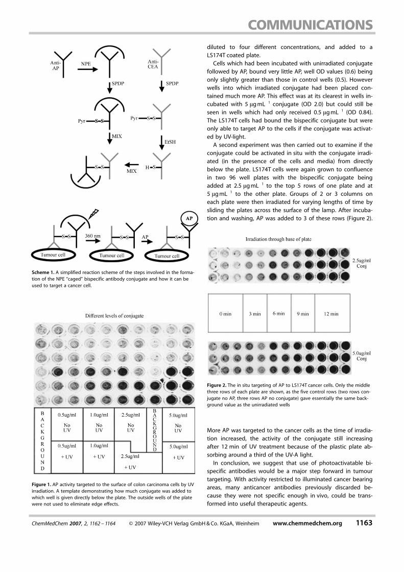
diluted to four different concentrations, and added to aLS174T coated plate.Cells which had been incubated with unirradiated conjugate
followed by AP, bound very little AP, well OD values (0.6) beingonly slightly greater than those in control wells (0.5). Howeverwells into which irradiated conjugate had been placed con-tained much more AP. This effect was at its clearest in wells in-cubated with 5 mgmL�1 conjugate (OD 2.0) but could still beseen in wells which had only received 0.5 mgmL�1 (OD 0.84).The LS174T cells had bound the bispecific conjugate but wereonly able to target AP to the cells if the conjugate was activat-ed by UV-light.A second experiment was then carried out to examine if the
conjugate could be activated in situ with the conjugate irradi-ated (in the presence of the cells and media) from directlybelow the plate. LS174T cells were again grown to confluencein two 96 well plates with the bispecific conjugate beingadded at 2.5 mgmL�1 to the top 5 rows of one plate and at5 mgmL�1 to the other plate. Groups of 2 or 3 columns oneach plate were then irradiated for varying lengths of time bysliding the plates across the surface of the lamp. After incuba-tion and washing, AP was added to 3 of these rows (Figure 2).
More AP was targeted to the cancer cells as the time of irradia-tion increased, the activity of the conjugate still increasingafter 12 min of UV treatment because of the plastic plate ab-sorbing around a third of the UV-A light.In conclusion, we suggest that use of photoactivatable bi-
ACHTUNGTRENNUNGspecific antibodies would be a major step forward in tumourtargeting. With activity restricted to illuminated cancer bearingareas, many anticancer antibodies previously discarded be-cause they were not specific enough in vivo, could be trans-formed into useful therapeutic agents.
Scheme 1. A simplified reaction scheme of the steps involved in the forma-tion of the NPE “caged” bispecific antibody conjugate and how it can beused to target a cancer cell.
Figure 1. AP activity targeted to the surface of colon carcinoma cells by UVirradiation. A template demonstrating how much conjugate was added towhich well is given directly below the plate. The outside wells of the platewere not used to eliminate edge effects.
Figure 2. The in situ targeting of AP to LS174T cancer cells. Only the middlethree rows of each plate are shown, as the five control rows (two rows con-jugate no AP, three rows AP no conjugate) gave essentially the same back-ground value as the unirradiated wells
ChemMedChem 2007, 2, 1162 – 1164 F 2007 Wiley-VCH Verlag GmbH&Co. KGaA, Weinheim www.chemmedchem.org 1163

Experimental Section
3 mL of the anti-AP monoclonal antibody (Zymed labs,0.25 mgmL�1gmL�1 in 0.1m NaHCO3 pH 8.3) was inactivated bythe addition of 20 mL NPE-carbonylchloride in dioxan.[2] After dialy-sis and centrifugation the NPE coated-anti-AP antibody(0.18 mgmL�1) was obtained as a clear solution. On absorption, theconcentration of the NPE-coated antibody reduced to0.11 mgmL�1.Measurement of NPE coated anti-AP antibody activity in an ELISA:The amount of NPE-coated-anti-AP capable of binding to APcoated ELISA plates was quantitated by the addition of Horseradishperoxidase-anti-MouseIgG followed by colour development at492 nm using o-phenyldiamine as substrate.The CEA specific antibody T84.66, was concentrated from serumfree medium by (NH4)2SO4 precipitation. T84.66 reacts stronglywith colonic cancer tissue and some other carcinomas but onlyweakly with normal tissues.[9]
Construction of the photoactivatable anti-CEA–anti-AP bispecificconjugate: The anti-CEA and NPE-coated anti-AP antibodies (eachat 0.18 mgmL�1) were derivatised by the addition of a 60-foldmolar excess of SPDP. The SPDP–anti-CEA antibody was reducedwith 0.5m Dithiothreitol (DTT, 30 min) and dialysed to removeexcess DTT. This reduced SPDP–anti-CEA was added to the unre-duced SPDP-derivatised NPE–anti-AP antibody and they were leftto crosslink. During the formation of the bispecific conjugates, wediscovered that much better conjugation at higher yields was ob-tained if the NPE coated anti-AP antibody was not preabsorbedprior to addition of the SPDP.The ability of T84.66–anti-AP conjugates to bind to CEA was as-sessed using CEA coated ELISA plates and a anti-mouse IgG(AP la-belled, 1/1000) second layer. This was then quantified by the hy-drolysis of p-nitrophenolphosphate ACHTUNGTRENNUNG(pNPP) to p-nitrophenol at405 nm. Anti-AP activity was measured by adding AP as a secondlayer and again measuring colour development at 405 nm.The targeting of LS174T colonic cancer cells: LS174T cells weregrown to 95–100% confluence in 96 well plates and 100 mL of SFMcontaining the bispecific conjugates (0.5–5 mgmL�1), was added toeach well. After irradiation and incubation AP was added, and theamount of AP targeted to the cells was quantitated using pNPP.Photolysis of conjugates: The NPE–anti-AP antibody conjugates(20 mgmL�l in 0.9% saline) were irradiated in quartz cuvettes with
UV-A light from a hand held Spectroline EN-16/F UV lamp. In laterexperiments the conjugates were added to the plates and irradiat-ed in the presence of the cells. This type of lamp produces UVlight over the wavelength range of 325–395 nm with a mercuryvapour peak at 365 nm.[10] The total UV-A irradiance of this handheld lamp was 5.45 mWcm�2 at a working distance of 0.5 cm.
Keywords: antibody · caging · cancer · photoactivation · UV-alight
[1] a) O. H. Brekke, I. Sandlie, Nat. Rev. Drug Discovery 2003, 2, 52–62; b) D.Schrama, R. A. Reisfeld, J. C. Becker, Nat. Rev. Drug Discovery 2006,5,147–159.
[2] a) R. H. J. Begent, Int. J. Cancer 1993, 55, 355–358; b) C. H. Self, S.Thompson, Lancet 2006, 367,1038–1039.
[3] a) S. Thompson, M-C. Fawcett, J. A. Spoors, C. H. Self, Biochem. Soc.Trans. 1995, 23,155S; b) C. H. Self, S. Thompson, Nat. Med. 1996, 2, 817–820.
[4] a) P. Carter, J. Immunol. Methods 2001, 248, 7–15; b) S. Withoff, W. Hel-frich, L. F. M. H. de Leij, G. Molema, Curr. Opin. Mol. Ther. 2001, 3, 53–62; c) P. A. Baeuerle, P. Kufer, R. Lutterbuse, Curr. Opin. Mol. Ther. 2003,5, 413–419.
[5] a) P. D. Senter, M. G. Saulnier, G. J. Schreiber, D. L. Hirschberg, J. P.Brown, I. Hellstrom, K. E. Hellstrom, Proc. Natl. Acad. Sci. USA 1988, 85,4842–4846; b) M. P. Deonarain, A. A. Epenetos, Br. J. Cancer 1994, 70,786–794; c) M. Rooseboom, J. N. M. Commandeur, N. P. E. Vermeulen,Pharmacol. Rev. 2004, 56, 53–102.
[6] a) S. Thompson, J. A. Spoors, M-C Fawcett, C. H. Self, Biochem. Biophys.Res. Commun. 1994, 201, 1213–1219; b) S. Thompson, M-C. Fawcett,L. B. Pulman, C. H. Self, Photochem. Photobiol. Sci. 2006, 5, 326–330.
[7] Dynamic Studies in Biology: Phototriggers, Photoswitches and Caged Bio-molecules (Eds: M.Goldner, R. Givens), Wiley-VCH, Weinheim, 2005,Whole book.
[8] Z. R. Shi, D. Tsao, Y. S. Kim, Cancer Res. 1983, 43, 4045–4049.[9] C. Wagener, Y. H. J. Yang, F. G. Crawford, J. E. Shively, J. Immunol. 1983,
130, 2308–2315.[10] C. H. Self, M-C Fawcett, J. A. Spoors, L. B. Pulman, S. Thompson, Bio-
chem. Soc. Trans. 1995, 23,156S.
Received: April 4, 2007Revised: April 23, 2007Published online on May 15, 2007
1164 www.chemmedchem.org F 2007 Wiley-VCH Verlag GmbH&Co. KGaA, Weinheim ChemMedChem 2007, 2, 1162 – 1164
MED