The Comparison of Four Different Methods of Perioperative ... · anestezi altında peroperatif göz...
3
r A a l þ a t n ý i r j m i r a O O h r c i r g a i n e a s l e R Özlem Kocatürk 1 , Tolga Kocatürk 2 , Nil Kaan 1 , Volkan Dayanır 2 1 Anesteziyoloji ve Reanimasyon AD, 2 Göz Hastalıkları AD, Adnan Menderes Üniversitesi, Tıp Fakültesi, Aydın, Türkiye Yüzüstü Pozisyonda Göz Koruma Yöntemleri / Eye Protection Methods in Prone Position The Comparison of Four Different Methods of Perioperative Eye Protection under General Anesthesia in Prone Position Yüzüstü Pozisyonda Genel Anestezi Altında Peroperatif Göz Korumada Dört Ayrı Yöntemin Karşılaştırılması DOI: 10.4328/JCAM.607 Received: 08.02.2011 Accepted: 19.02.2011 Printed: 01.04.2012 J Clin Anal Med 2012;3(2): 163-5 Corresponding Author: Tolga Kocatürk, Department of Ophthalmology, Bartın State Hospital, 74100, Aydın, Turkey GSM: +905333444111 E-Mail: [email protected] Özet Amaç: Bu çalışmanın amacı hipoalerjen yapışkan bant, antibiyotik merhem, suni göz yaşı jeli ve oküler nemlendirici pomadın yüzüstü pozisyonda genel anestezi altında peroperatif göz korumasındaki etkinliğinin karşılaştırılma- sı ve değerlendirilmesidir. Gereç ve Yöntem: Genel anestezi altında en az >90 dakika sürmüş olan spinal cerrahi geçiren 184 hasta (368 göz) rastgele ola- rak dört gruba ayrıldı. Gözlere hipoalerjen yapışkan bant, antibiyotik merhem, suni gözyaşı jeli veya oküler nemlendirici pomad uygulandı. Hastalara, ameli- yattan önce ve sonra, düzeltilmiş görme keskinliği ölçümü, bazal gözyaşı üre- timi, korneal ve konjunktival boyanma testlerini içeren detaylı göz muayenesi uygulandı. Bulgular: Bazal gözyaşı üretimi tüm gruplarda preoperatif değer- lere göre azaldı (P<0.001 ). Derlenme odasında, korneal epitelyal defekt in- sidansı grup 1’de %2.72, grup 2’de %2.72, grup 3’de %5.16, grup 4’de %2.17 olmak üzere tüm gruplarda toplam %12.77 olarak bulundu. Göz kapaklarında- ki yapışıklık yönünden gruplar arasında anlamlı fark yoktu (p>0.05). Preope- ratif ve postoperatif görme keskinlikleri arasında anlamlı fark yoktu. Sonuç: Korneal hasarlardan korunmada tüm yöntemler uygundur. Ancak postopera- tif dönemdeki geçici semptomların önlenmesinde hiçbiri tek başına yeterince iyi değildir. Genel anestezi sırasında korneal hasar insidansını azaltmak için gözlerin bant, gel, merhem veya pomad ile korunması gerekir. Anahtar Kelimeler Genel Anestezi; Yüzüstü Pozisyon; Korneal Epiteliyal Defekt; Göz Koruması Abstract Aim: The purpose of this study was to compare and assess the efficacy of hypoallergen adhesive tape, antibiotic ointment, artificial tear liquid gel and ocular lubricant pomade for perioperative protection of eyes under general anesthesia in prone position. Material and Method: One hundred and eighty four patients (368 eyes) undergoing general anesthesia for >90 min for spi- nal procedures were divided randomly into four groups. Hypoallergen ad- hesive tape, antibiotic ointment, artificial tear liquid gel or ocular lubricant pomade was applied into the eyes. The patients underwent complete oph- thalmic examination, including corrected visual acuity measurement, basal tear production, corneal and conjunctival staining both before and aſter sur- gery. Results: Basal tear production was reduced in all groups compared to preoperative values (P<0.001 ). The overall incidence of corneal epithelial defects was 12.77% in recovery room of which 2.72% occurred in group 1, 2.72% in group 2, 5.16% in group 3 and 2.17% in group 4. There was no sig- nificant difference between groups according to adhesive lids (p>0.05). There was no difference between pre and postoperative visual acuity. Discussion: All of the methods are suitable for protecting corneal injuries. But none of them is good enough to avoid temporary symptoms in postoperative period. During general anesthesia eyes need protection either by tape, gel, ointment or pomade to reduce the incidence of corneal injuries. Keywords General Anesthesia; Prone Position; Corneal Epithelial Defect; Eye Protection Journal of Clinical and Analytical Medicine | 163
Transcript of The Comparison of Four Different Methods of Perioperative ... · anestezi altında peroperatif göz...

| Journal of Clinical and Analytical Medicine
r
A
a
l
þ
a
t
n
ý
i
r
j
m
i
r
a
O
O
h
r
c
i
r
g
a
i
n
e
a
s
l
e
R
1
Özlem Kocatürk1, Tolga Kocatürk2, Nil Kaan1, Volkan Dayanır2
1Anesteziyoloji ve Reanimasyon AD, 2Göz Hastalıkları AD, Adnan Menderes Üniversitesi, Tıp Fakültesi, Aydın, Türkiye
Yüzüstü Pozisyonda Göz Koruma Yöntemleri / Eye Protection Methods in Prone Position
The Comparison of Four Different Methods of Perioperative Eye Protection under General Anesthesia in Prone Position
Yüzüstü Pozisyonda Genel Anestezi Altında Peroperatif Göz Korumada Dört Ayrı Yöntemin Karşılaştırılması
DOI: 10.4328/JCAM.607 Received: 08.02.2011 Accepted: 19.02.2011 Printed: 01.04.2012 J Clin Anal Med 2012;3(2): 163-5 Corresponding Author: Tolga Kocatürk, Department of Ophthalmology, Bartın State Hospital, 74100, Aydın, TurkeyGSM: +905333444111 E-Mail: [email protected]
ÖzetAmaç: Bu çalışmanın amacı hipoalerjen yapışkan bant, antibiyotik merhem, suni göz yaşı jeli ve oküler nemlendirici pomadın yüzüstü pozisyonda genel anestezi altında peroperatif göz korumasındaki etkinliğinin karşılaştırılma-sı ve değerlendirilmesidir. Gereç ve Yöntem: Genel anestezi altında en az >90 dakika sürmüş olan spinal cerrahi geçiren 184 hasta (368 göz) rastgele ola-rak dört gruba ayrıldı. Gözlere hipoalerjen yapışkan bant, antibiyotik merhem, suni gözyaşı jeli veya oküler nemlendirici pomad uygulandı. Hastalara, ameli-yattan önce ve sonra, düzeltilmiş görme keskinliği ölçümü, bazal gözyaşı üre-timi, korneal ve konjunktival boyanma testlerini içeren detaylı göz muayenesi uygulandı. Bulgular: Bazal gözyaşı üretimi tüm gruplarda preoperatif değer-lere göre azaldı (P<0.001 ). Derlenme odasında, korneal epitelyal defekt in-sidansı grup 1’de %2.72, grup 2’de %2.72, grup 3’de %5.16, grup 4’de %2.17 olmak üzere tüm gruplarda toplam %12.77 olarak bulundu. Göz kapaklarında-ki yapışıklık yönünden gruplar arasında anlamlı fark yoktu (p>0.05). Preope-ratif ve postoperatif görme keskinlikleri arasında anlamlı fark yoktu. Sonuç: Korneal hasarlardan korunmada tüm yöntemler uygundur. Ancak postopera-tif dönemdeki geçici semptomların önlenmesinde hiçbiri tek başına yeterince iyi değildir. Genel anestezi sırasında korneal hasar insidansını azaltmak için gözlerin bant, gel, merhem veya pomad ile korunması gerekir.
Anahtar KelimelerGenel Anestezi; Yüzüstü Pozisyon; Korneal Epiteliyal Defekt; Göz Koruması
AbstractAim: The purpose of this study was to compare and assess the efficacy of hypoallergen adhesive tape, antibiotic ointment, artificial tear liquid gel and ocular lubricant pomade for perioperative protection of eyes under general anesthesia in prone position. Material and Method: One hundred and eighty four patients (368 eyes) undergoing general anesthesia for >90 min for spi-nal procedures were divided randomly into four groups. Hypoallergen ad-hesive tape, antibiotic ointment, artificial tear liquid gel or ocular lubricant pomade was applied into the eyes. The patients underwent complete oph-thalmic examination, including corrected visual acuity measurement, basal tear production, corneal and conjunctival staining both before and after sur-gery. Results: Basal tear production was reduced in all groups compared to preoperative values (P<0.001 ). The overall incidence of corneal epithelial defects was 12.77% in recovery room of which 2.72% occurred in group 1, 2.72% in group 2, 5.16% in group 3 and 2.17% in group 4. There was no sig-nificant difference between groups according to adhesive lids (p>0.05). There was no difference between pre and postoperative visual acuity. Discussion: All of the methods are suitable for protecting corneal injuries. But none of them is good enough to avoid temporary symptoms in postoperative period. During general anesthesia eyes need protection either by tape, gel, ointment or pomade to reduce the incidence of corneal injuries.
KeywordsGeneral Anesthesia; Prone Position; Corneal Epithelial Defect; Eye Protection
Journal of Clinical and Analytical Medicine | 163

| Journal of Clinical and Analytical Medicine
Yüzüstü Pozisyonda Göz Koruma Yöntemleri / Eye Protection Methods in Prone Position
2
IntroductionPatients undergoing prolonged non-ocular surgery with general anesthesia (GA) may develop ocular complications. [1,2] The factors contributing to these ocular complications are abolition of protective corneal reflex, decreased basal tear production [3,4] and absence of pain perception. [1] Among them the most common ophthalmologic injury is corneal abrasion.Corneal protection may in principles be conveyed by taping the eye lids closed, and by application of ocular lubricants into the conjunctiva sac.The incidence of corneal epithelial defect during GA has been reported to be 44% [5] in unprotected eyes and 2.1% [6] in eyes protected with ointment. Though there is a lot of study in the literature, comparing the efficiency of the different eye protection methods in supine po-sition, there are only few documented study comparing the eye protection methods in the face-down prone position and to the best of our knowledge there are no studies comparing the ef-ficacy of eye protection strategies on preventing the corneal abrasions and to compare the side effects of these methods under similar conditions with regard to severity of the lesions in cornea and conjunctiva by a scoring system in prone position .We compare four eye protection methods: antibiotic ointment, artificial tear gel, ocular lubricant pomade and adhesive tape with regard to the severity of ocular surface injury according to a scoring system [7] in prone position.
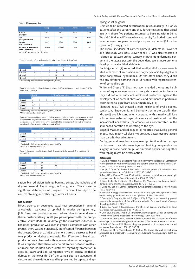
Material and Method184 ASA (American Society of Anesthesiologists Physical Status classification system) I adult patients (368 eyes) of 92 women and 92 men undergoing spinal surgery GA for >90 min in the prone position were included in the study. Patients were divided into four groups of 46 using a randomization chart. The groups were sex and aged matched. The study protocol was approved by the local ethical committee, and the study was carried out in accordance with the Declaration of Helsinki. Written informed consent was obtained from each patient. The patients were divided into four groups of 46 patients and one of the following methods was applied to the eyes. Group 1: hypoallergen adhesive tape (Hypafix®; Smith and Nephew, France); Group 2: antibiotic ointment (Terramycine®; Pfizer, Turkey); Group 3: artificial tear liquid gel containing polyacrylic acid (Viscotears®; Novartis, Turkey); Group 4: ocular lubricant pomade (Duratears®; Liba, Turkey). Anesthesia protocol was standardized for all study patients. Anesthesia induction consisted of administration of propofol (2-2.5 mg/kg) and rocuronium (1 mg/kg) to facilitate endotra-cheal intubation. After endotracheal intubation, anesthesia was maintained with isoflurane (1%), 50% nitrous oxide in oxygen, remifentanyl infusion (1 μg/kg/h), and rocuronium as needed. After the patients were intubated, the eye protection methods were performed according to the groups. The ophthalmologist was blinded to the groups of patients and protection methods. Then all patients were turned prone and their heads positioned in the neutral position with two pieces of silicone donut-shaped, 4 cm thick pad fit for forehead and chic frees the face and eyes, to prevent any extraocular pressure. Patients were evaluated in preoperative period, in post anes-thesia care unit (PACU) and 12 and 24 h after operation. Visual acuity, cornea and conjunctiva were assessed by the ophthal-mologist with Fluorescein and Rose-Bengal staining test and Schirmer-1 test 1 day before operation and 12 and 24 h after
operation. Post-operatively in PACU and 12 and 24 h after op-eration the patients were evaluated by a questionnaire form (adhesive lids, foreign body sensation, itching, burning, stings, photophobia, blurred vision and dryness) and conjunctival con-gestion and chemosis. For statistical analysis, Kruskal-Wallis test (followed by U-test) and Analysis of Variance with Bonfer-roni correction were applied. The severity of corneal damage was evaluated by intensity of staining (0, none; 1, mild; 2, moderate; 3, dense) and lesion size (0, none; 1, if the lesion was <1 mm; 2, if the lesion was >1 and <3 mm; 3, if the lesion was >3 mm). Hyperaemia of conjunctiva was assessed using a “scoring system (1, hyperaemia located only in the temporal or nasal areas of bulbar conjunctiva; 2, hyperaemia located in the nasal or temporal areas plus hyper-aemia and extension to the bulbar conjunctiva near to the upper or the lower fornixes; 3, hyperaemia including all of the areas of bulbar conjunctiva). Chemosis was assessed also using a scor-ing system [0, absent; 1, mild (to grey line); 2, moderate (to lid margin); 3, severe (over lid margin)].
ResultsThree hundred and sixty eight eyes (184 patients) were ob-served. Patients’ characteristics and duration of anaesthesia were not different between the groups (Table 1). There was a reduction in basal tear production postoperatively in all the four groups (P<0.001) but there was no difference in visual acu-ity for near and distant visions. Of the 368 eyes subjected to fluorescein and Rose-Bengal staining, none of the eyes showed corneal epithelial defect preoperatively. Immediate post-oper-ative examination in PACU revealed 47 eyes (12.77%) as the overall incidence of corneal epithelial defects of which 2.72% occurred in Group 1, 2.72% in Group 2, 5.16% in Group 3 and 2.17% in Group 4. Corneal epithelial defect was observed in 10 eyes (10.86%) in Group 1, 10 eyes (10.86%) in Group 2, 19 eyes (20.65%) in Group 3 and 8 eyes (8.69%) in Group 4.Conjunctival hyperemia was observed in 12 (13.04%), 14 (15.21%), 21 (22.82%) and 14 (15.21%) eyes of Groups 1, 2, 3 and 4, respectively. Chemosis was seen in 16 (17.39%), 24 (26.08%), 31 (33.69%) and 21 (22.82%) eyes of Groups 1, 2, 3 and 4, respectively. In Group 3 the number of eyes with mild intensity of corneal staining and with corneal lesion size of <1mm were significantly high in the PACU and at 12 and 24 h after the operation than those of the other groups (p<0.05) (Table 2 and 3). However there were not significant difference between the groups with regard to lesion size of >1mm and moderate or severe corne-al staining. Moderate and severe conjunctival hyperemia was observed more frequently in Group 3 than other groups in the PACU (p<0.05) (Table 4). After 12 hours, moderate conjunctival hyperemia was still high in Group 3, however there was no sig-nificant difference with regard to severe conjunctival hyperemia (Table 4). The highest scores of corneal lesion and conjunctival hyperaemia were observed in Group 3. There was significantly less moderate chemosis in Group 1 than the other groups in PACU (p <0.05) (Table 5). However, after 12 hours, moderate chemosis was observed more frequently in Group 3 (p <0.05) (Table 5). In PACU, blurred vision was high in Group 2 and 4 compared to other groups (p<0.05). Conjunctival hyperemia and itching were seen more frequently in group 3 (p<0.05). Foreign body sensation were seen in group 1 (p<0.05). 24 h after operation, there was no difference between the groups according to all symptoms such as foreign body sen-
| Journal of Clinical and Analytical Medicine164
Yüzüstü Pozisyonda Göz Koruma Yöntemleri / Eye Protection Methods in Prone Position
| Journal of Clinical and Analytical Medicine
Yüzüstü Pozisyonda Göz Koruma Yöntemleri / Eye Protection Methods in Prone Position
3
sation, blurred vision, itching, burning, stings, photophobia and dryness were similar among the four groups. There were no significant differences with regard to size or intensity of the corneal staining and other signs after 24 hours.
DiscussionDirect trauma or decreased basal tear production in general anesthesia may cause of ophthalmic injuries during surgery. [2,8] Basal tear production was reduced due to general anes-thesia postoperatively in all groups compared with the preop-erative values (P<0.0003). Although the maximum reduction in basal tear production was seen in group 1 compared with other groups, there was no statistically significant difference between the groups. Cross et al. [8] also demonstrated a decreased basal tear production during anesthesia. No difference in basal tear production was observed with increased duration of surgery. It was reported that there was no difference between methyl-cellulose and paraffin-based ointment regarding protection in eyes. [6] Batra and Bali [5] reported 44% of corneal epithelial defects in the lower third of the cornea due to inadequate lid closure and these defects could be prevented by taping and ap-
plying vaseline gauze. Orlin et al. [9] reported deterioration in visual acuity in 5 of 76 patients after the surgery and they further observed that visual acuity in these five patients returned to baseline within 24 hr. We didn’t find any difference in visual acuity for both distant and near between preoperative and postoperative period (24 h after operation) in any groups. The overall incidence of corneal epithelial defects in Grover et al.’s [10] study was 10%. Grover et al. [10] was also reported in relation to posture during surgery, in patients undergoing sur-gery in the lateral posture, the dependent eye is more prone to develop corneal epithelial defects. Ganidağlı et al. [7] reported that methylcellulose was associ-ated with more blurred vision and polyacrylic acid liquid gel with more conjunctival hyperaemia. On the other hand, they didn’t find any difference among these lubricants with regard to sever-ity of corneal lesion. White and Crosse [11] has not recommended the routine instil-lation of aqueous solutions, viscous gels or ointments, because they did not offer sufficient additional protection against the development of corneal abrasions, and ointments in particular contributed to significant ocular morbidity. [11]Manecke et al. [12] showed a high incidence of eyelid edema, conjunctival hyperaemia and blurred vision in the paraffin (lip-id-based) eye lubricant when compared with a methylcellulose solution (water-based) eye lubricants and postulated that the inhalational anaesthetic (halothane) was concentrated in the lipid-based paraffin and irritating to the eye. Boggild-Madsen and colleagues [1] reported that during general anaesthesia methylcellulose 4% provides better eye protection than paraffin-based ointment.During general anesthesia eyes need protection either by tape or ointment to avoid corneal injuries. Avoiding complaints after surgery in prone position gel or ointment application together with taping might be better option.
References1. Boggild-Madsen NB, Bundgarrd-Nielsen P, Hammer U, Jakobsen B. Comparison of eye protection with methylcellulose and paraffin ointments during general an-esthesia. Can Anaesth Soc J, 1981; 28: 575-8.2. Krupin T, Cross DA, Becket B. Decreased basal tear production associated with general anesthesia. Arch Ophthalmol, 1977; 95: 107-8.3. Terry HR Jr, Kearns TP, Love JG, Orwoll G. Untoward ophthalmic and neurologic events of anesthesia. Surg Clin North Am, 1965; 45: 927-9.4. Snow JC, Kripke BJ, Norton MI o Chandra 19, Woodcome HA. Corneal injuries during general anesthesia. Anesth Analg, 1975; 54: 465-7.5. Batra YK, Bali IM. Corneal abrasions during general anesthesia. Anesth Analg, 1977; 56: 363-5.6. Schimdt P, Boggild-Madsen NB. Protection of the eyes with ophthalmic oint-ments during general anesthesia. Acta Ophthalmol, 1981; 59: 422-7.7. Ganidağlı S, Cengiz M, Becerik C, Oğuz H, Kılıç A. “Eye protection during general anaesthesia: comparison of four different methods”, European Journal of Anaes-thesiology, 2004; 21: 665-7.8. Cross DA, Krupin T. Implications of the effects of general anesthesia on basal tear production. Anesth Analg, 1977; 56: 35-7.9. Orlin SE, Kurata FK, Krupin T, Schneider M, Glendrange RR. Ocular lubricants and corneal injury during anesthesia. Anesth Analg, 1989; 69: 384-5. 10. Grover VK, Kuma KVM, Sharma S, Sethi N, Grewal SPS. Comparison of meth-ods of eye protection under general anaesthesia. Can J Anesth, 1998; 45: 575-7.11. White E, Crosse MM. The aetiology and prevention of peri-operative corneal abrasions. Anaesthesia, 1998; 53: 157-61.12. Manecke GR Jr, Tannenbaum DP, McCoy BE. Severe bilateral corneal injury attributed to a preservative-containing eye lubricant. Anesthesiology, 2000; 93: 1545-6.
Tablo 1.
Journal of Clinical and Analytical Medicine | 165
Yüzüstü Pozisyonda Göz Koruma Yöntemleri / Eye Protection Methods in Prone Position