Modelling And Control of 6MG Siemens Wind Turbine Blades ...
Research ArticleThe Clinical Significance of Increased Serum ProinflammatoryCytokines, C-Reactive Protein, and Erythrocyte SedimentationRate in Patients with Hidradenitis Suppurativa
D. Jiménez-Gallo,1 R. de la Varga-Martínez,2 L. Ossorio-García,1 C. Albarrán-Planelles,1
C. Rodríguez,2 and M. Linares-Barrios1
1Unidad de Gestión Clínica de Dermatología, Hospital Universitario Puerta del Mar, Cádiz, Spain2Unidad de Gestión Clínica de Hematología e Inmunología, Hospital Universitario Puerta del Mar, Cádiz, Spain
Correspondence should be addressed to D. Jiménez-Gallo; [email protected]
Received 11 January 2017; Revised 22 May 2017; Accepted 13 June 2017; Published 10 July 2017
Academic Editor: Hermann Gram
Copyright © 2017 D. Jiménez-Gallo et al. This is an open access article distributed under the Creative Commons AttributionLicense, which permits unrestricted use, distribution, and reproduction in any medium, provided the original work isproperly cited.
Objectives. To assess inflammatory serum markers including serum proinflammatory cytokines, C-reactive protein (CRP), anderythrocyte sedimentation rate (ESR) according to the clinical inflammatory activity of patients with hidradenitis suppurativa(HS). Patients and Methods. Seventy-four patients with HS were studied based on the Hidradenitis Suppurativa-PhysicianGlobal Assessment (HS-PGA) score and Hurley staging system. Proinflammatory cytokines were measured using a multiplexcytokine assay. Twenty-two healthy volunteers were recruited. Results. Serum interleukin- (IL-) 6, IL-23, soluble tumournecrosis factor alpha (TNF-α) receptor I (sTNF-RI), CRP, and ESR were different in the patients with HS compared with thosein the healthy controls (P < 0 05). The levels of IL-1β, IL-6, IL-8, IL-10, IL-12p70, IL-17A, sTNF-RII, CRP, and ESR weresignificantly elevated according to inflammatory activity based on HS-PGA scores (r > 0 25, P < 0 05). The levels of IL-6(r = 0 53, P < 0 001), CRP (r = 0 54, P < 0 001), and ESR (r = 0 60, P < 0 001) were especially well correlated with clinicalinflammatory activity based on HS-PGA scores. The levels of IL-6, IL-8, sTNF-RI, sTNF-RII, CRP, and ESR were significantlyelevated according to Hurley staging system. Conclusions. Serum proinflammatory cytokines, CRP, and ESR are increased inrelation to the clinical inflammatory activity of patients with HS compared with healthy controls. Serum IL-6, CRP, and ESR areeffective biomarkers for evaluating the severity of HS.
1. Introduction
Hidradenitis suppurativa (HS) is a chronic inflammatorydisease originating in the follicular infundibulum [1]. HS ischaracterized by the formation of multiple inflammatorylesions such as nodules, abscesses, and fistulae, which pre-dominantly affect the intertriginous regions. These lesionsare painful and suppurative and result in scar formation inthe most severe cases. The most commonly affected areasare the axillae and groin as well as the submammary, gluteal,and perianal regions [2, 3]. The prevalence of HS is estimatedto be 1% [2]. Obesity and tobacco use are strongly associatedwith HS [4, 5]. Pathogenesis of HS is not completely under-stood. The proposed mechanism of the pathogenesis of HS
begins with an alteration of the innate immune response tothe commensal microbiota, which leads to epidermal hyper-plasia. This epidermal hyperplasia occludes the follicularinfundibulum through the formation of cysts, which eventu-ally break, free the keratin fibres, and release the commensalbacteria into the dermis. These steps amplify the inflamma-tory response through inflammasome activation and theinterleukin- (IL-) 1β-IL-23/T-helper (Th) 17/IL-17 pathway[3, 6]. Zee et al. [7] studied samples of lesional and perile-sional skin from patients with HS and observed elevatedlevels of IL-1β, tumour necrosis factor alpha (TNF-α), andIL-10. They also noted that the TNF-α levels were 5 timeshigher in the skin affected by HS than the skin affected bypsoriasis. This result is interesting as it implies a greater
HindawiMediators of InflammationVolume 2017, Article ID 2450401, 8 pageshttps://doi.org/10.1155/2017/2450401
inflammatory load in HS compared with other dermatologi-cal diseases [8, 9]. In this sense, HS continues to be achallenging dermatological disorder for dermatologists [10].
IL-1β is a key inflammatory mediator in HS. IL-1β levelsin skin affected with HS show a trend towards a positivecorrelation with disease severity [6, 7]. Lima et al. [11] dem-onstrated the presence of IL-17 in lesional and perilesionalskin from patients with HS. IL-17A is the defining cytokineof Th17 lymphocytes [12]. IL-1β, IL-6, IL-23, and transform-ing growth factor-β (TGF-β) are cytokines that are producedby activated innate cells, and they drive the differentiationof Th17 lymphocytes [6]. IL-17-stimulated keratinocytessecrete IL-1β through a mechanism involving the Nod-likereceptor protein 3 (NLRP3) inflammasome [13]. Further-more, IL-10 is an anti-inflammatory cytokine that has alsobeen observed in the skin of patients with HS [6]. Otherproinflammatory cytokines such as IL-22, IL-23, IL-6, andIL-8 have shown contradictory results in skin affected withHS [14–16]. In the later phases of HS, infiltration of thedermis by neutrophils leads to the amplification of theinflammatory response with higher production of IL-17[12]. All of the above provokes an inflammatory loop thatis manifested clinically by inflammatory lesions and thesuppuration characteristics of HS.
Hessam et al. [17] recently demonstrated that C-reactiveprotein (CRP), which is an acute phase protein (APP), iseffective for evaluating the severity and degree of inflamma-tion in patients with HS. Pascual et al. [18] evaluated subclin-ical arteriosclerosis in patients with HS and found that theerythrocyte sedimentation rate (ESR) was higher in thesepatients. The ESR, which is an indirect acute phase reactant,reflects plasma viscosity and the presence of APP, especiallyfibrinogen [19]. Matusiak et al. [20, 21] described elevatedserum levels of chitinase-3-like protein 1 (YKL-40) andIL-17 in patients with HS based on Hurley staging system.
The study of inflammatory markers in immunologicaldiseases with high inflammatory loads, such as rheumatoidarthritis and systemic lupus erythematosus, has resultedin the identification of biomarkers that are used in clinicalpractice, including serum inflammatory cytokines and CRP[22, 23]. In this study, we analysed the clinical significanceof serum proinflammatory cytokines, CRP, and ESR inpatients with HS.
2. Materials and Methods
2.1. Patients and Controls. In total, 74 patients with HS(36 males and 38 females) and a mean± standard deviation(SD) age of 37.4± 12.0 years (range 18–62) were enrolled.HS was diagnosed according to well-established criteria[24]. The exclusion criteria were the following: patientsyounger than 18 years of age; patients suffering from infec-tious, tumoural or autoimmune diseases; and patients whowere receiving ongoing local or systemic treatment for atleast 3 months prior to entering the study. The clinicalseverity of the disease was evaluated by the same dermatolo-gist using the HS-PGA score (mean± SD 3.0± 0.9) andHurley staging system. The HS-PGA score is a simple anddynamic scale that allows for the tracking of the clinical
progression of inflammation in HS [9, 25]. HS-PGA scorescategorize the severity of HS in 6 degrees as a function ofthe number of noninflammatory nodules, inflammatorynodules, abscesses, and draining fistulae [9, 26, 27]. Wedivided the patients with HS into the following 3 groupsaccording to clinical inflammatory activity: a group withlow inflammatory activity (LIA) (n = 22), including patientswith HS-PGA scores from 0 to 2 (clear, minimal, and mild);a group with moderate inflammatory activity (MIA) (n = 35),including patients with HS-PGA scores of 3 (moderate); anda group with high inflammatory activity (HIA) (n = 17),including patients with HS-PGA scores of 4 and 5. Addition-ally, we divided the patients with HS into the three-degreescale proposed by Hurley [9]. As a control group, 22 healthyvolunteers were recruited for this study. This group com-prised healthy volunteers without HS who also had no familyhistory of HS. The characteristics of the patients and healthycontrols are shown in Table 1. This study was conductedin accordance with the Helsinki Conference (64th WorldMedical Association General Assembly, Fortaleza, Brasil,October 2013) and with the Spanish legislation on clinicalresearch and personal data protection. The participants’rights and safety took precedence, and approval was obtainedfrom the Hospital Puerta del Mar Ethics Committee. Allof the participants were provided detailed informationregarding the purpose and method of this study, as well asthe expected results, and informed consent was obtainedbefore screening.
2.2. Serum. Venous blood samples (5–10mL) were collectedfrom the patients and healthy controls into vacuum tubesunder sterile conditions. The serum was separated by centri-fugation while the samples were fresh, immediately frozen at−80°C, and then stored until processing.
2.3. Cytokine Assays. Multiple cytokine analyses were per-formed using xMAP technology (Luminex Corporation,Austin, TX, USA) to measure the serum levels of IL-1β,IL-6, IL-8, IL-10, IL-12p70, IL-17A, IL-22, IL-23, and sol-uble TNF receptors I (sTNF-RI) and II (sTNF-RII). TheMilliplex MAP multiplex assay was conducted in a 96-wellmicroplate according to the manufacturer’s recommenda-tions (Millipore, Billerica, MA, USA). The levels of the seruminflammatory cytokines were calculated using a standardcurve, and the minimum detectable concentrations were0.18 pg/mL for IL-1β, 0.23 pg/mL for IL-6, 0.79 pg/mL forIL-8, 0.94 pg/mL for IL-10, 0.33 pg/mL for IL-12p70,0.58 pg/mL for IL-17A, 0.04 pg/mL for IL-22, 0.12 pg/mLfor IL-23, 12.2 pg/mL for sTNF-RI, and 12.2 pg/mL forsTNF-RII. Due to the short half-life of TNF-α, the sTNF-R(sTNF-RI, 55 kDa, and sTNF-RII, 75 kDa) levels were quan-tified because their half-lives are longer. The serum concen-trations of the sTNF-Rs correlate with those of TNF-α [28].All of the samples were analysed simultaneously at the endof recruitment, and the researchers were blinded to theclinical data.
2.4. Measurement of C-Reactive Protein Levels and theErythrocyte Sedimentation Rate. The CRP serum levels were
2 Mediators of Inflammation
measured using a turbidimetric assay with the Cobas 8000(Roche Diagnostics, Mannheim, Germany). The Westergrenmethod involving the collection of 2mL of venous blood intoa tube containing 0.5mL of sodium citrate was used to mea-sure the ESR (VES-MATIC 60, Menarini). A CRP level ofgreater than 6mg/L and an ESR of greater than 20mm/hourwere regarded as abnormal values.
2.5. Statistical Analysis. All data were analysed using SPSS19.0 for Windows (SPSS, Chicago, IL, USA). All data arepresented as the mean± SD (median). A two-tailed testwas used. We used the Mann–Whitney U test to evaluatethe differences between 2 groups (healthy controls versuspatients with HS). We then divided the patients into 3groups according to clinical inflammatory activity based ontheir HS-PGA scores. The Kruskal-Wallis test was used tomake comparisons among the healthy controls, patients withLIA HS, patients with MIA HS, and patients with HIA HS.To examine whether there were any statistically significantdifferences among the 4 groups, Bonferroni’s post hoc testfor pairwise comparisons was used. The correlation analysiswas performed by calculating the Spearman’s correlationcoefficient. Thus, the effect size was small (0.10–0.29),medium (0.30–0.49), and large (above 0.50), as defined byCohen [29]. Additionally, the Kruskal-Wallis test was usedto make comparisons among the healthy controls andpatients with HS based on Hurley staging system. Statisticalsignificance was accepted as P < 0 05.
3. Results
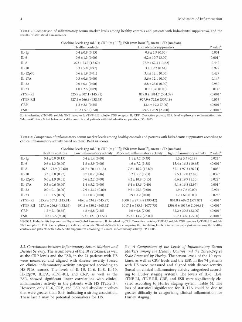
3.1. A Comparison of the Levels of Inflammatory SerumMarkers between Patients with Hidradenitis Suppurativaand the Control Group. The serum levels of 10 cytokinesand CRP, as well as the ESR, were analysed in 74 patients
with HS and 22 healthy controls (Table 2). The levels ofIL-6, IL-23, sTNF-RI, and CRP, as well as the ESR, were sig-nificantly different in our experimental group (P < 0 05).These findings are interesting because they reveal the sys-temic inflammatory characteristics of the patients with HS.
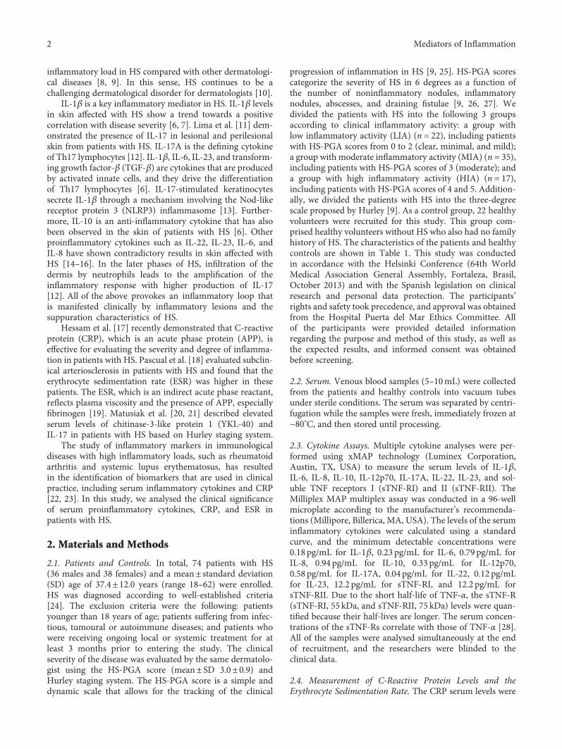
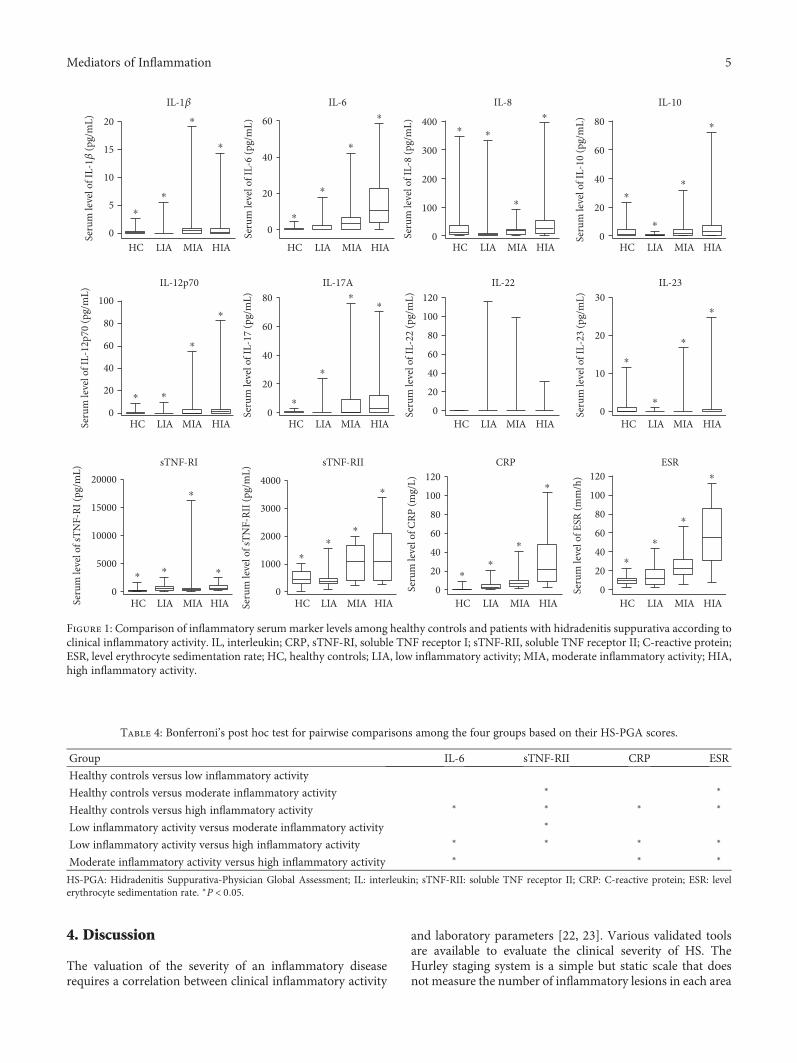
3.2. A Comparison of the Levels of Inflammatory SerumMarkers among the Healthy Control, Low InflammatoryActivity, Moderate Inflammatory Activity, and HighInflammatory Activity Groups. We divided the patients withHS into the following 3 groups according to clinical inflam-matory activity based on their HS-PGA scores: a LIA HSgroup (n = 22) including patients with HS-PGA scores from0 to 2 (clear, minimal, and mild); a MIA HS group (n = 35)including patients with HS-PGA scores of 3 (moderate);and a HIA HS group (n = 17) including patients with HS-PGA scores of 4 and 5 (severe and very severe, resp.)(Table 3). When comparing the 4 groups (3 groups of HSpatients and 1 control group), statistically significant differ-ences in the levels of IL-1β, IL-6, IL-8, IL-10, IL-12p70, IL-17A, IL-23, sTNF-RI, sTNF-RII, and CRP, as well as theESR, were observed (Figure 1). The cytokines that were sig-nificantly different when comparing the 4 groups were thenanalysed using Bonferroni’s post hoc test for pairwise com-parisons (Table 4). Interestingly, there were no significantdifferences in the levels of inflammatory serum markersbetween the healthy control group and the LIA HS group.Additionally, sTNF-RII levels and ESR were the most preco-cious inflammatory markers, as they showed the earliestchanges with increasing severity of HS. Finally, the changesin IL-6 and CRP levels, as well as the ESR, were significantin the MIA HS group and the HIA HS group, highlightingthe progression of inflammation in the groups with higherinflammatory loads.
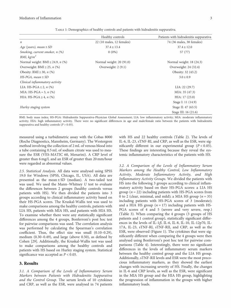
Table 1: Demographics of healthy controls and patients with hidradenitis suppurativa.
Healthy controls Patients with hidradenitis suppurativa
n 22 (10 males, 12 females) 74 (36 males, 38 females)
Age (years), mean± SD 37.4± 13.4 37.4± 12.0Smoking, current smoker, n (%) 0 (0%) 57 (77)
BMI, kg/m2
Normal weight: BMI≤ 24.9, n (%) Normal weight: 20 (91.0) Normal weight: 18 (24.3)
Overweight: BMI≥ 25, n (%) Overweight: 2 (9.1) Overweight: 24 (32.4)
Obesity: BMI≥ 30, n (%) Obesity: 32 (43.2)
HS-PGA, mean± SD 3.0± 0.9Clinical inflammatory activity
LIA: HS-PGA≤ 2, n (%) LIA: 22 (29.7)
MIA: HS-PGA= 3, n (%) MIA: 35 (47.3)
HIA: HS-PGA≥ 4, n (%) HIA: 17 (23.0)
Hurley staging system
Stage I: 11 (14.9)
Stage II: 47 (63.5)
Stage III: 16 (21.6)
BMI: body mass index; HS-PGA: Hidradenitis Suppurativa-Physician Global Assessment; LIA: low inflammatory activity; MIA: moderate inflammatoryactivity; HIA: high inflammatory activity. There were no significant differences in age and male/female ratio between the patients with hidradenitissuppurativa and healthy controls (P > 0 05).
3Mediators of Inflammation
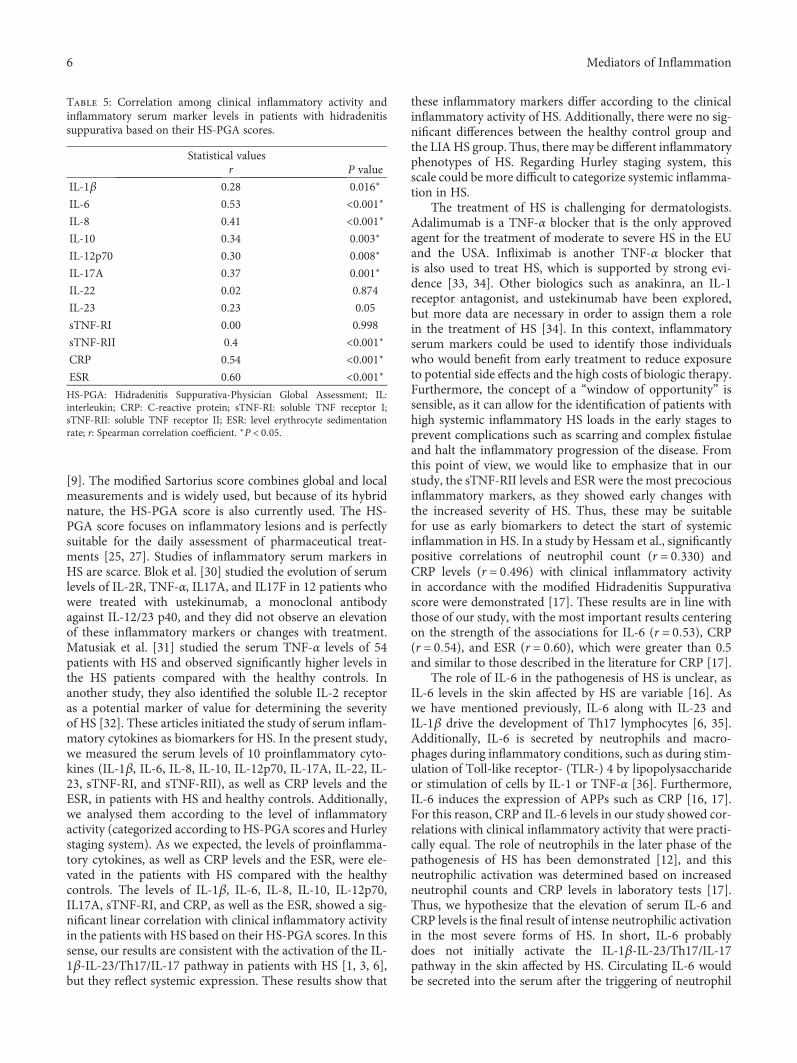
3.3. Correlations between Inflammatory Serum Markers andDisease Severity. The serum levels of the 10 cytokines, as wellas the CRP levels and the ESR, in the 74 patients with HSwere measured and aligned with disease severity (basedon clinical inflammatory activity categorized according toHS-PGA scores). The levels of IL-1β, IL-6, IL-8, IL-10,IL-12p70, IL17A, sTNF-RII, and CRP, as well as theESR, showed significant linear correlations with clinicalinflammatory activity in the patients with HS (Table 5).However, only IL-6, CRP, and ESR had absolute r valuesthat were greater than 0.49, indicating a strong association.These last 3 may be potential biomarkers for HS.
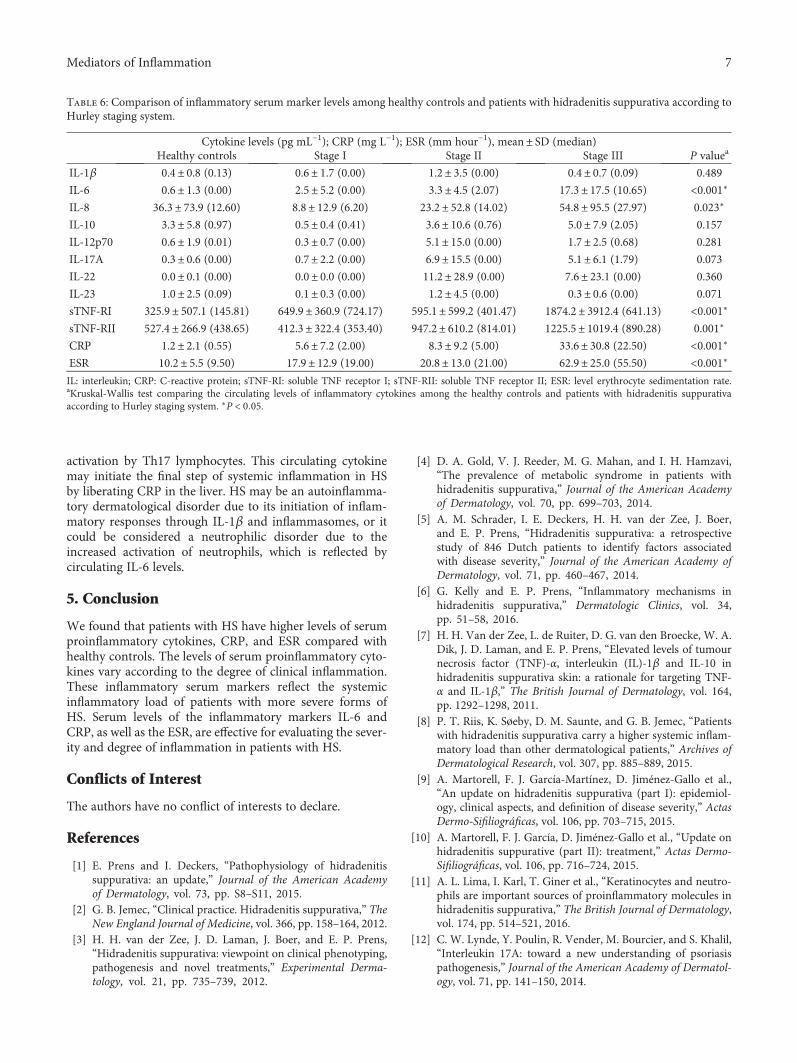
3.4. A Comparison of the Levels of Inflammatory SerumMarkers among the Healthy Control and the Three-DegreeScale Proposed by Hurley. The serum levels of the 10 cyto-kines, as well as CRP levels and the ESR, in the 74 patientswith HS were measured and aligned with disease severity(based on clinical inflammatory activity categorized accord-ing to Hurley staging system). The levels of IL-6, IL-8,sTNF-RI, sTNF-RII, CRP, and ESR were significantly ele-vated according to Hurley staging system (Table 6). Theloss of statistical significance for IL-17A could be due togreater difficulty in categorizing clinical inflammation forHurley staging.
Table 3: Comparison of inflammatory serum marker levels among healthy controls and patients with hidradenitis suppurativa according toclinical inflammatory activity based on their HS-PGA scores.
Cytokine levels (pg mL−1); CRP (mg L−1); ESR (mm hour−1), mean± SD (median)Healthy controls Low inflammatory activity Moderate inflammatory activity High inflammatory activity P valuea
IL-1β 0.4± 0.8 (0.13) 0.4± 1.4 (0.00) 1.1± 3.2 (0.39) 1.3± 3.5 (0.19) 0.022∗
IL-6 0.6± 1.3 (0.00) 1.8± 3.9 (0.00) 4.6± 7.2 (3.38) 15.4± 16.3 (10.65) <0.001∗IL-8 36.3± 73.9 (12.60) 21.7± 70.4 (4.13) 17.6± 16.2 (17.89) 57.1± 97.3 (26.24) 0.003∗
IL-10 3.3± 5.8 (0.97) 0.7± 0.7 (0.46) 3.2± 5.7 (1.63) 7.5± 17.0 (2.82) 0.032∗
IL-12p70 0.6± 1.9 (0.01) 0.6± 2.2 (0.00) 4.2± 10.8 (0.15) 6.6± 19.9 (1.20) 0.022∗
IL-17A 0.3± 0.6 (0.00) 1.4± 5.2 (0.00) 6.4± 13.6 (0.40) 9.1± 16.8 (2.97) 0.001∗
IL-22 0.0± 0.1 (0.00) 12.9± 33.7 (0.00) 9.5± 25.3 (0.00) 1.9± 7.6 (0.00) 0.904
IL-23 1.0± 2.5 (0.09) 0.1± 0.3 (0.00) 0.9± 3.2 (0.00) 1.7± 6.0 (0.00) 0.026∗
sTNF-RI 325.9± 507.1 (145.81) 746.0± 634.2 (645.27) 1000.3± 2714.8 (390.42) 804.8± 689.2 (577.87) <0.001∗sTNF-RII 527.4± 266.9 (438.65) 491.4± 380.2 (368.32) 1017.1± 583.3 (1077.75) 1309.0± 1017.6 (1096.81) <0.001∗CRP 1.2± 2.1 (0.55) 4.8± 5.8 (2.25) 9.6± 9.8 (7.00) 32.2± 30.5 (22.00) <0.001∗ESR 10.2± 5.5 (9.50) 15.3± 12.3 (12.50) 25.2± 13.2 (23.00) 56.7± 30.6 (55.00) <0.001∗HS-PGA: Hidradenitis Suppurativa-Physician Global Assessment; IL: interleukin; CRP: C-reactive protein; sTNF-RI: soluble TNF receptor I; sTNF-RII: solubleTNF receptor II; ESR: level erythrocyte sedimentation rate. aKruskal-Wallis test comparing the circulating levels of inflammatory cytokines among the healthycontrols and patients with hidradenitis suppurativa according to clinical inflammatory activity. ∗P < 0 05.
Table 2: Comparison of inflammatory serum marker levels among healthy controls and patients with hidradenitis suppurativa, and theresults of statistical assessments.
Cytokine levels (pg mL−1); CRP (mg L−1); ESR (mm hour−1), mean± SD (median)Healthy controls Hidradenitis suppurativa P valuea
IL-1β 0.4± 0.8 (0.13) 0.9± 2.9 (0.00) 0.801
IL-6 0.6± 1.3 (0.00) 6.2± 10.7 (3.00) 0.001∗
IL-8 36.3± 73.9 (12.60) 27.9± 62.3 (13.62) 0.442
IL-10 3.3± 5.8 (0.97) 3.4± 9.2 (0.64) 0.979
IL-12p70 0.6± 1.9 (0.01) 3.4± 12.1 (0.00) 0.427
IL-17A 0.3± 0.6 (0.00) 5.6± 12.1 (0.00) 0.147
IL-22 0.0± 0.1 (0.00) 8.8± 25.6 (0.00) 0.950
IL-23 1.0± 2.5 (0.09) 0.9± 3.6 (0.00) 0.014∗
sTNF-RI 325.9± 507.1 (145.81) 879.8± 1914.7 (504.39) <0.001∗sTNF-RII 527.4± 266.9 (438.65) 927.9± 722.6 (507.19) 0.053
CRP 1.2± 2.1 (0.55) 13.4± 19.2 (7.00) <0.001∗ESR 10.2± 5.5 (9.50) 29.5± 23.9 (23.00) <0.001∗IL: interleukin; sTNF-RI: soluble TNF receptor I; sTNF-RII: soluble TNF receptor II; CRP: C-reactive protein; ESR: level erythrocyte sedimentation rate.aMann–Whitney U test between healthy controls and patients with hidradenitis suppurativa. ∗P < 0 05.
4 Mediators of Inflammation
4. Discussion
The valuation of the severity of an inflammatory diseaserequires a correlation between clinical inflammatory activity
and laboratory parameters [22, 23]. Various validated toolsare available to evaluate the clinical severity of HS. TheHurley staging system is a simple but static scale that doesnot measure the number of inflammatory lesions in each area
Table 4: Bonferroni’s post hoc test for pairwise comparisons among the four groups based on their HS-PGA scores.
Group IL-6 sTNF-RII CRP ESR
Healthy controls versus low inflammatory activity
Healthy controls versus moderate inflammatory activity ∗ ∗
Healthy controls versus high inflammatory activity ∗ ∗ ∗ ∗
Low inflammatory activity versus moderate inflammatory activity ∗
Low inflammatory activity versus high inflammatory activity ∗ ∗ ∗ ∗
Moderate inflammatory activity versus high inflammatory activity ∗ ∗ ∗
HS-PGA: Hidradenitis Suppurativa-Physician Global Assessment; IL: interleukin; sTNF-RII: soluble TNF receptor II; CRP: C-reactive protein; ESR: levelerythrocyte sedimentation rate. ∗P < 0 05.
IL-8
Seru
m le
vel o
f IL-
8 (p
g/m
L)
HC LIA MIA HIA0
100
200
300
400
IL-1�훽Se
rum
leve
l of I
L-1�훽
(pg/
mL)
HC LIA MIA HIA0
5
10
15
20
⁎
IL-6
Seru
m le
vel o
f IL-
6 (p
g/m
L)HC LIA MIA HIA
0
20
40
60
IL-10
Seru
m le
vel o
f IL-
10 (p
g/m
L)
HC LIA MIA HIA0
20
40
60
80
IL-12p70
Seru
m le
vel o
f IL-
12p7
0 (p
g/m
L)
HC LIA MIA HIA0
20
40
60
80
100
IL-17A
Seru
m le
vel o
f IL-
17 (p
g/m
L)
HC LIA MIA HIA0
20
40
60
80IL-22
Seru
m le
vel o
f IL-
22 (p
g/m
L)HC LIA MIA HIA
0
20
40
60
80
100
120IL-23
Seru
m le
vel o
f IL-
23 (p
g/m
L)
HC LIA MIA HIA0
10
20
30
sTNF-RI
Seru
m le
vel o
f sTN
F-RI
(pg/
mL)
HC LIA MIA HIA0
5000
10000
15000
20000sTNF-RII
Seru
m le
vel o
f sTN
F-RI
I (pg
/mL)
HC LIA MIA HIA0
1000
2000
3000
4000
CRP
Seru
m le
vel o
f CRP
(mg/
L)
HC LIA MIA HIA0
20
40
60
80
100
120ESR
Seru
m le
vel o
f ESR
(mm
/h)
HC LIA MIA HIA0
20
40
60
80
100
120
⁎
⁎
⁎
⁎
⁎
⁎
⁎⁎ ⁎
⁎
⁎
⁎
⁎
⁎
⁎
⁎ ⁎
⁎
⁎
⁎
⁎
⁎⁎
⁎
⁎
⁎
⁎
⁎ ⁎ ⁎
⁎
⁎
⁎⁎
⁎
⁎⁎
⁎
⁎
⁎
⁎
⁎
⁎
Figure 1: Comparison of inflammatory serummarker levels among healthy controls and patients with hidradenitis suppurativa according toclinical inflammatory activity. IL, interleukin; CRP, sTNF-RI, soluble TNF receptor I; sTNF-RII, soluble TNF receptor II; C-reactive protein;ESR, level erythrocyte sedimentation rate; HC, healthy controls; LIA, low inflammatory activity; MIA, moderate inflammatory activity; HIA,high inflammatory activity.
5Mediators of Inflammation
[9]. The modified Sartorius score combines global and localmeasurements and is widely used, but because of its hybridnature, the HS-PGA score is also currently used. The HS-PGA score focuses on inflammatory lesions and is perfectlysuitable for the daily assessment of pharmaceutical treat-ments [25, 27]. Studies of inflammatory serum markers inHS are scarce. Blok et al. [30] studied the evolution of serumlevels of IL-2R, TNF-α, IL17A, and IL17F in 12 patients whowere treated with ustekinumab, a monoclonal antibodyagainst IL-12/23 p40, and they did not observe an elevationof these inflammatory markers or changes with treatment.Matusiak et al. [31] studied the serum TNF-α levels of 54patients with HS and observed significantly higher levels inthe HS patients compared with the healthy controls. Inanother study, they also identified the soluble IL-2 receptoras a potential marker of value for determining the severityof HS [32]. These articles initiated the study of serum inflam-matory cytokines as biomarkers for HS. In the present study,we measured the serum levels of 10 proinflammatory cyto-kines (IL-1β, IL-6, IL-8, IL-10, IL-12p70, IL-17A, IL-22, IL-23, sTNF-RI, and sTNF-RII), as well as CRP levels and theESR, in patients with HS and healthy controls. Additionally,we analysed them according to the level of inflammatoryactivity (categorized according to HS-PGA scores and Hurleystaging system). As we expected, the levels of proinflamma-tory cytokines, as well as CRP levels and the ESR, were ele-vated in the patients with HS compared with the healthycontrols. The levels of IL-1β, IL-6, IL-8, IL-10, IL-12p70,IL17A, sTNF-RI, and CRP, as well as the ESR, showed a sig-nificant linear correlation with clinical inflammatory activityin the patients with HS based on their HS-PGA scores. In thissense, our results are consistent with the activation of the IL-1β-IL-23/Th17/IL-17 pathway in patients with HS [1, 3, 6],but they reflect systemic expression. These results show that
these inflammatory markers differ according to the clinicalinflammatory activity of HS. Additionally, there were no sig-nificant differences between the healthy control group andthe LIAHS group. Thus, there may be different inflammatoryphenotypes of HS. Regarding Hurley staging system, thisscale could be more difficult to categorize systemic inflamma-tion in HS.
The treatment of HS is challenging for dermatologists.Adalimumab is a TNF-α blocker that is the only approvedagent for the treatment of moderate to severe HS in the EUand the USA. Infliximab is another TNF-α blocker thatis also used to treat HS, which is supported by strong evi-dence [33, 34]. Other biologics such as anakinra, an IL-1receptor antagonist, and ustekinumab have been explored,but more data are necessary in order to assign them a rolein the treatment of HS [34]. In this context, inflammatoryserum markers could be used to identify those individualswho would benefit from early treatment to reduce exposureto potential side effects and the high costs of biologic therapy.Furthermore, the concept of a “window of opportunity” issensible, as it can allow for the identification of patients withhigh systemic inflammatory HS loads in the early stages toprevent complications such as scarring and complex fistulaeand halt the inflammatory progression of the disease. Fromthis point of view, we would like to emphasize that in ourstudy, the sTNF-RII levels and ESR were the most precociousinflammatory markers, as they showed early changes withthe increased severity of HS. Thus, these may be suitablefor use as early biomarkers to detect the start of systemicinflammation in HS. In a study by Hessam et al., significantlypositive correlations of neutrophil count (r = 0 330) andCRP levels (r = 0 496) with clinical inflammatory activityin accordance with the modified Hidradenitis Suppurativascore were demonstrated [17]. These results are in line withthose of our study, with the most important results centeringon the strength of the associations for IL-6 (r = 0 53), CRP(r = 0 54), and ESR (r = 0 60), which were greater than 0.5and similar to those described in the literature for CRP [17].
The role of IL-6 in the pathogenesis of HS is unclear, asIL-6 levels in the skin affected by HS are variable [16]. Aswe have mentioned previously, IL-6 along with IL-23 andIL-1β drive the development of Th17 lymphocytes [6, 35].Additionally, IL-6 is secreted by neutrophils and macro-phages during inflammatory conditions, such as during stim-ulation of Toll-like receptor- (TLR-) 4 by lipopolysaccharideor stimulation of cells by IL-1 or TNF-α [36]. Furthermore,IL-6 induces the expression of APPs such as CRP [16, 17].For this reason, CRP and IL-6 levels in our study showed cor-relations with clinical inflammatory activity that were practi-cally equal. The role of neutrophils in the later phase of thepathogenesis of HS has been demonstrated [12], and thisneutrophilic activation was determined based on increasedneutrophil counts and CRP levels in laboratory tests [17].Thus, we hypothesize that the elevation of serum IL-6 andCRP levels is the final result of intense neutrophilic activationin the most severe forms of HS. In short, IL-6 probablydoes not initially activate the IL-1β-IL-23/Th17/IL-17pathway in the skin affected by HS. Circulating IL-6 wouldbe secreted into the serum after the triggering of neutrophil
Table 5: Correlation among clinical inflammatory activity andinflammatory serum marker levels in patients with hidradenitissuppurativa based on their HS-PGA scores.
Statistical valuesr P value
IL-1β 0.28 0.016∗
IL-6 0.53 <0.001∗IL-8 0.41 <0.001∗IL-10 0.34 0.003∗
IL-12p70 0.30 0.008∗
IL-17A 0.37 0.001∗
IL-22 0.02 0.874
IL-23 0.23 0.05
sTNF-RI 0.00 0.998
sTNF-RII 0.4 <0.001∗CRP 0.54 <0.001∗ESR 0.60 <0.001∗HS-PGA: Hidradenitis Suppurativa-Physician Global Assessment; IL:interleukin; CRP: C-reactive protein; sTNF-RI: soluble TNF receptor I;sTNF-RII: soluble TNF receptor II; ESR: level erythrocyte sedimentationrate; r: Spearman correlation coefficient. ∗P < 0 05.
6 Mediators of Inflammation
activation by Th17 lymphocytes. This circulating cytokinemay initiate the final step of systemic inflammation in HSby liberating CRP in the liver. HS may be an autoinflamma-tory dermatological disorder due to its initiation of inflam-matory responses through IL-1β and inflammasomes, or itcould be considered a neutrophilic disorder due to theincreased activation of neutrophils, which is reflected bycirculating IL-6 levels.
5. Conclusion
We found that patients with HS have higher levels of serumproinflammatory cytokines, CRP, and ESR compared withhealthy controls. The levels of serum proinflammatory cyto-kines vary according to the degree of clinical inflammation.These inflammatory serum markers reflect the systemicinflammatory load of patients with more severe forms ofHS. Serum levels of the inflammatory markers IL-6 andCRP, as well as the ESR, are effective for evaluating the sever-ity and degree of inflammation in patients with HS.
Conflicts of Interest
The authors have no conflict of interests to declare.
References
[1] E. Prens and I. Deckers, “Pathophysiology of hidradenitissuppurativa: an update,” Journal of the American Academyof Dermatology, vol. 73, pp. S8–S11, 2015.
[2] G. B. Jemec, “Clinical practice. Hidradenitis suppurativa,” TheNew England Journal of Medicine, vol. 366, pp. 158–164, 2012.
[3] H. H. van der Zee, J. D. Laman, J. Boer, and E. P. Prens,“Hidradenitis suppurativa: viewpoint on clinical phenotyping,pathogenesis and novel treatments,” Experimental Derma-tology, vol. 21, pp. 735–739, 2012.
[4] D. A. Gold, V. J. Reeder, M. G. Mahan, and I. H. Hamzavi,“The prevalence of metabolic syndrome in patients withhidradenitis suppurativa,” Journal of the American Academyof Dermatology, vol. 70, pp. 699–703, 2014.
[5] A. M. Schrader, I. E. Deckers, H. H. van der Zee, J. Boer,and E. P. Prens, “Hidradenitis suppurativa: a retrospectivestudy of 846 Dutch patients to identify factors associatedwith disease severity,” Journal of the American Academy ofDermatology, vol. 71, pp. 460–467, 2014.
[6] G. Kelly and E. P. Prens, “Inflammatory mechanisms inhidradenitis suppurativa,” Dermatologic Clinics, vol. 34,pp. 51–58, 2016.
[7] H. H. Van der Zee, L. de Ruiter, D. G. van den Broecke, W. A.Dik, J. D. Laman, and E. P. Prens, “Elevated levels of tumournecrosis factor (TNF)-α, interleukin (IL)-1β and IL-10 inhidradenitis suppurativa skin: a rationale for targeting TNF-α and IL-1β,” The British Journal of Dermatology, vol. 164,pp. 1292–1298, 2011.
[8] P. T. Riis, K. Søeby, D. M. Saunte, and G. B. Jemec, “Patientswith hidradenitis suppurativa carry a higher systemic inflam-matory load than other dermatological patients,” Archives ofDermatological Research, vol. 307, pp. 885–889, 2015.
[9] A. Martorell, F. J. García-Martínez, D. Jiménez-Gallo et al.,“An update on hidradenitis suppurativa (part I): epidemiol-ogy, clinical aspects, and definition of disease severity,” ActasDermo-Sifiliográficas, vol. 106, pp. 703–715, 2015.
[10] A. Martorell, F. J. García, D. Jiménez-Gallo et al., “Update onhidradenitis suppurative (part II): treatment,” Actas Dermo-Sifiliográficas, vol. 106, pp. 716–724, 2015.
[11] A. L. Lima, I. Karl, T. Giner et al., “Keratinocytes and neutro-phils are important sources of proinflammatory molecules inhidradenitis suppurativa,” The British Journal of Dermatology,vol. 174, pp. 514–521, 2016.
[12] C. W. Lynde, Y. Poulin, R. Vender, M. Bourcier, and S. Khalil,“Interleukin 17A: toward a new understanding of psoriasispathogenesis,” Journal of the American Academy of Dermatol-ogy, vol. 71, pp. 141–150, 2014.
Table 6: Comparison of inflammatory serum marker levels among healthy controls and patients with hidradenitis suppurativa according toHurley staging system.
Cytokine levels (pg mL−1); CRP (mg L−1); ESR (mm hour−1), mean± SD (median)Healthy controls Stage I Stage II Stage III P valuea
IL-1β 0.4± 0.8 (0.13) 0.6± 1.7 (0.00) 1.2± 3.5 (0.00) 0.4± 0.7 (0.09) 0.489
IL-6 0.6± 1.3 (0.00) 2.5± 5.2 (0.00) 3.3± 4.5 (2.07) 17.3± 17.5 (10.65) <0.001∗IL-8 36.3± 73.9 (12.60) 8.8± 12.9 (6.20) 23.2± 52.8 (14.02) 54.8± 95.5 (27.97) 0.023∗
IL-10 3.3± 5.8 (0.97) 0.5± 0.4 (0.41) 3.6± 10.6 (0.76) 5.0± 7.9 (2.05) 0.157
IL-12p70 0.6± 1.9 (0.01) 0.3± 0.7 (0.00) 5.1± 15.0 (0.00) 1.7± 2.5 (0.68) 0.281
IL-17A 0.3± 0.6 (0.00) 0.7± 2.2 (0.00) 6.9± 15.5 (0.00) 5.1± 6.1 (1.79) 0.073
IL-22 0.0± 0.1 (0.00) 0.0± 0.0 (0.00) 11.2± 28.9 (0.00) 7.6± 23.1 (0.00) 0.360
IL-23 1.0± 2.5 (0.09) 0.1± 0.3 (0.00) 1.2± 4.5 (0.00) 0.3± 0.6 (0.00) 0.071
sTNF-RI 325.9± 507.1 (145.81) 649.9± 360.9 (724.17) 595.1± 599.2 (401.47) 1874.2± 3912.4 (641.13) <0.001∗sTNF-RII 527.4± 266.9 (438.65) 412.3± 322.4 (353.40) 947.2± 610.2 (814.01) 1225.5± 1019.4 (890.28) 0.001∗
CRP 1.2± 2.1 (0.55) 5.6± 7.2 (2.00) 8.3± 9.2 (5.00) 33.6± 30.8 (22.50) <0.001∗ESR 10.2± 5.5 (9.50) 17.9± 12.9 (19.00) 20.8± 13.0 (21.00) 62.9± 25.0 (55.50) <0.001∗IL: interleukin; CRP: C-reactive protein; sTNF-RI: soluble TNF receptor I; sTNF-RII: soluble TNF receptor II; ESR: level erythrocyte sedimentation rate.aKruskal-Wallis test comparing the circulating levels of inflammatory cytokines among the healthy controls and patients with hidradenitis suppurativaaccording to Hurley staging system. ∗P < 0 05.
7Mediators of Inflammation
[13] K. A. Cho, J. W. Suh, K. H. Lee, J. L. Kang, and S. Y. Woo,“IL-17 and IL-22 enhance skin inflammation by stimulatingthe secretion of IL-1β by keratinocytes via the ROS-NLRP3-caspase-1 pathway,” International Immunology, vol. 24,pp. 147–158, 2012.
[14] C. Schlapbach, T. Hänni, N. Yawalkar, and R. E. Hunger,“Expression of the IL-23/Th17 pathway in lesions of hidra-denitis suppurativa,” Journal of the American Academy ofDermatology, vol. 65, pp. 790–798, 2011.
[15] E. J.Giamarellos-Bourboulis,A.Antonopoulou,C.Petropoulouet al., “Altered innate and adaptive immune responses inpatients with hidradenitis suppurativa,” The British Journalof Dermatology, vol. 156, pp. 51–56, 2007.
[16] G. Kelly, C. M. Sweeney, A. M. Tobin, and B. Kirby, “Hidrade-nitis suppurativa: the role of immune dysregulation,” Interna-tional Journal of Dermatology, vol. 53, pp. 1186–1196, 2014.
[17] S. Hessam, M. Sand, T. Gambichler, and F. G. Bechara,“Correlation of inflammatory serum markers with diseaseseverity in patients with hidradenitis suppurativa (HS),”Journal of the American Academy of Dermatology, vol. 73,pp. 998–1005, 2015.
[18] J. C. Pascual, I. González, D. Corona et al., “Assessment ofsubclinical atherosclerosis in hidradenitis suppurativa,” Jour-nal of the European Academy of Dermatology and Venereology,vol. 2, 2016.
[19] S. E. Bedell and B. T. Bush, “Erythrocyte sedimentation rate.From folklore to facts,” The American Journal of Medicine,vol. 78, p. 1001, 1985.
[20] Ł. Matusiak, J. Salomon, D. Nowicka-Suszko, A. Bieniek,and J. C. Szepietowski, “Chitinase-3-like protein 1 (YKL-40):novel biomarker of hidradenitis suppurativa disease activity?,”Acta Dermato-Venereologica, vol. 95, pp. 736-737, 2015.
[21] Ł. Matusiak, J. Szczęch, A. Bieniek, D. Nowicka-Suszko, andJ. C. Szepietowski, “Increased interleukin (IL)-17 serumlevels in patients with hidradenitis suppurativa: implicationsfor treatment with anti-IL-17 agents,” Journal of the AmericanAcademy of Dermatology, vol. 76, pp. 670–675, 2017.
[22] B. I. Gavrilă, C. Ciofu, and V. Stoica, “Biomarkers in rheuma-toid arthritis, what is new?,” Journal of Medicine and Life,vol. 9, pp. 144–148, 2016.
[23] Z. Rezaieyazdi, M. Sahebari, M. R. Hatef et al., “Is thereany correlation between high sensitive CRP and diseaseactivity in systemic lupus erythematosus?,” Lupus, vol. 20,pp. 1494–1500, 2011.
[24] J. Revuz, “Hidradenitis suppurativa,” Journal of the EuropeanAcademy of Dermatology and Venereology, vol. 23, pp. 985–998, 2009.
[25] J. Revuz, “Évaluation clinique de la sévérité de l’hidradénitesuppurée – maladie de Verneuil,” Annales de Dermatologie etde Vénéréologie, vol. 142, pp. 729–735, 2015.
[26] H. H. van der Zee and G. B. Jemec, “New insights into the diag-nosis of hidradenitis suppurativa: clinical presentations andphenotypes,” Journal of the American Academy of Dermatol-ogy, vol. 73, pp. S23–S26, 2015.
[27] A. B. Kimball, F. Kerdel, D. Adams et al., “Adalimumab forthe treatment of moderate to severe hidradenitis suppurativa: aparallel randomized trial,” Annals of Internal Medicine,vol. 157, pp. 846–855, 2012.
[28] F. Porteu and C. Nathan, “Shedding of tumor necrosis factorreceptors by activated human neutrophils,” The Journal ofExperimental Medicine, vol. 172, pp. 599–607, 1990.
[29] J. Cohen, Statistical Power Analysis for the Behavioral Sciences,Lawrence Erlbaum Associates, Hilsdale, New Jersey, USA,1988.
[30] J. L. Blok, K. Li, C. Brodmerkel, P. Horvátovich,M. F. Jonkman,and B. Horváth, “Ustekinumab in hidradenitis suppurativa:clinical results and a search for potential biomarkers inserum,” The British Journal of Dermatology, vol. 174,pp. 839–846, 2016.
[31] L. Matusiak, A. Bieniek, and J. C. Szepietowski, “Increasedserum tumour necrosis factor-alpha in hidradenitis suppura-tiva patients: is there a basis for treatment with anti-tumournecrosis factor-alpha agents?,” Acta Dermato-Venereologica,vol. 89, pp. 601–603, 2009.
[32] Ł. Matusiak, A. Bieniek, and J. C. Szepietowski, “Solubleinterleukin-2 receptor serum level is a useful marker ofhidradenitis suppurativa clinical staging,” Biomarkers, vol. 14,pp. 432–437, 2009.
[33] E. S. Kim, K. P. Garnock-Jones, and S. J. Keam, “Adalimumab:a review in hidradenitis suppurativa,” American Journal ofClinical Dermatology, vol. 17, pp. 545–552, 2016.
[34] R. A. Lee and D. B. Eisen, “Treatment of hidradenitis suppur-ativa with biologic medications,” Journal of the AmericanAcademy of Dermatology, vol. 73, pp. S82–S88, 2015.
[35] G. Kelly, R. Hughes, T. McGarry et al., “Dysregulated cytokineexpression in lesional and nonlesional skin in hidradenitissuppurativa,” The British Journal of Dermatology, vol. 173,pp. 1431–1439, 2015.
[36] D. Schmidt-Arras and S. Rose-John, “IL-6 pathway in the liver:from physiopathology to therapy,” Journal of Hepatology,vol. 64, pp. 1403–1415, 2016.
8 Mediators of Inflammation

Submit your manuscripts athttps://www.hindawi.com
Stem CellsInternational
Hindawi Publishing Corporationhttp://www.hindawi.com Volume 2014
Hindawi Publishing Corporationhttp://www.hindawi.com Volume 2014
MEDIATORSINFLAMMATION
of
Hindawi Publishing Corporationhttp://www.hindawi.com Volume 2014
Behavioural Neurology
EndocrinologyInternational Journal of
Hindawi Publishing Corporationhttp://www.hindawi.com Volume 2014
Hindawi Publishing Corporationhttp://www.hindawi.com Volume 2014
Disease Markers
Hindawi Publishing Corporationhttp://www.hindawi.com Volume 2014
BioMed Research International
OncologyJournal of
Hindawi Publishing Corporationhttp://www.hindawi.com Volume 2014
Hindawi Publishing Corporationhttp://www.hindawi.com Volume 2014
Oxidative Medicine and Cellular Longevity
Hindawi Publishing Corporationhttp://www.hindawi.com Volume 2014
PPAR Research
The Scientific World JournalHindawi Publishing Corporation http://www.hindawi.com Volume 2014
Immunology ResearchHindawi Publishing Corporationhttp://www.hindawi.com Volume 2014
Journal of
ObesityJournal of
Hindawi Publishing Corporationhttp://www.hindawi.com Volume 2014
Hindawi Publishing Corporationhttp://www.hindawi.com Volume 2014
Computational and Mathematical Methods in Medicine
OphthalmologyJournal of
Hindawi Publishing Corporationhttp://www.hindawi.com Volume 2014
Diabetes ResearchJournal of
Hindawi Publishing Corporationhttp://www.hindawi.com Volume 2014
Hindawi Publishing Corporationhttp://www.hindawi.com Volume 2014
Research and TreatmentAIDS
Hindawi Publishing Corporationhttp://www.hindawi.com Volume 2014
Gastroenterology Research and Practice
Hindawi Publishing Corporationhttp://www.hindawi.com Volume 2014
Parkinson’s Disease
Evidence-Based Complementary and Alternative Medicine
Volume 2014Hindawi Publishing Corporationhttp://www.hindawi.com