THE CIRCULATORY SYSTEM Table of Contents · PDF fileTHE CIRCULATORY SYSTEM Table of Contents...
29
THE CIRCULATORY SYSTEM Table of Contents Types of Circulatory Systems | Vertebrate Cardiovascular System | Vertebrate Vascular Systems The Heart | The Vascular System | Blood | The Lymphatic System | Learning Objectives | Links Types of Circulatory Systems | Back to Top Living things must be capable of transporting nutrients, wastes and gases to and from cells. Single-celled organisms use their cell surface as a point of exchange with the outside environment. Multicellular organisms have developed transport and circulatory systems to deliver oxygen and food to cells and remove carbon dioxide and metabolic wastes. Sponges are the simplest animals, yet even they have a transport system. Seawater is the medium of transport and is propelled in and out of the sponge by ciliary action. Simple animals, such as the hydra and planaria, lack specialized organs such as hearts and blood vessels, instead using their skin as an exchange point for materials. This, however, limits the size an animal can attain. To become larger, they need specialized organs and organ systems.
Transcript of THE CIRCULATORY SYSTEM Table of Contents · PDF fileTHE CIRCULATORY SYSTEM Table of Contents...

THE CIRCULATORY SYSTEM
Table of Contents
Types of Circulatory Systems | Vertebrate Cardiovascular System | Vertebrate Vascular
Systems
The Heart | The Vascular System | Blood | The Lymphatic System | Learning Objectives |
Links
Types of Circulatory Systems | Back to Top
Living things must be capable of transporting nutrients, wastes and gases to and from
cells. Single-celled organisms use their cell surface as a point of exchange with the
outside environment. Multicellular organisms have developed transport and
circulatory systems to deliver oxygen and food to cells and remove carbon dioxide
and metabolic wastes. Sponges are the simplest animals, yet even they have a
transport system. Seawater is the medium of transport and is propelled in and out of
the sponge by ciliary action. Simple animals, such as the hydra and planaria, lack
specialized organs such as hearts and blood vessels, instead using their skin as an
exchange point for materials. This, however, limits the size an animal can attain. To
become larger, they need specialized organs and organ systems.
Structures that serve some of the functions of the circulatory system in animals that
lack the system. Image from Purves et al., Life: The Science of Biology, 4th Edition, by
Sinauer Associates (http://www.sinauer.com/) and WH Freeman (http://www.whfreeman.com/),
used with permission.
Multicellular animals do not have most of their cells in contact with the external
environment and so have developed circulatory systems to transport nutrients, oxygen,
carbon dioxide and metabolic wastes. Components of the circulatory system include
blood: a connective tissue of liquid plasma and cells
heart: a muscular pump to move the blood
blood vessels: arteries, capillaries and veins that deliver blood to all tissues
There are several types of circulatory systems. The open circulatory system is
common to molluscs and arthropods. Open circulatory systems (evolved in insects,
mollusks and other invertebrates) pump blood into a hemocoel with the blood
diffusing back to the circulatory system between cells. Blood is pumped by a heart
into the body cavities, where tissues are surrounded by the blood. The resulting blood
flow is sluggish.

Circulatory systems of an insect (top) and mollusc (bottom). Images from Purves et al.,
Life: The Science of Biology, 4th Edition, by Sinauer Associates (http://www.sinauer.com/) and
WH Freeman (http://www.whfreeman.com/), used with permission.
Details of the circulatory system of an insect. The above image is from
http://www.biosci.uga.edu/almanac/bio_104/notes/may_7.html.
Vertebrates, and a few invertebrates, have a closed circulatory system. Closed
circulatory systems (evolved in echinoderms and vertebrates) have the blood closed at
all times within vessels of different size and wall thickness. In this type of system,
blood is pumped by a heart through vessels, and does not normally fill body cavities.
Blood flow is not sluggish. Hemoglobin causes vertebrate blood to turn red in the
presence of oxygen; but more importantly hemoglobin molecules in blood cells
transport oxygen. The human closed circulatory system is sometimes called the
cardiovascular system.
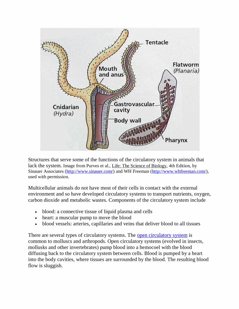
The circulatory system of an earthworm. The above image is from
http://www.biosci.uga.edu/almanac/bio_104/notes/may_7.html.
A secondary circulatory system, the lymphatic circulation, collects fluid and cells and
returns them to the cardiovascular system.
Vertebrate Cardiovascular System | Back to Top
The vertebrate cardiovascular system includes a heart, which is a muscular pump that
contracts to propel blood out to the body through arteries, and a series of blood
vessels. The upper chamber of the heart, the atrium (pl. atria), is where the blood
enters the heart. Passing through a valve, blood enters the lower chamber, the
ventricle. Contraction of the ventricle forces blood from the heart through an artery.
The heart muscle is composed of cardiac muscle cells.

The basic circulatory patterns of blood flow in a mammal. The above image is from
http://johns.largnet.uwo.ca/shine/health/heart.htm.
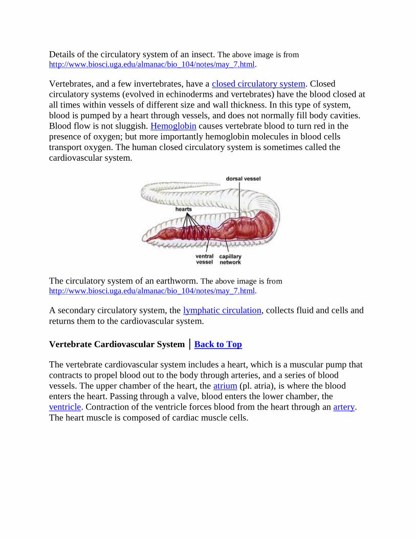
Arteries are blood vessels that carry blood away from heart. Arterial walls are able to
expand and contract. Arteries have three layers of thick walls. Smooth muscle fibers
contract, another layer of connective tissue is quite elastic, allowing the arteries to
carry blood under high pressure.
Structure of an artery. Image from Purves et al., Life: The Science of Biology, 4th Edition, by
Sinauer Associates (http://www.sinauer.com/) and WH Freeman (http://www.whfreeman.com/),
used with permission.
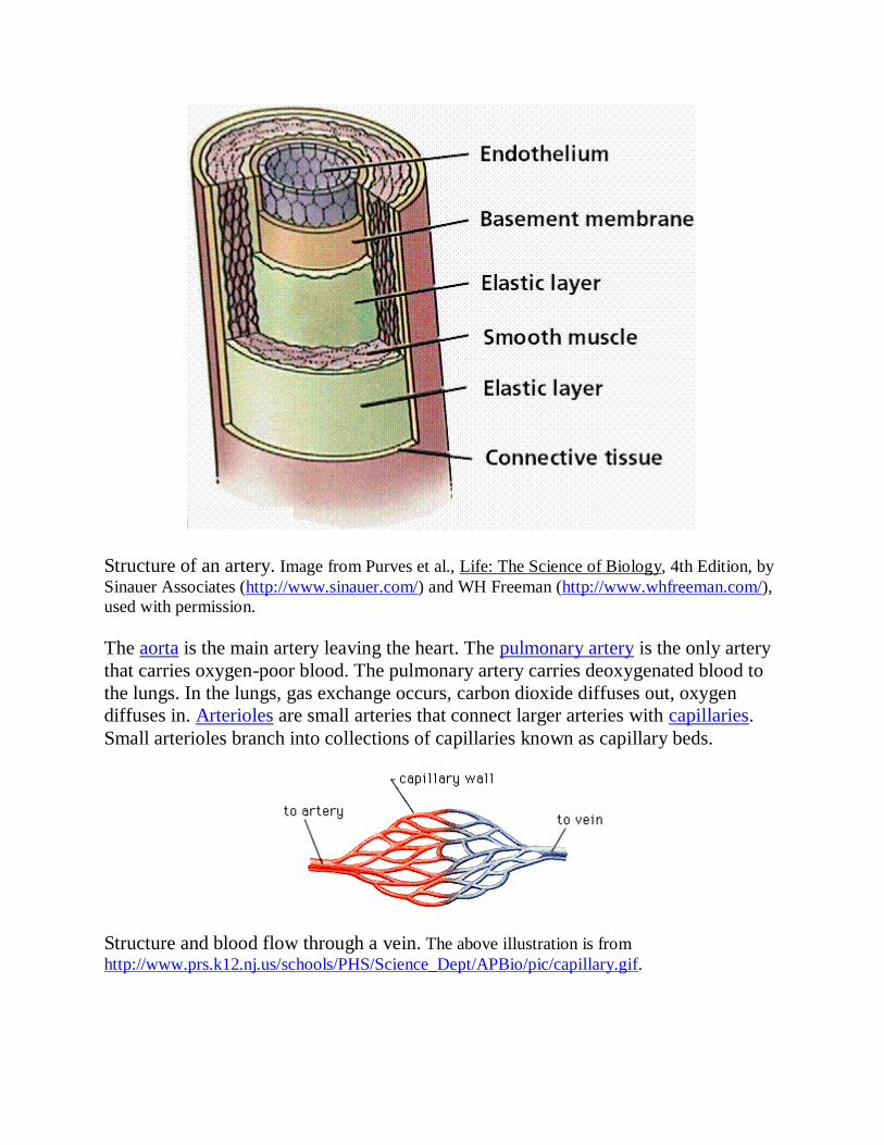
The aorta is the main artery leaving the heart. The pulmonary artery is the only artery
that carries oxygen-poor blood. The pulmonary artery carries deoxygenated blood to
the lungs. In the lungs, gas exchange occurs, carbon dioxide diffuses out, oxygen
diffuses in. Arterioles are small arteries that connect larger arteries with capillaries.
Small arterioles branch into collections of capillaries known as capillary beds.
Structure and blood flow through a vein. The above illustration is from
http://www.prs.k12.nj.us/schools/PHS/Science_Dept/APBio/pic/capillary.gif.
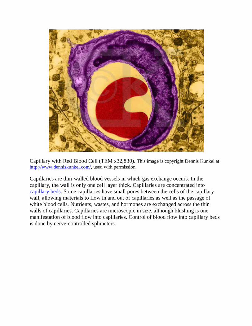
Capillary with Red Blood Cell (TEM x32,830). This image is copyright Dennis Kunkel at
http://www.denniskunkel.com/, used with permission.
Capillaries are thin-walled blood vessels in which gas exchange occurs. In the
capillary, the wall is only one cell layer thick. Capillaries are concentrated into
capillary beds. Some capillaries have small pores between the cells of the capillary
wall, allowing materials to flow in and out of capillaries as well as the passage of
white blood cells. Nutrients, wastes, and hormones are exchanged across the thin
walls of capillaries. Capillaries are microscopic in size, although blushing is one
manifestation of blood flow into capillaries. Control of blood flow into capillary beds
is done by nerve-controlled sphincters.

Changes in blood pressure, velocity, and the area of the arteries, capillaries, and veins
of the circulatory system. Image from Purves et al., Life: The Science of Biology, 4th Edition,
by Sinauer Associates (http://www.sinauer.com/) and WH Freeman
(http://www.whfreeman.com/), used with permission.
The circulatory system functions in the delivery of oxygen, nutrient molecules, and
hormones and the removal of carbon dioxide, ammonia and other metabolic wastes.
Capillaries are the points of exchange between the blood and surrounding tissues.
Materials cross in and out of the capillaries by passing through or between the cells
that line the capillary.
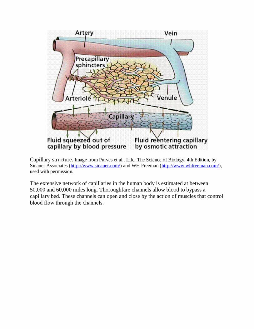
Capillary structure. Image from Purves et al., Life: The Science of Biology, 4th Edition, by
Sinauer Associates (http://www.sinauer.com/) and WH Freeman (http://www.whfreeman.com/),
used with permission.
The extensive network of capillaries in the human body is estimated at between
50,000 and 60,000 miles long. Thoroughfare channels allow blood to bypass a
capillary bed. These channels can open and close by the action of muscles that control
blood flow through the channels.
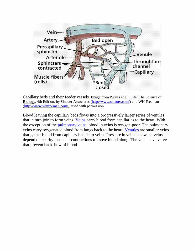
Capillary beds and their feeder vessels. Image from Purves et al., Life: The Science of
Biology, 4th Edition, by Sinauer Associates (http://www.sinauer.com/) and WH Freeman
(http://www.whfreeman.com/), used with permission.
Blood leaving the capillary beds flows into a progressively larger series of venules
that in turn join to form veins. Veins carry blood from capillaries to the heart. With
the exception of the pulmonary veins, blood in veins is oxygen-poor. The pulmonary
veins carry oxygenated blood from lungs back to the heart. Venules are smaller veins
that gather blood from capillary beds into veins. Pressure in veins is low, so veins
depend on nearby muscular contractions to move blood along. The veins have valves
that prevent back-flow of blood.
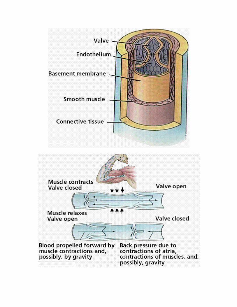
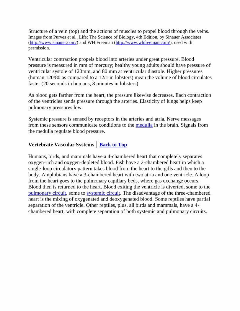
Structure of a vein (top) and the actions of muscles to propel blood through the veins. Images from Purves et al., Life: The Science of Biology, 4th Edition, by Sinauer Associates
(http://www.sinauer.com/) and WH Freeman (http://www.whfreeman.com/), used with
permission.
Ventricular contraction propels blood into arteries under great pressure. Blood
pressure is measured in mm of mercury; healthy young adults should have pressure of
ventricular systole of 120mm, and 80 mm at ventricular diastole. Higher pressures
(human 120/80 as compared to a 12/1 in lobsters) mean the volume of blood circulates
faster (20 seconds in humans, 8 minutes in lobsters).
As blood gets farther from the heart, the pressure likewise decreases. Each contraction
of the ventricles sends pressure through the arteries. Elasticity of lungs helps keep
pulmonary pressures low.
Systemic pressure is sensed by receptors in the arteries and atria. Nerve messages
from these sensors communicate conditions to the medulla in the brain. Signals from
the medulla regulate blood pressure.
Vertebrate Vascular Systems | Back to Top
Humans, birds, and mammals have a 4-chambered heart that completely separates
oxygen-rich and oxygen-depleted blood. Fish have a 2-chambered heart in which a
single-loop circulatory pattern takes blood from the heart to the gills and then to the
body. Amphibians have a 3-chambered heart with two atria and one ventricle. A loop
from the heart goes to the pulmonary capillary beds, where gas exchange occurs.
Blood then is returned to the heart. Blood exiting the ventricle is diverted, some to the
pulmonary circuit, some to systemic circuit. The disadvantage of the three-chambered
heart is the mixing of oxygenated and deoxygenated blood. Some reptiles have partial
separation of the ventricle. Other reptiles, plus, all birds and mammals, have a 4-
chambered heart, with complete separation of both systemic and pulmonary circuits.
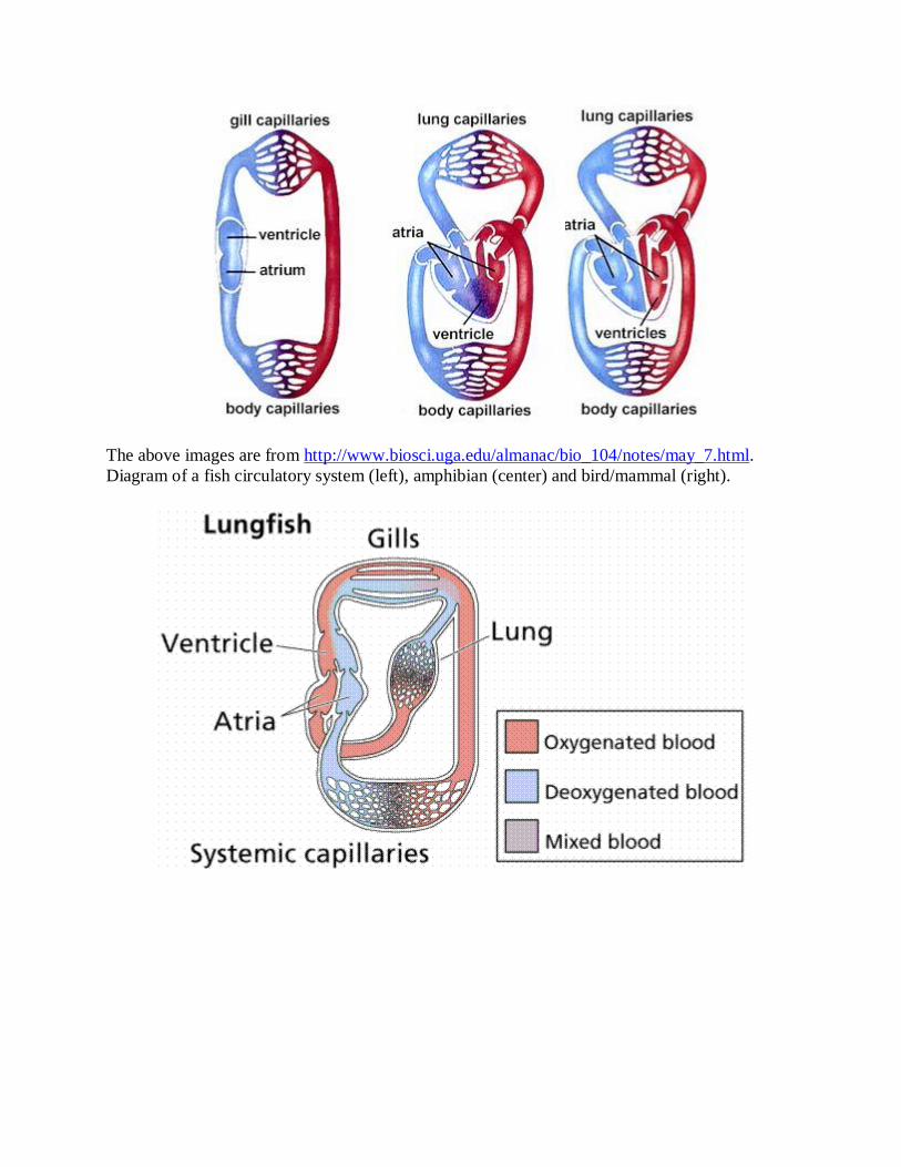
The above images are from http://www.biosci.uga.edu/almanac/bio_104/notes/may_7.html.
Diagram of a fish circulatory system (left), amphibian (center) and bird/mammal (right).

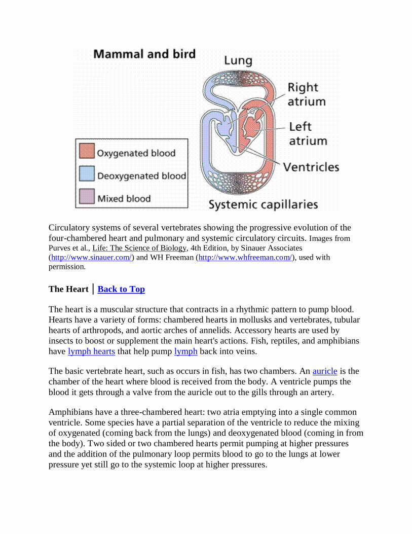
Circulatory systems of several vertebrates showing the progressive evolution of the
four-chambered heart and pulmonary and systemic circulatory circuits. Images from
Purves et al., Life: The Science of Biology, 4th Edition, by Sinauer Associates
(http://www.sinauer.com/) and WH Freeman (http://www.whfreeman.com/), used with
permission.
The Heart | Back to Top
The heart is a muscular structure that contracts in a rhythmic pattern to pump blood.
Hearts have a variety of forms: chambered hearts in mollusks and vertebrates, tubular
hearts of arthropods, and aortic arches of annelids. Accessory hearts are used by
insects to boost or supplement the main heart's actions. Fish, reptiles, and amphibians
have lymph hearts that help pump lymph back into veins.
The basic vertebrate heart, such as occurs in fish, has two chambers. An auricle is the
chamber of the heart where blood is received from the body. A ventricle pumps the
blood it gets through a valve from the auricle out to the gills through an artery.
Amphibians have a three-chambered heart: two atria emptying into a single common
ventricle. Some species have a partial separation of the ventricle to reduce the mixing
of oxygenated (coming back from the lungs) and deoxygenated blood (coming in from
the body). Two sided or two chambered hearts permit pumping at higher pressures
and the addition of the pulmonary loop permits blood to go to the lungs at lower
pressure yet still go to the systemic loop at higher pressures.
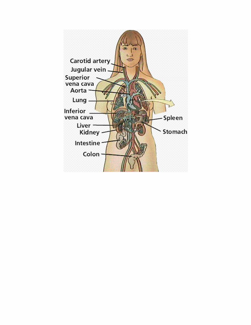

The relationship of the heart and circulatory system to major visceral organs. Below:
the structure of the heart. Images from Purves et al., Life: The Science of Biology, 4th
Edition, by Sinauer Associates (http://www.sinauer.com/) and WH Freeman
(http://www.whfreeman.com/), used with permission.
Establishment of the four-chambered heart, along with the pulmonary and systemic
circuits, completely separates oxygenated from deoxygenated blood. This allows
higher the metabolic rates needed by warm-blooded birds and mammals.

The above image is from http://www.biosci.uga.edu/almanac/bio_104/notes/may_7.html.
The human heart is a two-sided, 4 chambered structure with muscular walls. An
atrioventricular (AV) valve separates each auricle from ventricle. A semilunar (also
known as arterial) valve separates each ventricle from its connecting artery.
The heart beats or contracts 70 times per minute. The human heart will undergo over 3
billion contraction cycles during a normal lifetime. The cardiac cycle consists of two
parts: systole (contraction of the heart muscle) and diastole (relaxation of the heart
muscle). Atria contract while ventricles relax. The pulse is a wave of contraction
transmitted along the arteries. Valves in the heart open and close during the cardiac
cycle. Heart muscle contraction is due to the presence of nodal tissue in two regions of
the heart. The SA node (sinoatrial node) initiates heartbeat. The AV node
(atrioventricular node) causes ventricles to contract. The AV node is sometimes called
the pacemaker since it keeps heartbeat regular. Heartbeat is also controlled by the
autonomic nervous system.
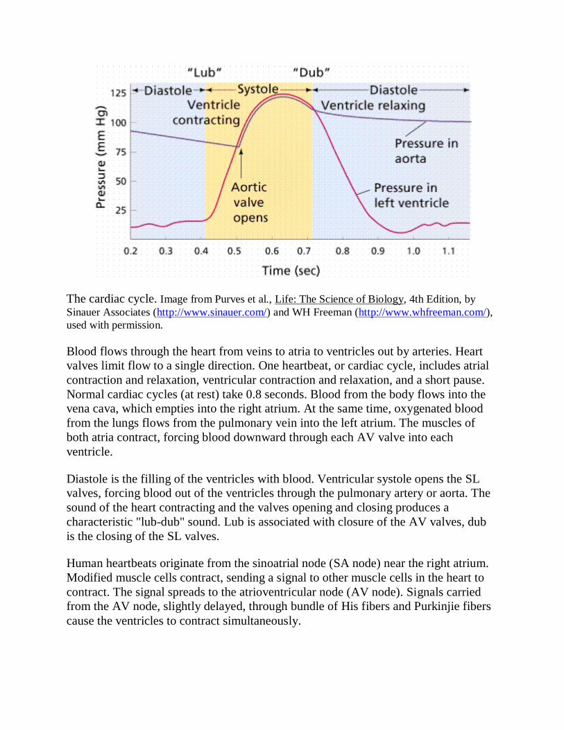
The cardiac cycle. Image from Purves et al., Life: The Science of Biology, 4th Edition, by
Sinauer Associates (http://www.sinauer.com/) and WH Freeman (http://www.whfreeman.com/),
used with permission.
Blood flows through the heart from veins to atria to ventricles out by arteries. Heart
valves limit flow to a single direction. One heartbeat, or cardiac cycle, includes atrial
contraction and relaxation, ventricular contraction and relaxation, and a short pause.
Normal cardiac cycles (at rest) take 0.8 seconds. Blood from the body flows into the
vena cava, which empties into the right atrium. At the same time, oxygenated blood
from the lungs flows from the pulmonary vein into the left atrium. The muscles of
both atria contract, forcing blood downward through each AV valve into each
ventricle.
Diastole is the filling of the ventricles with blood. Ventricular systole opens the SL
valves, forcing blood out of the ventricles through the pulmonary artery or aorta. The
sound of the heart contracting and the valves opening and closing produces a
characteristic "lub-dub" sound. Lub is associated with closure of the AV valves, dub
is the closing of the SL valves.
Human heartbeats originate from the sinoatrial node (SA node) near the right atrium.
Modified muscle cells contract, sending a signal to other muscle cells in the heart to
contract. The signal spreads to the atrioventricular node (AV node). Signals carried
from the AV node, slightly delayed, through bundle of His fibers and Purkinjie fibers
cause the ventricles to contract simultaneously.

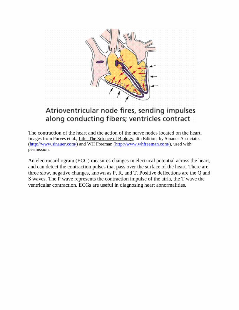
The contraction of the heart and the action of the nerve nodes located on the heart. Images from Purves et al., Life: The Science of Biology, 4th Edition, by Sinauer Associates
(http://www.sinauer.com/) and WH Freeman (http://www.whfreeman.com/), used with
permission.
An electrocardiogram (ECG) measures changes in electrical potential across the heart,
and can detect the contraction pulses that pass over the surface of the heart. There are
three slow, negative changes, known as P, R, and T. Positive deflections are the Q and
S waves. The P wave represents the contraction impulse of the atria, the T wave the
ventricular contraction. ECGs are useful in diagnosing heart abnormalities.
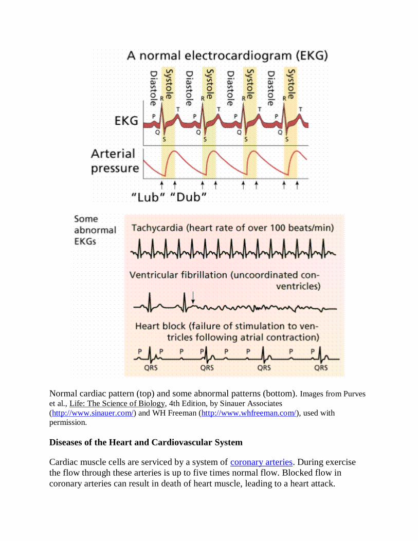
Normal cardiac pattern (top) and some abnormal patterns (bottom). Images from Purves
et al., Life: The Science of Biology, 4th Edition, by Sinauer Associates
(http://www.sinauer.com/) and WH Freeman (http://www.whfreeman.com/), used with
permission.
Diseases of the Heart and Cardiovascular System
Cardiac muscle cells are serviced by a system of coronary arteries. During exercise
the flow through these arteries is up to five times normal flow. Blocked flow in
coronary arteries can result in death of heart muscle, leading to a heart attack.
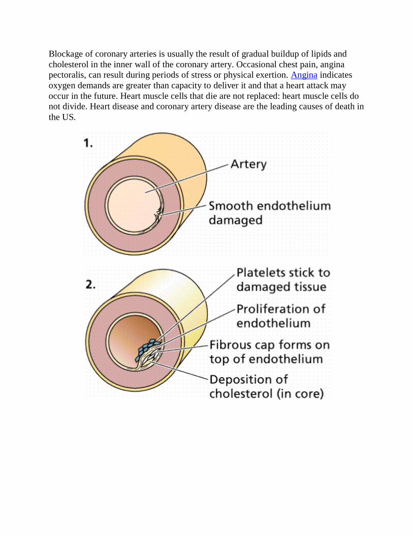
Blockage of coronary arteries is usually the result of gradual buildup of lipids and
cholesterol in the inner wall of the coronary artery. Occasional chest pain, angina
pectoralis, can result during periods of stress or physical exertion. Angina indicates
oxygen demands are greater than capacity to deliver it and that a heart attack may
occur in the future. Heart muscle cells that die are not replaced: heart muscle cells do
not divide. Heart disease and coronary artery disease are the leading causes of death in
the US.
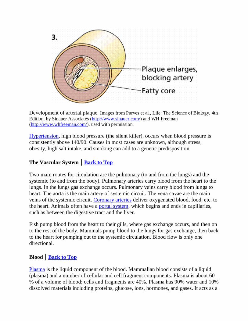
Development of arterial plaque. Images from Purves et al., Life: The Science of Biology, 4th
Edition, by Sinauer Associates (http://www.sinauer.com/) and WH Freeman
(http://www.whfreeman.com/), used with permission.
Hypertension, high blood pressure (the silent killer), occurs when blood pressure is
consistently above 140/90. Causes in most cases are unknown, although stress,
obesity, high salt intake, and smoking can add to a genetic predisposition.
The Vascular System | Back to Top
Two main routes for circulation are the pulmonary (to and from the lungs) and the
systemic (to and from the body). Pulmonary arteries carry blood from the heart to the
lungs. In the lungs gas exchange occurs. Pulmonary veins carry blood from lungs to
heart. The aorta is the main artery of systemic circuit. The vena cavae are the main
veins of the systemic circuit. Coronary arteries deliver oxygenated blood, food, etc. to
the heart. Animals often have a portal system, which begins and ends in capillaries,
such as between the digestive tract and the liver.
Fish pump blood from the heart to their gills, where gas exchange occurs, and then on
to the rest of the body. Mammals pump blood to the lungs for gas exchange, then back
to the heart for pumping out to the systemic circulation. Blood flow is only one
directional.
Blood | Back to Top
Plasma is the liquid component of the blood. Mammalian blood consists of a liquid
(plasma) and a number of cellular and cell fragment components. Plasma is about 60
% of a volume of blood; cells and fragments are 40%. Plasma has 90% water and 10%
dissolved materials including proteins, glucose, ions, hormones, and gases. It acts as a

buffer, maintaining pH near 7.4. Plasma contains nutrients, wastes, salts, proteins, etc.
Proteins in the blood aid in transport of large molecules such as cholesterol.
Red blood cells, also known as erythrocytes, are flattened, doubly concave cells about
7 µm in diameter that carry oxygen associated in the cell's hemoglobin. Mature
erythrocytes lack a nucleus. They are small, 4 to 6 million cells per cubic millimeter
of blood, and have 200 million hemoglobin molecules per cell. Humans have a total of
25 trillion (about 1/3 of all the cells in the body). Red blood cells are continuously
manufactured in red marrow of long bones, ribs, skull, and vertebrae. Life-span of an
erythrocyte is only 120 days, after which they are destroyed in liver and spleen. Iron
from hemoglobin is recovered and reused by red marrow. The liver degrades the heme
units and secretes them as pigment in the bile, responsible for the color of feces. Each
second 2 million red blood cells are produced to replace those taken out of circulation.
White blood cells, also known as leukocytes, are larger than erythrocytes, have a
nucleus, and lack hemoglobin. They function in the cellular immune response. White
blood cells (leukocytes) are less than 1% of the blood's volume. They are made from
stem cells in bone marrow. There are five types of leukocytes, important components
of the immune system. Neutrophils enter the tissue fluid by squeezing through
capillary walls and phagocytozing foreign substances. Macrophages release white
blood cell growth factors, causing a population increase for white blood cells.
Lymphocytes fight infection. T-cells attack cells containing viruses. B-cells produce
antibodies. Antigen-antibody complexes are phagocytized by a macrophage. White
blood cells can squeeze through pores in the capillaries and fight infectious diseases in
interstitial areas
Platelets result from cell fragmentation and are involved with clotting. Platelets are
cell fragments that bud off megakaryocytes in bone marrow. They carry chemicals
essential to blood clotting. Platelets survive for 10 days before being removed by the
liver and spleen. There are 150,000 to 300,000 platelets in each milliliter of blood.
Platelets stick and adhere to tears in blood vessels; they also release clotting factors. A
hemophiliac's blood cannot clot. Providing correct proteins (clotting factors) has been
a common method of treating hemophiliacs. It has also led to HIV transmission due to
the use of transfusions and use of contaminated blood products.

Human Red Blood Cells, Platelets and T-lymphocyte (erythocytes = red; platelets =
yellow; T-lymphocyte = light green) (SEM x 9,900). This image is copyright Dennis
Kunkel at http://www.denniskunkel.com/, used with permission.
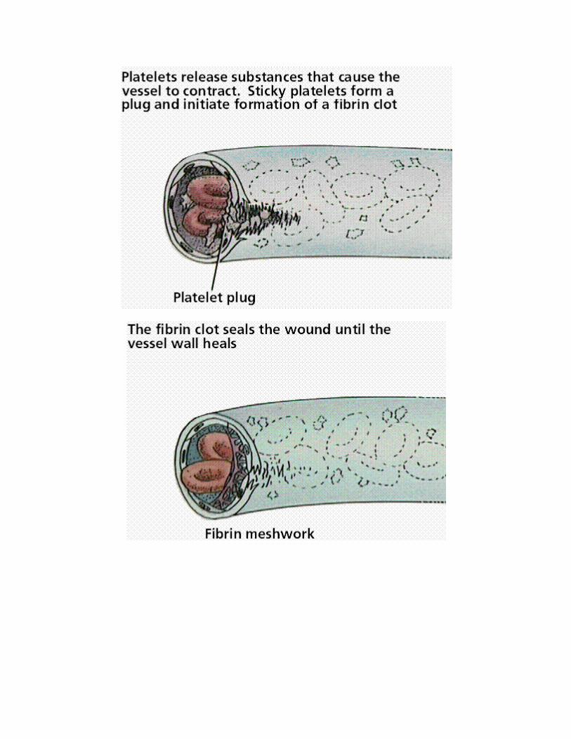

The formation and actions of blood clots. Images from Purves et al., Life: The Science of
Biology, 4th Edition, by Sinauer Associates (http://www.sinauer.com/) and WH Freeman
(http://www.whfreeman.com/), used with permission.
Blood Clot Formation (blood cells, platelets, fibrin clot) (SEM x10,980). This image is
copyright Dennis Kunkel at http://www.denniskunkel.com/, used with permission.

The Lymphatic System | Back to Top
Water and plasma are forced from the capillaries into intracellular spaces. This
interstitial fluid transports materials between cells. Most of this fluid is collected in
the capillaries of a secondary circulatory system, the lymphatic system. Fluid in this
system is known as lymph.
Lymph flows from small lymph capillaries into lymph vessels that are similar to veins
in having valves that prevent backflow. Lymph vessels connect to lymph nodes,
lymph organs, or to the cardiovascular system at the thoracic duct and right lymphatic
duct.
Lymph nodes are small irregularly shaped masses through which lymph vessels flow.
Clusters of nodes occur in the armpits, groin, and neck. Cells of the immune system
line channels through the nodes and attack bacteria and viruses traveling in the lymph.
Learning Objectives | Back to Top
List three functions of blood.
Distinguish between open and closed circulatory systems.
Describe the composition and functions of blood.
Trace the path of blood in the human body. Begin with the aorta and name all major
components of the circulatory system through which the blood passes before it returns to
the aorta.
Links | Back to Top
The Heart: An Online Exploration
American Heart Association Page
The Circulatory System A health-related view of the heart and its associated organs.
The Heart Clear text and some nice graphics, including an animated beating heart.
The Respiratory and Circulatory Systems A clickable map from Japan.
The Circulatory System Yes, another page with the same good descriptive title.
The Heart and the Circulatory System Roger B. Phillips from Access Excellence
Collection. Historical overview of the development of our understanding of circulatory
systems.
Text ©1992, 1994, 1997, 1998, 2000, 2001, by M.J. Farabee, all rights reserved. Use for
educational purposes is encouraged.