The Chorda Tympani and Middle Ear in Reptiles, Birds, and Mammals
27
CHOBDA TYMPANI AND MIDDLE EAE IN REPTILES. 137 The Chorda Tympani and Middle Ear in Reptiles, Birds, and Mammals. By Tldwin S. Goodi-icli, F.R.S., Fellow of Merton College, Oxford. With Plates 11, 12 and 13, and 5 Text-figures. A GREAT deal has been written of late years about the development and homology of the columella auris of reptiles and the chain of auditory ossicles of mammals; a mass of evidence has gradually been gathered from all sides supporting the view put forward by Reichert that the stapes and the columella are derived from the dorsal end of the hyoid arch, and that the incus and malleus, derived from the mandibular arch, correspond to the quadrate and articular. In the search for evidence not only has the development of the skeletal elements been studied, but also the origin of the tympanum and tympanic cavity, and the disposition of the blood-vessels, muscles, and nerves of the middle ear. It was with the intention of comparing the exact relation of these various parts in reptiles, birds, and mammals that the present work was undertaken. Since it was begun, however, so admirable and convincing a summary of the facts in favour of Reichert's view has been given by Gaupp (17) that it would seem as if the question were finally settled and little remained to be said. Yet some doubts and obscurities still remain, especially with regard to the exact relation of the chorda tympani to the first gill-slit, tympanum, and surrounding structures ; so I decided to publish this paper as a small contribution to the discussion of a most important morphological problem. The VOL. 6 1 , PART 2 . NEW SERIES. 10
Transcript of The Chorda Tympani and Middle Ear in Reptiles, Birds, and Mammals

CHOBDA TYMPANI AND MIDDLE EAE IN REPTILES. 137
The Chorda Tympani and Middle Ear inReptiles, Birds, and Mammals.
ByTldwin S. Goodi-icli, F.R.S.,Fellow of Merton College, Oxford.
With Plates 11, 12 and 13, and 5 Text-figures.
A GREAT deal has been written of late years about thedevelopment and homology of the columella auris of reptilesand the chain of auditory ossicles of mammals; a mass ofevidence has gradually been gathered from all sides supportingthe view put forward by Reichert that the stapes and thecolumella are derived from the dorsal end of the hyoid arch,and that the incus and malleus, derived from the mandibulararch, correspond to the quadrate and articular. In the searchfor evidence not only has the development of the skeletalelements been studied, but also the origin of the tympanumand tympanic cavity, and the disposition of the blood-vessels,muscles, and nerves of the middle ear. It was with theintention of comparing the exact relation of these variousparts in reptiles, birds, and mammals that the present work wasundertaken. Since it was begun, however, so admirable andconvincing a summary of the facts in favour of Reichert's viewhas been given by Gaupp (17) that it would seem as if thequestion were finally settled and little remained to be said.Yet some doubts and obscurities still remain, especially withregard to the exact relation of the chorda tympani to thefirst gill-slit, tympanum, and surrounding structures ; so Idecided to publish this paper as a small contribution to thediscussion of a most important morphological problem. The
VOL. 6 1 , PART 2 . NEW SERIES. 10

1,38 EDW.IX S. G00D1UCH.
results of these researches have mostly been given in figuresof reconstructions in which I have endeavoured to representclearly the t rue relation of the par ts dealt with, and to enablethe reader easily to compare the different forms studied. Thefigures are no mere diagrams, but carefully made graphicreconstructions of transverse or longitudinal sections drawnwith the camera lucid*. As far as possible they have beenshown from corresponding points of view, in uniform style,with consistent colouring; unessential details and irregu-larities being omitted to avoid unnecessary complication. Iam indebted to various friends for the opportunity of studyingmany series of sections besides my own. Dr. Verslnys I haveto thank for lending me a series of sections of an embryo-Platj^dactylus, and Prof. Dendy for sections of Sphenodon ;Dr. Jenkinson for the loan of series of lizard, chick, and mouseembryos (from which figs. 1, 2, 3, 13, 22-26, were drawn),and Prof. J . Hill for valuable series of Ornitborhynchus andTrichosnrns embryos of various stages (figs. 5, 14-21).
The chorda tj 'mpani, a twig of the main or hyomandibularbranch of the facial nerve, supplies the organs of taste nearthe base of the tongue and the salivary glands in the region ofthe lower jaw. In adult mammals it issues from the seventhnerve behind the tympanic cavily, turns forward over thiscavity, and makes its way to the lower jaw, passing below thechain of ossicles. Thus the chorda tympani runs downwardsanterior to the tympanum and tympanic cavity and posteriorto the incus and malleus. While the importance of thechorda in determining the homology of the par ts of themiddle ear has become more and more apparent throughthe work of Gaupp (16, 17), Kingsley (25), and others, therehas been considerable confusion about its development andhomology. I t can easily be identified in birds and reptileswhere it follows much the same course as in mammals ; butalthough in these it has the same origin and destination, italways passes over the colnmella, being anterior to it and thetympanum, and posterior to the quadrate and articular.Stannius first suggested tha t the chorda tympani is homo-
CHOJiUA TYMPANI AND MIDDLE KAU IN HJJH'LLKS. 139
logus with the post-spiracular ratnus mandibularis internusof the facial nerve in Amphibia and Pisces. Balfour (1) andDison (7), however, compare it to a pre-spiracular branch,being doubtless misled by the then" prevailing view that thetympanum represents the closing membrane of the spiracularslit. So we find Cole (11) and Herrick (20) arguing that, in spiteof its similar function and peripheral distribution, the chordacannot be homologous with the rainus mandibularis interuus,because the latter is post-spiracular in position. However,it is now known that the tympanum is not developed at thepoint of closure of the first gill-slit, but behind it; so that anerve may be post-spiracular and yet pre-tympanic. Longago Froriep (15) correctly described the chorda tympani in anembryo calf as post-spiracular in origin, aud the same resultwas recorded by Kastschenko in the pig (23), by Eunnel (13)in Microtns, and Druner in the mouse (10), while Hoffmann(21) and Versluys (31) have shown that in lizards it develops iuthe same way. That the chorda tympani is really homologouswith the post-spiracular ratnus maudibularis internus of fishand amphibians may now be considered as established, chieflyowing to the work of G-aupp (16) and Bender (2). Iu hisrecently published monograph Bender traces this nervethrough the whole vertebrate series, aud his conclusion isfurther strengthened by the results of Strong (30), Herrick(20), and others who have shown that gustatory fibres passup this rainus in the lower forms.
My own observations on the development of the chorda,and the various reconstructions figured on Pis. 11, 12, 13may now be described.
Reptilia.—Lacerta is the t}Tpe studied. At a stage whenthe spiracular gill-cleft still opens to the exterior by a smallpore at the dorsal edge of the first gill-pouch (PI. 11, fig. 3), theskeleton of the first two visceral arches becomes visible as avaguely defined blastema extending along the mandibular barin front of the fh'St gill-pouch, and a similar blastema in thehyoid bar behind the gill-pouch. The latter blastema reachesup to the scarcely yet defined blastema of the auditory cap-

140 EDWIN S. GOODEICH.t
sule, pushing in the posterior dorsal surface of the pouch andpassing inwards between the .facial nerve and vena capitislateralis above and the internal carotid below. This stagecorresponds nearly to that described in Platydactylus byVersluys (31), though perhaps a little earlier. Later on, asso well shown by Versluys, the continuous hyoid blastemabends sharply to form the horizontal columellar region andthe more ventral cornual region curved backwards and down-wards. For a considerable time these two regions remainconnected by a band of blastema in Lacerta (PI. 11, fig. 8),though eventually sepai'ating. The more dorsal columellarregion develops into a stout cartilage rod, with a foot orstapedial .base fitting into the fenestra ovalis of the auditorycapsule, and an expanded extra-columella applied to thatregion of the body-wall which will form the middle part ofthe tympanum (PI. 11, figs. 6, 8, 11 ; PI. 13, fig. 30). On thecolumella1 develops about midway an upstanding dorsalprocess and an anterior internal process. The former has aswollen dorsal extremity which later separates off from thecolumella and becomes fixed on to the parotic process of theskull (PI. 11, fig. 8). A fine ligament remains, indicating theoriginal cartilaginous connection of this "intercalary" withthe columella, The internal process (figs. 6, 9-11, 29, 30)projects towards the quadrate, with which it becomes con-nected by ligament.
Meanwhile, the mand-ibular blastema has given rise to thequadrate and Meckel's cartilage. The quadrate articulateswith the parotic process above, the intercalary being wedgediu between them at this point. Extending downwards tomeet the articular region of Meckel's cartilage the quadratepasses outside the facial nerve, vena capitis lateralis, andfacial artery (PI. 11, figs. 7, 9).
In the earliest stage here figured, the first or spiracular1 The term " columella " is used to denote the whole rod, stretching
from tympanum to fenestra ovalis. Prom the point where theremainder of the hyoid arch separates off, the inner or stapedial portionruns inwards, and the outer or extra-columellar portion runs outwards.
CHORDA TYMPANI AND MIDDLE EAR IN REPTILES. 141
gill-split is widely open to the exterior and the first gill-pouchis in the form of a wide, somewhat obliquely flattened out-growth (PL 11, figs. 1, 2). As explained above, by.the timethehyoid blastema is differentiated the slit has almost closed, butstill remains open by a small pore at the upper corner of thepouch (PI. 11, fig. 3). Now, it is well known that the gangliaof the fifth, seventh, ninth, and tenth cranial nerves receivecontributions from the epiblast at the top edge of the corre-sponding gill-slits. These are the so-called branchial senseorgans (Froriep, 15; Kastschenko, 23a). Such an epidermalthickening can be seen above the first gill-slit iu Lacerta inearly stages (PI. 11, figs. 1, 3 ; PI. 13, fig. 27), and later onwill sink in, contributing to form the geniculate ganglion. Ittherefore marks a fixed point very useful in the comparisonboth of different stages and of different animals. In Lacertait can be followed for some little time after the closure of thefirst gill-slit and the separation of the first gill-pouch from theepiblast, and is seen to correspond to the anterior dorsal, inneror medial, corner of the pouch which gives rise to the recessusmedialis (fig. 6 air.}. A careful description of the develop-ment of the tympanic cavity in Lacerta has been given byEl. Cords (12), and my own observations are in agreementwith hers, as shown in PL 11, figs. 6, 8-11 ; PL 13, figs. 29and 30. The flattened first gill-pouch separates off from theepiblast from below upwards. At the same time the lowerposterior region grows outwards and forwards, and pushinginwards the original upper part of the pouch which lastopened to the extei'ior it expands behind the quadrate toform the adult tympanic cavity. Three outgrowths or re-cesses of the cavity teud to surround the columella. Theseare an anterior inner or medial, an anterior outer or lateral,and a posterior recess. The two first grow upwards andthen backwards over the columella, having between themthe dorsal process, the internal process, and the chordatympani (figs. 6 and 29). At a later stage the lateral recessmeeting and opening into the posterior recess enables thetympanic cavity to completely surround the extra-columella.
142 EDWIN S. GOODEICH.
By a thiuning-out of the mesenchymatous wall separatingthe tympanic' diverticulam of the gill-pouch from the super-ficial epidermis the tympanic membrane is framed. Thetympanum is really developed, then, not from a membraneclosing the sph-acular slit, but, as clearly stated by Versluys(31), by the outgrowth of the first gill-pouch posterior andsomewhat venti-al to the spiracle and immediately in front ofthe hyoid arch. This outgrowth we may call the tympanicdiverticulum (see diagram A, .B, and c). The whole columella,and the hyoid corim as well, are morphologically posterior tothe tympanum and tympanic cavity.
Coming now to the blood-vessels, we find that the venacapitis lateralis passes forwards outside the tenth, ninth, andseventh nerves, above the columella, and o.n the inner side ofthe fifth nerve (PL 11, figs. 2, 3, 9). It runs on the innerside of the process dorsalis in the space included between thequadrate and the auditory capsule (PI. 11, fig. 9; PI. 13, figs.29, 30). Accompanying the vein for this part of its course isthe facial or stapedial artery (figs. 3, 6, 8, 9,30). Starting fromthe internal carotid, which, of course, runs above the pharynxand gill-pouches, this artery passes outwards and forwardsover the columella, then upwards and forwards on the outerside of the vena capitis latei-alis and below the articulation ofthe quadrate with the skull. Versluys (31) has shown thatiu Piatydactylus the facial artery pierces the foot of thecolumella, and his suggestion that it is homologous with thestapedial artery of the mammal seems to be fully justified,siuce it develops like the latter from the top end of the hyoidarterial arch aud has the same relations (figs. 3, 14).
The position of the seventh or facial nerve in relation tothe above-mentioned structures is well shown in PI. 11, figs.6, 8, 9, and PL 13, figs. 29, 30. Passing outwards and back-wards from its ganglionic masses in front of the auditorycapsule, the main or hyomandibular branch of the facial isseen to run across and under the vena capitis lateralis justdorsal to the first gill-slit or pouch, and then backwards anddownwards behind the spiracular slit into the hyoid bar. As

CHOLfDA TYMPANf AND MIDDLE EAR IN liEP'l'lLES. 1 4 3
already described, a connection exists in early stages betweenthe facial and inner dorsal region of the epiblastic ingrowthforming the opening of the first gill-slit. An epidermal pro-liferation here contributes to the geniculate ganglion, andimmediately in front of it runs forward the incipient palatinenerve. At a stage when the spiracular slit is still widely open{figs. 1, 2), the chorda tympani is already seen to arise from thehyomaudibular branch of the facial behind the opening, and torun below the opening obliquely downwards and forwards intothe mandibular bar. The chorda tympani is, therefore, asdescribed in lizards by Hoffmann (21) and Versluys (31), dis-tinctly a post-spiracular or post-trematic nerve. It first runsnear the epidermis until it passes the lower edge of the gill-slit, when it turns inwards and runs along the mandibular barand close to the floor of the buccal cavity, to which most ofits fibres are doubtless distributed.
As the spiracular slit closes from below upwards the chordatympani keeps near its lower edge, passing at first diagonallydownwards to the lower jaw across the future tympauic region(figs. 3, 27). When the first gill-slit separates off from theepidermis, and as the tympanic diverticulum enlarges, thechorda tympani becomes more and more pushed up in frontof the developing tympanic membrane (diagrams A, B, and c).Finally it slips inwards, so to speak, over the tympanic cavityso as to pass over the extra-columella on the inner side of thelateral recess, but outside both the dorsal and the internalprocess of the columella. The chorda tympani in repbiles is,therefore, primarily post-trematic and pre-tympanic, beingsituated between the opening of the first gill-slit and thetympanic membrane (diagram D, p. 153) as described byVersluys in Platydactylus.
A muscle has been described by Killian (24).in reptilesextending backwards from the extra-columellar to the paroticprocess of the skull. He calls it the stapedial muscle, andhouiologises it with the muscle of the same name in theMammalia. It is the muscle called " m. extra-columellaris "by Versluys (31) in adult Geckonidse, and also in the embryo
U4> EDWIN S. GOODRICH.
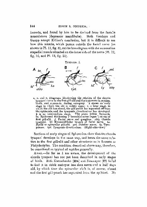
Lacerta, and found by him to be derived from the facialismusculature (depressor mandibulae). Both Versluys andGaupp accept Killian's conclusion, but it is difficult to seehow this muscle, which passes outside the facial nerve (asshown in PL 11, fig. 8), can be homologous with the mammalianstapedial muscle situated on the inner side of the nerve (PL 12,fig. 16, and PL 13, fig. 25).
cfity. t'y. chty.
A, B, and c, Diagrams illustrating the relation of the chordatyrnpani nerve to the first gill-slit and the trypannm in reptiles,bh-ds, and mammals, during ontogeny. A shows an earlystage in which the slit is widely open j C, a later stage, inwhich the slit has closed, the gill-pouch has separated off fromthe epidermis, and the tympanic diverticulum has developed.B is an intermediate stage. The arrow points forwards.bo. Epidermal thickening (" branchial sense organ ") at top offirst gill-slit. / . Facial nerve and ganglion, chty. Chordatympani. hf. Hyomandibular branch of facial nerve, hs.Hyoid or spiracular gill-slit, pal. Palatine nerve, ty. Tym-panum, tyd. Tympanic diverticulum. (Right-side view.)
Sections of early stages of Sphenodon show that the chordatympani develops in the same way, and bears the same rela-tion to the first gill-slit and other structures as in Lacerta orPlatydactylus. The condition described above may, therefore,be considered as typical of reptiles generally.
Aves.—So far as I am aware, the development of thechorda tympani has not yet been described in early stagesof birds. Both Kastschenko (23a) and Neumayer (27) failedto find it in chick embryos less than seven and a half daysold, by which time the spiracular cleft is, of course, closedand the first gill-pouch has separated from the epiblast. No

CHORDA TYMPANI AND MIDDLE EAR IN REPTILES. 1 4 5
doubt these observers looked for the nerve where we shouldexpect to find it, but where, as a matter of fact, it is not inthe chick, as will be explained below.
In addition to the chick I have studied early stages in theduck, and find that this bird agrees in every essential withLacerta. Fig. 4, PI. 11, is a reconstruction of a four and ahalf-day duck embryo, where the spiracular region of the firstgill-pouch is still quite continuous with the epidermis, but theskeletal blastema can hardly yet be distinguished. The venacapitis lateralis and the internal carotid bear the same rela-tion to surrounding structures as in Lacerta. A beginningof the facial artery can be seen arising from the root of thereduced hyoid arterial arch. The outer lower region of thegeniculate ganglion is attached by an epidermal proliferation,the so-called branchial sense-organ, to the dorsal part of thefirst gill-pouch, and the hyomandibular branch of the facialnerve passes out below the vena capitis lateralis and down intothe hyoid bar (PI. 13, fig. 28). Coming off close to the originof the facial can be seen the chorda tympani running behindthe spiracular slit, and pursuing a curved course below itinto the mandibular bar, along which it can be traced nearthe floor of the buccal cavity. Behind the chorda tympanithe first gill-pouch is growing out to form the tympanicdiverticulum. Since the position of the chorda tympani inthe adult duck agrees with that found in the majority of birds—Magnien (26), Bender (2), Smith (29)—we may assume thatthis is the normal structure and development of these parts inthe Aves. The chorda tympani in every respect behaves asin Lacerta, passing over the extra-columella (extra-stapedialcartilage) and distal to the dorsal process (supra-stapedinl),Smith (29).
Strangely enough, in the fowl quite another relatiou isborne by the chorda tympani to surrounding structures. Tobegin with, Hasse has shown that in the adult Grallus (19),and Magnien (26) that in the adult turkey (Meleagris gallo-pavo), this nerve takes a very unusual course. Originatingquite far forwards from the geniculate ganglion, it passes
,146 EDWIN S. GOODRICH.
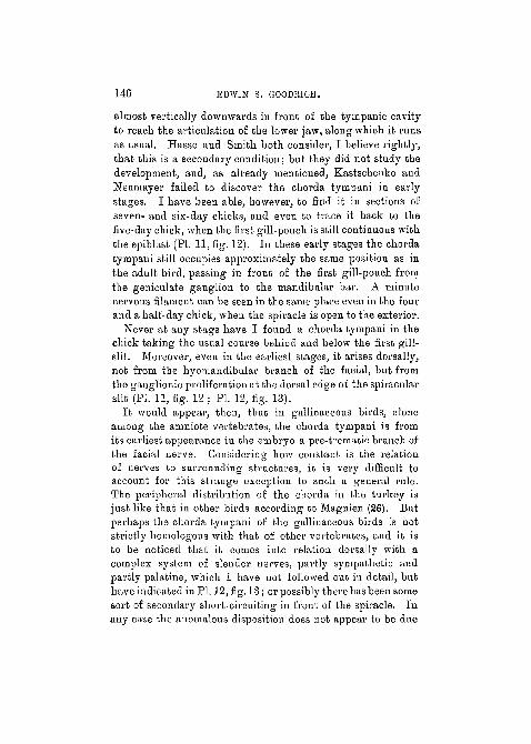
almost vertically downwards in front o£ the tympanic cavityto reach the articulation of the lower jaw, along which it runsas usual. Hasse aud Smith both consider, I believe rightly,that this is a secondary condition; but they did not study thedevelopment, and, as already mentioned, Kastschenko andNeumayer failed to discover the chorda tytnpani in eai'lystages. I have been able, however, to find it in sections ofseven- and six-day chicks, and even to trace it back to thefive-day chick, when the first gill-pouch is still continuous withthe epiblast (PI. 11, fig. 12), In these early stages the chordatyinpani still occupies approximately the same position as inthe adult bird, passing in front of the first gill-pouch fromthe geniculate ganglion to the mandibular bar. A minutenervous filament can be seen in the same place even in the fourand a half-day chick, when the spiracle is open to the exterior.
Never at any stage have I found a chorda tympani in thechick taking the usual course behiad and below the first gill-slit. Moreover, even in the earliest stages, it arises dorsally,not from the hyomandibular branch of the facial, but fromthe ganglionic proliferation at the dorsal edge of the spiracularslit (PI. 11, fig. 12 ; PI. 12, fig. 13).
It would appear, then, that in gallinaceous birds, aloneamong the amniote vertebrates, the chorda tympani is fromits earliest appearance iu the embryo a pre-trematic branch ofthe facial nerve. Considering how constant is the relationof nerves to surrounding structures, it is very difficult toaccount for this strange exception to such a general rule.The peripheral distribution of the chorda in the turkey isjust like that in other birds according to Magnien (26). Butperhaps the chorda tyinpani of the gallinaceous birds is notstrictly homologous with that of other vertebrates, aud it isto be noticed that it comes into relation dorsally with acomplex system of slender nerves, partly sympathetic andpartly palatine, which I have not followed out iu detail, buthave indicated in PI. 12,fig.l3j orpossibly there has been somesort of secondary short-circuiting in front of the spiracle. Inany case the anomalous disposition does not appear to be due
CHORDA TYMPANI AND MIDDLE BAR IN EEPTILES. 147
to a mere shifting of the chorda tympaui, as suggested bySmith (29), analogous to the shifting inwards of the nerveaccompanying the reduction of the dorsal and internal pro-cesses of the colutnella found to occur in the Lacertilia, andso well described by Versluys (31). The whole questionrequires further investigation, and it would be interesting toknow whether the chorda tympani develops in front of thefirst gill-slit in any other birds.
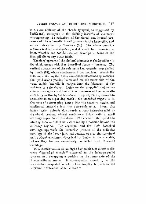
The development of the skeletal elements of the hyoid bar inthe chick agrees with that described above in Lacerta. Theearliest appearance of the columella has recently been studiedby Smith (29), whose conclusions I can confirm. About thefifth and sixth day there is a continuous blastema representingthe hyoid arch; passing below and on the inner side of thevena capitis lateralis it merges into the blastema of theauditory capsule above. Later on the stapedial and extra-columellar regions and the various processes of the columellachondrify in this hyoid blastema. Pig. 13, PI. 12, shows thecondition in an eight-day chick : the stapedial region is inthe form of a stout plug fitting into the fenestra ovalis, andcontinued outwards into the extra-columella. Prom thislatter region extends downwards a long infra-stapedial orStylohyal process, almost continuous below with a smallcartilage separate at this stage. The coruu of the hyoid hasalready become detached, and taken up a position behind theauditory region. The stylohyal and the little detached•cartilage approach the posterior process of the articular•cartilage of the lower jaw, and remind one of the interhyaland epihyal cartilages described by Parker in the crocodile,where they become secondarily connected with Meckel'scartilage.
This reconstruction of an eight-day chick also shows a dis-tinct " stapedial muscle" attached to the infra-stapedialprocess, and occupying a position on the inner side of thehyomandibular nerve. It corresponds, therefore, to themammalian stapedial muscle in this respect, but not to thereptilian " extra-colnmellar muscle."

148 EDWIN S. GOODIUCH.
The first gill-pouch is also seen, now, of course, detachedfrom the epidermis. The tympanic diverticulum, however, isbut little developed, the mesenchymatous tissue, into whichextends the extra-columellar, between it and the ingrowingexternal auditory meatus, being still quite thick.
Mammalia.—As already mentioned (p. 139) the positionof the chorda tympani was correctly figured and described byFroriep in early calf embryos as long ago as 1885. Bromau(4), Emtnel (13), and others, have since confirmed this obser-vation, and it may now be considered as firmly establishedthat, just as in the Reptilia EO in the Mammalia, the chordatympani arises as a post-trematic branch of the facial nerve.
My own observations fully support this conclusion. Asseen in the reconstructions of early stages of Trichosurus,when the gill-pouch is still in continuity with the epidermisat a point corresponding to the open spiracular cleft oflower vertebrates, the chorda passes from the hyomandibularbranch of the facial nerve round behind aud below the gill-pouch to reach its destination in the mandibular arch (figs.5 and 15). At a later stage, when the first gill-pouch isseparating off from the epidermis, the chorda tympani stilloccupies much the same position; and it is only later whenthe tympanic diverticulum develops and expands that thenerve is pushed upwards and forwards just as in Lacerta(PI. 12, figs. 14 and 1G).
Little remains to be said about the development of theauditory ossicles of the Mammalia, which has been lately soaccurately described by Dreyfuss (8), Broman (4), Jenkinson(22) and others. My observations entirely support the viewsof these and other authors who contend that the stapesis derived from the upper end of the hyoid arch, andthe incus and malleus from the upper end of the mandibulararch; as against Fuchs and others who believe otherwise(see Gaupp (17)). From the time when it first appears as ablastema in the mouse the stapes is distinct from the auditorycapsule and continuous with the blastema of the remainderof the hyoid arch, passing as usual below the vena capitis
CHORDA TYMl'ANI AND MIDDLE UAH, IN KEPl'ILES. 149
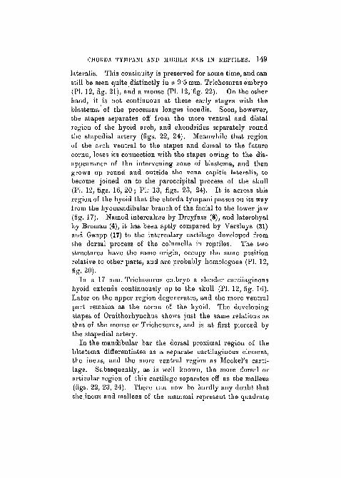
lateralis. This continuity is preserved for some time, and canstill be seen quite distinctly in a 9*5 mm. Trichosurus embryo(Fl. 12, fig. 21), and a mouse (PI. 12, fig. 22). On the otherhand, it is not continuous at these early stages with theblastema of the processus longus iucudis. Soon, however,the stapes separates off from the more ventral and distalregion of the hyoid arch, and chondrifies separately roundthe stapedial artery (figs. 22, 24). Meanwhile that regionof the arch ventral to the stapes and dorsal to the futurecornu, loses its connection with the stapes owing to the dis-appearance of the intervening zone of blastema, and thengrows up round and outside the vena capitis latei-alis, tobecome joined on to the paroccipital process of the skull(PI. 12, figs. 16, 20 j PI; 13, figs. 23, 24). It is across thisregion of the hyoid that the chorda tympani passes on its wayfrom the hyomandibular branch of the facial to the lower jaw(fig. 17). Named intercalare by Dreyfuss (8), and laterohyalby Broman (4), it has been aptly compared by Veisluys (31)and Gaupp (17) to the intercalary cartilage developed fromthe dorsal process of the columella in reptiles. The twostructures have the same origin, occupy the same positionrelative to other parts, and are pi-obably homologous (PI. 12,fig. 20).
In a 17 mm. Trichosurus embryo a slender cartilaginoushyoid extends continuously up to the skull (PI. 12, tig. 16).Later on the upper region degenerates, and the more ventralpart remains as the cornu of the hyoid. The developingstapes of Ornithorhynchus shows just the same relations asthat of the mouse or Trichosurus, and is at first pierced bythe stapedial artery.
In the mandibular bar the dorsal proximal region of theblastema differentiates as a separate cartilaginous element,the incus, and the more ventral region as Meckel's carti-lage. Subsequently, as is well known, the more dorsal orarticular region of this cartilage separates off as the malleus(figs. 22, 23, 24). There can now be hardly auy doubt thatthe incus and malleus of the .mammal represent the quadrate
150 EDWIN S. GOODRICH.
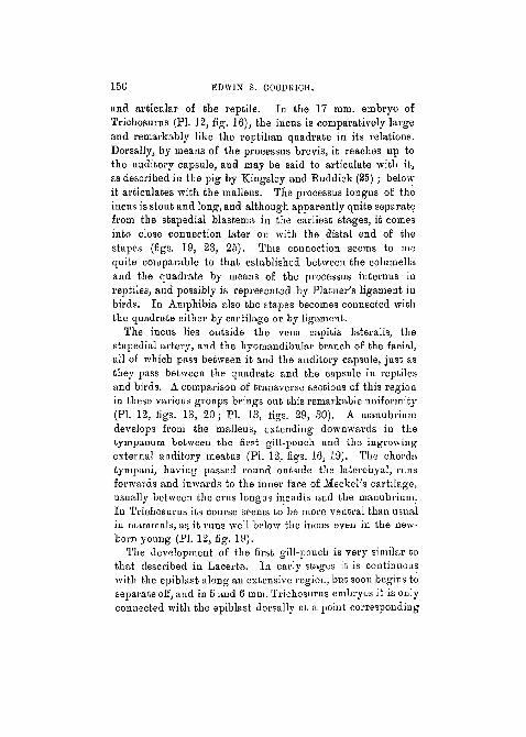
and articular of the reptile. In the 17 mm. embryo ofTrichosurus (PL 12, fig. 16), the incus is comparatively largeand remarkably like the reptilian quadrate in its relations.Dorsally, by means of the processus brevis, it reaches up tothe auditory capsule, and may be said to articulate with it,as described in the pig by Kingsley and Ruddick (25) ; belowit articulates with the malleus. The processus longus of theincus is stout and long, and although apparently quite separatefrom the stapedial blastema in the earliest stages, it comesinto close connection later on with the distal end of thestapes (figs. 19, 23, 25). This connection seems to mequite comparable to that established between the columellaand the quadrate by means of the processus internus inreptiles, and possibly is represented by Platner's ligament inbirds. In Amphibia also the stapes becomes connected withthe quadrate either by cartilage or by ligament.
The incus lies outside the vena capitis lateralis, thestapedial artery, and the hyomandibular branch of the facial,all of which pass between it and the auditory capsule, just asthey pass between the quadrate and the capsule in reptilesand birds. A comparison of transverse sections of this regionin these various groups brings out this remarkable uniformity(PI. 12, figs. 18, 20; PI. 13, figs. 29, 30). A manubriumdevelops from the malleus, extending downwards in thetympanum between the first gill-pouch and the ingrowingexternal auditory meatus (PI. 12, figs. 16, 19). The chordatympani, having passed round outside the laterohyal, runsforwards and inwards to the inner face of MeckePs cartilage,usually between the cms longus incudis and the manubrium.In Trichosurus its course seems to be more ventral than usualin mammals, as it runs well below the incus even in the new-born young (PI. 12, fig. 19).
The development of the first gill-pouch is very similar tothat described in Lacerta. In early stages it is continuouswith the epiblast along an extensive region, but soon begins toseparate off, and in 5 and 6 mm. Trichosurus embryos it is onlyconnected with the epiblast dorsally at a point corresponding
CHORDA TYMPANI AND MIDDLE EAK IN BKPT1LKS. 1 5 1
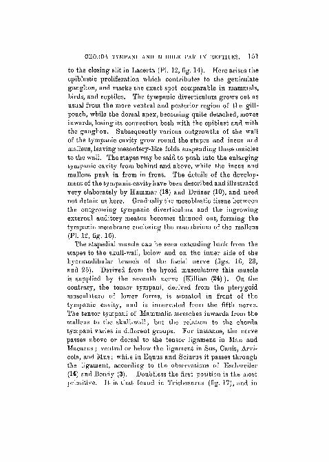
to the closing slit in Lacerta (PI. 12, fig. 14). Here arises theepiblastic proliferation which contributes to the geniculateganglion, and marks the exact spot comparable in mammals,birds, and reptiles. The tympanic diverticulum grows out asusual from the more ventral and posterior region of the gill-pouch, while the dorsal apex, becoming quite detached, movesinwards, losing its connection both with the epiblast and withthe ganglion. Subsequently various outgrowths of the wallof the tympanic cavity grow round the stapes and incus andmalleus, leaving mesentery-like folds suspending these ossiclesto the wall. The stapes may be said to push into the enlargingtympanic cavity from behind and above, while the incus andmalleus push in from in front. The details of the develop-ment of the tympanic cavity have been described and illustratedvery elaborately by Hammar (18) and Driiner (10), and neednot detain us here. Gradually the mesoblasfcic tissue betweenthe outgrowing tympanic diverticulum and the ingrowingexternal auditory meatus becomes thinned out, forming thetympanic membrane enclosing the manubrium of the malleus(PI. 12, fig. 16).
The stapedial muscle can be seen extending back from thestapes to the skull-wall, below and on the inner side of thehyomandibular branch of the facial nerve (figs. 16, 23,and 25). Derived from the hyoid musculature this muscleis supplied by the seventh nerve (Killian (24) ). On thecontrary, the tensor tympaui, derived from the pterygoidmusculature of lower forms, is situated in front of thetympanic cavity, and is innervated from the fifth nerve.The tensor tympani of Mammalia stretches inwards from themalleus to the skull-wall; but the relation to the choi'datympani varies in different groups. For instance, the nervepasses above or dorsal to the tensor ligament in Man andMacacus; ventral or below the ligament in Sus, Canis, Arvi-cola, and Mus; while in Equus and Sciurus it passes throughthe ligament, according to the observations of Eschweiler(14) and Bondy (3). Doubtless the first position is the mostprimitive. It is that found in Trichosurus (fig. 17), and in

152 EDWIN S. GOODRICH.
Mus (fig. 26), and corresponds to the relation of nerve and7nuscle in reptiles.
Conclusion.—In the preceding pages it has been shownthat the chorda tympani in reptiles, birds, and mammals,develops as a post-trematic branch of the facial nerve behindthe first gill-slit. As may be seen in the diagrams A, B, andc (p. 144), owing to the outgrowth of the tympanic diverticu-luin from the hinder region of the first gill-pouch, the chordacomes to lie in front of this pouch when the slit closes and thepouch separates off from the epidermis. The only knownexception to this rule is that of the gallinaceous birds, inwhich the chorda is found to pass in front of the first gill-pouch from its very first appearance. In the adult amniotethe chorda necessarily passes down anterior to the tympanum.Although in certain forms like Sphenodon, where the mem-brane is not very clearly delimited, the chorda may seem topass across the dorsal and anterior region of the tympanum,the relative position of these parts remains essentiallyunaltered.
It has also been shown how very constant throughoutontogeny are the relations of the spiracular slit, vena capitislateralis, facial or stapedial artery, facial nerve, and chordatympani, in mammals, birds, and reptiles. Indeed, the earlystages of the lizard, duck, and Ti-ichosurus, when the firstgill-pouch is still continuous with the epidermis, are sosimilar that figures of any one of them would apply almostequally well to the other two. Only unimportant differencesof relative size and proportion can be detected between them.Later on divergencies occur owing to the great developmentof the extra-columella in birds and reptiles, and to theposition taken up in mammals by the incus and malleus,where they come to lie between the stapes and the tympanum;but even then the blood-vessels, muscles, nerves, and skeletalparts retain their essential morphological relations. Only onthe supposition that the incus represents the quadrate, andthe malleus the articular, is this structure intelligible. As aglance at diagrams D and E will show, the chorda tympani

CHORDA TYMPANI AND MITDDLE EAR IN REPTILES. 1 5 3
really follows essentially the.same course in reptiles andmammals. In both 'tlie hyoid arch, at its dorsal end, hastwo diverging branches : a stapedial fitting into the feuestraovalis and an intercalary branch coming into contact withthe skull. The hyomandibular "branch of tho facial nervepasses back over the stapes, arid the chorda tympani runsfrom it outside the arch, then forwards between the spiracular
.../
D and E, Diagrams sliowing the i-el^tion of the first and secondvisceral arches and the branches of the facial nerve to theclosing first gill-slit and the developing tympanum in areptile (D) and a mammal (E). a. Articular cartilage, chty.Chorda tympani. ec. Extra cplumella. / . Facial ganglion.lie. Hyoid comn. hf. Hyoma«dibular bramcli of facial nerve.hs. Hyoid or spii-acular gijl-slit. i. Incus, int. Intercalaiycartilage of processus dorsalis. Hi, Intercalary or laterohyalcartilage. '/». Malleus,, pi. Processus intemus. q. Quadrate.st. Stapes, ty. Tympamiin. (Right-side view.)
opening (virtual in mammals) and the tympanum to the innerside of the articular or malleus. But, whereas in the reptilethe columella has given rise to an extra-columella whichgrows into the tympanum below the chorda, in the mammalno such extra-columella is developed, and it is the malleuswhich comes into relation with the tympaiuim. Thus therelation of the chorda to the hyoid arch is really the same inthe two cases. The articulation of the incus with the stapesis paralleled by the connection, cartilaginous or ligamentous,so frequently established between the columella and the
VOL. 6 J , TART 2.—NEW SERIES. 11
154 EDWIN S. GOODRICH.
quadrate in reptiles and birds, and even in Amphibia."Whether the extra-columella be primitive among Reptilia is adoubtful point; but on the whole it seems probable that theMammalia have lost it, on the gradual assumption of itsfunctions by the incus and malleus. Accompanying thismodification of the quadrate and articular to transmit vibra-tions from the tympanum, the jaws in mammals must, ofcourse, have acquired a new mode of articulation. But thisdifficulty often urged against Reichert's theory may now besaid to have disappeared owing chiefly to the discoveries ofSeeley and Broom (5, 6). There is no need to .go into thisquestion in this paper. But it may be pointed out that these
/ authors have shown that, in fossil Reptilia related to theancestors of the Mammalia, the squamosal and dentary boneshave gradually increased in importance, contributing moreand more to the support of the lower jaw j-while the quadrateand articular have dwindled in size, become loosened fromthe surrounding bones, and have, so to speak, been drawninto the service of the middle ear.
While the homology of these structures may now be con-sidered as well established in the amniote vertebrates, theirdisposition in the Amphibia still presents serious difficulties.For in the only group which possesses a tympanum, the Anura,the ramus mandibularis internus (chorda tympani) is posteriorto it. Driiner, indeed, concludes that the tympanum andtympanic cavity of the Amphibia and Amniota are not homo-logous (9). The evidence for such an extreme view seemsquite insufficient, and Bender (2) has brought forward impor-tant facts with regard to the nerve-supply of the wall of thetympanitic cavity, which go far to prove that it is homologousthroughout the terrestrial vertebrates and with the spiracularslit of fishes, a conclusion which is in agreement with theresults of embryology. While accepting Bender's conclusionGaupp still considers that the tympanum itself has becomeindependently developed in Amphibia, reptiles, and mammals(17). But, while admitting that the position of the chorda isa serious difficulty in comparing the amphibian with the

CHORDA TYMPANI AND MIDDLE EAE IN REPTILES. 355
reptilian structure, it would seem much more probable thatthere has been some relative shifting of parts. Moreover,the presence of a characteristic notch, behind the quadrate infossil Stegocephalia indicates the possession of a tympanum,and those modern forms (Apoda and Urodela) which do notpossess one have probably lost it, being secondarily adaptedto a burrowing or aquatic mode of life. The view of Gauppthat the tympanum of Reptilia ia not homologous with thatof Mammalia, chiefly because the former is situated above theMeckelian cartilage .and the latter below it, seems to megreatly to exaggerate ike importance of a comparativelytrivial difference. If the manubrium of the malleus repre-sents the posterior process of the articular, the tympanumextends both above and below it, and the difference betweenthe two types is small, and just such as we should expect tofind accompanying the change of size and function of theincus and malleus. Rather should we consider the modernreptilian and mammalian plan as showing two divergenttypes derived from some intermediate plan of structureperhaps to be discovered among the Theromorpha.
Summary.—A comparison of the development of thevarious structures of the middle-ear region in the lizard, duck,and mammal, shows a remarkable uniformity in their originand relation. The first gill-pouch separates off from theepidermis from below upwards; at its dorsal edge is an epi-blastic proliferation contributing to the geniculate ganglion.The tympanum is formed between the outer epidermis and anoutgrowing diverticulum of the hinder lower region of the firstgill-pouch. The chorda tympani is a post-trematic branch ofthe facial nerve, developing behind the first or spiraculargill-slit, and passing down to the lower jaw between the tym-panum and the closing spiracle. The relation of these partsto the skeleton and blood-vessels is (with the exceptionmentioned below) constant throughout the Amniota, and isonly intelligible on the view of Reichert that the proximalregion of the columella corresponds to the stapes, the quad-rate to the incus, and the articular to the malleus.
156 . EDWIN S. GOODRICH.
In the chick the chorda tympani develops as a pre-tvematio
branch of the facial nerve from its first appearance. In adult
gallinaceous birds the chorda passes down directly from the
geniculate ganglion in front of the tympanic cavity. This
exceptional position is probably due to some secondary modi-
fication at present unexplained.
September 2nd, 1914.
LITERATURE.
1. Balfour, F. M.—' Coinp. Embryology,' vol. ii, London, 1881.2. Bender, O.—" Die Schleimhautnerven des Facialis," etc., 'Denkschr.
d. Med.-nat. Ges. Jena,' Bd. vii, 1906.3. Bondy, G.—" Beitr. z. vergl. Anat. d. Gehororgane der Siiuger,"
' Anat. Hefte,' Bd. xxxv, 1907.4. Broman, I.—"Die Entw. der Gehbrknoclielehen beim Menscken,"
' Anat. Hefte,' Bd. xi, 1899.5. Broom, R.—" On the Structure of tlie Theriodont Mandible," etc.,
' Proc. Zool. Soc.,' London, vol. i, 1904.6. " On the Structure of the Skull in Oynodont Reptiles,"
ibid., 1911.
7. Dixon, A. F.—" The Facial Nerve in Man," ' Journ. Anat. andPkys.,' vol. xxxiii, 1899.
8. Dreyfuss, R.—" Beitr. z. Enfrw. des Mittelohres u. d. Trommelfels,"' Moi-ph. Arb.,' Bd. ii, 1893.
9. Driiner, L.—" Ueber d. Muskulatur d. Tisceral-skelettes cl. Uro-delen," ' Anat. Anz.,' Bd. xxiii, 1903.
10. "Ueber d. Anat. u. Entw. des Mittelohres b. Menschen vi.b. d. Maus," ibid., bd. xxiv, 1904.
11. Cole, F. J.—" The Cranial Nerves and Lat. Sense Organs of Fishes,"' Trans. Linn. Soc.,' London, vol. vii, 1898.
12. Cords, El.—"Die Entw. d. Paukenhbhle von L a c e r t a agi l is ,"'Anat. Hefte,' Bd. xxxviii, 1909.
13. Emmel, V. E.—"The Relation of the Chorda Tympani to theYisceral Arches in Microtue," ' Journ. Compt. Neurol.,' vol. xiv,1904.
14. Eschweiler, R.—" Z. vergl. Anat. d. Muskeln u. d. Topographie d.Mittelohres versch. Siiugetiere," ' Arch. f. mik. Anat. n. Entw.,'Bd. liii, 1899,
CHORDA TYMPANI AND MIDDLE KAR IN BEbTlLES. 157
15. Froriep, A.—" Anlagen von Sinnesorganen am Facialis," etc.,' Avch. Anat. u. Phys. Anat. Abt.,' 1885.
16. Gaupp, E.—"Anat. Unters. ii. d. Nervensorgung d. Mund. n.Nasenhohlendriisen," ' Morph. Jahrb.,' Bd. xiv, 1888.
17. " Die Reichertsehe Theorie," ' Arch. Anat. u. Phys.,' Suppl.,1912.
18. Hammar, J. A.—"Die Entw. des Vorderdarms," etc., ' Arch. f. rnik.Anat. u. Entw.,1 Bd. lix, 1912.
19. Hasse, C.—" Z. Morph. d. Labyrinths der Vogel," ' Anat. Studien,'Bd. i, 1873.
20. Herrick, C. J.—" The Cranial and First Spinal Nerves of Menidia,"' Journ. Oompt. Neurol.,' vol. ix, 1899.
21. Hoffmann, C. K.—" Beptilien," ' Bronn's Klassen. u. Ordnungen,'Bd. vi,1891.
22. Jenkinson, J. W.—"Development of the Ear-bones in the Mouse,"' Journ. Anat. and Phys.,' vol. xlv, 1911.
23. Kastschenko, N.—"Das Schicksal d. einbr. Schlundspalten beiSfingethiere," ' Arch. f. mik. Anat.,' vol. xxx, 1887.
23a. " Das Schlundspaltsystem des Hiihnchens," ' Arch. f. Anat.Anat. u. Phys., Anat. Abt.,' 1887.
24. Killian, G.—'Z. vergl. Anat. u. vergl. Entw. der Ohrmuskeln."'Anat. Anz.,' vol. v, 1890.
25. Kingsley, J. S., and Ruddick, W. H.—" The Ossicula Auditus andMammalian Ancestry," ' Am. Naturalist,' vol. xxxiii, 1899-
26. Magnien, L.—" Recherches s. I'anat. comp, de la corde du tympandes Oiseaux," ' C. R. Ac. Sc.,' t. ci, 1885.
27. Neumayer, L.—Hertwig's ' Handb. d. vergl. Entw. d. Wirbeltiere,'Bd. ii, 1906.
28. Sheldon, R. E.—" The Phylogeny of the Facial Nerve and ChordaTynipani," 'Anat. Record,' vol. iii, 1909.
29. Smith, G. W.—' The Middle Ear and Columella of Birds," ' Quart.Journ. Micr. Sci.,' vol. 48, 1904.
30. Strong, O. S.—" The Cranial Nerves of Amphibia," 'Journ. Morph.,'vol. x, 1895.
31. Versluys, J.—' Entw. d. Columella auris b. d. Lacertiliern," ' Zool.Jahrb., Abt. Anat. u. Ont.,1 vol. xix, 1903.
158 EDWJN S. GOODRICH.
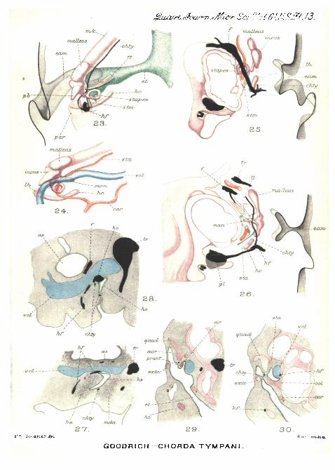
EXPLANATION OF PLATES 11, 12, AND 13,
Illustrating Mr. Edwin S. Goodrich's paper on "The ChordaTympani and Middle Ear in Reptiles, Birds, andMammals."
LETTERING OF PLATES.
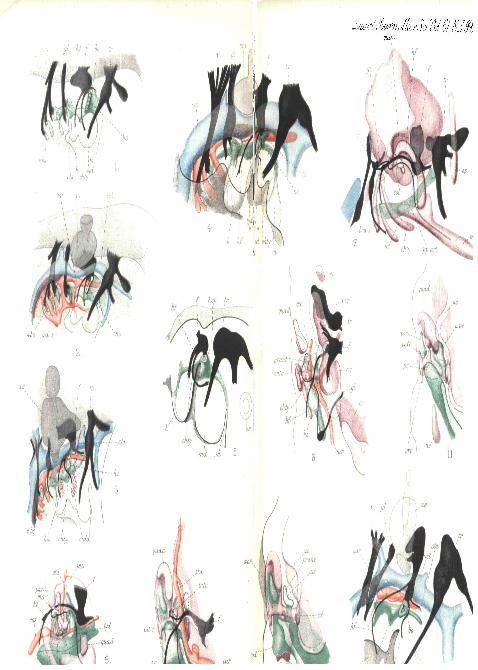
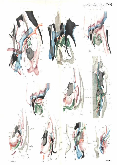
a. a. Aortic arch. a. c. Auditory capsule, a. i. r. Anterior innerrecess of tympanic cavity, a. o. r. Anterior outer recess, art. Arti-cular cartilage, a. s. Auditory sac. b. Blastema extending fromstapes to hyoid. bo. Bpiblastic proliferation or branchial " sense organ"of facial nerve, bra. Branchial arch, bs 1-4. First to fourth branchialslits, c. Small cartilage of hyoid arch, = epihyal? car. Internalcarotid artery, col. Columella auris. earn. Invagination to form ex-ternal auditory meatus and external wall of tympanum, ep. Epiptery-goid. et. Eustachian tube. / . Facial ganglion and nerve, fb. Fore-bvain. gl. Glosso-pharyngeal ganglion and nerve, h. Hyoid bar. ha.Blastema of hyoid ai-ch. hart. Hyoid arterial arch. hb. Hindbrain.he. Hyoid cornu. hf. Hyomandibular branch of facial nerve, /is.Hyoid or spiracular slit. hsp. Pouch of hyoid slit. intc. Intercalarycartilage from dorsal process. Ish. Lateral cranial wall. man. Manu-brrain mallei, md. Mandibular bar. mda. Blastema of mandibulararch. mk. Meckel's cartilage, ms. Extra-columellar muscle, oc. Occi-pital cartilage, os. Optic stalk, par. Paroccipital process, pb. Pro-cessus brevis inctidis. pi. Processus longus incudis. ppart. Posteriorprocess of articular cartilage, pr. Posterior recess of tympanic cavity.prant. Anterior process of columella. prp. Posterior process of extra-columella. psk. Process of skull, quad. Quadrate cartilage, sta.Stapedial artery or facial artery, sth. Stylohyal cartilage, stm. Sta-pedial nuiscle. tr. Trigeminal nerve and ganglion, tt. Tensor tym-pani muscle, tyd. Tympanic diverticulum. v. Vagus ganglion andnerve, vcl. Vena capitis lateralis.
[In the reconstructions here figured the nerves are in black; thepharynx, gill-pouches, and tympanic cavity in green; the veins inblue ; the arteries in red ; the cartilage in purple. In the earliest stagesthe blastema representing the skeleton is indicated by purple dots.]
PLATE 11.
Fig. 1.—Reconstruction of a portion of the right side of the head ofa Lacerta embryo at an early stage when the first or spiracular slit isstill widely open to the exterior. The nerves and gill-slits are recon-structed on the outline of a section near the middle line.

CHORDA TYMPAN1 AND MIDDLE EAR IN REPTILES. 159
Fig. 2.—The same with the blood-vessels added.Fig. 3.—Similar view of a later stage of Lacerta, when the first gill-
slit is partially closed, and the blastemata of the mandibular and hyoidarches are visible.
Fig. 4.—Similar view of a con-esponding stage of an embryo duck (fourand a half days). In this, in fig. 6, and in some of the following figures,the epidermis indicating the surface of the embryo has been shown.
Fig. 5.—Reconstruction of a portion of the right side of an embryoTr ichoaurus vulpecula 5 mm. long (I, A, '01). The endodermalpouch of the first slit is still continuous with the epidermis at a point notrepresented in the figure, the surface of the head having been cut away.
Fig. 6.—Reconstruction of the right side of a thick horizontalsection of the head of Lacerta at a stage when the hyoid arch is stillcontinuous with the columella by means of procartilage. View fromabove (dorsal) the vena, capitis lateralis having been removed. The firstgill-pouch has separated from the epidermis.
Fig. 7.—Right side of the head of an older Lacerta embryo. Onlythe skeletal and nervous systems have been reconstructed, but thepharynx and blood-vessels are shown as cut in the section nearest themedian line. For the sake of clearness the distal region of thecolumella has been cut away.
Fig. 8.—More complete reconstruction of a portion of the samespecimen as shown in fig. 7, with the complete columella, the arteriesand the gill-pouch. A narrow strip of vaguely defined tissue stillconnects the top of the hyoid cornu -with the extra-columella.
Figs. 9, 10, 11.—Reconstructions of the quadrate region of a lateLacerta embryo seen from behind (posterior view). In figs. 10 and 11only the skeleton and tympanic cavity are shown, the columella beingcompleted in both. Fig. 11 fits on to fig. 10, and shows the posteriorupper part of the quadrate and the tympanic recesses. Fig. 9 resemblesfig. 10, but has the nerves, tympanic cavity, and arteries included. Theposition of the vena capitis lateralis is indicated by a dotted ring.
Fig. 12.—Reconstruction of the right auditory region of a five-daychick embryo. The first gill-pouch is still continuous with the epi-blast, and the chorda tympani runs down anterior to it. The skeletonis present only as a vaguely defined blastema at this stage.
PLATE 12.Fig. 13.—The right a\iditory region of an eight-day chick embryo.
In the thick slice reconstructed only the inner region of the ingrowingexternal auditory meatus appeal's. The upper posterior region of thequadrate has been cut away to expose the underlying structures.
160 EDWIN S. GOODBICH, ...,. . .,,,
Fig. 14.—Reconstruction of a portion of the right side of the head ofan embryo of Tr ichosurus vulpeciila'7'25 mm. long (XII, A, J01).The first gill-pouch has just separated" front the:epiblast a? a pointbelow the " branchial sense organ " bo. * '•'•"-' ••'*'• •->'—•_*
Fig. 15.—Dorsal view of a reconstruction from transverse sections ofthe right side of the head of an embryo Tr ichbsurus vulpecula6 mm. long. The first gill-pouch is still continuous with the epiblast,and the chorda tympani passes Behind and below it. The thick slicedoes not include the whole vena capitis late'ralis, and only the mostventral part of the auditory sac lying above the other structures.
Figs. 16 and 17.—Reconstructions of the right auditory regions of thehead of an embryo Tr ichosurus v i i lpeonla l7 mm. long. To exposethe stapes and other structures the incus and malleus have not beenincluded in fig. 17.
Figs. 18,19, 20.—Reconstruction of three consecutive thick transverseslices through the left auditory region of the head of a newbornTr i chosu rus vulpecula. The reconstructions are seen from, infront (anterior view), and the hind surface of the slices 18 and 19 fit onto the front surface of the slices 19 and 20 respectively.
Fig. 21.—Yentral view of a thick slice of the right auditory regionof the head of an embryo of Tr ichosurus vulpecula 95 mm. long,reconstructed from transverse sections. The skeleton is procartilagenous.The stapes is seen through the first gill-pouch, and is continuous withthe procartilage of the hyoid arch.
Fig. 22.—Similar view, but from the dorsal surface, of the left auditoryregion of an embryo mouse at a slightly later stage.
PLATE 13.
Figs. 23 and 24.—Similar dorsal views of the same region of afourteen-day embryo mouse. A portion of the skull has been cut awayto expose the stapedial muscle in fig. 23. In fig. 24 the skeleton of thevisceral arches and the blood-vessels are shown.
Figs. 25 and 26.—Two consecutive thick slices of the right auditoryregion of an older embryo mouse 22 mm. long, viewed from the dorsalsurface and reconstructed from transverse sections.
Fig. 27.—Longitudinal sagittal section through the middle-ear regionof the Lacerta embryo drawn in fig. 3. Cain,
Fig. 2S.—Similar section of the embryo duck drawn in fig. 4. Cam.Figs. 29 and 30.—Transverse sections of the ear region of the Lacerta
embryo shown in fig. 9. Fig. 29 represents a section in front of thecolumella, and fig. 30 through the columella. Cam.
f ac
col
carHuth London
GOODRICH - CHORDA TYMPANI.
W$.&. 13.
£ S GondricVi del.27. 29.
GOODRICH-CHORDA TYMPANI.
3 0 .