Biology 484 – Ethology Chapter 4a – Neural Mechanisms Controlling Behavior
JCB
JCB: Review
617
The Rockefeller University Press $30.00J. Cell Biol. Vol. 213 No. 6 617–629www.jcb.org/cgi/doi/10.1083/jcb.201602089
IntroductionInflammasomes are multimeric complexes formed in response to a variety of physiological and pathogenic stimuli. Inflam-masome activation is an essential component of the innate im-mune response and is critical for the clearance of pathogens or damaged cells. However, overt inflammasome activation is also a major driver of autoimmune and metabolic disorders, underly-ing the importance of understanding this process in physiologi-cal and pathological contexts.
The inflammasome sensors are grouped according to their structural features into nucleotide-binding domain–like recep-tors (NLRs), absent in melanoma 2–like receptors (ALRs), and the recently identified pyrin. These receptors have the ability to assemble inflammasomes and activate the cysteine protease caspase-1. In addition to the sensor (NLR, ALR, or pyrin) and enzymatic component (caspase-1), most inflammasomes also use an adaptor molecule known as ASC (apoptosis-associated speck-like protein containing a caspase activation and recruit-ment domain). Upon detecting specific stimuli, the activated sen-sor nucleates ASC to form a discrete foci, or “speck,” within the activated cell. Nucleated ASC sequentially recruits caspase-1,
which undergoes proximity induced autocatalytic cleavage to produce the active subunits p10 and p20. These active caspase-1 subunits can then proteolytically process cytokines, IL-1β and IL-18, and induce a specific form of inflammatory cell death termed pyroptosis. Activated caspase-1 thus provides the host cell with dual defense mechanisms through the release of ma-ture cytokines and removal of the infected or damaged cell. Inflammasome assembly is thus a coordinated signaling event essential for mounting an appropriate immune response after pathogenic or sterile insults. Although most studies have used in vitro culture systems and reconstitution assays to analyze inflammasome activation, recent advances have allowed visu-alization of these processes in vivo after an infectious insult at the level of a single cell (Sagoo et al., 2016). Recent studies have also highlighted the existence of an NLRP3 inflammasome pathway, mediated by caspase-11 (Kayagaki et al., 2011).
Inflammasome activation is tightly regulated to provide defense against pathogenic insults and avoid damage to the host. Multiple molecular and cellular signals are therefore involved in maintaining the balance between inflammatory response and resolution. In this review, we provide an overview of the cel-lular and molecular mechanisms involved in the regulation of inflammasome activation.
NLR familyNLR family members all share a central nucleotide-binding do-main (NBD), and most members have a C-terminal leucine-rich repeat (LRR) domain and a variable N-terminal domain. The NLR family can be subdivided into NLRP or NLRC based on whether the N terminus contains a pyrin or caspase activation and recruitment domain (CARD), respectively. Certain mem-bers of the family, including NLRP1, NLRP3, and NLRC4, have been well established as NLRs capable of forming inflam-masomes, whereas other members, like NLRP6 and NLRP12, are still considered putative inflammasome sensors. It remains to be seen if other members of the NLR family are capable of forming or regulating inflammasome assembly in response to some unknown stimuli.
NLRP1NLRP1 was the first cytosolic receptor identified for its ability to form a caspase-1 activating platform (Martinon et al., 2002). Human NLRP1 contains the canonical NBD and LRR domains, a pyrin domain (PYD), as well as a function-to-find and a
Over the past decade, numerous advances have been made in the role and regulation of inflammasomes during pathogenic and sterile insults. An inflammasome complex comprises a sensor, an adaptor, and a zymogen pro-caspase-1. The functional output of inflammasome activa-tion includes secretion of cytokines, IL-1β and IL-18, and induction of an inflammatory form of cell death called pyroptosis. Recent studies have highlighted the intersec-tion of this inflammatory response with fundamental cellu-lar processes. Novel modulators and functions of inflammasome activation conventionally associated with the maintenance of homeostatic biological functions have been uncovered. In this review, we discuss the biological processes involved in the activation and regulation of the inflammasome.
The cell biology of inflammasomes: Mechanisms of inflammasome activation and regulation
Deepika Sharma and Thirumala-Devi Kanneganti
Department of Immunology, St. Jude Children’s Research Hospital, Memphis, TN 38105
© 2016 Sharma and Kanneganti This article is distributed under the terms of an Attribution–Noncommercial–Share Alike–No Mirror Sites license for the first six months after the publication date (see http ://www .rupress .org /terms). After six months it is available under a Creative Commons License (Attribution–Noncommercial–Share Alike 3.0 Unported license, as described at http ://creativecommons .org /licenses /by -nc -sa /3 .0 /).
Correspondence to Thirumala-Devi Kanneganti: [email protected] used in this paper: ALR, absent in melanoma 2–like receptor; CARD, caspase activation and recruitment domain; dsDNA, double-stranded DNA; GSD MD, gasdermin D; HIN200, hematopoietic interferon-inducible nuclear proteins with 200–amino acid repeat; LPS, lipopolysaccharide; LRR, leucine-rich repeat; NBD, nucleotide-binding domain; NLR, nucleotide-binding domain–like receptor; PYD, pyrin domain; ROS, reactive oxygen species; TRIF, Toll/Interleukin receptor domain-containing adapter-inducing interferon β.
TH
EJ
OU
RN
AL
OF
CE
LL
BIO
LO
GY
Dow
nloaded from http://w
ww
.rupress.org/jcb/article-pdf/213/6/617/1372251/jcb_201602089.pdf by guest on 06 Decem
ber 2021

JCB • Volume 213 • NumBer 6 • 2016618
C-terminal CARD domain. The mouse genome encodes three paralogs, NLRP1(a–c), which all lack the PYD. NLRP1b is ac-tivated by anthrax lethal toxin produced by Bacillus anthrax. Lethal toxin comprises protective antigen and lethal factor; protective antigen generates pores in the host cell membrane, whereas lethal factor enters the cell and cleaves NLRP1b at an N-terminal site, leading to the induction of inflammasome acti-vation (Fig. 1; Chavarría-Smith and Vance, 2013). This cleavage is essential and sufficient to induce NLRP1b activation (Cha-varría-Smith and Vance, 2013). Proteolytic cleavage within the NLRP1b function-to-find domain is also required for assembly of a functional inflammasome (Frew et al., 2012). Based on these observations, it has been hypothesized that NLRP1b could act as a sensor of protease activity caused by various bacterial toxins, although this has not been rigorously tested or shown. NLRP1b activation also promotes parasite clearance and pro-vides protection against mortality in response to Toxoplasma gondii infection in mice (Gorfu et al., 2014). Toxoplasma- induced NLRP1 activation is observed in susceptible rat strains (Cirelli et al., 2014) and is a major determinant of parasite con-trol in infected macrophages (Cavailles et al., 2014), although activation of NLRP1b through this parasite does not result in detectable levels of an NLRP1b cleavage product (Ewald et al., 2014). This suggests that either NLRP1b processing is not a pre-requisite for inflammasome activation as posited or a thorough analysis for a potentially unstable cleavage product is required
to reconcile this discrepancy. The link between immune re-sponse to Toxoplasma infection and NLRP1 inflammasome is clinically relevant even in humans, as congenital toxoplasmo-sis is associated with single-nucleotide polymorphisms in the NLRP1 gene and loss of NLRP1 in human monocytic cell lines promotes Toxoplasma infectivity (Witola et al., 2011).
Evidence for the ability of NLRP1a to induce inflam-masome activation comes from a genetic model, wherein mice harboring a mutation in the NLRP1a gene (Q593P) develop a caspase-1– and IL-1β–mediated systemic inflammatory dis-ease (Masters et al., 2012). These mice also exhibit a change in myelopoiesis, such that the number and function of hema-topoietic progenitor cells are markedly altered in a caspase-1–dependent but IL-1R signaling–independent manner. Thus, a significant loss of hematopoietic progenitors is observed be-cause of aberrant inflammasome activation and cell-intrinsic pyroptosis. However, the specific triggers or mechanisms regu-lating the NLRP1a inflammasome are as yet unknown. Genetic studies in humans have identified NLRP1 mutations linked to autoinflammatory diseases including vitiligo (Jin et al., 2007), Addison’s disease (Zurawek et al., 2010), rheumatoid arthritis, systemic sclerosis, and Crohn’s disease (Finger et al., 2012); however, further studies that define the molecular mechanisms and structural conformations involved in NLRP1 activation will be needed to deduce the link between NLRP1 and these autoinflammatory diseases.
Figure 1. Canonical inflammasomes NLRP1, NLRP3, NLRC4, AIM2, and pyrin. Ligands and upstream mediators involved in inflammasome activation. The NLRP1b inflammasome responds to lethal factor produced by B. anthracis and assembles an inflammasome by recruiting caspase-1 through its CARD domain or through ASC as an adaptor. The NLRP3 inflammasome responds to intracellular damage induced by pathogenic or sterile insults. The NLRC4 inflammasome assembles in response to recognition of bacterial flagellin or components of the type III secretion system via NAIPs and can form complexes with or without recruiting ASC. AIM2 inflammasome senses double-stranded DNA through its HIN200 domain. Pyrin responds to Rho modification induced by bacterial toxins. Inflammasome activation leads to caspase-1 activation that in turn cleaves its downstream effectors: the newly identified pyroptosis executioner gasdermin D and pro-form of cytokines IL-1β and IL-18. DAMP, danger associated molecular pattern; GBP, guanylate binding protein; PAMP, pathogen associated molecular pattern; T3SS, type III secretion systems.
Dow
nloaded from http://w
ww
.rupress.org/jcb/article-pdf/213/6/617/1372251/jcb_201602089.pdf by guest on 06 Decem
ber 2021
Inflammasome activation and regulation • Sharma and Kanneganti 619
NLRP3NLRP3 was first shown to be associated with hereditary auto-inflammatory syndromes called cryopyrin-associated periodic syndromes, which are characterized by skin rashes and epi-sodes of fever (Hoffman et al., 2001). In fact, over 90 disease- associated mutations have since been observed in humans in and around the NBDs that render NLRP3 constitutively active (Masters et al., 2009). NLRP3 is an inflammasome-forming NLR that responds to a wide range of infectious and endoge-nous ligands and is implicated in the pathogenesis of several autoinflammatory diseases, including arthritis, gout, diabetes, obesity, and Alzheimer’s disease (Guo et al., 2015).
The triggers that have been shown to induce NLRP3 acti-vation include pathogen-derived ligands such as microbial cell wall components, nucleic acids, and pore-forming toxins; envi-ronmental crystalline pollutants like silica, asbestos, and alum; and endogenous danger signals like ATP, serum amyloid A, and uric acid crystals (Man and Kanneganti, 2015). In fact, the di-versity of its activators is one of the most distinctive features of the NLRP3 inflammasome and makes the possibility of a direct interaction with each activator unlikely. It is therefore assumed that the NLRP3 inflammasome either senses a common second-ary activator downstream of these stimuli or responds to cel-lular stress associated with infection or physiological damage. Production of reactive oxygen species (ROS), potassium efflux, changes in cell volume, calcium signaling, and lysosomal dis-ruption have all been proposed as critical upstream signals re-quired for NLRP3 activation (Cruz et al., 2007; Pétrilli et al., 2007; Cassel et al., 2008; Dostert et al., 2008; Halle et al., 2008; Hornung et al., 2008; Schorn et al., 2011; Zhou et al., 2011; Compan et al., 2012; Lee et al., 2012; Murakami et al., 2012; Muñoz-Planillo et al., 2013). Even though potassium efflux has been reported by multiple groups to be the unifying mechanism regulating NLRP3 activation, potassium efflux is also observed in response to NLRP1b activation (Pétrilli et al., 2007), raising doubt over the specificity of potassium efflux for NLRP3 in-flammasome activation. Nevertheless, all of these features are associated with cellular stress, suggesting NLRP3 could be a global sensor of cellular damage (Fig. 1).
Although the precise trigger is unknown, recent studies have identified multiple cellular mechanisms that regulate ac-tivation of the NLRP3 inflammasome. Under resting condi-tions, ASC is found in the mitochondria, cytosol, and nucleus, whereas NLRP3 associates with the ER (Zhou et al., 2011). Nucleation of the inflammasome requires a change in the sub-cellular localization of these molecules to facilitate their in-teraction. This change in spatial arrangement is brought about by dynein-mediated transport of mitochondria toward the ER, bringing ASC and NLRP3 in close proximity (Misawa et al., 2013). Further, mitochondrial dysfunction induces microtubule alterations that enable the juxtaposition of NLRP3 and ASC, suggesting that diverse cellular functions involved in mitochon-drial homeostasis and cytoskeletal modulations coordinate and converge on NLRP3 activation (Misawa et al., 2013). Although colocalization of inflammasome components is required for all inflammasomes to be activated, inhibitors of microtubule alter-ations specifically block the NLRP3 inflammasome, suggesting that distinct cytoskeletal signatures are involved in activation of certain inflammasomes (Misawa et al., 2013).
In addition to cytoskeletal rearrangements, recent studies have identified NEK7, a protein involved in cell-cycle progres-sion, as a novel regulator of the NLRP3 inflammasome (He et
al., 2016; Schmid-Burgk et al., 2016; Shi et al., 2016). NEK7 is a serine/threonine kinase that is known to promote mitotic spindle formation and cytokinesis and is required for progres-sion through mitosis (O’Regan and Fry, 2009). NEK7 interacts with the LRR domain of NLRP3, independent of its kinase ac-tivity, and is required for NLRP3 inflammasome activation (He et al., 2016). This interaction additionally restricts NLRP3 in-flammasome activation to cells in interphase (Shi et al., 2016). It was hypothesized that this cell cycle–mediated restriction of NLRP3 activation is caused by the limited cellular concentra-tion of NEK7, such that once a cell enters mitosis, NEK7 is localized to mitotic spindles and NLRP3 activation is attenu-ated (Shi et al., 2016). The significance of this intersection be-tween NLRP3 activation and cell cycle progression is currently unknown, though it could be posited that this is a safeguard cells use to dampen the inflammasome while undergoing cell division, during which the cell may be enriched in endogenous ligands that could be misrepresented as cellular stress.
NLRC4NLRC4 activates procaspase-1 through its CARD domain to induce cell death (Poyet et al., 2001) in response to Salmonella infection (Mariathasan et al., 2004). Bacterial flagellin (Fran-chi et al., 2006; Miao et al., 2006; Molofsky et al., 2006; Ren et al., 2006) and multiple components of the bacterial type III secretion systems (Miao et al., 2010b; Zhao et al., 2011; Yang et al., 2013) activate the NLRC4 inflammasome, enabling it to provide host defense against a diverse range of pathogens. However, NLRC4 does not interact directly with its activa-tors; instead, a family of proteins, termed NAIPs (NLR fam-ily, apoptosis inhibitory proteins), act as sensors that recognize the ligand and induce activation of the NLRC4 inflammasome. Mouse NAIP1 and NAIP2 recognize bacterial needle and inner rod proteins, respectively, whereas NAIP5 and NAIP6 bind to flagellin (Fig. 1; Kofoed and Vance, 2011; Zhao et al., 2011; Rayamajhi et al., 2013; Yang et al., 2013). The human genome encodes only one known NAIP (Endrizzi et al., 2000), which can sense both flagellin and needle proteins, suggesting a simi-larity in the mode of recognition of the two structurally related ligands (Yang et al., 2013; Kortmann et al., 2015).
NLRC4 contains a winged-helix domain, which together with the NBD stabilizes NLRC4 in a closed conformation under basal conditions, whereas the LRR domain provides steric hin-drance to its oligomerization (Tenthorey et al., 2014). These two inhibitory mechanisms thus maintain NLRC4 in an inac-tive state in the absence of a ligand. Support for this theory comes from structural modeling based on cryo-EM analysis of NAIP5-NLRC4 multimers and a crystal structure of dormant NLRC4 (Diebolder et al., 2015). This study suggested that ro-tation of the LRR domain would be required to allow NLRC4 to fit into the active oligomeric form detected by cryo-EM (Die-bolder et al., 2015). A recently characterized cryo-EM structure of the NLRC4 inflammasome in response to bacterial type III secretion systems rod protein PrgJ revealed that nucleation is initiated from a single, activated NAIP2 molecule that subse-quently induces NLRC4 polymerization. The inflammasome assembly follows a domino-like reaction, wherein the activated NAIP2 molecule provides a platform for self-oligomerization of NLRC4 to form a wheel- or disc-like structure (Hu et al., 2015; Zhang et al., 2015).
In addition to removal of infected cells through pyropto-sis and perpetuation of inflammation through IL-1β and IL-18
Dow
nloaded from http://w
ww
.rupress.org/jcb/article-pdf/213/6/617/1372251/jcb_201602089.pdf by guest on 06 Decem
ber 2021

JCB • Volume 213 • NumBer 6 • 2016620
release, NLRC4 activation affects other aspects of cell biology that are critical in host defense. In macrophages infected with Salmonella, NLRC4 induces an actin polymerization response that prevents further bacterial uptake and increases intracellular ROS production to enhance intracellular killing and lower bac-terial dissemination (Man et al., 2014a). NLRC4 activation in epithelial cells is specifically required to control pathogen load during Salmonella infection. In accordance with this, epithelium- specific deletion of inflammasome components (Naip 1–6, NLRC4, and caspase-1/caspase-11) results in higher bacterial load and extraintestinal dissemination (Sellin et al., 2014). A surprising feature of NLRC4 activation is observed in a mouse model using intraperitoneal delivery of flagellin, which induces rapid host mortality (within 30 min). This striking inflam-masome-dependent response is independent of IL-1β or IL-18 and instead relies on the production of eicosanoids, including prostaglandins and leukotrienes. This inflammasome-dependent eicosanoid production leads to rapid vascular fluid loss and mor-tality (von Moltke et al., 2012). How the NLRC4 inflammasome activates biosynthesis of eicosanoids and whether this function of eicosanoid production can be extended to all inflammasomes are currently unknown. Overall, these studies highlight the im-portance of NLRC4 inflammasome in cellular responses be-yond the conventional roles of cytokine release and pyroptosis.
Human genetic studies have identified gain-of-function mutations in NLRC4 that are associated with autoinflammation and enterocolitis (Canna et al., 2014; Kitamura et al., 2014; Romberg et al., 2014). Because NLRC4 has been ascribed such diverse functions in animal models of infection, it remains to be seen whether human patients recapitulate any specific fea-tures of NLRC4 activation.
NLRP6NLRP6 is uncharacteristic by virtue of the unusual ligand it recognizes and its observed downstream effects. One of the earliest studies identified NLRP6 as a negative regulator of the mitogen-activated protein kinase and NF-κB pathway in macrophages infected with Listeria monocytogenes, Esche-richia coli, and Salmonella (Anand et al., 2012). Thereafter, the role of NLRP6 in activation of caspase-1 was hypothesized as being required to maintain optimal mucosal response to chem-ical damage and pathogens (Elinav et al., 2011; Wlodarska et al., 2014). In the gut, NLRP6 antagonizes microbial dysbiosis through mucus production (Elinav et al., 2011). In the absence of NLRP6, the intestinal barrier integrity is compromised, ren-dering the animal susceptible to pathogenic and chemical in-sults (Chen et al., 2011; Elinav et al., 2011; Huber et al., 2012; Hu et al., 2013; Nowarski et al., 2015). The identity of the li-gand that activates NLRP6 remained elusive until a recent study identified taurine, a microbial metabolite, as its activator (Levy et al., 2015). The authors demonstrated that NLRP6–ASC–caspase-1 axis, through IL-18 production, promotes induction of antimicrobial peptides that resist microbial dysbiosis. The normal microbiota in turn produces a specific metabolite profile enriched in taurine that engages NLRP6 in the gut to provide mucosal immunity (Levy et al., 2015). NLRP6 therefore has a central role in gut homeostasis and associated disorders (Henao- Mejia et al., 2012). It should be noted that altered or dysbiotic microbiota is also observed in mice lacking inflammasome sen-sors NLRP3 (Henao-Mejia et al., 2012) and AIM2 (Man et al., 2015b), suggesting that the inflammasome plays a nonredun-dant role in maintaining gut bacterial symbiosis. The role of
NLRP6 at mucosal sites is relevant even in the context of viral infection. During systemic infection with encephalomyocarditis and norovirus, NLRP6 affected viral loads specifically in the gastrointestinal tract, and oral infection with the same viruses required NLRP6-mediated defense to confer survival (Wang et al., 2015). However, direct proof for the importance of NLRP6 at nonmucosal sites is yet to be demonstrated and further inves-tigation is needed to uncover the molecular mechanisms gov-erning this putative inflammasome.
NLRP12NLRP12 was initially characterized as an inhibitor of nonca-nonical NF-κB signaling (Williams et al., 2005; Lich et al., 2007). It was also involved in caspase-1 activation in response to Yersinia pestis and Plasmodium infection (Vladimer et al., 2012; Ataide et al., 2014). The role of NLRP12 in NF-κB– mediated signaling makes it an important regulator of immune response after Salmonella infection and during colorectal cancer (Zaki et al., 2011, 2014). Further, it modulates NF-κB signaling intrinsically in T cells during their activation, thereby affecting their colitogenic and encephalitogenic potential (Lukens et al., 2015). However, the ligands that activate NLRP12 and its regu-latory mechanisms remain poorly understood.
ALR familyThe ALR family is named for its most well-characterized member, AIM2 (absent in melanoma 2). The members of the ALR family are characterized by an N-terminal PYD and a C-terminal hematopoietic interferon-inducible nuclear protein with 200–amino acid repeat (HIN200) domain. The expression of ALRs is restricted to and conserved among mammalian spe-cies (Cridland et al., 2012). AIM2 has a distinct cytosolic local-ization by virtue of lacking a nuclear localization signal that is present in all other members. Further, the PYD of AIM2 is func-tionally distinct from other ALRs in its ability to interact with the PYD of ASC (Hornung et al., 2009). It is because of this specific interaction that AIM2 is the only member of this fam-ily known to be capable of forming an inflammasome. Other mouse ALRs have been shown to interact with ASC, making them potential regulators of inflammasome responses (Brunette et al., 2012), and another human ALR, γ-IFN–inducible protein 16, is considered a putative inflammasome (Kerur et al., 2011). However, most major studies have concerned the identification and regulation of the AIM2 inflammasome.
AIM2AIM2 was initially identified as a γ-IFN–inducible protein in a tumor suppressor screen. A later study redefined it as a nucleic acid sensor that assembles into an inflammasome, specifically in response to double-stranded DNA (dsDNA; Fig. 1; Bürck-stümmer et al., 2009; Fernandes-Alnemri et al., 2009; Hornung et al., 2009; Roberts et al., 2009). AIM2 is critical to immune responses after infection with various viral and bacterial infec-tious agents, such as vaccinia virus, mouse cytomegalovirus (Rathinam et al., 2010), Francisella tularensis (Fernandes- Alnemri et al., 2010; Jones et al., 2010; Rathinam et al., 2010), and Listeria (Kim et al., 2010; Rathinam et al., 2010).
The HIN200 domain of AIM2 is involved in ligand bind-ing and mediates dsDNA recognition, independent of sequence identity, but requires the dsDNA to be of a certain length (∼80 bp; Jin et al., 2012). Structural and biophysical studies suggest existence of sequence-neutral electrostatic interactions between
Dow
nloaded from http://w
ww
.rupress.org/jcb/article-pdf/213/6/617/1372251/jcb_201602089.pdf by guest on 06 Decem
ber 2021

Inflammasome activation and regulation • Sharma and Kanneganti 621
the HIN200 domain and DNA bases (Jin et al., 2012; Li et al., 2014). Structural analysis also ascribes an autoinhibitory role to the HIN200 domain, because in the absence of a ligand, it inter-acts with the PYD of AIM2. DNA binding by the HIN200 do-main relieves this autoinhibition, allowing the PYD of AIM2 to undergo homotypic interaction with the adaptor ASC (Jin et al., 2013b). Reconstituted AIM2–ASC–caspase-1 inflammasome, viewed by EM, exhibits a star-shaped structure with multiple filaments radiating from a central hub. In this complex, AIM2 and ASC form the center, whereas caspase-1 makes up the fila-ments (Lu et al., 2014).
Although AIM2 binds to both viral and bacterial DNA, there is a distinct difference in their inflammasome engage-ment. Type I IFN signaling is required for AIM2 activation in response to bacterial stimuli, such as Francisella, but not viral stimuli, like mouse cytomegalovirus (Man et al., 2015a; Meu-nier et al., 2015). Type I IFN signaling induces expression of IFN regulatory genes that mediate bacterial cytosolic escape and bacterial lysis, releasing the ligand for AIM2 activation (Man et al., 2015a; Meunier et al., 2015). Despite the ubiqui-tous nature of this ligand, not all bacteria and viruses activate the AIM2 inflammasome. Recent studies indicate that certain bacteria, such as Mycobacterium and Legionella, encode viru-lence factors that can reduce dsDNA release and thereby evade detection by AIM2 (Peng et al., 2011; Ge et al., 2012; Shah et al., 2013). The AIM2 inflammasome is also inhibited by host-encoded decoy molecules and dominant-negative regula-tors (Dombrowski et al., 2011; Morizane et al., 2012; Khare et al., 2014). Another member of the HIN200 family, p202, lacks the N-terminal PYD and acts as a negative regulator of the AIM2 inflammasome, perhaps by sequestering dsDNA and/or AIM2 through heterodimerization (Roberts et al., 2009; Yin et al., 2013). Recognition of nucleic acids also grants AIM2 the ability to respond to host DNA released in response to cellular damage. This recognition and the ensuing inflammasome ac-tivation underlie various autoinflammatory diseases, such as psoriasis, systemic lupus erythematosus, and abdominal aortic aneurysm (Dombrowski et al., 2011; Choubey, 2012; Dihlmann et al., 2014), raising the possibility of AIM2 as a therapeutic target for these autoimmune disorders.
PyrinPyrin is associated with an autoinflammatory disorder called fa-milial Mediterranean fever and was only recently identified as an inflammasome-forming protein. Pyrin was first implicated in inflammasome activation from a mouse model expressing familial Mediterranean fever mutation–containing pyrin; these mice exhibited an ASC and IL-1–mediated autoinflammatory disorder (Chae et al., 2011). A more recent study provided de-finitive proof by showing that the pyrin inflammasome assem-bles in response to Rho-modifying toxins produced by various bacterial species, including Clostridium difficile (TcdB), Vibrio parahemolyticus (VopS), Histophilus somni (IbpA), Clostrid-ium botulinum (C3), and Burkholderia cenocepacia (Fig. 1; Xu et al., 2014). These toxins induce covalent modifications that include glycosylation, adenylylation, and ADP ribosylation within the Switch I region of Rho family members, suggesting a universal signature detected by the pyrin inflammasome. In another study, pertussis toxin, through its ADP-ribosyltrans-ferase activity, was shown to engage the pyrin inflammasome (Dumas et al., 2014). Although a direct interaction between Rho and pyrin has not been detected, Rho modification seems to be
essential for pyrin inflammasome activation, suggesting that pyrin responds to the functional activity of Rho. However, the consequence of Rho modification and the intermediary steps in-volved in pyrin activation are currently unknown. As Rho modi-fications are closely associated with multiple cellular functions, including division and migration, pyrin activation is likely kept in check during these processes. A recent study identified 14-3-3 proteins as regulators of pyrin activity until a pyrin-ac-tivating stimuli releases pyrin from this inhibition (Masters et al., 2016). Pyrin phosphorylation targets 14-3-3 for binding under basal conditions and dephosphorylation attenuates 14-3-3 binding. Further, a human mutation in this phosphosite reca-pitulates pyrin activation, even in the absence of an exogenous stimuli (Masters et al., 2016). The molecular regulators con-trolling pyrin phosphorylation and dephosphorylation are still being elucidated. 14-3-3 proteins are critical scaffolds involved in cell cycle progression and cell death, so it remains to be seen whether 14-3-3 interaction forms a nexus between these cellular processes and pyrin activation.
ASC oligomerization and the “ASC speck”The interaction between major inflammasome components oc-curs through homotypic PYD–PYD or CARD–CARD interac-tions. The PYD and CARD domains possess nucleating ability that allows them to induce oligomerization, forming the struc-tural basis for assembly of inflammasomes (Cai et al., 2014; Lu et al., 2014; Sborgi et al., 2015). Based on these domains, inflammasome sensors assemble into complexes with or with-out the adaptor ASC. CARD-containing NLRP1 and NLRC4 directly recruit caspase-1 (Nour et al., 2009; Jin et al., 2013a; Ponomareva et al., 2013), whereas PYD-containing NLRP3, AIM2, and pyrin recruit ASC for inflammasome assembly. Even though NLRP1 and NLRC4 can induce pyroptosis in an ASC-independent manner, efficient cytokine processing de-pends on ASC-mediated supramolecular complex assembly (Mariathasan et al., 2004; Broz et al., 2010b; Guey et al., 2014; Van Opdenbosch et al., 2014). Because of the homotypic in-teractions underlying inflammasome assembly, pyrin-only pro-teins and CARD-only proteins can act as dominant-negative regulators to block transduction of inflammasome signaling (Matusiak et al., 2015).
ASC-dependent NLRP3 and AIM2 activation have been recapitulated in vitro and proceeds through two subsequent nucleation events. First, the sensor nucleates ASC to form oligomeric filaments. Second, ASC nucleates caspase-1, such that the sensor, the adaptor, and the enzyme are present at in-creasing concentrations within the larger complex to effectuate signal amplification (Lu et al., 2014). ASC specks generated in response to inflammasome activation can be visualized as micrometer-sized foci and can be recapitulated in vitro. Under physiological pH, ASC assembles into filaments through elec-trostatic and hydrophobic interactions among the PYD domains, as observed by nuclear magnetic resonance spectroscopy and cryo-EM (Moriya et al., 2005; Sborgi et al., 2015). The CARD domain in these filaments is dynamic to facilitate interaction with the CARD domain of caspase-1. Mutational studies pin-pointed multiple interacting surfaces between ASC–CARD and caspase-1–CARD, and inflammasome activity is dependent on these interfaces (Proell et al., 2013). Although large strides have been made in determining the structural features of in-flammasomes, the architecture of the inflammasome in intact cells remains unresolved.
Dow
nloaded from http://w
ww
.rupress.org/jcb/article-pdf/213/6/617/1372251/jcb_201602089.pdf by guest on 06 Decem
ber 2021

JCB • Volume 213 • NumBer 6 • 2016622
Activation of inflammasome is an all-or-none phenome-non (Liu et al., 2014), and a pathogen can simultaneously en-gage multiple inflammasome sensors within a single cell (Broz et al., 2010a; Man et al., 2014b; Karki et al., 2015). It is nota-ble that irrespective of activation of multiple inflammasomes, they all converge on a single supramolecular structure and ASC speck per cell (Broz et al., 2010a; Man et al., 2014b; Karki et al., 2015). It was recently demonstrated that in response to Salmonella infection, NLRC4 recruits NLRP3 and ASC to pro-mote inflammasome activation, highlighting crosstalk between the inflammasome sensors (Qu et al., 2016). Salmonella acti-vates NLRP3 and NLRC4 through distinct stimuli (Broz et al., 2010a), so it remains to be seen how and at what stage these signals are integrated. It is possible that the two sensors, once in close proximity, somehow interact and merge to form one complex, though this remains to be confirmed through direct visualization of such events.
In addition to driving pyroptosis and cytokine matura-tion, inflammasome activation leads to release of ASC specks that are phagocytosed by adjacent cells. This drives ASC nu-cleation, demonstrating the ability of specks to act as inflam-masome-perpetuating signals in recipient cells (Baroja-Mazo et al., 2014; Franklin et al., 2014). Given the ability of PYD and CARD domains to assemble into filamentous structures, the initial nucleation event might allow an ASC speck to act as a scaffold for addition of soluble ASC monomers, via a pri-onic, self-propagating mechanism (Cheng et al., 2010; Cai et al., 2014; Franklin et al., 2014). Thus, the ASC speck can poten-tiate inflammation by amplifying the inflammasome response through multiple mechanisms (Fig. 2).
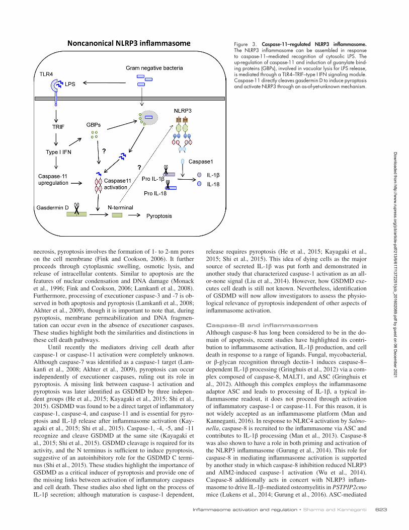
Caspase-11 and the NLRP3 inflammasomeCaspase-11 is an inflammatory caspase identified as a binding partner and mediator of caspase-1 activation (Wang et al., 1998). The functional significance of caspase-11 was masked for a long time because of its high genetic linkage to caspase-1 (Kay-agaki et al., 2011). It was eventually identified as an inducer of pyroptosis independent of caspase-1 and was further required for NLRP3 inflammasome activation specifically in response to gram-negative bacteria through its CARD domain (Shi et al., 2014). Caspase-11 acts as both the sensor and the inducer of
lipopolysaccharide (LPS)-induced pyroptotic responses and further triggers assembly of the NLRP3 inflammasome. This distinguishes the two functions of inflammasome activation, such that pyroptosis proceeds through activation of caspase-11, whereas cytokine processing occurs through NLRP3-mediated caspase-1 activation (Fig. 3).
How caspase-11 promotes the noncanonical NLRP3 activation is not entirely known. Although caspase-11 can in-teract with caspase-1 (Wang et al., 1998), loss of caspase-11 does not affect canonical NLRP3–ASC complex assembly, disproving its role as an essential adaptor. Another possibility is that caspase-11 employs a downstream molecule to instigate NLRP3 inflammasome assembly. For example, caspase-11 ac-tivation causes a drop in intracellular potassium levels, suggest-ing that potassium efflux could bridge caspase-11 and NLRP3 activation (Rühl and Broz, 2015; Schmid-Burgk et al., 2015). Pannexin-1 is also a caspase-11 target, and pannexin-1 cleavage induces activation of the NLRP3 inflammasome through a drop in intracellular potassium levels (Yang et al., 2015). However, the specificity of potassium efflux to NLRP3 activation has not been established (Pétrilli et al., 2007), so it remains to be seen whether intracellular potassium is the exclusive link between caspase-11 and NLRP3 activation.
Pyroptosis and GSD MDInflammasome activation leads to a distinct form of programmed cell death termed pyroptosis, characterized by cellular lysis, re-lease of intracellular components, and an inflammatory response. To date, caspase-induced pyroptosis has been demonstrated in macrophages (Fink et al., 2008), dendritic cells (Edgeworth et al., 2002), enterocytes (Sellin et al., 2014), and hematopoietic pro-genitors (Masters et al., 2012), whereas neutrophils and mono-cytes do not undergo pyroptosis after inflammasome activation (Miao et al., 2010a; Chen et al., 2014; Gaidt et al., 2016). Pyro-ptosis also appears to be evolutionarily conserved, as loss of mac-rophages caused by pyroptosis has been visualized in zebrafish larvae after Listeria infection, marked by ASC speck and nuclear condensation (Vincent et al., 2016). Pyroptosis is therefore a crit-ical feature of inflammasome activation in a wide variety of cells.
Pyroptosis is a distinct form of cell death but shares certain features with both apoptosis and necrosis. Similar to
Figure 2. ASC-mediated amplification of inflammasome activation. ASC mediates am-plification of inflammasome signals through at least three distinct mechanisms. (1) Signal transduction during inflammasome assembly, such that the sensor, ASC, and caspase-1 are present at increasing concentrations and each sensor activates a much greater number of the enzyme molecules. (2) Caspase-1–mediated maturation and release of bioactive cytokines affects activation and further infiltration of im-mune cells, eventually amplifying the overall inflammatory response. (3) The released ASC speck can be taken up by neighboring cells and promotes ASC assembly in the recipient cells, consequently providing another mode of inflammasome amplification.
Dow
nloaded from http://w
ww
.rupress.org/jcb/article-pdf/213/6/617/1372251/jcb_201602089.pdf by guest on 06 Decem
ber 2021
Inflammasome activation and regulation • Sharma and Kanneganti 623
necrosis, pyroptosis involves the formation of 1- to 2-nm pores on the cell membrane (Fink and Cookson, 2006). It further proceeds through cytoplasmic swelling, osmotic lysis, and release of intracellular contents. Similar to apoptosis are the features of nuclear condensation and DNA damage (Monack et al., 1996; Fink and Cookson, 2006; Lamkanfi et al., 2008). Furthermore, processing of executioner caspase-3 and -7 is ob-served in both apoptosis and pyroptosis (Lamkanfi et al., 2008; Akhter et al., 2009), though it is important to note that, during pyroptosis, membrane permeabilization and DNA fragmen-tation can occur even in the absence of executioner caspases. These studies highlight both the similarities and distinctions in these cell death pathways.
Until recently the mediators driving cell death after caspase-1 or caspase-11 activation were completely unknown. Although caspase-7 was identified as a caspase-1 target (Lam-kanfi et al., 2008; Akhter et al., 2009), pyroptosis can occur independently of executioner caspases, ruling out its role in pyroptosis. A missing link between caspase-1 activation and pyroptosis was later identified as GSD MD by three indepen-dent groups (He et al., 2015; Kayagaki et al., 2015; Shi et al., 2015). GSD MD was found to be a direct target of inflammatory caspase-1, caspase-4, and caspase-11 and is essential for pyro-ptosis and IL-1β release after inflammasome activation (Kay-agaki et al., 2015; Shi et al., 2015). Caspase-1, -4, -5, and -11 recognize and cleave GSD MD at the same site (Kayagaki et al., 2015; Shi et al., 2015). GSD MD cleavage is required for its activity, and the N terminus is sufficient to induce pyroptosis, suggestive of an autoinhibitory role for the GSD MD C termi-nus (Shi et al., 2015). These studies highlight the importance of GSD MD as a critical inducer of pyroptosis and provide one of the missing links between activation of inflammatory caspases and cell death. These studies also shed light on the process of IL-1β secretion; although maturation is caspase-1 dependent,
release requires pyroptosis (He et al., 2015; Kayagaki et al., 2015; Shi et al., 2015). This idea of dying cells as the major source of secreted IL-1β was put forth and demonstrated in another study that characterized caspase-1 activation as an all-or-none signal (Liu et al., 2014). However, how GSD MD exe-cutes cell death is still not known. Nevertheless, identification of GSD MD will now allow investigators to assess the physio-logical relevance of pyroptosis independent of other aspects of inflammasome activation.
Caspase-8 and inflammasomesAlthough caspase-8 has long been considered to be in the do-main of apoptosis, recent studies have highlighted its contri-bution to inflammasome activation, IL-1β production, and cell death in response to a range of ligands. Fungal, mycobacterial, or β-glycan recognition through dectin-1 induces caspase-8– dependent IL-1β processing (Gringhuis et al., 2012) via a com-plex composed of caspase-8, MALT1, and ASC (Gringhuis et al., 2012). Although this complex employs the inflammasome adaptor ASC and leads to processing of IL-1β, a typical in-flammasome readout, it does not proceed through activation of inflammatory caspase-1 or caspase-11. For this reason, it is not widely accepted as an inflammasome platform (Man and Kanneganti, 2016). In response to NLRC4 activation by Salmo-nella, caspase-8 is recruited to the inflammasome via ASC and contributes to IL-1β processing (Man et al., 2013). Caspase-8 was also shown to have a role in both priming and activation of the NLRP3 inflammasome (Gurung et al., 2014). This role for caspase-8 in mediating inflammasome activation is supported by another study in which caspase-8 inhibition reduced NLRP3 and AIM2-induced caspase-1 activation (Wu et al., 2014). Caspase-8 additionally acts in concert with NLRP3 inflam-masome to drive IL-1β–mediated osteomyelitis in PST PIP2cmo mice (Lukens et al., 2014; Gurung et al., 2016). ASC-mediated
Figure 3. Caspase-11–regulated NLRP3 inflammasome. The NLRP3 inflammasome can be assembled in response to caspase-11–mediated recognition of cytosolic LPS. The up-regulation of caspase-11 and induction of guanylate bind-ing proteins (GBPs), involved in vacuolar lysis for LPS release, is mediated through a TLR4–TRIF–type I IFN signaling module. Caspase-11 directly cleaves gasdermin D to induce pyroptosis and activate NLRP3 through an as-of-yet-unknown mechanism.
Dow
nloaded from http://w
ww
.rupress.org/jcb/article-pdf/213/6/617/1372251/jcb_201602089.pdf by guest on 06 Decem
ber 2021

JCB • Volume 213 • NumBer 6 • 2016624
caspase-8 recruitment and activation additionally leads to apop-tosis in response to AIM2 and NLRP3 activation. This study proposed that although pyroptosis is a rapid form of cell death, cells lacking caspase-1 undergo caspase-8–mediated apop-tosis induced with delayed kinetics (Sagulenko et al., 2013). This ASC/caspase-8–mediated apoptosis in response to AIM2 stimulation is also observed in response to Francisella infec-tion (Pierini et al., 2012). Even though caspase-8 is known to be a determinant of cell survival or cell death through apopto-sis, these studies have ascribed a pivotal role for caspase-8 in inflammasome activation.
Autophagy and inflammasomesAutophagy involves the degradation of damaged organelles and recycling of cellular metabolites. Inflammasomes, on the other hand, respond to stimuli that mark cellular damage through aber-rant physiological functions or in response to infectious agents. The intersection of these two cellular responses is therefore in-evitable and critical to the maintenance of cellular physiology. Cells activate autophagy in response to a pathogenic insult to contain the infection and promote clearance of the pathogen (Shi et al., 2012). Autophagy also clears damaged organelles generated as a byproduct of homeostatic or activatory conditions (Jabir et al., 2015) and thereby attenuates the inflammasome re-sponse. Mechanisms so far identified to mediate inflammasome inhibition via autophagy include the dampening of ROS produc-tion (Zhou et al., 2011; Lupfer et al., 2013), removal of damaged mitochondria (Jabir et al., 2015), degradation of ASC aggregates (Shi et al., 2012), and sequestration of pro–IL-1β (Harris et al., 2011). Conversely, blocking autophagy promotes inflammasome activation through accumulation of ROS-generating mitochon-dria (Zhou et al., 2011; Jabir et al., 2015). Further, cells lack-ing components of autophagosome assembly, such as ATG16L, LC3B, and beclin-1 (Saitoh et al., 2008; Nakahira et al., 2011), show increased inflammasome activation. In addition, IL-1β promotes autophagy, highlighting a negative feedback mecha-nism for inflammasome activation (Peral de Castro et al., 2012).
Conversely, autophagy can be regulated by inflammasome components. NLRP3 and NLRC4 can interact with beclin 1, an important regulator of autophagy. Association with NLRC4 in-hibits autophagy under both physiological conditions and bac-terial infection. NLRC4 additionally interacts with the class C vacuolar protein-sorting complex and inhibits autophagosome maturation (Jounai et al., 2011). Caspase-1 activation can also inhibit autophagy in response to inflammasome activation after bacterial stimuli. Toll/Interleukin receptor domain-containing adapter-inducing interferon β (TRIF) is a caspase-1 substrate, such that caspase-1 activation reduces TRIF-dependent type I IFN–mediated autophagy (Jabir et al., 2014). The functional consequence of this modulation is not entirely known and seems to be a mechanism to amplify the inflammatory response by blocking autophagy-mediated bacterial clearance and removal of endogenous inflammasome activators. In addition to auto-phagy, inflammasome activation can affect other physiological processes associated with cellular metabolism. Glycolytic en-zymes are direct caspase-1 substrates, and reduced glycolytic flux is observed in response to inflammasome activation (Shao et al., 2007). Inflammasome activation additionally induces lipid synthesis and membrane biogenesis through activation of sterol regulatory element binding proteins (Gurcel et al., 2006). This serves as a means to promote cell membrane repair and consequent cell survival.
These studies highlight that inflammasome activation controls multiple processes involved in cellular biology. In ad-dition to inducing cell death, caspase-1 activation affects sig-nals regulating cell metabolism and survival. It is tempting to hypothesize that in response to a cellular stress, multiple sig-naling modules are affected, including pyroptosis/apoptosis and cellular metabolism, such that based on the degree of damage, the overall fate of the affected cell could be repair or death.
Conclusion and future directionsInflammasome activation is intricately intertwined with basic cellular functions. In addition to removal of damaged cells, inflammasomes are also involved in cell repair, metabolism, and proliferation. Various molecules believed to be involved in the maintenance of cellular homeostasis have been demon-strated to act as critical regulators of inflammasome function and vice versa. Newly uncovered functions for the inflam-masome in cell metabolism and proliferation require further investigation. In addition, several questions still remain: What are the endogenous triggers of inflammasome activation, and how are they generated during physiological processes? What is the link between cell division and inflammasome activa-tion? What regulates inflammasome assembly in response to multiple-sensor activation to permit one ASC speck per cell? What are the cytoskeletal changes involved in inflammasome activation, and how are they brought about? What are the mol-ecules involved in GSD MD-mediated cell rupture? What reg-ulates the crosstalk between different cell death mechanisms? Investigating these and other research avenues highlighted in this review will expand our current understanding of the cell biology of inflammasomes and fuel the interest of the research community for years to come.
Acknowledgments
We would like to thank Drs. Si Ming Man and Prajwal Gurung for their helpful suggestions in writing and editing this manuscript. We apologize to our colleagues whose work we could not cite because of space limitations.
Work from the laboratory is supported by grants from the National Institutes of Health (AR056296, CA163507, AI101935, and AI124346) and the American Lebanese Syrian Associated Charities (to T.-D. Kanneganti).
The authors declare no competing financial interests.
Submitted: 27 February 2016Accepted: 27 May 2016
ReferencesAkhter, A., M.A. Gavrilin, L. Frantz, S. Washington, C. Ditty, D. Limoli, C. Day,
A. Sarkar, C. Newland, J. Butchar, et al. 2009. Caspase-7 activation by the Nlrc4/Ipaf inflammasome restricts Legionella pneumophila infection. PLoS Pathog. 5:e1000361. http ://dx .doi .org /10 .1371 /journal .ppat .1000361
Anand, P.K., R.K. Malireddi, J.R. Lukens, P. Vogel, J. Bertin, M. Lamkanfi, and T.D. Kanneganti. 2012. NLRP6 negatively regulates innate immunity and host defence against bacterial pathogens. Nature. 488:389–393. http ://dx .doi .org /10 .1038 /nature11250
Ataide, M.A., W.A. Andrade, D.S. Zamboni, D. Wang, M.C. Souza, B.S. Franklin, S. Elian, F.S. Martins, D. Pereira, G. Reed, et al. 2014. Malaria-induced NLRP12/NLRP3-dependent caspase-1 activation mediates inflammation and hypersensitivity to bacterial superinfection.
Dow
nloaded from http://w
ww
.rupress.org/jcb/article-pdf/213/6/617/1372251/jcb_201602089.pdf by guest on 06 Decem
ber 2021
Inflammasome activation and regulation • Sharma and Kanneganti 625
PLoS Pathog. 10:e1003885. (published erratum appears in PLoS Pathog. 2014. 10:e1004258) http ://dx .doi .org /10 .1371 /journal .ppat .1003885
Baroja-Mazo, A., F. Martín-Sánchez, A.I. Gomez, C.M. Martínez, J. Amores-Iniesta, V. Compan, M. Barberà-Cremades, J. Yagüe, E. Ruiz-Ortiz, J. Antón, et al. 2014. The NLRP3 inflammasome is released as a particulate danger signal that amplifies the inflammatory response. Nat. Immunol. 15:738–748. http ://dx .doi .org /10 .1038 /ni .2919
Broz, P., K. Newton, M. Lamkanfi, S. Mariathasan, V.M. Dixit, and D.M. Monack. 2010a. Redundant roles for inflammasome receptors NLRP3 and NLRC4 in host defense against Salmonella. J. Exp. Med. 207:1745–1755. http ://dx .doi .org /10 .1084 /jem .20100257
Broz, P., J. von Moltke, J.W. Jones, R.E. Vance, and D.M. Monack. 2010b. Differential requirement for Caspase-1 autoproteolysis in pathogen-induced cell death and cytokine processing. Cell Host Microbe. 8:471–483. http ://dx .doi .org /10 .1016 /j .chom .2010 .11 .007
Brunette, R.L., J.M. Young, D.G. Whitley, I.E. Brodsky, H.S. Malik, and D.B. Stetson. 2012. Extensive evolutionary and functional diversity among mammalian AIM2-like receptors. J. Exp. Med. 209:1969–1983. http ://dx .doi .org /10 .1084 /jem .20121960
Bürckstümmer, T., C. Baumann, S. Blüml, E. Dixit, G. Dürnberger, H. Jahn, M. Planyavsky, M. Bilban, J. Colinge, K.L. Bennett, and G. Superti-Furga. 2009. An orthogonal proteomic-genomic screen identifies AIM2 as a cytoplasmic DNA sensor for the inflammasome. Nat. Immunol. 10:266–272. http ://dx .doi .org /10 .1038 /ni .1702
Cai, X., J. Chen, H. Xu, S. Liu, Q.X. Jiang, R. Halfmann, and Z.J. Chen. 2014. Prion-like polymerization underlies signal transduction in antiviral immune defense and inflammasome activation. Cell. 156:1207–1222. http ://dx .doi .org /10 .1016 /j .cell .2014 .01 .063
Canna, S.W., A.A. de Jesus, S. Gouni, S.R. Brooks, B. Marrero, Y. Liu, M.A. DiMattia, K.J. Zaal, G.A. Sanchez, H. Kim, et al. 2014. An activating NLRC4 inflammasome mutation causes autoinflammation with recurrent macrophage activation syndrome. Nat. Genet. 46:1140–1146. http ://dx .doi .org /10 .1038 /ng .3089
Cassel, S.L., S.C. Eisenbarth, S.S. Iyer, J.J. Sadler, O.R. Colegio, L.A. Tephly, A.B. Carter, P.B. Rothman, R.A. Flavell, and F.S. Sutterwala. 2008. The Nalp3 inflammasome is essential for the development of silicosis. Proc. Natl. Acad. Sci. USA. 105:9035–9040. http ://dx .doi .org /10 .1073 /pnas .0803933105
Cavailles, P., P. Flori, O. Papapietro, C. Bisanz, D. Lagrange, L. Pilloux, C. Massera, S. Cristinelli, D. Jublot, O. Bastien, et al. 2014. A highly conserved Toxo1 haplotype directs resistance to toxoplasmosis and its associated caspase-1 dependent killing of parasite and host macrophage. PLoS Pathog. 10:e1004005. http ://dx .doi .org /10 .1371 /journal .ppat .1004005
Chae, J.J., Y.H. Cho, G.S. Lee, J. Cheng, P.P. Liu, L. Feigenbaum, S.I. Katz, and D.L. Kastner. 2011. Gain-of-function Pyrin mutations induce NLRP3 protein-independent interleukin-1β activation and severe autoinflammation in mice. Immunity. 34:755–768. http ://dx .doi .org /10 .1016 /j .immuni .2011 .02 .020
Chavarría-Smith, J., and R.E. Vance. 2013. Direct proteolytic cleavage of NLRP1B is necessary and sufficient for inflammasome activation by anthrax lethal factor. PLoS Pathog. 9:e1003452. http ://dx .doi .org /10 .1371 /journal .ppat .1003452
Chen, G.Y., M. Liu, F. Wang, J. Bertin, and G. Núñez. 2011. A functional role for Nlrp6 in intestinal inflammation and tumorigenesis. J. Immunol. 186:7187–7194. http ://dx .doi .org /10 .4049 /jimmunol .1100412
Chen, K.W., C.J. Groß, F.V. Sotomayor, K.J. Stacey, J. Tschopp, M.J. Sweet, and K. Schroder. 2014. The neutrophil NLRC4 inflammasome selectively promotes IL-1β maturation without pyroptosis during acute Salmonella challenge. Cell Reports. 8:570–582. http ://dx .doi .org /10 .1016 /j .celrep .2014 .06 .028
Cheng, J., A.L. Waite, E.R. Tkaczyk, K. Ke, N. Richards, A.J. Hunt, and D.L. Gumucio. 2010. Kinetic properties of ASC protein aggregation in epithelial cells. J. Cell. Physiol. 222:738–747. http ://dx .doi .org /10 .1002 /jcp .22005
Choubey, D. 2012. Interferon-inducible Ifi200-family genes as modifiers of lupus susceptibility. Immunol. Lett. 147:10–17. http ://dx .doi .org /10 .1016 /j .imlet .2012 .07 .003
Cirelli, K.M., G. Gorfu, M.A. Hassan, M. Printz, D. Crown, S.H. Leppla, M.E. Grigg, J.P. Saeij, and M. Moayeri. 2014. Inflammasome sensor NLRP1 controls rat macrophage susceptibility to Toxoplasma gondii. PLoS Pathog. 10:e1003927. http ://dx .doi .org /10 .1371 /journal .ppat .1003927
Compan, V., A. Baroja-Mazo, G. López-Castejón, A.I. Gomez, C.M. Martínez, D. Angosto, M.T. Montero, A.S. Herranz, E. Bazán, D. Reimers, et al. 2012. Cell volume regulation modulates NLRP3 inflammasome activation. Immunity. 37:487–500. http ://dx .doi .org /10 .1016 /j .immuni .2012 .06 .013
Cridland, J.A., E.Z. Curley, M.N. Wykes, K. Schroder, M.J. Sweet, T.L. Roberts, M.A. Ragan, K.S. Kassahn, and K.J. Stacey. 2012. The mammalian PYH IN gene family: phylogeny, evolution and expression. BMC Evol. Biol. 12:140. http ://dx .doi .org /10 .1186 /1471 -2148 -12 -140
Cruz, C.M., A. Rinna, H.J. Forman, A.L. Ventura, P.M. Persechini, and D.M. Ojcius. 2007. ATP activates a reactive oxygen species-dependent oxidative stress response and secretion of proinflammatory cytokines in macrophages. J. Biol. Chem. 282:2871–2879. http ://dx .doi .org /10 .1074 /jbc .M608083200
Diebolder, C.A., E.F. Halff, A.J. Koster, E.G. Huizinga, and R.I. Koning. 2015. Cryoelectron tomography of the NAIP5/NLRC4 inflammasome: implications for NLR Activation. Structure. 23:2349–2357. http ://dx .doi .org /10 .1016 /j .str .2015 .10 .001
Dihlmann, S., P. Erhart, A. Mehrabi, A. Nickkholgh, F. Lasitschka, D. Böckler, and M. Hakimi. 2014. Increased expression and activation of absent in melanoma 2 inflammasome components in lymphocytic infiltrates of abdominal aortic aneurysms. Mol. Med. 20:230–237. http ://dx .doi .org /10 .2119 /molmed .2013 .00162
Dombrowski, Y., M. Peric, S. Koglin, C. Kammerbauer, C. Göss, D. Anz, M. Simanski, R. Gläser, J. Harder, V. Hornung, et al. 2011. Cytosolic DNA triggers inflammasome activation in keratinocytes in psoriatic lesions. Sci. Transl. Med. 3:82ra38. http ://dx .doi .org /10 .1126 /scitranslmed .3002001
Dostert, C., V. Pétrilli, R. Van Bruggen, C. Steele, B.T. Mossman, and J. Tschopp. 2008. Innate immune activation through Nalp3 inflammasome sensing of asbestos and silica. Science. 320:674–677. http ://dx .doi .org /10 .1126 /science .1156995
Dumas, A., N. Amiable, J.P. de Rivero Vaccari, J.J. Chae, R.W. Keane, S. Lacroix, and L. Vallières. 2014. The inflammasome pyrin contributes to pertussis toxin-induced IL-1β synthesis, neutrophil intravascular crawling and autoimmune encephalomyelitis. PLoS Pathog. 10:e1004150. http ://dx .doi .org /10 .1371 /journal .ppat .1004150
Edgeworth, J.D., J. Spencer, A. Phalipon, G.E. Griffin, and P.J. Sansonetti. 2002. Cytotoxicity and interleukin-1beta processing following Shigella flexneri infection of human monocyte-derived dendritic cells. Eur. J. Immunol. 32:1464–1471. http ://dx .doi .org /10 .1002 /1521 -4141(200205)32 :5<1464::AID-IMMU1464>3.0.CO;2-G
Elinav, E., T. Strowig, A.L. Kau, J. Henao-Mejia, C.A. Thaiss, C.J. Booth, D.R. Peaper, J. Bertin, S.C. Eisenbarth, J.I. Gordon, and R.A. Flavell. 2011. NLRP6 inflammasome regulates colonic microbial ecology and risk for colitis. Cell. 145:745–757. http ://dx .doi .org /10 .1016 /j .cell .2011 .04 .022
Endrizzi, M.G., V. Hadinoto, J.D. Growney, W. Miller, and W.F. Dietrich. 2000. Genomic sequence analysis of the mouse Naip gene array. Genome Res. 10:1095–1102. http ://dx .doi .org /10 .1101 /gr .10 .8 .1095
Ewald, S.E., J. Chavarria-Smith, and J.C. Boothroyd. 2014. NLRP1 is an inflammasome sensor for Toxoplasma gondii. Infect. Immun. 82:460–468. http ://dx .doi .org /10 .1128 /IAI .01170 -13
Fernandes-Alnemri, T., J.W. Yu, P. Datta, J. Wu, and E.S. Alnemri. 2009. AIM2 activates the inflammasome and cell death in response to cytoplasmic DNA. Nature. 458:509–513. http ://dx .doi .org /10 .1038 /nature07710
Fernandes-Alnemri, T., J.W. Yu, C. Juliana, L. Solorzano, S. Kang, J. Wu, P. Datta, M. McCormick, L. Huang, E. McDermott, et al. 2010. The AIM2 inflammasome is critical for innate immunity to Francisella tularensis. Nat. Immunol. 11:385–393. http ://dx .doi .org /10 .1038 /ni .1859
Finger, J.N., J.D. Lich, L.C. Dare, M.N. Cook, K.K. Brown, C. Duraiswami, J. Bertin, and P.J. Gough. 2012. Autolytic proteolysis within the function to find domain (FII ND) is required for NLRP1 inflammasome activity. J. Biol. Chem. 287:25030–25037. http ://dx .doi .org /10 .1074 /jbc .M112 .378323
Fink, S.L., and B.T. Cookson. 2006. Caspase-1-dependent pore formation during pyroptosis leads to osmotic lysis of infected host macrophages. Cell. Microbiol. 8:1812–1825. http ://dx .doi .org /10 .1111 /j .1462 -5822 .2006 .00751 .x
Fink, S.L., T. Bergsbaken, and B.T. Cookson. 2008. Anthrax lethal toxin and Salmonella elicit the common cell death pathway of caspase-1-dependent pyroptosis via distinct mechanisms. Proc. Natl. Acad. Sci. USA. 105:4312–4317. http ://dx .doi .org /10 .1073 /pnas .0707370105
Franchi, L., A. Amer, M. Body-Malapel, T.D. Kanneganti, N. Ozören, R. Jagirdar, N. Inohara, P. Vandenabeele, J. Bertin, A. Coyle, et al. 2006. Cytosolic flagellin requires Ipaf for activation of caspase-1 and interleukin 1beta in salmonella-infected macrophages. Nat. Immunol. 7:576–582. http ://dx .doi .org /10 .1038 /ni1346
Franklin, B.S., L. Bossaller, D. De Nardo, J.M. Ratter, A. Stutz, G. Engels, C. Brenker, M. Nordhoff, S.R. Mirandola, A. Al-Amoudi, et al. 2014. The adaptor ASC has extracellular and ‘prionoid’ activities that propagate inflammation. Nat. Immunol. 15:727–737. http ://dx .doi .org /10 .1038 /ni .2913
Dow
nloaded from http://w
ww
.rupress.org/jcb/article-pdf/213/6/617/1372251/jcb_201602089.pdf by guest on 06 Decem
ber 2021
JCB • Volume 213 • NumBer 6 • 2016626
Frew, B.C., V.R. Joag, and J. Mogridge. 2012. Proteolytic processing of Nlrp1b is required for inflammasome activity. PLoS Pathog. 8:e1002659. http ://dx .doi .org /10 .1371 /journal .ppat .1002659
Gaidt, M.M., T.S. Ebert, D. Chauhan, T. Schmidt, J.L. Schmid-Burgk, F. Rapino, A.A. Robertson, M.A. Cooper, T. Graf, and V. Hornung. 2016. Human monocytes engage an alternative inflammasome pathway. Immunity. 44:833–846. http ://dx .doi .org /10 .1016 /j .immuni .2016 .01 .012
Ge, J., Y.N. Gong, Y. Xu, and F. Shao. 2012. Preventing bacterial DNA release and absent in melanoma 2 inflammasome activation by a Legionella effector functioning in membrane trafficking. Proc. Natl. Acad. Sci. USA. 109:6193–6198. http ://dx .doi .org /10 .1073 /pnas .1117490109
Gorfu, G., K.M. Cirelli, M.B. Melo, K. Mayer-Barber, D. Crown, B.H. Koller, S. Masters, A. Sher, S.H. Leppla, M. Moayeri, et al. 2014. Dual role for inflammasome sensors NLRP1 and NLRP3 in murine resistance to Toxoplasma gondii. MBio. 5: e01117–e01113. http ://dx .doi .org /10 .1128 /mBio .01117 -13
Gringhuis, S.I., T.M. Kaptein, B.A. Wevers, B. Theelen, M. van der Vlist, T. Boekhout, and T.B. Geijtenbeek. 2012. Dectin-1 is an extracellular pathogen sensor for the induction and processing of IL-1β via a noncanonical caspase-8 inflammasome. Nat. Immunol. 13:246–254. http ://dx .doi .org /10 .1038 /ni .2222
Guey, B., M. Bodnar, S.N. Manié, A. Tardivel, and V. Petrilli. 2014. Caspase-1 autoproteolysis is differentially required for NLRP1b and NLRP3 inflammasome function. Proc. Natl. Acad. Sci. USA. 111:17254–17259. http ://dx .doi .org /10 .1073 /pnas .1415756111
Guo, H., J.B. Callaway, and J.P. Ting. 2015. Inflammasomes: mechanism of action, role in disease, and therapeutics. Nat. Med. 21:677–687. http ://dx .doi .org /10 .1038 /nm .3893
Gurcel, L., L. Abrami, S. Girardin, J. Tschopp, and F.G. van der Goot. 2006. Caspase-1 activation of lipid metabolic pathways in response to bacterial pore-forming toxins promotes cell survival. Cell. 126:1135–1145. http ://dx .doi .org /10 .1016 /j .cell .2006 .07 .033
Gurung, P., P.K. Anand, R.K. Malireddi, L. Vande Walle, N. Van Opdenbosch, C.P. Dillon, R. Weinlich, D.R. Green, M. Lamkanfi, and T.D. Kanneganti. 2014. FADD and caspase-8 mediate priming and activation of the canonical and noncanonical Nlrp3 inflammasomes. J. Immunol. 192:1835–1846. http ://dx .doi .org /10 .4049 /jimmunol .1302839
Gurung, P., A. Burton, and T.D. Kanneganti. 2016. NLRP3 inflammasome plays a redundant role with caspase 8 to promote IL-1β-mediated osteomyelitis. Proc. Natl. Acad. Sci. USA. 113:4452–4457. http ://dx .doi .org /10 .1073 /pnas .1601636113
Halle, A., V. Hornung, G.C. Petzold, C.R. Stewart, B.G. Monks, T. Reinheckel, K.A. Fitzgerald, E. Latz, K.J. Moore, and D.T. Golenbock. 2008. The NALP3 inflammasome is involved in the innate immune response to amyloid-beta. Nat. Immunol. 9:857–865. http ://dx .doi .org /10 .1038 /ni .1636
Harris, J., M. Hartman, C. Roche, S.G. Zeng, A. O’Shea, F.A. Sharp, E.M. Lambe, E.M. Creagh, D.T. Golenbock, J. Tschopp, et al. 2011. Autophagy controls IL-1beta secretion by targeting pro-IL-1beta for degradation. J. Biol. Chem. 286:9587–9597. http ://dx .doi .org /10 .1074 /jbc .M110 .202911
He, W.T., H. Wan, L. Hu, P. Chen, X. Wang, Z. Huang, Z.H. Yang, C.Q. Zhong, and J. Han. 2015. Gasdermin D is an executor of pyroptosis and required for interleukin-1β secretion. Cell Res. 25:1285–1298. http ://dx .doi .org /10 .1038 /cr .2015 .139
He, Y., M.Y. Zeng, D. Yang, B. Motro, and G. Núñez. 2016. NEK7 is an essential mediator of NLRP3 activation downstream of potassium efflux. Nature. 530:354–357. http ://dx .doi .org /10 .1038 /nature16959
Henao-Mejia, J., E. Elinav, C. Jin, L. Hao, W.Z. Mehal, T. Strowig, C.A. Thaiss, A.L. Kau, S.C. Eisenbarth, M.J. Jurczak, et al. 2012. Inflammasome-mediated dysbiosis regulates progression of NAF LD and obesity. Nature. 482:179–185. http ://dx .doi .org /10 .1038 /nature10809
Hoffman, H.M., J.L. Mueller, D.H. Broide, A.A. Wanderer, and R.D. Kolodner. 2001. Mutation of a new gene encoding a putative pyrin-like protein causes familial cold autoinflammatory syndrome and Muckle-Wells syndrome. Nat. Genet. 29:301–305. http ://dx .doi .org /10 .1038 /ng756
Hornung, V., F. Bauernfeind, A. Halle, E.O. Samstad, H. Kono, K.L. Rock, K.A. Fitzgerald, and E. Latz. 2008. Silica crystals and aluminum salts activate the NALP3 inflammasome through phagosomal destabilization. Nat. Immunol. 9:847–856. http ://dx .doi .org /10 .1038 /ni .1631
Hornung, V., A. Ablasser, M. Charrel-Dennis, F. Bauernfeind, G. Horvath, D.R. Caffrey, E. Latz, and K.A. Fitzgerald. 2009. AIM2 recognizes cytosolic dsDNA and forms a caspase-1-activating inflammasome with ASC. Nature. 458:514–518. http ://dx .doi .org /10 .1038 /nature07725
Hu, B., E. Elinav, S. Huber, T. Strowig, L. Hao, A. Hafemann, C. Jin, C. Wunderlich, T. Wunderlich, S.C. Eisenbarth, and R.A. Flavell. 2013. Microbiota-induced activation of epithelial IL-6 signaling links inflammasome-driven inflammation with transmissible cancer. Proc.
Natl. Acad. Sci. USA. 110:9862–9867. http ://dx .doi .org /10 .1073 /pnas .1307575110
Hu, Z., Q. Zhou, C. Zhang, S. Fan, W. Cheng, Y. Zhao, F. Shao, H.W. Wang, S.F. Sui, and J. Chai. 2015. Structural and biochemical basis for induced self-propagation of NLRC4. Science. 350:399–404. http ://dx .doi .org /10 .1126 /science .aac5489
Huber, S., N. Gagliani, L.A. Zenewicz, F.J. Huber, L. Bosurgi, B. Hu, M. Hedl, W. Zhang, W. O’Connor Jr., A.J. Murphy, et al. 2012. IL-22BP is regulated by the inflammasome and modulates tumorigenesis in the intestine. Nature. 491:259–263. http ://dx .doi .org /10 .1038 /nature11535
Jabir, M.S., N.D. Ritchie, D. Li, H.K. Bayes, P. Tourlomousis, D. Puleston, A. Lupton, L. Hopkins, A.K. Simon, C. Bryant, and T.J. Evans. 2014. Caspase-1 cleavage of the TLR adaptor TRIF inhibits autophagy and β-interferon production during Pseudomonas aeruginosa infection. Cell Host Microbe. 15:214–227. http ://dx .doi .org /10 .1016 /j .chom .2014 .01 .010
Jabir, M.S., L. Hopkins, N.D. Ritchie, I. Ullah, H.K. Bayes, D. Li, P. Tourlomousis, A. Lupton, D. Puleston, A.K. Simon, et al. 2015. Mitochondrial damage contributes to Pseudomonas aeruginosa activation of the inflammasome and is downregulated by autophagy. Autophagy. 11:166–182. http ://dx .doi .org /10 .4161 /15548627 .2014 .981915
Jin, T., A. Perry, J. Jiang, P. Smith, J.A. Curry, L. Unterholzner, Z. Jiang, G. Horvath, V.A. Rathinam, R.W. Johnstone, et al. 2012. Structures of the HIN domain :DNA complexes reveal ligand binding and activation mechanisms of the AIM2 inflammasome and IFI16 receptor. Immunity. 36:561–571. http ://dx .doi .org /10 .1016 /j .immuni .2012 .02 .014
Jin, T., J. Curry, P. Smith, J. Jiang, and T.S. Xiao. 2013a. Structure of the NLRP1 caspase recruitment domain suggests potential mechanisms for its association with procaspase-1. Proteins. 81:1266–1270. http ://dx .doi .org /10 .1002 /prot .24287
Jin, T., A. Perry, P. Smith, J. Jiang, and T.S. Xiao. 2013b. Structure of the absent in melanoma 2 (AIM2) pyrin domain provides insights into the mechanisms of AIM2 autoinhibition and inflammasome assembly. J. Biol. Chem. 288:13225–13235. http ://dx .doi .org /10 .1074 /jbc .M113 .468033
Jin, Y., C.M. Mailloux, K. Gowan, S.L. Riccardi, G. LaBerge, D.C. Bennett, P.R. Fain, and R.A. Spritz. 2007. NALP1 in vitiligo-associated multiple autoimmune disease. N. Engl. J. Med. 356:1216–1225. http ://dx .doi .org /10 .1056 /NEJMoa061592
Jones, J.W., N. Kayagaki, P. Broz, T. Henry, K. Newton, K. O’Rourke, S. Chan, J. Dong, Y. Qu, M. Roose-Girma, et al. 2010. Absent in melanoma 2 is required for innate immune recognition of Francisella tularensis. Proc. Natl. Acad. Sci. USA. 107:9771–9776. http ://dx .doi .org /10 .1073 /pnas .1003738107
Jounai, N., K. Kobiyama, M. Shiina, K. Ogata, K.J. Ishii, and F. Takeshita. 2011. NLRP4 negatively regulates autophagic processes through an association with beclin1. J. Immunol. 186:1646–1655. http ://dx .doi .org /10 .4049 /jimmunol .1001654
Karki, R., S.M. Man, R.K. Malireddi, P. Gurung, P. Vogel, M. Lamkanfi, and T.D. Kanneganti. 2015. Concerted activation of the AIM2 and NLRP3 inflammasomes orchestrates host protection against Aspergillus infection. Cell Host Microbe. 17:357–368. http ://dx .doi .org /10 .1016 /j .chom .2015 .01 .006
Kayagaki, N., S. Warming, M. Lamkanfi, L. Vande Walle, S. Louie, J. Dong, K. Newton, Y. Qu, J. Liu, S. Heldens, et al. 2011. Non-canonical inflammasome activation targets caspase-11. Nature. 479:117–121. http ://dx .doi .org /10 .1038 /nature10558
Kayagaki, N., I.B. Stowe, B.L. Lee, K. O’Rourke, K. Anderson, S. Warming, T. Cuellar, B. Haley, M. Roose-Girma, Q.T. Phung, et al. 2015. Caspase-11 cleaves gasdermin D for non-canonical inflammasome signalling. Nature. 526:666–671. http ://dx .doi .org /10 .1038 /nature15541
Kerur, N., M.V. Veettil, N. Sharma-Walia, V. Bottero, S. Sadagopan, P. Otageri, and B. Chandran. 2011. IFI16 acts as a nuclear pathogen sensor to induce the inflammasome in response to Kaposi Sarcoma-associated herpesvirus infection. Cell Host Microbe. 9:363–375. http ://dx .doi .org /10 .1016 /j .chom .2011 .04 .008
Khare, S., R.A. Ratsimandresy, L. de Almeida, C.M. Cuda, S.L. Rellick, A.V. Misharin, M.C. Wallin, A. Gangopadhyay, E. Forte, E. Gottwein, et al. 2014. The PYR IN domain-only protein POP3 inhibits ALR inflammasomes and regulates responses to infection with DNA viruses. Nat. Immunol. 15:343–353. http ://dx .doi .org /10 .1038 /ni .2829
Kim, S., F. Bauernfeind, A. Ablasser, G. Hartmann, K.A. Fitzgerald, E. Latz, and V. Hornung. 2010. Listeria monocytogenes is sensed by the NLRP3 and AIM2 inflammasome. Eur. J. Immunol. 40:1545–1551. http ://dx .doi .org /10 .1002 /eji .201040425
Kitamura, A., Y. Sasaki, T. Abe, H. Kano, and K. Yasutomo. 2014. An inherited mutation in NLRC4 causes autoinflammation in human and mice. J. Exp. Med. 211:2385–2396. http ://dx .doi .org /10 .1084 /jem .20141091
Dow
nloaded from http://w
ww
.rupress.org/jcb/article-pdf/213/6/617/1372251/jcb_201602089.pdf by guest on 06 Decem
ber 2021

Inflammasome activation and regulation • Sharma and Kanneganti 627
Kofoed, E.M., and R.E. Vance. 2011. Innate immune recognition of bacterial ligands by NAIPs determines inflammasome specificity. Nature. 477:592–595. http ://dx .doi .org /10 .1038 /nature10394
Kortmann, J., S.W. Brubaker, and D.M. Monack. 2015. Cutting edge: inflammasome activation in primary human macrophages is dependent on flagellin. J. Immunol. 195:815–819. http ://dx .doi .org /10 .4049 /jimmunol .1403100
Lamkanfi, M., T.D. Kanneganti, P. Van Damme, T. Vanden Berghe, I. Vanoverberghe, J. Vandekerckhove, P. Vandenabeele, K. Gevaert, and G. Núñez. 2008. Targeted peptidecentric proteomics reveals caspase-7 as a substrate of the caspase-1 inflammasomes. Mol. Cell. Proteomics. 7:2350–2363. http ://dx .doi .org /10 .1074 /mcp .M800132 -MCP200
Lee, G.S., N. Subramanian, A.I. Kim, I. Aksentijevich, R. Goldbach-Mansky, D.B. Sacks, R.N. Germain, D.L. Kastner, and J.J. Chae. 2012. The calcium-sensing receptor regulates the NLRP3 inflammasome through Ca2+ and cAMP. Nature. 492:123–127. http ://dx .doi .org /10 .1038 /nature11588
Levy, M., C.A. Thaiss, D. Zeevi, L. Dohnalová, G. Zilberman-Schapira, J.A. Mahdi, E. David, A. Savidor, T. Korem, Y. Herzig, et al. 2015. Microbiota-modulated metabolites shape the intestinal microenvironment by regulating NLRP6 inflammasome signaling. Cell. 163:1428–1443. http ://dx .doi .org /10 .1016 /j .cell .2015 .10 .048
Li, H., J. Wang, J. Wang, L.S. Cao, Z.X. Wang, and J.W. Wu. 2014. Structural mechanism of DNA recognition by the p202 HINa domain: insights into the inhibition of Aim2-mediated inflammatory signalling. Acta Crystallogr. F Struct. Biol. Commun. 70:21–29. http ://dx .doi .org /10 .1107 /S2053230X1303135X
Lich, J.D., K.L. Williams, C.B. Moore, J.C. Arthur, B.K. Davis, D.J. Taxman, and J.P. Ting. 2007. Monarch-1 suppresses non-canonical NF-kappaB activation and p52-dependent chemokine expression in monocytes. J. Immunol. 178:1256–1260. http ://dx .doi .org /10 .4049 /jimmunol .178 .3 .1256
Liu, T., Y. Yamaguchi, Y. Shirasaki, K. Shikada, M. Yamagishi, K. Hoshino, T. Kaisho, K. Takemoto, T. Suzuki, E. Kuranaga, et al. 2014. Single-cell imaging of caspase-1 dynamics reveals an all-or-none inflammasome signaling response. Cell Reports. 8:974–982. http ://dx .doi .org /10 .1016 /j .celrep .2014 .07 .012
Lu, A., V.G. Magupalli, J. Ruan, Q. Yin, M.K. Atianand, M.R. Vos, G.F. Schröder, K.A. Fitzgerald, H. Wu, and E.H. Egelman. 2014. Unified polymerization mechanism for the assembly of ASC-dependent inflammasomes. Cell. 156:1193–1206. http ://dx .doi .org /10 .1016 /j .cell .2014 .02 .008
Lukens, J.R., P. Gurung, P. Vogel, G.R. Johnson, R.A. Carter, D.J. McGoldrick, S.R. Bandi, C.R. Calabrese, L. Vande Walle, M. Lamkanfi, and T.D. Kanneganti. 2014. Dietary modulation of the microbiome affects autoinflammatory disease. Nature. 516:246–249. http ://dx .doi .org /10 .1038 /nature13788
Lukens, J.R., P. Gurung, P.J. Shaw, M.J. Barr, M.H. Zaki, S.A. Brown, P. Vogel, H. Chi, and T.D. Kanneganti. 2015. The NLRP12 Sensor negatively regulates autoinflammatory disease by modulating interleukin-4 production in T cells. Immunity. 42:654–664. http ://dx .doi .org /10 .1016 /j .immuni .2015 .03 .006
Lupfer, C., P.G. Thomas, P.K. Anand, P. Vogel, S. Milasta, J. Martinez, G. Huang, M. Green, M. Kundu, H. Chi, et al. 2013. Receptor interacting protein kinase 2-mediated mitophagy regulates inflammasome activation during virus infection. Nat. Immunol. 14:480–488. http ://dx .doi .org /10 .1038 /ni .2563
Man, S.M., and T.D. Kanneganti. 2015. Regulation of inflammasome activation. Immunol. Rev. 265:6–21. http ://dx .doi .org /10 .1111 /imr .12296
Man, S.M., and T.-D. Kanneganti. 2016. Converging roles of caspases in inflammasome activation, cell death and innate immunity. Nat. Rev. Immunol. 16:7–21. http ://dx .doi .org /10 .1038 /nri .2015 .7
Man, S.M., P. Tourlomousis, L. Hopkins, T.P. Monie, K.A. Fitzgerald, and C.E. Bryant. 2013. Salmonella infection induces recruitment of Caspase-8 to the inflammasome to modulate IL-1β production. J. Immunol. 191:5239–5246. http ://dx .doi .org /10 .4049 /jimmunol .1301581
Man, S.M., A. Ekpenyong, P. Tourlomousis, S. Achouri, E. Cammarota, K. Hughes, A. Rizzo, G. Ng, J.A. Wright, P. Cicuta, et al. 2014a. Actin polymerization as a key innate immune effector mechanism to control Salmonella infection. Proc. Natl. Acad. Sci. USA. 111:17588–17593. http ://dx .doi .org /10 .1073 /pnas .1419925111
Man, S.M., L.J. Hopkins, E. Nugent, S. Cox, I.M. Glück, P. Tourlomousis, J.A. Wright, P. Cicuta, T.P. Monie, and C.E. Bryant. 2014b. Inflammasome activation causes dual recruitment of NLRC4 and NLRP3 to the same macromolecular complex. Proc. Natl. Acad. Sci. USA. 111:7403–7408. http ://dx .doi .org /10 .1073 /pnas .1402911111
Man, S.M., R. Karki, R.K. Malireddi, G. Neale, P. Vogel, M. Yamamoto, M. Lamkanfi, and T.D. Kanneganti. 2015a. The transcription factor IRF1 and guanylate-binding proteins target activation of the AIM2
inflammasome by Francisella infection. Nat. Immunol. 16:467–475. http ://dx .doi .org /10 .1038 /ni .3118
Man, S.M., Q. Zhu, L. Zhu, Z. Liu, R. Karki, A. Malik, D. Sharma, L. Li, R.K. Malireddi, P. Gurung, et al. 2015b. Critical role for the DNA sensor AIM2 in stem cell proliferation and cancer. Cell. 162:45–58. http ://dx .doi .org /10 .1016 /j .cell .2015 .06 .001
Mariathasan, S., K. Newton, D.M. Monack, D. Vucic, D.M. French, W.P. Lee, M. Roose-Girma, S. Erickson, and V.M. Dixit. 2004. Differential activation of the inflammasome by caspase-1 adaptors ASC and Ipaf. Nature. 430:213–218. http ://dx .doi .org /10 .1038 /nature02664
Martinon, F., K. Burns, and J. Tschopp. 2002. The inflammasome: a molecular platform triggering activation of inflammatory caspases and processing of proIL-beta. Mol. Cell. 10:417–426. http ://dx .doi .org /10 .1016 /S1097 -2765(02)00599 -3
Masters, S.L., A. Simon, I. Aksentijevich, and D.L. Kastner. 2009. Horror autoinflammaticus: the molecular pathophysiology of autoinflammatory disease. Annu. Rev. Immunol. 27:621–668. http ://dx .doi .org /10 .1146 /annurev .immunol .25 .022106 .141627
Masters, S.L., M. Gerlic, D. Metcalf, S. Preston, M. Pellegrini, J.A. O’Donnell, K. McArthur, T.M. Baldwin, S. Chevrier, C.J. Nowell, et al. 2012. NLRP1 inflammasome activation induces pyroptosis of hematopoietic progenitor cells. Immunity. 37:1009–1023. http ://dx .doi .org /10 .1016 /j .immuni .2012 .08 .027
Masters, S.L., V. Lagou, I. Jéru, P.J. Baker, L. Van Eyck, D.A. Parry, D. Lawless, D. De Nardo, J.E. Garcia-Perez, L.F. Dagley, et al. 2016. Familial autoinflammation with neutrophilic dermatosis reveals a regulatory mechanism of pyrin activation. Sci. Transl. Med. 8:332ra45. http ://dx .doi .org /10 .1126 /scitranslmed .aaf1471
Matusiak, M., N. Van Opdenbosch, and M. Lamkanfi. 2015. CARD- and pyrin-only proteins regulating inflammasome activation and immunity. Immunol. Rev. 265:217–230. http ://dx .doi .org /10 .1111 /imr .12282
Meunier, E., P. Wallet, R.F. Dreier, S. Costanzo, L. Anton, S. Rühl, S. Dussurgey, M.S. Dick, A. Kistner, M. Rigard, et al. 2015. Guanylate-binding proteins promote activation of the AIM2 inflammasome during infection with Francisella novicida. Nat. Immunol. 16:476–484. http ://dx .doi .org /10 .1038 /ni .3119
Miao, E.A., C.M. Alpuche-Aranda, M. Dors, A.E. Clark, M.W. Bader, S.I. Miller, and A. Aderem. 2006. Cytoplasmic flagellin activates caspase-1 and secretion of interleukin 1beta via Ipaf. Nat. Immunol. 7:569–575. http ://dx .doi .org /10 .1038 /ni1344
Miao, E.A., I.A. Leaf, P.M. Treuting, D.P. Mao, M. Dors, A. Sarkar, S.E. Warren, M.D. Wewers, and A. Aderem. 2010a. Caspase-1-induced pyroptosis is an innate immune effector mechanism against intracellular bacteria. Nat. Immunol. 11:1136–1142. http ://dx .doi .org /10 .1038 /ni .1960
Miao, E.A., D.P. Mao, N. Yudkovsky, R. Bonneau, C.G. Lorang, S.E. Warren, I.A. Leaf, and A. Aderem. 2010b. Innate immune detection of the type III secretion apparatus through the NLRC4 inflammasome. Proc. Natl. Acad. Sci. USA. 107:3076–3080. http ://dx .doi .org /10 .1073 /pnas .0913087107
Misawa, T., M. Takahama, T. Kozaki, H. Lee, J. Zou, T. Saitoh, and S. Akira. 2013. Microtubule-driven spatial arrangement of mitochondria promotes activation of the NLRP3 inflammasome. Nat. Immunol. 14:454–460. http ://dx .doi .org /10 .1038 /ni .2550
Molofsky, A.B., B.G. Byrne, N.N. Whitfield, C.A. Madigan, E.T. Fuse, K. Tateda, and M.S. Swanson. 2006. Cytosolic recognition of flagellin by mouse macrophages restricts Legionella pneumophila infection. J. Exp. Med. 203:1093–1104. http ://dx .doi .org /10 .1084 /jem .20051659
Monack, D.M., B. Raupach, A.E. Hromockyj, and S. Falkow. 1996. Salmonella typhimurium invasion induces apoptosis in infected macrophages. Proc. Natl. Acad. Sci. USA. 93:9833–9838. http ://dx .doi .org /10 .1073 /pnas .93 .18 .9833
Moriya, M., S. Taniguchi, P. Wu, E. Liepinsh, G. Otting, and J. Sagara. 2005. Role of charged and hydrophobic residues in the oligomerization of the PYR IN domain of ASC. Biochemistry. 44:575–583. http ://dx .doi .org /10 .1021 /bi048374i
Morizane, S., K. Yamasaki, B. Mühleisen, P.F. Kotol, M. Murakami, Y. Aoyama, K. Iwatsuki, T. Hata, and R.L. Gallo. 2012. Cathelicidin antimicrobial peptide LL-37 in psoriasis enables keratinocyte reactivity against TLR9 ligands. J. Invest. Dermatol. 132:135–143. http ://dx .doi .org /10 .1038 /jid .2011 .259
Muñoz-Planillo, R., P. Kuffa, G. Martínez-Colón, B.L. Smith, T.M. Rajendiran, and G. Núñez. 2013. K+ efflux is the common trigger of NLRP3 inflammasome activation by bacterial toxins and particulate matter. Immunity. 38:1142–1153. http ://dx .doi .org /10 .1016 /j .immuni .2013 .05 .016
Murakami, T., J. Ockinger, J. Yu, V. Byles, A. McColl, A.M. Hofer, and T. Horng. 2012. Critical role for calcium mobilization in activation of the NLRP3 inflammasome. Proc. Natl. Acad. Sci. USA. 109:11282–11287. http ://dx .doi .org /10 .1073 /pnas .1117765109
Dow
nloaded from http://w
ww
.rupress.org/jcb/article-pdf/213/6/617/1372251/jcb_201602089.pdf by guest on 06 Decem
ber 2021

JCB • Volume 213 • NumBer 6 • 2016628
Nakahira, K., J.A. Haspel, V.A.K. Rathinam, S.-J. Lee, T. Dolinay, H.C. Lam, J.A. Englert, M. Rabinovitch, M. Cernadas, H.P. Kim, et al. 2011. Autophagy proteins regulate innate immune responses by inhibiting the release of mitochondrial DNA mediated by the NALP3 inflammasome. Nat. Immunol. 12:222–230. http ://dx .doi .org /10 .1038 /ni .1980
Nour, A.M., Y.G. Yeung, L. Santambrogio, E.D. Boyden, E.R. Stanley, and J. Brojatsch. 2009. Anthrax lethal toxin triggers the formation of a membrane-associated inflammasome complex in murine macrophages. Infect. Immun. 77:1262–1271. http ://dx .doi .org /10 .1128 /IAI .01032 -08
Nowarski, R., R. Jackson, N. Gagliani, M.R. de Zoete, N.W. Palm, W. Bailis, J.S. Low, C.C. Harman, M. Graham, E. Elinav, and R.A. Flavell. 2015. Epithelial IL-18 equilibrium controls barrier function in colitis. Cell. 163:1444–1456. http ://dx .doi .org /10 .1016 /j .cell .2015 .10 .072
O’Regan, L., and A.M. Fry. 2009. The Nek6 and Nek7 protein kinases are required for robust mitotic spindle formation and cytokinesis. Mol. Cell. Biol. 29:3975–3990. http ://dx .doi .org /10 .1128 /MCB .01867 -08
Peng, K., P. Broz, J. Jones, L.M. Joubert, and D. Monack. 2011. Elevated AIM2-mediated pyroptosis triggered by hypercytotoxic Francisella mutant strains is attributed to increased intracellular bacteriolysis. Cell. Microbiol. 13:1586–1600. http ://dx .doi .org /10 .1111 /j .1462 -5822 .2011 .01643 .x
Peral de Castro, C., S.A. Jones, C. Ní Cheallaigh, C.A. Hearnden, L. Williams, J. Winter, E.C. Lavelle, K.H.G. Mills, and J. Harris. 2012. Autophagy regulates IL-23 secretion and innate T cell responses through effects on IL-1 secretion. J. Immunol. 189:4144–4153. http ://dx .doi .org /10 .4049 /jimmunol .1201946
Pétrilli, V., S. Papin, C. Dostert, A. Mayor, F. Martinon, and J. Tschopp. 2007. Activation of the NALP3 inflammasome is triggered by low intracellular potassium concentration. Cell Death Differ. 14:1583–1589. http ://dx .doi .org /10 .1038 /sj .cdd .4402195
Pierini, R., C. Juruj, M. Perret, C.L. Jones, P. Mangeot, D.S. Weiss, and T. Henry. 2012. AIM2/ASC triggers caspase-8-dependent apoptosis in Francisella-infected caspase-1-deficient macrophages. Cell Death Differ. 19:1709–1721. http ://dx .doi .org /10 .1038 /cdd .2012 .51
Ponomareva, L., H. Liu, X. Duan, E. Dickerson, H. Shen, R. Panchanathan, and D. Choubey. 2013. AIM2, an IFN-inducible cytosolic DNA sensor, in the development of benign prostate hyperplasia and prostate cancer. Mol. Cancer Res. 11:1193–1202. http ://dx .doi .org /10 .1158 /1541 -7786 .MCR -13 -0145
Poyet, J.L., S.M. Srinivasula, M. Tnani, M. Razmara, T. Fernandes-Alnemri, and E.S. Alnemri. 2001. Identification of Ipaf, a human caspase-1-activating protein related to Apaf-1. J. Biol. Chem. 276:28309–28313. http ://dx .doi .org /10 .1074 /jbc .C100250200
Proell, M., M. Gerlic, P.D. Mace, J.C. Reed, and S.J. Riedl. 2013. The CARD plays a critical role in ASC foci formation and inflammasome signalling. Biochem. J. 449:613–621. http ://dx .doi .org /10 .1042 /BJ20121198
Qu, Y., S. Misaghi, K. Newton, A. Maltzman, A. Izrael-Tomasevic, D. Arnott, and V.M. Dixit. 2016. NLRP3 recruitment by NLRC4 during Salmonella infection. J. Exp. Med. 213:877–885. http ://dx .doi .org /10 .1084 /jem .20132234
Rathinam, V.A., Z. Jiang, S.N. Waggoner, S. Sharma, L.E. Cole, L. Waggoner, S.K. Vanaja, B.G. Monks, S. Ganesan, E. Latz, et al. 2010. The AIM2 inflammasome is essential for host defense against cytosolic bacteria and DNA viruses. Nat. Immunol. 11:395–402. http ://dx .doi .org /10 .1038 /ni .1864
Rayamajhi, M., D.E. Zak, J. Chavarria-Smith, R.E. Vance, and E.A. Miao. 2013. Cutting edge: Mouse NAIP1 detects the type III secretion system needle protein. J. Immunol. 191:3986–3989. http ://dx .doi .org /10 .4049 /jimmunol .1301549
Ren, T., D.S. Zamboni, C.R. Roy, W.F. Dietrich, and R.E. Vance. 2006. Flagellin-deficient Legionella mutants evade caspase-1- and Naip5-mediated macrophage immunity. PLoS Pathog. 2:e18. http ://dx .doi .org /10 .1371 /journal .ppat .0020018
Roberts, T.L., A. Idris, J.A. Dunn, G.M. Kelly, C.M. Burnton, S. Hodgson, L.L. Hardy, V. Garceau, M.J. Sweet, I.L. Ross, et al. 2009. HIN-200 proteins regulate caspase activation in response to foreign cytoplasmic DNA. Science. 323:1057–1060. http ://dx .doi .org /10 .1126 /science .1169841
Romberg, N., K. Al Moussawi, C. Nelson-Williams, A.L. Stiegler, E. Loring, M. Choi, J. Overton, E. Meffre, M.K. Khokha, A.J. Huttner, et al. 2014. Mutation of NLRC4 causes a syndrome of enterocolitis and autoinflammation. Nat. Genet. 46:1135–1139. http ://dx .doi .org /10 .1038 /ng .3066
Rühl, S., and P. Broz. 2015. Caspase-11 activates a canonical NLRP3 inflammasome by promoting K+ efflux. Eur. J. Immunol. 45:2927–2936. http ://dx .doi .org /10 .1002 /eji .201545772
Sagoo, P., Z. Garcia, B. Breart, F. Lemaître, D. Michonneau, M.L. Albert, Y. Levy, and P. Bousso. 2016. In vivo imaging of inflammasome activation reveals a subcapsular macrophage burst response that mobilizes innate and adaptive immunity. Nat. Med. 22:64–71. http ://dx .doi .org /10 .1038 /nm .4016
Sagulenko, V., S.J. Thygesen, D.P. Sester, A. Idris, J.A. Cridland, P.R. Vajjhala, T.L. Roberts, K. Schroder, J.E. Vince, J.M. Hill, et al. 2013. AIM2 and NLRP3 inflammasomes activate both apoptotic and pyroptotic death pathways via ASC. Cell Death Differ. 20:1149–1160. http ://dx .doi .org /10 .1038 /cdd .2013 .37
Saitoh, T., N. Fujita, M.H. Jang, S. Uematsu, B.G. Yang, T. Satoh, H. Omori, T. Noda, N. Yamamoto, M. Komatsu, et al. 2008. Loss of the autophagy protein Atg16L1 enhances endotoxin-induced IL-1β production. Nature. 456:264–268. http ://dx .doi .org /10 .1038 /nature07383
Sborgi, L., F. Ravotti, V.P. Dandey, M.S. Dick, A. Mazur, S. Reckel, M. Chami, S. Scherer, M. Huber, A. Böckmann, et al. 2015. Structure and assembly of the mouse ASC inflammasome by combined NMR spectroscopy and cryo-electron microscopy. Proc. Natl. Acad. Sci. USA. 112:13237–13242. http ://dx .doi .org /10 .1073 /pnas .1507579112
Schmid-Burgk, J.L., M.M. Gaidt, T. Schmidt, T.S. Ebert, E. Bartok, and V. Hornung. 2015. Caspase-4 mediates non-canonical activation of the NLRP3 inflammasome in human myeloid cells. Eur. J. Immunol. 45:2911–2917. http ://dx .doi .org /10 .1002 /eji .201545523
Schmid-Burgk, J.L., D. Chauhan, T. Schmidt, T.S. Ebert, J. Reinhardt, E. Endl, and V. Hornung. 2016. A genome-wide CRI SPR (clustered regularly interspaced short palindromic repeats) screen identifies NEK7 as an essential component of NLRP3 inflammasome activation. J. Biol. Chem. 291:103–109. http ://dx .doi .org /10 .1074 /jbc .C115 .700492
Schorn, C., B. Frey, K. Lauber, C. Janko, M. Strysio, H. Keppeler, U.S. Gaipl, R.E. Voll, E. Springer, L.E. Munoz, et al. 2011. Sodium overload and water influx activate the NALP3 inflammasome. J. Biol. Chem. 286:35–41. http ://dx .doi .org /10 .1074 /jbc .M110 .139048
Sellin, M.E., A.A. Müller, B. Felmy, T. Dolowschiak, M. Diard, A. Tardivel, K.M. Maslowski, and W.D. Hardt. 2014. Epithelium-intrinsic NAIP/NLRC4 inflammasome drives infected enterocyte expulsion to restrict Salmonella replication in the intestinal mucosa. Cell Host Microbe. 16:237–248. http ://dx .doi .org /10 .1016 /j .chom .2014 .07 .001
Shah, S., A. Bohsali, S.E. Ahlbrand, L. Srinivasan, V.A. Rathinam, S.N. Vogel, K.A. Fitzgerald, F.S. Sutterwala, and V. Briken. 2013. Cutting edge: Mycobacterium tuberculosis but not nonvirulent mycobacteria inhibits IFN-β and AIM2 inflammasome-dependent IL-1β production via its ESX-1 secretion system. J. Immunol. 191:3514–3518. http ://dx .doi .org /10 .4049 /jimmunol .1301331
Shao, W., G. Yeretssian, K. Doiron, S.N. Hussain, and M. Saleh. 2007. The caspase-1 digestome identifies the glycolysis pathway as a target during infection and septic shock. J. Biol. Chem. 282:36321–36329. http ://dx .doi .org /10 .1074 /jbc .M708182200
Shi, C.-S., K. Shenderov, N.-N. Huang, J. Kabat, M. Abu-Asab, K.A. Fitzgerald, A. Sher, and J.H. Kehrl. 2012. Activation of autophagy by inflammatory signals limits IL-1β production by targeting ubiquitinated inflammasomes for destruction. Nat. Immunol. 13:255–263. http ://dx .doi .org /10 .1038 /ni .2215
Shi, H., Y. Wang, X. Li, X. Zhan, M. Tang, M. Fina, L. Su, D. Pratt, C.H. Bu, S. Hildebrand, et al. 2016. NLRP3 activation and mitosis are mutually exclusive events coordinated by NEK7, a new inflammasome component. Nat. Immunol. 17:250–258. http ://dx .doi .org /10 .1038 /ni .3333
Shi, J., Y. Zhao, Y. Wang, W. Gao, J. Ding, P. Li, L. Hu, and F. Shao. 2014. Inflammatory caspases are innate immune receptors for intracellular LPS. Nature. 514:187–192. http ://dx .doi .org /10 .1038 /nature13683
Shi, J., Y. Zhao, K. Wang, X. Shi, Y. Wang, H. Huang, Y. Zhuang, T. Cai, F. Wang, and F. Shao. 2015. Cleavage of GSD MD by inflammatory caspases determines pyroptotic cell death. Nature. 526:660–665. http ://dx .doi .org /10 .1038 /nature15514
Tenthorey, J.L., E.M. Kofoed, M.D. Daugherty, H.S. Malik, and R.E. Vance. 2014. Molecular basis for specific recognition of bacterial ligands by NAIP/NLRC4 inflammasomes. Mol. Cell. 54:17–29. http ://dx .doi .org /10 .1016 /j .molcel .2014 .02 .018
Van Opdenbosch, N., P. Gurung, L. Vande Walle, A. Fossoul, T.D. Kanneganti, and M. Lamkanfi. 2014. Activation of the NLRP1b inflammasome independently of ASC-mediated caspase-1 autoproteolysis and speck formation. Nat. Commun. 5:3209. http ://dx .doi .org /10 .1038 /ncomms4209
Vincent, W.J., C.M. Freisinger, P.Y. Lam, A. Huttenlocher, and J.D. Sauer. 2016. Macrophages mediate flagellin induced inflammasome activation and host defense in zebrafish. Cell. Microbiol. 18:591–604. http ://dx .doi .org /10 .1111 /cmi .12536
Vladimer, G.I., D. Weng, S.W. Paquette, S.K. Vanaja, V.A. Rathinam, M.H. Aune, J.E. Conlon, J.J. Burbage, M.K. Proulx, Q. Liu, et al. 2012. The NLRP12 inflammasome recognizes Yersinia pestis. Immunity. 37:96–107. http ://dx .doi .org /10 .1016 /j .immuni .2012 .07 .006
von Moltke, J., N.J. Trinidad, M. Moayeri, A.F. Kintzer, S.B. Wang, N. van Rooijen, C.R. Brown, B.A. Krantz, S.H. Leppla, K. Gronert, and R.E. Vance. 2012. Rapid induction of inflammatory lipid mediators by
Dow
nloaded from http://w
ww
.rupress.org/jcb/article-pdf/213/6/617/1372251/jcb_201602089.pdf by guest on 06 Decem
ber 2021

Inflammasome activation and regulation • Sharma and Kanneganti 629
the inflammasome in vivo. Nature. 490:107–111. http ://dx .doi .org /10 .1038 /nature11351
Wang, P., S. Zhu, L. Yang, S. Cui, W. Pan, R. Jackson, Y. Zheng, A. Rongvaux, Q. Sun, G. Yang, et al. 2015. Nlrp6 regulates intestinal antiviral innate immunity. Science. 350:826–830. http ://dx .doi .org /10 .1126 /science .aab3145
Wang, S., M. Miura, Y.K. Jung, H. Zhu, E. Li, and J. Yuan. 1998. Murine caspase-11, an ICE-interacting protease, is essential for the activation of ICE. Cell. 92:501–509. http ://dx .doi .org /10 .1016 /S0092 -8674(00)80943 -5
Williams, K.L., J.D. Lich, J.A. Duncan, W. Reed, P. Rallabhandi, C. Moore, S. Kurtz, V.M. Coffield, M.A. Accavitti-Loper, L. Su, et al. 2005. The CAT ERP ILL ER protein monarch-1 is an antagonist of toll-like receptor-, tumor necrosis factor α-, and Mycobacterium tuberculosis-induced pro-inflammatory signals. J. Biol. Chem. 280:39914–39924. http ://dx .doi .org /10 .1074 /jbc .M502820200
Witola, W.H., E. Mui, A. Hargrave, S. Liu, M. Hypolite, A. Montpetit, P. Cavailles, C. Bisanz, M.F. Cesbron-Delauw, G.J. Fournié, and R. McLeod. 2011. NALP1 influences susceptibility to human congenital toxoplasmosis, proinflammatory cytokine response, and fate of Toxoplasma gondii-infected monocytic cells. Infect. Immun. 79:756–766. http ://dx .doi .org /10 .1128 /IAI .00898 -10
Wlodarska, M., C.A. Thaiss, R. Nowarski, J. Henao-Mejia, J.P. Zhang, E.M. Brown, G. Frankel, M. Levy, M.N. Katz, W.M. Philbrick, et al. 2014. NLRP6 inflammasome orchestrates the colonic host-microbial interface by regulating goblet cell mucus secretion. Cell. 156:1045–1059. http ://dx .doi .org /10 .1016 /j .cell .2014 .01 .026
Wu, Y.H., W.C. Kuo, Y.J. Wu, K.T. Yang, S.T. Chen, S.T. Jiang, C. Gordy, Y.W. He, and M.Z. Lai. 2014. Participation of c-FLIP in NLRP3 and AIM2 inflammasome activation. Cell Death Differ. 21:451–461. http ://dx .doi .org /10 .1038 /cdd .2013 .165
Xu, H., J. Yang, W. Gao, L. Li, P. Li, L. Zhang, Y.-N. Gong, X. Peng, J.J. Xi, S. Chen, et al. 2014. Innate immune sensing of bacterial modifications of Rho GTPases by the Pyrin inflammasome. Nature. 513:237–241. http ://dx .doi .org /10 .1038 /nature13449
Yang, D., Y. He, R. Muñoz-Planillo, Q. Liu, and G. Núñez. 2015. Caspase-11 requires the pannexin-1 channel and the purinergic P2X7 pore to mediate
pyroptosis and endotoxic shock. Immunity. 43:923–932. http ://dx .doi .org /10 .1016 /j .immuni .2015 .10 .009
Yang, J., Y. Zhao, J. Shi, and F. Shao. 2013. Human NAIP and mouse NAIP1 recognize bacterial type III secretion needle protein for inflammasome activation. Proc. Natl. Acad. Sci. USA. 110:14408–14413. http ://dx .doi .org /10 .1073 /pnas .1306376110
Yin, Q., D.P. Sester, Y. Tian, Y.S. Hsiao, A. Lu, J.A. Cridland, V. Sagulenko, S.J. Thygesen, D. Choubey, V. Hornung, et al. 2013. Molecular mechanism for p202-mediated specific inhibition of AIM2 inflammasome activation. Cell Reports. 4:327–339. http ://dx .doi .org /10 .1016 /j .celrep .2013 .06 .024
Zaki, M.H., P. Vogel, R.K. Malireddi, M. Body-Malapel, P.K. Anand, J. Bertin, D.R. Green, M. Lamkanfi, and T.D. Kanneganti. 2011. The NOD-like receptor NLRP12 attenuates colon inflammation and tumorigenesis. Cancer Cell. 20:649–660. http ://dx .doi .org /10 .1016 /j .ccr .2011 .10 .022
Zaki, M.H., S.M. Man, P. Vogel, M. Lamkanfi, and T.D. Kanneganti. 2014. Salmonella exploits NLRP12-dependent innate immune signaling to suppress host defenses during infection. Proc. Natl. Acad. Sci. USA. 111:385–390. http ://dx .doi .org /10 .1073 /pnas .1317643111
Zhang, L., S. Chen, J. Ruan, J. Wu, A.B. Tong, Q. Yin, Y. Li, L. David, A. Lu, W.L. Wang, et al. 2015. Cryo-EM structure of the activated NAIP2-NLRC4 inflammasome reveals nucleated polymerization. Science. 350:404–409. http ://dx .doi .org /10 .1126 /science .aac5789
Zhao, Y., J. Yang, J. Shi, Y.N. Gong, Q. Lu, H. Xu, L. Liu, and F. Shao. 2011. The NLRC4 inflammasome receptors for bacterial flagellin and type III secretion apparatus. Nature. 477:596–600. http ://dx .doi .org /10 .1038 /nature10510
Zhou, R., A.S. Yazdi, P. Menu, and J. Tschopp. 2011. A role for mitochondria in NLRP3 inflammasome activation. Nature. 469:221–225. http ://dx .doi .org /10 .1038 /nature09663
Zurawek, M., M. Fichna, D. Januszkiewicz-Lewandowska, M. Gryczyńska, P. Fichna, and J. Nowak. 2010. A coding variant in NLRP1 is associated with autoimmune Addison’s disease. Hum. Immunol. 71:530–534. http ://dx .doi .org /10 .1016 /j .humimm .2010 .02 .004
Dow
nloaded from http://w
ww
.rupress.org/jcb/article-pdf/213/6/617/1372251/jcb_201602089.pdf by guest on 06 Decem
ber 2021