The Brain and Cranial Nerves - Napa Valley College 218/15...The Brain and Cranial Nerves ... Figure...
101
C h a p t e r 15 The Nervous System: The Brain and Cranial Nerves PowerPoint ® Lecture Slides prepared by Jason LaPres North Harris College Houston, Texas Copyright © 2009 Pearson Education, Inc., publishing as Pearson Benjamin Cummings
Transcript of The Brain and Cranial Nerves - Napa Valley College 218/15...The Brain and Cranial Nerves ... Figure...
C h a p t e r
15
The Nervous System:
The Brain and Cranial Nerves
PowerPoint® Lecture Slides
prepared by Jason LaPres
North Harris College
Houston, Texas
Copyright © 2009 Pearson Education, Inc.,
publishing as Pearson Benjamin Cummings
Introduction
The brain is far more complex than the spinal
cord.
The brain contains roughly 20 billion neurons.
Excitatory and inhibitory interactions among the
extensively interconnected neuronal pools ensure
that the response can vary to meet changing
circumstances.
Copyright © 2009 Pearson Education, Inc., publishing as Pearson Benjamin Cummings

An Introduction to the Organization of the Brain
Figure 15.1 Major Divisions of the BrainCopyright © 2009 Pearson Education, Inc., publishing as Pearson Benjamin Cummings

An Introduction to the Organization of the Brain
Copyright © 2009 Pearson Education, Inc., publishing as Pearson Benjamin Cummings

An Introduction to the Organization of the Brain
Figure 15.2a Ventricles of the Brain (Lateral View)
Copyright © 2009 Pearson Education, Inc., publishing as Pearson Benjamin Cummings
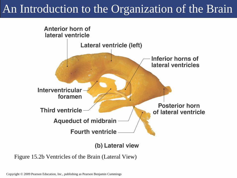
An Introduction to the Organization of the Brain
Figure 15.2b Ventricles of the Brain (Lateral View)
Copyright © 2009 Pearson Education, Inc., publishing as Pearson Benjamin Cummings

An Introduction to the Organization of the Brain
Figure 15.2c Ventricles of the Brain (Anterior View)
Copyright © 2009 Pearson Education, Inc., publishing as Pearson Benjamin Cummings
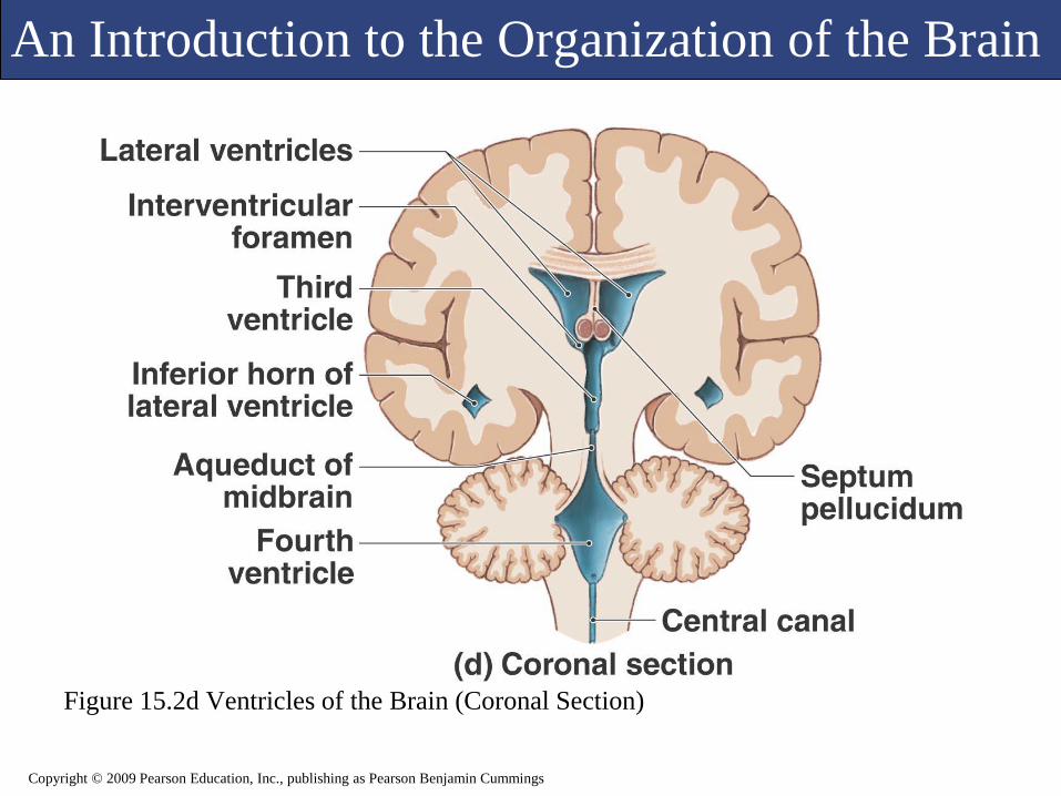
An Introduction to the Organization of the Brain
Figure 15.2d Ventricles of the Brain (Coronal Section)
Copyright © 2009 Pearson Education, Inc., publishing as Pearson Benjamin Cummings
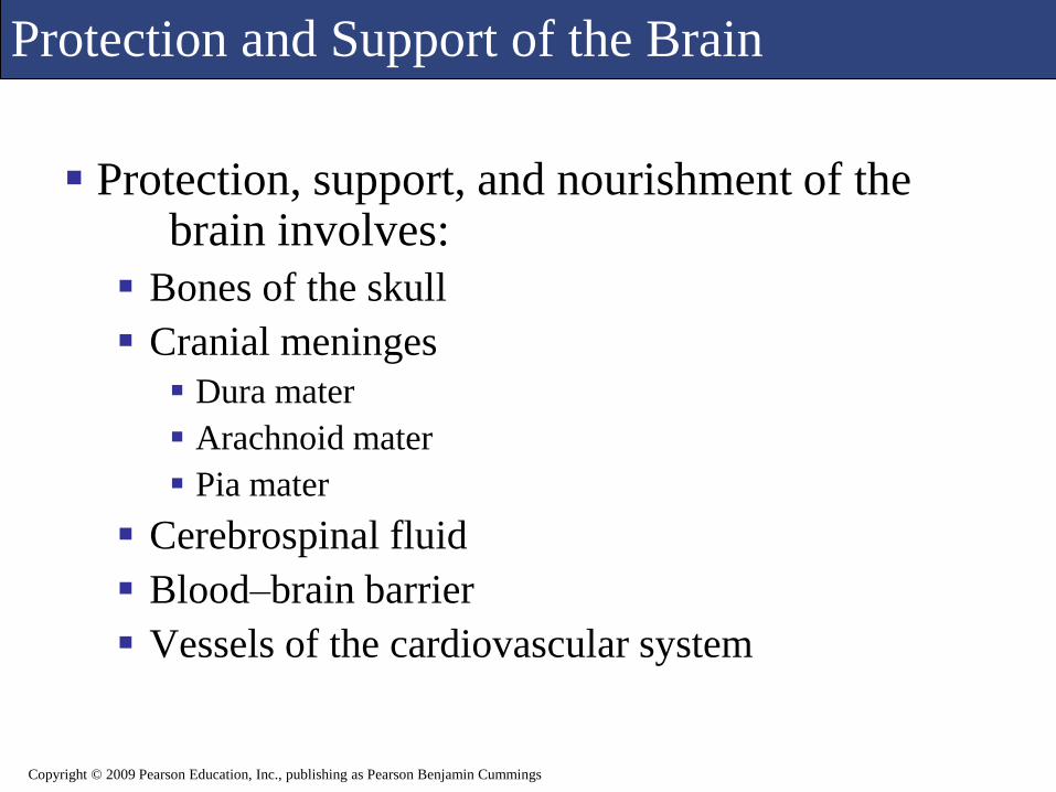
Protection and Support of the Brain
Protection, support, and nourishment of the brain involves:
Bones of the skull
Cranial meninges
Dura mater
Arachnoid mater
Pia mater
Cerebrospinal fluid
Blood–brain barrier
Vessels of the cardiovascular system
Copyright © 2009 Pearson Education, Inc., publishing as Pearson Benjamin Cummings
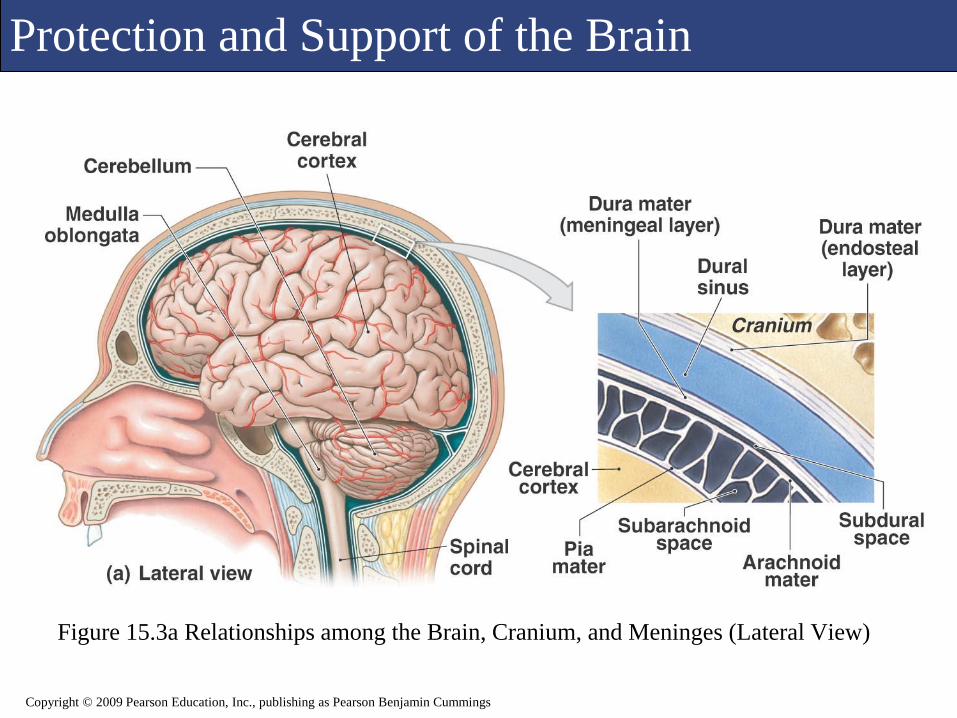
Protection and Support of the Brain
Figure 15.3a Relationships among the Brain, Cranium, and Meninges (Lateral View)
Copyright © 2009 Pearson Education, Inc., publishing as Pearson Benjamin Cummings
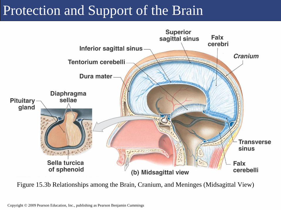
Protection and Support of the Brain
Figure 15.3b Relationships among the Brain, Cranium, and Meninges (Midsagittal View)
Copyright © 2009 Pearson Education, Inc., publishing as Pearson Benjamin Cummings

Protection and Support of the Brain
Figure 15.4a The Cranial Meninges
Copyright © 2009 Pearson Education, Inc., publishing as Pearson Benjamin Cummings
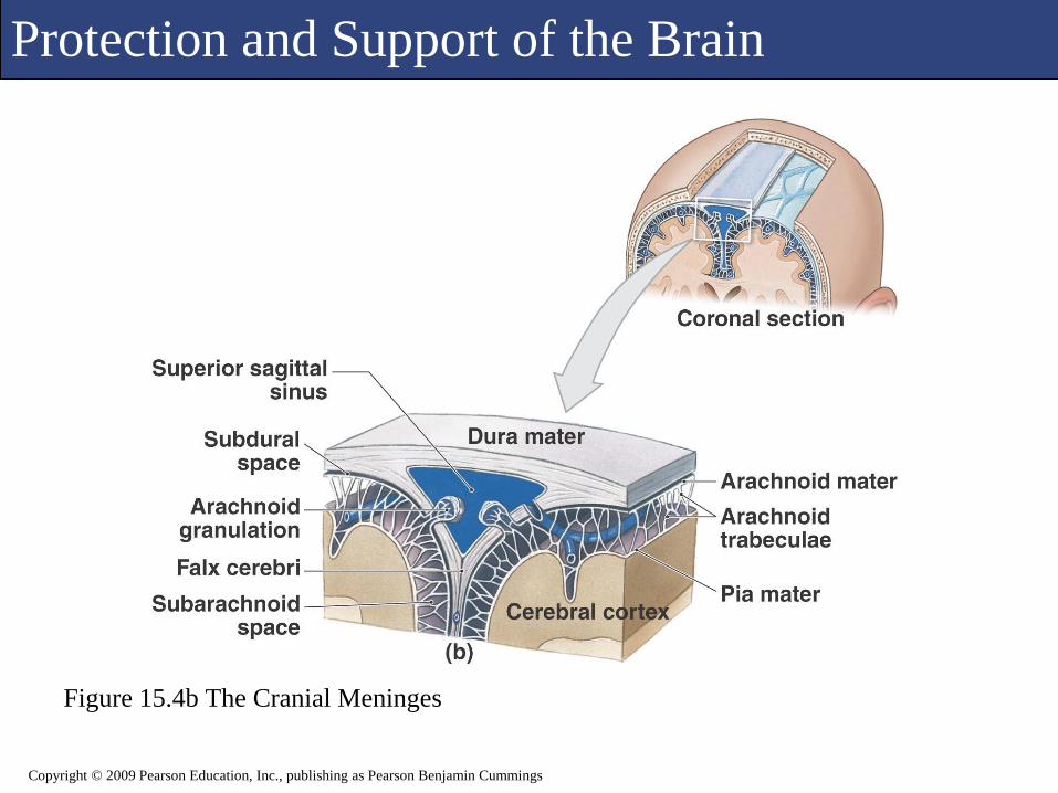
Protection and Support of the Brain
Figure 15.4b The Cranial Meninges
Copyright © 2009 Pearson Education, Inc., publishing as Pearson Benjamin Cummings

Protection and Support of the Brain
Figure 15.4c The Cranial Meninges
Copyright © 2009 Pearson Education, Inc., publishing as Pearson Benjamin Cummings
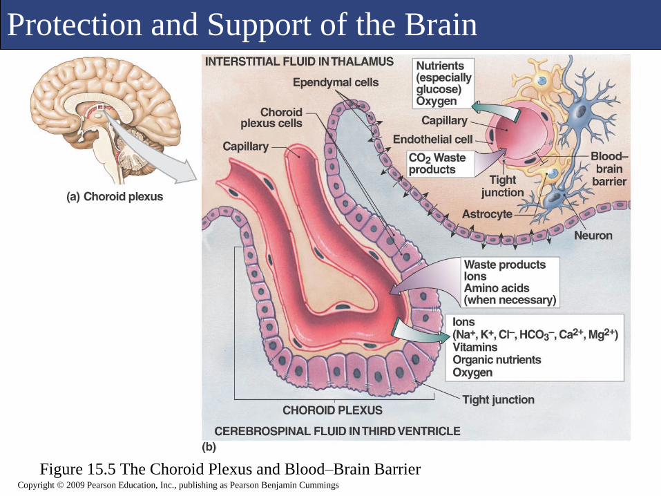
Protection and Support of the Brain
Figure 15.5 The Choroid Plexus and Blood–Brain BarrierCopyright © 2009 Pearson Education, Inc., publishing as Pearson Benjamin Cummings

Protection and Support of the Brain
Figure 15.6 Circulation of Cerebrospinal FluidCopyright © 2009 Pearson Education, Inc., publishing as Pearson Benjamin Cummings

Protection and Support of the Brain Blood Supply to the Brain
Copyright © 2009 Pearson Education, Inc., publishing as Pearson Benjamin Cummings
Figure 22.13a Arteries of the Neck and Head
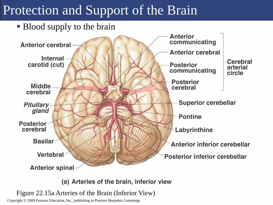
Protection and Support of the Brain Blood supply to the brain
Copyright © 2009 Pearson Education, Inc., publishing as Pearson Benjamin Cummings
Figure 22.15a Arteries of the Brain (Inferior View)

Protection and Support of the Brain Blood supply to the brain
Copyright © 2009 Pearson Education, Inc., publishing as Pearson Benjamin Cummings
Figure 22.22a Major Veins of the Head and Neck (Lateral View)

Protection and Support of the Brain Blood Supply to the Brain
Copyright © 2009 Pearson Education, Inc., publishing as Pearson Benjamin Cummings
Figure 22.22b Venous Drainage of the Brain (Inferior view)
Protection and Support of the Brain
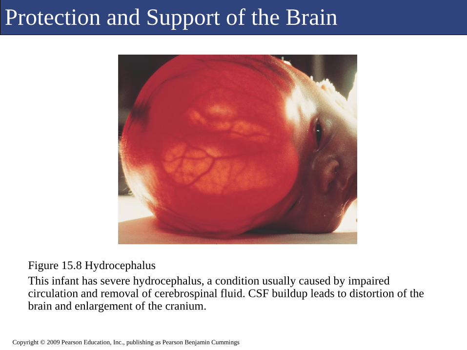
Figure 15.8 Hydrocephalus
This infant has severe hydrocephalus, a condition usually caused by impaired circulation and removal of cerebrospinal fluid. CSF buildup leads to distortion of the brain and enlargement of the cranium.
Copyright © 2009 Pearson Education, Inc., publishing as Pearson Benjamin Cummings

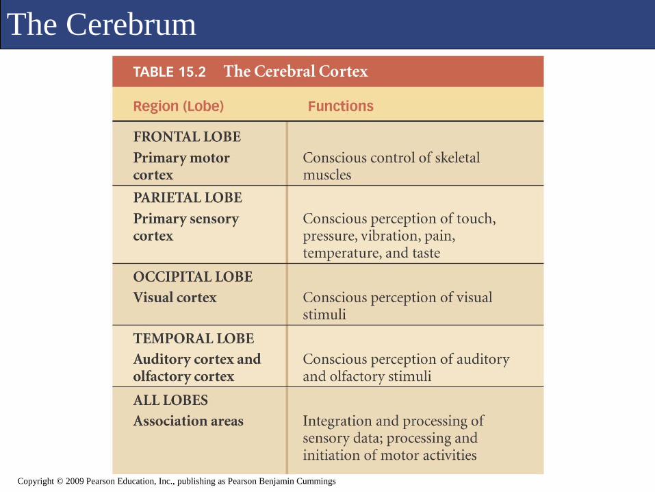
The Cerebrum
The cerebrum is the largest, most superior portion of the human brain.
Each cerebral hemisphere receives sensory information from and generates motor commands to the opposite side of the body.
The two hemispheres have some functional differences, although anatomically they appear to be identical.
Copyright © 2009 Pearson Education, Inc., publishing as Pearson Benjamin Cummings
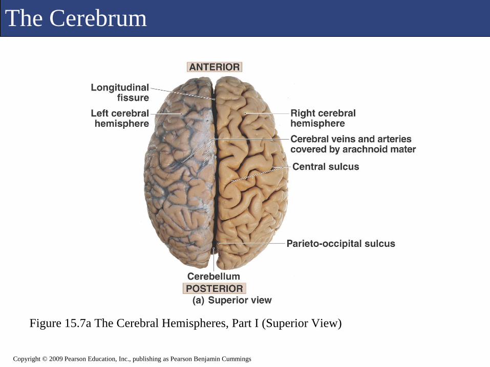
The Cerebrum
Figure 15.7a The Cerebral Hemispheres, Part I (Superior View)
Copyright © 2009 Pearson Education, Inc., publishing as Pearson Benjamin Cummings

The Cerebrum
Figure 15.7b The Cerebral Hemispheres, Part I (Anterior View)
Copyright © 2009 Pearson Education, Inc., publishing as Pearson Benjamin Cummings
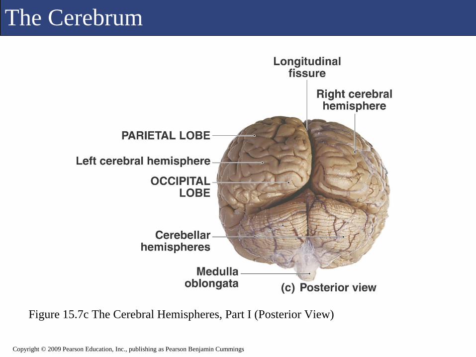
The Cerebrum
Figure 15.7c The Cerebral Hemispheres, Part I (Posterior View)
Copyright © 2009 Pearson Education, Inc., publishing as Pearson Benjamin Cummings

The Cerebrum
Figure 15.9a The Cerebral Hemispheres, Part II (Lateral View of Intact Brain)
Copyright © 2009 Pearson Education, Inc., publishing as Pearson Benjamin Cummings

The Cerebrum
Figure 15.9b The Cerebral Hemispheres, Part II (The Left Cerebral Hemisphere)
Copyright © 2009 Pearson Education, Inc., publishing as Pearson Benjamin Cummings
The Cerebrum
Copyright © 2009 Pearson Education, Inc., publishing as Pearson Benjamin Cummings

The Cerebrum
Figure 15.10a The Central White Matter (Lateral View)
Copyright © 2009 Pearson Education, Inc., publishing as Pearson Benjamin Cummings

The Cerebrum
Figure 15.10b The Central White Matter (Anterior View)
Copyright © 2009 Pearson Education, Inc., publishing as Pearson Benjamin Cummings

The Cerebrum
Copyright © 2009 Pearson Education, Inc., publishing as Pearson Benjamin Cummings
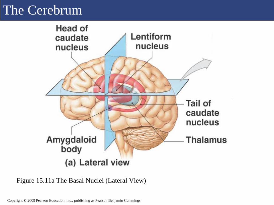
The Cerebrum
Figure 15.11a The Basal Nuclei (Lateral View)
Copyright © 2009 Pearson Education, Inc., publishing as Pearson Benjamin Cummings

The Cerebrum
Figure 15.11b The Basal Nuclei (Horizonatal View, Dissected)
Copyright © 2009 Pearson Education, Inc., publishing as Pearson Benjamin Cummings
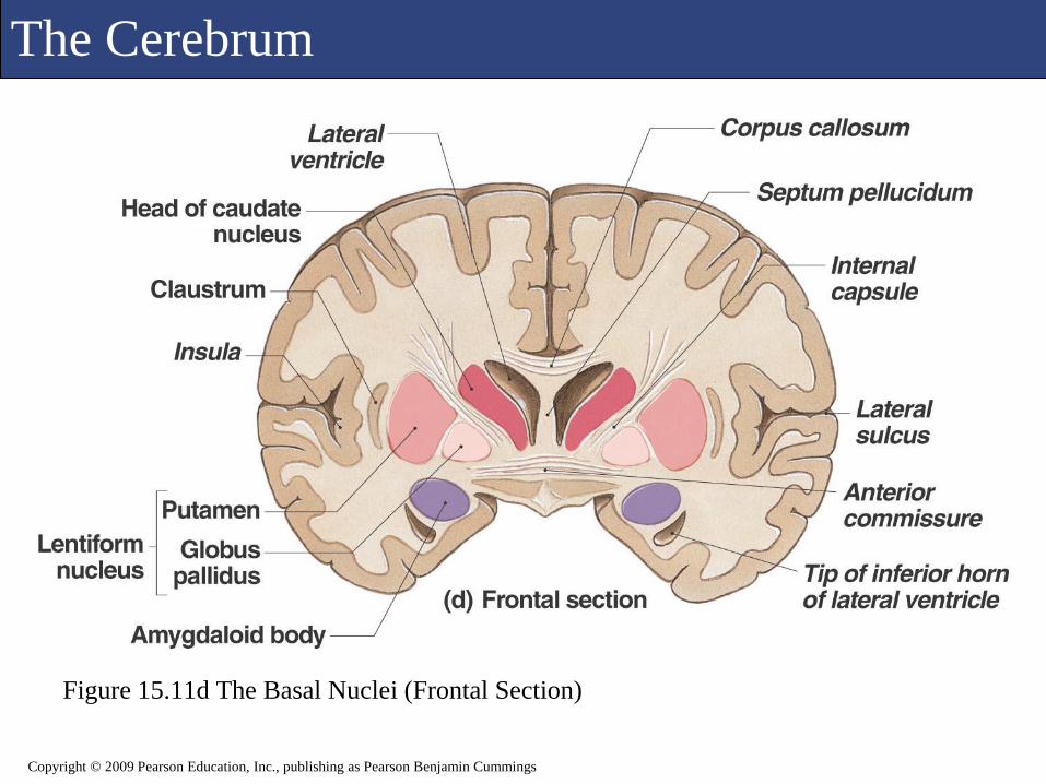
The Cerebrum
Figure 15.11d The Basal Nuclei (Frontal Section)
Copyright © 2009 Pearson Education, Inc., publishing as Pearson Benjamin Cummings

The Cerebrum
Figure 15.11c The Basal Nuclei (Horizontal Section)
Copyright © 2009 Pearson Education, Inc., publishing as Pearson Benjamin Cummings

The Cerebrum
Figure 15.11e The Basal Nuclei (Frontal Section)
Copyright © 2009 Pearson Education, Inc., publishing as Pearson Benjamin Cummings
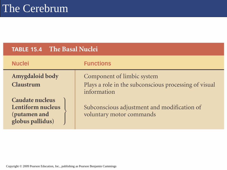
The Cerebrum
Copyright © 2009 Pearson Education, Inc., publishing as Pearson Benjamin Cummings

The Cerebrum
Figure 15.12a The Limbic System
Copyright © 2009 Pearson Education, Inc., publishing as Pearson Benjamin Cummings

The Cerebrum
Figure 15.12b The Limbic System
Copyright © 2009 Pearson Education, Inc., publishing as Pearson Benjamin Cummings

The Cerebrum
Copyright © 2009 Pearson Education, Inc., publishing as Pearson Benjamin Cummings
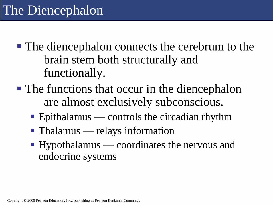
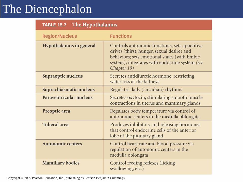
The Diencephalon
The diencephalon connects the cerebrum to the brain stem both structurally and functionally.
The functions that occur in the diencephalon are almost exclusively subconscious.
Epithalamus — controls the circadian rhythm
Thalamus — relays information
Hypothalamus — coordinates the nervous and endocrine systems
Copyright © 2009 Pearson Education, Inc., publishing as Pearson Benjamin Cummings

The Diencephalon
Figure 15.15a Sectional Views of the Brain (Midsagittal Section)
Copyright © 2009 Pearson Education, Inc., publishing as Pearson Benjamin Cummings
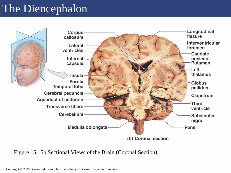
The Diencephalon
Figure 15.15b Sectional Views of the Brain (Coronal Section)
Copyright © 2009 Pearson Education, Inc., publishing as Pearson Benjamin Cummings
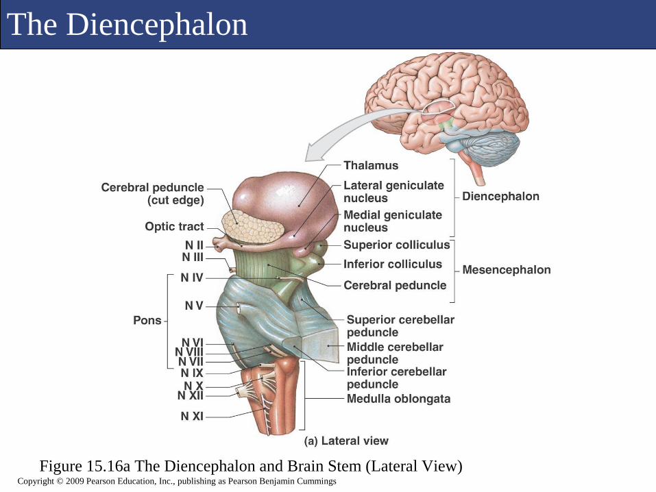
The Diencephalon
Figure 15.16a The Diencephalon and Brain Stem (Lateral View)Copyright © 2009 Pearson Education, Inc., publishing as Pearson Benjamin Cummings
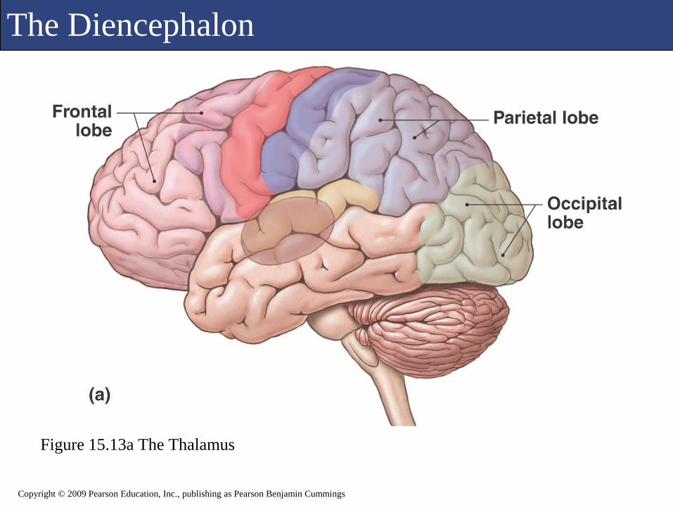
The Diencephalon
Figure 15.13a The Thalamus
Copyright © 2009 Pearson Education, Inc., publishing as Pearson Benjamin Cummings
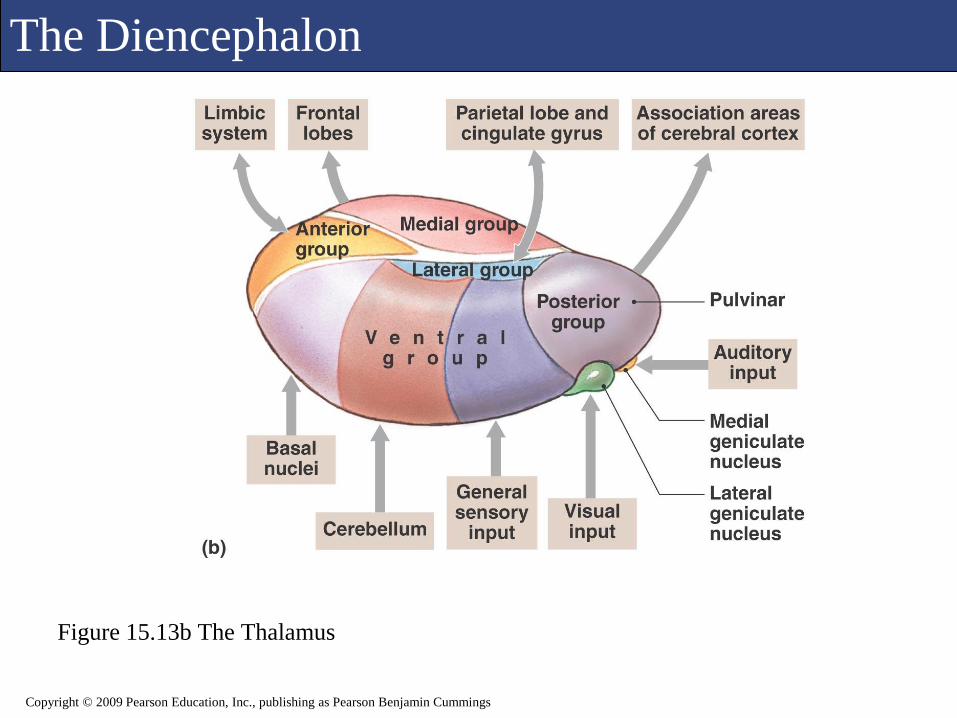
The Diencephalon
Figure 15.13b The Thalamus
Copyright © 2009 Pearson Education, Inc., publishing as Pearson Benjamin Cummings

The Diencephalon
Copyright © 2009 Pearson Education, Inc., publishing as Pearson Benjamin Cummings
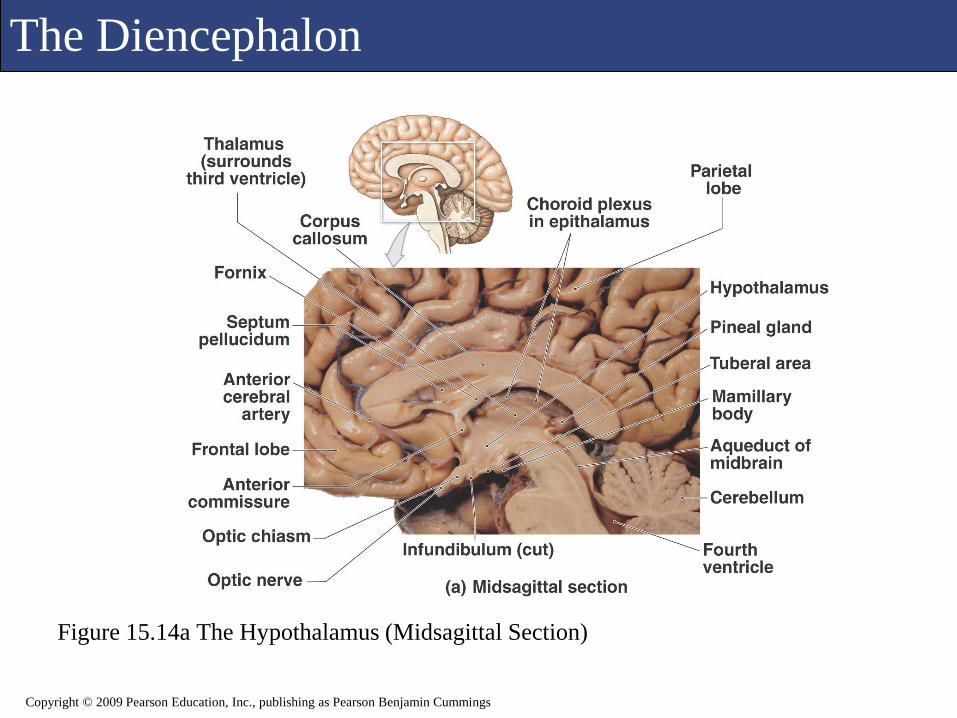
The Diencephalon
Figure 15.14a The Hypothalamus (Midsagittal Section)
Copyright © 2009 Pearson Education, Inc., publishing as Pearson Benjamin Cummings

The Diencephalon
Figure 15.14b The Hypothalamus
Copyright © 2009 Pearson Education, Inc., publishing as Pearson Benjamin Cummings
The Diencephalon
Copyright © 2009 Pearson Education, Inc., publishing as Pearson Benjamin Cummings

The Mesencephalon
The mesencephalon, or midbrain, is the most
superior portion of the brain stem.
Nuclei coordinate visual and auditory reflexes.
Corpora quadregemina
Superior colliculi — visual
Inferior colliculi — auditory
Limbic system nuclei
Coordinate involuntary movements of skeletal muscles
Cerebral peduncles
Nerve bundles to and from the brain/spinal cord
Copyright © 2009 Pearson Education, Inc., publishing as Pearson Benjamin Cummings
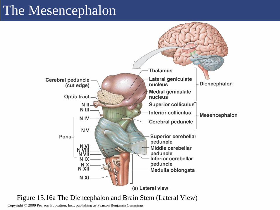
The Mesencephalon
Figure 15.16a The Diencephalon and Brain Stem (Lateral View)Copyright © 2009 Pearson Education, Inc., publishing as Pearson Benjamin Cummings

The Mesencephalon
Figure 15.16b The Diencephalon and Brain Stem (Sagittal Section)Copyright © 2009 Pearson Education, Inc., publishing as Pearson Benjamin Cummings
The Mesencephalon
Figure 15.16c The Diencephalon and Brain Stem (Posterior View)
Copyright © 2009 Pearson Education, Inc., publishing as Pearson Benjamin Cummings
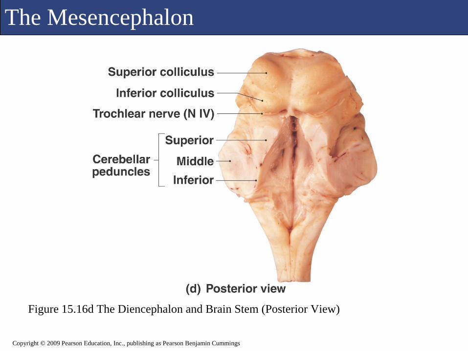
The Mesencephalon
Figure 15.16d The Diencephalon and Brain Stem (Posterior View)
Copyright © 2009 Pearson Education, Inc., publishing as Pearson Benjamin Cummings
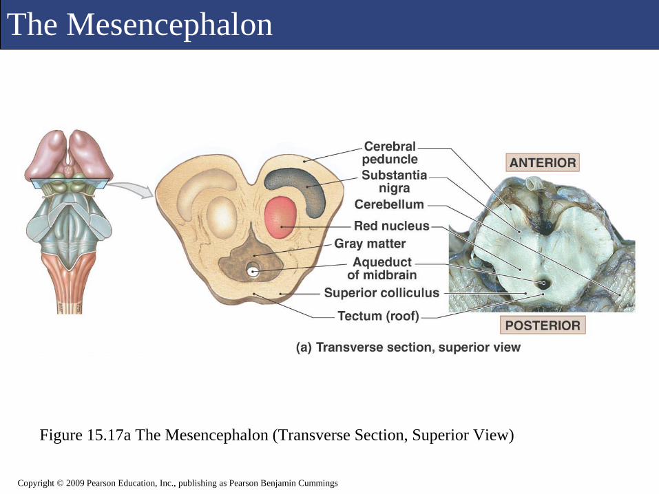
The Mesencephalon
Figure 15.17a The Mesencephalon (Transverse Section, Superior View)
Copyright © 2009 Pearson Education, Inc., publishing as Pearson Benjamin Cummings
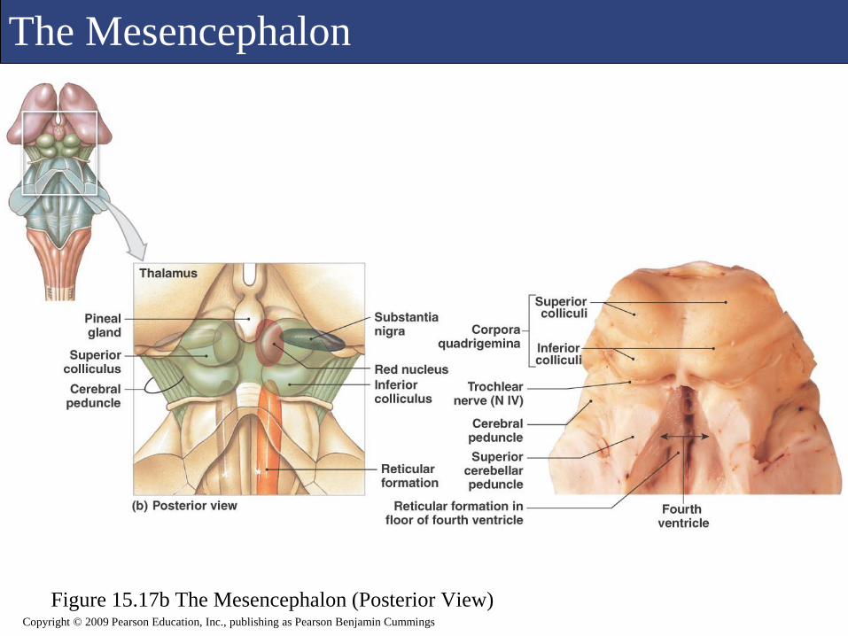
The Mesencephalon
Figure 15.17b The Mesencephalon (Posterior View)Copyright © 2009 Pearson Education, Inc., publishing as Pearson Benjamin Cummings

The Mesencephalon
Copyright © 2009 Pearson Education, Inc., publishing as Pearson Benjamin Cummings
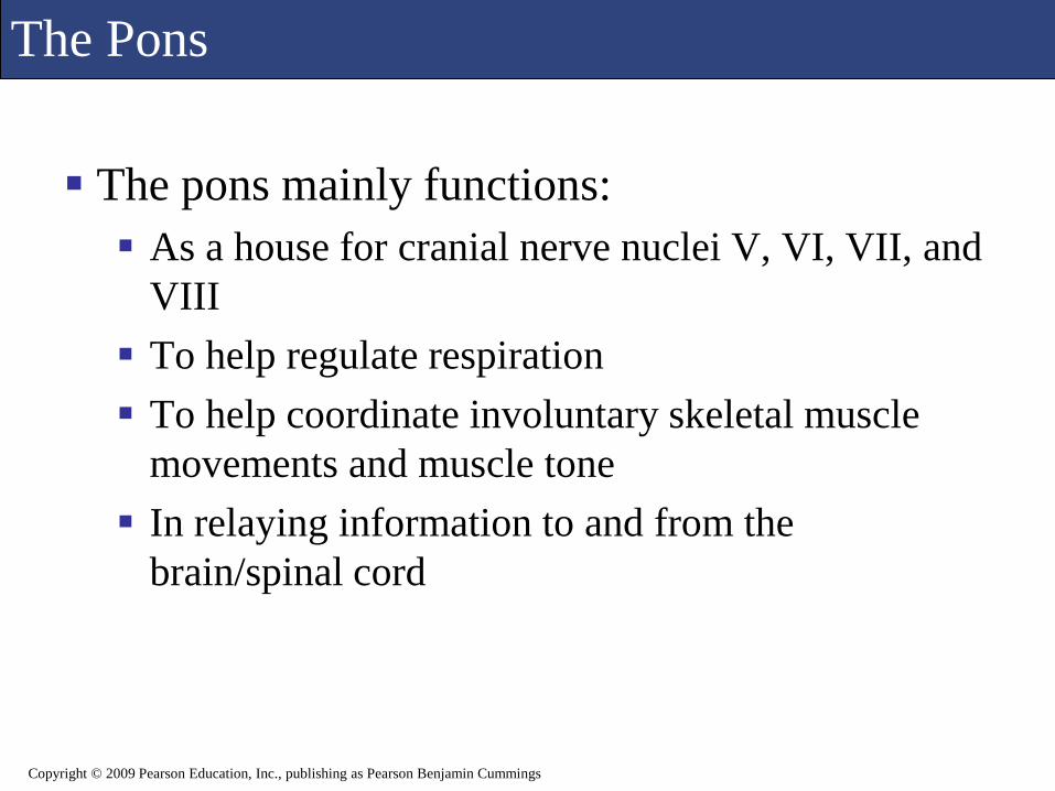
The Pons
The pons mainly functions:
As a house for cranial nerve nuclei V, VI, VII, and
VIII
To help regulate respiration
To help coordinate involuntary skeletal muscle
movements and muscle tone
In relaying information to and from the
brain/spinal cord
Copyright © 2009 Pearson Education, Inc., publishing as Pearson Benjamin Cummings
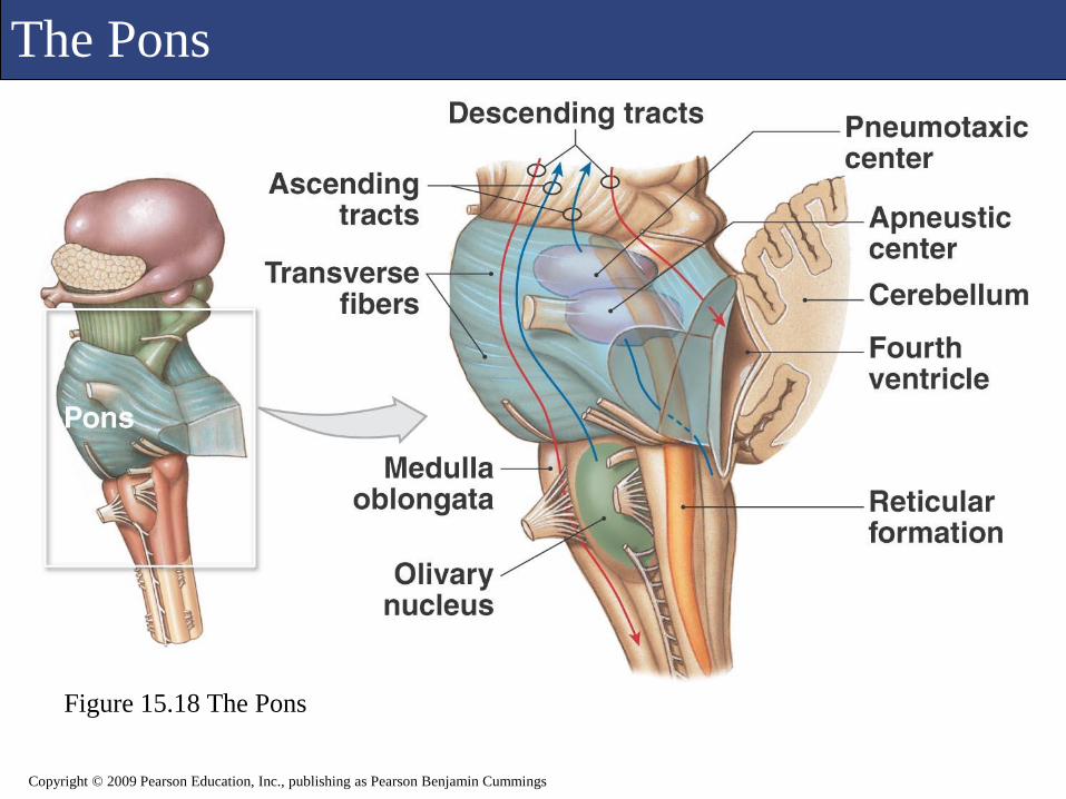
The Pons
Figure 15.18 The Pons
Copyright © 2009 Pearson Education, Inc., publishing as Pearson Benjamin Cummings

The Pons
Copyright © 2009 Pearson Education, Inc., publishing as Pearson Benjamin Cummings
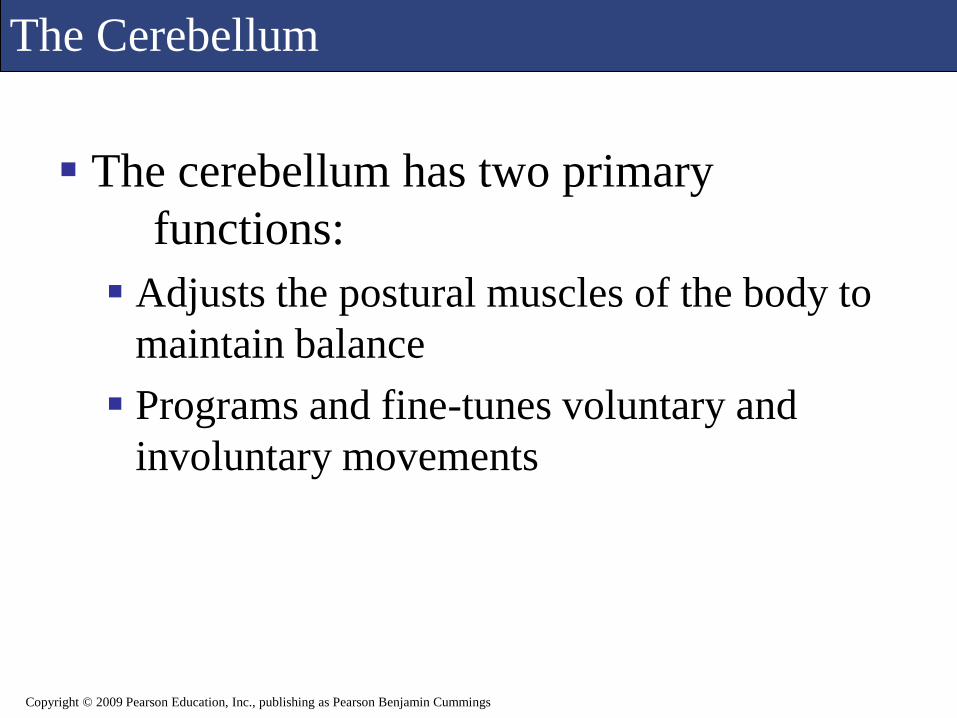
The Cerebellum
The cerebellum has two primary
functions:
Adjusts the postural muscles of the body to
maintain balance
Programs and fine-tunes voluntary and
involuntary movements
Copyright © 2009 Pearson Education, Inc., publishing as Pearson Benjamin Cummings

The Cerebellum
Figure 15.19a The Cerebellum (Posterior, Superior Surface)
Copyright © 2009 Pearson Education, Inc., publishing as Pearson Benjamin Cummings
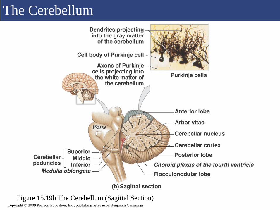
The Cerebellum
Figure 15.19b The Cerebellum (Sagittal Section)Copyright © 2009 Pearson Education, Inc., publishing as Pearson Benjamin Cummings

The Cerebellum
Copyright © 2009 Pearson Education, Inc., publishing as Pearson Benjamin Cummings

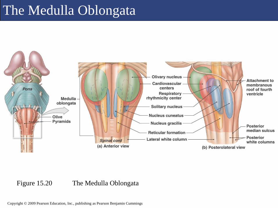
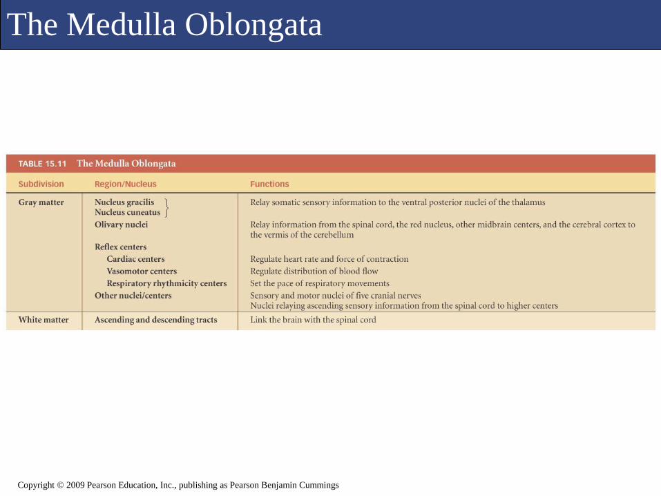
The Medulla Oblongata
The medulla oblongata physically connects the
brain with the spinal cord.
It is so important that, if it is severely
compromised, the victim will likely die.
The medulla oblongata is a relay station, house
for cranial nerve nuclei, and most
importantly, controls visceral functions like
blood pressure, breathing, and heart rate.
Copyright © 2009 Pearson Education, Inc., publishing as Pearson Benjamin Cummings
The Medulla Oblongata
Figure 15.20 The Medulla Oblongata
Copyright © 2009 Pearson Education, Inc., publishing as Pearson Benjamin Cummings
The Medulla Oblongata
Copyright © 2009 Pearson Education, Inc., publishing as Pearson Benjamin Cummings
M
Copyright © 2009 Pearson Education, Inc., publishing as Pearson Benjamin Cummings
Brain Animation Review
The Brain
The Cranial Nerves
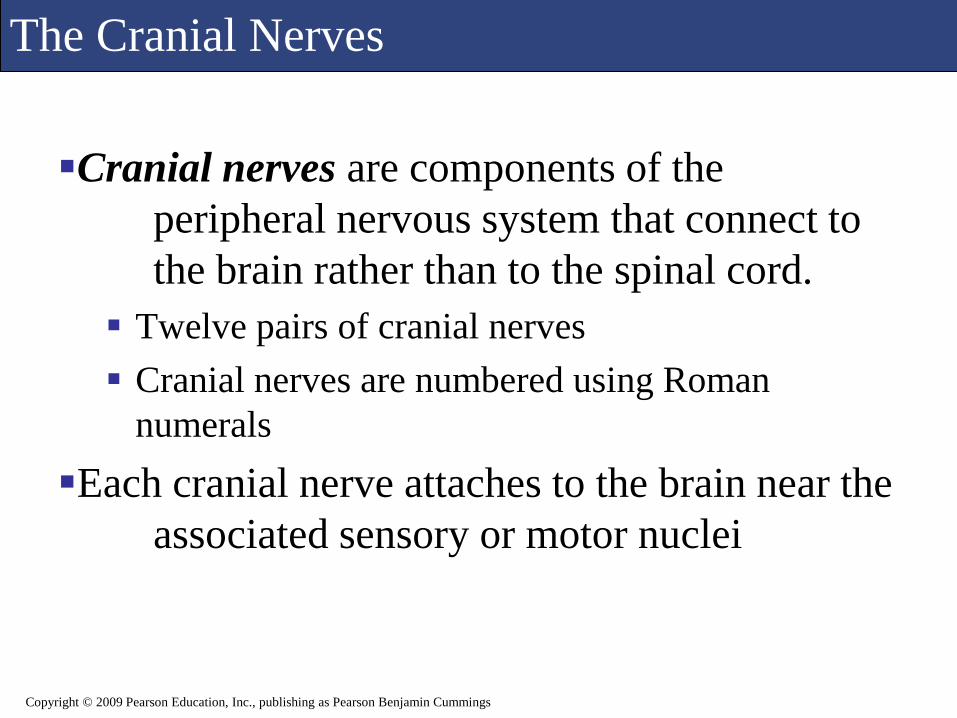
Cranial nerves are components of the
peripheral nervous system that connect to
the brain rather than to the spinal cord.
Twelve pairs of cranial nerves
Cranial nerves are numbered using Roman
numerals
Each cranial nerve attaches to the brain near the
associated sensory or motor nuclei
Copyright © 2009 Pearson Education, Inc., publishing as Pearson Benjamin Cummings

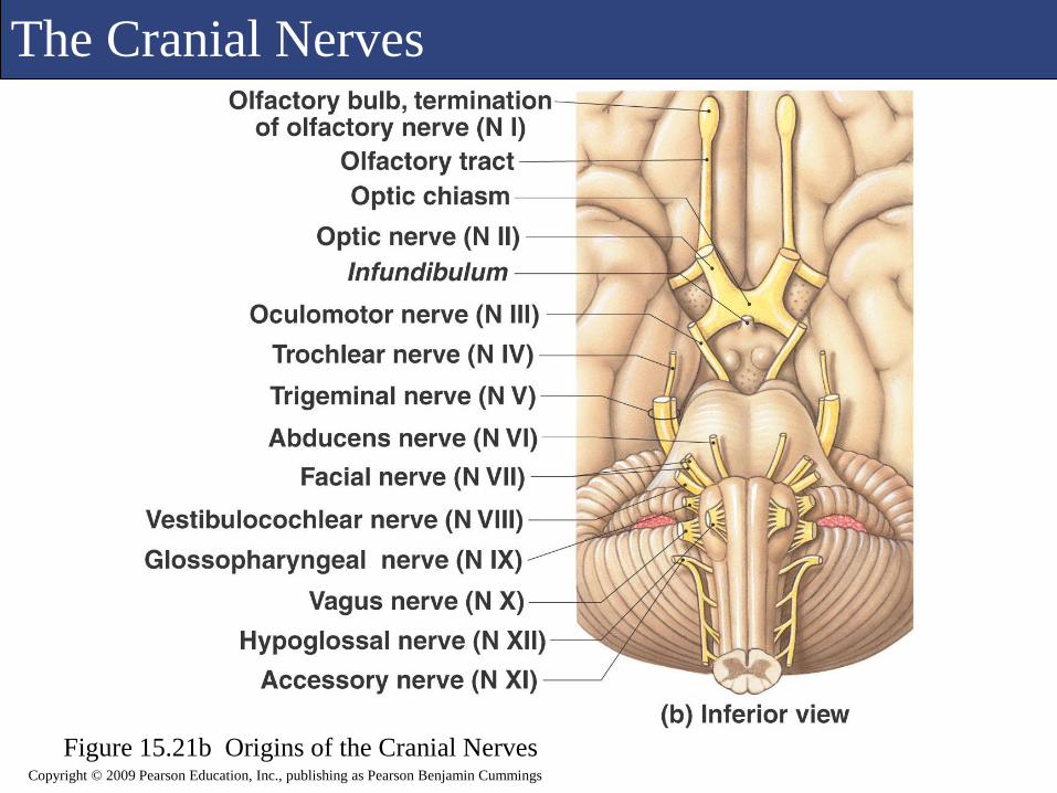
The Cranial Nerves
Figure 15.21a Origins of the Cranial NervesCopyright © 2009 Pearson Education, Inc., publishing as Pearson Benjamin Cummings
The Cranial Nerves
Copyright © 2009 Pearson Education, Inc., publishing as Pearson Benjamin Cummings
Figure 15.21b Origins of the Cranial Nerves
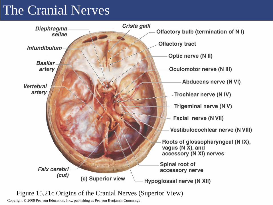
The Cranial Nerves
Figure 15.21c Origins of the Cranial Nerves (Superior View)Copyright © 2009 Pearson Education, Inc., publishing as Pearson Benjamin Cummings
The Cranial Nerves
Copyright © 2009 Pearson Education, Inc., publishing as Pearson Benjamin Cummings
The Cranial Nerves
Copyright © 2009 Pearson Education, Inc., publishing as Pearson Benjamin Cummings

The Cranial Nerves
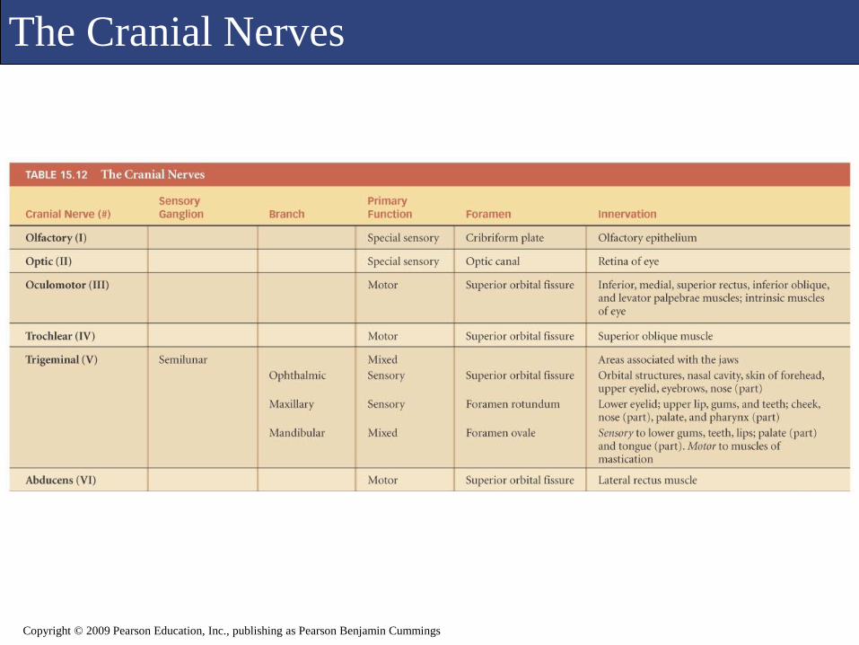
Olfactory Nerve (N I)
Primary function: special sensory (smell)
Origin: receptors of olfactory epithelium
Passes through: cribriform plate of ethmoid
Destination: olfactory bulbs
Copyright © 2009 Pearson Education, Inc., publishing as Pearson Benjamin Cummings
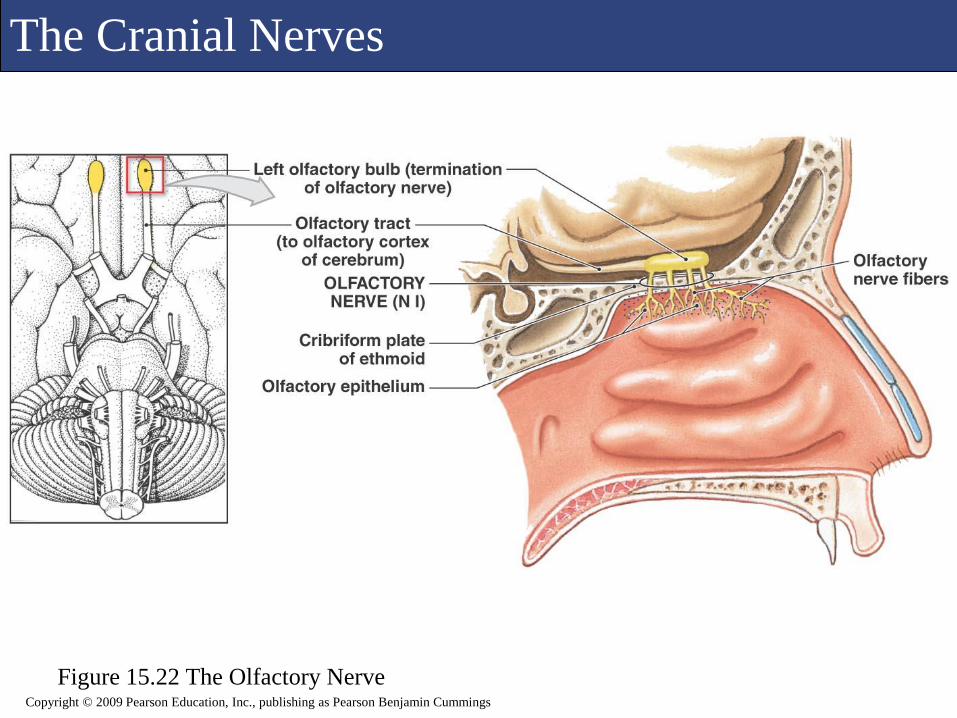
The Cranial Nerves
Figure 15.22 The Olfactory NerveCopyright © 2009 Pearson Education, Inc., publishing as Pearson Benjamin Cummings
The Cranial Nerves
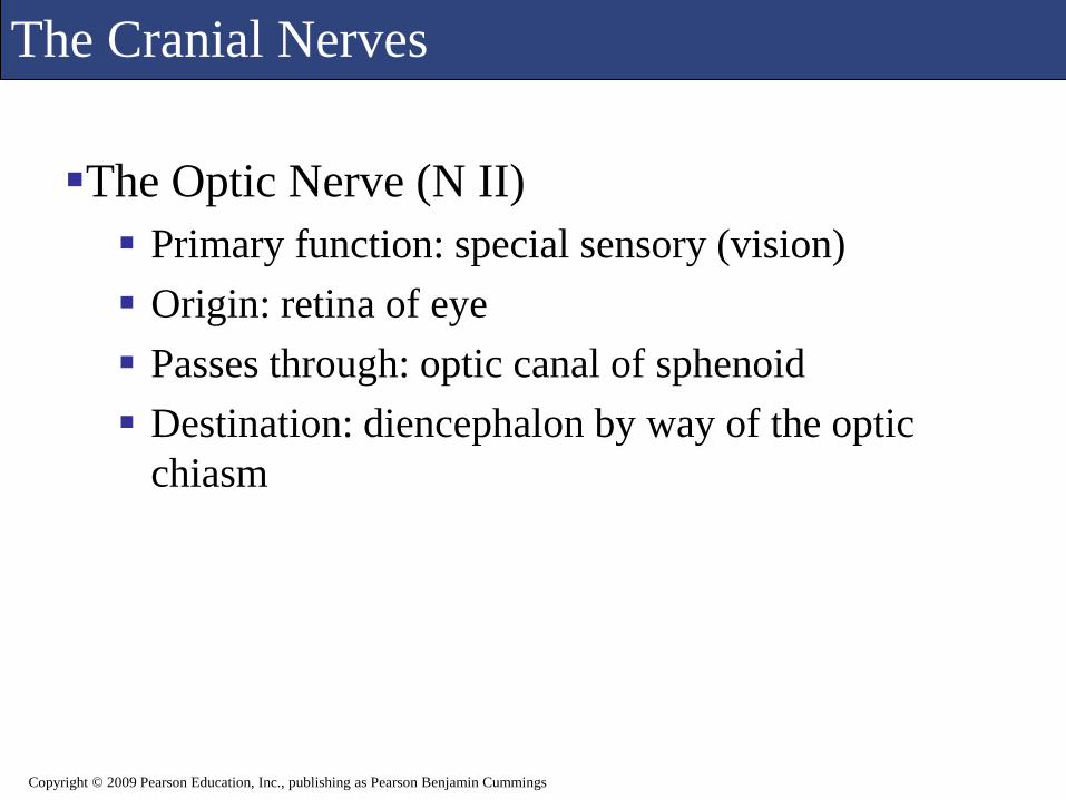
The Optic Nerve (N II)
Primary function: special sensory (vision)
Origin: retina of eye
Passes through: optic canal of sphenoid
Destination: diencephalon by way of the optic
chiasm
Copyright © 2009 Pearson Education, Inc., publishing as Pearson Benjamin Cummings

The Cranial Nerves
Figure 15.23 The Optic NerveCopyright © 2009 Pearson Education, Inc., publishing as Pearson Benjamin Cummings

The Cranial Nerves
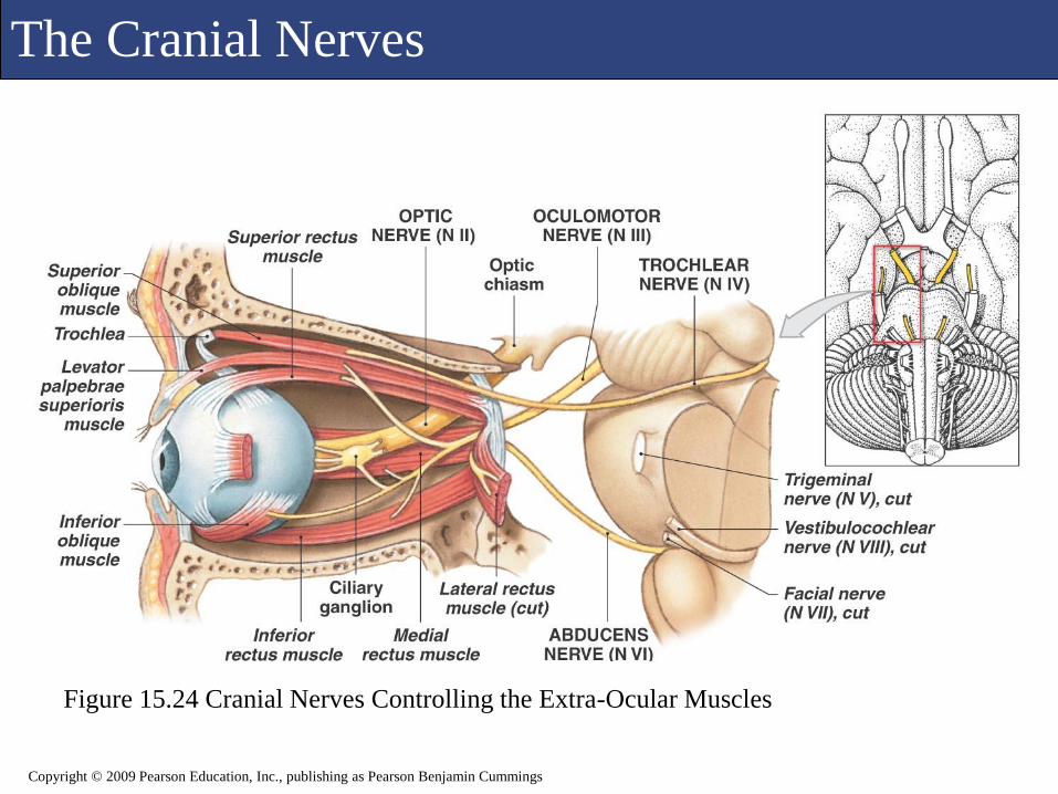
The Oculomotor Nerve (N III)
Primary function: motor, eye movements
Origin: mesencephalon
Passes through: superior orbital fissure of sphenoid
Destination:
Somatic motor: superior, inferior, and medial rectus muscles; the inferior oblique muscle; the levator palpebrae superioris muscle
Visceral motor: intrinsic eye muscles
Copyright © 2009 Pearson Education, Inc., publishing as Pearson Benjamin Cummings
The Cranial Nerves
Figure 15.24 Cranial Nerves Controlling the Extra-Ocular Muscles
Copyright © 2009 Pearson Education, Inc., publishing as Pearson Benjamin Cummings
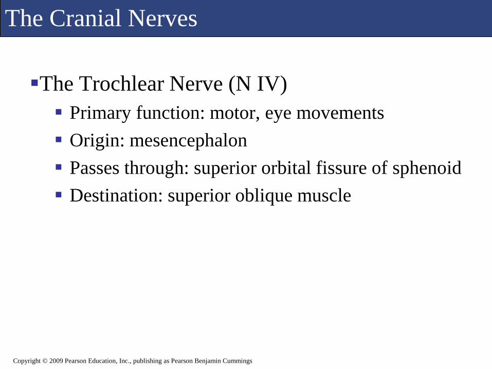
The Cranial Nerves
The Trochlear Nerve (N IV)
Primary function: motor, eye movements
Origin: mesencephalon
Passes through: superior orbital fissure of sphenoid
Destination: superior oblique muscle
Copyright © 2009 Pearson Education, Inc., publishing as Pearson Benjamin Cummings

The Cranial Nerves
Figure 15.24 Cranial Nerves Controlling the Extra-Ocular Muscles
Copyright © 2009 Pearson Education, Inc., publishing as Pearson Benjamin Cummings

The Cranial Nerves
The Trigeminal Nerve (N V) Primary function: Mixed (sensory and motor)
Ophthalmic and maxillary branches sensory
Mandibular branch mixed
Origin: Ophthalmic branch (sensory): orbital structures, nasal
cavity, skin of forehead, superior eyelid, eyebrow, and part of the nose
Maxillary branch (sensory): inferior eyelid, upper lip, gums, and teeth; cheek; nose, palate, and part of the pharynx
Mandibular branch (mixed): sensory from lower gums, teeth, and lips; palate and tongue (part); motor from motor nuclei of pons
Copyright © 2009 Pearson Education, Inc., publishing as Pearson Benjamin Cummings

The Cranial Nerves
The Trigeminal Nerve (N V)
Passes through:
Ophthalmic branch through superior orbital fissure
Maxillary branch through foramen rotundum
Mandibular branch through foramen ovale
Destination:
Ophthalmic, maxillary, and mandibular branches to
sensory nuclei in the pons
Mandibular branch also innervates muscles of
mastication
Copyright © 2009 Pearson Education, Inc., publishing as Pearson Benjamin Cummings

The Cranial Nerves
Figure 15.25 The Trigeminal Nerve
Copyright © 2009 Pearson Education, Inc., publishing as Pearson Benjamin Cummings
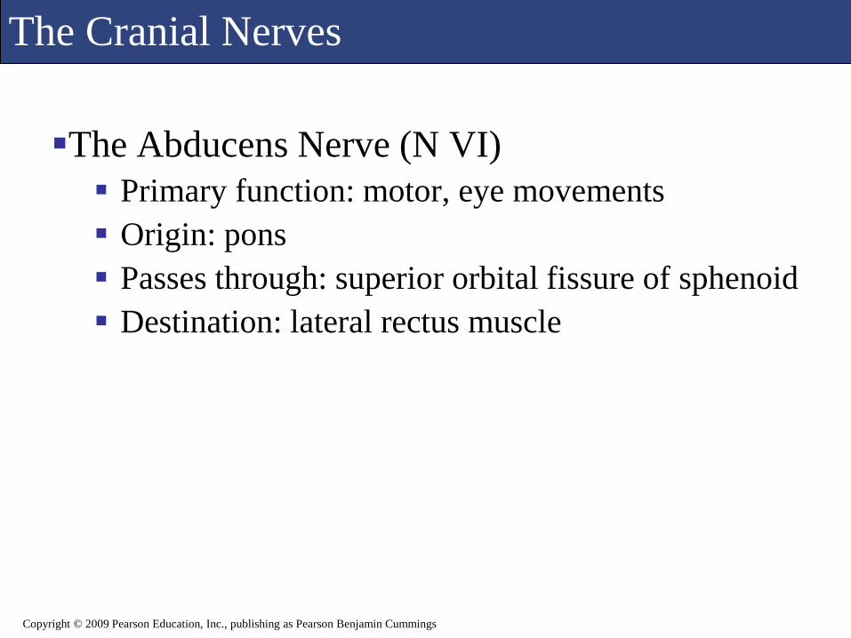
The Cranial Nerves
The Abducens Nerve (N VI)
Primary function: motor, eye movements
Origin: pons
Passes through: superior orbital fissure of sphenoid
Destination: lateral rectus muscle
Copyright © 2009 Pearson Education, Inc., publishing as Pearson Benjamin Cummings

The Cranial Nerves
Figure 15.24 Cranial Nerves Controlling the Extra-Ocular Muscles
Copyright © 2009 Pearson Education, Inc., publishing as Pearson Benjamin Cummings

The Cranial Nerves
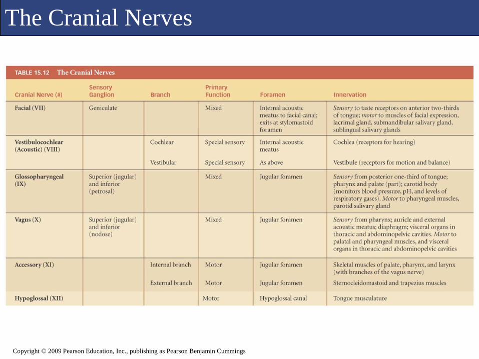
The Facial Nerve (N VII) Primary function: mixed (sensory and motor)
Origin:
Sensory from taste receptors on anterior two thirds of tongue
Motor from motor nuclei of pons
Passes through: internal acoustic meatus of temporal bone,
along facial canal to reach stylomastoid foramen
Destination:
Sensory to sensory nuclei of pons
Somatic motor: muscles of facial expression
Visceral motor: lacrimal (tear) gland and nasal mucous glands via
pterygopalatine ganglion; submandibular and sublingual salivary
glands via submandibular ganglion
Copyright © 2009 Pearson Education, Inc., publishing as Pearson Benjamin Cummings

The Cranial Nerves
Figure 15.26a The Facial NerveCopyright © 2009 Pearson Education, Inc., publishing as Pearson Benjamin Cummings

The Cranial Nerves
Copyright © 2009 Pearson Education, Inc., publishing as Pearson Benjamin Cummings
Figure 15.26b The Facial Nerve
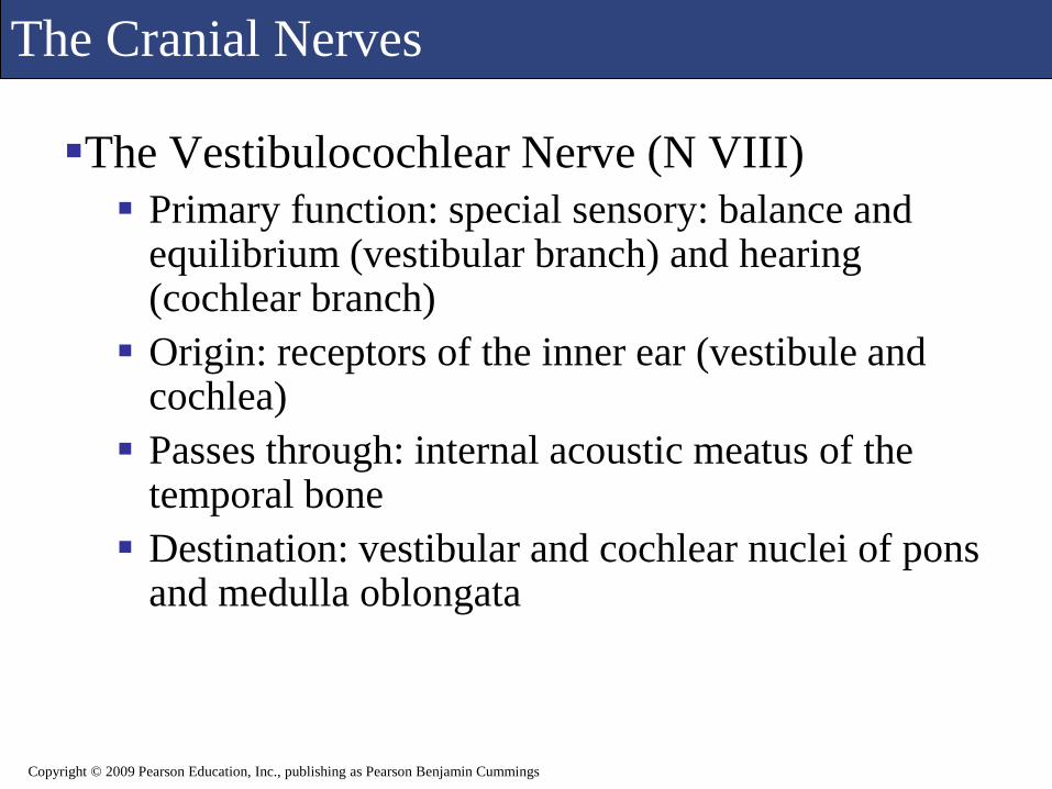
The Cranial Nerves
The Vestibulocochlear Nerve (N VIII)
Primary function: special sensory: balance and equilibrium (vestibular branch) and hearing (cochlear branch)
Origin: receptors of the inner ear (vestibule and cochlea)
Passes through: internal acoustic meatus of the temporal bone
Destination: vestibular and cochlear nuclei of pons and medulla oblongata
Copyright © 2009 Pearson Education, Inc., publishing as Pearson Benjamin Cummings

The Cranial Nerves
Figure 15.27 The Vestibulocochlear Nerve
Copyright © 2009 Pearson Education, Inc., publishing as Pearson Benjamin Cummings

The Cranial Nerves
The Glossopharyngeal Nerve (N IX) Primary function: mixed (sensory and motor)
Origin:
Sensory from posterior one third of the tongue, part of the pharynx
and palate, the carotid arteries of the neck
Motor from motor nuclei of medulla oblongata
Passes through: jugular foramen between occipital and
temporal bones
Destination:
Sensory fibers to sensory nuclei of medulla oblongata
Somatic motor: pharyngeal muscles involved in swallowing
Visceral motor: parotid salivary gland, after synapsing in the otic
ganglion
Copyright © 2009 Pearson Education, Inc., publishing as Pearson Benjamin Cummings
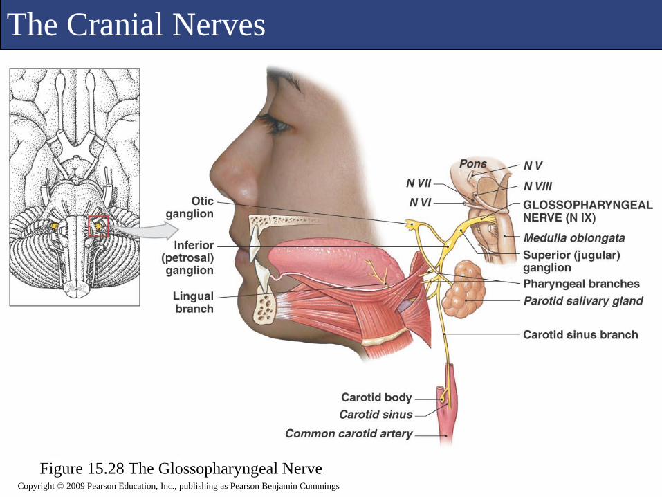
The Cranial Nerves
Figure 15.28 The Glossopharyngeal NerveCopyright © 2009 Pearson Education, Inc., publishing as Pearson Benjamin Cummings

The Cranial Nerves
The Vagus Nerve (N X) Primary function: mixed (sensory and motor)
Origin:
Visceral sensory from pharynx (part), auricle, external acoustic meatus, diaphragm, and visceral organs in thoracic and abdominopelvic cavities
Visceral motor from motor nuclei in the medulla oblongata
Passes through: jugular foramen between occipital and temporal bones
Destination:
Sensory fibers to sensory nuclei and autonomic centers of medulla oblongata
Somatic motor to muscles of the palate and pharynx
Visceral motor to respiratory, cardiovascular, and digestive organs in the thoracic and abdominal cavities.
Copyright © 2009 Pearson Education, Inc., publishing as Pearson Benjamin Cummings

The Cranial Nerves
Figure 15.29 The Vagus NerveCopyright © 2009 Pearson Education, Inc., publishing as Pearson Benjamin Cummings

The Cranial Nerves
The Accessory Nerve (N XI)
Primary function: motor
Origin: motor nuclei of spinal cord and medulla oblongata
Passes through: jugular foramen between occipital and temporal bones
Destination:
Internal branch innervates voluntary muscles of palate, pharynx, and larynx
External branch controls sternocleidomastoid and trapezius muscles
Copyright © 2009 Pearson Education, Inc., publishing as Pearson Benjamin Cummings
The Cranial Nerves
The Hypoglossal Nerve (XII)
Primary function: motor, tongue movements
Origin: motor nuclei of the medulla oblongata
Passes through: hypoglossal canal of occipital
bone
Destination: muscles of the tongue
Copyright © 2009 Pearson Education, Inc., publishing as Pearson Benjamin Cummings

The Cranial Nerves
Figure 15.30 The Accessory and Hypoglossal Nerves
Copyright © 2009 Pearson Education, Inc., publishing as Pearson Benjamin Cummings
The Cranial Nerves
Copyright © 2009 Pearson Education, Inc., publishing as Pearson Benjamin Cummings

![14 [chapter 14 the brain and cranial nerves]](https://static.fdocuments.us/doc/165x107/5a6496117f8b9a2c568b5ff1/14-chapter-14-the-brain-and-cranial-nerves.jpg)