The BMP pathway acts to directly regulate Tbx20 in the developing ...
11
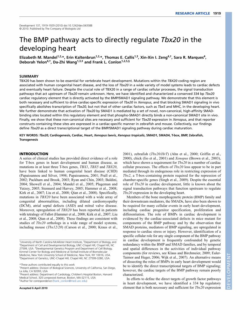
1919 RESEARCH ARTICLE INTRODUCTION A series of clinical studies has provided direct evidence of a role for T-box genes in heart development and human disease, as mutations in at least three T-box genes, TBX1, TBX5 and TBX20, have been linked to human congenital heart disease (CHD) (Papaioannou and Silver, 1998; Papaioannou, 2001; Prall et al., 2002; Packham and Brook, 2003; Ryan and Chin, 2003; Baldini, 2004; Showell et al., 2004; Mandel et al., 2005; Plageman and Yutzey, 2005; Stennard and Harvey, 2005; Hammer et al., 2008; Kirk et al., 2007; Liu et al., 2008; Qian et al., 2008). Specifically, mutations in TBX20 have been associated with a wide array of congenital abnormalities, including dilated cardiomyopathy (DCM), atrial septal defects (ASD) and mitral valve disease. Moreover, upregulation of TBX20 has been reported in patients with tetralogy of Fallot (Hammer et al., 2008; Kirk et al., 2007; Liu et al., 2008; Qian et al., 2008). These findings are consistent with studies of Tbx20 orthologs in a wide range of model systems, including mouse (Tbx12/20) (Carson et al., 2000; Kraus et al., 2001), zebrafish (Tbx20/HrT) (Ahn et al., 2000; Griffin et al., 2000), chick (Iio et al., 2001) and Xenopus (Brown et al., 2003), which have shown a requirement for Tbx20 in a number of cardiac cellular processes. The effects of Tbx20 loss appear to be in part mediated through its endogenous role in restricting expression of Tbx2, a T-box-containing protein required for the repression of chamber-specific genes (Singh et al., 2009). Despite the essential role of Tbx20 in cardiac development, little is known about the signal transduction pathways that function upstream to regulate Tbx20 expression in the developing heart. Members of the bone morphogenetic protein (BMP) family and their downstream mediators, the SMADs, have also been shown to be required for many cellular events in early heart development, including cardiac progenitor specification, proliferation and differentiation. The role of BMPs in cardiac development is evidenced by the cardiac-associated defects in mice mutant for components of the BMP pathway and by the observation that SMAD proteins, mediators of BMP signaling, are upregulated in response to cardiac stress or injury. However, identification of a specific cellular role for any single component of the BMP pathway in cardiac development is frequently confounded by genetic redundancy within the BMP and SMAD families, and by temporal and spatial differences in the activities of individual pathway components (for reviews, see Klaus and Birchmeier, 2009; Euler- Taimor and Heger, 2006; Wijk et al., 2007). An alternative means of dissecting the roles of BMPs in early heart development would be to identify the direct transcriptional targets of BMP signaling; however, the cardiac targets of the BMP pathway remain poorly characterized. In efforts to define the direct targets of growth factor pathways in heart development, we have identified a 334 bp regulatory element that is both necessary and sufficient for Tbx20 expression Development 137, 1919-1929 (2010) doi:10.1242/dev.043588 © 2010. Published by The Company of Biologists Ltd 1 University of North Carolina McAllister Heart Institute, 2 Department of Biology, and 3 Department of Cell and Developmental Biology, UNC-Chapel Hill, Chapel Hill, NC 27599, USA. 4 Developmental Genetics Program and Department of Cell Biology, Kimmel Center for Biology and Medicine at Skirball Institute of Biomolecular Medicine, New York University School of Medicine, New York, NY 10016, USA. 5 Department of Genetics, UNC-Chapel Hill, Chapel Hill, NC 27599-3280, USA. *These authors contributed equally to this work † Present address: Division of Biological Sciences, University of California, San Diego, La Jolla, CA 92093, USA ‡ Present address: Department of Cardiology, Children’s Hospital Boston, Harvard Medical School, 320 Longwood Avenue, Boston, MA 02115, USA § Author for correspondence ([email protected]) Accepted 6 April 2010 SUMMARY TBX20 has been shown to be essential for vertebrate heart development. Mutations within the TBX20 coding region are associated with human congenital heart disease, and the loss of Tbx20 in a wide variety of model systems leads to cardiac defects and eventually heart failure. Despite the crucial role of TBX20 in a range of cardiac cellular processes, the signal transduction pathways that act upstream of Tbx20 remain unknown. Here, we have identified and characterized a conserved 334 bp Tbx20 cardiac regulatory element that is directly activated by the BMP/SMAD1 signaling pathway. We demonstrate that this element is both necessary and sufficient to drive cardiac-specific expression of Tbx20 in Xenopus, and that blocking SMAD1 signaling in vivo specifically abolishes transcription of Tbx20, but not that of other cardiac factors, such as Tbx5 and MHC, in the developing heart. We further demonstrate that activation of Tbx20 by SMAD1 is mediated by a set of novel, non-canonical, high-affinity SMAD- binding sites located within this regulatory element and that phospho-SMAD1 directly binds a non-canonical SMAD1 site in vivo. Finally, we show that these non-canonical sites are necessary and sufficient for Tbx20 expression in Xenopus, and that reporter constructs containing these sites are expressed in a cardiac-specific manner in zebrafish and mouse. Collectively, our findings define Tbx20 as a direct transcriptional target of the BMP/SMAD1 signaling pathway during cardiac maturation. KEY WORDS: Tbx20, Cardiogenesis, Cardiac, Heart, Xenopus laevis, Xenopus tropicalis, SMAD1, SMAD4, T-box, BMP, Zebrafish, Transgenesis The BMP pathway acts to directly regulate Tbx20 in the developing heart Elizabeth M. Mandel 1,2, *, Erin Kaltenbrun 1,2, *, Thomas E. Callis 1,3 , Xin-Xin I. Zeng 4,† , Sara R. Marques 4 , Deborah Yelon 4,† , Da-Zhi Wang 1,3,‡ and Frank L. Conlon 1,2,5,§ DEVELOPMENT
Transcript of The BMP pathway acts to directly regulate Tbx20 in the developing ...
1919RESEARCH ARTICLE
INTRODUCTIONA series of clinical studies has provided direct evidence of a rolefor T-box genes in heart development and human disease, asmutations in at least three T-box genes, TBX1, TBX5 and TBX20,have been linked to human congenital heart disease (CHD)(Papaioannou and Silver, 1998; Papaioannou, 2001; Prall et al.,2002; Packham and Brook, 2003; Ryan and Chin, 2003; Baldini,2004; Showell et al., 2004; Mandel et al., 2005; Plageman andYutzey, 2005; Stennard and Harvey, 2005; Hammer et al., 2008;Kirk et al., 2007; Liu et al., 2008; Qian et al., 2008). Specifically,mutations in TBX20 have been associated with a wide array ofcongenital abnormalities, including dilated cardiomyopathy(DCM), atrial septal defects (ASD) and mitral valve disease.Moreover, upregulation of TBX20 has been reported in patientswith tetralogy of Fallot (Hammer et al., 2008; Kirk et al., 2007; Liuet al., 2008; Qian et al., 2008). These findings are consistent withstudies of Tbx20 orthologs in a wide range of model systems,including mouse (Tbx12/20) (Carson et al., 2000; Kraus et al.,
2001), zebrafish (Tbx20/HrT) (Ahn et al., 2000; Griffin et al.,2000), chick (Iio et al., 2001) and Xenopus (Brown et al., 2003),which have shown a requirement for Tbx20 in a number of cardiaccellular processes. The effects of Tbx20 loss appear to be in partmediated through its endogenous role in restricting expression ofTbx2, a T-box-containing protein required for the repression ofchamber-specific genes (Singh et al., 2009). Despite the essentialrole of Tbx20 in cardiac development, little is known about thesignal transduction pathways that function upstream to regulateTbx20 expression in the developing heart.
Members of the bone morphogenetic protein (BMP) family andtheir downstream mediators, the SMADs, have also been shown tobe required for many cellular events in early heart development,including cardiac progenitor specification, proliferation anddifferentiation. The role of BMPs in cardiac development isevidenced by the cardiac-associated defects in mice mutant forcomponents of the BMP pathway and by the observation thatSMAD proteins, mediators of BMP signaling, are upregulated inresponse to cardiac stress or injury. However, identification of aspecific cellular role for any single component of the BMP pathwayin cardiac development is frequently confounded by geneticredundancy within the BMP and SMAD families, and by temporaland spatial differences in the activities of individual pathwaycomponents (for reviews, see Klaus and Birchmeier, 2009; Euler-Taimor and Heger, 2006; Wijk et al., 2007). An alternative meansof dissecting the roles of BMPs in early heart development wouldbe to identify the direct transcriptional targets of BMP signaling;however, the cardiac targets of the BMP pathway remain poorlycharacterized.
In efforts to define the direct targets of growth factor pathwaysin heart development, we have identified a 334 bp regulatoryelement that is both necessary and sufficient for Tbx20 expression
Development 137, 1919-1929 (2010) doi:10.1242/dev.043588© 2010. Published by The Company of Biologists Ltd
1University of North Carolina McAllister Heart Institute, 2Department of Biology, and3Department of Cell and Developmental Biology, UNC-Chapel Hill, Chapel Hill, NC27599, USA. 4Developmental Genetics Program and Department of Cell Biology,Kimmel Center for Biology and Medicine at Skirball Institute of BiomolecularMedicine, New York University School of Medicine, New York, NY 10016, USA.5Department of Genetics, UNC-Chapel Hill, Chapel Hill, NC 27599-3280, USA.
*These authors contributed equally to this work†Present address: Division of Biological Sciences, University of California, San Diego,La Jolla, CA 92093, USA‡Present address: Department of Cardiology, Children’s Hospital Boston, HarvardMedical School, 320 Longwood Avenue, Boston, MA 02115, USA§Author for correspondence ([email protected])
Accepted 6 April 2010
SUMMARYTBX20 has been shown to be essential for vertebrate heart development. Mutations within the TBX20 coding region areassociated with human congenital heart disease, and the loss of Tbx20 in a wide variety of model systems leads to cardiac defectsand eventually heart failure. Despite the crucial role of TBX20 in a range of cardiac cellular processes, the signal transductionpathways that act upstream of Tbx20 remain unknown. Here, we have identified and characterized a conserved 334 bp Tbx20cardiac regulatory element that is directly activated by the BMP/SMAD1 signaling pathway. We demonstrate that this element isboth necessary and sufficient to drive cardiac-specific expression of Tbx20 in Xenopus, and that blocking SMAD1 signaling in vivospecifically abolishes transcription of Tbx20, but not that of other cardiac factors, such as Tbx5 and MHC, in the developing heart.We further demonstrate that activation of Tbx20 by SMAD1 is mediated by a set of novel, non-canonical, high-affinity SMAD-binding sites located within this regulatory element and that phospho-SMAD1 directly binds a non-canonical SMAD1 site in vivo.Finally, we show that these non-canonical sites are necessary and sufficient for Tbx20 expression in Xenopus, and that reporterconstructs containing these sites are expressed in a cardiac-specific manner in zebrafish and mouse. Collectively, our findingsdefine Tbx20 as a direct transcriptional target of the BMP/SMAD1 signaling pathway during cardiac maturation.
KEY WORDS: Tbx20, Cardiogenesis, Cardiac, Heart, Xenopus laevis, Xenopus tropicalis, SMAD1, SMAD4, T-box, BMP, Zebrafish,Transgenesis
The BMP pathway acts to directly regulate Tbx20 in thedeveloping heartElizabeth M. Mandel1,2,*, Erin Kaltenbrun1,2,*, Thomas E. Callis1,3, Xin-Xin I. Zeng4,†, Sara R. Marques4,Deborah Yelon4,†, Da-Zhi Wang1,3,‡ and Frank L. Conlon1,2,5,§
DEVELO
PMENT

1920
during cardiac chamber formation in Xenopus. We further showthat the Tbx20 cardiac element is a direct transcriptional target ofthe BMP/SMAD1 arm of the transforming growth factor b (TGFb)pathway and that its activation is independent of theTGFb/activin/nodal/SMAD3 pathway. We further demonstrate thatTbx20 is co-expressed with nuclear SMAD1 in cardiomyocytesduring cardiac chamber formation and that blocking SMAD1activity in vivo leads to a specific loss of cardiac Tbx20 but notother markers of cardiac tissue. We go on to demonstrate that theminimal cardiac Tbx20 element contains four crucial non-canonical, high-affinity SMAD-binding sites, which are directlybound by phospho-SMAD1 and which are necessary for the propercombinatorial regulation of Tbx20. Finally, we demonstrate that theability to recognize the non-canonical SMAD1 sites is not specificto Xenopus by showing that reporter constructs containing theseelements are expressed in a cardiac-specific manner in zebrafishand mouse. Collectively, our studies define a direct target of theBMP/SMAD1 signaling pathways in heart development andindicate a role for BMP signaling in cardiac maturation.
MATERIALS AND METHODSBAC library screen and RLM-RACEThe ISB-1 Xenopus tropicalis bacterial artificial chromosome (BAC)library [Children’s Hospital Oakland Research Institute (CHORI)] wasscreened with the 5� terminus of the X. laevis Tbx20 coding region, andBAC DNA prepared according to CHORI. DNA was initiallycharacterized by field inversion gel electrophoresis (FIGE) and Southernblot analysis using a panel of Tbx20-specific probes. Following FIGE andSouthern blotting, a 4114 bp EcoRI fragment of Tbx20 was isolated andligated into the pBSII-KS+ vector (Stratagene). From this fragment, theelement ranging from base pair position –2464 to +142 was used in thesubsequent cloning of Tbx20-EGFP transgenic constructs. Thetranscriptional start site of X. tropicalis Tbx20 was identified using theFirst Choice RLM-RACE Kit (Ambion) and 5� RLM-RACE as describedby the manufacturer using whole X. tropicalis embryos (n25), as well asbrain-enriched and heart-enriched tissues (approximately 250 embryos foreach), at stage 28. Primer sequences and details are given in Tables S1 andS2 in the supplementary material.
Tbx20-EGFP and Xenopus transgenesisTbx20-EGFP reporter constructs were generated by introducing EGFP in-frame into exon 1 of Tbx20 at position +142. A Tbx20-EGFP deletionseries was generated by substituting elements of Tbx20 ranging from 471-2106 bp, each containing a 5� EcoRI linker and a 3� BamHI linker, for theoriginal 2601 bp of the Tbx20-EGFP construct. Details and primersequences are given in Table S1 in the supplementary material. All Tbx20reporter constructs were linearized by KpnI and transgenesis performedaccording to Kroll and Amaya (Kroll and Amaya, 1996). For eachconstruct, more than 10 EGFP-positive embryos were examined from atleast three independent sets of injections.
XTbx20-EGFP transgenic miceThe XTbx20(–2464)-EGFP plasmid was prepared for microinjection bydigestion with SacII and KpnI to release the linear transgene. The transgeneDNA was purified by agarose gel electrophoresis and injected into thepronuclei of C57BL/6�DBA2 hybrid embryos at the UNC Animal Modelcore facility. Fertilized ova were subsequently implanted into pseudo-pregnant females and offspring were analyzed for the presence of thetransgene. Founders were identified by PCR analysis of tail DNA, usingthe following primers: 5�-CCCTATTTGATCAGCAAACG-3� and 5�-CACTTCCATGGGCTGATGCT-3�. Embryos resulting from timedmatings between one of the male founders and a wild-type C57BL/6female were screened for EGFP expression on a Leica MZ16Fstereomicroscope. Animal care and animal experiments were in accordancewith the Animal Care Committee at the University of North Carolina-Chapel Hill.
Xenopus embryo and explant cultureXenopus embryos were obtained and staged according to Nieuwkoop andFaber (Nieuwkoop and Faber, 1967). For tissue explants, tissue posteriorto the cement gland and including the heart field was excised at stage 35/36and cultured in 1�MBS (Chemicon) at 23°C until stage 40. The cardiacexplants included overlying pharyngeal endoderm and some foregutendoderm. Anterior regions of whole embryos were excised and culturedin identical conditions as cardiac explants. Explants were treated at stage40 with either 7 M DMSO or 5 M dorsomorphin (also referred to asCompound C; Calbiochem) in 1�MBS for 6 hours at 23°C (Hao et al.,2008; Yu et al., 2008). Explants were then fixed for 2 hours at roomtemperature in either Dent’s Fix (80% methanol in DMSO) for whole-mount antibody staining, MEMFA for in situ hybridization, or 4%paraformaldehyde for immunohistochemistry.
Zebrafish embryo culture and transgenesisFor transient expression in zebrafish, the XTbx20(–334)-EGFP reporterconstruct was flanked by Tol2 arms in a pT2 vector for Tol2 transposase-mediated transgenesis (Fisher et al., 2006). Embryos were injected at the10-cell stage with 100 pg of capped mRNA encoding Tol2 transposase and50-100 pg of the transgene plasmid. Injected embryos were examined andphotographed at 48 hours post-fertilization (hpf) on Zeiss M2Bio andAxioplan microscopes.
Cell culture and luciferase assaysTransient transfections were conducted as previously described (Wang etal., 2001). Each assay was conducted in triplicate at least two times in 12-well plates using the following expression plasmids: myocardin (Wang etal., 2001), SRF (Wang et al., 2001), Mef2c (Wang et al., 2001), SMAD3(Feng et al., 2000), SMAD4 (Feng et al., 2000), pRK5 N-Flag Smad1 (Liuet al., 1996), pGL3-Nkx2.5 (Lien et al., 2002), Gata4 (Oh et al., 2004) andSM22 (Li et al., 1996). Fold induction was calculated as inductioncompared with that of reporter alone, and error bars refer to the standarddeviation of fold induction.
In situ hybridization and immunohistochemistryIn situ hybridization and immunohistochemistry were conducted aspreviously described (Goetz et al., 2006), with the following addition:anti-Phospho-Smad1(Ser463/465) / Smad5(Ser463/465) / Smad8(Ser426/428) (1:100; Cell Signaling).
Protein-DNA binding assaysFor 2� coverage of the Tbx20(–334) regulatory element, 21 double-stranded, 30 basepair, 5�-FAM oligonucleotides were designed to overlapby 15 bases beginning from base –1 (Fig. 6A), and XVent and SRF bindingsite oligonucleotides were designed as positive and negative controls,respectively, based on previously published work (Chang et al., 2001;Henningfeld et al., 2000). All fluorescence polarization experiments wereperformed in a PHERAstar microplate reader (BMG Labtechnologies) withreactions performed in a 50 l volume containing 250 nM 5�-FAMoligonucleotide and increasing concentrations of GST-SMAD1 (0-7.41667mM) in 10 mM Tris-HCl (pH 8.0), 100 mM NaCl. Each assay wasperformed in triplicate at 25°C. Anisotropy was measured by excitationwith vertically polarized light, using 490 nm excitation and 520 nmemission filters with the gain optimized for maximum signal andnormalized to ‘no protein’ controls. Data analysis was performed usingSigmaPlot 8.0 software, and dissociation constants (Kd) determined foreach oligonucleotide using the single rectangular I, three parameterequation yyo+ax/(b+x), where b is equal to Kd.
Chromatin immunoprecipitation in X. tropicalis embryosStage 41 embryos (n30) were cross-linked in 1% formaldehyde in PBSfor 60 minutes and washed in 0.125 M glycine for 10 minutes and thenthree times in PBS. Embryos were homogenized in 500 l cell lysis buffer[50 mM Tris-HCl (pH 8), 2 mM EDTA, 0.1% NP-40, 10% glycerol, andprotease/phosphatase inhibitors], centrifuged, and the pellet rinsed twice incold PBS. Nuclei were lysed in 200 l nuclei lysis buffer [50 mM Tris-HCl(pH 8), 10 mM EDTA, 1% SDS, and protease/phosphatase inhibitors] andnuclear extracts were diluted in 400 l IP dilution buffer [20 mM Tris-HCl
RESEARCH ARTICLE Development 137 (11)
DEVELO
PMENT

(pH 8), 2 mM EDTA, 150 mM NaCl, 1% Triton X-100, andprotease/phosphatase inhibitors]. ChIP extracts were sonicated three timesfor 30 seconds on ice, centrifuged, and supernatants were pre-cleared with50 l Protein A/G agarose beads (Santa Cruz Biotechnology) at 4°C for1.5 hours. Pre-cleared ChIP extracts were diluted in 400 l IP dilutionbuffer and incubated with 2 g antibody on a rotating wheel at 4°Covernight. Protein A/G agarose beads (50 l) were added to the ChIPsamples for 2 hours at 4°C, and beads were subsequently washed in IPdilution buffer, ChIP wash buffer [10 mM Tris-HCl (pH 8), 1 mM EDTA,1% sodium deoxycholate, 1% NP-40, 0.25 M LiCl, and protease/phosphatase inhibitors], IP dilution buffer, and TE buffer [10 mM Tric-HCl(pH 8) and 1 mM EDTA]. The material was eluted in elution buffer [50mM Tris-HCl (pH 8), 10 mM EDTA, and 1% SDS] at 65°C, digested withRNase A for 3 hours at 37°C, and incubated in 0.3 M NaCl overnight at65°C to reverse cross links. ChIP samples were subsequently digested withproteinase K for 4 hours at 55°C, phenol extracted, and precipitated, andthe recovery of specific DNA sequences was determined by quantitativePCR using SYBR Green PCR reagents (Sigma) and an AppliedBiosystems 7900 HT Fast Real-Time PCR machine. Anti-Phospho-Smad1(Ser463/465) / Smad5(Ser463/465) / Smad8(Ser426/428) (CellSignaling) antibody was used. As a control for this procedure, ChIP wasalso performed on stage 9 X. laevis embryos (n50) with a rabbit anti-b-catenin antibody (Cocalico Laboratories; Reamstown, PA, USA), aspreviously reported (Blythe et al., 2009). Fold enrichment relative to a noantibody control was calculated using the comparative CT method (Ct).See Table S3 in the supplementary material for ChIP primer sequences.
RESULTSA Tbx20-EGFP transgene recapitulatesendogenous expression of Tbx20 in mid-tadpolestage embryosGiven the evolutionarily conserved role for Tbx20 in heartdevelopment and its role in human congenital heart disease, wesought to determine the regulatory pathways that are required forthe proper spatial and temporal expression pattern of Tbx20. To thisend, we mapped the cardiac transcriptional start site of Tbx20 andinserted an EGFP reporter cassette in-frame with the TBX20
translational start site (see Materials and methods) in a 4116 bpfragment corresponding to the 5� end of the Xenopus tropicalisTbx20 locus (Fig. 1A,B). Based on our observations that Tbx20 isexpressed in an identical pattern in X. tropicalis and X. laevis(Brown et al., 2003; Showell et al., 2006), we introduced theTbx20(–2464)-EGFP reporter into X. laevis embryos by restrictionenzyme-mediated integration (REMI) transgenesis. Consistentwith endogenous Tbx20 expression, the Tbx20 reporter directedexpression of EGFP to the developing heart and cement gland (≥5rounds of injections; n≥20 EGFP-expressing embryos perexperiment; Fig. 1C-F; see also Fig. S1 in the supplementarymaterial). Specifically, EGFP expression was first observed in thecement gland at stage 24 and in the heart at stage 32. Identical toendogenous Tbx20 expression, as the cells of the cement glandbegan to undergo apoptosis, expression of EGFP decreased and itwas completely absent by late tadpole stages (stages 48). Bycontrast, EGFP expression in the heart was maintained throughoutchamber differentiation and heart looping, and continued to beexpressed until later stages of cardiac development (>stage 46; Fig.1C-F; data not shown). We did not observe EGFP expression in theheart prior to early tadpole stages (stage 32) nor in any other tissuetypes, including those that endogenously express Tbx20, such asthe hindbrain and eye.
A 334 bp regulatory element is sufficient forcardiac Tbx20 expressionTo define the minimal regulatory element necessary to drive Tbx20cardiac-specific expression, we generated transgenic embryos witha panel of 5� deletion constructs. Results from these injectionsshowed that –2464 bp, –1483 bp and –334 bp Tbx20 reporters (≥3rounds of injections; ≥20 EGFP-expressing embryos perexperiment; Fig. 2A-J) all recapitulated endogenous Tbx20expression in cardiac tissue and the cement gland at stage 46 (8/8EGFP-expressing embryos per construct; Fig. 2K-P). Collectively,these data show that sequences within 334 bp upstream of the
1921RESEARCH ARTICLESMAD1 regulates Tbx20 cardiac expression
Fig. 1. A regulatory element 5� to the Tbx20 genomic locus is sufficient to drive gene expression in the Xenopus cement gland andheart. (A)Schematic of the X. tropicalis Tbx20 genomic locus. X. tropicalis Tbx20 consists of eight exons spanning approximately 20 kB. The Tbx20transcriptional start site is located 287 bp upstream of the translation start site in exon 1. A putative cardiac regulatory element is located at the 5�end of the Tbx20 locus (dashed box). (B)Schematic of the 2464 bp region of the 5� end of Tbx20 cloned in frame to the EGFP reporter to examineits regulatory capacity in X. laevis transgenics. (C-F)As with endogenous Xenopus Tbx20 expression, the Tbx20 EGFP reporter is expressed in thecement gland and heart of living X. laevis transgenic embryos. (C)Ventral views of the anterior ends of stage 46 sibling non-transgenic (left) andtransgenic (right) embryos. (D)Fluorescence views of siblings in C. (E,F)Magnified view of the EGFP expression driven by the Tbx20 regulatoryelement in the cement gland (E) and heart (F) of the transgenic embryo in D. D
EVELO
PMENT

1922
Tbx20 transcriptional start site contain elements that function toregulate Tbx20 cardiac expression at this stage of heartdevelopment.
Tbx20 reporter expression is conserved in mouseand is regulated by SMAD1/SMAD4 but notSMAD3We observed that offspring from a mouse transgenic foundercarrying the Xenopus Tbx20(–2464)-EGFP reporter showed anexpression pattern analogous to that observed in Xenopus, withstrong EGFP expression throughout the developing heart but not inother tissues where Tbx20 is endogenously expressed in the mouse,such as the hindbrain and the lateral plate mesoderm (Carson et al.,2000; Kraus et al., 2001) (Fig. 3A-D). Based on these findings, wedetermined whether a set of murine cardiac transcription factors forwhich binding sites were identified in the minimal 334 bp Tbx20regulatory element by ConSite, Jaspar and Transfac software,including SRF, NKX2.5, MEF2C, GATA4, SMAD1 and TBX5,were capable of activating Xenopus cardiac-specific Tbx20reporters (Fig. 3E-G). Of the potential transcription factors,SMAD1, SRF, and the SRF co-factor myocardin were found to becapable of activating Tbx20 (Fig. 3E-G). We further note that the334 bp reporter has a greater response to SMAD1 and SRF than dothe 1483 bp or 2464 bp elements, suggesting that sequencesupstream of the 334 bp element can attenuate the response toSMAD1 and SRF. Although SRF and myocardin were able toinduce Tbx20, upon further analysis we found that the Tbx20reporters did not respond to myocardin or SRF in a dose-dependent
fashion and that mutation of putative SRF-binding sites had noeffect on the temporal or spatial expression of the Tbx20 reporterin vivo (data not shown). Although we cannot formally rule out arole for SRF or myocardin in the regulation of Tbx20 expression,we did not analyze the effects of SRF or myocardin on Tbx20expression in greater detail.
To also characterize the activation of Tbx20 in response to theTGFb family of signaling molecules, we tested the ability ofSMAD1, a mediator of BMP signaling, SMAD3, a mediator ofTGFb/activin/nodal signaling, and the common SMAD, SMAD4,to activate Tbx20 luciferase reporters. We observed a dose-dependent activation of both the largest (–2464) and the smallest(–334) Tbx20 regulatory elements with increasing amounts ofSMAD1 and SMAD4 (Fig. 3H,I,K,L). Further deletions of theTbx20 upstream region to –251 and –81 bps greatly decreased theresponsiveness to SMAD4 and led to an increase in EGFP in non-specific regions in Xenopus transgenics (see Fig. S2 in thesupplementary material).
We further observed that SMAD3 failed to activate any Tbx20reporter although it did induce expression of the SMAD3 controlreporter SM22 (Fig. 3J) (Qiu et al., 2003). Consistent withthese findings, treatment of cells with the TGFb/activin/nodal smallmolecule inhibitor SB431542 had no effect on the induction ofTbx20 in response to SMAD4 (Fig. 3M). Taken together, theseresults suggest that Tbx20 cardiac activation occurs in a SMAD1-dependent, SMAD3-independent manner that is, at least in part,mediated by sequences that lie between 81 and 334 bp upstream ofthe Tbx20 cardiac transcriptional start site.
RESEARCH ARTICLE Development 137 (11)
Fig. 2. A 334 bp regulatory element recapitulates the endogenous expression of Tbx20 throughout the X. laevis heart. A deletion seriesof the 5� regulatory element was created to determine a reduced element sufficient to drive EGFP transgene expression. (A)Schematic of thedeletion series of Tbx20 elements fused to EGFP for X. laevis transgenesis. (B,E,H) Ventral view of the anterior regions of living stage 46 (latetadpole) X. laevis embryos (left) and siblings transgenic for constructs shown in A (right) under white light. (C,F,I) Embryos in B, E and H as viewedunder fluorescent light. Green autofluorescence in the gut can be noted in both control and transgenic embryos. (D,G,J) Magnified views of theEGFP-expressing hearts of embryos in C, F and I demonstrating that EGFP expression in the heart is maintained under the control of a Tbx20(–334)element. (K-P)Transverse sections were cut through the embryos expressing Tbx20-EGFP shown in B-J, and expression of the Tbx20(–2464)-EGFP(K,L), Tbx20(–1483)-EGFP (M,N) and Tbx20(–334)-EGFP (O,P) transgenes was demonstrated by antibody staining for EGFP. Anterior (K,M,O) andposterior (L,N,P) sections show EGFP transgene expression throughout the heart. CA, carotid arch; EC, endocardial cushion; LA, left atrium; OFT,outflow tract; PA, pulmocutaneous arch; RA, right atrium; SA, systemic arch; T, trabeculae; TA, truncus arteriosis; V, ventricle.
DEVELO
PMENT

Tbx20 and SMAD1 are colocalized during cardiacchamber formationUpon activation of the BMP signaling pathway, SMAD1 isphosphorylated and translocates to the nucleus where it binds DNAand regulates transcription of neighboring genes (for reviews, seeKretzschmar and Massague, 1998; Massague et al., 2005). Todetermine whether SMAD1 is nuclear localized and co-expressedwith Tbx20 in cardiac tissue and, therefore, could functionendogenously to regulate Tbx20 expression, we serial sectioned X.laevis hearts (stage 46) and examined Tbx20 expression by in situhybridization. On adjacent sections, we examined phospho-
SMAD1 expression and its cellular localization byimmunohistochemistry. Our results demonstrate that at these stagesTbx20 and phospho-SMAD1 are co-expressed throughout themyocardium of the developing heart, including in the ventricle, theatria, the outflow tract and the truncus arteriosus (Fig. 4A-F).
SMAD signaling is required for the maintenanceof Tbx20 expression in vivoTo verify that BMP signaling regulates Tbx20 expression in vivo,we determined the effects of inhibiting SMAD1 activation onTbx20 expression. In order to bypass the requirements for
1923RESEARCH ARTICLESMAD1 regulates Tbx20 cardiac expression
Fig. 3. XTbx20 5� regulatory elements are activated by TGFb/BMP signaling via SMAD1 and SMAD4 but not SMAD3. (A,B)The XenopusTbx20 5� element is expressed in a cardiac-specific manner in E10.5 mouse embryos derived from a transgenic mouse founder expressing theXTbx20(–2464)-EGFP reporter. (C,D)Magnified view of EGFP fluorescence in the heart of a E10.5 XTbx20(–2464)-EGFP+/– mouse embryo.(E-G)Luciferase reporters controlled by three Tbx20 deletion elements were transfected into COS7 cells with a panel of cardiac factor expressionplasmids. (H,I,K,L) The Tbx20(–2464) (H,I) and Tbx20(–334) (K,L) reporters are both activated by SMAD1 and SMAD4 in a dose-dependent mannerwhen transfected with increasing amounts of SMAD expression plasmid. (J)SMAD3 transfection does not induce the Tbx20(–2464) reporter,though the control SM22 reporter is dramatically induced. (M)Treatment of COS7 cells with increasing doses of a small molecule inhibitor of activinsignaling SB431542 does not affect the activation of the Tbx20(–334) plasmid by SMAD4. Values are the fold increase in luciferase activity relativeto that driven by the reporter alone. Error bars represent the standard deviation of fold induction for three trials. LV, left ventricle; OFT, outflow tract;RV, right ventricle. Scale bars: 1 mm in A-D.DEVELO
PMENT

1924
SMAD1 during the early stages of embryogenesis, we made useof the SMAD1 inhibitor Dorsomorphin and a tissue explant assay(Fukuda et al., 2009; Hao et al., 2008; Langdon et al., 2007; Yuet al., 2008). As reported for tissue culture cells, treatment ofstage 40 anterior explants with Dorsomorphin clearly showed thatDorsomorphin blocks the nuclear localization of SMAD1 (Fig.5A-F,A�-F�) and completely inhibits the expression of Tbx20 inthe developing heart, as compared with controls (Fig. 5G,H,K,L).Moreover, the effects of blocking SMAD1 are specific to thecardiac expression of Tbx20, as we saw little change inexpression of Tbx20 in the hindbrain (Fig. 5I,J). Furthermore,SMAD1 inhibition had little to no effect on other cardiacmarkers, including MHC, tropomyosin and Tbx5 (Fig. 5M-P; seealso Fig. S3 in the supplementary material). Taken together, theseresults strongly imply that the BMP pathway signals throughSMAD1 to directly regulate the cardiac expression of Tbx20 invivo.
SMAD activation of Tbx20 occurs through directbinding of SMAD1To identify the specific endogenous SMAD1-binding sites withinthe minimal 334 bp Tbx20 cardiac regulatory element, and toaccurately determine the binding affinity of SMAD1 to therespective elements, we used fluorescence polarization assays.Double-stranded, 30 bp, 5� carboxyfluorescein-labeledoligonucleotides overlapping by 15 bp were designed across the334 bp Tbx20 cardiac regulatory element for full 2� coverage
(Fig. 6A). Based on the premise that oligonucleotides tumblemore slowly in solution when bound by protein as compared withunbound oligonucleotides, we combined fluorescentoligonucleotides with increasing concentrations of GST-SMAD1fusion protein to evaluate the changes in light depolarization asanisotropy. From the anisotropy data, we plotted binding curvesand calculated dissociation constants [Kd(mM)] for eacholigonucleotide interaction with SMAD1, and, as controls, thebinding of GST-SMAD1 to oligonucleotides containing knownSMAD1-binding sequences (XVent) or known SRF-bindingsequences (Fig. 6B; see also Table S4 in the supplementarymaterial) (Chang et al., 2001; Henningfeld et al., 2000). SMAD1bound oligonucleotide 8, covering bases –105 to –135, with thehighest affinity (Kd2.078 mM). However, SMAD1 bound sixadditional oligonucleotides (2, 6, 9, 13, 16 and 19) with affinitiesthat were equal to or greater than twice that of XVent (Kd2.078mM to Kd3.758 mM; Fig. 6B; see Table S4 in thesupplementary material). Results from these studies show thatSMAD1 binds the Tbx20 cardiac regulatory element at sevenindividual sites with an affinity at least twice that of the XVentcontrol oligonucleotide (Kd7.829 mM), suggesting that theseseven sites are high-affinity SMAD1-binding sites (Fig. 6C).Sequence analysis of the oligonucleotides bound by SMAD1revealed two conserved, consensus SMAD-binding sites with thesequences GTCT and CAGAC in oligos 16 and 8, respectively.We also observed that one region containing a putative bindingsite failed to bind SMAD1 protein in vitro. From this, we proposethe presence of non-traditional SMAD-binding elements betweenbases –15 and –45 (oligonucleotide 2), –75 and –105(oligonucleotide 6), –120 and –150 (oligonucleotides 8/9), –180and –210 (oligonucleotide 13), and –270 and –300(oligonucleotide 19). We further note that SMAD1 is capable ofbinding non-canonical sites with affinities equal to those ofcanonical sites.
The ability of SMAD1 protein to bind non-canonical sites withinthe Tbx20 334 bp cardiac regulatory element suggests the presenceof a common SMAD1 motif within these oligonucleotides.Accordingly, sequence analysis by MEME software revealed anovel SMAD1-binding site with the sequence AGGA/CA/TGwithin oligonucleotides 19, 13, 9, 6 and 2 (Fig. 6D). Of theoligonucleotides containing the non-canonical SMAD site,SMAD1 bound oligo 6, containing the binding site AGGCAG,with the highest affinity (Fig. 6B; see also Table S4 in thesupplementary material). To determine whether SMAD1 directlybinds a portion of the Tbx20 cardiac element containing bothcanonical and non-canonical SMAD1 sites in vivo, we performedChIP on stage 41 X. tropicalis tadpoles with a phospho-SMAD1/5/8 antibody (Fig. 6C). In parallel and as a positivecontrol, we tested the occupancy of b-catenin on the Xnr6 locus instage 9 X. laevis embryos, as Xnr6 has been demonstrated by othersto be a direct target of b-catenin by ChIP in Xenopus (Blythe et al.,2009). A 6.8-fold enrichment above background of phospho-SMAD1 was observed on the endogenous SMAD1 sites within theTbx20 cardiac element (Fig. 6E, Amplicon 1). We next tested theoccupancy of endogenous phospho-SMAD1 on a single non-canonical SMAD1 site within the Tbx20 cardiac element (Fig. 6C,Amplicon 2). Strikingly, phospho-SMAD1 was enriched 9.4-foldabove background on the non-canonical SMAD1 site, which wascomparable to the enrichment of b-catenin we saw at the Xnr6locus (10.6-fold; Fig. 6E). These data are consistent with SMAD1directly binding a novel non-canonical site within the Tbx20cardiac element in vivo.
RESEARCH ARTICLE Development 137 (11)
Fig. 4. XTbx20 is expressed throughout the myocardium andendocardium of the X. laevis heart. (A,B)Tbx20 is expressed in boththe anterior and posterior regions of the X. laevis stage 46 heart.(C-F)Immunohistochemistry of serial sections shows that Tbx20expression overlaps with that of the myocardial marker tropomyosin(C,D) and with phospho-SMAD1/5/8 expression in the endocardium(E,F); anti-tropomyosin (Tm) staining is labeled green, anti-pSMAD1/5/8is labeled red, and nuclei are labeled blue with DAPI. LA, left atrium;OFT, outflow tract; TA, truncus arteriosus; V, ventricle.
DEVELO
PMENT

Canonical SMAD sites alone are not sufficient forTbx20 activation by SMAD1To determine which SMAD1-binding sites are crucial for cardiac-specific expression of Tbx20, we mutated the two consensusSMAD1-binding sites, alone or in combination, in the context ofthe Tbx20(–2464)-EGFP or –luc reporter constructs (Fig. 7A,B),and the Tbx20(–334)-EGFP or –luc constructs (Fig. 7C,D). Resultsfrom these assays show that constructs lacking SMAD1 consensussites are still SMAD1 responsive. Thus, these data strongly implythat SMAD1 responsiveness is mediated by non-canonicalSMAD1-binding sites. Finally, when we deleted the 334 bpminimal element in the context of the original Tbx20(–2464)cardiac reporter, both the response to SMAD1/4 in tissue cultureassays and EGFP expression were greatly reduced (Fig. 7E,F; datanot shown). Together, these data show that the 334 bp regiondirectly upstream of the Tbx20 start site is necessary and sufficientfor cardiac expression of Tbx20, and that Tbx20 cardiac expressionis dependent on SMAD1/4 activity.
Xenopus Tbx20 reporter constructs are expressedin a cardiac-specific fashion in zebrafishAs a further test of whether Tbx20 is a general target of theBMP/SMAD1 pathway, we analyzed expression of Tbx20 inzebrafish mutant for the BMP receptor Alk8. Consistent with our
findings in Xenopus, we observed a significant reduction in Tbx20expression in zebrafish alk8 (lost-a-fin) mutants (Fig. 8A,B).Zygotic alk8 mutants are weakly dorsalized, exhibiting the effectsof a mild disruption of BMP signaling (Bauer et al., 2001; Mintzeret al., 2001). Correspondingly, Tbx20 expression was diminishedin the bilateral heart fields (Fig. 8B).
The observation that the BMP/SMAD1 pathway directlyregulates the cardiac expression of Tbx20 through a set of non-canonical SMAD1-binding sites, and the observation that Tbx20 isdownregulated in alk8 mutants, led us to question whetherrecognition of the canonical and non-canonical SMAD1-bindingsites is specific to Xenopus or whether these sites can serve as ageneral response element to BMP/SMAD1 signaling in othervertebrates. To address these issues, we generated a Tbx20(–334)-EGFP fusion construct flanked by Tol2 transposase sites andinjected this transgene together with Tol2 transposase RNA intozebrafish. Injection of reporter constructs in this fashion yieldsrelatively efficient, yet highly mosaic, transgene expression (Fisheret al., 2006). The Tbx20(–334)-EGFP transgene was highlyefficient at driving EGFP expression in the zebrafish heart (Fig.8C-E). Ninety-three percent (92/99) of the injected embryosexpressed the transgene, and 100% (92/92) of the expressingembryos displayed mosaic EGFP expression in the heart. As withXenopus, expression outside the heart was inconsistent and
1925RESEARCH ARTICLESMAD1 regulates Tbx20 cardiac expression
Fig. 5. SMAD1 activation is required for cardiac-specific expression of Tbx20 in X. laevis. (A-F�)Immunohistochemistry of transversesections through the heart of stage 40 anterior explants shows loss of nuclear phospho-SMAD1/5/8 (arrows) in the myocardium of dorsomorphin-treated explants (D-F,D�-F�) compared with DMSO-treated controls (A-C,A�-C�). In the merged images, anti-phospho-SMAD1/5/8 (pSMAD1/5/8)staining is labeled red, anti-myosin heavy chain (MHC) is labeled green, and nuclei are labeled blue with DAPI. (G-L)In situ hybridization for Tbx20performed on stage 40 anterior and cardiac explants shows complete loss of Tbx20 expression in the heart (H,L) but not the hindbrain (J) ofdorsomorphin-treated anterior and cardiac explants compared with DMSO-treated controls (G,I,K). (M-P)Whole-mount antibody staining of stage40 anterior explants shows normal expression of the myocardial marker MHC in dorsomorphin-treated explants (N,P) compared to DMSO-treatedcontrols (M,O). Dorso, dorsomorphin. Scale bars: 20m in A-F�; 1 mm in G-P.
DEVELO
PMENT

1926
appeared as irreproducible ectopic expression of EGFP. Insummary, our data demonstrate that the 334 bp element from theXenopus Tbx20 locus is sufficient for cardiac-specific expressionin Xenopus and zebrafish.
DISCUSSIONStudies of cardiac gene regulation have suggested that heart-specific transcription is regulated temporally and spatially via a setof distinct modular cis-acting elements (Schwartz and Olson,1999). Here, we report that SMAD signaling is required in vivo forexpression of Tbx20 during cardiac chamber formation. We havealso identified a 334 bp element in Xenopus that contains a seriesof seven high-affinity SMAD1/4-binding sites that are necessaryand sufficient for the evolutionarily conserved cardiac expressionof Tbx20 in mouse and zebrafish. Complementary to this finding,we have identified additional sequences that attenuate theBMP/SMAD1 response. Collectively, our studies demonstrate twodistinct temporal requirements for BMP signaling during heartdevelopment: BMP signaling is required during the early phases of
vertebrate heart development for the establishment of the cardiaclineage and is required later during cardiac chamber formation andmaturation through the direct transcriptional regulation of Tbx20.
Tbx20 cardiac expression requires canonical andnon-canonical SMAD1-binding sitesOur data demonstrate a requirement for a set of high-affinity,canonical and non-canonical SMAD-binding sites in the regulationof Tbx20 expression. Sequence analysis of regions within the 334bp Tbx20 cardiac element that were demonstrated to bind SMAD1reveals two conserved consensus SMAD-binding sites containingthe sequences GTCT and CAGAC, as well as a novel non-canonical SMAD-binding motif containing the sequenceAGGA/CA/TG. We observed that SMAD1 occupies a combinationof these sites in vivo during cardiac maturation. We havedemonstrated that mutation of SMAD1/4 canonical-binding sites,either singly or in combination, has little effect on the expressionof Tbx20 either in vitro or in vivo, implying that it is thecomplement of canonical and non-canonical SMAD1-binding sites
RESEARCH ARTICLE Development 137 (11)
Fig. 6. SMAD1 binds to seven regions within the 334 bp Tbx20 regulatory element in vitro and occupies a combination of canonicaland non-canonical SMAD1-binding sites in vivo. (A)Double stranded, 5� carboxyfluorescein-labeled, 30 bp oligonucleotides designed for 2�coverage of the 334 bp Tbx20 cardiac regulatory element for use in fluorescence polarization assays. (B)The dissociation constants (Kd) for eacholigonucleotide analyzed in fluorescence polarization studies. Bold type indicates oligonucleotides bound by SMAD1. (C)Schematic of the locationof seven putative SMAD1-binding sites located within the 334 bp cardiac regulatory element, including the regions to be amplified by two separatesets of ChIP PCR primers. (D)Position weight matrix generated by MEME software from the sequence analysis of oligonucleotides 19, 13, 9, 6 and2 reveals a novel non-canonical SMAD1-binding site within the 334 bp cardiac regulatory element. (E)Phospho-SMAD1 occupies a combination ofcanonical and non-canonical SMAD1-binding sites within the 334 bp cardiac regulatory element. ChIP assay was performed on stage 41 X.tropicalis tadpoles with a phospho-SMAD1/5/8 antibody, and precipitated DNA was probed with primers against either a combination of canonicaland non-canonical SMAD1 sites (Amplicon 1) or a single non-canonical SMAD1 site (Amplicon 2). For comparison, ChIP assay was performed onstage 9 X. laevis embryos with a b-catenin antibody, and precipitated DNA was probed with primers against Xnr6. Values are fold enrichmentrelative to background (no antibody control).
DEVELO
PMENT

that is required for cardiac expression of Tbx20. Furthermore, ourresults suggest that the ability of SMAD1 to bind to DNA is notbased on sequence alone. This hypothesis is supported by ourobservation that a region of the Tbx20 minimal element containinga putative SMAD1/4-binding site failed to bind to SMAD1 in vitro.Taken together, our data support a model in which Tbx20expression is regulated during the later stages of heart developmentby BMP signaling. In this model, the BMP pathway acts through acombinatorial set of unique SMAD-binding elements, theindividual elements of which differ in their contributions to theresponse to growth factor signaling, and therefore, totranscriptional output.
Our finding that a complement of SMAD1/4-binding sites isrequired for Tbx20 cardiac expression is broadly consistent withthe results of studies on two other BMP-responsive genes, XVentand Nkx2.5. Early mesodermal expression of XVent is dependenton five putative SMAD1/4-binding sites, whereas cardiacexpression of Nkx2.5 is dependent on twelve individualSMAD1/4-binding sites (Henningfeld et al., 2000; Liberatore etal., 2002; Lien et al., 2002). Similar to our findings, pointmutations or deletions in multiple SMAD1/4-binding sites in theXVent promoter have no effect on SMAD responsiveness(Henningfeld et al., 2000). However, the regulation of cardiac-specific expression of Nkx2.5 appears to be unique in that it ismediated by a direct interaction of a SMAD1/SMAD4 complexand a member of the GATA transcription factor family (Caban etal., 2004; Liberatore et al., 2002). Although we have identified a
GATA consensus site within the minimal Tbx20 cardiac element,none of the Tbx20 reporters respond to GATA4 (Fig. 3) anddeletion of the GATA site in the 5� deletion series has no effect onthe cardiac-specific expression of Tbx20 (Fig. 7D). Thus, tissue-specific expression of Tbx20, unlike that of Nkx2.5, appears tooccur through a GATA-independent mechanism. The activation ofcardiac gene expression via BMP signaling has also been shownto be dependent on additional cardiac transcription factors. Forexample, the myocardin-dependent expression of cardiac genes issynergistically activated by the direct interaction of SMAD1 withmyocardin (Callis et al., 2005). However, data we obtained usinga large panel of cardiac transcription factors demonstrate that, withthe exception of SRF, none of these factors significantly induceTbx20 expression in transient transcriptional assays (Fig. 3).Although we cannot formally rule out a potential role of SRF inTbx20 expression, mutation of the SRF-binding site had no effecton the temporal or spatial expression of Tbx20 reporter constructsin vivo (data not shown).
Cardiac-specific Tbx20 expressionWhat, then, is the mechanism underlying the cardiac-specificexpression of Tbx20? We note that nuclear localization of SMAD1during heart development is temporally regulated. Based on thisobservation and the results of reporter analyses, we favor a modelin which the complement of SMAD1/4-binding sites directs apattern of broad temporal expression of Tbx20 in the embryo thatis further spatially refined by restriction of expression to the
1927RESEARCH ARTICLESMAD1 regulates Tbx20 cardiac expression
Fig. 7. SMAD1 activation is mediated through non-canonical SMAD1-binding sites. (A-D)Mutation of the two consensus SMAD1-bindingsites alone or in combination, in the context of the Tbx20 (–2464)-EGFP or -luc reporter constructs (A,B), and the Tbx20(–334)-EGFP or -lucconstructs (C,D), led to a decrease but not loss of activation in response to SMAD1. (E,F)Deletion of the 334 bp regulatory element from the 2464bp reporters, Tbx20(–2464/–334)-luc and Tbx20(–2464/–334)-EGFP, led to a substantial decrease in response to SMAD1. Fold induction reflectschanges in induction relative to induction of the reporter alone; error bars represent standard deviation of three replicates.
DEVELO
PMENT

1928
developing cardiac tissue as a result of the action of as yetunidentified transcriptional repressors. This idea is supported byour observations that: (1) the response of Tbx20 reporters toSMAD1 and SMAD4 is enhanced by deletion of regions bothoutside and within the 334 bp element; and (2) upon reduction ofthe 334 bp Tbx20 regulatory element to 81 bp, reporter expressionsubstantially increased in non-cardiac tissues in X. laevis transgenicanimals (see Fig. S2 in the supplementary material). These findingsare consistent with studies that have demonstrated that the BMParm of the SMAD signaling pathway is associated with theregulation of genes involved in early heart development, whereasthe TGFb/activin/nodal arm of the SMAD pathway appears todrive cardiac regulation of factors associated with fibrotic,apoptotic and anti-hypertrophic events related to progression toheart failure (for a review, see Euler-Taimor and Heger, 2006).Furthermore, it has been suggested that BMP signals act as long-range diffusible morphogens originating from multiple locations inthe embryo, including the endoderm, ectoderm, or the cardiac cellsthemselves (Schlange et al., 2000; Schultheiss et al., 1997). It istherefore interesting to speculate that the regulation of the novel334 bp Tbx20 cardiac element during late cardiogenesis is a resultof a continued or second wave of BMP signaling from theunderlying endoderm or from the myocardial cells, mediated bySMAD1/4.
Although the SMAD1/4-binding sites are crucial for expressionof Tbx20 during cardiac chamber formation, the regulatory elementdescribed here does not activate endogenous expression of Tbx20in other regions of the embryo. Thus, our minimal Tbx20 elementdoes not comprise all of the information necessary for the completeexpression of Tbx20, and elements regulating early cardiac andneural expression of Tbx20 remain to be identified. Based on thisobservation and on the modular nature of the BMP/SMADresponse elements described here, it appears that, as for regulationof Nkx2.5 expression, regulation of Tbx20 occurs in a modular
manner. Finally, taking into consideration (1) that the minimalelement we have identified is required for expression of Tbx20during cardiac chamber formation, (2) the established correlationbetween mutations in Tbx20 and human congenital heart disease,and (3) that not all human mutations that map to Tbx20 occur in thecoding region of the gene (Hammer et al., 2008; Kirk et al., 2007;Liu et al., 2008; Qian et al., 2008), it will be interesting todetermine if an association exists between mutations in the Tbx20minimal element and congenital heart disease and/or cardiachypertrophy.
AcknowledgementsWe thank Dr Shoko Ishibashi, Dr Enrique Amaya, Scott A. Lujan, Dr Laura M.Guogas and Dr Matthew R. Redinbo for technical assistance, and DrChristopher Showell for critical reading of the manuscript and helpfulsuggestions. We also thank Dr Yonqin Wu and the UNC In Situ HybridizationCore Facility for valuable assistance. We thank N. A. Thomas for hercontributions to the zebrafish work and Lauren Waldron for her technicalassistance with Xenopus transgenesis. The tropomyosin antibody developed byJ.-C. Lin was obtained from the Developmental Studies Hybridoma Bankdeveloped under the auspices of the NICHD and maintained by the Universityof Iowa, Department of Biological Sciences, Iowa City, IA 52242, USA. Thiswork is supported by grants to F.L.C. from the NIH/NHLBI and the AmericanHeart Association, and by an award from the UNC Medical AlumniAssociation. E.M.M. was supported by a National Science FoundationGraduate Research Fellowship and a UNC Graduate School DissertationCompletion Fellowship. E.K. was supported by the UNC DevelopmentalBiology Training Grant and an award from the American Heart Association.Work in the Yelon lab was supported by grants from the National Institutes ofHealth, the American Heart Association and the March of Dimes. Work in theWang lab was supported by the NIH/NHLBI and the American HeartAssociation. Deposited in PMC for release after 12 months.
Competing interests statementThe authors declare no competing financial interests.
Supplementary materialSupplementary material for this article is available athttp://dev.biologists.org/lookup/suppl/doi:10.1242/dev.043588/-/DC1
ReferencesAhn, D. G., Ruvinsky, I., Oates, A. C., Silver, L. M. and Ho, R. K. (2000). tbx20,
a new vertebrate T-box gene expressed in the cranial motor neurons anddeveloping cardiovascular structures in zebrafish. Mech. Dev. 95, 253-258.
Baldini, A. (2004). DiGeorge syndrome: an update. Curr. Opin. Cardiol. 19, 201-204.
Bauer, H., Lele, Z., Rauch, G. J., Geisler, R. and Hammerschmidt, M. (2001).The type I serine/threonine kinase receptor Alk8/Lost-a-fin is required forBmp2b/7 signal transduction during dorsoventral patterning of the zebrafishembryo. Development 128, 849-858.
Blythe, S. A., Reid, C. D., Kessler, D. S. and Klein, P. S. (2009). Chromatinimmunoprecipitation in early Xenopus laevis embryos. Dev. Dyn. 238, 1422-1432.
Brown, D. D., Binder, O., Pagratis, M., Parr, B. A. and Conlon, F. L. (2003).Developmental expression of the Xenopus laevis Tbx20 orthologue. Dev. GenesEvol. 212, 604-607.
Caban, A. J., Hama, A. T., Lee, J. W. and Sagen, J. (2004). Enhancedantinociception by nicotinic receptor agonist epibatidine and adrenalmedullary transplants in the spinal subarachnoid space. Neuropharmacology47, 106-116.
Callis, T. E., Cao, D. and Wang, D. Z. (2005). Bone morphogenetic proteinsignaling modulates myocardin transactivation of cardiac genes. Circ. Res. 97,992-1000.
Carson, C. T., Kinzler, E. R. and Parr, B. A. (2000). Tbx12, a novel T-box gene, isexpressed during early stages of heart and retinal development. Mech. Dev. 96,137-140.
Chang, P. S., Li, L., McAnally, J. and Olson, E. N. (2001). Muscle specificityencoded by specific serum response factor-binding sites. J. Biol. Chem. 276,17206-17212.
Euler-Taimor, G. and Heger, J. (2006). The complex pattern of SMAD signaling inthe cardiovascular system. Cardiovasc. Res. 69, 15-25.
Feng, X. H., Lin, X. and Derynck, R. (2000). Smad2, Smad3 and Smad4cooperate with Sp1 to induce p15(Ink4B) transcription in response to TGF-beta.EMBO J. 19, 5178-5193.
Fisher, S., Grice, E. A., Vinton, R. M., Bessling, S. L., Urasaki, A., Kawakami,K. and McCallion, A. S. (2006). Evaluating the biological relevance of putative
RESEARCH ARTICLE Development 137 (11)
Fig. 8. The Xenopus Tbx20 334 bp cardiac regulatory elementdrives EGFP expression in a cardiac-specific manner in zebrafish.(A,B)In situ hybridization depicts expression of Tbx20 in wild-typezebrafish embryos and alk8sk42 (Marques and Yelon, 2009) mutantsiblings at the 10-somite stage; dorsal views, anterior to the top. Tbx20expression is reduced in both the anterior lateral plate mesoderm,including the bilateral cardiac primordia, and the midline mesenchymeof zygotic alk8 mutants. (C-E)Lateral views of a live zebrafish embryo at48 hpf, following injection with the XTbx20(–334)-EGFP transgene.Injected embryos express EGFP in the myocardium (arrows).
DEVELO
PMENT
enhancers using Tol2 transposon-mediated transgenesis in zebrafish. Nat.Protoc. 1, 1297-1305.
Fukuda, T., Kohda, M., Kanomata, K., Nojima, J., Nakamura, A., Kamizono,J., Noguchi, Y., Iwakiri, K., Kondo, T., Kurose, J. et al. (2009). Constitutivelyactivated ALK2 and increased SMAD1/5 cooperatively induce bonemorphogenetic protein signaling in fibrodysplasia ossificans progressiva. J. Biol.Chem. 284, 7149-7156.
Goetz, S. C., Brown, D. D. and Conlon, F. L. (2006). TBX5 is required forembryonic cardiac cell cycle progression. Development 133, 2575-2584.
Griffin, K. J., Stoller, J., Gibson, M., Chen, S., Yelon, D., Stainier, D. Y. andKimelman, D. (2000). A conserved role for H15-related T-box transcriptionfactors in zebrafish and Drosophila heart formation. Dev. Biol. 218, 235-247.
Hammer, S., Toenjes, M., Lange, M., Fischer, J. J., Dunkel, I., Mebus, S.,Grimm, C. H., Hetzer, R., Berger, F. and Sperling, S. (2008). Characterizationof TBX20 in human hearts and its regulation by TFAP2. J. Cell. Biochem. 104,1022-1033.
Hao, J., Daleo, M. A., Murphy, C. K., Yu, P. B., Ho, J. N., Hu, J., Peterson, R. T.,Hatzopoulos, A. K. and Hong, C. C. (2008). Dorsomorphin, a selective smallmolecule inhibitor of BMP signaling, promotes cardiomyogenesis in embryonicstem cells. PLoS One 3, e2904.
Henningfeld, K. A., Rastegar, S., Adler, G. and Knochel, W. (2000). Smad1and Smad4 are components of the bone morphogenetic protein-4 (BMP-4)-induced transcription complex of the Xvent-2B promoter. J. Biol. Chem. 275,21827-21835.
Iio, A., Koide, M., Hidaka, K. and Morisaki, T. (2001). Expression pattern ofnovel chick T-box gene, Tbx20. Dev. Genes Evol. 211, 559-562.
Kirk, E. P., Sunde, M., Costa, M. W., Rankin, S. A., Wolstein, O., Castro, M. L.,Butler, T. L., Hyun, C., Guo, G., Otway, R. et al. (2007). Mutations in cardiacT-box factor gene TBX20 are associated with diverse cardiac pathologies,including defects of septation and valvulogenesis and cardiomyopathy. Am. J.Hum. Genet. 81, 280-291.
Klaus, A. and Birchmeier, W. (2009). Developmental signaling in myocardialprogenitor cells: a comprehensive view of Bmp- and Wnt/beta-catenin signaling.Pediatr. Cardiol. 30, 609-616.
Kraus, F., Haenig, B. and Kispert, A. (2001). Cloning and expression analysis ofthe mouse T-box gene tbx20. Mech. Dev. 100, 87-91.
Kretzschmar, M. and Massague, J. (1998). SMADs: mediators and regulators ofTGF-beta signaling. Curr. Opin. Genet. Dev. 8, 103-111.
Kroll, K. L. and Amaya, E. (1996). Transgenic Xenopus embryos from spermnuclear transplantations reveal FGF signalling requirements during gastrulation.Development 122, 3173-3183.
Langdon, Y. G., Goetz, S. C., Berg, A. E., Swanik, J. T. and Conlon, F. L.(2007). SHP-2 is required for the maintenance of cardiac progenitors.Development 134, 4119-4130.
Li, L., Miano, J. M., Cserjesi, P. and Olson, E. N. (1996). SM22 alpha, a markerof adult smooth muscle, is expressed in multiple myogenic lineages duringembryogenesis. Circ. Res. 78, 188-195.
Liberatore, C. M., Searcy-Schrick, R. D., Vincent, E. B. and Yutzey, K. E.(2002). Nkx-2.5 gene induction in mice is mediated by a Smad consensusregulatory region. Dev. Biol. 244, 243-256.
Lien, C. L., McAnally, J., Richardson, J. A. and Olson, E. N. (2002). Cardiac-specific activity of an Nkx2-5 enhancer requires an evolutionarily conservedSmad binding site. Dev. Biol. 244, 257-266.
Liu, C., Shen, A., Li, X., Jiao, W., Zhang, X. and Li, Z. (2008). T-box transcriptionfactor TBX20 mutations in Chinese patients with congenital heart disease. Eur. J.Med. Genet. 51, 580-587.
Liu, F., Hata, A., Baker, J. C., Doody, J., Carcamo, J., Harland, R. M. andMassague, J. (1996). A human Mad protein acting as a BMP-regulatedtranscriptional activator [see comments]. Nature 381, 620-623.
Mandel, E. M., Callis, T. E., Wang, D. Z. and Conlon, F. L. (2005).Transcriptional mechanisms of congenital heart disease. Drug Disc. Today: Dis.Mech. 2, 33-38.
Marques, S. R. and Yelon, D. (2009). Differential requirement for BMP signalingin atrial and ventricular lineages establishes cardiac chamber proportionality. Dev.Biol. 328, 472-482.
Massague, J., Seoane, J. and Wotton, D. (2005). Smad transcription factors.Genes Dev. 19, 2783-2810.
Mintzer, K. A., Lee, M. A., Runke, G., Trout, J., Whitman, M. and Mullins, M.C. (2001). Lost-a-fin encodes a type I BMP receptor, Alk8, acting maternally andzygotically in dorsoventral pattern formation. Development 128, 859-869.
Nieuwkoop, P. D. and Faber, J. (1967). Normal Table of Xenopus laevis (Daudin).Amsterdam: North Holland.
Oh, J., Wang, Z., Wang, D. Z., Lien, C. L., Xing, W. and Olson, E. N. (2004).Target gene-specific modulation of myocardin activity by GATA transcriptionfactors. Mol. Cell. Biol. 24, 8519-8528.
Packham, E. A. and Brook, J. D. (2003). T-box genes in human disorders. Hum.Mol. Genet. 12, R37-R44.
Papaioannou, V. E. (2001). T-box genes in human disorders. Hum. Mol. Genet.12, R37-R44.
Papaioannou, V. E. and Silver, L. M. (1998). The T-box gene family. BioEssays 20,9-19.
Plageman, T. F., Jr and Yutzey, K. E. (2005). T-box genes and heart development:putting the ‘T’ in hearT. Dev. Dyn. 232, 11-20.
Prall, O. W., Elliot, D. A. and Harvey, R. P. (2002). Developmental paradigms inheart disease: insights from tinman. Ann. Med. 34, 148-156.
Qian, L., Mohapatra, B., Akasaka, T., Liu, J., Ocorr, K., Towbin, J. A. andBodmer, R. (2008). Transcription factor neuromancer/TBX20 is required forcardiac function in Drosophila with implications for human heart disease. Proc.Natl. Acad. Sci. USA 105, 19833-19838.
Qiu, P., Feng, X. H. and Li, L. (2003). Interaction of Smad3 and SRF-associatedcomplex mediates TGF-beta1 signals to regulate SM22 transcription duringmyofibroblast differentiation. J. Mol. Cell. Cardiol. 35, 1407-1420.
Ryan, K. and Chin, A. J. (2003). T-box genes and cardiac development. BirthDefects Res. C Embryo Today 69, 25-37.
Schlange, T., Andree, B., Arnold, H. H. and Brand, T. (2000). BMP2 is requiredfor early heart development during a distinct time period. Mech. Dev. 91, 259-270.
Schultheiss, T. M., Burch, J. B. and Lassar, A. B. (1997). A role for bonemorphogenetic proteins in the induction of cardiac myogenesis. Genes Dev. 11,451-462.
Schwartz, R. J. and Olson, E. N. (1999). Building the heart piece by piece:modularity of cis-elements regulating Nkx2-5 transcription. Development 126,4187-4192.
Showell, C., Binder, O. and Conlon, F. L. (2004). T-box genes in earlyembryogenesis. Dev. Dyn. 229, 201-218.
Showell, C., Christine, K. S., Mandel, E. M. and Conlon, F. L. (2006).Developmental expression patterns of Tbx1, Tbx2, Tbx5, and Tbx20 in Xenopustropicalis. Dev. Dyn. 235, 1623-1630.
Singh, R., Horsthuis, T., Farin, H. F., Grieskamp, T., Norden, J., Petry, M.,Wakker, V., Moorman, A. F., Christoffels, V. M. and Kispert, A. (2009).Tbx20 interacts with smads to confine Tbx2 expression to the atrioventricularcanal. Circ. Res. 105, 442-452.
Stennard, F. A. and Harvey, R. P. (2005). T-box transcription factors and their rolesin regulatory hierarchies in the developing heart. Development 132, 4897-4910.
Wang, D., Chang, P. S., Wang, Z., Sutherland, L., Richardson, J. A., Small, E.,Krieg, P. A. and Olson, E. N. (2001). Activation of cardiac gene expression bymyocardin, a transcriptional cofactor for serum response factor. Cell 105, 851-862.
Wijk, B. V., Moorman, A. F. M. and Hoff, M. J. B. (2007). Role of bonemorphogenetic proteins in cardiac differentiation. Cardiovasc. Res. 74, 244-255.
Yu, P. B., Hong, C. C., Sachidanandan, C., Babitt, J. L., Deng, D. Y., Hoyng, S.A., Lin, H. Y., Bloch, K. D. and Peterson, R. T. (2008). Dorsomorphin inhibitsBMP signals required for embryogenesis and iron metabolism. Nat. Chem. Biol.4, 33-41.
1929RESEARCH ARTICLESMAD1 regulates Tbx20 cardiac expression
DEVELO
PMENT