The Black Band Disease of Atlantic Reef Corals.
19
P. S. Z. N. I: Marine Ecology, 4 (4): 301-319 (1983) © 1983 Paul Parey Scientific Publishers, Berlin and Hamburg ISSN 0173-9565IInterCode: MAECDR Accepted: 11.4.1983 The Black Band Disease of Atlantic Reef Corals. I. Description of the Cyanophyte Pathogen KLAUS RtiTzLER & DEBORAH L. SANTAVY* Department of Invertebrate Zoology, National Museum of Natural History, Smithsonian Institution; Washington, D. C. 20560, U. S. A. With 8 figures and 1 table Key words: Coral pathogen, Cyanophyta, Cyanobacteria, Scleractinia, Phormidium corallyticum, morphology, finestructure, pigments, systematics, new species. Abstrad. The cyanophyte (Cyanobacterium) that causes black band disease of Atlantic reef corals is described under the name Phormidium corallyticum, new species (family Oscillatoriaceae), and its generic placement is discussed from the standpoint of the GEITLERian (classical) and DROUET systems. Distinguishing characters include densely interwoven filaments that form a blackish mat and trichomes without significant cell wall constrictions, almost isodiametric cells (4.2I!m mean width, 4.01!m mean length) tapering end cells, and thin (O.ll!m or less) mucilaginous wall coating. Transmission electron microscopy shows typical cyanophyte cell walls, sheath, nucleoplasm, and cytoplasmic inclusions, but an unusual thylakoid of straight, and, as seen in cell cross section, radiating lamellae. The dark coloration is due to a high concentration of phycocyanin and some phycoerythrin. The species is similar to Oscillatoria (= Phormidium) submembranacea, which differs in several morphological features and does not infect coral tissue. It is concluded that Phormidium should be used for this and related species that have external mucilage but not the distinct sheath found in Lyngbya. Problem Black band (or black line) disease affects many Atlantic reef corals, particularly of the Scleractinia genera Montastrea and Diploria (ANTONIUS, 1981). This common and widely distributed disorder was named for the darkly pigmented, interwoven filaments of a bluegreen alga (Cyanobacterium) that characteristi- cally form a band (5-30 mm wide) separating live coral tissue from recently exposed, brilliant white skeleton (Fig. 1). Observations of this phenomenon by other workers are summarized in an ecological study of the black band disease by RtiTZLER et at., 1983. It has long been suspected that the blackish cyanophyte identified as Oscil- latoria submembranacea ARDIssONE & STRAFFORELLO is not only associated with the disease but actually causes it (ANTONIUS, 1973; identification of the organ- ism by D. L. TAYLOR; A. ANTONIUS, pers. comm.). Our experimental work at • Present address: Department of Microbiology, University of Maryland; College Park, Maryland 20740, U. S. A.
Transcript of The Black Band Disease of Atlantic Reef Corals.

P. S. Z. N. I: Marine Ecology, 4 (4): 301-319 (1983)© 1983 Paul Parey Scientific Publishers, Berlin and HamburgISSN 0173-9565IInterCode: MAECDR
Accepted: 11.4.1983
The Black Band Disease ofAtlantic Reef Corals.I. Description of the Cyanophyte Pathogen
KLAUS RtiTzLER & DEBORAH L. SANTAVY*
Department of Invertebrate Zoology, National Museum of Natural History, SmithsonianInstitution; Washington, D. C. 20560, U. S. A.
With 8 figures and 1 table
Key words: Coral pathogen, Cyanophyta, Cyanobacteria, Scleractinia, Phormidiumcorallyticum, morphology, finestructure, pigments, systematics, new species.
Abstrad. The cyanophyte (Cyanobacterium) that causes black band disease of Atlantic reef coralsis described under the name Phormidium corallyticum, new species (family Oscillatoriaceae), and itsgeneric placement is discussed from the standpoint of the GEITLERian (classical) and DROUETsystems. Distinguishing characters include densely interwoven filaments that form a blackish matand trichomes without significant cell wall constrictions, almost isodiametric cells (4.2I!m meanwidth, 4.01!m mean length) tapering end cells, and thin (O.ll!m or less) mucilaginous wall coating.Transmission electron microscopy shows typical cyanophyte cell walls, sheath, nucleoplasm, andcytoplasmic inclusions, but an unusual thylakoid of straight, and, as seen in cell cross section,radiating lamellae. The dark coloration is due to a high concentration of phycocyanin and somephycoerythrin. The species is similar to Oscillatoria (= Phormidium) submembranacea, whichdiffers in several morphological features and does not infect coral tissue. It is concluded thatPhormidium should be used for this and related species that have external mucilage but not thedistinct sheath found in Lyngbya.
Problem
Black band (or black line) disease affects many Atlantic reef corals, particularlyof the Scleractinia genera Montastrea and Diploria (ANTONIUS, 1981). Thiscommon and widely distributed disorder was named for the darkly pigmented,interwoven filaments of a bluegreen alga (Cyanobacterium) that characteristically form a band (5-30 mm wide) separating live coral tissue from recentlyexposed, brilliant white skeleton (Fig. 1). Observations of this phenomenon byother workers are summarized in an ecological study of the black band diseaseby RtiTZLER et at., 1983.
It has long been suspected that the blackish cyanophyte identified as Oscillatoria submembranacea ARDIssONE & STRAFFORELLO is not only associated withthe disease but actually causes it (ANTONIUS, 1973; identification of the organism by D. L. TAYLOR; A. ANTONIUS, pers. comm.). Our experimental work at
• Present address: Department of Microbiology, University of Maryland; College Park, Maryland20740, U. S. A.

302 RUTZLER & SANTAVY
Fig. 1. Black band formed by intermeshed Phormidium coral/yticum filament advancing onMontastrea annularis (Belize); live polyps of the coral are seen to the lower right, recently exposedcoral rock to the upper left. (Scale = 10 mm).
Carrie Bow Cay, Belize, and in Bermuda supports this earlier assumption butalso raises some new ideas about the systematic status of the disease agent(RUTZLER et al., 1982). Although O. submembranacea (also reported as Phormidium submembranaceum) is recognized by both the classical and the DROUETsystems (GEITLER, 1932; DROUET, 1968) and another specialist considers Phormidium submembranaceum the best suited available binomial (A. B. WHITTO ,in lit.), most features of our pathogen do not correspond well with thatdiagnosis. Furthermore, we have collected an intertidal black cyanophyte at theCarrie Bow Cay study site (RUTZLER & FERRARIS, 1982) - only tens of metersaway from black band diseased corals - that was identified as Oscillatoriasubmembranacea (S. BRAWLEY, pers. comrn.) and that agrees well with thepublished diagnosis. Our attempts to infect corals with the intertidal filamentswas unsuccessful and confirmed our suspicion that the black band cyanophyte istaxonomically different.
After some hesitation - in view of the confusion over the systematic status ofCyanophyta - we decided to establish a new species because the organism hasstable characters that are distinct from those described in related taxa andbecause it has a very unusual mode of life. Our description includes quantitativedata, finestructure observations, and detailed illustrations to facilitate futurerevisional work, such as numerical taxonomic studies, proposed some time ago(GOLUBIC, 1969; WHITTO , 1969); such work should not rely on the subjectiveinterpretations found in most of the literature.
Part I of our study of black band disease examines the systematic position ofthe new pathogen by means of traditional phycolocial methods as well as

Cyanophyte pathogen of reef corals 303
electron microscopy. Parts II and III (TAYLOR, 1983; RthZLER et al., 1983) willintroduce complementary characteristics revealed by ecological observations,field and laboratory experiments and culture attempts.
Material and Methods
Tenths of black band samples were taken, observed alive, cultured under various conditions, andfixed or frozen for later study. Locations included shallow (1-3 m) back reef areas and patch reefsnear Carrie Bow Cay and Curlew Bank (16°48'N, 88°05'W), Belize (RUTZLER & MACINTYRE, 1982),and similar reef areas (2-4 m deep) north and east of St. George's, Bermuda, site of the BermudaBiological Station. Samples were collected in Mayor June of 1980, 1981, and 1982 in Belize, andJuly of 1981, 1982 in Bermuda; one small, inactive tuft of the alga was collected in Bermuda inJanuary 1982.
Live and fixed material was viewed and measured by light microscope (100 x oil immersion,regular or phase contrast). Fixation media for light microscopy included 4 % hexamine bufferedformalin-seawater (pH 7.0-7.5) and 3.5 % glutaraldehyde followed by 1 % osmium tetroxide (seebelow). Stains used were 1 % aqueous anilin blue, 1 % aqueous methylene blue (for sheathdetection, in particular), and Gram's iodine (iodine saturated in 1 % aqueous potassium iodite).Semipermanent slide mounts were embedded in 70 % corn syrup.
Material for electron microscopy was fixed in 3 % glutaraldehyde in 0.1 M phosphate buffer withthe addition of 0.45 M sucrose (90 min. at 29°C) and post-fixed in 1 % osmium tetroxide in the samebuffer-sucrose mixture (60 min. at 29°C). Fixation was immediately followed by dehydration in anethanol series to 95 %, and the samples were then stored and transported to the laboratory from thefield. Dehydration was completed ten days after fixation and the material embedded in Epon 812resin. Sections were stained with uranyl acetate and examined and photographed through a Zeisselectron microscope EM 9 S-2.
Specimens for scanning electron microscopy (SEM) were fixed by the above EM technique,dehydrated in an ethyl alcohol series, and critical point dried (in liquid CO2); they were coated by20 nm of gold and photographed by a Cambridge Stereoscan Mark II A. Samples for pigmentanalysis were frozen and analysed by spectrophotometry in the laboratory of E. GANTT, SmithsonianRadiation Biology Laboratory. Experimental methods examining responses to salinity, light,temperature, nutrients, and antibiotics are outlined in another paper of this series (RUTZLER el at.,1983).
Results
1. Systematic position
a. Genus Phormidium KUTZING
The family Oscillatoriaceae, order Hormogonales, includes filamentous bluegreen algae lacking heterocysts and trichome ("true") branching. Phormidium isa typical representative with the following characteristics: Plants forming feltlike layers over the substrate; trichomes cylindrical, flexed but never spiraled;sheaths always present but thin, watery mucuous, never branched, at timesconfluent; end cells commonly tapering, outer membrane in some speciesthickened (GEITLER, 1932; COKE, 1967; HUMM & WICKS, 1980).
On the basis of its sheath, Phormidium is intermediate between Oscillatoria,without sheath, and Lyngbya, with distinct sheath (see discussion for properdefinitions). BOURRELLY (1970), a modern proponent of the "classical"Cyanophyta system, follows THURET (1875) by including Phormidium in the

304 RUTZLER & SANTAVY
genus Lyngbya. We find, however, that Phormidium is a valid and useful taxonas long as it is properly defined.
b. Phormidium corallyticum, new species (Figs. 1-4)
Diagnosis. Trichoma cylindricum non ramosum, atro-brunneum, 0.5-2.0mmlong.; vagina aquosa, gracilis sed definita (10-100 nm); cellulae 4.0-4.5 (4.2) IJ-mlat., 2.5-5.0 (4.0) IJ-m long. (2.5 IJ-m post divisionem; 5.0 IJ-m ante divisionem);cellula apicalis conica parum flexaque, sine calyptra; protoplasma homogeneum;filum non constrictum.
Blackish brown, cylindrical, unbranched trichomes coated by a thin(10-100 nm) mucilaginous layer. Fast gliding movement with reversible polarityand oscillating free end but not rotation. One end cell tapering, the opposite onehemispherical; less commonly, both end cells hemispherical; rarely, both endcells tapering. Tip attenuation over 1-3 cells. Without thickened end walls,pronounced cross wall constrictions, or light microscopically conspicuousgranules. Thylakoid membranes radiating outward from nucleoplasm, perpendicular to both outer cell wall and cross walls. Measurements (live): 0.5-2.0mmtrichome length; 4.0-4.5 (4.2) I-lm trichome (cell) width; 2.5-5.0 (4.0) I-lffi celllength (2.5I-lm after division, 5.01-lm before division). Width to length ratio (livemean) is 1.1 (Table 1).
Table 1. Cell measurements (!-lm) for samples of Phormidium corallyticum; means and standard error aregiven where 5 or more measurements (most commonly 10) are available (- = no data). Abbreviations areused to indicate localities (BDA = Bermuda, CBC = Carrie Bow Cay), preparation of material (L = live; F= formalin fixed, observed in water; EM = glutaraldehyde-osmic acid fixed, critical point dried), andmethod of measurement (LM = light microscope, SEM = scanning electron microscope); double cells areincomplete dividing stages (not included in the width: length index).
Width Lengh Width:
Sample No.; Method (all except end cell) single cell double cell end cell length
range mean ±s.e. range mean ±s.e. range mean ±s.e. range ratio
BOA 82.01; L,LM 4.Q-4.5 4.24 0.07 3.1-5.0 3.93 0.11 5.0 5.00 - 4.0--7.0 1.1CBC 82.05; L,LM 4.Q-4.5 4.20 0.12 2.5-5.0 4.00 0.21 5.0 5.00 - 5.0--7.5 1.1
-BOA 81.08 A; F,LM 4.Q-4.5 4.21 0.05 2.2-3.5 3.01 0.16 4.0--5.0 4.50 0.10 - 1.4BOA 81.08B; F,LM 3.5-4.5 3.95 0.12 2.3-4.0 3.20 0.11 - - - 3.5-9.0 1.2CBC 80.AA; F,LM 3.8-4.3 4.12 0.05 2.5-4.0 3.07 0.16 4.Q-4.5 4.30 0.10 4.0--5.0 1.3CBC 81.05; F,LM 3.6-4.0 3.92 0.04 2.5-3.0 2.68 0.11 - - - - 1.5CBC 81-EM5; EM,SEM 2.3-4.8 3.93 0.10 0.8-3.8 2.42 0.08 1.8-5.0 3.53 0.19 3.0--10.0 1.6CBC 81-EM11; EM,SEM 3.5-4.6 4.15 0.09 2.0--3.3 2.67 0.05 3.Q-4.5 3.49 0.16 5.0--10.0 1.6CBC 80--EMC; EM,LM 3.5-3.8 3.71 0.05 3.0--3.8 3.48 0.10 3.8-4.5 4.02 0.10 6.0 1.1
- Holotype
Description. Single Phormidium filaments are best studied by letting a small tuftof the organisms spread out on a microscope slide, under a cover slip supportedby wax feet (Fig. 2). The cylindrical, unbranched trichomes are blackish brownand are covered by a thin sheath that can only be discerned by staining(methylene blue). One end cell of a trichome is usually tapering, the other one

Cyanophyte pathogen of reef corals 305
hemispherical (Figs. 2; 3; 4 a-c); however, two pointed or two hemisphericalends also occur. Tapering of attenuated tips is restricted mainly to the apical cell(Figs. 2, 3), but it can also take place gradually over two, three, or rarely fourcells (Fig. 4c). A tapering elongate apical cell can flex in all directions but neverchanges to hemispherical shape. Filaments appear to move preferentiallytowards the pointed end but direction is readily reversed and there are noobvious differences in gliding performance that could be correlated with apicalcell shape.
Live filaments are typically 0.5-2.0mm long, but fragmented trichomes ofonly 3-20 cells are quite common during the stressed condition of microscopeexamination. The filaments are difficult to measure because they are denselyintertwined. Teasing and gradual desiccation help to isolate them but alsopromote fragmentation; desiccation, increased salinity, and oxygen depletioncause dense coiling of the separated filaments. While experimenting withantibiotics to obtain axenic culture, we accidentally found that 12 h exposure toseawater treated with gentamicin sulfate (1 mg I-I) resulted in wellstraightenedunbroken filaments that can be accurately measured.
Living cells appear to be almost isodiametric, 4 x 4 !lm in average, excludingapical cells. Close examination shows that filament width is quite constant, justover 4 !lm, but that the length depends on the state of division (Fig. 4 d).Recently separated daughter cells are only about 2.5!lm long, whereas analmost divided mother cell ("double cell") measures close to 5!lm (Table 1). Afew single cells were found to be as much as 5!lm long, but chances are thatincomplete intercellular membranes are not always clearly discernible by lightmicroscopy. Cell counts within a measured distance (50-1OO!lm) along atrichome convert to average cell lengths of just under 4 !lm. The latter methodwas employed primarily in live preparations where filament movements makeaccurate individual measurements difficult. A tapering apical cell (Figs. 2; 3; 4 a,c) can reach a length of lO!lm, but cells of 5-7!lm are more common; thickeningof the outer wall of terminal cells (calyptra) was never observed. Cell cross wallsare only slightly constricted, except under adverse conditions, such as gradualdesiccation, just before fragmentation occurs. The cell content appears quitehomogeneous under a light microscope. Granules, where present, are notconspicuous enough in size or location within the cell to make them usefulsystematic characters. "Pseudovacuoles" (Fig. 2), too, are not diagnostic and donot appear clearly except under stress or certain undefined physiological conditions.
Fixation, dehydration, staining, and mounting in glycerin or corn syrup allinfluence the dimensions and appearance of the trichomes. Preparations inwhich these effects were obvious and strong were excluded from our measurements. Despite this precaution our data show an average shrinkage of 5 % inwidth and 23 % in length, in comparison with live and carefully fixed cells.
The blackish brown color of Phormidium corallyticum is a stable feature inthe healthy organism. Bleaching to greenish or light brown is caused by adverseconditions and is due to loss of the phycobilin pigments. Aqueous extracts(0.1 M phosphate buffer, pH 6.8) that were examined with a spectrophotometershowed copious amounts of phycocyanin (618 nm peak) and lesser quantities ofphycoerythrin (543 nm and 565 nm peaks) (E. GANTI, pers. comm.).

306 RUTZLER & SANTAVY
Remarks. Phormidium corallyticum can be considered close to P. submembranacea, as already pointed out. P. submembranacea, however, is diagnosedby: cells that are 5 f!m wide and 4-10 f!m long, constrictions at the trichomenodes, rounded terminal cells, and a depressed conical calyptra that develops
Fig. 2. Phormidium cora/lyricum filaments as seen by light microscopy, unstained. Inset: Enlarged(100 x oil immersion objective) view showing round and tapering end cells. (Scales = 10 !!m) .
.0 •••••...: , .
~I:D.. , ",::".::,..~.: \:::.:.) ..
.",".. ',.',,' ,.',.,••••• .0':::.,., :::'..
,0' ••••0.. :: :.,0.:... .... ~ . . 0.. 0.
.0' ,0- ••••••••• 0. • :'::': ~"'f,0' .0 •• •••••• ; •• ~ .;.:
~~"""""~L~."._.......~""'--"'--'I.....l:.:..:..:,;~~.......
~ , , ..
.::,.,,:::;:.~:r.j:7?G~".::,::' .,::, ...
.........
.'
Fig. 3. Phormidium cora/lyricum, filament tip variations ketched from light photomicrographs.(Scale = 10!!rn).
Cyanophyte pathogen of reef coral 307
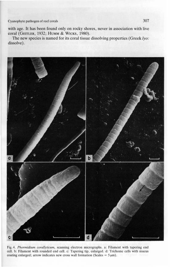
with age. It has been found only on rocky shores, never in association with livecoral (GEITLER, 1932; HUMM & WICKS, 1980).
The new species is named for its coral tissue dissolving properties (Greek [yo:dissolve).
Fig. 4. Phormidium corallylicum, scanning electron micrographs. a: Filament with tapering endcell. b: Filament with rounded end cell. c: Tapering tip, enlarged. d: Trichome cell with mucucoating enlarged; arrow indicate new cros wall formation (Scales = 5 ~m).

308 ROTzLER & SANTA
Material and distribution. Holotype BDA 81.08 A [microscope slide] on £,ploria strigosa (DANA), 3 m, Three .Hill Shoals, Bermuda, K. RtiTZLER, col. :July 1981; deposited in the U. S. National Herbarium, Smithsonian InstitutioWashington, D. C., USA. Paratypes from Bermuda and Carrie Bow CaBelize, are also deposited at the U. S. National Herbarium and in the DepaJment of Invertebrate Zoology (Lower Invertebrates) of the same institutioPhormidium corallyticum is also known from the reefs of Florida, the FloricKeys, the Bahamas, and Jamaica (A. ANTONIUS, pers. comm.; TAYLOR, 1983
2. Cellular organization
Cell walls and sheath. Outer walls are 35-40 run thick and show the character:tic sequence of four layers (LI-LIV) over the plasmalemma (Fig. 7). Layer Iis covered by thin sheath material. Cross walls (septa) between cells aseparated only by the two inner layers (Lf-LII). Cell shrinkage during embeding of the material is evident from the undulating appearance of wall layers alfrom clear spaces between cells (Figs. 5, 6a, 7b). New septa form one per cat a time, by symmetrical annular ingrowth (Figs. 5, 6a, 7b). Small circul(60-ZOO nm) electron transparent areas (granules?) appear regularly in crcsections on either side of the base of newly developing cross walls ("seplgranule", Fig. 7 b); these are distinct from clear areas between cross walls alplasmalemma caused by shrinkage. Photosynthetic lamellae are cut by develoing septa without changing their longitudinal trend. Neither plasmodesmata njunctional pores could be detected.
A thin mucilaginous layer coats the outer cell wall but does not formprominent sheath. Its fibrils are without noticeable orientation, and are intemeshed in a network that is much denser than that of the coral mucus that alappears on the photomicrographs (Figs. 5, 7b). The mucous layer is 10-10011thick, the average being 40 nm and thus similar in dimension to the cell wal
Thylakoid and nucleoplasm. The photosynthetic apparatus consists of thin fllamellae radiating from a longitudinal axis or core - the nucleoplasm - towarthe peripheral plasmalemma (Figs. 6 b, 7 a). In a cell cross section the melbranes resemble the spokes of a wheel; although they do not penetrate tnucleoplasm, they are arranged as if they originated at a common axis.tangential longitudinal sections the thylakoids appear as parallel lines extendi:from one cross wall to the next. On all sections we examined, the lamellae aflat, without intrathylakoid vacuolization, and oriented in one plane, except j
a few locations where large granules cause deflections from the straight tre:(Fig. 7 a). Stacking of membranes does not occur; branching or anastomoses aeither rare or absent. The nucleoplasm occupies 30-40 % of the cell lumeThere are about 80 thylakoids per cell cross section; each is 10 nm thi(distance between membranes) and 65-80nm from the next (at the circurnflence). Width of the lamellae, extending from the nucleoplasmic region to wittZ0-30nm of the plasmalemma, is 0.3-1.0I!m, 0.71!m on the average; no dinconnection with the plasma membrane is evident. The membrane sheets are tsame length as the cell (about 4I!m), although the gener~l trend appe;
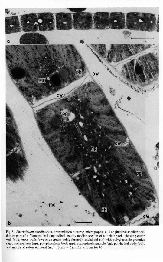
Fig. 5. Phormidium corallyticum, transmission electron micrograph. a: Longitudinal median section of part of a filament. b: Longitudinal, nearly median ection of a dividing cell, showing outerwall (ow), cross walls (cw; one septum being formed), thylakoid (th) with polygJucoside granules(pg), nucleoplasm (np), polypho phate body (pp), cyanophycin granule (cg), polyhedral body (pb),and mucus of substrate coral (mc). (Scale = 5 J.lm for a; 1 J.lm for b).

b
Fig. 6. Phormidium coral/yticum, tran mj sion electron micrograph. a: Longitudinal tangentialsection of two cells showing thylakoid (th) arrangement at peripheral chromatoplasm; trend oflamellae across septum is evident; nucleoplasm (np) is exposed in left cell. b: Cross section of celldepicting regular arrangement of thylakoid (th) membranes radiating from nucleoplasm (np) towardouter cell wall (ow). (Scales = ll!m).

Cyanophyte pathogen of reef corals 311
Fig. 7. Phormidium corallyticum, transmission electron micrographs. a: Cross section showingradial thylakoid (th) with polyglucoside granules (pg), outer cell waH (ow) with sheath (sh), andnucleoplasm (np). b: Longitudinal ection illustrating outer cell wall (ow) with sheath and mucusfrom substrate coral (me), cross wall (cw) with septal granule (?) (sg), cyanophycin granule (cg),polyhedral body (pb), polyphosphate body (pp), and nucleoplasm (np). (Scales = O.ll!m).
312 RiiTZLER & SANTAVY
continuous and is interrupted only by the cross walls. Phycobilisome granulesline the thylakoid membranes in the usual manner but the available resolutiondoes not offer additional detail.
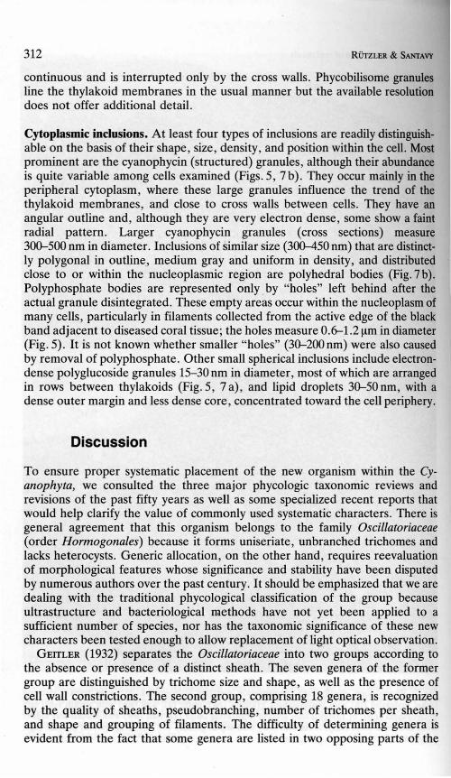
Cytoplasmic inclusions. At least four types of inclusions are readily distinguishable on the basis of their shape, size, density, and position within the cell. Mostprominent are the cyanophycin (structured) granules, although their abundanceis quite variable among cells examined (Figs. S, 7 b). They occur mainly in theperipheral cytoplasm, where these large granules influence the trend of thethylakoid membranes, and close to cross walls between cells. They have anangular outline and, although they are very electron dense, some show a faintradial pattern. Larger cyanophycin granules (cross sections) measure300-S00 nm in diameter. Inclusions of similar size (300-4S0 nm) that are distinctly polygonal in outline, medium gray and uniform in density, and distributedclose to or within the nucleoplasmic region are polyhedral bodies (Fig. 7b).Polyphosphate bodies are represented only by "holes" left behind after theactual granule disintegrated. These empty areas occur within the nucleoplasm ofmany cells, particularly in filaments collected from the active edge of the blackband adjacent to diseased coral tissue; the holes measure 0.6-1.2!-tm in diameter(Fig. S). It is not known whether smaller "holes" (30-200 nm) were also causedby removal of polyphosphate. Other small spherical inclusions include electrondense polyglucoside granules IS-30nm in diameter, most of which are arrangedin rows between thylakoids (Fig. S, 7 a), and lipid droplets 30-S0 om, with adense outer margin and less dense core, concentrated toward the cell periphery.
Discussion
To ensure proper systematic placement of the new organism within the Cyanophyta, we consulted the three major phycologic taxonomic reviews andrevisions of the past fifty years as well as some specialized recent reports thatwould help clarify the value of commonly used systematic characters. There isgeneral agreement that this organism belongs to the family Oscillatoriaceae(order Hormogonales) because it forms uniseriate, unbranched trichomes andlacks heterocysts. Generic allocation, on the other hand, requires reevaluationof morphological features whose significance and stability have been disputedby numerous authors over the past century. It should be emphasized that we aredealing with the traditional phycological classification of the group becauseultrastructure and bacteriological methods have not yet been applied to asufficient number of species, nor has the taxonomic significance of these newcharacters been tested enough to allow replacement of light optical observation.
GEITLER (1932) separates the Oscillatoriaceae into two groups according tothe absence or presence of a distinct sheath. The seven genera of the formergroup are distinguished by trichome size and shape, as well as the presence ofcell wall constrictions. The second group, comprising 18 genera, is recognizedby the quality of sheaths, pseudobranching, number of trichomes per sheath,and shape and grouping of filaments. The difficulty of determining genera isevident from the fact that some genera are listed in two opposing parts of the

Cyanophyte pathogen of reef corals 313
key (Gomontiella: with and without sheath; PoLychLamydum, Schizothrix: oneversus several trichomes per sheath) and that many exceptions to qualifyingcharacters are listed in footnotes.
In the latest thorough phycological revision of the family, DROUET (1968)considers the sheath an environmentally influenced and therefore variable andtaxonomically useless character and reduces the Oscillatoriceae to only sixgenera. Except for the aberrant SpiruLina (without apparent cross walls'), thegeneric separation is based on just a few characters, such as trichome tapering,presence or absence of granules, and thickening of terminal cell walls.
DROUET'S extreme views on oscillatoriacean variability and systematics arecriticized by BOURRELLY (1970: 430, 431), who employs a classical systematicconcept, but he concedes that numerous controlled cultures are still needed toclarify the validity of certain species. Eleven freshwater genera are dealt with byBOURRELLY and separated on the basis of sheath, number of trichomes persheath, spores, trichome shape, cell shape, and membrane thickness.
The principal question ·raised by these two opposing views is to what extentmorphological features resolvable by light microscopy - such as sheaths, celltapering, shape and membrane thickening of end cells, cell wall constrictions,granules, vacuoles, and pseudobranching - are genetically controlled and towhat extent they are influenced by environmental conditions. Ambiguity as aresult of nonquantitative evaluation of characters (e. g., thin: thick; few: many;indistinct: distinct; more: less) is common to both classification systems and is asubstantial element of misinterpretation.
The attempts to determine environment-induced variability in Cyanophytaare summarized by BAKER & BOLD (1970). Unfortunately, in the past manyworkers employed only field observations to determine variability and thereforecould not improve upon taxonomic judgment. Supporting culture studies wererare or incidental, and were poorly controlled until a series of investigationsduring the 1960s - cited in BAKER & BOLD (1970) - examined the influence ofenvironmental parameters (such as concentration of organic materials and othernutrients, moisture, pH, salt concentration, and media composition) on morphological features (including sheath structure and thickness, sheath coloration,trichome structure, cell shape and proportions, granulation, filament motility,and false branching, and plant mass configuration). In essence, all theseexperiments showed that some characters, particularly sheath formation, terminal cell shape, trichome structure, and cell proportions, are to be consideredtaxonomically important because they do not change beyond recognition withinone taxon, despite their obvious variability. This observation supports GaMONT'S (1892) view of almost a century ago that environmental modifications donot change one species into another (assuming a well-founded species concept).Examples of less reliable characters are numbers and types of false branchingthat seem entirely dependent (in those species considered) on moisture contentof the substrate.
BAKER & BOLD (1970) themselves made a significant contribution to oscillatoriacean taxonomic methodology by culturing isolates from soil, air, marine,
I Spirulina does not strictly fit the diagnosis of the family because trichomes are multicellular bydefinition.

314 RiiTZLER & SANTAVY
and freshwater samples under controlled conditions on nutrient agar. This workdetermined or confirmed that some morphological features, particularly themacroscopic structure of the plant mass, the sheath formation, and the shape ofthe terminal cell, are constant characteristics whose nature cannot be mistakendespite a certain degree of variability. Type of granulation and vacuolization, onthe other hand, as well as cell size and shape, and thickening of the outer wall ofthe terminal cell - all considered important attributes in DROUET'S (1968)classification - were found to fluctuate considerably with age and amongdifferent isolates of a monospecific population.
At the beginning of our investigation, we had difficulty deciding whether tocall the delicate mucous polysaccharide layer enveloping the trichomes of theblack band cyanophyte a proper sheath. To avoid the issue, we applied theDROUET system (DROUET, 1968; HUMM & WICKS, 1980), which places theorganism in the genus Schizothrix because it lacks granules along the cross wallsand thickening of the outer membrane of the terminal cell, and because, iftrichome tapering occurs, only the terminal cell is attenuated. This characterization seems unsatisfactory as it is based mainly on characters (or rather absenceof characters) that are experimentally proven to be unreliable. Furthermore,members of Schizothrix are known to have distinct, firm, even colored sheathsalthough they can leave these and survive as naked trichomes, as DROUET (1963)demonstrated - and to form multiple trichomes per sheath and pseudobranches,characters that do not occur in our organism.
Pure culture material was used by RIPPKA et al. (1979) in an attempt todistinguish between phenotypic and genotypic expression in cyanobacteria.Forty-four strains of oscillatorians ("Section III genera") are examined andclassified by characters seen with the light microscope. Nineteen of these strainsare placed in three distinct sheathless genera (Spirulina, Oscillatoria,Pseudanabaena), the remaining twenty-five are assigned to the provisional LPP(Lyngbya-, Phormidium-, Plectonema-like) groups. Our organism falls in theheterogeneous LPP group B (possessing isodiametrical or cylindrical cells, highmotility, unpronounced sheaths, cell width under 5 !J.m); neither physiologicalnor genetic testing of strains in this group by RIPPKA and colleagues allowedfurther meaningful breakdown into useful taxonomic units.
Encouraged by BAKER & BOLD'S (1970) assessment, we reconsidered theclassical systematics, particularly, the significance of the sheath. Accordingly,we noted that our organism has affinities to three genera, Oscillatoria, Phormidium, and Lyngbya, which are distinguished solely by the presence anddevelopment of sheaths. Like GOMONT (1892), we reserve Oscillatoria forspecies that do not develop a sheath, as judged by light microscope staining andelectron microscopy of numerous specimens in different growth phases. Phormidium, as defined here, has a mucilaginous envelope that can be stained bymethylene blue, contrasted by India ink, or made visible by electron microscopy; this envelope does not remain intact and distinct if separated from thetrichome. Lyngbya, on the other hand, has a "distinct" sheath because theenvelope is solid and discrete, allowing trichomes to glide back and forth, andremains intact even after the trichome has left (this quality is independent ofpossible layered structure); sheaths are visible without staining or contrasting bysimple light microscopy.

Cyanophyte pathogen of reef corals 315
It can be argued that our definitions are arbitrary and that borderline casesand transitions are Likely to occur. However, we are focusing on one species thatwe have studied at two distant geographical locations and under many differentecological conditions and growth phases. In view of these observations and theliterature based on light microscope studies it seems logical and convenient toplace our organism in the genus Phormidium. A similar conclusion was reachedby PEARSON & KINGSBURY (1966) in their experimental study of a "Lyngbya sp."that displays an indefinite and thin sheath under all culture conditions. Thesheath of Oscillatoria rubescens DE CANDOLLE, studied under the electronmicroscope by lOST (1965), also corresponds closely to the mucus envelope ofPhormidium corallyticum and would justify transfer of that species to Phormidium.
Our own electron microscope studies have limited value for this discussionbecause our observations could not be related to the finestructural morphologyof a sufficient number of known oscillatoriacean species. Our findings can beused, however, as a data bank for future investigations in this direction (Fig. 8).In examining the finestructure of Phormidium corallyticum, we have paidparticular attention to details that have already been identified as actually orpotentially significant characters for taxonomic use (WHITTON, 1972). The cellwall is composed of four layers (LI-LIV), as is typical for bluegreen algae(lOST, 1965). Only the inner layers (LI, LII) participate in the formation of
pg cw
pb
Fig. 8. Phormidium corallyticum, semischematic, three dimensional reconstruction of cell organiza.tion; parts of outer cell wall and thylakoid region are removed for clarity. Cell components shownare: Outer cell wall (ow), sheath (sh), cross wall (cw), thylakoid membranes (th), nucleoplasm (np),cyanophycin granules (cg), polyhedral bodies (pb), polyglucoside granules (pg), and polyphosphatebodies (pp).

316 RiiTZLER & SANTAVY
cross walls, septa close in symmetrically, and there are no outer wall pores orplasmodesmata. Membrane undulations and other shrinkage phenomena werealso noted by JOST (1965) and shown to take place during polymerization ofembedding media as they are absent in freeze etching preparations of the samematerial. Freeze etchings by JOST also reveal the structure of mucilage or sheathmaterial better than epoxy resin sections. The mucous coating of Oscil/atoriarubescens in an epoxy thin section looks identical to that of Phormidiu/'!lcoral/yticum, but in freeze etchings the fibrils (3 x500nm) of O. rubescens areclearly perpendicular to the cell wall; a high water content of the mucilaginouslayer can also be demonstrated by this technique if antifreeze treatment isomitted (JOST, 1965: Figs. 2, 10-12, 20). A comparative study of solid Lyngbyatype sheaths, particularly by freeze etching, would be most useful becausedifferences in microfibril orientation in some tbick sbeaths bave already beendemonstrated (LEAK, 1967: Fig. 4; LAMONT, 1969: Fig.6c).
The thylakoid may be the most important taxonomic feature made visible byelectron microscopy, although, as with any other character, a certain variabilityhas to be anticipated with age, cell inclusions, and environmental conditions(WHITTON, 1972; LANG & WHITTON, 1973). From a list of eighteen cytoplasmicmembrane characters considered systematically significant (WHITTON, 1972:Table I), only one applies directly to the organization of Phormidium coral/yticum; that is, the tbylakoids run at right angles to the [outer] wall. Thisdiagnosis applies to membrane arrangement that is parallel or perpendicular totbe cross walls, botb of which are demonstrated in the literature. Lamellae thatare perpendicular to longitudinal and parallel to cross walls, occur in anunnamed filamentous form illustrated by LANG & WHITTON (1973). Thylakoidsperpendicular to both outer and cross walls have been noted in a few Osciilatoriaceae, such as Oscil/atoria rubescens (JOST, 1965: Figs. 17, 19, 27) and O.spongeliae (BERTHOLD et al., 1982: Figs. 2,9-11). The thylakoid of O. rubescensdiffers from that of P. coral/yticum in that its membranes are stacked (up to 17per group) and not radial; that is, tbey would not meet at one point if projectedtoward the cell center. Pbotosynthetic membranes of O. spongeliae are radialbut their somewhat fragmented appearance in cross section indicates elaboratebranching in one plane; they also show extensive intrathylakoidal vacuolization.Generic assignment of the latter species, possibly to Borzia as suggested byFELDMANN (1958), should be reconsidered in tbe light of recent work with theelectron microscope (WILKINSON, 1980; BERTHOLD et at., 1982; RfuzLER, inprep.).
The taxonomic value of inclusion bodies is probably limited in comparisonwith features of the thylakoids, although shape and distribution of some - forinstance, polyglucoside granules - are considered useful (WHITTON, 1972), andothers - such as stallar bodies (BERTHOLD et at., 1982) - are promising but needmore comparative study. However, all inclusions found by us in Phormidiumcoral/yticum are common types and correspond to figures and descriptions givenfor a great variety of prokaryotes (LANG, 1968; FOGG et at., 1973; SHIVELY,1974).
These and other new characters are clearly needed before another revision ofOscillatoriaceae can be attempted. The ultrastructure of cell detail in these smallorganisms opens a new field. The improved method of visual examination at this
Cyanophyte pathogen of reef corals 317
level of study could be supplemented by a series of biochemical tests, whichhave become standard procedure in bacteriology. Even the most advancedtechniques will fail to clarify systematic relationships, however, unless variability of old and new characters is studied under carefully controlled conditions.
Summary
Phormidium corallyticum, a new species of the cyanophyte family Oscillatoriaceae found in shallow reefs of the subtropical and tropical westernAtlantic, such as those of Bermuda, Florida, and Belize, is notable for itsdestruction of coral. Traditional phycological methods based on light microscopy alone make it difficult to classify the new organism within the existingoscillatoriacean systems. We have studied finestructure and responses to different ecological conditions in nature and in culture, but these characters havereceived no attention in the monographs on the group and will not assist inclassification until an appropriate revision is made. Since generic allocation isunsatisfactory under the DROUET classification, the classical system employed byGEITLER and BOURRELLY should be reconsidered. Experiments show that severalcharacters used in the classical system are more stable and reliable than fieldworkers have interpreted them to be. The mucilaginous trichome envelopesdiffer from proper solid sheaths but appear distinctive enough to justify revivalof the genus Phormidium, which many authors treat as a junior synonym ofLyngbya. More and better defined taxonomic features could be made availablethrough comparative study of this species group by electron microscopy andbiochemical reactions. If these new characters, along with the traditional ones,are submitted to controlled testing of environmentally induced variability, wecan hope to arrive at an improved taxonomic hierarchy.
Acknowledgements
We would like to thank A. ANTONIUS for introducing us to the black band disease that he firstdiscovered on Carrie Bow Cay reefs more than a decade ago (1972). P. ARCHIBALD, A. BAKER,S. BRAWLEY, and B. WHIITON advised us on. Cyanophycea taxonomy, but we take full responsibilityfor the conclusions reached; A. BAKER kindly provided the Latin diagnosis. Specimens werecontributed by A. ANTONIUS (Carrie Bow Cay) and R. SMITH and T. JICKELLS (Bermuda); we areindebted to E. GANIT for pigment analysis. We thank C. ROTzLER for preparing the tissue thinsections, W. BROWN and M.-J. MAN for taking the scanning electron micrographs, M. PARRISH forartwork and typing, M. CARPENTER for photographic printing, D. TAYLOR for his review, andV. MACINTYRE for editorial help. Part of the field work was conducted at the Bermuda BiologicalStation (Contribution No. 951) and partly supported by a STORROW Fellowship to K. R. Support forfield work in Belize came from the Smithsonian Fluid Research Fund and a grant from the ExxonCorporation. This is Contribution No. 127, Smithsonian Institution Coral Reef and Mangrove StudyProgram at Carrie Bow Cay, Belize.

318
References
RUTZLER & SANTAVY
ANTONIUS, A., 1973: New observations on coral destruction in reefs. Tenth Meeting, Assoc. IslandMar. Lab. Caribb., University of Puerto Rico, Mayagiiez. Abstracts: p.3.1981 [1982]: The "band" diseases in coral reefs. In: E. D. GOMEZ et al. (Eds.), Proceedings,
Fourth International Coral Reef Symposium. Marine Science Center, Quezon City, Philippines, 2: 7-14.
BAKER, A. F. & H. C. BOLD, 1970: Taxonomic studies in the Oscillatoriaceae. Phycological StudiesX, The University of Texas Publication, 7004; 105 pp.
BERTHOLD, M. A., M. A. BOROWITZKA & M. A. MACKAY, 1982: The ultrastructure of Oscillatoriaspongeliae, the blue-green algal endosymbiont of the sponge Dysidea herbacea. Phycologia, 21:327-335.
BOURRELLY, P., 1970: Les algues d'eau douce, initiation a la systematique, ill (5) Les algues bleuesou Cyanophycees. N. Boobee & Cie, Paris: 285-497.
COCKE, E. C., 1967: The Myxophyceae of North Carolina. Publ. by the author, Wake Forest Univ.,Winston-Salem, N. c.; 206 pp.
DROUET, F., 1963: Ecophenes of Schizothrix calcicola (Oscillatoriaceae). Proc. Acad. Nat. Sci.Philadelphia, 115: 261-281. .
- -, 1968: Revision of the Oscillatoriaceae. Monograph 15, Acad. Nat. Sci. Philadelphia; 370pp.FELDMANN, J., 1958: Les Cyanophycees marines de la Guadeloupe (Antilles Franc;aises). Rev.
Algol. N. S., 4: 32-40.FOGG, G. E., W. D. P. STEWART, P. FAY & A. E. WALSBY, 1973: The blue-green algae. AcadeInic
Press, London and New York; 459pp.GETTLER, L., 1932: Cyanophyceae. In: L. RABENHORST (Ed.), Kryptogamen-Flora von Deutschland,
Osterreich und der Schweiz. Second edition, vol. 14; Akademische Verlagsgesellschaft, Leipzig; 1196 pp.
GOLUBlt, S., 1969: Tradition and revision in the system of the Cyanophyta. Verh. Int. Ver. Limnol.,17: 752-756.
GOMONT, M., 1892: Monographie des Oscillariees. Ann. Sci. Nat., Bot., Ser. 7, 15: 263-368; 16:91-264; 16pl.
HUMM, H. J. & S. R. WICKS, 1980: Introduction and guide to the marine bluegreen algae. JohnWiley and Sons, New York; 194pp.
JOST, M., 1965: Die Ultrastruktur von Oscillatoria rubescens D. C. Arch. Mikrobiol., 50: 211-245.LAMONT, H. c., 1969: Shear oriented microfibrils in the mucilaginous investments of two motile
oscillatoriacean blue-green algae. J. Bacteriol., 97: 350-361.LANG, N. J., 1968: The fine structure of blue-green algae. Annu. Rev. Microbiol., 22: 15-46.
& B. A. WHITTON, 1973: Arrangement and structure of thylakoids. In: N. G. CARR &B. A. WHITTON (Eds.), The Biology of Blue-Green Algae. Botanical Monographs, 9. University of California Press, Berkely and Los Angeles: 66-79.
LEAK, L. V., 1967: Finestructure of the mucilaginous sheath of Anabaena sp. J. Ultrastruct. Res.,21: 61-74.
PEARSON, J. E. & J. M. KINGSBURY, 1966: Culturally induced variation in four morphologicallydiverse bluegreen algae. Am. J. Bot., 53: 192-200.
RIPPKA, R., J. DERVELLES, J. B. WATERBURY, M. HERDMAN & R. Y. STANlER, 1979: Generic assignments, strain histories and properties of pure cultures of Cyanobacteria. J. Gen. Microbiol.,111: 1-61.
RiiTZLER, K., P. ARCHIBALD & D. SANTAVY-RoBERTSON, 1982: A new species of Lyngbya AGARDHemend. BOURRELLY (Cyanophyta, Oscillatoriaceae) cause of black band disease of tropical reefcorals. First International Phycological Congress, St. John's, Newfoundland. Abstracts:p.a42.& J. D. FERRARIS, 1982: Terrestrial environment and climate, Carrie Bow Cay, Belize. In:K. RUTZLER & I. G. MACfNTYRE (Eds.), The Atlantic Barrier Reef Ecosystem at Carrie BowCay, Belize, I: Structure and Communities. Smithson. Contrib. Mar. Sci., 12: 77-91.& I. G. MACINTYRE, 1982: The habitat distribution and community structure of the barrier reefcomplex at Carrie Bow Cay, Belize. In: K. RiiTZLER & I. G. MAcJNlYRE (Eds.), The AtlanticBarrier Reef Ecosystem at Carrie Bow Cay, Belize, I: Structure and Communities. Smithson.Contrib. Mar. Sci., 12: 9-45.

Cyanophyte pathogen of reef corals 319
- -, D. L. SANTAVY & A. ANTONIUS, 1983: The Black Band Disease of Atlantic Reef Corals. III.Distribution, Ecology, and Development. P. S. Z. N. I: Marine Ecology, 4 (4): 329-358.
SHIVELY, J. M., 1974: Inclusion bodies. Annu. Rev. Microbiol., 28: 167-187.TAYLOR, D. L., 1983: The Black Band Disease of Atlantic Reef Corals. II. Isolation, Cultivation,
and Growth of Phormidium corallyticum. P. S. Z. N. I: Marine Ecology, 4 (4): 321-328.THURET, M. G., 1875: Essai de classification des Nostochinees. Ann. Sci. Nat., Bot., Ser. 6, 1:
372-382.WHIlTON, B. A., 1969: The taxonomy of blue-green algae. Br. Phycol. J., 4: 121-123.- -, 1972: Finestructure and taxonomy in the blue-green algae. In: T. V. DESIKACHARY (Ed.),
Taxonomy and Biology of Blue-Green Algae. University of Madras, Madras: 18-26.WrLKINSON, C. R., 1980: Cyanobacteria symbiotic in marine sponges. In: W. SCHWEMMLER &
H. E. A. SCHENK (Eds.), Endocytobiology: Endosymbiosis and Cell Biology. Walter de Gryter& Co., Berlin: 553-563.