
The biology of brain metastases—translation to new therapies
13
344 | JUNE 2011 | VOLUME 8 www.nature.com/nrclinonc Stephen E. and Catherine Pappas Center for Neuro- oncology (A. F. Eichler), and Deparment of Radiation Oncology (E. Chung, D. P. Kodack, J. S. Loeffler, D. Fukumura, R. K. Jain), Massachusetts General Hospital and Harvard Medical School, Boston, MA 02114, USA. Correspondence to: R. K. Jain jain@ steele.mgh.harvard.edu The biology of brain metastases—translation to new therapies April F. Eichler, Euiheon Chung, David P. Kodack, Jay S. Loeffler, Dai Fukumura and Rakesh K. Jain Abstract | Brain metastases are a serious obstacle in the treatment of patients with solid tumors and contribute to the morbidity and mortality of these cancers. It is speculated that the frequency of brain metastasis is increasing for several reasons, including improved systemic therapy and survival, and detection of metastases in asymptomatic patients. The lack of preclinical models that recapitulate the clinical setting and the exclusion of patients with brain metastases from most clinical trials have slowed progress. Molecular factors contributing to brain metastases are being elucidated, such as genes involved in cell adhesion, extravasation, metabolism, and cellular signaling. Furthermore, the role of the unique brain microenvironment is beginning to be explored. Although the presence and function of the blood–brain barrier in metastatic tumors is still poorly understood, it is likely that some tumor cells are protected from therapeutics by the blood–tumor barrier, creating a sanctuary site. This Review discusses what is known about the biology of brain metastases, what preclinical models are available to study the disease, and which novel therapeutic strategies are being studied in patients. Eichler, A. F. et al. Nat. Rev. Clin. Oncol. 8, 344–356 (2011); published online 12 April 2011; doi:10.1038/nrclinonc.2011.58 Introduction Metastatic brain tumors are the most frequently occur‑ ring intracranial neoplasms in adults with the annual incidence in the USA estimated to be 200,000 cases. 1 Furthermore, an estimated 8–10% of adults with cancer develop symptomatic brain metastases. 2,3 The majority of brain metastases originate from primary cancers in the lung (40–50%) or breast (15–25%), or from mela‑ noma (5–20%). 2,3 The frequency of diagnosis of meta‑ static brain tumors seems to be increasing as a result of improved imaging modalities and earlier detection as well as longer survival after primary cancer diagnosis because of more‑effective treatment of systemic disease. The distribution of brain metastases correlates with blood flow and tissue volume, with 80% detected in the cerebral hemispheres, 15% in the cerebellum, and 5% in the brainstem. 4 The majority of patients exhibit multiple tumors at the time of brain‑metastasis diagnosis. Clinical features include headache, neurological deficit, and sei‑ zures. Neuropsychological testing demonstrates cognitive impairment in 65% of patients with brain metastases, 5,6 which might be a result of destruction or displacement of brain tissue by the expanding tumor, peritumoral edema leading to further disruption of surrounding white matter tracts, increased intracranial pressure, and/or vascular compromise. Therapeutic approaches for brain metastases include surgery, whole‑brain radiation therapy (WBRT), stereotactic radiosurgery (SRS), chemotherapy, growth factor inhibitors, or a combination of these therapies. Survival for patients with brain metastases treated with WBRT typically ranges from 4–6 months, but can be as long as 12–24 months for selected patients. 6 Strong posi‑ tive prognostic factors include good functional status, age <65 years, no sites of metastasis outside of the central nervous system (CNS), controlled primary tumor, 7 the presence of a single metastasis in the brain, long inter‑ val from primary diagnosis to brain relapse, and certain cancer subtypes such as HER2‑positive breast cancer and EGFR‑mutant non‑small‑cell lung cancer (NSCLC). 8–10 In up to half of patients the cause of death is attrib‑ uted to CNS progression. 11 Randomized clinical trials have shown that surgery or SRS combined with WBRT improves overall survival compared with WBRT alone in patients with a single metastasis in the brain. In patients with four or fewer brain metastases, SRS results in equiva‑ lent overall survival but worse intracranial disease control compared with SRS plus WBRT. 11–15 The combination of radiotherapy and chemotherapy improves response rate and/or progression‑free survival in some studies, but not overall survival. 16–18 We review our current understanding of the biology of brain metastasis, present preclinical approaches that are available to model the disease, including emerging imaging techniques, and discuss novel therapeutic strategies to target the unique features of CNS metastases. Brain metastasis formation The metastatic cascade, whereby cancer cells escape from the primary tumor site, invade surrounding tissue, intra‑ vasate into the bloodstream or lymphatics, and arrest, Competing interests R. K. Jain declares associations with the following companies: Astellas, AstraZeneca, Dyax, Enlight Biosciences, Genzyme, MedImmune, Millenium, MPM Capital, Noxxon, Roche, SynDevRx. The other authors declare no competing interests. REVIEWS © 2011 Macmillan Publishers Limited. All rights reserved
Transcript of The biology of brain metastases—translation to new therapies

344 | JUNE 2011 | VOLUME 8 www.nature.com/nrclinonc
Stephen E. and Catherine Pappas Center for Neuro-oncology (A. F. Eichler), and Deparment of Radiation Oncology (E. Chung, D. P. Kodack, J. S. Loeffler, D. Fukumura, R. K. Jain), Massachusetts General Hospital and Harvard Medical School, Boston, MA 02114, USA.
Correspondence to: R. K. Jain jain@ steele.mgh.harvard.edu
The biology of brain metastases—translation to new therapiesApril F. Eichler, Euiheon Chung, David P. Kodack, Jay S. Loeffler, Dai Fukumura and Rakesh K. Jain
Abstract | Brain metastases are a serious obstacle in the treatment of patients with solid tumors and contribute to the morbidity and mortality of these cancers. It is speculated that the frequency of brain metastasis is increasing for several reasons, including improved systemic therapy and survival, and detection of metastases in asymptomatic patients. The lack of preclinical models that recapitulate the clinical setting and the exclusion of patients with brain metastases from most clinical trials have slowed progress. Molecular factors contributing to brain metastases are being elucidated, such as genes involved in cell adhesion, extravasation, metabolism, and cellular signaling. Furthermore, the role of the unique brain microenvironment is beginning to be explored. Although the presence and function of the blood–brain barrier in metastatic tumors is still poorly understood, it is likely that some tumor cells are protected from therapeutics by the blood–tumor barrier, creating a sanctuary site. This Review discusses what is known about the biology of brain metastases, what preclinical models are available to study the disease, and which novel therapeutic strategies are being studied in patients.
Eichler, A. F. et al. Nat. Rev. Clin. Oncol. 8, 344–356 (2011); published online 12 April 2011; doi:10.1038/nrclinonc.2011.58
IntroductionMetastatic brain tumors are the most frequently occur‑ring intracranial neoplasms in adults with the annual incidence in the USA estimated to be 200,000 cases.1 Furthermore, an estimated 8–10% of adults with cancer develop sympto matic brain metastases.2,3 The majority of brain metastases originate from primary cancers in the lung (40–50%) or breast (15–25%), or from mela‑noma (5–20%).2,3 The frequency of diagnosis of meta‑static brain tumors seems to be increasing as a result of improved imaging modalities and earlier detection as well as longer survival after primary cancer diagnosis because of more‑effective treatment of systemic disease.
The distribution of brain metastases correlates with blood flow and tissue volume, with 80% detected in the cerebral hemispheres, 15% in the cerebellum, and 5% in the brainstem.4 The majority of patients exhibit multiple tumors at the time of brain‑metastasis diagnosis. Clinical features include headache, neurological deficit, and sei‑zures. Neuropsychological testing demonstrates cognitive impairment in 65% of patients with brain metastases,5,6 which might be a result of destruction or displacement of brain tissue by the expanding tumor, peritumoral edema leading to further disruption of surrounding white matter tracts, increased intracranial pressure, and/or vascular compromise.
Therapeutic approaches for brain metastases include surgery, whole‑brain radiation therapy (WBRT),
stereotactic radiosurgery (SRS), chemotherapy, growth factor inhibitors, or a combination of these therapies. Survival for patients with brain metastases treated with WBRT typically ranges from 4–6 months, but can be as long as 12–24 months for selected patients.6 Strong posi‑tive prognostic factors include good functional status, age <65 years, no sites of metastasis outside of the central nervous system (CNS), controlled primary tumor,7 the presence of a single metastasis in the brain, long inter‑val from primary diagnosis to brain relapse, and certain cancer subtypes such as HER2‑positive breast cancer and EGFR‑mutant non‑small‑cell lung cancer (NSCLC).8–10 In up to half of patients the cause of death is attrib‑uted to CNS progression.11 Randomized clinical trials have shown that surgery or SRS combined with WBRT improves overall survival compared with WBRT alone in patients with a single metastasis in the brain. In patients with four or fewer brain metastases, SRS results in equiva‑lent overall survival but worse intracranial disease control compared with SRS plus WBRT.11–15 The combination of radio therapy and chemo therapy improves response rate and/or progression‑free survival in some studies, but not overall survival.16–18
We review our current understanding of the biology of brain metastasis, present preclinical approaches that are available to model the disease, including emerging imaging techniques, and discuss novel therapeutic strat egies to target the unique features of CNS metastases.
Brain metastasis formationThe metastatic cascade, whereby cancer cells escape from the primary tumor site, invade surrounding tissue, intra‑vasate into the bloodstream or lymphatics, and arrest,
Competing interestsR. K. Jain declares associations with the following companies: Astellas, AstraZeneca, Dyax, Enlight Biosciences, Genzyme, MedImmune, Millenium, MPM Capital, Noxxon, Roche, SynDevRx. The other authors declare no competing interests.
REVIEWS
© 2011 Macmillan Publishers Limited. All rights reserved
NATURE REVIEWS | CLINICAL ONCOLOGY VOLUME 8 | JUNE 2011 | 345
extravasate, survive and proliferate within a secondary site (Figure 1), is an inherently inefficient process.19 Certain tumor types demonstrate an organ‑specific pattern of spread; for example, prostate cancer frequently meta‑stasizes to bone and melanoma frequently meta stasizes to the lung, liver and brain.20,21 Stephen Paget was the first to hypothesize that the propensity of cancer cells to spread to specific sites was dependent upon two factors: the cancer cell (the ‘seed’) and the receiving organ envi‑ronment (the ‘soil’).22 An alternative hypo thesis attributed to pathologist James Ewing proposes that circu latory pat‑terns between the primary tumor and specific second ary organs are sufficient to explain the majority of organ‑specific metastatic spread.23 A newer hypo thesis is that cancer cells can bring their own soil—stromal compo‑nents from the primary site including activated fibro‑blasts—to secondary sites.24 Researchers have sought to understand how molecular and genetic features of the primary cancer cell, the brain micro environment, and the angiogenic pathways influence spread of cancer to the brain and its growth there (Table 1).
Brain microenvironmentOnce metastatic cancer cells enter the brain circulation, they might arrest in sites of slow flow within the capillary bed at vascular branch points,25 which is then followed by early changes in the brain microenvironment.26 The arrested cancer cells encounter brain vascular endo‑thelial cells, which seem to promote metastatic tumor cell growth and invasion.25,27 In addition, stromal cells such as fibroblasts associated with the primary tumor are involved in metastatic nodules in the brain (Figure 1).24 These co‑disseminating stromal cells provide survival and prolifera tive advantages to the tumor cells and facilitate early colonization of metastatic foci. The brain also pro‑vides an environment that differs from most other organs, and the factors that either promote or suppress coloniza‑tion and proliferation are poorly understood (Table 1). Thus, successful treatment of CNS metastases may require targeting of both tumor and host responses.
Response of host stromal cellsAfter infiltration into the brain tissue, cancer cells encounter a number of host cell types, including micro‑glia and astrocytes. Microglia displaying stellate morpho‑logy with thick cellular processes, characteristic of an activated state, have been observed around extra vastated cancer cells within 7 days of intracarotid injection of breast cancer cell lines into mice.26 Activated astrocytes with thick processes and upregulated expression of glial fibrillary acidic protein can be seen even earlier, when cancer cells are still in the intravascular space, and they remain closely associated with cancer cells throughout their growth into macrometastases.26 In a xenograft model, reactive astrocytes and microglia were in direct contact with tumor cells along the border of the tumors as well as infiltrating the inner tumor mass. Similar find‑ings were reported in human specimens. In addition, in vitro co‑cultures demonstrated that glia induced a fivefold increase in metastatic cell proliferation, leading
Key points
■ Longer survival of patients and more-sensitive detection of metastatic disease by improved imaging modalities might contribute to the increased incidence of detected brain metastases
■ Novel preclinical models that more accurately represent clinical brain metastases and imaging techniques that allow study of the formation of brain metastases and their response to treatments are emerging
■ Brain metastases grow by co-opting existing blood vessels and/or by forming new blood vessels; the brain microenvironment promotes tumor cell survival, tumor growth and resistance to therapy
■ Although a lesser problem in large metastases, the blood–brain barrier (BBB) could prevent therapeutic access to micrometastases; strategies to enhance drug delivery across the BBB are under investigation
■ Differential expression of genes involved in intravasation and extravasation, metabolism, cell adhesion, and cellular signaling in brain-specific metastatic clones have been identified
■ Targeted therapies, including inhibitors of EGFR, HER2, PI3K and BRAF, have shown promise in the treatment of brain metastases, but require testing in randomized trials
the researchers to hypothesize that reactive glia creates an altered brain microenvironment that is more permissive to tumor growth and invasion.28 Consistent with these findings, expression of endo thelin 1, a regulator of numer‑ous transforming processes, was observed in peritumoral astrocytes in 85% of hematogeneous metastases of the human brain.29 Furthermore, additional studies suggest that microglia can enhance the invasion and coloni zation in the brain tissue by breast cancer cells similar to other tissue‑specific stromal cells such as osteoblasts and osteo‑clasts in the bone.30,31 Most likely, during the early stages of brain metastasis formation, there is a balance between the protective or trophic, and cytotoxic functions of microglia, dependent on the release of a variety of factors and ultimately determined by signals from tumor cells.32
Protective roles of astrocytesAstrocytes within the brain microenvironment might serve to protect brain metastases from cytotoxicity induced by chemotherapeutic drugs.33 In co‑culture experiments, the presence of astrocytes but not fibro‑blasts dramatically reduced 5‑fluorouracil‑induced and cisplatin‑induced apoptosis in human tumor cells.34 The protective effect was lost when cells were separated with a trans‑well membrane, suggesting that direct contact of astrocytes with tumor cells, rather than secreted factors, was mediating the chemoprotective effect. Similar effects have also been demonstrated in human melanoma cell lines,35 breast cancer cells, and lung cancer cells.36
Soluble factors also have an important role in tumor cell–astrocyte interaction. Activated astrocytes sur‑round brain metastatic lesions from lung cancer, both in an experimental mouse model and in the human brain.37 Tumor cell‑derived factors (including migration inhibi‑tory factor, interleukin [IL]‑8, and plasminogen activa‑tor inhibitor 1) induced astrocytic activation and release of molecules (IL‑6, TNF‑α, and IL‑1β) that promoted tumor‑cell proliferation in vitro. Furthermore, neuro‑trophins and their receptors have a role in the invasion and colonization of brain‑prone melanoma cells.38,39
FOCUS ON METASTASIS
© 2011 Macmillan Publishers Limited. All rights reserved

346 | JUNE 2011 | VOLUME 8 www.nature.com/nrclinonc
Figure 1 | Steps in the formation of hematogenous metastasis to the brain. Once tumor cells shed from primary tumors arrive in brain vasculature, a | they arrest in the capillary bed primarily owing to size restriction, and b | subsequently extravasate across the BBB and enter the brain parenchyma. Three genes mediate cancer cell trans-BBB migration: HBEGF, COX2, and ST6GALNAC5.78 Also, activation of integrins, such as αvβ3
54,139 and β1,27 is suggested to control tumor-cell arrest and adhesion
to the vasculature. c | Metastasizing tumor cells (seeds) may bring their own host cells (soil) in metastasis.24 After extravasation, tumor cells either d | grow along pre-existing blood vessels (perivascular growth),49 or e | recruit new blood vessels (angiogenesis) to obtain sufficient nutrients to support their proliferation. Abbreviation: BBB, blood–brain barrier.
AngiogenesisThe growth and proliferation of primary and metastatic tumors is dependent on the establishment of an adequate blood supply.40–42 A tumor can recruit blood vessels via different mechanisms: angiogenesis (sprouting from existing blood vessels), vasculogenesis (formation of a de novo vascular system from endothelial precursor cells), co‑option (growth of cancer cells along existing blood vessels), intussusception (vessel remodeling and expansion by the insertion of interstitial tissue columns into the lumen of pre‑existing vessels), vasculogenic mimicry (cancer cells lining blood vessels), cancer cells that transdifferentiate into endothelial cells,43 and cancer stem‑like cells that form an inner lining of blood vessels in the brain.44–47
Changes in brain vasculatureKienast et al.25 used multiphoton laser scanning micro‑scopy and a mouse cranial window model to follow in real time brain metastasis formation from both lung cancer and melanoma cell lines. After extravasation, there was a persistent correlation between tumor cells with micro‑vessels and either vessel co‑option (with melanoma) or
angiogenesis (with lung carcinoma). Previous studies showed a similar association between meta stasizing tumor cells and blood vessels.27,28,48,49 Kusters et al.48 showed that a melanoma brain metastasis could grow up to 3 mm through co‑opting pre‑existing blood vessels. Similar observations in murine experimental brain metastases models from breast cancer and melanoma cells indicate that an active adhesion mechanism exists between tumor cells and the vascular basement membrane.27
Metastatic brain tumor vessels in preclinical models and human surgical specimens have significantly larger diameters and thicker basement membranes when com‑pared with the vessels of normal brain.50 Brain meta stases from murine melanoma, murine fibrosarcoma, human lung carcinoma, and human colon carcinoma have a lower microvascular density than the surrounding normal brain parenchyma, and they all contain dilated blood vessels with large lumens.50 The mechanism for this enlargement seems to involve endothelial cell proliferation.
Angiogenic factors in brain metastasisThe progressive growth of the majority of metastatic brain tumors is critically dependent on the expression of
Primary tumor
Brain metastasis
Perivascular growthco-option
Post-antiangiogenictreatment?
Angiogenesis
ExtravasationArrest in thecapillary bed
Stromal cellmetastasis
ST6GALNAC5,COX2, HBEGF
Brain vasculature
BBB intact
BBBdisrupted
Cancer cell Stroma cell Astrocyte Microglia
αv, β3, β1integrin
ab
c
d
e
REVIEWS
© 2011 Macmillan Publishers Limited. All rights reserved
NATURE REVIEWS | CLINICAL ONCOLOGY VOLUME 8 | JUNE 2011 | 347
VEGF.51 In a brain metastatic variant of MDA‑MB‑231 cells, there was a significant increase in VEGF‑A pro‑duction, and inhibition of VEGFR activity significantly reduced brain tumor burden after intracarotid injec‑tion in mice.52 Moreover, when human lung cancer cells transfected with antisense VEGF165 were injected into the carotid artery of nude mice, the formation of brain metastases was substantially decreased compared with untransfected cells.51 However, in cells transfected with VEGF121 or VEGF165, no increase in metastasis was observed, indicating that VEGF expression is necessary but not sufficient for the production of brain metastases. In another study, VEGFR inhibition with cediranib did
not impede tumor cell extravasation in a murine model of brain metastasis from a prostate cancer primary.53 These data suggest hetero geneity between different tumor cells and types.
Activation of integrin αvβ3 on tumor cells strongly promotes metastatic breast tumor growth in the brain by enabling tumor cells to attract blood vessels independent of hypoxia, an effect mediated by post‑transcriptional control of VEGF expression.54 Other growth factors including angiopoietin 1 and 2, bFGF, PlGF, SDF1α, PDGF and IL‑8 have been implicated in the angiogenesis pathway in primary tumors and gliomas, but their role remains to be established in metastatic brain tumors.43,55
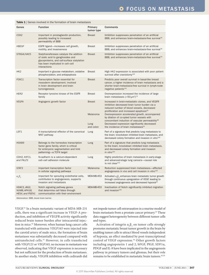
Table 1 | Genes involved in the formation of brain metastasis
Genes Function Primary tumor type
Comments
COX2 Important in prostaglandin production, possibly leading to increased permeability of BBB
Breast Inhibition suppresses penetration of an artificial BBB, and enhances brain-metastasis-free survival78
HBEGF EGFR ligand—increases cell growth, motility, and invasiveness
Breast Inhibition suppresses penetration of an artificial BBB, and enhances brain-metastasis-free survival78
ST6GALNAC5 Sialyltransferases catalyze the addition of sialic acid to gangliosides and glycoproteins, and cell-surface sialylation has been implicated in cell–cell interactions
Breast Inhibition suppresses penetration of an artificial BBB, and enhances brain-metastasis-free survival78
HK2 Important in glucose metabolism, oxidative phosphorylation, and antiapoptosis
Breast High HK2 expression is associated with poor patient survival after craniotomy140
FOXC1 Transcription factor essential for mesoderm development; involved in brain development and brain tumorigenesis
Breast Predicts poor overall survival in basal-like breast cancer, a higher incidence of brain metastasis and a shorter brain-metastasis-free survival in lymph-node-negative patients141
HER2 Receptor tyrosine kinase of the EGFR family
Breast Overexpression increased the incidence of large brain metastases (>50 μm2)73
VEGFA Angiogenic growth factor Breast
Melanoma
Lung and colon
Increased in brain-metastatic clones, and VEGFR inhibition decreased brain tumor burden via a reduced number of blood vessels, decreased proliferation and increased apoptosis52
Overexpression accelerated growth, accompanied by dilation of co-opted tumor vessels with concomitant induction of vascular permeability48
Decreased expression significantly decreased the incidence of brain metastases51
LEF1 A transcriptional effector of the canonical WNT pathway
Lung Part of a signature that predicts lung metastasis to the brain; knockdown inhibited brain metastasis, and decreased colony formation and invasion in vitro142
HOXB9 Belongs to the homeobox transcription factor gene family, which is critical for embryonic segmentation and limb patterning—a TCF4 target
Lung Part of a signature that predicts lung metastasis to the brain; knockdown inhibited brain metastasis, and decreased colony formation and invasion in vitro142
CDH2, KIFC1, and FALZ3
N-cadherin is a calcium-dependent cell–cell adhesion molecule
Lung Highly predictive of brain metastasis in early-stage and advanced-stage lung cancers—causal role is not clear143
STAT3 Important transcription factor in cellular signaling pathways
Melanoma Reduction suppressed brain metastases—affected angiogenesis in vivo and cell invasion in vitro144
αv β3Important for sprouting endothelial cells, contributes to angiogenesis, supports invasion and metastasis
MDA-MB-453 Activated αvβ3 enhances brain metastatic tumor growth through continuous upregulation of VEGF, leading to increased angiogenesis and decreased hypoxia54
HDAC3, JAG2, NUMB, APH1B, HES4, and PSEN1
Notch signaling pathway genes that determine cell fates through communication with their environment
MDA-MB-453 Inactivation of Notch significantly inhibited migration and invasion145
Abbreviation: BBB, blood–brain barrier.
FOCUS ON METASTASIS
© 2011 Macmillan Publishers Limited. All rights reserved

348 | JUNE 2011 | VOLUME 8 www.nature.com/nrclinonc
Hostile microenvironment impairs drug deliveryIn cancer, the balance between proangiogenic and anti‑angiogenic factors tips towards angiogenesis owing to the overexpression of proangiogenic growth factors. This overexpression leads to blood vessels that are structurally and functionally abnormal, exhibiting heterogeneous and often sluggish blood flow and hyperpermeability.50,56–59 A heterogeneous and inefficient blood vessel network creates regions of hypoperfusion or no perfusion where reduced nutrients and oxygen, and impaired waste removal, creates an abnormal metabolic environment, characterized by hypoxia and acidosis. Furthermore, vascular hyperpermeability induces tumor interstitial hypertension, which hinders drug penetration owing to the lack of convective transport.60,61
Clearly, host–tumor interactions including angio genesis have an important role in the formation and expansion of brain metastases. However, the knowledge of the molecu‑lar mechanisms required during the formation of meta‑static lesions is still lacking. What is certain is that the molecular and cellular changes within metastatic brain tumor vasculature and the micro environment should be considered when designing strategies to improve the delivery and efficacy of therapeutic agents.
Modeling and imaging brain metastasesSubstantial progress has been made over the past two decades in modeling human cancer in the mouse and, more recently, model systems have emerged that recapitu‑late many aspects of metastatic disease to the CNS. An
important barrier to the development of reliable CNS mouse models is that in most orthotopically transplanted primary tumors that give rise to metastatic foci, the animals succumb to systemic disease before the brain can be reliably studied. Therefore, current models often rely on either direct implantation of tumors into the brain or iterative selection of brain‑seeking clones resulting from experimental hematogeneous dissemination to the brain to enrich metastatic disease in the CNS. These experi‑mental metastasis models inevitably miss the initial steps of the metastatic cascade and thus may not reflect the full clinical manifestation of the disease.
Animal models of brain metastasisVarious preclinical animal models for the study of brain metastases and their advantages and limitations are summarized in Table 2. Rodent model systems for brain metastasis have been reported for multiple cancer types, including melanoma,62,63 lung carcinoma,64 and breast carcinoma.65–71 The emergence of new animal models of brain metastasis that more closely mimic human disease provides the tools necessary to understand all of the steps involved in the process. Unfortunately, the field is still limited by the few spontaneous models available and the complexity of more clinically relevant models.
MelanomaMelanoma mouse models have been established using a variety of murine and human melanoma cell lines injected via an intracardiac, intracarotid or, less successfully,
Table 2 | Preclinical rodent animal models of brain metastasis
Tumor dissemination routes
Methods Advantages Disadvantages
Direct implantation model
Leptomeningeal metastasis75,146,147
Inoculation of tumor cells or fragments into the subarachnoid space, or subarachnoid catheter for delivery to the cerebrospinal fluid space
Relatively simple procedure; convenient for longitudinal imaging study with molecular or intravital microscopic imaging
Bypasses precolonization steps of metastasis; does not model cerebral brain metastases
Intraparenchymal implantation27,148,149
Inoculation of tumor cells or fragments directly into the brain parenchyma by hand or stereotactic guidance
Simple way to study metastatic tumor growth inside the brain; the procedure has been extended by placement of a cranial window or burr hole (at the site of inoculation)
Bypasses precolonization steps of metastasis
Hematogeneous metastasis model
Intracardiac injection25,78
Injection of single-cell suspension of tumor into the left ventricle of an animal
Relatively simple procedure and can bypass pulmonary arrest and retention
Dissemination of tumor cells to sites other than cerebral target; decreased reproducibility due to the blind nature of the inoculation compared with direct implantation
Intracarotid artery injection72,150,151
Injection of metastatic cells into the internal carotid artery followed by permanent ligation
Produces predominately cerebral tumors and minimal noncerebral metastasis
Murine carotid is very small and significant microsurgical skill is required
Spontaneous metastasis model
Spontaneous formation63,77,152
Spontaneous brain metastases subsequent to removal of primary tumor from either parental or metastatic variants
Recapitulates complete metastatic steps; generated from other simpler model systems; useful for confirming hypotheses
Longer duration requiring resection of primary tumors or multiple preselection processes; most complicated method to run a controlled experiment
REVIEWS
© 2011 Macmillan Publishers Limited. All rights reserved
NATURE REVIEWS | CLINICAL ONCOLOGY VOLUME 8 | JUNE 2011 | 349
intravenous route. Most models show a combination of parenchymal and leptomeningeal deposits at the time of sacrifice, with occasional dural deposits.72
Breast cancerYoneda et al.70 established a brain‑seeking clone (MDA‑MB‑231BR) of the human breast cancer cell line MDA‑MB‑231 by serial in vivo and in vitro passages. In a mouse xenograft, when the MDA‑MB‑231BR cell line was transfected with HER2, the HER2‑overexpressing clones demonstrated a threefold increase in the number of large brain metastases compared with untransfected MDA‑MB‑231BR cells; however, the number of micro‑metastases was unaffected.73 This model has also been used to probe the effect of anti‑HER2 therapies on the development and propagation of brain metastases.74 Alternatively, human breast cancer cells implanted near the surface of the brain provide a model to directly observe tumor and vessel growth using a cranial window and intravital microscopy (Figure 2a–c).75,76
Spontaneous metastasisFew reports describe cell lines capable of reproducibly spontaneously metastasizing to the CNS from a primary tumor. These include a variant of the murine B16 mela‑noma cell line (named G3.5), generated through succes‑sive rounds of selection of brain metastases following intravenous injection, which leads to spontaneous meta‑stasis to the CNS from subcutaneous primary tumors in 80% of mice.77 Using a similar method, a brain metastatic
variant of the triple‑negative breast cancer cell line CN34 (CN34‑BrM2) capable of metastasizing to the CNS from a primary mammary tumor in about 42% of mice was generated.78 A procedure that might more accurately replicate the selection mechanisms of clinical brain meta stasis formation was established from long‑term, low‑dose metro nomic chemotherapy treatment of mice injected subdermally with a highly metastatic variant of the WM239A human melanoma cell line.63 Among long‑term surviving mice, 20% had spontaneous brain metastases, and cell lines generated from these meta‑stases spontaneously metastasized to the brain paren‑chyma after ortho topic transplantation and removal of the primary tumor.63
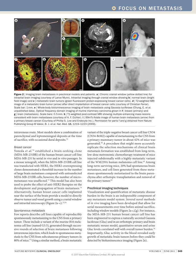
Preclinical imaging techniquesVisualization and quantification of metastatic disease burden in the brain is an indispensible component of any metastasis model system. Several novel methods of in vivo imaging have been developed that allow for serial measurements over time before animal sacrifice, including window models (Figure 2a‑c,f,g). For instance, the MDA‑MB‑231 human breast cancer cell line has been engineered to express a naturally secreted Gaussia luciferase (Gluc) and in an orthotopic primary and brain metastatic mouse model, quantitative serum and urine Gluc levels correlated well with overall tumor burden.76 Importantly, Gluc activity in the blood revealed early growth of metastatic brain tumors before they could be detected by bioluminescence imaging (Figure 2e).
a
c
d e
f g h i
b
Figure 2 | Imaging brain metastases in preclinical models and patients. a | Chronic cranial window (yellow dotted line) for intravital brain imaging (courtesy of Lance Munn). Intravital imaging through cranial window showing b | normal brain (bright field image) and c | metastatic brain tumors (green fluorescent protein-expressing breast cancer cells). d | T2-weighted MRI image of a metastatic brain tumor (arrow) after direct implantation of breast cancer cells (courtesy of Christian Farrar). Scale bar: 1 mm. e | Whole-body bioluminescence imaging of brain metastasis using Gaussia luciferase (Chung, E. et al. unpublished data). Optical frequency domain imaging of murine mammary carcinoma grown in f | breast (primary) and g | brain (metastasis). Scale bars: 0.5 mm. h | T1-weighted post-contrast MRI showing multiple enhancing mass lesions consistent with brain metastases (courtesy of A. F. Eichler). i | 99mTc-folate image of human brain metastasis (arrow) from a primary breast cancer (Courtesy of Phillip S. Low and Endocyte Inc.). Permission for parts f and g obtained from Nature Publishing Group © Vakoc, B. J. et al. Nat. Med. 15, 1219–1223 (2009).
FOCUS ON METASTASIS
© 2011 Macmillan Publishers Limited. All rights reserved

350 | JUNE 2011 | VOLUME 8 www.nature.com/nrclinonc
An alternative adaptation of quantitative fluorescent and phosphorescent imaging techniques has recently been developed that interrogates blood–tumor barrier (BTB) permeability within and surrounding metastatic brain tumors in mice, using tumor cells expressing enhanced green fluorescent protein (eGFP) and Texas red dextran as a marker of passive permeability.79 This model also allows simultaneous quantification of BTB permeability using radiolabeled chemotherapeutic drugs. Combining these techniques with the MDA‑MB‑231BR HER2‑overexpressing mouse model previously described,70 Lockman et al.79 analyzed over 2,000 brain metastases and demonstrated that BTB permeability was compromised in over 89% of lesions.79 However, 14C‑paclitaxel and 14C‑doxorubicin only reached cytotoxic concentrations in about 10% of metastases.79
Other powerful imaging technologies include MRI, PET and near‑infrared optical imaging.80,81 In the past, the majority of MRI studies of brain tumors in mice were per‑formed using high‑field‑strength MRI (≥7 Tesla) because of its high spatial resolution and signal‑to‑noise ratio (Figure 2d).82,83 However, several techniques enhance the sensitivity of clinical‑strength MRI scanners (1.5–3 Tesla)
for the detection of small brain metastases in rodents. When MDA‑BR‑231‑eGFP cells are labeled with fluo‑rescent μm‑sized superparamagnetic iron oxide (MPIO) particles, single labeled breast cancer cells can be detected in the rat brain parenchyma as small dark or hypointense voxels on T2*‑weighted MRI that then develop into metastases after 4 weeks.84 The same cells labeled with ferumoxides‑protamine sulfate (FEPro) could be detected and quantified within 2 weeks of intracardiac injection using T2*‑weighted 3‑Tesla MRI with better sensitivity than bioluminescence imaging, providing a powerful tool for both spatial and temporal monitoring of metastatic tumor growth and progression.85,86
Novel therapeutic strategiesThe role of chemotherapy in the treatment of brain metastasis has been limited owing to multiple factors: poor blood–brain barrier (BBB) penetrability of many systemically active chemotherapeutic drugs; the ten‑dency for many patients to have had multiple rounds of chemotherapy prior the development of CNS metastatic disease; and the historical exclusion of patients with brain metastases from clinical trials testing new agents. New
Ef�ux pumps of the BBB
Pathways across the BBB
Albumin,plasma proteins
Insulin,transferrin
Glucose,amino acidsnucleosides
AbsorptivetranscytosisTransport
protein pathwayReceptor-mediatedtranscytosis
+ +++++
+ +++++
––– ––
–––
–
–––
–
–– –
–
––– –
–
–
–
–
+ +++++
+ ++++
++++
++ ++
+
MRPTightjunction
Blood
Endothelium
Brain
PgP ABCG2
Tightjunction
Blood
Endothelium
Brain
Endothelialcell
Pericyte
Lipid-solubleagents
Water-solubleagents
Transcellularlipophilic pathway
Intercellularpathway
Astrocyte
a b c
e
f
g d
Figure 3 | Approaches to enhance drug delivery to the brain. Schematic diagram of the BBB with an enhanced illustration of the brain capillary endothelial cell. The main drug efflux transporters of brain capillary endothelial cells include MRPs, PgP, and ABCG2. All of these transport proteins have been targeted for pharmacological inhibition. a | probenecid, sulfinpyrazone and MK-571; b | verapamil, cyclosporin A, quinidine, valspodar, elacridar, biricodar, zosuquidar, and tariquidar; and c | GF120918, elacridar, and fumitremorgin C. Tight junctions normally restrict penetration of water-soluble compounds across the BBB, but they can be disrupted by mechanical and pharmacological methods, via d | ultrasound and e | bradykinin analogs, respectively. f | Receptor-mediated transcytosis of transferrin or insulin has been used to increase transport of drugs across the BBB, and g | cationization (that is, antibodies) can increase uptake of molecules by absorptive transcytosis. Abbreviations: ABCG2, breast cancer resistant protein; BBB, blood–brain barrier; MRPs, multidrug resistant proteins; PgP, P-glycoprotein.
REVIEWS
© 2011 Macmillan Publishers Limited. All rights reserved

NATURE REVIEWS | CLINICAL ONCOLOGY VOLUME 8 | JUNE 2011 | 351
therapies and strategies are increasingly being explored that aim to enhance drug delivery to the brain, comp‑lement the effects of radiation therapy, substitute the role of radiation therapy, or act as preventative agents against the development of new metastases. Furthermore, with the development of new models and imaging techniques (Figure 2h,i), preclinical treatment strategies can become more focused on established brain metastases, necessary for translational potential.
Crossing the blood–brain barrierThe BBB is a selective barrier between the systemic circulation and cerebrospinal fluid that is formed by specialized endothelial cells lining the cerebral micro‑vasculature, together with pericytes and astrocytic perivascular endfeet (Figure 3).87 Tight junctions between adjacent cells force most molecules to pass through, rather than around, endothelial cells, creating a physical barrier. Transport systems on the luminal and ablumi‑nal membranes regulate the passage of small hydrophilic molecules. Large hydrophilic molecules, including many chemotherapeutic and molecular‑targeted drugs, are excluded from the CNS unless they can be actively trans‑ported by receptor‑mediated transcytosis. In addition, the BBB expresses high levels of drug efflux pumps such as the P‑glycoprotein (PgP)/multi‑drug resistance pro‑teins,88 which actively remove some chemotherapeutic drugs from the brain.
When metastatic tumors grow beyond 1–2 mm in diameter within the brain parenchyma, the BBB becomes structurally and functionally compromised.56,58,75,89–92 In an experimental brain metastasis model of eight human tumor lines, lesions smaller than 0.2 mm2 contained an intact BBB; however, larger tumor‑cell clusters resulted in leakage of sodium fluorescein indicating that the func‑tion of the BBB is directly related to the size of the lesion, and a growing tumor mass may disrupt the interaction of astrocytes and endothelial cells.93 In addition to changes in blood vessel permeability, there was a significant reduction in PgP expression to 5% and 40% of normal levels in brain metastases from melanoma and lung carci‑noma, respectively.94 The disruption of the BBB might not be homogeneous, and the BBB might remain intact at least in parts of tumors. Thus, barriers to drug delivery to such lesions might still need to be overcome.
It is widely thought that macroscopic metastatic brain tumors have a disrupted BBB, as evidenced by homo‑geneous contrast enhancement on MRI. Brain meta‑stases, distinct from infiltrative gliomas, have sharp borders and can usually be completely resected if their location allows. However, not all single metastases are amenable to surgical resection, and even those tumors that are assumed to be totally resected at the time of surgery have a recurrence rate of up to 50% when post‑operative radiation therapy is not employed.95 In addi‑tion, there are likely micrometastatic deposits in many patients at the time of diagnosis that are not well visual‑ized by MRI, have an intact BBB, and contribute to CNS recurrence after initial local therapy, and some macro‑scopic tumors have a relatively intact BBB.79 Therefore,
strategies to circumvent the challenges of an intact BBB are likely to be needed in some patients for improved intracranial disease control (Table 3).
An alternative approach to physical and local dis‑ruption techniques is to design drugs that can be shut‑tled across the BBB using receptors that are naturally expressed on the endothelial cells of the BBB (Figure 3). Preclinical studies have demonstrated feasibility using anti bodies against transferrin and insulin receptors, as well as peptides targeting the receptor lipoprotein receptor‑ related protein (LRP‑1).96–98 LRP‑1 is highly expressed at the BBB, is involved in the transport of pro‑teins and peptides such as β‑amyloid, tissue plasmino‑gen activator, melano transferrin and receptor‑associated peptide, and is upregulated in brain tumors.96,97,99–101 GRN1005 (Angiochem, Montreal, QC, Canada), is paclitaxel linked to angiopep2 (a ligand for LRP‑1) and is in phase I clinical trials in patients with metastatic brain tumors or malignant gliomas.102,103 GRN1005 has antitumor acti vity in sub cutaneously implanted glio‑blastoma and lung tumors and extends the survival of mice with intra cerebral tumors.104 In addition, in the MDA‑MB‑231BR mouse model, there was a fourfold to 54‑fold increase in in vivo uptake of GRN1005 into brain and brain meta stases compared with paclitaxel.105 Another mechanism to increase the concentration of anticancer agent in the CNS is to inhibit their efflux by targeting the efflux transporters that comprise the BBB (Figure 3a–c).
Sensitizing tumor cells to radiationAlthough WBRT leads to stabilization or shrinkage of tumors in at least half of patients, many patients have tumor recurrence either at sites of original disease or in new sites, some of which may have been present at the time of initial treatment but below the threshold of detection. There is, therefore, continued interest in developing drugs that act to sensitize tumor cells to radiation therapy, with the goal of improving local and distant control rates over radiation alone while sparing toxicity to normal tissue. Multiple agents with preclinical radiosensitizing properties have failed to show benefit in randomized controlled trials, including lonidamine, metronidazole, misonidazole, motexafin gadolinium, bromodeoxyuridine, and efiproxiral.6,106–111 Several chemo therapeutic agents with preclinical evidence of both radiosensitizing properties and BBB penetration have been tested in trials in combination with WBRT, including temozolomide and topotecan, but none has emerged as clearly superior to WBRT alone.16,18,112 The histone deacetylase inhibitor vorinostat has shown promise as a radiosensitizer in multiple cells lines as well as in brain using the MDA‑MB‑231BR mouse model, where an improvement in both tumor growth delay and overall survival was seen compared with radiation alone.113 In a separate study using the MDA‑MB‑231BR mouse model, vorinostat as a single agent reduced the size and number of brain metastases formed compared with vehicle‑treated controls, raising the possibility that this agent might be useful as a prophylactic agent
FOCUS ON METASTASIS
© 2011 Macmillan Publishers Limited. All rights reserved

352 | JUNE 2011 | VOLUME 8 www.nature.com/nrclinonc
in patients who are at high‑risk of tumor relapse in the brain.114 The mechanism of the prevention of metastasis by vorinostat seems to be the induction of DNA double‑strand breaks associated with the downregulation of the DNA repair gene RAD52, indicating that vorinostat sensitizes the tumor to DNA‑targeting therapies such as radiation.114 Vorinostat in combination with WBRT is being tested in a phase I clinical trial in patients with brain metastases from multiple solid tumor types and in a phase I trial in combination with SRS in patients with brain metastases from NSCLC.115,116
Targeting angiogenesisThere are conflicting preclinical data as to whether inhi‑bition of angiogenesis results in tumor growth delay and a reduction in metastatic potential, or promotes altered growth patterns via vessel co‑option and increased metastatic potential.48,49,52 There are limited clinical data to support the phenomenon of vessel co‑option and infiltrative growth of metastatic brain tumors under the influence of antiangiogenic therapy,46 but angio‑genesis inhibitors have not been systematically studied in brain metastases mainly owing to concerns about the potential for intracranial hemorrhage. Such concerns have been substantially allayed with the publication of reviews of large clinical trial datasets and two prospec‑tive clinical trials. These studies show that the risk of CNS hemorrhage in patients with solid tumors that have not spread to the CNS, as well as in patients with stable brain meta stases at the time of initiation of anti‑angiogenic therapy, is low (0.8–3.3%) and not above rates that would be expected independent of anti angiogenic agent exposure.117–120 Therefore, clinical trials have been launched to determine the safety and efficacy of various anti angiogenic agents in combination with either radiation therapy or single‑agent chemotherapy for the
treatment of new or progressive brain metastases from solid tumors.121
Other targeted therapiesNSCLCBrain metastases from NSCLC have been shown to respond to the EGFR inhibitors gefitinib and erlotinib. In patients with unselected NSCLC brain metastasis, there was a response rate of 10–38% to gefitinib (com‑plete and partial), with a median duration of response of 9–13.5 months.122,123 Similar findings have been docu‑mented with erlotinib.124–126 As for extracranial disease, response is highly dependent on the presence of an acti‑vating EGFR mutation.122,123 Patients who were treatment naive were particularly responsive: in 23 Asian never‑smokers with brain metastases from a NSCLC primary treated with first‑line erlotinib or gefitinib, a 70% CNS response rate was observed,127 and all seven patients with brain metastases enrolled on a phase II study of erlo‑tinib for chemotherapy‑naive advanced‑stage NSCLC achieved an objective CNS response.128 Retrospective data indicate that patients with brain metastases from EGFR‑mutant NSCLC might have improved overall survival compared with EGFR wild‑type cancers, par‑ticularly when they receive treatment with an EGFR inhibitor.10 As new genetic subpopulations of NSCLC are identified that can be targeted with small‑molecule inhibitors, such as cancers harboring an ALK rearrange‑ment that are responsive to crizotinib,129 it will be impor‑tant to assess whether there is a differential response to targeted therapies in the CNS compared with extra‑cranial sites of disease. The CNS responses seen with the first generation of small‑molecule EGFR inhibitors in properly selected patients suggest that the use of drugs that are highly effective is at least as important as drug delivery for treating patients with brain metastases.
Table 3 | Strategies to circumvent the blood–brain barrier
Strategy Methods Potential applications
Physical
Convection-enhanced delivery153
Catheters placed around the resection cavity at the time of surgery, left in place for infusion of drug for 1–2 days
Large brain metastases that cannot be totally resected; to improve local control rates after gross total resection
Osmotic BBB disruption154 Intra-arterial infusion of hyperosmotic agent before infusion of chemotherapeutic, antibody, or nanoparticle drug
Multiple brain metastases from a chemosensitive primary tumor
Targeted ultrasound BBB disruption153
Intravenous injection of preformed gas bubbles before pulsed ultrasound treatment
Single or limited number of brain metastases from a chemosensitive primary; single refractory or recurrent brain metastasis
Pharmacological
Bradykinin analogs155–157 Intravenous delivery to transiently increase the permeability of the BBB
In combination with chemotherapy (for example, RMP-7 increased the delivery of carboplatinum into intracranial brain tumors)
Exploiting RMT:158 TfR, IR, IGF-1R, LRP-1
To achieve RMT, chemotherapy of choice linked to an antibody that targets the TfR, IR, IGF-1R, or LRP-1
Broad applicability for single or multiple brain metastases
PgP inhibitors159–161 Inhibiting the drug efflux pump (for example, HM30181A, cyclosporine A, valspodar, elacridir, zosuquidar)
Administration concurrently with chemotherapy for broad applications
Abbreviations: BBB, blood–brain barrier; IGF-1R, insulin-like growth factor-1 receptor; IR, insulin receptor; LRP-1, low density lipoprotein receptor-related protein 1; PgP, P-glycoprotein; RMT, receptor-mediated transcytosis; TfR, transferrin receptor.
REVIEWS
© 2011 Macmillan Publishers Limited. All rights reserved

NATURE REVIEWS | CLINICAL ONCOLOGY VOLUME 8 | JUNE 2011 | 353
HER2-amplified breast cancerPatients with HER2‑positive breast tumors are at high risk of developing brain metastases, with a frequency as high as 35% in patients with advanced‑stage disease.130,131 A combination of factors likely explains the increased incidence of CNS disease in these patients, including the ability of HER2 to increase brain colonization73 via its downstream molecules including heparanase—a target of microRNA‑1258132—and improved control of systemic disease with the anti‑HER2 monoclonal antibody, tras‑tuzumab, which has poor BBB penetration.130 Lapatinib, a small‑molecule tyrosine kinase inhibitor of EGFR and HER2, has limited acti vity as a single agent for patients with refractory brain metastases,133 but slightly higher activity in combination with capecitabine.134 In a phase III randomized study of capecitabine plus lapatinib versus capecitabine alone for advanced‑stage, trastuzumab‑ refractory breast cancer, fewer patients in the combina‑tion arm had sympto matic CNS progression as part of the first progression event compared with those not receiv‑ing lapatinib.135 Furthermore, in a preclinical study using an experimental model of HER2‑positive breast cancer brain meta stases, lapatinib inhibited the formation of large brain metastases by 54%, suggesting preventative activity and supporting the clinical findings.74 It should be noted that trastuzumab not only directly affects HER2‑expressing tumor cells but also acts as an antiangiogenic cocktail by downregulating multiple proangiogenic factors and inducing endo genous antiangiogenic factors, and normalizes blood vessels of breast cancer grown in the leptomeningeal space.75 Unfortunately, the decrease in VEGF expression in cancer cells is compensated by an increase in VEGF expression in host cells. These findings suggest that an anti‑VEGF agent (for example bevacizumab) might be beneficial when combined with trastuzumab or lapatinib.75 Ongoing clinical trials of com‑bined anti‑VEGF and anti‑HER2 therapies in patients with metastatic breast cancer will likely shed on whether this hypothesis is supported by the data.136
MelanomaMelanoma accounts for 5–20% of all brain metastases, and 40–60% of melanomas carry an activating mutation in the gene encoding BRAF, a serine–threonine protein
kinase.137 In a phase I study, administration of an inhibi‑tor of mutated BRAF, PLX4032 (Plexxikon, Berkeley, CA, USA), to patients with metastatic melanoma har‑boring this mutation resulted in complete or partial tumor regression in the majority of patients. However, patients with active brain metastases were excluded from this study.137 In a separate study using the BRAF inhibi‑tor GSK2118436 (GlaxoSmithKline, Brentford, UK), all seven evaluable patients with previously untreated brain metastases showed CNS tumor shrinkage, includ‑ing three complete responses, and parallel extracranial responses were noted in most patients.138
ConclusionsMany genes whose products are necessary for the steps of brain metastases formation—in particular extravasation and colonization—have been identified. New develop‑ments of more clinically relevant models and advanced imaging techniques over the past couple of years have begun to facilitate understanding of the intricacies of brain metastases. It is increasingly evident that the brain microenvironment has a key role in the metastatic growth process, as well as in resistance to antitumor ther‑apies. Delivery of drugs across the BBB is a challenge, albeit more so in smaller lesions. Although the BTB is not as tight as the BBB, concentrations of drugs in brain lesions are usually lower compared with extracranial sites. The grand challenge now is to integrate this know‑ledge and develop novel strategies to target the unique microenviron ment of brain metastasis.
Review criteria
Information for this Review was compiled by searching the PubMed database for articles published before 1 March 2011. Search terms included “brain metastasis animal model”, “brain metastasis blood–brain barrier”, “brain metastasis genes”, “brain metastasis microenvironment”, “brain metastasis therapy”, “brain tumor angiogenesis”, “central nervous system metastasis animal model”, ”intravital microscopy”, and ”small animal imaging”. Full articles were checked for additional material when appropriate, and articles that cite key publications were also checked.
1. Gavrilovic, I. T. & Posner, J. B. Brain metastases: epidemiology and pathophysiology. J. Neurooncol. 75, 5–14 (2005).
2. Barnholtz-Sloan, J. S. et al. Incidence proportions of brain metastases in patients diagnosed (1973 to 2001) in the Metropolitan Detroit Cancer Surveillance System. J. Clin. Oncol. 22, 2865–2872 (2004).
3. Schouten, L. J., Rutten, J., Huveneers, H. A. & Twijnstra, A. Incidence of brain metastases in a cohort of patients with carcinoma of the breast, colon, kidney, and lung and melanoma. Cancer 94, 2698–2705 (2002).
4. Delattre, J. Y., Krol, G., Thaler, H. T. & Posner, J. B. Distribution of brain metastases. Arch. Neurol. 45, 741–744 (1988).
5. Chang, E. L. et al. A pilot study of neurocognitive function in patients with one to three new brain
metastases initially treated with stereotactic radiosurgery alone. Neurosurgery 60, 277–283 (2007).
6. Mehta, M. P. et al. Survival and neurologic outcomes in a randomized trial of motexafin gadolinium and whole-brain radiation therapy in brain metastases. J. Clin. Oncol. 21, 2529–2536 (2003).
7. Gaspar, L. et al. Recursive partitioning analysis (RPA) of prognostic factors in three Radiation Therapy Oncology Group (RTOG) brain metastases trials. Int. J. Radiat. Oncol. Biol. Phys. 37, 745–751 (1997).
8. Melisko, M. E., Moore, D. H., Sneed, P. K., De Franco, J. & Rugo, H. S. Brain metastases in breast cancer: clinical and pathologic characteristics associated with improvements in survival. J. Neurooncol. 88, 359–365 (2008).
9. Eichler, A. F. et al. Survival in patients with brain metastases from breast cancer: the importance of HER-2 status. Cancer 112, 2359–2367 (2008).
10. Eichler, A. F. et al. EGFR mutation status and survival after diagnosis of brain metastasis in nonsmall cell lung cancer. Neuro Oncol. 12, 1193–1199 (2010).
11. Patchell, R. A. et al. A randomized trial of surgery in the treatment of single metastases to the brain. N. Engl. J. Med. 322, 494–500 (1990).
12. Vecht, C. J. et al. Treatment of single brain metastasis: radiotherapy alone or combined with neurosurgery? Ann. Neurol. 33, 583–590 (1993).
13. Andrews, D. W. et al. Whole brain radiation therapy with or without stereotactic radiosurgery boost for patients with one to three brain
FOCUS ON METASTASIS
© 2011 Macmillan Publishers Limited. All rights reserved

354 | JUNE 2011 | VOLUME 8 www.nature.com/nrclinonc
metastases: phase III results of the RTOG 9508 randomised trial. Lancet 363, 1665–1672 (2004).
14. Aoyama, H. et al. Stereotactic radiosurgery plus whole-brain radiation therapy vs stereotactic radiosurgery alone for treatment of brain metastases: a randomized controlled trial. JAMA 295, 2483–2491 (2006).
15. Kocher, M. et al. Adjuvant whole-brain radiotherapy versus observation after radiosurgery or surgical resection of one to three cerebral metastases: results of the EORTC 22952–26001 study. J. Clin. Oncol. 29, 134–141 (2011).
16. Antonadou, D. et al. Phase II randomized trial of temozolomide and concurrent radiotherapy in patients with brain metastases. J. Clin. Oncol. 20, 3644–3650 (2002).
17. Robinet, G. et al. Results of a phase III study of early versus delayed whole brain radiotherapy with concurrent cisplatin and vinorelbine combination in inoperable brain metastasis of non-small-cell lung cancer: Groupe Francais de Pneumo-Cancerologie (GFPC) Protocol 95–1. Ann. Oncol. 12, 59–67 (2001).
18. Verger, E. et al. Temozolomide and concomitant whole brain radiotherapy in patients with brain metastases: a phase II randomized trial. Int. J. Radiat. Oncol. Biol. Phys. 61, 185–191 (2005).
19. Talmadge, J. E. & Fidler, I. J. AACR centennial series: the biology of cancer metastasis: historical perspective. Cancer Res. 70, 5649–5669 (2010).
20. Hess, K. R. et al. Metastatic patterns in adenocarcinoma. Cancer 106, 1624–33 (2006).
21. Patel, J. K., Didolkar, M. S., Pickren, J. W. & Moore, R. H. Metastatic pattern of malignant melanoma. A study of 216 autopsy cases. Am. J. Surg. 135, 807–10 (1978).
22. Paget, S. The distribution of secondary growths in cancer of the breast. Lancet 133, 571–573 (1889).
23. Ewing, J. Neoplastic Diseases. A Treatise on Tumors (W. B. Saunders Co., Philadelphia and London, 1928).
24. Duda, D. G. et al. Malignant cells facilitate lung metastasis by bringing their own soil. Proc. Natl Acad. Sci. USA 107, 21677–21682 (2010).
25. Kienast, Y. et al. Real-time imaging reveals the single steps of brain metastasis formation. Nat. Med. 16, 116–122 (2010).
26. Lorger, M. & Felding-Habermann, B. Capturing changes in the brain microenvironment during initial steps of breast cancer brain metastasis. Am. J. Pathol. 176, 2958–2971 (2010).
27. Carbonell, W. S., Ansorge, O., Sibson, N. & Muschel, R. The vascular basement membrane as “soil” in brain metastasis. PLoS ONE 4, e5857 (2009).
28. Fitzgerald, D. P. et al. Reactive glia are recruited by highly proliferative brain metastases of breast cancer and promote tumor cell colonization. Clin. Exp. Metastasis 25, 799–810 (2008).
29. Zhang, M. & Olsson, Y. Reactions of astrocytes and microglial cells around hematogenous metastases of the human brain. Expression of endothelin-like immunoreactivity in reactive astrocytes and activation of microglial cells. J. Neurol. Sci. 134, 26–32 (1995).
30. Joyce, J. A. & Pollard, J. W. Microenvironmental regulation of metastasis. Nat. Rev. Cancer 9, 239–252 (2009).
31. Pukrop, T. et al. Microglia promote colonization of brain tissue by breast cancer cells in a Wnt-dependent way. Glia 58, 1477–1489 (2010).
32. He, B. P. et al. Differential reactions of microglia to brain metastasis of lung cancer. Mol. Med. 12, 161–170 (2006).
33. Fidler, I. J. The role of the organ microenvironment in brain metastasis. Semin. Cancer Biol. 21, 107–112 (2011).
34. Langley, R. R. et al. Generation of an immortalized astrocyte cell line from H-2Kb-tsA58 mice to study the role of astrocytes in brain metastasis. Int. J. Oncol. 35, 665–672 (2009).
35. Lin, Q. et al. Reactive astrocytes protect melanoma cells from chemotherapy by sequestering intracellular calcium through gap junction communication channels. Neoplasia 12, 748–754 (2010).
36. Kim, S. J. et al. Astrocytes upregulate survival genes in tumor cells and induce protection from chemotherapy Neoplasia 13, 286–298 (2011).
37. Seike, T. et al. Interaction between lung cancer cells and astrocytes via specific inflammatory cytokines in the microenvironment of brain metastasis. Clin. Exp. Metastasis 28, 13–25 (2010).
38. Denkins, Y. et al. Brain metastases in melanoma: roles of neurotrophins. Neuro Oncol. 6, 154–165 (2004).
39. Menter, D. G., Herrmann, J. L. & Nicolson, G. L. The role of trophic factors and autocrine/paracrine growth factors in brain metastasis. Clin. Exp. Metastasis 13, 67–88 (1995).
40. Carmeliet, P. & Jain, R. K. Angiogenesis in cancer and other diseases. Nature 407, 249–257 (2000).
41. Folkman, J. Angiogenesis: an organizing principle for drug discovery? Nat. Rev. Drug Discov. 6, 273–286 (2007).
42. Carmeliet, P. & Jain, R. K. Molecular mechanisms and clinical applications of angiogenesis. Nature (in press).
43. Jain, R. K. et al. Angiogenesis in brain tumours. Nat. Rev. Neurosci. 8, 610–622 (2007).
44. Ricci-Vitiani, L. et al. Tumour vascularization via endothelial differentiation of glioblastoma stem-like cells. Nature 468, 824–828 (2010).
45. Wang, R. et al. Glioblastoma stem-like cells give rise to tumour endothelium. Nature 468, 829–833 (2010).
46. Di Tomaso, E. et al. Glioblastoma recurrence after cediranib therapy in patients: lack of “rebound” revascularization as mode of escape. Cancer Res. 71, 19–28 (2011).
47. Soda, Y. et al. Transdifferentiation of glioblastoma cells into vascular endothelial cells. Proc. Natl Acad. Sci. USA 108, 4274–4280 (2011).
48. Kusters, B. et al. Vascular endothelial growth factor-A165 induces progression of melanoma brain metastases without induction of sprouting angiogenesis. Cancer Res. 62, 341–345 (2002).
49. Leenders, W. P. et al. Antiangiogenic therapy of cerebral melanoma metastases results in sustained tumor progression via vessel co-option. Clin. Cancer Res. 10, 6222–6230 (2004).
50. Fidler, I. J., Yano, S., Zhang, R. D., Fujimaki, T. & Bucana, C. D. The seed and soil hypothesis: vascularisation and brain metastases. Lancet Oncol. 3, 53–57 (2002).
51. Yano, S. et al. Expression of vascular endothelial growth factor is necessary but not sufficient for production and growth of brain metastasis. Cancer Res. 60, 4959–4967 (2000).
52. Kim, L. S., Huang, S., Lu, W., Lev, D. C. & Price, J. E. Vascular endothelial growth factor expression promotes the growth of breast cancer brain metastases in nude mice. Clin. Exp. Metastasis 21, 107–118 (2004).
53. JuanYin, J. et al. Noninvasive imaging of the functional effects of anti-VEGF therapy on tumor cell extravasation and regional blood volume in
an experimental brain metastasis model. Clin. Exp. Metastasis 26, 403–414 (2009).
54. Lorger, M., Krueger, J. S., O’Neal, M., Staflin, K. & Felding-Habermann, B. Activation of tumor cell integrin αvβ3 controls angiogenesis and metastatic growth in the brain. Proc. Natl Acad. Sci. USA 106, 10666–10671 (2009).
55. Hiratsuka, S. et al. C-X-C receptor type 4 promotes metastasis by activating p38 mitogen-activated protein kinase in myeloid differentiation antigen (Gr-1)-positive cells. Proc. Natl Acad. Sci. USA 108, 302–307 (2011).
56. Bullitt, E. et al. Vessel tortuosity and brain tumor malignancy: a blinded study. Acad. Radiol. 12, 1232–1240 (2005).
57. Feigin, I., Allen, L. B., Lipkin, L. & Gross, S. W. The endothelial hyperplasia of the cerebral blood vessels with brain tumors, and its sarcomatous transformation. Cancer 11, 264–277 (1958).
58. Yuan, F. et al. Vascular permeability and microcirculation of gliomas and mammary carcinomas transplanted in rat and mouse cranial windows. Cancer Res. 54, 4564–4568 (1994).
59. Hiratsuka, S. et al. Endothelial focal adhesion kinase mediates cancer cell homing to discrete regions of the lungs via E-selectin upregulation. Proc. Natl Acad. Sci. USA 108, 3725–3730 (2011).
60. Jain, R. K. Normalization of tumor vasculature: an emerging concept in antiangiogenic therapy. Science 307, 58–62 (2005).
61. Fukumura, D., Duda, D. G., Munn, L. L. & Jain, R. K. Tumor microvasculature and microenvironment: novel insights through intravital imaging in pre-clinical models. Microcirculation 17, 206–225 (2010).
62. Cranmer, L. D., Trevor, K. T., Bandlamuri, S. & Hersh, E. M. Rodent models of brain metastasis in melanoma. Melanoma Res. 15, 325–356 (2005).
63. Cruz-Munoz, W., Man, S., Xu, P. & Kerbel, R. S. Development of a preclinical model of spontaneous human melanoma central nervous system metastasis. Cancer Res. 68, 4500–4505 (2008).
64. Mathieu, A. et al. Development of a chemoresistant orthotopic human nonsmall cell lung carcinoma model in nude mice: analyses of tumor heterogenity in relation to the immunohistochemical levels of expression of cyclooxygenase-2, ornithine decarboxylase, lung-related resistance protein, prostaglandin E synthetase, and glutathione-S-transferase-alpha (GST)-α, GST-μ, and GST-π. Cancer 101, 1908–1918 (2004).
65. Chen, E. I. et al. Adaptation of energy metabolism in breast cancer brain metastases. Cancer Res. 67, 1472–1486 (2007).
66. Monsky, W. L. et al. Role of host microenvironment in angiogenesis and microvascular functions in human breast cancer xenografts: mammary fat pad versus cranial tumors. Clin. Cancer Res. 8, 1008–1013 (2002).
67. Price, J. E. Metastasis from human breast cancer cell lines. Breast Cancer Res. Treat. 39, 93–102 (1996).
68. Price, J. E., Polyzos, A., Zhang, R. D. & Daniels, L. M. Tumorigenicity and metastasis of human breast carcinoma cell lines in nude mice. Cancer Res. 50, 717–721 (1990).
69. Rye, P. D. et al. Brain metastasis model in athymic nude mice using a novel MUC1-secreting human breast-cancer cell line, MA11. Int. J. Cancer 68, 682–687 (1996).
70. Yoneda, T., Williams, P. J., Hiraga, T., Niewolna, M. & Nishimura, R. A bone-seeking clone exhibits different biological properties from the
REVIEWS
© 2011 Macmillan Publishers Limited. All rights reserved
NATURE REVIEWS | CLINICAL ONCOLOGY VOLUME 8 | JUNE 2011 | 355
MDA-MB-231 parental human breast cancer cells and a brain-seeking clone in vivo and in vitro. J. Bone Miner. Res. 16, 1486–1495 (2001).
71. Zhang, R. D., Fidler, I. J. & Price, J. E. Relative malignant potential of human breast carcinoma cell lines established from pleural effusions and a brain metastasis. Invasion Metastasis 11, 204–215 (1991).
72. Fidler, I. J., Schackert, G., Zhang, R. D., Radinsky, R. & Fujimaki, T. The biology of melanoma brain metastasis. Cancer Metastasis Rev. 18, 387–400 (1999).
73. Palmieri, D. et al. HER-2 overexpression increases the metastatic outgrowth of breast cancer cells in the brain. Cancer Res. 67, 4190–4198 (2007).
74. Gril, B. et al. Effect of lapatinib on the outgrowth of metastatic breast cancer cells to the brain. J. Natl Cancer Inst. 100, 1092–1103 (2008).
75. Izumi, Y., Xu, L., di Tomaso, E., Fukumura, D. & Jain, R. K. Tumour biology: herceptin acts as an anti-angiogenic cocktail. Nature 416, 279–280 (2002).
76. Chung, E. et al. Secreted Gaussia luciferase as a biomarker for monitoring tumor progression and treatment response of systemic metastases. PLoS ONE 4, e8316 (2009).
77. Alterman, A. L. & Stackpole, C. W. B16 melanoma spontaneous brain metastasis: occurrence and development within leptomeninges blood vessels. Clin. Exp. Metastasis 7, 15–23 (1989).
78. Bos, P. D. et al. Genes that mediate breast cancer metastasis to the brain. Nature 459, 1005–1009 (2009).
79. Lockman, P. R. et al. Heterogeneous blood-tumor barrier permeability determines drug efficacy in mouse brain metastases of breast cancer. Clin. Cancer Res. 16, 5664–5678 (2010).
80. Weissleder, R. Scaling down imaging: molecular mapping of cancer in mice. Nat. Rev. Cancer 2, 11–18 (2002).
81. Weissleder, R. & Pittet, M. J. Imaging in the era of molecular oncology. Nature 452, 580–589 (2008).
82. Anderson, S. A. & Frank, J. A. MRI of mouse models of neurological disorders. NMR Biomed. 20, 200–215 (2007).
83. Frank, J. A. et al. Methods for magnetically labeling stem and other cells for detection by in vivo magnetic resonance imaging. Cytotherapy 6, 621–625 (2004).
84. Heyn, C. et al. In vivo MRI of cancer cell fate at the single-cell level in a mouse model of breast cancer metastasis to the brain. Magn. Reson. Med. 56, 1001–1010 (2006).
85. Song, H. T. et al. Quantitative T2* imaging of metastatic human breast cancer to brain in the nude rat at 3 T. NMR Biomed. 24, 325–334 (2011).
86. Song, H. T. et al. Rat model of metastatic breast cancer monitored by MRI at 3 tesla and bioluminescence imaging with histological correlation. J. Transl. Med. 7, 88 (2009).
87. Abbott, N. J., Rönnbäck, L. & Hansson, E. Astrocyte-endothelial interactions at the blood-brain barrier. Nat. Rev. Neurosci. 7, 41–53 (2006).
88. Neuwelt, E. A. Mechanisms of disease: the blood-brain barrier. Neurosurgery 54, 131–140 (2004).
89. Deeken, J. F. & Loscher, W. The blood-brain barrier and cancer: transporters, treatment, and Trojan horses. Clin. Cancer Res. 13, 1663–1674 (2007).
90. Fukumura, D. et al. Hypoxia and acidosis independently up-regulate vascular endothelial
growth factor transcription in brain tumors in vivo. Cancer Res. 61, 6020–6024 (2001).
91. Hobbs, S. K. et al. Regulation of transport pathways in tumor vessels: role of tumor type and microenvironment. Proc. Natl Acad. Sci. USA 95, 4607–4612 (1998).
92. Monsky, W. L. et al. Augmentation of transvascular transport of macromolecules and nanoparticles in tumors using vascular endothelial growth factor. Cancer Res. 59, 4129–4135 (1999).
93. Zhang, R. D., Price, J. E., Fujimaki, T., Bucana, C. D. & Fidler, I. J. Differential permeability of the blood-brain barrier in experimental brain metastases produced by human neoplasms implanted into nude mice. Am. J. Pathol. 141, 1115–1124 (1992).
94. Regina, A. et al. Multidrug resistance in brain tumors: roles of the blood-brain barrier. Cancer Metastasis Rev. 20, 13–25 (2001).
95. Patchell, R. A. et al. Postoperative radiotherapy in the treatment of single metastases to the brain: a randomized trial. JAMA 280, 1485–1489 (1998).
96. de Boer, A. G. & Gaillard, P. J. Strategies to improve drug delivery across the blood-brain barrier. Clin. Pharmacokinet. 46, 553–576 (2007).
97. Lillis, A. P., Van Duyn, L. B., Murphy-Ullrich, J. E. & Strickland, D. K. LDL receptor-related protein 1: unique tissue-specific functions revealed by selective gene knockout studies. Physiol. Rev. 88, 887–918 (2008).
98. Pardridge, W. M. Re-engineering biopharmaceuticals for delivery to brain with molecular Trojan horses. Bioconjug. Chem. 19, 1327–1338 (2008).
99. Bu, G., Maksymovitch, E. A., Geuze, H. & Schwartz, A. L. Subcellular localization and endocytic function of low density lipoprotein receptor-related protein in human glioblastoma cells. J. Biol. Chem. 269, 29874–29882 (1994).
100. Demeule, M. et al. High transcytosis of melanotransferrin (P97) across the blood-brain barrier. J. Neurochem. 83, 924–933 (2002).
101. Pan, W. et al. Efficient transfer of receptor-associated protein (RAP) across the blood-brain barrier. J. Cell Sci. 117, 5071–5078 (2004).
102. US National Library of Medicine. Clinicaltrials.gov [online], http://clinicaltrials.gov/ct2/show/NCT00539383 (2010).
103. US National Library of Medicine. Clinicaltrials.gov [online], http://clinicaltrials.gov/ct2/show/NCT00539344 (2010).
104. Regina, A. et al. Antitumour activity of ANG1005, a conjugate between paclitaxel and the new brain delivery vector Angiopep-2. Br. J. Pharmacol. 155, 185–197 (2008).
105. Thomas, F. C. et al. Uptake of ANG1005, a novel paclitaxel derivative, through the blood-brain barrier into brain and experimental brain metastases of breast cancer. Pharm. Res. 26, 2486–2494 (2009).
106. DeAngelis, L. M. et al. The combined use of radiation therapy and lonidamine in the treatment of brain metastases. J. Neurooncol. 7, 241–247 (1989).
107. Eyre, H. J. et al. Randomized trial of radiotherapy versus radiotherapy plus metronidazole for the treatment metastatic cancer to brain. A Southwest Oncology Group study. J. Neurooncol. 2, 325–330 (1984).
108. Komarnicky, L. T. et al. A randomized phase III protocol for the evaluation of misonidazole combined with radiation in the treatment of patients with brain metastases (RTOG-7916). Int. J. Radiat. Oncol. Biol. Phys. 20, 53–58 (1991).
109. Mehta, M. P. et al. Motexafin gadolinium combined with prompt whole brain radiotherapy prolongs time to neurologic progression in non-small-cell lung cancer patients with brain metastases: results of a phase III trial. Int. J. Radiat. Oncol. Biol. Phys. 73, 1069–1076 (2009).
110. Phillips, T. L., Scott, C. B., Leibel, S. A., Rotman, M. & Weigensberg, I. J. Results of a randomized comparison of radiotherapy and bromodeoxyuridine with radiotherapy alone for brain metastases: report of RTOG trial 89-05 Int. J. Radiat. Oncol. Biol. Phys. 33, 339–348 (1995).
111. Suh, J. H. et al. Phase III study of efaproxiral as an adjunct to whole-brain radiation therapy for brain metastases. J. Clin. Oncol. 24, 106–114 (2006).
112. Neuhaus, T. et al. A phase III trial of topotecan and whole brain radiation therapy for patients with CNS-metastases due to lung cancer. Br. J. Cancer 100, 291–297 (2009).
113. Baschnagel, A. et al. Vorinostat enhances the radiosensitivity of a breast cancer brain metastatic cell line grown in vitro and as intracranial xenografts. Mol. Cancer Ther. 8, 1589–1595 (2009).
114. Palmieri, D. et al. Vorinostat inhibits brain metastatic colonization in a model of triple-negative breast cancer and induces DNA double-strand breaks. Clin. Cancer Res. 15, 6148–6157 (2009).
115. US National Library of Medicine. Clinicaltrials.gov [online], http://clinicaltrials.gov/ct2/show/NCT00838929 (2011).
116. US National Library of Medicine. Clinicaltrials.gov [online], http://clinicaltrials.gov/ct2/show/NCT00946673 (2010).
117. Besse, B. et al. Bevacizumab safety in patients with central nervous system metastases. Clin. Cancer Res. 16, 269–278 (2010).
118. Socinski, M. A. et al. Safety of bevacizumab in patients with non-small-cell lung cancer and brain metastases. J. Clin. Oncol. 27, 5255–5261 (2009).
119. Polikoff, J. et al. Safety of bevacizumab (Bv) therapy in combination with chemotherapy in subjects with non-small cell lung cancer (NSCLC) treated on ATLAS [abstract]. J. Clin. Oncol. 26 (15 Suppl.), a8079 (2008).
120. De Braganca, et al. Efficacy and safety of bevacizumab in active brain metastases from non-small cell lung cancer. J. Neurooncol. 100, 443–447 (2010).
121. Eichler, A. F. et al. A phase I study of cediranib plus whole-brain radiation therapy in patients with brain metastases from non-small cell lung cancer [abstract]. J. Clin. Oncol. 28 (15 Suppl.), TPS177 (2010).
122. Ceresoli, G. L. et al. Gefitinib in patients with brain metastases from non-small-cell lung cancer: a prospective trial. Ann. Oncol. 15, 1042–1047 (2004).
123. Wu, C. et al. Gefitinib as palliative therapy for lung adenocarcinoma metastatic to the brain. Lung Cancer 57, 359–364 (2007).
124. Fekrazad, M. H., Ravindranathan, M. & Jones, D. V. Jr. Response of intracranial metastases to erlotinib therapy. J. Clin. Oncol. 25, 5024–5026 (2007).
125. Lai, C. S., Boshoff, C., Falzon, M. & Lee, S. M. Complete response to erlotinib treatment in brain metastases from recurrent NSCLC. Thorax 61, 91 (2006).
126. Popat, S. et al. Recurrent responses to non-small cell lung cancer brain metastases with erlotinib. Lung Cancer 56, 135–137 (2007).
127. Kim, J. E. et al. Epidermal growth factor receptor tyrosine kinase inhibitors as a first-line therapy
FOCUS ON METASTASIS
© 2011 Macmillan Publishers Limited. All rights reserved

356 | JUNE 2011 | VOLUME 8 www.nature.com/nrclinonc
for never-smokers with adenocarcinoma of the lung having asymptomatic synchronous brain metastasis. Lung Cancer 65, 351–354 (2009).
128. Paz-Ares, L. et al. Phase III study of gemcitabine and cisplatin with or without aprinocarsen, a protein kinase C-alpha antisense oligonucleotide, in patients with advanced-stage non-small-cell lung cancer. J. Clin. Oncol. 24, 1428–1434 (2006).
129. Kwak, E. L. et al. Anaplastic lymphoma kinase inhibition in non-small-cell lung cancer. N. Engl. J. Med. 363, 1693–1703 (2010).
130. Leyland-Jones, B. Human epidermal growth factor receptor 2-positive breast cancer and central nervous system metastases. J. Clin. Oncol. 27, 5278–5286 (2009).
131. Lin, N. U. & Winer, E. P. Brain metastases: the HER2 paradigm. Clin. Cancer Res. 13, 1648–1655 (2007).
132. Zhang, L., Sullivan, P. S., Goodman, J. C., Gunaratne, P. H. & Marchetti, D. MicroRNA-1258 suppresses breast cancer brain metastasis by targeting heparanase. Cancer Res. 71, 645–654 (2011).
133. Lin, N. U. et al. Phase II trial of lapatinib for brain metastases in patients with human epidermal growth factor receptor 2-positive breast cancer. J. Clin. Oncol. 26, 1993–1999 (2008).
134. Lin, N. U. et al. Multicenter phase II study of lapatinib in patients with brain metastases from HER2-positive breast cancer. Clin. Cancer Res. 15, 1452–1459 (2009).
135. Cameron, D. et al. A phase III randomized comparison of lapatinib plus capecitabine versus capecitabine alone in women with advanced breast cancer that has progressed on trastuzumab: updated efficacy and biomarker analyses. Breast Cancer Res. Treat. 112, 533–543 (2008).
136. US National Library of Medicine. Clinicaltrials.gov [online], http://clinicaltrials.gov/ct2/show/NCT01004172 (2011).
137. Flaherty, K. T. et al. Inhibition of mutated, activated BRAF in metastatic melanoma. N. Engl. J. Med. 363, 809–819 (2010).
138. Long, G. V. et al. Phase 1/2 study of GSK2118436, a selective inhibitor of V600 mutant (MUT) BRAF kinase; evidence of activity in melanoma brain metastases (METS) [abstract]. Ann. Oncol. 21, viii12 (2010).
139. Felding-Habermann, B. et al. Integrin activation controls metastasis in human breast cancer. Proc. Natl Acad. Sci. USA 98, 1853–1858 (2001).
140. Palmieri, D. et al. Analyses of resected human brain metastases of breast cancer reveal the association between up-regulation of
hexokinase 2 and poor prognosis. Mol. Cancer Res. 7, 1438–1445 (2009).
141. Ray, P. S. et al. FOXC1 is a potential prognostic biomarker with functional significance in basal-like breast cancer. Cancer Res. 70, 3870–3876 (2010).
142. Nguyen, D. X. et al. WNT/TCF signaling through LEF1 and HOXB9 mediates lung adenocarcinoma metastasis. Cell 138, 51–62 (2009).
143. Grinberg-Rashi, H. et al. The expression of three genes in primary non-small cell lung cancer is associated with metastatic spread to the brain. Clin. Cancer Res. 15, 1755–1761 (2009).
144. Xie, T. X. et al. Activation of Stat3 in human melanoma promotes brain metastasis. Cancer Res. 66, 3188–3196 (2006).
145. Nam, D. H. et al. Activation of notch signaling in a xenograft model of brain metastasis. Clin. Cancer Res. 14, 4059–4066 (2008).
146. Siegal, T. et al. Alteration of blood-brain-CSF barrier in experimental meningeal carcinomatosis. A morphologic and adriamycin-penetration study. J. Neurooncol. 4, 233–242 (1987).
147. Schabet, M. & Herrlinger, U. Animal models of leptomeningeal metastasis. J. Neurooncol. 38, 199–205 (1998).
148. Schackert, G., Price, J. E., Bucana, C. D. & Fidler, I. J. Unique patterns of brain metastasis produced by different human carcinomas in athymic nude mice. Int. J. Cancer 44, 892–897 (1989).
149. Broder, H., Anderson, A., Kremen, T. J., Odesa, S. K. & Liau, L. M. MART-1 adenovirus-transduced dendritic cell immunization in a murine model of metastatic central nervous system tumor. J. Neurooncol. 64, 21–30 (2003).
150. Schackert, G. & Fidler, I. J. Development of in vivo models for studies of brain metastasis. Int. J. Cancer 41, 589–594 (1988).
151. Schackert, G. & Fidler, I. J. Site-specific metastasis of mouse melanomas and a fibrosarcoma in the brain or meninges of syngeneic animals. Cancer Res. 48, 3478–3484 (1988).
152. Yang, M. et al. Genetically fluorescent melanoma bone and organ metastasis models. Clin. Cancer Res. 5, 3549–3559 (1999).
153. Muldoon, L. L. et al. Chemotherapy delivery issues in central nervous system malignancy: a reality check. J. Clin. Oncol. 25, 2295–2305 (2007).
154. Bellavance, M. A., Blanchette, M. & Fortin, D. Recent advances in blood-brain barrier disruption as a CNS delivery strategy. AAPS J. 10, 166–177 (2008).
155. Gregor, A. et al. Phase II studies of RMP-7 and carboplatin in the treatment of recurrent high grade glioma. RMP-7 European Study Group. J. Neurooncol. 44, 137–145 (1999).
156. Matsukado, K. et al. Enhanced tumor uptake of carboplatin and survival in glioma-bearing rats by intracarotid infusion of bradykinin analog, RMP-7. Neurosurgery 39, 125–133 (1996).
157. Prados, M. D. et al. A randomized, double-blind, placebo-controlled, phase 2 study of RMP-7 in combination with carboplatin administered intravenously for the treatment of recurrent malignant glioma. Neuro Oncol. 5, 96–103 (2003).
158. Jones, A. R. & Shusta, E. V. Blood-brain barrier transport of therapeutics via receptor-mediation. Pharm. Res. 24, 1759–1771 (2007).
159. Joo, K. M. et al. Oral paclitaxel chemotherapy for brain tumors: ideal combination treatment of paclitaxel and p-glycoprotein inhibitor. Oncol. Rep. 19, 17–23 (2008).
160. Joo, K. M. et al. Response of brain specific microenvironment to p-glycoprotein inhibitor: an important factor determining therapeutic effect of p-glycoprotein inhibitor on brain metastatic tumors. Int. J. Oncol. 33, 705–712 (2008).
161. Bauer, B., Hartz, A. M., Fricker, G. & Miller, D. S. Modulation of p-glycoprotein transport function at the blood-brain barrier. Exp. Biol. Med. 230, 118–127 (2005).
AcknowledgmentsWe thank J. Engelman, S. Goel, L. Xu, I. J. Fidler, R. S. Kerbel, W. Cruz-Munoz and S. Mohla for their helpful comments on the manuscript. This work was supported by the National Institutes of Health grants P01-CA080124 (R. K. Jain and D. Fukumura), R01-CA085140, R01-CA115767 and R01-CA126642 (R. K. Jain), R01-CA096915 (D. Fukumura), R21-CA135605 and U01-CA062490 (A. F. Eichler), a Federal Share Income Grant (R. K. Jain and D. Fukumura), T32-CA073479 (D. P. Kodak) and Department of Defense Breast Cancer Research Innovator Award W81XWH-10-1-0016 (R. K. Jain). E. Chung was supported by a Tosteson postdoctoral fellowship award from the Massachusetts Biomedical Research Corporation. E. Chung also acknowledges support from his current institute: Gwangju Institute of Science and Technology, Gwangju, Republic of Korea.
Author contributionsA. F. Eichler and R. K. Jain contributed equally to the preparation of this manuscript. All authors contributed to researching data for the article, discussions of content, writing of the manuscript, and to reviewing and editing of the article before submission.
REVIEWS
© 2011 Macmillan Publishers Limited. All rights reserved


![Brain metastases - University at Buffalo · related, and cancer remains the second leading cause of death [1]. Brain metastases are among the most feared ... Metastases from breast,](https://static.fdocuments.us/doc/165x107/5b0afd697f8b9aba628d14a0/brain-metastases-university-at-and-cancer-remains-the-second-leading-cause-of.jpg)


