Chemistry and Biological Activity of Steroidal Glycosides ...

THE BIOLOGICAL ACTIVITY OF
SPECIFIC
ESSENTIAL OIL CONSTITUENTS
Sammy Tsietsi Seatlholo
A dissertation submitted to the Faculty of Health Sciences, University of the
Witwatersrand, in fulfilment of the requirements for the degree of Master of Science in
Medicine
Johannesburg, South Africa, 2007
DECLARATION
I, Sammy Tsietsi Seatlholo declare that this dissertation is my own work. It is being submitted
for the degree of Master of Science in Medicine at the University of the Witwatersrand,
Johannesburg. It has not been submitted before for any degree or examination at this or any
other University.
……………………..
………. day of …………, 2007
ii
DEDICATIONS
We become so busy in our everyday life such that everything we have been through becomes
a memorable moment in time. This is because people who are very close to our hearts make
the true meaning and essence of how life is like. This work is in loving memory of my late,
caring, supportive and beloved sister, Ellen Sameeng, who left this world while I was still
busy with this wonderful project. Unequivocally, I also dedicate this to the rest of my loving,
caring and supportive family, the Seatlholos. These wonderful people never gave up on me a
single moment of their lives and always believed that I could. In life we meet people with
different wonderful characters whom we associate ourselves with, to lend us their lives to lean
on. This special dedication is to my loving and caring fiancée for her love and support which
kept me strong and helped me to conquer all the obstacles on my way. What would my life
have been like without you people? I am even reminded to thank above all, God our father,
who have always been near and taking a lead in my life ever since I was born. Thank you
father God very much once more, for when happy or difficult moments come my way I shall
not fear, because I know that you are always my strength, wisdom and understanding. A
million thanks once again to my people, I love you all.
iii
PUBLICATIONS AND PRESENTATIONS ARISING FROM THIS STUDY
PUBLICATIONS
1. van Zyl RL, Seatlholo ST, van Vuuren SF and Viljoen AM. The biological activity of
20 nature identical essential oil constituents. Journal of Essential Oil Research 2006;
18: 129-133.
PRESENTATIONS
1. Seatlholo ST, van Zyl RL, van Vuuren SF and Viljoen AM. The biological activity of
specific essential oil constituents. BAASA Conference and SAAB Congress –
University of KwaZulu-Natal, Durban, South Africa. 18- 22 January 2004.
iv

ABSTRACT
Twenty essential oil constituents (EOC′s) from seven structural groups were tested for their
antimalarial, antimicrobial (both bacterial and fungal), anti-oxidant, anticholinesterase and
toxicity properties. To test for their antimalarial property, the tritiated hypoxanthine
incorporation assay was used, while the disc diffusion and minimum inhibitory concentration
(MIC) microplate assays were employed for the antimicrobial properties. The 2,2-diphenyl-1-
picrylhydrazyl (DPPH) method was used to test the anti-oxidant property and their toxicity
profile was assessed with the 3-(4,5-dimethyl-2-thiazol-2-yl)-2,5-diphenyl-2H-tetrazolium
bromide (MTT) cellular viability assay. The anticholinesterase activity was determined using
the thin layer chromatography (TLC) bioautographic method. The EOC´s were found to
inhibit the growth of Plasmodium falciparum with IC50 values ranging between 0.9 to
1528.8μM with E- and Z-(±)-nerolidol, (-)-pulegone, (+)-α-pinene and linalyl acetate being
the most active. In combination p-cymene (the least active) and E- and Z-(±)-nerolidol (the
most active) displayed the most synergistic interaction (∑FIC = 0.09), with their antimalarial
activity comparable to that of the interaction between E- and Z-(±)-nerolidol and quinine
(∑FIC = 0.01). Eugenol had the most favourable safety index and was the only EOC with
anti-oxidant activity comparable to vitamin C. Combination studies showed that
E- and Z-(±)-nerolidol and (-)-pulegone or quinine, p-cymene and γ-terpinene or (-)-pulegone
potentiated each other′s toxicity. The EOC´s inhibited the growth of Gram-positive, Gram-
negative bacteria and yeast with MIC values ranging from 1.66 to >238.4mM. When
combined, synergism was observed between (+)-β-pinene and carvacrol or γ-terpinene;
γ-terpinene and geranyl acetate when tested against Staphylococcus aureus, while
(+)-β-pinene and (-)-menthone showed antagonism against C. albicans. The combinations of
EOC′s and a standard antimicrobial resulted in synergistic interactions between carvacrol and
ciprofloxacin against Bacillus cereus, eugenol and ciprofloxacin against Eschericia coli,
carvacrol and amphotericin B against C. albicans. The trans-geraniol and
E- and Z-(±)-nerolidol combination demonstrated an additive interaction against B. cereus,
while for eugenol and E- and Z-(±)-nerolidol an indifferent interaction against E. coli was
noted. These results show that the biological activities of EOC′s can vary when used alone
and in combination. They do have the potential to be used as templates for novel drugs and as
adjuncts to modern medicines in the combat against drug resistance.
v

ACKNOWLEDGEMENTS
1) I am greatly indebted to Dr. R.L. van Zyl, my supervisor, who has been encouraging and
supporting me through this work, and for her unfailing guidance. Thank you so much Dr. van
Zyl.
2) A special thanksgiving to Prof. A.M. Viljoen for also being my supervisor and for his
support, encouragement and motivation, and for always insisting that we should finish what
we started no matter what. We would not do better without you.
3) Mrs. S.F. van Vuuren, how can I forget your generosity, devotion and unselfish help with
the antimicrobial work. Thank you so much for sharing your expertise with me in this work
and for your support.
4) Thank you to the staff of The Department of Pharmacy and Pharmacology and my
colleagues at the University of the Witwatersrand. For the sake of brevity, I could not mention
all your names, because the list is so long. I really appreciate your wonderful support and help
throughout the entire course of this project, without you I would not have reached the
finishing line.
5) I would also like to thank even those who told me that I would not make it this far in my
studies, believe it or not, they too were a part of my motivation. May God bless those who
come against S. Seatlholo or Sammy, the road was long but I made it through, thank you.
vi
TABLE OF CONTENTS
Page(s)
DECLARATION ii
DEDICATIONS iii
PUBLICATIONS AND PRESENTATIONS ARISING FROM THIS STUDY iv
ABSTRACT v
ACKNOWLEDGEMENTS vi
TABLE OF CONTENTS vii-xx
LIST OF FIGURES xiv-xvi
LIST OF TABLES xvii
LIST OF ABBREVIATIONS xviii-xx 1. CHAPTER ONE - GENERAL INTRODUCTION 1-17
1.1. Plants and essential oils 1-3
1.2. Use of essential oils in plants 3
1.3. History of essential oils 3-4
1.4. Isolation procedures of essential oils from plants 5-6
1.5. The chemistry of essential oils 7-10
1.5.1. Terpenes 8
1.5.1.1. Monoterpenes 8
1.5.1.2. Sesquiterpenes 8
1.5.2. Terpenoids 9-10
1.5.2.1. Alcohols 10
1.5.2.2. Aldehydes 10
1.5.2.3. Esters 10
1.5.2.4. Ketones 10
1.5.2.5. Oxides 10
1.5.2.6. Phenols 10
1.6. Medicinal applications and adverse effects of essential oils 10-12
1.7. Route of administration 12
1.8. The commercial importance of essential oils 12-13
1.9. Public interest in the use of essential oils 13-14
1.10. Essential oils in the twentieth century 14
vii
1.11. Single versus combination therapy 14-16
1.12. Future of essential oils 16
1.13. Study objectives 17
2. CHAPTER TWO - SELECTION OF ESSENTIAL OIL
CONSTITUENTS 18-24
2.1. Introduction 18
2.2. Methodology 19
2.2.1. Materials used 19
2.2.2. Gas chromatographic analysis of essential oil constituents 19
2.2.3. The chromatogram 19
3. CHAPTER THREE – ANTIMALARIAL ACTIVITY 25-51
3.1. Definition of malaria 25-32
3.1.1. Malaria burden 25
3.1.2. Malaria distribution 25-26
3.1.3. Signs and symptoms of a malaria infection 26
3.1.4. Diagnosis 26-27
3.1.5. Life cycle of Plasmodium 27-29
3.1.5.1. Human stage 27-29
3.1.5.2. Vector stage 29
3.1.6. Parasite resistance to classical antimalarial drugs 29-31
3.1.7. Antimalarial drug development 31-32
3.2. Methodology 32-39
3.2.1. Culture maintenance 32-33
3.2.1.1. Preparation of complete culture medium 32-33
3.2.1.2. Plasma preparation 33
3.2.1.3. Red blood cell preparation 33
3.2.1.4. Assessment of parasite growth using light microscopy 33
3.2.2. Synchronization of the culture 34
3.2.3. Tritiated hypoxanthine incorporation assay 34-37
3.2.3.1. Preparation of uninfected red blood cells 34
3.2.3.2. Preparation of the parasites 34
3.2.3.3. Preparation of the drug and essential oil constituents 35
viii
3.2.3.3.1. Preparation of the classical antimalarial drug 35
3.2.3.3.2. Preparation of essential oil constituents 35
3.2.3.4. Preparation of microtitre plates 35
3.2.3.5. Incubation in a micro-aerobic environment 35-36
3.2.3.6. Preparation of the isotope and labelling of parasites 36
3.2.3.7. Harvesting and scintillation counting 36-37
3.2.4. The combinations between the standard antimalarial and
essential oil constituents 37-38
3.2.4.1. Essential oil constituent and drug preparation 37-38
3.2.5. Data analysis 38-39
3.2.5.1. Isobologram construction 38
3.2.5.2. Fractional inhibitory concentration determination 39
3.2.6. Statistics 40
3.3. Results 40-44
3.3.1. The antimalarial activity of individual essential oil constituents 40
3.3.2. The relationship between essential oil constituent density and
antimalarial activity 42
3.3.3. The antimalarial activity of the combined essential oil
constituents 42
3.3.4. The antimalarial activity of quinine and the most active
essential oil constituent 44
3.4. Discussion 44-51
3.4.1. The antimalarial activity of individual essential oil
constituents 44-50
3.4.2. The antimalarial activity of the combined essential oil
constituents 50-51
3.4.3. The combined effect of quinine and E- and Z-(±)-nerolidol 51
4. CHAPTER FOUR - ANTI-OXIDANT AND ANTICHOLINESTERASE
ACTIVITIES 52-67
4.1. Introduction 52-55
4.1.1. Free radicals 53-55
4.2. Anti-oxidant activity 55-60
4.2.1. Methodology 56-58
4.2.1.1. 2,2-Diphenyl-1-picrylhydrazyl assay 56
ix
4.2.1.2. 2,2-Diphenyl-1-picrylhydrazyl preparation 57
4.2.1.3. Preparation of essential oil constituents 57
4.2.1.4. Preparation of microtitre plates 57
4.2.1.5. Reading of the plate and data analysis 58
4.2.2. Results 58
4.2.3. Discussion 59-60
4.2.3.1. The anti-oxidant activity of essential oil constituents 59-60
4.3. Acetylcholinesterase activity 60-67
4.3.1. Cholinesterase inhibitors and alzheimer′s disease 60-62
4.3.2. Methodology 62-63
4.3.2.1. Preparation of essential oil constituents 62
4.3.2.2. Preparation of acetylcholinesterase 62
4.3.2.3. Bioautography 62-63
4.3.3. Results 63
4.3.4. Discussion 64-67
5. CHAPTER FIVE - CYTOTOXICITY PROFILE 68-87
5.1. Introduction 68-73
5.1.1. Topical effects 69-70
5.1.2. Systemic effects 70-73
5.1.2.1. Hepatotoxicity 71
5.1.2.2. Carcinogenecity 71
5.1.2.3. Cardiac problems 72-73
5.2. Methodology 74-78
5.2.1. Principles of the method 74
5.2.2. Culture maintenance 74-75
5.2.2.1. Preparation of complete culture medium 74
5.2.2.2. Preparation of foetal calf serum 75
5.2.2.3. Assessment of cell growth 75
5.2.2.4. Trypsinization 75
5.2.3. MTT cytotoxicity assay 75-76
5.2.3.1. Preparation of the cells 75-76
5.2.4. Preparation of essential oil constituents and control drugs 76
5.2.5. Selection and preparation of essential oil constituents used in
combination assays 76
x

5.2.6. Preparation of microtitre plates 77
5.2.7. Addition of MTT and DMSO 77
5.2.8. Data analysis 77-78
5.2.9. Statistics 78
5.3. Results 78-83
5.3.1. The toxicity profiles of the individual essential oil constituents 78
5.3.2. The combination studies of selected essential oil constituents 78-80
5.3.3. The toxicity profile of the essential oil constituent and quinine 80
5.3.4. The relationship between essential oil constituent toxicity profiles
and density 82
5.3.5. The relationship between essential oil constituent toxicity profiles
and antimalarial activity 82
5.4. Discussion 83-87
5.4.1. The toxicity profiles of the individual essential oil constituents 83-85
5.4.2. The toxicity profile of the essential oil constituent and quinine 86-87
6. CHAPTER SIX – ANTIMICROBIAL ACTIVITY 88-117
6.1 Introduction 88-91
6.1.1 Microbial infections 89-90
6.1.2 Microbial resistance 91
6.2 Methodology 91-95
6.2.1 Preparation of microbial culture 91-92
6.2.2 Disc diffusion assay 92-93
6.2.2.1 Preparation of individual essential oil constituents 92
6.2.2.2 Preparation of the combinations of two or three essential oil
constituents 92
6.2.2.3 Preparation of agar and bioassay agar plates 92
6.2.2.4 Data analysis and interpretation 93
6.2.3 Minimum inhibitory concentration microplate assay 93-95
6.2.3.1 Preparation of individual essential oil constituents 93
6.2.3.2 Preparation of essential oil constituents or standard
antimicrobials for the combination studies 93
6.2.3.3 Preparation of microtitre plates 94
6.2.3.4 Determination of minimum inhibitory concentration 94
6.2.3.5 Data analysis and interpretation 94-95
xi
6.2.3.5.1 Isobologram construction 94
6.2.3.5.2 Fractional inhibitory concentration determination 95
6.3 Results 95-107
6.3.1 Disc diffusion assay 95-100
6.3.1.1 The disc diffusion assay: individual essential oil constituents 95
6.3.1.2 The disc diffusion assay: the combined effects of two
essential oil constituents 95-100
6.3.1.3 The disc diffusion assay: the combined effects of three
essential oil constituents 100
6.3.2 The minimum inhibitory concentration microplate assay 101-103
6.3.2.1 The minimum inhibitory concentration assay: individual
essential oil constituents 101-103
6.3.2.2 The minimum inhibitory concentration assay: combined
effects of two selected essential oil constituents 103
6.3.2.3 The minimum inhibitory concentration assay: the combined
effect of selected essential oil constituents and standard
antimicrobial agents 103
6.4 Discussion 107-117
6.4.1 The disc diffusion assay: the antimicrobial activity of 10 individual
essential oil constituents 107-108
6.4.2 The disc diffusion assay: the combined effects of two essential oil
constituents 108-111
6.4.3 The disc diffusion assay: the combined effects of three essential oil
constituents 111
6.4.4 Minimum inhibitory concentration assay: the antimicrobial activity
of the individual essential oil constituents 111-114
6.4.5 Minimum inhibitory concentration assay: the antimicrobial activity
of the two combined essential oil constituents 114-115
6.4.6 Minimum inhibitory concentration assay: the antimicrobial activity
of essential oil constituents with the standard antimicrobial agents 115-116
6.4.7 The correlation between the disc diffusion and minimum
inhibitory concentration assays 116-117
7. CHAPTER SEVEN - CONCLUSIONS 118-119
8. CHAPTER EIGHT - RECOMMENDATIONS 120
xii
9. REFERENCES 121-130
APPENDICES 131-137
APPENDIX A : The combined antimicrobial disc diffusion activity of
selected essential oil constituents 131-134
APPENDIX B: The combined antimicrobial minimum inhibitory
concentration activity of selected essential oil constituents
and standard antimicrobials 135-137
xiii

LIST OF FIGURES
Figure Page
1.1: A schematic diagram showing a distillation apparatus used for the isolation
of essential oils. 5
1.2: An industrial machine used for isolating essential oils during cold pressing
process. 6
1.3: An overview of isoprenoids biosynthesis pathway in plants. 7
1.4: The chemical structures of (a) an isoprene unit (b) α-pinene and
(c) β-caryophyllene. 8
1.5: Various chemical structures of essential oil constituents and a secondary
metabolite showing some degree of cyclization. 9
2.1: The chemical structures of 20 essential oil constituents from seven different
structural groups. 20
3.1: The various stages of the life cycle of the malaria parasites. 28
3.2: A schematic representation of the 96-well microtitre plate. 36
3.3: An isobologram showing possible interactions of the essential oil
constituents. 39
3.4: The log sigmoidal dose response curves of quinine, p-cymene and
E- and Z-(±)-nerolidol. 40
3.5: The antimalarial activity of the 20 essential oil constituents and quinine. 41
3.6: The linear relationship between the density of the essential oil constituents
and their antimalarial activity. 42
3.7: p-Cymene interacted in a synergistic manner when combined with γ-terpinene
and E- and Z-(±)-nerolidol. 43
3.8: An antagonistic interaction between E- and Z- (±)-nerolidol and (-)-pulegone. 44
3.9: The biosynthetic pathway of isoprenoids in Plasmodium falciparum. 46
3.10: The effect of chloroquine on haem detoxification in the lysosome of a
chloroquine-resistant malaria parasite. 48
4.1: A stepwise reduction of an oxygen molecule resulting in different
free radicals. 54
4.2: A schematic representation of the setup of the microtitre plate used in the
2,2-diphenyl-1-picrylhydrazyl assay. 57
4.3: A thin layer chromatographic plate showing the anticholinesterase activity
xiv
of 19 essential oil constituents. 63
5.1: The toxicity profile of the individual essential oil constituents. 79
5.2: The synergistic interaction between E- and Z-(±)-nerolidol and
(-)-pulegone indicating an increase in their toxicity profile. 80
5.3: p- Cymene interacted in a synergistic manner with both γ-terpinene and
(-)-pulegone to potentiate the toxicity profile of the essential oil constituents. 81
5.4: The synergistic interaction between quinine and E- and Z-(±)-nerolidol. 81
5.5: The linear relationship between the density of the essential oil constituents
and their toxic effects on the human kidney epithelial cells. 82
5.6: The linear relationship between the antimalarial activity of the essential oil
constituents and their toxic effects on the human kidney epithelial cells. 83
6.1: The antimicrobial disc diffusion activity of six selected combinations of
essential oil constituents against Staphylococcus aureus. 98
6.2: The antimicrobial disc diffusion activity of six selected combinations of
essential oil constituents against Eschericia coli. 99
6.3: The antimicrobial disc diffusion activity of six selected combinations of
essential oil constituents against Bacillus cereus. 99
6.4: The antimicrobial disc diffusion activity of six selected combinations of
essential oil constituents against Candida albicans. 100
6.5: The antimicrobial disc diffusion activity of four selected combinations of
essential oil constituents against four test microorganisms. 101
6.6: An antagonistic interaction between (+)-β-pinene and (-)-menthone against
C. albicans. 104
6.7: A synergistic interaction between E- and Z-(±)-nerolidol and γ-terpinene
against B. cereus. 105
6.8: Contrasting interactions against E. coli, with an indifferent interaction
between E- and Z-(±)-nerolidol and eugenol; compared to the additive
interaction between E- and Z-(±)-nerolidol and geranyl acetate 105
6.9: A synergistic interaction between ciprofloxacin and eugenol against
S. aureus and E. coli. 106
6.10: A synergistic interaction between amphotericin B and carvacrol against
C. albicans. 107
B.1: A synergistic interaction between carvacrol and (+)-β-pinene against
S. aureus. 135
B.2: An additive interaction was observed between 1,8-cineole and geranyl
xv

acetate; compared to a synergistic interaction between 1,8-cineole and
(+)-β-pinene against C. albicans. 135
B.3: The synergistic and additive interaction between eugenol and trans-geraniol
against B. cereus and E. coli, respectively 136
B.4: γ-Terpinene interacted in a synergistic manner with both (+)-β-pinene and
geranyl acetate against S. aureus. 136
B.5: A synergistic interaction between trans-geraniol and E- and Z-(±)-nerolidol
against B. cereus. 137
B.6: A synergistic interaction between ciprofloxacin and carvacrol against
B. cereus. 137
xvi

LIST OF TABLES
Table Page
1.1: Distinctive oil cells are found in various aromatic medicinal plant families. 1
1.2: Essential oil secretory glands of medicinal plants. 2
2.1: Properties of 20 selected essential oil constituents. 21-23
2.2: Verification of percentage purity of 20 selected essential oil constituents. 24
3.1: Essential oil constituents used in the combination study. 37
3.2: The combined interaction between two essential oil constituents. 43
4.1: The anti-oxidant activity (IC50 values) of essential oil constituents. 58
4.2: The anticholinesterase activity of 19 essential oil constituents. 65
5.1: IFRA banned aromatic raw materials. 72
5.2: IFRA restricted aromatic raw materials. 73
5.3: The toxicity profile of the combined essential oil constituents. 80
5.4: The safety indices of 20 essential oil constituents based on their IC50 values
as a measure of the toxicity and antimalarial properties. 84
6.1: The antimicrobial disc diffusion activity of 10 essential oil constituents. 96
6.2: The antimicrobial disc diffusion interactions between two selected EOC´s. 97
6.3: The antimicrobial disc diffusion interactions among three selected
essential oil constituents. 101
6.4: In vitro antimicrobial minimum inhibitory concentration activity of 20
individual essential oil constituents and the standard antimicrobial agents. 102
6.5: The combined antimicrobial minimum inhibitory concentration activity of
selected essential oil constituents. 104
6.6: The antimicrobial minimum inhibitory concentration activity of an
essential oil constituent combined with a standard antimicrobial agent. 106
A.1: The interaction between two selected essential oil constituents as determined
by the disc diffusion assay. 131-134
A.2: The interaction among three selected essential oil constituents as determined
by the disc diffusion assay. 134
xvii
LIST OF ABBREVIATIONS
% Percent
∑ FIC The sum of the fractional inhibitory concentrations
°C Degree celsius
µl Microlitre
µm Micrometre
µM Micromolar
Ach Acetylcholine
AChE Acetylcholinesterase
AIDS Autoimmune deficiency syndrome
AMD Age-related macular degeneration
An Antagonism
AREDS Age-related eye disease study
ATCC American type culture collection
cAMP Cyclic adenosine monophosphate
CFU Colony forming units
CO2 Carbon dioxide
CPDA Citrate phosphate dextrose adenine
CPM Count per minute
CYP Cytochrome P450 enzyme
dhfr Dihydrofolate reductase
dhps Dihydropteroate synthetase
DMAPP Dimethylallyl diphosphate
DMSO Dimethyl sulfur oxide
DPPH 2,2-Diphenyl-1-picrylhydrazyl
EDTA Ethylene diamine tetra acetic acid
EOC Essential oil constituent
FCR-3 Falciparum chloroquine resistant strain
FCS Foetal calf serum
FDA Food drug administration
FIC Fractional inhibitory concentration
FPP Farnesyl pyrophosphate
g Gram
G6PD Glucose-6-phosphate dehydrogenase
xviii

GC Gas chromatography
GC-MS Gas chromatography coupled to mass spectrometry
GI Growth index
GPP Geranyl pyrophosphate
h Hour
HEPES N-2-hydroxyethyl-piperazine-N-2-ethane-sulfonic acid
HPLC High performance liquid chromatography
HRP-2 Histidine rich protein
I Indifferent
IC50 Concentration that inhibited 50% of parasite or cell growth or
biological activity
IFRA International fragrances research association
INT p-Iodonitrotetrazolium violet
IPP Isopentyl diphosphate
l Litre
m Metre
M Molar
MIC Minimum inhibitory concentration
min Minute
ml Millilitre
mm Millimetre
mM Millimolar
MPO Myeloperoxide
Mr Molecular weight
MS Mass spectrometry
MTT 3-(4,5-Dimethyl-2-thiazol-2yl)-2,5-diphenyl-2H-tetrazolium bromide
N2 Nitrogen
NHLS National health laboratory services
nM Nanomolar
nm Nanometre
NTCC National type culture collection
O2 Oxygen
PBS Phosphate buffer solution
pH Potential hydrogen
pLDH Parasite specific lactate dehydrogenase
xix

ppm Parts per million
r2 Correlation coefficient
RBC Red blood cell
Rf Relative to the front
ROS Reactive oxygen species
rpm Revolution per minute
RPMI-1640 Roswell park memorial institute medium type 1640
Rt Retention time
S Synergism
s.d. Standard deviation of the mean
SABS South African bureau of standards
TLC Thin layer chromatography
UV-Vis Ultraviolet visible
WHO World health organization
xx
1
CHAPTER ONE
GENERAL INTRODUCTION
1.1 Plants and essential oils
An essential oil is the volatile fraction of an aromatic plant that is borne in that plant within
distinctive oil cells. The oil is a concentrated, hydrophobic liquid containing a complex
mixture of volatile components of certain secondary plant metabolites. A wide range of plant
species including herbs, shrubs and trees synthesise essential oils as secondary metabolites
(Table 1.1). The type of essential oil-containing cell is an important characteristic of the plant
family which can be used to taxonomically verify a species. The essential oil and its aroma
associated with a plant can also be distinctive, e.g. “oil of sandalwood” or “oil of clove”
(Lawrence, 2000).
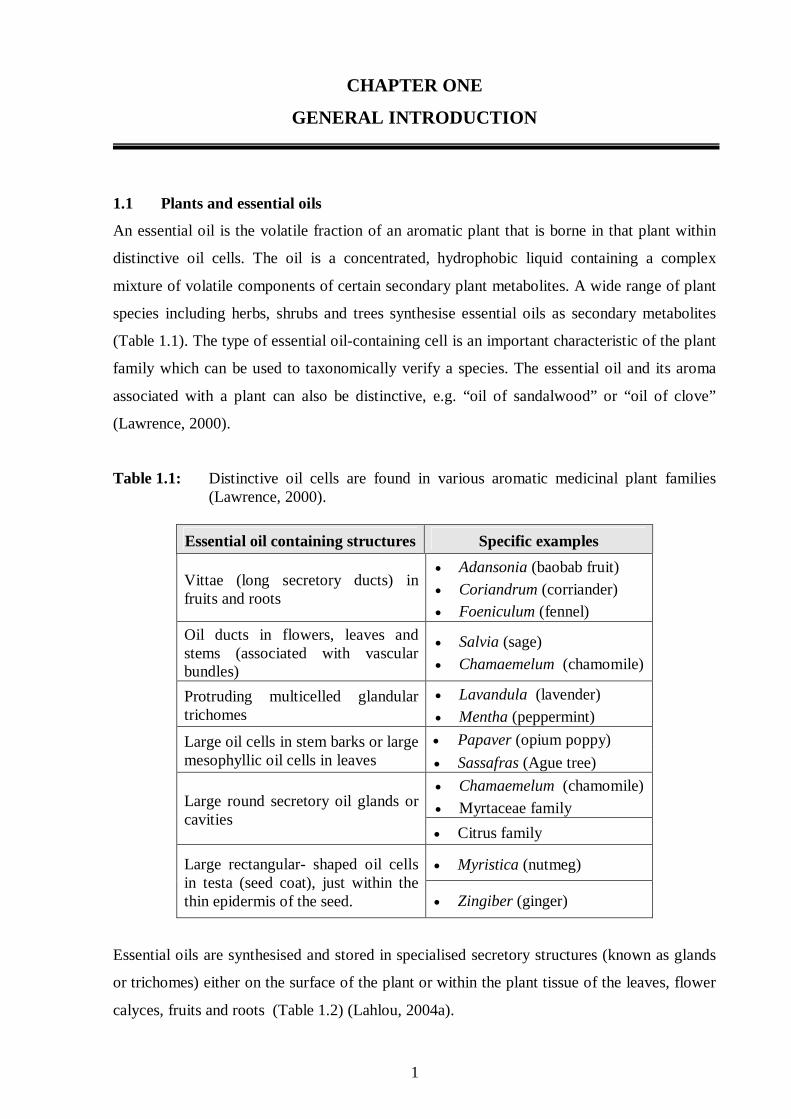
Table 1.1: Distinctive oil cells are found in various aromatic medicinal plant families (Lawrence, 2000).
Essential oil containing structures Specific examples
Vittae (long secretory ducts) in fruits and roots
• Adansonia (baobab fruit)
• Coriandrum (corriander) • Foeniculum (fennel)
Oil ducts in flowers, leaves and stems (associated with vascular bundles)
• Salvia (sage)
• Chamaemelum (chamomile)
Protruding multicelled glandular trichomes
• Lavandula (lavender)
• Mentha (peppermint)
• Papaver (opium poppy) Large oil cells in stem barks or large mesophyllic oil cells in leaves • Sassafras (Ague tree)
• Chamaemelum (chamomile)
• Myrtaceae family Large round secretory oil glands or cavities
• Citrus family
• Myristica (nutmeg) Large rectangular- shaped oil cells in testa (seed coat), just within the thin epidermis of the seed. • Zingiber (ginger)
Essential oils are synthesised and stored in specialised secretory structures (known as glands
or trichomes) either on the surface of the plant or within the plant tissue of the leaves, flower
calyces, fruits and roots (Table 1.2) (Lahlou, 2004a).
2
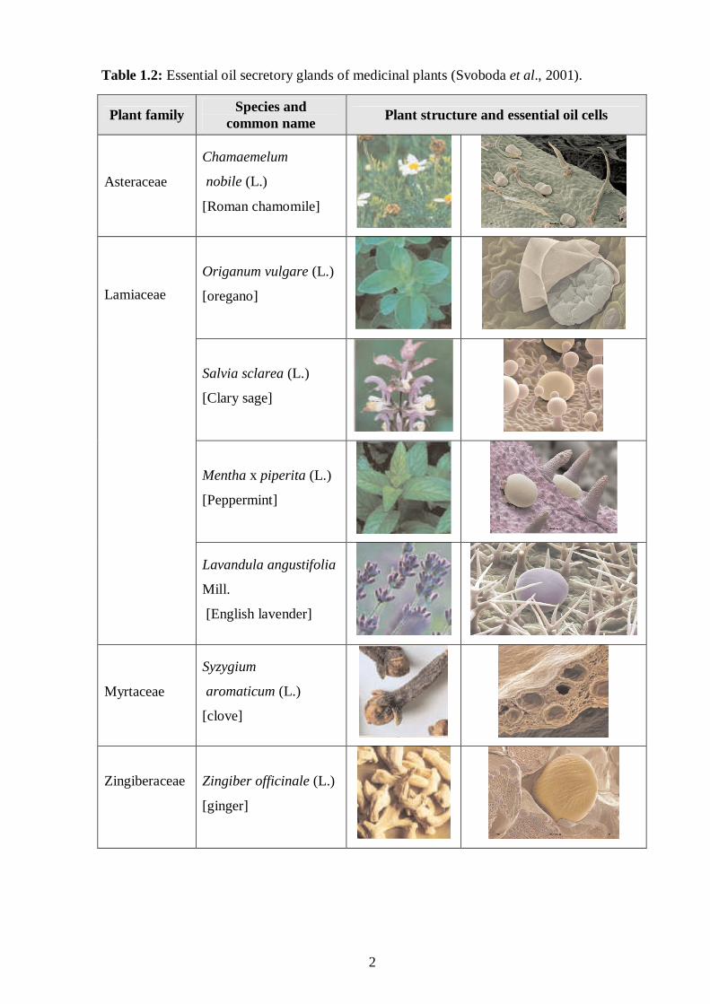
Table 1.2: Essential oil secretory glands of medicinal plants (Svoboda et al., 2001).
Plant family Species and
common name Plant structure and essential oil cells
Asteraceae
Chamaemelum
nobile (L.)
[Roman chamomile]
Origanum vulgare (L.)
[oregano]
Salvia sclarea (L.)
[Clary sage]
Mentha x piperita (L.)
[Peppermint]
Lamiaceae
Lavandula angustifolia
Mill.
[English lavender]
Myrtaceae
Syzygium
aromaticum (L.)
[clove]
Zingiberaceae
Zingiber officinale (L.)
[ginger]
3
There are various types of secretory structures where the oil content, larger size and thick
cuticularised lining differentiate them from adjacent non-secretory cells. These cell types are
found for example in ginger (Zingiber officinale) and nutmeg (Myristica fragrance). In
contrast to secretory cavities which consist of spherical structures lined with secretory cells
that produce the essential oil. Included in this group are the fruits and leaves from the citrus
and eucalyptus family and flower buds of cloves (Table 1.2) (Svoboda et al., 2001). Secretory
ducts, found in the Apiaceae and Asteraceae families, are formed once the cavities between
adjacent cells fuse. Cells in these cavities differentiate to form secretory epithelial cells.
Glandular trichomes found in lavender, oregano and mint are modified epidermal hairs with a
toughened cuticle that covers the trichome and encapsulates the essential oils (Table 1.2).
1.2 Use of essential oils in plants
The biological role of these secondary metabolites are still not fully understood, but they are
known to protect the plants against diseases, attract or repel insects, birds and other animals
via complex chemical mimicry (Halcon, 2002). For example, certain plants appear to protect
themselves from overgrazing by producing sesquiterpene lactones which are known to be
potent antifeedants. If eaten by herbivores, these substances affect the normal gastrointestinal
flora and interfere with digestion (Onawunmi et al., 1984). Corniferous plants secrete a
complex mixture of monoterpenes and sesquiterpenes in response to attack by insects and
predators (Mahmoud and Croteau, 2002).
1.3 History of essential oils
Essential oils have been used since antiquity by different cultures in a variety of ways as
medicines, in ritual worship and as perfumes (Halcon, 2002; Nakatsu et al., 2000). They were
very precious in the ancient world and were traded for gold, silver and even slaves. The first
nation to use essential oils were the Egyptians who created fragrances for personal use as well
as for ritual and ceremonial use in the temples and pyramids (Gollin, 1999). Some oils,
particularly frankincense, are cited repeatedly in religious texts of Judeo-Christians and
Muslims (Inouye et al., 2001). In 1817, an 870 foot long Ebers Papyrus dating back to 1500
B.C. was discovered listing over 800 herbal prescriptions and remedies (Jirovertz et al.,
1990). Many listed herbals contained oils which were used for embalming due to their
effective antibacterial properties and it is reported that the corpses of the Pharaohs were
embalmed with these oils (Gollin, 1999). Other oils such as lotus and sandalwood were also
widely used in ancient Egyptian purification and embalming rituals. The importance of these
essential oils is best displayed by the discovery of 350 litres of essential oils sealed and
4
preserved in alabaster jars found in Tutankhamen′s tomb when opened in 1922 (Inouye et al.,
2001). Even 3000 years after being encased in the jars, there was a faint odour of lotus oil and
frankincense.
The “Father of Modern Medicine,” Hippocrates (460BC) stated that “a daily aromatic bath
followed by a scented massage is a proper way to good health” (Lis-Balchin, 1997). This
practice was also employed by the Romans and Greeks who in addition to using aromatic oils
in their bath houses, diffused essential oils in their temples and political buildings. Physicians
of Greece including Hippocrates attended the Egyptian school of Cas to learn about the oils.
Amongst the medicinal properties taught were the antiseptic properties of clove and lemon
essential oils, hundreds of years prior to the discovery of modern antimicrobials used today
(Jirovertz et al., 1990).
The ancient Arabian people researched the chemical properties of essential oils and further
developed and refined the distillation process. Arabian perfumes were first discovered by
western man and brought back to Europe by the crusaders, where they became well known
and fashionable with the aristocracy (Buchbauer, 1993). During the Middle Ages the
antiseptic properties of essential oils became popular to the doctors of that time, such that they
carried these antiseptic aromatic oils in the handles of their walking sticks, which they held to
their noses during their visits to patients (Buchbauer, 1993).
The rediscovery of the antiseptic properties of essential oils is attributed to French cosmetic
chemist, René-Maurice Gattefossé, as a consequence of his personal experiences (Halcon,
2002). It was in July of 1910 when a laboratory explosion set him aflame. “It is reported that,
after extinguishing the flames his hands were quickly developing gas gangrene”. However,
“one rinse with lavender essential oil stopped this process and the next day healing began”.
This incident motivated Gattefossé to investigate the healing components of essential oils.
The research on essential oils prompted their clinical use on wounded World War II soldiers,
and wounds responded better to the oils than to the antibiotics of that time (Jirovertz et al.,
1990). Around this time another medical scientist Royal R. Rife discovered that essential oils
could effectively treat various bacterial diseases such as coughs and flu (Jirovertz et al.,
1990).

5
1.4 Isolation procedures of essential oils from plants
For an essential oil to be a true essential oil, it must only be isolated by physical means
(Nakatsu et al., 2000). The methods of isolating the essential oils from plants significantly
affect the chemical constituents and composition of the essential oil (Lahlou, 2004b). For
instance, α-pinene and β-pinene can be isomerized during the process and analysis, forming
camphene and possibly other monoterpenes in the process (Geron et al., 2000). Lawrence
(2000) stated that the most appropriate and convenient method to concentrate the essential oil
should be selected. If the activity is based on the mixture, and not on a single compound, then
all components should be concentrated from the isolates (Lawrence, 2000). Generally most
essential oil constituents are small, volatile and lipophilic compounds, thus a key
consideration is the need to separate them from aqueous plant materials (Lahlou, 2004b;
Lawrence, 2000; Nakatsu et al., 2000).
The physical methods used include distillation (hydrodistillation, steam distillation and
hydrodiffusion) (Figure 1.1), cold pressing (expression), maceration/distillation and
microwave irradiation (Lahlou, 2004b). In the hydrodistillation process, the plant material is
heated in two or three times its weight of water with indirect steam from outside the still. In
contrast, in steam distillation the plant material is isolated by direct steam produced in the
still, or by indirect steam produced outside and fed into the still. In hydrodiffusion, low
pressure steam (0.1 bar) replaces the volatile oil from the intact plant material by an osmotic
action (Lahlou, 2004b; Lawrence, 2000).
Figure 1.1: A schematic diagram showing a distillation apparatus used for the isolation of
essential oils (adapted from Nakatsu et al., 2000).

6
Cold pressing (Figure 1.2) is a technique used to isolate citrus peel oil and many other
essential oils from seeds, grains, kernels and fruits (Inouye et al., 2001; Nakatsu et al., 2000).
During this process, machines score the fruit/seed releasing the oil. Nakatsu et al. (2000)
mentioned that this method has recently been used to great advantage in isolating oxygen-
containing terpenes, which tend to degrade or rearrange when heat is applied.
Figure 1.2: An industrial machine used for isolating essential oils during cold pressing
process (Lahlou, 2004b).
During maceration/distillation the plant material is macerated in warm water to release the
enzyme-bound essential oil. Examples of essential oils produced by this technique are garlic,
wintergreen and bitter almond (Carson and Riley, 1995). Microwave irradiation is a modern
technique using microwaves to excite water molecules in the plant tissues, thus causing the
plant cells to rupture and release the essential oils trapped in the extracellular tissues of the
plant (Lahlou, 2004b).
Solvent isolation is used to isolate essential oils from those plants that cannot withstand the
process of distillation and heat (e.g. jasmine). A solvent such as hexane or supercritical CO2
can be used to isolate the essential oil and is then removed with alcohol and distilled to obtain
the required amount of essential oil (Lahlou, 2004b).

7
1.5 The chemistry of essential oils
Various factors influence the quantity and composition of the essential oil synthesised by
plants. These include soil conditions, geographical and seasonal variations, climate (rainfall)
and altitude; as well as harvest method, isolation technique and which part of the plant is used
(Lahlou, 2004b). In addition, each plant can produce several different chemotypes with
biochemical variations that change the composition and ratio of essential oil constituents and
ultimately influence its therapeutic efficacy (Viljoen et al., 2005).
Essential oils are volatile, complex mixtures, composed of numerous terpene hydrocarbons
and oxygenated compounds (Griffin et al., 1999; Lis-Balchin, 1997). These constituents are
not only volatile, but also lipophilic (Griffin et al., 1999). All terpenes are synthesized
through the condensation of isopentyl diphosphate (IPP) and its allylic isomer, dimethylallyl
diphosphate (DMAPP as seen in Figure 1.3) (Mahmoud and Croteau, 2002).
OPP OPP
IPP DMAPP
OPP
GPP
OPP
FPP Sesquiterpenes (C15)
Triterpenes (C30)
Monoterpenes (C10)
Figure 1.3: An overview of isoprenoids biosynthesis pathway in plants (adapted from
Mahmoud and Croteau, 2002). Where IPP = isopentyl diphosphate;
GPP = geranyl pyrophosphate; FPP = farnesyl pyrophosphate and DMAPP =
dimethylallyl diphosphate.
8
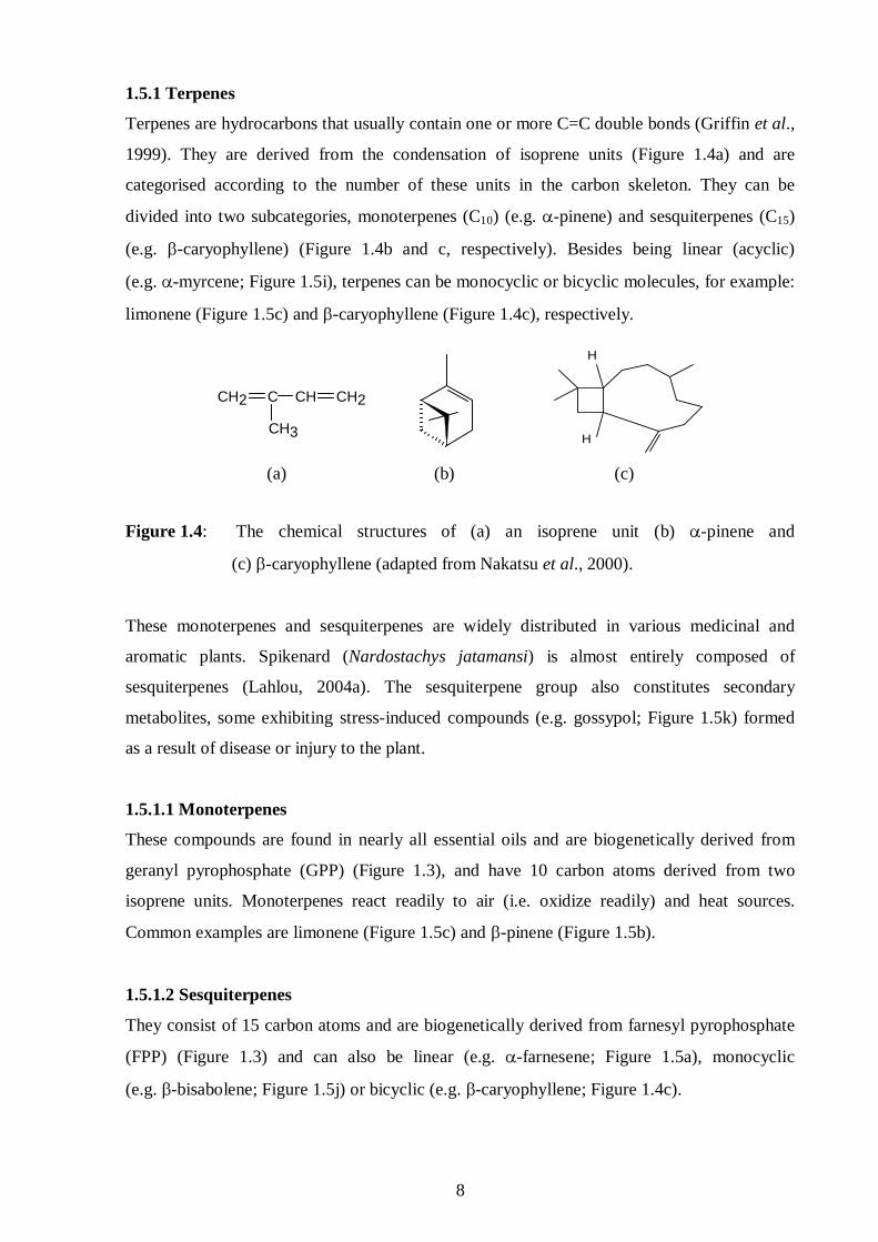
1.5.1 Terpenes
Terpenes are hydrocarbons that usually contain one or more C=C double bonds (Griffin et al.,
1999). They are derived from the condensation of isoprene units (Figure 1.4a) and are
categorised according to the number of these units in the carbon skeleton. They can be
divided into two subcategories, monoterpenes (C10) (e.g. α-pinene) and sesquiterpenes (C15)
(e.g. β-caryophyllene) (Figure 1.4b and c, respectively). Besides being linear (acyclic)
(e.g. α-myrcene; Figure 1.5i), terpenes can be monocyclic or bicyclic molecules, for example:
limonene (Figure 1.5c) and β-caryophyllene (Figure 1.4c), respectively.
Figure 1.4: The chemical structures of (a) an isoprene unit (b) α-pinene and
(c) β-caryophyllene (adapted from Nakatsu et al., 2000).
These monoterpenes and sesquiterpenes are widely distributed in various medicinal and
aromatic plants. Spikenard (Nardostachys jatamansi) is almost entirely composed of
sesquiterpenes (Lahlou, 2004a). The sesquiterpene group also constitutes secondary
metabolites, some exhibiting stress-induced compounds (e.g. gossypol; Figure 1.5k) formed
as a result of disease or injury to the plant.
1.5.1.1 Monoterpenes
These compounds are found in nearly all essential oils and are biogenetically derived from
geranyl pyrophosphate (GPP) (Figure 1.3), and have 10 carbon atoms derived from two
isoprene units. Monoterpenes react readily to air (i.e. oxidize readily) and heat sources.
Common examples are limonene (Figure 1.5c) and β-pinene (Figure 1.5b).
1.5.1.2 Sesquiterpenes
They consist of 15 carbon atoms and are biogenetically derived from farnesyl pyrophosphate
(FPP) (Figure 1.3) and can also be linear (e.g. α-farnesene; Figure 1.5a), monocyclic
(e.g. β-bisabolene; Figure 1.5j) or bicyclic (e.g. β-caryophyllene; Figure 1.4c).
H
H
(a) (b) (c)
CH2 C
CH3
CH CH2

9
Figure 1.5: Various chemical structures of essential oil constituents (a - j) and a secondary
metabolite (k = gossypol) showing some degree of cyclization (adapted from
Nakatsu et al., 2000).
1.5.2 Terpenoids
These are oxygen-containing analogues of the terpenes and are subdivided according to the
number of carbon atoms in the same manner as are terpenes and can be linear (e.g. nerolidol;
Figure 1.5g), monocyclic (e.g. pulegone; Figure 1.5e) or bicyclic (α+β thujone; Figure 1.5d).
The skeleton of terpenoids may differ from just adding isoprene units by the loss or shift of a
fragment, generally a methyl group is replaced or shifted by an O2 in various functional
groups (Nakatsu et al., 2000). These oxygenated compounds belong to a number of different
chemical groups including the alcohols (nerolidol), aldehydes (citronellal), esters (linalyl
acetate), ketones (fenchone), oxides (1,8-cineole) and phenols (eugenol). Terpenoids are
usually very expensive, because like most secondary metabolites they are synthesized by
plants in relatively small amounts (Lawrence, 2000). In addition, due to their complex
O
(a) α-Farnesene b) β-Pinene (c) Limonene (d) α+β-Thujone
o
OH
OH
(e) Pulegone (f) Linalool (g) Nerolidol (h) α-Bisabolol
HO
HO
CHO CHO
HO
HO
(i) α-Myrcene (j) β-Bisabolene (k) Gossypol
OH
H

10
structures, most terpenoids cannot be obtained in a profitable way through chemical synthesis
and as are normally isolated by steam distillation (Lahlou, 2004b).
1.5.2.1 Alcohols
Alcohols are not water soluble and are less prone to oxidation and for this reason they
evaporate quite slowly (Nakatsu et al., 2000). They can further be subdivided into
monoterpene alcohols (e.g. linalool; Figure 1.5f) and sesquiterpene alcohols (e.g. α-bisabolol;
Figure 1.5h). Other examples are nerol, geraniol, citronellol and patchoulol.
1.5.2.2 Aldehydes
The majority of the lemon scented oils fall into this group (Onawumni et al., 1984), and
include citral, geranial, neral and citronellal.
1.5.2.3 Esters
Esters sometimes have a trademark fruity aroma and are formed when acids react with
alcohols (Cowan, 1996). Linalyl acetate, neryl acetate and geranyl acetate are common
examples.
1.5.2.4 Ketones
The ketones consist of some of the most toxic elements of essential oils (Zhou et al., 2004).
Some of these include α+β-thujone (Figure 1.5d) and pulegone (Figure 1.5e).
1.5.2.5 Oxides
Oils containing oxides are generally camphoraceous in nature (Nakatsu et al., 2000). This
group contains essential oil constituents like asarone and ascaridol and some non-toxic oxides
like 1,8-cineole (eucalyptol), bisabolol oxide and linalool oxide.
1.5.2.6 Phenols
Phenols found in essential oils normally have a carbon side chain and are more hydrophilic
and evaporate more quickly than oils that do not contain phenols (Nakatsu et al., 2000).
Examples of phenols are eugenol, thymol, carvacrol, gaiacol, chavicol and australol.
1.6 Medicinal applications and adverse effects of essential oils
In humans, essential oils exhibit a wide spectrum of pharmacological activities such as
infection control, wound healing, pain relief, nausea, inflammation and anxiety
11
(Onawunmi et al., 1984; Halcon, 2002; Kalemba and Kunicka, 2003; Shin and Kang, 2003).
Traditional medicines containing essential oils have been scientifically proven to be effective
in treating various ailments like malaria and others of microbial origin (Campbell et al., 1997;
Cragg et al., 1997; Lopes et al., 1999; Nakatsu et al., 2000; Goulart et al., 2004).
Nakatsu et al. (2000) reported that although each essential oil is different in composition,
there is considerable overlap in their actions. For example, essential oils of Lippia javanica,
Tetradenia riparia and Virola surinamensis have reported antimalarial activities yet they have
different essential oil profiles (Lukwa, 1994; Campbell et al., 1997; Lopes et al., 1999).
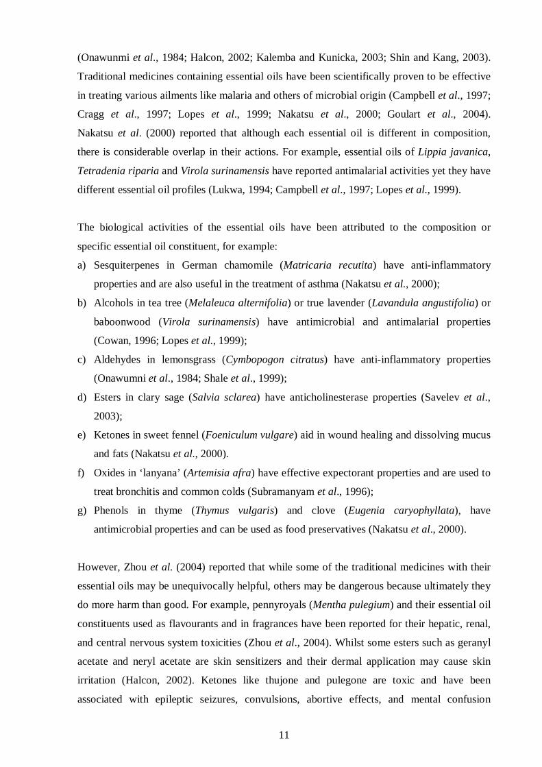
The biological activities of the essential oils have been attributed to the composition or
specific essential oil constituent, for example:
a) Sesquiterpenes in German chamomile (Matricaria recutita) have anti-inflammatory
properties and are also useful in the treatment of asthma (Nakatsu et al., 2000);
b) Alcohols in tea tree (Melaleuca alternifolia) or true lavender (Lavandula angustifolia) or
baboonwood (Virola surinamensis) have antimicrobial and antimalarial properties
(Cowan, 1996; Lopes et al., 1999);
c) Aldehydes in lemonsgrass (Cymbopogon citratus) have anti-inflammatory properties
(Onawumni et al., 1984; Shale et al., 1999);
d) Esters in clary sage (Salvia sclarea) have anticholinesterase properties (Savelev et al.,
2003);
e) Ketones in sweet fennel (Foeniculum vulgare) aid in wound healing and dissolving mucus
and fats (Nakatsu et al., 2000).
f) Oxides in ‘lanyana’ (Artemisia afra) have effective expectorant properties and are used to
treat bronchitis and common colds (Subramanyam et al., 1996);
g) Phenols in thyme (Thymus vulgaris) and clove (Eugenia caryophyllata), have
antimicrobial properties and can be used as food preservatives (Nakatsu et al., 2000).
However, Zhou et al. (2004) reported that while some of the traditional medicines with their
essential oils may be unequivocally helpful, others may be dangerous because ultimately they
do more harm than good. For example, pennyroyals (Mentha pulegium) and their essential oil
constituents used as flavourants and in fragrances have been reported for their hepatic, renal,
and central nervous system toxicities (Zhou et al., 2004). Whilst some esters such as geranyl
acetate and neryl acetate are skin sensitizers and their dermal application may cause skin
irritation (Halcon, 2002). Ketones like thujone and pulegone are toxic and have been
associated with epileptic seizures, convulsions, abortive effects, and mental confusion

12
(Zhou et al., 2004). Some oxides such as asarone and ascaridol may cause convulsions
(Nakatsu et al., 2000). Another example of an essential oil containing oxides is peppermint
oil. In high oral doses it has produced severe toxic reactions, and its major component,
menthol, has been associated with apnoea and severe jaundice in babies and patients with
glucose-6-phosphate dehydrogenase (G6PD) deficiency, and hepatotoxicity (Halcon, 2002;
Zhou et al., 2004).
1.7 Route of administration
When applying essential oils for any medical reasons, the route of administration is selected
based on pathophysiology, desired outcome, safety and toxicity data, professional practice
parameters, and cultural preferences (Cowan, 1996; Halcon, 2002). For example, although
inhalation is the best route of administration for treating respiratory symptoms and for
affecting mood or cognition, topical application is likely to work best for burns, wound care
and most dermatological conditions. For instance, essential oils of ‘lanyana’ (Artemisia afra)
can be inhaled or used as a chest rub (topically) to treat common colds and bronchial
disorders (Viljoen et al., 2006). Whilst essential oils of lavender (Lavandula angustifolia)
may be applied topically to treat burns and wound infections (Nakatsu et al., 2000).
Some essential oils, such as Eucalyptus globulus, can be toxic even when a teaspoon is
ingested. Thus, the patient or provider should choose another route of administration or
another species of Eucalyptus that has similar therapeutic effects, but presents less danger of
toxicity (Cowan, 1996; Halcon, 2002). Internal application of essential oils by suppository or
oral ingestion is very common in places such as France, where essential oils are prescribed by
physicians. While in the United Kingdom and the United States topical preparations and
inhalation are common, where they are employed by nurses and other health care
professionals (Halcon, 2002). In Africa, oral ingestion, topical application and inhalation of
essential oils are common routes of administration and are commonly used by traditional
healers.
1.8 The commercial importance of essential oils
Essential oil terpenes such as α-pinene, d-limonene and turpentine are used as a component of
semi-aqueous cleaning solutions or by themselves (Mahmoud and Corteau, 2002; Nakatsu et
al., 2000). Terpenes are volatile organic compounds, thus flammable. Regardless, in the
industrial world they are used as solvents for rosin fluxes, fingerprints, heavy petroleum
greases, and oils (Constantin et al., 1991). In addition, they can be used with immersion and

13
ultrasonic systems, often working well at room temperature (Constantin et al., 1991).
Terpenes may produce explosive mists when sprayed, thus they should only be used in spray
applications with proper safety precautions, such as inert gas blanketing (Halcon, 2002).
Moreover, they need to be incinerated at an approved facility and not be discarded via
drainage system (Constantin et al., 1991).
Overall essential oils are pertinent for pharmaceutical, cosmetic and food research and
development, and are widely viewed as templates for structure optimization programs with
the goal of creating new drugs (Cragg et al., 1997). Essential oils forming part of the bioactive
molecules of medicinal plants have been in existence for much longer than any drug company
(De Smet, 1997). Because most essential oil constituents like terpenoids are biologically
active, they are employed for medical purposes. For instance, the antimalarial drug
artemisinin (from Artemisia annua) and the anticancer drug paclitaxel (Taxol®) are of a
terpene nature with an established medical application (Cowan, 1996; Cragg et al., 1997; De
Smet, 1997).
With respect to the pharmacokinetic profile of essential oils, it is believed that they are
absorbed and metabolized according to their chemical composition, dose and route of
administration, as is the case with other substances (Halcon, 2002). However, this cannot be
assumed. To avert this serious dilemma such that essential oils can be clinically used,
scientific evidence confirming their safety and efficacy including their pharmacokinetics (i.e.
their bioavailabilty, distribution rate and dosage regimen) are necessary (Cragg et al., 1997).
This requires phytotherapeutic and pharmacological studies to confirm their use as medical
drugs or lead molecules for novel drug development.
1.9 Public interest in the use of essential oils
The use of essential oils forms the basis of a fascinating plant-based traditional medicine
system that has been utilized in countries such as China, France and India, but predominantly
in the developing countries of the African continent where finance to buy classical synthetic
drugs is a major challenge (Nakatsu et al., 2000). In addition, one of the issues driving the
interest in essential oils is the increased public use of complementary therapies. As a result,
health sciences education and training in complementary therapies have taken a lead to
encourage medical personnel to be aware of the risk and benefits of essential oils and to also
educate their patients regarding these issues (Shale et al., 1999).

14
In general, public interest in the therapeutic uses of essential oils has grown much faster than
the available scientific research to back it up. There is, however, a growing body of published
laboratory and human studies on specific essential oils that are employed for selected health
outcomes. This information can aid practitioners in advising patients and can provide
direction for future clinical research (Halcon, 2002). Among South African medicinal plants
‘lanyana’ (Artemisia afra) is a common example that is used extensively as an antibacterial
and an antifungal and is also used as a component of fragrances in toiletries, cosmetics and
perfumes (Subramanyam et al., 1996).
1.10 Essential oils in the twentieth century
The volatile compounds from plants have played an important part in the medical field
throughout the twentieth century (Cragg et al., 1997; De Smet, 1997). Peppermint oil is a
good example of an essential oil that has a history of continuous wide use and holds promise
for medicine, but also warrants caution because of its potential side effects (De Smet, 1997;
Shale et al., 1999). Peppermint in a variety of forms can be found in toothpaste and countless
other products and foods (Halcon, 2002). Although, numerous studies on essential oils have
been undertaken by the food, cosmetics, and tobacco industries, access to the information by
the healthcare community has been limited (Halcon, 2002; Mahmoud and Corteau, 2002;
Nakatsu et al., 2000). Essential oils of thyme and clove are also ubiquitous as food and
cosmetic additives, drawing on their well known preservative (antimicrobial) properties
(Nakatsu et al., 2000).
1.11 Single versus combination therapy
From the historical perspective, people traditionally using medicines believed that complex
diseases could be treated with a 'single magic herb'. A perfect example is that of Melaleuca
alternifolia (tea tree) and its essential oils, which has been widely used over the past 30 years
(Carson and Riley, 1995; Cox et al., 2001). Numerous studies have reported the efficacy of
M. alternifolia essential oil against a variety of pathogenic microorganisms including many
Staphylococcus aureus isolates (including methicillin-resistant S. aureus), as well as other
bacteria and fungi (Carson and Riley, 1995; Cox et al., 2001). However, it is also important to
note that many plants are referred to as “tea tree,” but that the above applies only to M.
alternifolia (Carson and Riley, 1995).
Traditionally in South Africa, aromatic plants used in combination include the following few
examples. The Cape snowbush (Eriocephalus africanus) can be blended with sandalwood
15
(Santalum album) or bergamot (Citrus bergamia) and the mixtures applied topically or via
inhalation for the treatment of depression, colds and flu, menstrual problems, muscular aches
and pain (Constantin et al., 1991). Another example is the combination of buchu (Agathosma
betulina) with lavender (Lavandula angustifolia), when applied topically or via inhalation are
useful in the treatment of burns, wound infections and as an anxiolytic/sedative
(Subramanyam et al., 1996).
It is unequivocally incorrect and unwise to rely on monotherapy, as a possible drawback is the
tendency of the organisms to develop resistance to the antimicrobial and antimalarial drug
(Cox et al., 2001). If medical science has to rely on a single therapeutic compound to
eradicate disease, it would mean a serious catastrophe to human health as microbial resistance
is already increasing at an unprecedented rate (Cassella et al., 2002). It is widely accepted that
agents should be used in combination to avoid treatment failure (Bell, 2005). Thus, the
discovery of new and effective chemical entities is urgently required. Essential oil
constituents are possible candidates to be used in combination and together with standard
antimicrobials to combat this dilemma.
Cassella et al. (2002) reported that it is an accepted premise by practising aromatherapists,
that essential oils act better when used in combination in order to optimise their efficacy. The
interaction of two or more drugs is said to be synergistic if the potency of the combination is
higher than the expected activity of the individual drug activity. However, if the potency of
the combination is lower, then the end result is defined as an antagonistic interaction
(Bell, 2005). It has been claimed that the most dominating constituents in the essential oil are
the ones eliciting the activity attributed to the whole oil (Nakatsu et al., 2000). Despite this
fact, constituents in very small amounts are often found to be as useful as the principal
constituent (Cassella et al., 2002; Nakatsu et al., 2000). For example, when the antibacterial
activity of the whole oil of thyme (Thymus vulgaris) was compared to its principal
constituents (borneol, carvacrol, camphene, thymol, p-cymene and α-pinene) the whole oil
was more active than these individual constituents (Nakatsu et al., 2000; Viljoen et al., 2006).
This suggests that minor essential oil constituents played a significant role in the biological
activity of this essential oil.
Moreover, synergistic and antagonistic effects could also be achieved by using the essential
oils in combination, but also by combining the essential oils with other standard drugs.
However, there are few published studies investigating such interactions (Cassella et al.,

16
2002), which are vital since essential oils have great potential as adjuvants in the symptomatic
management or treatment of a wide range of health problems (Cowan, 1996; De Smet, 1997).
1.12 Future of essential oils
Essential oils applied at the correct concentrations and appropriately administered have
resulted in few known complications (Gollin, 1999). As such, physicians who want to include
the use of essential oils in their therapies must review literature, participate in established
courses, or partner with a qualified and experienced individual (Gollin, 1999). Halcon (2002)
mentioned that conservatism is essential to protect patients, but it is also vital to recognize
that exposure to essential oils is neither new nor rare. The increasing public and professional
interest in essential oils is prompting an urgent need for laboratory and clinical research to
expand and clarify the evidence base as well as for additional research on safety and toxicity.
The World Health Organization (WHO) Guidelines for the Assessment of Herbal Medicines
(WHO, 2003) allows variation in the usual clinical trial path in the case of therapeutic
substances that have a long history of apparent safe usage. Some essential oils including those
of M. alternifolia may fit this criterion (Cox et al., 2001). Aromatic plants will continue to be
used in South Africa as long as the majority of people keep on consulting traditional healers.
Thus, the layperson as well as the medical and traditional healthcare workers need to be better
informed in order to optimize safety and therapeutic applications of essential oil preparations
(Nchinda, 1998).
While the reported biological properties are quite varied, there are numerous studies that
report on the efficacy of essential oils against bacteria, fungi and parasites (e.g. Plasmodium
falciparum) and their use as anti-oxidants and acetylcholinesterase inhibitors (Burfiled and
Reekie, 2005; Goulart et al., 2004; Nakatsu et al., 2000; Atindehou et al., 2004; Pauli and
Schilcher, 2004). However, very little information is known about the contribution of the
individual essential oil constituents to the overall activity of the crude essential oils and their
combined interaction with each other and standard drugs. Thus, this project was undertaken to
contribute and enhance this versatile and growing reservoir of scientific knowledge on
essential oils.

17
1.13 Study objectives
1. To assess the biological activity of twenty specific essential oil constituents from seven
structural chemical groups. Activities included:
1.1 Antibacterial and antifungal
1.2 Antimalarial
1.3 Anti-oxidant
1.4 Anticholinesterase
2. To determine the toxicity profile of the essential oil constituents.
3. To determine whether the combination of selected essential oil constituents interact in a
synergistic, additive, indifferent or antagonistic manner with each other, as well as with
standard antimalarial and antimicrobial agents.
18
CHAPTER TWO
SELECTION OF ESSENTIAL OIL CONSTITUENTS
2.1 Introduction
Essential oils are complex mixtures comprising many single compounds, with each of these
constituents contributing to the beneficial or adverse effects of the oils (Dormans and Deans,
2000). Many essential oils have been applied for centuries in local healing rites, however,
their composition and biological activity are poorly recorded (Nakatsu et al., 2000; Zhou et
al., 2004). The analysis of essential oils is generally performed using gas chromatography
(GC; quantitative analysis) and gas chromatography-mass spectroscopy (GC-MS; qualitative
analysis) (Lahlou, 2004b).
Identification of the main components is carried out by the comparison of both the GC
retention times and mass spectroscopic (MS) data against those of the reference standards.
Lahlou (2004b) reported that, analytical conditions and procedures used should carefully be
described and these include:
• Apparatus of oil analysis (make and model number of the equipment);
• Column type and dimensions;
• Carrier gas flow rate;
• The temperature programming conditions including injector temperature, detector and
column temperatures, in addition to mass spectra (electronic impact).
Many essential oils are isolated, analysed and their main components are identified,
characterized and then published without any biological testing whatsoever (Campbell et al.,
1997; Nakatsu et al., 2000). Their useful biological activities can remain unknown for years
(Carson and Riley, 1995; Cowan, 1996). Therefore, there is an urgent need to increase the
knowledge base on the potential uses and benefits of essential oils and the specific
constituents. Guided by the fact that essential oil constituents vary quantitatively between
various plant species (Halcon, 2002), twenty essential oil constituents (EOC´s) were selected
based on the reported biological activity.

19
2.2 Methodology
2.2.1 Materials used
Twenty EOC´s were purchased from Sigma-Aldrich (USA) and Fluka (Switzerland). These
EOC´s included both terpene hydrocarbons and oxygenated terpenes (Figure 2.1 and Table
2.1).
2.2.2 Gas chromatographic analysis of essential oil constituents
Analysis of all standards was performed on a Shimadzu 17A gas chromatograph using the
following conditions; Column: J&W-DB1 (60m x 0.25mm id., 0.25µm film thickness);
Temperatures: injection port 230°C, column 60°C for 1min, 5°C/min to 180°C, 180°C for
2min, (total = 25min). Helium was used as a carrier gas.
2.2.3 The chromatogram
The chromatogram is observed as a series of peaks where each peak represents a chemical
compound. The x-axis represents the time scale and the time at which the peak is recorded is
called the retention time (Rt). The peak height and peak area is an indication of the quantity of
the compound in the mixture. The peak area is integrated as a percent of the total. The purity
of the purchased EOC´s was verified by GC to those supplied by Sigma-Aldrich and Fluka
(Figure 2.1 and Table 2.2). The specific chirality was not confirmed and the suppliers
specifications were considered correct.

20
(a) (-)-α-Bisabolol (b) trans-Geraniol (c) (±)-Linalool (d) E- and Z-(±)-Nerolidol (e) (-)-Citronellal (f) Geranyl acetate (g) Linalyl acetate (h) (±)-α+ β-Thujone (i) (+)-Carvone (j) (-)-Fenchone (k) (-)-Menthone (l) (-)-Pulegone (m) Carvacrol (n) Eugenol (o) (+)-α-Pinene (p) (+)-β-Pinene (q) γ-Terpinene (r) (R)-(+)-Limonene (s) p-Cymene (t) 1,8-Cineole
Figure 2.1: The chemical structures of 20 essential oil constituents from seven different
structural groups (Nakatsu et al., 2000).
o
OH
OH
o-Ac
o-AcO
O O
O O
OH
o-Me
CHO
OH
HOH
OH

21
Table 2.1: Properties of 20 selected essential oil constituents (values and synonyms have been obtained from the cited
references as well as from the supplied compounds, Sigma-Aldrich or Fluka, 2006 and Merck, 2001).
Essential oil constituents
Chemical structure
Empirical formula
Molecular weight (g/mol)
Density (g/l)
Boiling point (°C/mmHg) Synonyms References
Alcohols
(-)-α-Bisabolol Figure 2.1 a
C15H26O 222.37 0.929 157-158 6-Methyl-2-(4-methyl-3-cyclohexene-1-yl)-5-hepten-2-ol
Mahmoud and Croteau (2002)
trans-Geraniol Figure 2.1 b
C10H18O 154.25 0.889 229-230 trans-3,7-Dimethyl-2,6-octadien-1-ol; (±)-3,7-Dimethyl-1,6-octadien-3-ol
Nakatsu et al. (2000)
(±)-Linalool Figure 2.1 c
C10H18O 154.25 0.870 194-197 (±)-3,7-Dimethyl-3-hydroxy-1.6-octadien-1-ol
Lukwa (1994)
E- and Z-(±)Nerolidol Figure 2.1 d
C15H26O 222.37 0.875 114 3,7,11-Trimethyl-1,6,10-dodecatrien-3-ol
Nakatsu et al. (2000)
Aldehyde
(-)-Citronellal Figure 2.1 e
C10H18O 154.25 0.851 207 (S)-3,7-Dimethyl-6-octenal; Hydroxy-citronellal
Nakatsu et al. (2000)
Esters
Geranyl acetate Figure 2.1 f
C12H20O2 196.29 0.916 200-201 trans-3,7-Dimethyl-2,6-octadien-1-yl acetate; Geraniol acetate
Onawumni et al. (1984)
Linalyl acetate Figure 2.1 g
C12H20O2 196.29 0.895 115-116 3,7-Dimethyl-1,6-octadien-3-yl acetate
Onawumni et al. (1984)
21

22
Table 2.1 continued: Properties of 20 selected essential oil constituents.
Essential oil constituents
Chemical structure
Empirical formula
Molecular weight (g/mol)
Density (g/l)
Boiling point
(°C/mmHg) Synonyms References
Ketones
(cis+trans)-(±)α+β-Thujone
Figure 2.1 h
C10H16O 152.23 0.920 100 1-Isopropyl-4-methylbicyclo [3.1.0] hexan-3-one
Lahlou (2004b)
(+)-Carvone Figure 2.1 i
C10H14O 150.22 0.960 228-230 p-Mentha-6,8-diene-2-one; (S)-5-Isopropenyl-2-methyl-2-cyclohexenone
Campbell et al. (1997)
(-)-Fenchone
Figure 2.1 j
C10H16O 152.24 0.945 191-195
(1R)-1,3,3-Trimethylbicyclo[2.2.1] heptan-2-one; 2-Norbornanone; (−)-1,3,3-Trimethyl-2-norbornanone; (-)-1,3,3-Trimethylnorcamphor
Nakatsu et al. (2000)
(-)-Menthone Figure 2.1 k
C10H18O 154.25 0.896 85-88 ρ-Menthan-3-one; 2-Isopropyl-5-methylcyclohexanone
Zhou et al. (2004)
(-)-Pulegone Figure 2.1 l
C10H16O 152.24 0.936 223-224 (S)-2-Isopropylidene-5-methylcyclo-hexanone; ρ-Menth-4-(8)-en-3-one
Zhou et al. (2004)
Phenols
Carvacrol Figure 2.1 m
C10H14O 150.22 0.976 236-237 5-Isopropyl-2-methylphenol; ρ-Cymen-2-ol
Nakatsu et al. (2000)
Eugenol Figure 2.1 n
C10H12O2 164.21 1.067 254 2-Methoxy-4-(2-propenyl) phenol ; 4-Allylguaiacol
Nakatsu et al. (2000)
22
23
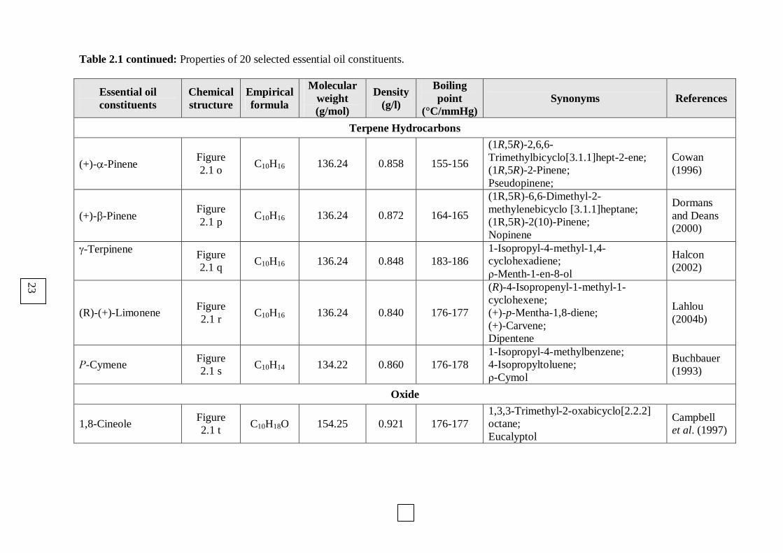
Table 2.1 continued: Properties of 20 selected essential oil constituents.
Essential oil constituents
Chemical structure
Empirical formula
Molecular weight (g/mol)
Density (g/l)
Boiling point
(°C/mmHg) Synonyms References
Terpene Hydrocarbons
(+)-α-Pinene Figure 2.1 o
C10H16 136.24 0.858 155-156
(1R,5R)-2,6,6-Trimethylbicyclo[3.1.1]hept-2-ene; (1R,5R)-2-Pinene; Pseudopinene;
Cowan (1996)
(+)-β-Pinene Figure 2.1 p
C10H16 136.24 0.872 164-165
(1R,5R)-6,6-Dimethyl-2-methylenebicyclo [3.1.1]heptane; (1R,5R)-2(10)-Pinene; Nopinene
Dormans and Deans (2000)
γ-Terpinene Figure 2.1 q
C10H16 136.24 0.848 183-186 1-Isopropyl-4-methyl-1,4-cyclohexadiene; ρ-Menth-1-en-8-ol
Halcon (2002)
(R)-(+)-Limonene Figure 2.1 r
C10H16 136.24 0.840 176-177
(R)-4-Isopropenyl-1-methyl-1-cyclohexene; (+)-p-Mentha-1,8-diene; (+)-Carvene; Dipentene
Lahlou (2004b)
Ρ-Cymene Figure 2.1 s
C10H14 134.22 0.860 176-178 1-Isopropyl-4-methylbenzene; 4-Isopropyltoluene; ρ-Cymol
Buchbauer (1993)
Oxide
1,8-Cineole Figure 2.1 t
C10H18O 154.25 0.921 176-177 1,3,3-Trimethyl-2-oxabicyclo[2.2.2] octane; Eucalyptol
Campbell et al. (1997)
23

24
Table 2.2: Verification of percentage purity of 20 selected essential oil constituents.
Essential oil constituents Determined Purity (%) Reported Purity (%)
Alcohols
(-)-α-Bisabolol 90.32 ~97.0
trans-Geraniol 95.65 98.0
(±)-Linalool 97.00 97.0
E- and Z-(±)-Nerolidol 94.43 98.0
Aldehyde
(-)-Citronellal 100.00 ≥98.0
Esters
Geranyl acetate 98.73 98.0
Linalyl acetate 96.81 97.0
Ketones
(±)-α+β-Thujone 98.73 ~98.0
(+)-Carvone 96.81 ≥99.0
(-)-Fenchone 96.81 ~98.0
(-)-Menthone 80.85 ≥97.0
(-)-Pulegone 98.62 ~99.0
Phenols
Carvacrol 95.48 ~98.0
Eugenol 96.32 ~97.0
Terpene Hydrocarbons
(+)-α-Pinene 95.48 ~99.0
(+)-β-Pinene 96.32 ≥97.0
γ-Terpinene 94.71 98.0
(R)-(+)-Limonene 92.98 97.0
Ρ-Cymene 93.19 ≥99.5
Oxide
1,8-Cineole 93.13 ≥99.7
25
CHAPTER THREE
ANTIMALARIAL ACTIVITY
3.1 Definition of malaria
Malaria is a common public health catastrophe affecting millions of people throughout the
tropics and subtropics (Nchinda, 1998). People thought that this life threatening disease came
from fetid marshes, hence the name ´mal aria` (bad air) (Hayward et al., 2000). Later in 1880,
scientists discovered the real cause, a one-cell protozoal parasite named Plasmodium
(Hayward et al., 2000). It was found to be transmitted from person to person through the bite
of a female Anopheles mosquito, which depends on a blood meal to mature her eggs
(Nchinda, 1998). Less common modes of transmission include: inoculation of infected blood,
use of contaminated needles and from an infected mother to her infant during pregnancy
(Nchinda, 1998).
3.1.1 Malaria burden
Today malaria causes more than 300 million acute illnesses and at least one million deaths
annually in the world (Atindehou et al., 2004; Benoit-Vical et al., 1999; WHO, 2005).
It continues to be a major obstacle in the social and economic development of developing
countries. In the last decade, the prevalence of malaria has been escalating at an
unprecedented and uncontrolled rate, especially in Africa (WHO, 2005). Cases in Africa
account for 90% of malaria cases in the world, and the disease reduces the working capacity
of those infected (Nchinda, 1998). It affects the overall quality of life and undermines efforts
at sustainable development. Young adults and adolescents are now dying of severe forms of
the disease, where in Africa alone it is estimated that more than a million children die of
malaria each year (Boyom et al., 2003). Between 1994 and 1996, malaria epidemics in 14
countries of Sub-Saharan Africa resulted in an unacceptably high number of deaths, in many
areas previously free of the disease (Nchinda, 1998). In South Africa alone about 13399
malaria cases and 89 fatal cases were reported in 2004, while in 2005, 4539 malaria cases and
35 deaths were reported (Department of Health, 2005).
3.1.2 Malaria distribution
Many of the tropical developing countries in Africa, America and Asia continue to experience
the occurrence of malaria (WHO, 2005). In South Africa, malaria occurs more commonly in
low altitude areas below 1000m in north eastern KwaZulu-Natal, Limpopo and Mpumalanga.

26
Other limited areas in which malaria may occasionally occur are the north west and northern
Cape Provinces along the Molopo and Orange rivers. Malaria is a distinctly seasonally based
problem with the highest risk occuring during the wet summer months (October to May)
(Department of Health, 2005). Malaria cases have also been reported in Gauteng with 366
cases and five deaths being reported since the beginning of 2006. These cases were due to
people returning from holiday in countries outside the borders of South Africa. But cases
reported in Johannesburg have been attributed to mosquitoes being imported from malaria
areas by being transported in planes, motor vehicles and trains (Department of Health, 2005).
3.1.3 Signs and symptoms of a malaria infection
Human malaria is an infectious disease caused by a protozoan of the genus Plasmodium and
the subspecies P. falciparum, P. vivax, P. malariae and P. ovale. In Sub-Saharan Africa over
90% of human malaria infections are due to P. falciparum and is accountable for severe
morbidity and mortality. The other three species cause a milder illness, with P. ovale and
P. vivax infections sometimes recurring if treatment is not appropriate. Infections are
characterized by a high degree of parasitaemia causing death through complications such as
renal failure, severe haemolysis and anaemia, pulmonary oedema and a variety of serious
neurological disorders (Noedl et al., 2003). The symptoms of a malaria infection develop after
10 – 14 days following a mosquito bite. These may initially resemble a “flu-like” illness with
one or more of the following; fever, rigors, headache, sweating, fatigue, mylagia, diarrhoea,
loss of appetite, nausea, vomiting and a cough. The most severe manifestations are cerebral
malaria (mainly in children and persons without previous immunity) and anaemia (mainly in
children and pregnant women) (Noedl et al., 2003). Persons repeatedly exposed to the disease
acquire a considerable degree of clinical immunity, but is unstable and disappears after a year
away from the endemic-disease environment (Nchinda, 1998).
3.1.4 Diagnosis
The diagnosis of malaria should be an early and serious consideration for any patient with
fever who has travelled to or lives in a malaria area, even if chemoprophylaxis has been taken
(Hayward et al., 2000; Noedl et al., 2003). An examination of blood for parasites can be
performed by a rapid malaria test (dipstick method), a blood smear or flow cytometry (Boyom
et al., 2003). In recent years a number of new techniques based on the ´dip-stick` format have
become available for diagnostic purposes. Hayward et al. (2000) reported that these methods
detect plasmodial histidine rich protein-2 (HRP-2) in P. falciparum or parasite-specific lactate

27
dehydrogenase (pLDH), which can be used to differentiate between P. falciparum and
P. vivax infections (Boyom et al., 2003).
3.1.5 Life cycle of Plasmodium
The life cycle of the Plasmodium malaria parasite involves two hosts (Figure 3.1), namely:
human (host stage) and the female Anopheles mosquito (vector stage). During a blood meal, a
malaria-infected female Anopheles mosquito inoculates sporozoites into the human host .
Sporozoites infect the liver cells and mature into schizonts , which rupture and release
merozoites . (Of note, in P. vivax and P. ovale a dormant stage [hypnozoites] can persist in
the liver and cause relapses by invading the bloodstream weeks, or even years later.) After
this initial replication in the liver (exo-erythrocytic schizogony ), the parasites undergo
asexual multiplication in the erythrocytes (erythrocytic schizogony ). Merozoites infect the
red blood cells . The ring stage trophozoites mature into schizonts, which rupture releasing
merozoites . Some parasites differentiate into sexual erythrocytic stages (gametocytes) .
Blood stage parasites are responsible for the clinical manifestations of the disease.
The gametocytes, male (microgametocytes) and female (macrogametocytes), are ingested by
an Anopheles mosquito during a blood meal . The parasites’ multiplication in the mosquito
is known as the sporogonic cycle . While in the mosquito's stomach, the microgametes fuse
with the macrogametes generating zygotes . The zygotes in turn become motile and
elongate into ookinetes which invade the midgut wall of the mosquito where they develop
into oocysts . The oocysts grow, rupture and release sporozoites , which make their way
to the mosquito's salivary glands. Inoculation of the sporozoites into a new human host
perpetuates the malaria life cycle.
3.1.5.1 Human stage
The life cycle within the human host occurs in both erythrocytes (intra-erythrocytic) and in
the hepatic cells (extra-erythrocytic) (Nchinda, 1998). The mosquito infected with
Plasmodium injects the sporozoites through the skin of human host, which travel within
seconds to the liver via the bloodstream (Figure 3.1).
In the hepatocytes, sporozoites develop into exo-erythrocytic schizonts and undergo nuclear
division to mature into approximately 32000 daughter merozoites that are released into the
blood stream. Within seconds the merozoites penetrate the erythrocytes, and initiate the
asexual intra-erythrocytic life cycle. The merozoites are small in size, approximately 1µm in

28
Figure 3.1: The various stages of the life cycle of the malaria parasites
(http://www.cdc.gov/malaria/biology/lifecycle.htm).
diameter and each consists of a single nucleus in the cytoplasm (Noedl et al., 2003). A
vacuole forms in the parasite as it transforms into the ring stage. After about 12 - 24h the
yellow-brown crystalline pigment appears in a food vacuole. The malaria pigment or
haemozoin is the inert end product of the digested red blood cell haemoglobin, which was
endocytosed as a source of amino acids essential for the parasites (Noedl et al., 2003). The
parasite appears as a solid body and occupies a variable portion in the host cell, and is now
known as a trophozoite (Figure 3.1).
When nuclear division starts to occur within the trophozoite, the parasite is referred to as a
schizont. The nucleus starts to divide very quickly in P. falciparum as compared to other
species. Nuclear division continues until the schizont matures with the formation of 16 - 32
daughter merozoites (Boyom et al., 2003). The erythrocyte-containing schizont ruptures
releasing the merozoites into the plasma, such that they reinvade new erythrocytes, thereby
initiating the next cycle of asexual development (Figure 3.1). Some of these merozoites

29
differentiate into the sexual forms of the parasite: the macrogametocytes (female) and the
microgametocytes (male) (Boyom et al., 2003). The male gametocyte is more easily
distinguishable than the female gametocyte by its larger nucleus and the pale cytoplasm.
3.1.5.2 Vector stage
This next phase that occurs in the female Anopheles mosquito is referred to as sporogony
(i.e. spore formation). The blood is ingested into the midgut of the insect, where the
macrogametocyte rapidly escapes the erythrocyte to form a macrogamete.
Microgametogenesis is a slower process, but after about ten minutes, the nucleus divides into
eight flagellated microgametes that are actively motile and break free from the parent body
and move quickly to fertilize the female macrogamete (Noedl et al., 2003). The two nuclei
fuse and a zygote is formed, which elongates into a slow motile ookinete (Nchinda, 1998).
The ookinete penetrates the epithelial layer of the midgut and comes to rest on the external
surface, where it develops into an oocyte. The oocyte is enclosed within a thin cyst wall, and
contains a single nucleus, pigment granules and small residual structures of the ookinete. The
oocyte expands and its nucleus divides repeatedly until maturity is reached. From a single
oocyte, nuclear division gives rise to approximately 10000 sporozoites (Noedl et al., 2003).
The mature oocyte ruptures and the sporozoites travel in the haemocoelomic fluid from the
midgut and accumulate in the acrinal cells of the salivary glands of the mosquito. Thus, when
the mosquito bites the human host, the sporozoites pass with the saliva into the host, and the
life cycle repeats itself.
3.1.6 Parasite resistance to classical antimalarial drugs
Parasite populations are known to adapt in an environment where a sub-therapeutic
concentration of an antimalarial agent is introduced and thereby initiating drug resistance
(Noedl et al., 2003). Malaria parasites are capable of passing their genetic information to
future generations to ensure the succession of drug-resistant malaria parasites (Goulart et al.,
2004). Drug resistance in malaria has therefore also been defined as the ability of the parasite
strain to survive and/or multiply, despite the administration and absorption of the drug given
in doses higher than those usually recommended, but within the limits of tolerance of the
subjects (Noedl et al., 2003).
A factor contributing to the development of resistance is drug failure, which involves
pharmacokinetic issues such as absorption, bioavailability (distribution and metabolism),

30
elimination and dosage regimen. If sufficient amounts (i.e. the low doses) of the active
metabolites of a drug cannot reach the target site, the drug will not be able to kill and clear the
parasite from the patient’s body (Noedl et al., 2003). But these sub-therapeutic doses allow
for the more resistant strains to survive, whilst the sensitive ones are killed. This contributes
to the ever increasing number of cases of severe malaria and even deaths. Treatment failure,
poor patient compliance and inappropriate drug regimens have all contributed to the
development of multi-drug resistant parasites.
Advances in molecular techniques, have allowed several markers to be identified and have
been investigated for their association with antimalarial drug resistance (Hayward et al.,
2000). Resistance has developed more rapidly against the folate antagonists, which include
pyrimethamine, sulfadoxine, proguanil and dapsone. The synergistic interaction between
pyrimethamine and sulfadoxine inhibiting the consecutive dihydrofolate reductase and
dihydropteroate synthetase enzymes, respectively, prevents the synthesis of folinic acid and
subsequent DNA/RNA synthesis (Gregson and Plowe, 2005). This combination has up until
the early 2000´s been the first line of treatment in uncomplicated cases, but the development
and spread of resistance to this effective combination have necessitated alternative therapeutic
agents (Noedl et al., 2003). The association between pyrimethamine resistance and point
mutations on the dihydrofolate reductase (dhfr) gene, as well as sulfadoxine resistance and
point mutations on dihydropteroate synthetase (dhps) gene, have been well documented
(Hayward et al., 2000; Gregson and Plowe, 2005).
The first cases of P. falciparum resistance to chloroquine were recorded in 1959 in Thailand,
and from then on there was rapid worldwide spread of chloroquine-resistant P. falciparum
malaria (Hayward et al., 2000). With the first case in Africa being reported in 1979 in Kenya
(WHO, 2003). However, despite the extensive spread of resistance in P. falciparum,
chloroquine is still widely used to treat P. vivax, as sporadic resistance cases have only
recently been reported. Currently in South Africa, the prophylactic combination of
chloroquine and proguanil is less favoured over mefloquine, doxycycline and the new
combination of atovaqoune and proguanil (Department of Health, 2005). But monotherapy of
chloroquine is still used in Mozambique to maintain asymptomatic parasitaemias in patients
to induce semi-immunity in the infected patients.
The resistance of the parasite to chloroquine ranges from a minimal loss of effect, detected
only by delayed recrudescence (RI), to a high level of resistance, at which the drug has no
31
suppressive effect (RIII) (WHO, 2005). These varying levels of resistance are attributed to
various genetic changes (Hayward et al., 2000; WHO, 2005).
3.1.7 Antimalarial drug development
Several attempts have been made to synthesize a vaccine or monoclonal antibodies to alter
immune response to a malaria infection (Noedl et al. 2003; Nchinda, 1998). However, no
therapeutic agent has as yet been successful throughout the continent. Thus, until the release
of a successful vaccine, therapeutic management includes preventative non-pharmacological
measures as well as antimalarial drugs.
In the last 30 years very few antimalarial drugs have been developed (Ridley, 2002), and it is
now common practice to use these drugs in combination in attempts to decrease the rate of
resistance from developing.
Looking at the history of antimalarial drug development, it could be noted that the majority
were plant-derived. One of the most important lead compounds against malaria is quinine,
isolated from Cinchona bark and was used as the template for the synthesis of chloroquine
and mefloquine (Schwikkard and van Heerden, 2002). More recently, artemisinin isolated
from the Chinese plant Artemisia annua, has successfully been used to synthesize a series of
derivatives to which chloroquine-resistant P. falciparum are susceptible.
With the realisation that medicinal plants could yield more lead compounds or structures
modelled on these molecules, there has been a renewed interest in phytomedicine and
traditional remedies (Anthony et al., 2005). According to the South African Health Ministry,
an estimated 70% of the population uses traditional medicines on a regular basis (Scott et al.,
2004). For example, most of the South Africans treat various illnesses including malaria and
its associated symptoms by relying on the use of herbal plant remedies (Watt and Breyer-
Brandwijk, 1962). Approximately 500 plant species are currently used as traditional
medicines in South Africa. A range of these medicinal plants have been scientifically
validated for their antimalarial properties and have shown potential to be as effective as
Western medicine (Prozesky et al., 2001; Clarkson et al., 2004; Kamatou et al., 2005; Waako
et al., 2005).
When evaluating the pharmacological effects of plants, most studies have concentrated on the
solvent extracts (aqueous or alcoholic), with little work carried out on the purified essential
32
oils. The essential oils could be as or more effective in treating or preventing a parasitic
disease (Anthony et al., 2005; Burfield and Reekie, 2005). Several studies have shown the
antimalarial activity of essential oils isolated from plants such as Salvia (Kamatou et al.,
2005), Helichrysum (van Vuuren et al., 2006), Cochlospermum (Benoit-Vical et al., 1999),
Lippia (Valentin et al., 1995), Virola (Lopes et al., 1999) and Tetradenia (Campbell et al.,
1997).
Although these studies have determined the composition of the essential oil, there has been
little or no work done with regards to which of the major and/or minor constituents are
responsible for the inhibitory activity. Thus, it was the aim of this study to investigate the
antimalarial effects of 20 essential oil constituents (EOC´s) that would be present in varying
quantities in these active plant essential oils. Since the essential oil is composed of numerous
EOC’s, it is likely that an interaction will exist between the EOC´s. To examine this
possibility, selected EOC´s with varying activity profiles were combined and their
pharmacological interaction recorded.
3.2 Methodology
3.2.1 Culture maintenance
The chloroquine-resistant strain of Plasmodium falciparum (FCR-3) was continuously
maintained in vitro according to the method of Jansen and Trager (1976) and the departmental
protocol. The parasites were maintained in culture in human red blood cells (RBC´s) to which
complete culture medium (Section 3.2.1.1) was added. Parasitised red blood cells (pRBC´s)
were maintained at a parasitaemia of 5-10% and haematocrit of 5%. The stock culture was
flushed with a gas mixture of 5% CO2, 3% O2 and 92% N2, sealed and incubated at 37ºC.
There was a fast growth rate of the stock culture as seen by the doubling of parasitaemia every
42 - 48h. In order to ensure optimal growth of the parasites, the culture medium was replaced
daily, and the culture was diluted with freshly washed red blood cells every second day when
the parasites were in the trophozoite-schizont stage.
3.2.1.1 Preparation of complete culture medium
The culture medium was prepared in autoclaved Milli-Q® water with: 10.4g/l RPMI-1640
(Highveld Biologicals, South Africa), 5.9g/l HEPES (N-2-hydroxyethyl-piperazine-N-2-
ethane-sulfonic acid) buffer (Merck), 4.0g/l D-glucose (Merck), 44mg/l hypoxanthine
(Sigma-Aldrich) and 50mg/l gentamicin sulphate (Sigma-Aldrich). The culture medium was
sterilized through a Sterivex-GS® 0.22µm filter and stored at 4ºC. Before addition to the
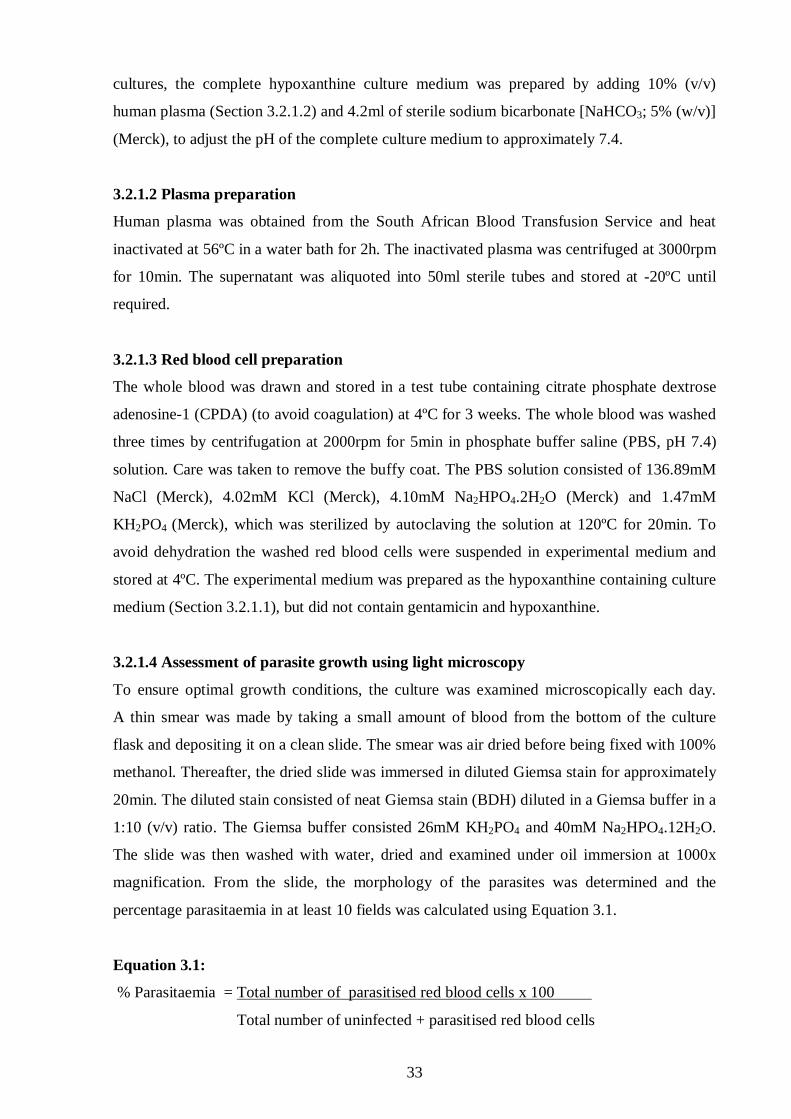
33
cultures, the complete hypoxanthine culture medium was prepared by adding 10% (v/v)
human plasma (Section 3.2.1.2) and 4.2ml of sterile sodium bicarbonate [NaHCO3; 5% (w/v)]
(Merck), to adjust the pH of the complete culture medium to approximately 7.4.
3.2.1.2 Plasma preparation
Human plasma was obtained from the South African Blood Transfusion Service and heat
inactivated at 56ºC in a water bath for 2h. The inactivated plasma was centrifuged at 3000rpm
for 10min. The supernatant was aliquoted into 50ml sterile tubes and stored at -20ºC until
required.
3.2.1.3 Red blood cell preparation
The whole blood was drawn and stored in a test tube containing citrate phosphate dextrose
adenosine-1 (CPDA) (to avoid coagulation) at 4ºC for 3 weeks. The whole blood was washed
three times by centrifugation at 2000rpm for 5min in phosphate buffer saline (PBS, pH 7.4)
solution. Care was taken to remove the buffy coat. The PBS solution consisted of 136.89mM
NaCl (Merck), 4.02mM KCl (Merck), 4.10mM Na2HPO4.2H2O (Merck) and 1.47mM
KH2PO4 (Merck), which was sterilized by autoclaving the solution at 120ºC for 20min. To
avoid dehydration the washed red blood cells were suspended in experimental medium and
stored at 4ºC. The experimental medium was prepared as the hypoxanthine containing culture
medium (Section 3.2.1.1), but did not contain gentamicin and hypoxanthine.
3.2.1.4 Assessment of parasite growth using light microscopy
To ensure optimal growth conditions, the culture was examined microscopically each day.
A thin smear was made by taking a small amount of blood from the bottom of the culture
flask and depositing it on a clean slide. The smear was air dried before being fixed with 100%
methanol. Thereafter, the dried slide was immersed in diluted Giemsa stain for approximately
20min. The diluted stain consisted of neat Giemsa stain (BDH) diluted in a Giemsa buffer in a
1:10 (v/v) ratio. The Giemsa buffer consisted 26mM KH2PO4 and 40mM Na2HPO4.12H2O.
The slide was then washed with water, dried and examined under oil immersion at 1000x
magnification. From the slide, the morphology of the parasites was determined and the
percentage parasitaemia in at least 10 fields was calculated using Equation 3.1.
Equation 3.1:
% Parasitaemia = Total number of parasitised red blood cells x 100
Total number of uninfected + parasitised red blood cells
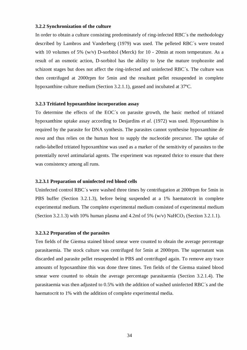
34
3.2.2 Synchronization of the culture
In order to obtain a culture consisting predominately of ring-infected RBC´s the methodology
described by Lambros and Vanderberg (1979) was used. The pelleted RBC´s were treated
with 10 volumes of 5% (w/v) D-sorbitol (Merck) for 10 - 20min at room temperature. As a
result of an osmotic action, D-sorbitol has the ability to lyse the mature trophozoite and
schizont stages but does not affect the ring-infected and uninfected RBC´s. The culture was
then centrifuged at 2000rpm for 5min and the resultant pellet resuspended in complete
hypoxanthine culture medium (Section 3.2.1.1), gassed and incubated at 37ºC.
3.2.3 Tritiated hypoxanthine incorporation assay
To determine the effects of the EOC´s on parasite growth, the basic method of tritiated
hypoxanthine uptake assay according to Desjardins et al. (1972) was used. Hypoxanthine is
required by the parasite for DNA synthesis. The parasites cannot synthesise hypoxanthine de
nova and thus relies on the human host to supply the nucleotide precursor. The uptake of
radio-labelled tritiated hypoxanthine was used as a marker of the sensitivity of parasites to the
potentially novel antimalarial agents. The experiment was repeated thrice to ensure that there
was consistency among all runs.
3.2.3.1 Preparation of uninfected red blood cells
Uninfected control RBC´s were washed three times by centrifugation at 2000rpm for 5min in
PBS buffer (Section 3.2.1.3), before being suspended at a 1% haematocrit in complete
experimental medium. The complete experimental medium consisted of experimental medium
(Section 3.2.1.3) with 10% human plasma and 4.2ml of 5% (w/v) NaHCO3 (Section 3.2.1.1).
3.2.3.2 Preparation of the parasites
Ten fields of the Giemsa stained blood smear were counted to obtain the average percentage
parasitaemia. The stock culture was centrifuged for 5min at 2000rpm. The supernatant was
discarded and parasite pellet resuspended in PBS and centrifuged again. To remove any trace
amounts of hypoxanthine this was done three times. Ten fields of the Giemsa stained blood
smear were counted to obtain the average percentage parasitaemia (Section 3.2.1.4). The
parasitaemia was then adjusted to 0.5% with the addition of washed uninfected RBC´s and the
haematocrit to 1% with the addition of complete experimental media.

35
3.2.3.3 Preparation of the drug and essential oil constituents
The effect of quinine and EOC´s on the resistant strain of P. falciparum was tested. Stock
solutions were freshly prepared before each experiment and the highest possible grades of the
drug and EOC´s were purchased from Sigma-Aldrich and Fluka.
3.2.3.3.1 Preparation of the classical antimalarial drug
Quinine sulphate (Mr = 324.43g/mol) (Sigma-Aldrich) served as the reference antimalarial
agent. The stock solution of quinine was prepared with a dilution factor of 10 taken into
account. This stock solution was prepared in autoclaved Milli-Q® water, to the concentration
of 0.1mM. The dilutions were prepared in plastic eppendorf tubes using incomplete
experimental medium.
3.2.3.3.2 Preparation of essential oil constituents
The stock solution of 10% (v/v) of each EOC was prepared by diluting 10µl of neat pure
EOC in 90µl of 100% DMSO (dimethyl sulfoxide) (Merck). Notably the density of each EOC
was taken into account when the stock solution was prepared. All further EOC dilutions were
made up in plastic eppendorfs using DMSO. The dilution factor resulting from addition to
wells in plates was taken into account and determined to be 125x by dividing the amount of
oil added (2µl) plus 23µl of experimental medium into the total volume of the wells (250µl).
3.2.3.4 Preparation of microtitre plates
Two microlitres of EOC dilution plus 23µl of incomplete experimental medium were added in
triplicate from A1 – G12 (Figure 3.2). Thereafter, 200µl of the parasite suspension (Section
3.2.3.2) was added to all the wells except the first four wells of row H. These wells served as
the uninfected RBC´s control, where 200µl of 1% (v/v) uninfected RBC suspension (Section
3.2.3.1) was added. The last eight wells served as the drug-free parasitised RBC´s control. To
ensure that the total volume in each well was 250µl, 25µl of experimental medium was added
to all wells of row H.
3.2.3.5 Incubation in a micro-aerobic environment
To create a micro-aerobic environment suitable for optimal parasite growth, the candle jar
method described by Jansen and Trager (1976) was used. After preparing the microtitre
plates, they were placed in an airtight humified glass desiccator with candles. These candles
were lit before the cover was fitted and when the flame died the gas content in the candle jar
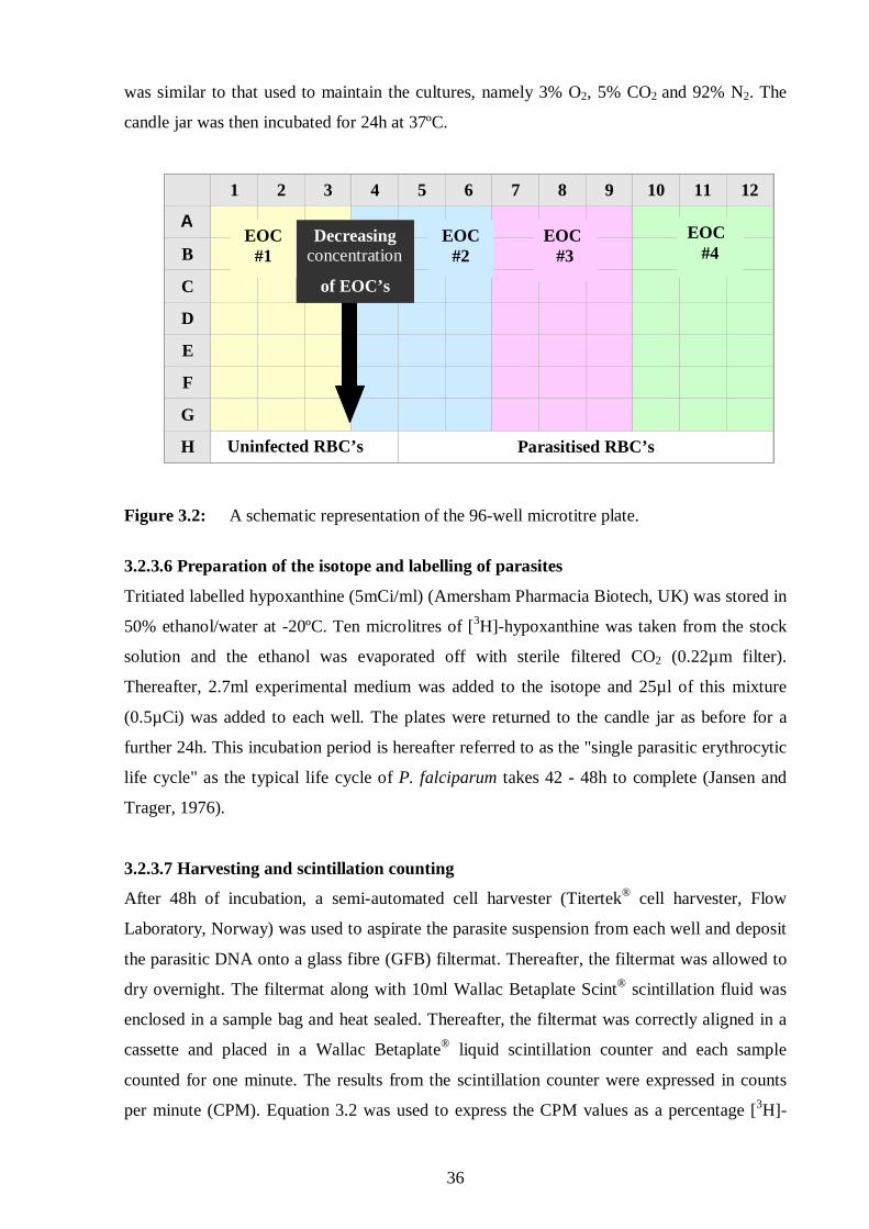
36
was similar to that used to maintain the cultures, namely 3% O2, 5% CO2 and 92% N2. The
candle jar was then incubated for 24h at 37ºC.
Figure 3.2: A schematic representation of the 96-well microtitre plate.
3.2.3.6 Preparation of the isotope and labelling of parasites
Tritiated labelled hypoxanthine (5mCi/ml) (Amersham Pharmacia Biotech, UK) was stored in
50% ethanol/water at -20ºC. Ten microlitres of [3H]-hypoxanthine was taken from the stock
solution and the ethanol was evaporated off with sterile filtered CO2 (0.22µm filter).
Thereafter, 2.7ml experimental medium was added to the isotope and 25µl of this mixture
(0.5µCi) was added to each well. The plates were returned to the candle jar as before for a
further 24h. This incubation period is hereafter referred to as the "single parasitic erythrocytic
life cycle" as the typical life cycle of P. falciparum takes 42 - 48h to complete (Jansen and
Trager, 1976).
3.2.3.7 Harvesting and scintillation counting
After 48h of incubation, a semi-automated cell harvester (Titertek® cell harvester, Flow
Laboratory, Norway) was used to aspirate the parasite suspension from each well and deposit
the parasitic DNA onto a glass fibre (GFB) filtermat. Thereafter, the filtermat was allowed to
dry overnight. The filtermat along with 10ml Wallac Betaplate Scint® scintillation fluid was
enclosed in a sample bag and heat sealed. Thereafter, the filtermat was correctly aligned in a
cassette and placed in a Wallac Betaplate® liquid scintillation counter and each sample
counted for one minute. The results from the scintillation counter were expressed in counts
per minute (CPM). Equation 3.2 was used to express the CPM values as a percentage [3H]-
1 2 3 4 5 6 7 8 9 10 11 12
A
B
C
D
E
F
G
H Uninfected RBC’s Parasitised RBC’s
Decreasing concentration
of EOC’s
EOC #1
EOC #2
EOC #4
EOC #3
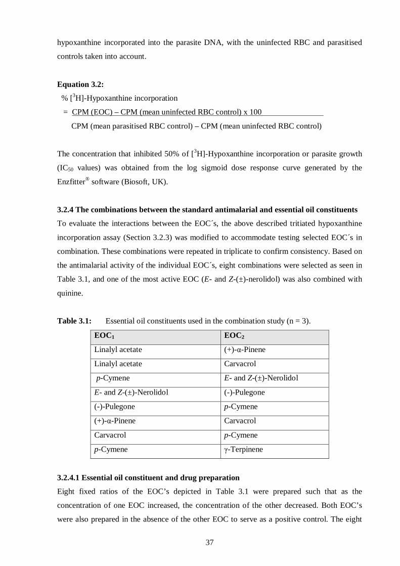
37
hypoxanthine incorporated into the parasite DNA, with the uninfected RBC and parasitised
controls taken into account.
Equation 3.2:
% [3H]-Hypoxanthine incorporation
= CPM (EOC) – CPM (mean uninfected RBC control) x 100
CPM (mean parasitised RBC control) – CPM (mean uninfected RBC control)
The concentration that inhibited 50% of [3H]-Hypoxanthine incorporation or parasite growth
(IC50 values) was obtained from the log sigmoid dose response curve generated by the
Enzfitter® software (Biosoft, UK).
3.2.4 The combinations between the standard antimalarial and essential oil constituents
To evaluate the interactions between the EOC´s, the above described tritiated hypoxanthine
incorporation assay (Section 3.2.3) was modified to accommodate testing selected EOC´s in
combination. These combinations were repeated in triplicate to confirm consistency. Based on
the antimalarial activity of the individual EOC´s, eight combinations were selected as seen in
Table 3.1, and one of the most active EOC (E- and Z-(±)-nerolidol) was also combined with
quinine.
Table 3.1: Essential oil constituents used in the combination study (n = 3).
EOC1 EOC2
Linalyl acetate (+)-α-Pinene
Linalyl acetate Carvacrol
p-Cymene E- and Z-(±)-Nerolidol
E- and Z-(±)-Nerolidol (-)-Pulegone
(-)-Pulegone p-Cymene
(+)-α-Pinene Carvacrol
Carvacrol p-Cymene
p-Cymene γ-Terpinene
3.2.4.1 Essential oil constituent and drug preparation
Eight fixed ratios of the EOC’s depicted in Table 3.1 were prepared such that as the
concentration of one EOC increased, the concentration of the other decreased. Both EOC’s
were also prepared in the absence of the other EOC to serve as a positive control. The eight

38
dilutions for each individual EOC were prepared in DMSO as described above (Section
3.2.3.3.2).
The dilutions were then combined in a 1:1 (v/v) ratio. A further dilution factor of 2 was taken
into account, so that the final dilution factor increased to 250x for the EOC’s and 20x for
quinine. In the wells of row A, 2µl of the combined EOC’s and 23µl of experimental medium
were plated out in triplicate. From row B to F, a 1:10 serial dilution was prepared in
experimental medium, while a 1:100 dilution was prepared in row G. After serially diluting
the combined EOC’s, 65µl of the content from row B – F was discarded and 175µl discarded
from row G. To the remaining 25µl EOC’s, 200µl parasitised suspension was plated out as
described in Section 3.2.3.3 and incubated for 24h (Section 3.2.3.5), before [3H]-
hypoxanthine was added for a further 24h (Section 3.2.3.6) and then the DNA harvested and
filtermats counted (Section 3.2.3.7).
3.2.5 Data analysis
The data was initially analysed as for the individual EOC´s (section 3.2.3). From the eight log
sigmoidal dose response curves, eight IC50 values were calculated, and in turn the IC50 values
for the other EOC’s were calculated using direct proportion. The drug ratio values
(or otherwise known as fractional inhibitory concentration (FIC) values) were calculated by
dividing the IC50 values of each EOC’s combination by the IC50 values of the individual EOC.
3.2.5.1 Isobologram construction
The drug ratio values (IC50 value of combined EOC’s/IC50 value of individual EOC) were
then used to construct an isobologram, from which the nature of the interaction between the
two EOC´s could be determined as seen in Figure 3.3 (Berenbaum, 1989; Meadows et al.,
2002). Each experimental run was done in triplicate to confirm consistency. In order to
observe the interaction between EOC´s and classical antimalarial agents used in the treatment
of malaria, one of the most active EOC´s was combined with quinine. Quinine was prepared
as described in Section 3.2.3.3.1. It must be noted that there may be some points on the
isobolograms overlying on each other and cannot be clearly seen sometimes. This can be the
case irrespective that six fixed ratios of the two EOC´s were combined.
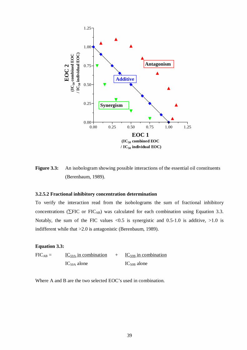
39
0.00 0.25 0.50 0.75 1.00 1.250.00
0.25
0.50
0.75
1.00
1.25
EOC 1(IC50 combined EOC
/ IC50 individual EOC)
EO
C 2
(IC
50 c
omb
ined
EO
C/
IC50
indi
vid
ual E
OC
)
Figure 3.3: An isobologram showing possible interactions of the essential oil constituents
(Berenbaum, 1989).
3.2.5.2 Fractional inhibitory concentration determination
To verify the interaction read from the isobolograms the sum of fractional inhibitory
concentrations (∑FIC or FICAB) was calculated for each combination using Equation 3.3.
Notably, the sum of the FIC values <0.5 is synergistic and 0.5-1.0 is additive, >1.0 is
indifferent while that >2.0 is antagonistic (Berenbaum, 1989).
Equation 3.3:
FICAB = IC50A in combination + IC50B in combination
IC50A alone IC50B alone
Where A and B are the two selected EOC’s used in combination.
Antagonism
Synergism
Additive
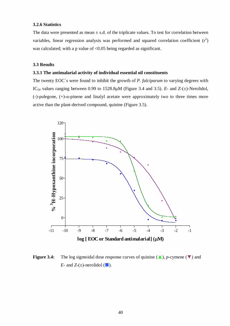
40
3.2.6 Statistics
The data were presented as mean ± s.d. of the triplicate values. To test for correlation between
variables, linear regression analysis was performed and squared correlation coefficient (r2)
was calculated; with a p value of <0.05 being regarded as significant.
3.3 Results
3.3.1 The antimalarial activity of individual essential oil constituents
The twenty EOC´s were found to inhibit the growth of P. falciparum to varying degrees with
IC50 values ranging between 0.99 to 1528.8µM (Figure 3.4 and 3.5). E- and Z-(±)-Nerolidol,
(-)-pulegone, (+)-α-pinene and linalyl acetate were approximately two to three times more
active than the plant-derived compound, quinine (Figure 3.5).
-11 -10 -9 -8 -7 -6 -5 -4 -3 -2 -1
0
25
50
75
100
120
log [ EOC or Standard antimalarial] (µM)
%3 H
-Hyp
oxa
nthi
ne in
corp
ora
tion
Figure 3.4: The log sigmoidal dose response curves of quinine (▲), p-cymene (▼) and
E- and Z-(±)-nerolidol (�).

Quinin
eα -B
isab
olol
trans
-Ger
anio
l(± )-
Linalo
ol
E- and
Z- (
± )-Ner
olido
l
(-)-C
itron
ellal
Geran
yl ac
etate
Linal
yl ac
etate
( ± )-α +β
-Thu
jone
(+)-C
arvo
ne
(-)-F
encho
ne
(-)-M
enth
one
(-)-P
ulego
neCar
vacr
olEug
enol
(+)-α -
Pinene
(+)-β -
Pinene
γ -Terp
inen
e
(R)-(
+)-L
imon
ene
Ρ -Cym
ene
1,8-
Cineol
e
0.0
2.55
255
505
755
1005
1255
15051600
Standard antimalarialAlcoholsAldehydeEstersKetonesPhenolsTerpene hydrocarbons
Oxide
0.99±0.30
254.40±10.20
698.5036.50
114.20±11.60
1.40±0.10
528.60±43.10
57.30±4.30
457.90±47.20
505.80±19.90
1.10±0.10
1067.90±44.50
753.50±33.80
1.20±0.20
294.77±20.55
1003.70±18.70
533.50±17.50
1528.80±44.90
70.20±4.00
307.30±24.00
135.40±7.00
0.29±0.03
Essential oil constituents
IC50
val
ues
( µM
)
Figure 3.5: The antimalarial activity of the 20 essential oil constituents and quinine.
41

42
3.3.2 The relationship between essential oil constituent density and antimalarial activity
There was no correlation (r2 = 0.008) between the antimalarial activity and the densities or
lipophilicity of the 20 EOC’s (Figure 3.6).
-0.100 -0.075 -0.050 -0.025 -0.000 0.025 0.050-1
0
1
2
3
4
5
r² = 0.008207
log Density (g/l)
log
An
tim
alar
ial
IC5
0 va
lues
Figure 3.6: The linear relationship between the density of the essential oil constituents and
their antimalarial activity.
3.3.3 The antimalarial activity of the combined essential oil constituents
After investigating the antimalarial activity of individual EOC´s, various combinations
depending on spectrum of activity of EOC´s were performed (Table 3.2). Representative
isobolograms showing the synergistic (between p-cymene and E- and Z-(±)-nerolidol or
γ-terpinene) and antagonistic (between E- and Z-(±)-nerolidol and (-)-pulegone) interactions
are illustrated in Figures 3.7 and 3.8, respectively.

43
Table 3.2: The combined interaction between two essential oil constituents (n = 3).
EOC1 EOC2 ∑FIC value Interaction
Linalyl acetate (+)-α-Pinene 0.91 Additive
Linalyl acetate Carvacrol 0.14 Synergistic
p-Cymene E- and Z-(±)-Nerolidol 0.09 Synergistic
E- and Z-(±)-Nerolidol (-)-Pulegone 2.03 Antagonistic
(-)-Pulegone p-Cymene 11.30 Antagonistic
(+)-α-Pinene Carvacrol 0.22 Synergistic
Carvacrol p-Cymene 0.02 Synergistic
p-Cymene γ-Terpinene 0.37 Synergistic
Figure 3.7: p-Cymene interacted in a synergistic manner when combined with
γ-terpinene. (�) and E- and Z-(±)-nerolidol (▲).
0.00 0.25 0.50 0.75 1.00 1.250.00
0.25
0.50
0.75
1.00
1.25
p-Cymene(IC50 combined EOC /IC50 individual EOC)
gam
ma-
Ter
pine
ne /
E-
and
Z-(± )
-Ner
olid
ol(I
C50
com
bine
d E
OC
/IC
50 in
divi
dual
EO
C)

44
Figure 3.8: An antagonistic interaction between E- and Z-(±)-nerolidol and (-)-pulegone.
3.3.4 The antimalarial activity of quinine and the most active essential oil constituent
A synergistic interaction (∑FIC = 0.01) was observed when the most active EOC, namely
E- and Z-(±)-nerolidol, was combined with the standard antimalarial agent, quinine.
3.4 Discussion
3.4.1 The antimalarial activity of individual essential oil constituents
Of the twenty EOC´s tested, E- and Z-(±)-nerolidol was found to be the most active
(IC50 = 0.2µg/ml = 0.9µM) (Figure 3.5). The antimalarial activity of the EOC’s did not
correlate with their density or lipophilicity (Figure 3.6), which indicated that all the EOC’s
crossed the various red blood cell and parasite membranes equally well in order to reach their
respective targets. But once across the membranes, their ability to interfere or bind with their
respective target differed greatly.
E- and Z-(±)-Nerolidol is also one of the EOC´s in the essential oils of numerous plant species
that have been reported to possess antimalarial activity. E- and Z-(±)-Nerolidol found in the
oil of Virola surinamensis (Myristiceae) has previously been reported to have antimalarial
0.0 0.5 1.0 1.5 2.0 2.5 3.0 3.50.0
0.5
1.0
1.5
2.0
2.5
3.0
3.5
E- and Z-(±)-Nerol idol(IC50 combined EOC /IC50 individual EOC)
(-)-
Pu
leg
one
(IC
50 c
om
bine
d E
OC
/IC
50 in
divi
dual
EO
C)

45
activity where 100, 10 and 1µg/ml inhibited the development of young trophozoites to the
schizont stage (without haemozoin formation) (Lopes et al., 1999). In trying to determine the
mechanism of action of E- and Z-(±)-nerolidol, Lopes et al. (1999) found that 100µM had no
effect on overall protein synthesis (as indicated by L-[35S]–methionine incorporation), but did
inhibit the incorporation of D-[U-14C]-glucose especially when young trophozoites were
differentiating into mature trophozoites (22 - 48h culture).
From this it was proposed that E- and Z-(±)-nerolidol interferes with malaria growth by
inhibiting glycoprotein biosynthesis (Lopes et al., 1999). Possibly by inhibiting specific
pathways in the biosynthesis of isoprenoids that play an important role as components of
structural cholesterol and in ubiquinones (Figure 3.9) (Goulart et al., 2004). It has been shown
that E- and Z-(±)-nerolidol inhibits the biosynthesis of coenzyme Q and other intermediates of
the isoprenoid pathway (dolichol) in all intra-erythrocytic stages (De Macedo et al., 2002).
This could be achieved by E- and Z-(±)-nerolidol competing with natural substrates in an
enzyme-substrate reaction (Goulart et al., 2004).
Terpenes such as E- and Z-(±)-nerolidol have a chemical structure similar to that of some
intermediates of the isoprenoid pathway such as farnesyl pyrophophate (FPP, C15) and
geranylgeranyl pyrophosphate (GGPP, C20) (Figure 3.9) (Holstein and Hohl, 2003). As such,
E- and Z-(±)-nerolidol could inhibit the biosynthesis of the polyprenyl pyrophosphate side and
chain attached to ubiquinone (De Macedo et al., 2002). Alternatively E- and Z-(±)-nerolidol
could inhibit the biosynthesis of both dolichol and the isoprenic chain of coenzyme Q. De
Macedo et al. (2002) proposed that E- and Z-(±)-nerolidol interfered with the mechanism of
elongation involved in the biosynthesis of dolichol and the isoprenic chain attached to the
benzoquinone ring of coenzyme Q.
In addition, E- and Z-(±)-nerolidol as well as the monoterpene (R)-(+)-limonene, have been
shown to inhibit the isoprenylation of proteins, resulting in the interference with
mitochondrial metabolic processes like pyrimidine biosynthesis (De Macedo et al., 2002).
The difference in the mechanism of action of these two constituents is probably due to the
difference in chemical structure, where the hydroxyl group and linear structure of
E- and Z-(±)-nerolidol could allow better interaction with the target enzyme compared to the
six-member ring backbone of (R)-(+)-limonene (Figure 2.1, d and s) (De Macedo et al.,
2002).

0
Figure 3.9: The biosynthetic pathway of isoprenoids in Plasmodium falciparum (http://sites.huji.ac.il/malaria/maps/isoprenoidmetpath.html).
PPi
Apicoplast membrane
Glyceraldehyde 3P Pyruvate
1-Deoxy-D-xylulose 5P
1-Hydroxy-2-methyl-2-butenyl 4-diphosphate
Geranyl-Geranyl-PP
Farnesyl-PP Geranyl-PP
Isopentenyl-2P isomerase
Dimethylallyl-PP Isopentenyl-PP
4-Hydroxy-3-methylbut-2-enyl diphosphate reductase
S-Geranylgeranyl-protein Protein- cysteine
S-Farnesyl-protein
PPi PPi PPi
Ubiquinone metabolism
Dimethylallyl-PP transferase
Geranyl transferase
Farnesyltransferase
Protein farnesyltransferase
Protein-cysteine
PPi
Protein geranylgeranyltransferase type II
Glycolysis
46

47
The antimalarial activities obtained in this study (0.9µM) are similar to those reported by
Goulart et al. (2004) (0.8µM), even though a different P. falciparum strain was used.
Regardless of this potent activity, the E- and Z-(±)-nerolidol-rich oil (45.2%) from Lippia
multiflora only displayed moderate antimalarial activity (47.6µg/ml) even in the presence of a
high concentration of (±)-linalool (20.2%) (Valentin et al., 1995). Thus, the interaction
between major and minor constituents dictates the overall activity of the oil and needs to be
taken into account.
Both (R)-(+)-limonene and (±)-linalool were found to inhibit the isoprenoid biosynthesis
pathway at a much earlier step compared to farnesol. It is possible that these monoterpenes
((R)-(+)-limonene and (±)-linalool) inhibit the biosynthesis of dolichol and ubiquinones by
interfering with the condensation between isopentenyl-PP and dimethylallyl-PP (Figure 3.6)
(Goulart et al., 2004).
In addition (R)-(+)-limonene was found to induce some inhibition of Ras-like protein
isoprenylation in the ring and schizont stages (Moura et al., 2001). Holstein and Hohl (2003)
noted that the (+)- and (-)-isomers of menthol displayed varying inhibitory activity on
isoprenylation, where (-)-menthol was active compared to the inactive (+)-isomer. It was also
suggested that alcohols with an open chain such as (±)-linalool and trans-geraniol are inactive
or have low inhibitory activity on isoprenylation (Holstein and Hohl, 2003).
The antimalarial activity of E- and Z-(±)-nerolidol, (±)-linalool and R-(+)-limonene are
comparable, even though a 3D-7 chloroquine-sensitive strain was used compared to FCR-3
chloroquine-resistant strain in this study. Goulart et al. (2004) reported values of 0.8µM,
0.28mM and 1.22mM respectively, and this study reported values of 0.9µM, 0.3mM and
0.5mM, respectively (Figure 3.5). The differences in antimalarial activity of these terpenes
could be attributed to their structural differences. While the (R)- and (S)-enantiomers
displayed similar activity to downregulate Ras-related proteins, overall it was found that the
order of activity was related to the presence of an oxygenated substituent at the C7 position;
where alcohol followed by aldehydes and acids were consequently more active (Holstein and
Hohl, 2003). Whilst the deoxygenated form of (R)-(+)-limonene was found to be inactive
(Holstein and Hohl, 2003).
Even though (±)-linalool is similar in structure to E- and Z-(±)-nerolidol and contains a
hydroxyl group, the shorter chain greatly reduces the antimalarial activity of (±)-linalool and
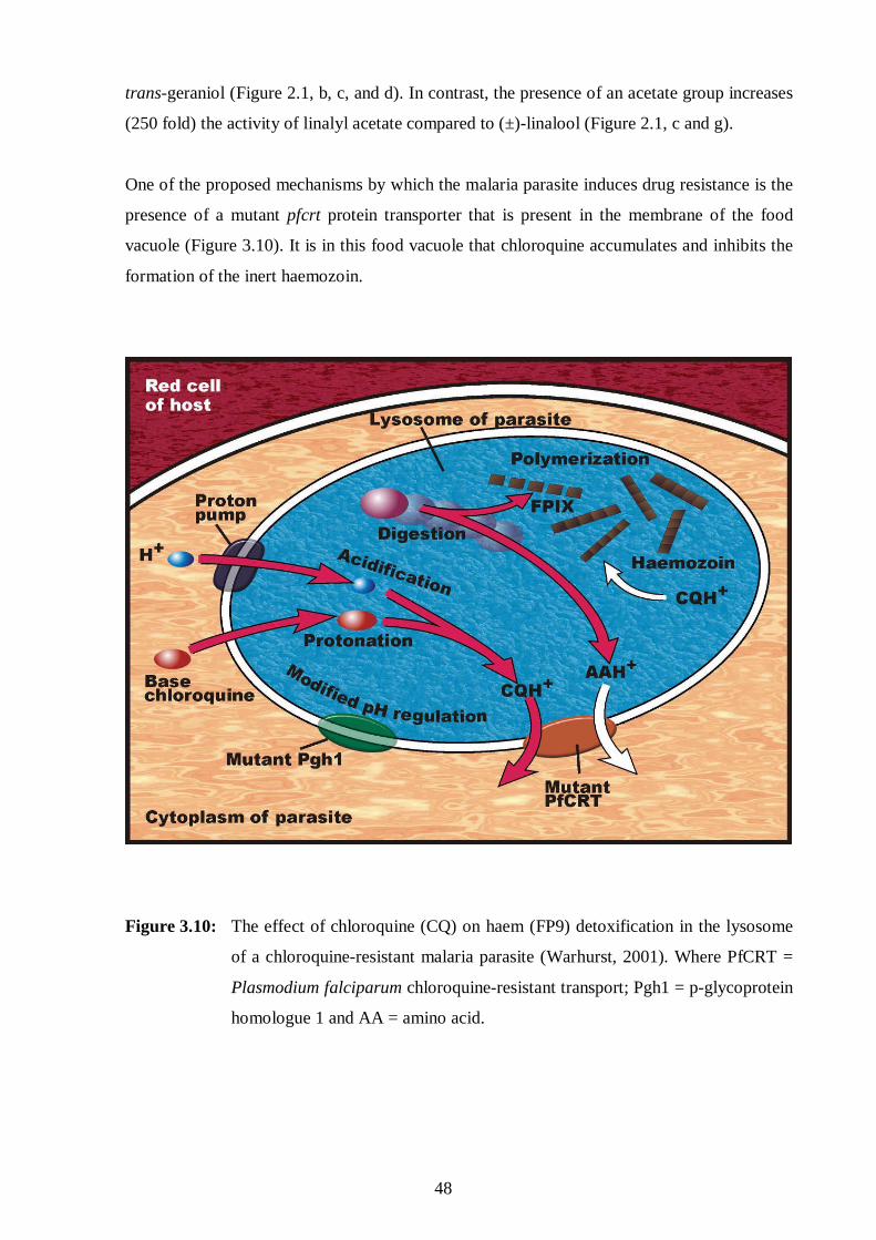
48
trans-geraniol (Figure 2.1, b, c, and d). In contrast, the presence of an acetate group increases
(250 fold) the activity of linalyl acetate compared to (±)-linalool (Figure 2.1, c and g).
One of the proposed mechanisms by which the malaria parasite induces drug resistance is the
presence of a mutant pfcrt protein transporter that is present in the membrane of the food
vacuole (Figure 3.10). It is in this food vacuole that chloroquine accumulates and inhibits the
formation of the inert haemozoin.
Figure 3.10: The effect of chloroquine (CQ) on haem (FP9) detoxification in the lysosome
of a chloroquine-resistant malaria parasite (Warhurst, 2001). Where PfCRT =
Plasmodium falciparum chloroquine-resistant transport; Pgh1 = p-glycoprotein
homologue 1 and AA = amino acid.

49
The chloroquine-haem complex is toxic to the parasite, resulting in lipid peroxidation and
parasite death (Warhurst, 2001). The pfcrt pump transports the accumulated chloroquine out
of the food vacuole, thereby decreasing its concentration and the possibility of interacting
with the haem units and results in the death of the parasite. This process closely resembles the
mechanisms by which cancer cells become multidrug resistant.
Yoshiba et al. (2005) reported that the essential oil of Zanthoxyli fructus inhibited the activity
of the p-glycoprotein transport pump, resulting in the accumulation of anticancer drug in the
cells and subsequent cell death. The chief components of this essential oil were the acyclic
monoterpenoids (geraniol, linalool, citronellal and citronellol) and the monocyclic
monoterpenoids (limonene and 1,8-cineole). It was found that the acyclic monoterpenoids,
(R)-(+)-citronellal and (S)-(-)-β-citonellol were potent inhibitors of p-glycoprotein compared
to the other EOC´s.
However, in this study (-)-citronellal was relatively inactive as an antimalarial agent
(IC50 = 698.5±36.5µM) (Figure 3.5). This aldehyde compound like drugs such as verapamil
(a calcium channel blocker) have the ability to block the action of p-glycoproteins in cancer
cells (Yoshiba et al., 2005). In addition, it can block transport pumps (pfcrt) in the malaria
parasite (Warhurst, 2001), even if it is relatively ineffective in directly inhibiting parasite
growth. These drugs including verapamil are termed “reversal agents” which, when combined
with an antimalarial agent such as chloroquine reverse the resistance in the strain, resulting in
the parasite being more sensitive to the inhibitory effects of chloroquine. To verify this
hypothesis, chloroquine should be combined with (-)-citronellal to determine if the interaction
is synergistic. If so, then (-)-citronellal could act as a “reversal agent” and could be used as
adjunct therapy in the treatment of malaria.
The oils of Tetradenia riparia (Lamiaceae) contain the ketone fenchone (13.6%) and have
also been reported to have moderate antimalarial activity (IC50 = 50.3µg/ml) (Campbell et al.,
1997). However, this activity is possibly due to interactions with other constituents present in
the oil, as (-)-fenchone alone has poor antimalarial activity (Figure 3.5).
It is interesting to observe that the two structural isomers, (+)-α-pinene and (+)-β-pinene
(Figure 2.1, o and p, respectively) displayed contrasting activity with (+)-α-pinene being 250
times more active than (+)-β-pinene against P. falciparum, although the two compounds

50
displayed identical toxicity profiles (Figure 5.4). This could indicate that the conformation of
(+)-α-pinene is crucial for its interaction and binding to its plasmodial target.
3.4.2 The antimalarial activity of the combined essential oil constituents
When two EOC´s were combined and assessed for their antiplasmodial activity the overall
interaction between the various combinations were found to be synergistic, although to
varying degrees (Table 3.2). The combination between the least active and most active
EOC´s, p-cymene (1528.8µM) and E- and Z-(±)-nerolidol (0.99µM) displayed a pronounced
synergistic interaction (Figure 3.7). E- and Z-(±)-Nerolidol with its reported mechanism of
action (Lopes et al., 1999) potentiated the antimalarial activity of p-cymene which had
minimal activity when used individually (or vice versa) (Figure 3.5). Goulart et al. (2004)
reported that most terpenes inhibit the isoprenoid biosynthetic pathway in P. falciparum. It is
likely that the interaction of E- and Z-(±)-nerolidol with p-cymene might assist p-cymene to
reach the site of action in the parasites (or vice versa), or they may have acted on the
complementary targets.
The volatile oils from V. surinamensis also showed the predominance of p-cymene (42.0%),
α-pinene (11.7%), and β-pinene (5.2%) and nerolidol (3.8%) (Lopes et al., 1999). From the
results of this study, it can be seen that p-cymene and β-pinene (which constitute 47% of the
whole oil) were both relatively poor inhibitors of parasite growth (Figure 3.6). However,
100µg/ml of this oil inhibited 100% parasite growth over 48h of treatment. Thus, the
remaining two major constituents, namely α-pinene and nerolidol contributed to the effective
antimalarial activity of this essential oil. This illustrates the importance of considering the
combined effect of EOC´s even though the active constituents only represent 15.5% of the
essential oil.
In this present study the two least active EOC´s, namely carvacrol (1067.9µM) and p-cymene
interacted synergistically (Table 3.2). p-Cymene is the biological precursor of carvacrol, but
lacks a phenolic hydroxyl group (Figure 2.1, m and r). While carvacrol is structurally similar
to (R)-(+)-limonene but has one less hydroxyl group and appeared to act well in combination
with p-cymene. Limonene has been found to inhibit the isoprenylation of proteins, resulting in
interference with mitochondrial metabolic processes like pyrimidine biosynthesis (De Macedo
et al., 2002).

51
When the least active EOC´s, γ-terpinene (1003.7µM) and p-cymene (1528.8µM) were
combined a synergistic interaction was observed. Whilst when the most active EOC´s,
E- and Z-(±)-nerolidol and (-)-pulegone were combined an antagonistic interaction occured
(Figure 3.8). Likewise the combinations of the two most active EOC’s [linalyl acetate
(1.4µM) and (+)-α-pinene (1.2µM)] showed an additive interaction (Table 3.2). While that of
the most and the least active EOC’s ((-)-pulegone and p-cymene) showed an antagonistic
interaction (Table 3.2). It may be that these two EOC’s competed for the same site in an
essential biochemical process in P. falciparum. Moreover, this study shows that the
interactions that exist when two EOC´s are combined, vary depending on the concentration
and ratio of the agents used. Boyom et al. (2003) stated that in the case of malaria, the
physical properties of essential oil constituents namely its low density (~0.94g/ml) and
lipophilicity which facilitates rapid diffusion across cell membranes, might enhance targeting
of the active components into the intracellular malaria parasite. But the final interaction
between the two EOC´s depends on their respective cellular targets.
3.4.3 The combined effect of quinine and E- and Z-(±)-nerolidol
The combination of a classical antimalarial drug, namely quinine with E- and Z-(±)-nerolidol
displayed a synergistic interaction (∑FIC value = 0.01). The ability of these two agents to
target separate cellular components may have contributed to their enhanced interaction; with
E- and Z-(±)-nerolidol able to inhibit isoprenoid biosynthesis (Goulart et al., 2004) and
quinine able to inhibit haemozoin formation (Warhurst, 2001). Quinine is also proposed to
inhibit malaria growth by decreasing the plasmodial DNA strand separation and transcription,
thereby inhibiting protozoal protein synthesis (Noedl et al., 2003). Thus, if a patient does
combine a volatile oil (for example through inhalation) with the more classical agents to treat
malaria, the patient will not be compromised by this combination.
In the absence of vaccines, people rely on chemotherapeutic agents to prevent and treat an
infection. These results indicate that plant-derived or chemically synthesised EOC´s have the
potential to be used in adjunct therapy with antimalarial agents. In addition, they could be
used as new templates for drug design.

52
CHAPTER FOUR
ANTI-OXIDANT AND ANTICHOLINESTERASE ACTIVITIES
4.1 Introduction
The study of anti-oxidants and anticholinesterases is producing a medical revolution that
promises a new age in health and disease management. In the past few years, there has been
renewed interest in studying the anti-oxidant activity of essential oils from aromatic plants
due to their health promoting benefits (Choi et al., 2000; Nakatsu et al., 2000). There have
been numerous reports on the anti-oxidant properties of essential oils (Sanchez-Moreno,
2002), however, there is little or no research conducted on the anticholinesterase activity of
essential oils (Graβmann et al., 2000). Natural anti-oxidants found in plants play a major role
in the prevention of complex pathological processes including cardiovascular diseases,
cerebrovascular diseases and carcinogenesis (Sanchez-Moreno, 2002). In addition, both
anti-oxidants and anticholinesterases play a vital role in minimising the progression of
neurodegenerative and chronic diseases like Alzheimer’s disease (Mi-Yeon et al., 2000).
In many pathological cases, reactive oxygen species (ROS) play an important role; where
ROS are generated by several types of inflammatory cells, for example: neutrophils and
macrophages in order to defend the host from invading microorganisms (Choi et al., 2000).
A continuous or over production of ROS and resultant inflammatory reactions may lead to
tissue damage. In the central nervous system of patients with Alzheimer’s disease, not only do
free radicals such as ROS damage the mitochondrial and nuclear DNA, but the development
of senile plaques and neurofibrillary tangles, all contribute to the pathology of Alzheimer’s
disease (Mi-Yeon et al., 2000).
It is also pertinent to note that apart from being therapeutically used in Alzheimer’s disease,
anticholinesterases are used as pesticides and to treat other medical conditions. These
anticholinesterases are able to inhibit cholinesterase reversibly or irreversibly (Ringman and
Cummings, 1999). Generally, the reversible anticholinesterases, such as edrophonium,
pyridostigmine, neostigmine, distigmine and physostigmine are used as medicines; whilst the
irreversible inhibitors such as parathion and malathion are used as pesticides. Although
ecothiophate, which is an irreversible inihibitor is used to treat glaucoma. It is important to
know that all pesticides are lipophilic and are readily absorbed through the skin and cross the
blood brain barrier causing central cholinergic effects, in addition to the full spectrum of

53
peripheral nicotinic and muscarinic effects (Pappano, 2004). These include markedly
impaired vision, copious lacrimation, nasal discharge and salivation, accompanied by slurred
speech, wheezing, impaired respiration, gastro-intestinal cramping, diarrhoea, urination,
profuse sweating, vomiting, dehydration, bradycardia and hypotension. These complications
are treated with atropine (an anticholinergic agent) accompanied by an acetylcholinesterase
enzyme regenerator, obidoxime (Pappano, 2004).
When studying any disease profile or in any situation where there is possible tissue damage,
one has to bear in mind that free radicals may be involved. Along with the knowledge that
essential oils possess anti-oxidant properties and Salvia essential oils have been used in the
treatment of Alzheimer’s disease (Savelev et al., 2003), the anti-oxidant and
anticholinesterase activities of the individual essential oil constituents were investigated.
4.1.1 Free radicals
Anti-oxidants play a vital role in preventing disease and strengthening the immune system,
particularly when one is suffering from a chronic disease. Their main function is to protect the
body against free radicals (also called oxidants), by neutralising or scavenging the oxidants
before they can damage cells and tissues (Sanchez-Moreno, 2002).
What are free radicals? A free radical can be any molecule containing one or more unpaired
electron(s) and is capable of independent existence (Sanchez-Moreno, 2002). Each atom has
two elements: a nucleus, consisting of protons and neutrons, and an electron that revolves
around the core (Peiwu et al., 1999). Free radicals are highly reactive, with the result that they
have a very limited lifespan. They are, however, not only produced within the body but also
from substances in the environment, such as tobacco smoke, exhaust fumes, compounds in
food, pollutants in drinking water and alcohol that may enter our bodies (Graβmann et al.,
2000).
Extensive research has shown that the body produces more free radicals under certain
conditions, which in turn places greater pressure on the immune system (Choi et al., 2000).
For example, prolonged or uncontrolled stress over a long period can significantly increase
free radical production (Graβmann et al., 2000). Sports participants also produce more free
radicals on account of increased catabolic metabolism (Graβmann et al., 2000).
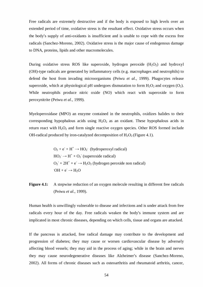
54
Free radicals are extremely destructive and if the body is exposed to high levels over an
extended period of time, oxidative stress is the resultant effect. Oxidative stress occurs when
the body′s supply of anti-oxidants is insufficient and is unable to cope with the excess free
radicals (Sanchez-Moreno, 2002). Oxidative stress is the major cause of endogenous damage
to DNA, proteins, lipids and other macromolecules.
During oxidative stress ROS like superoxide, hydrogen peroxide (H2O2) and hydroxyl
(OH)-type radicals are generated by inflammatory cells (e.g. macrophages and neutrophils) to
defend the host from invading microorganisms (Peiwu et al., 1999). Phagocytes release
superoxide, which at physiological pH undergoes dismutation to form H2O2 and oxygen (O2).
While neutrophils produce nitric oxide (NO) which react with superoxide to form
peroxynitrite (Peiwu et al., 1999).
Myeloperoxidase (MPO) an enzyme contained in the neutrophils, oxidizes halides to their
corresponding hypophalous acids using H2O2 as an oxidant. These hypophalous acids in
return react with H2O2 and form single reactive oxygen species. Other ROS formed include
OH-radical produced by iron-catalyzed decomposition of H2O2 (Figure 4.1).
O2 + e- + H+ → HO2. (hydroperoxyl radical)
HO2. → H+ + O2
- (superoxide radical)
O2- + 2H+ + e- → H2O2 (hydrogen peroxide non radical)
.OH + e- → H2O
Figure 4.1: A stepwise reduction of an oxygen molecule resulting in different free radicals
(Peiwu et al., 1999).
Human health is unwillingly vulnerable to disease and infections and is under attack from free
radicals every hour of the day. Free radicals weaken the body′s immune system and are
implicated in most chronic diseases, depending on which cells, tissue and organs are attacked.
If the pancreas is attacked, free radical damage may contribute to the development and
progression of diabetes; they may cause or worsen cardiovascular disease by adversely
affecting blood vessels; they may aid in the process of aging; while in the brain and nerves
they may cause neurodegenerative diseases like Alzheimer′s disease (Sanchez-Moreno,
2002). All forms of chronic diseases such as osteoarthritis and rheumatoid arthritis, cancer,
55
emphysema and HIV/AIDS result in an increased production of free radicals, which further
potentiate and prolong the damage caused by the disease. Thus, an increased intake of
anti-oxidants may prevent cellular damage by free radicals.
4.2 Anti-oxidant activity
Humans have their own natural anti-oxidant defenses in the form of enzyme systems.
However, we rely greatly on external anti-oxidants such as vitamin C and E, α-tocopherol,
β-carotene, glutathione and other flavonoids in our diet (Nakatsu et al., 2000). Non-enzymatic
anti-oxidants also play a role in resistance against oxidative stress. They protect normal
human body function from free radical damage by preventing the formation of excess free
radicals, scavenging free radicals and repairing damaged molecules (Sanchez-Moreno, 2002).
Vitamin E is considered the “master” anti-oxidant as it is a very efficient neutralizer of the
free radical damage, especially to the lipid structure of our cell membranes (Choi et al., 2000).
Furthermore, anti-oxidants complement each other’s activity, for instance, L-ascorbic acid
(vitamin C) helps to regenerate vitamin E. As a water-soluble anti-oxidant, L-ascorbic acid is
in a unique position to "scavenge" aqueous peroxyl radicals before these destructive
substances have a chance to damage the lipids. It works along with vitamin E, a fat-soluble
anti-oxidant, and the enzyme glutathione peroxidase to stop free radical chain reactions.
Anti-oxidants have been shown to be beneficial in preventing or delaying the progression of a
number of disease states such as cancer, Alzheimer’s disease, and macular degeneration
(Sramek and Cutler, 2000). A study in China including 29584 adults showed that
supplementation with α-tocopherol, β-carotene and selenium reduced stomach cancer by 21%
and oesophageal cancer by 42% (Sanchez-Moreno, 2002). An observational study of 991522
participants from 1982 to 1988 showed that regular long-term use of vitamin E reduced the
risk of death from bladder cancer by 40% (Sanchez-Moreno, 2002).
The evidence for anti-oxidants is strong enough for the Food and Drug Administration (FDA)
to allow a limited claim of anti-oxidants in the prevention of cancer (Graβmann et al., 2000).
The Age-Related Eye Disease Study (AREDS) underlined the benefits of anti-oxidant intake
in people who suffer from age-related macular degeneration (AMD), a leading cause of
blindness in the elderly (Sramek and Cutler, 2000). In this interventional trial, patients with
intermediate or advanced stage AMD who received anti-oxidant supplementation (vitamin C,
vitamin E, β-carotene and zinc) had a 28% reduced risk of further progression of this disease

56
(Sramek and Cutler, 2000). Furthermore, anti-oxidants showed benefits in immunity and
reduced the risk of infections in elderly patients in nursing homes.
A healthy diet rich in vegetables and fruits is the first step toward obtaining a well-balanced
anti-oxidant regimen. However, the beneficial doses used in many studies are higher than one
can obtain from diet alone, since essential nutrients are lost in the harvesting, processing and
cooking of food even before it is consumed (Nakatsu et al., 2000). So it is advisable that
consumers choose a supplement that has a wide spectrum of anti-oxidants in their most
natural and bioavailable form (Peiwu et al., 1999). The anti-oxidant dose should be sufficient
to aid in the defense against free radicals (above a multivitamin), but not mega-doses that may
be harmful (Nakatsu et al., 2000).
There has been much attention on using anti-oxidants to inhibit lipid peroxidation or at least
afford protection from damage by free radicals. Essential oils from Citrus paradisi and
Cymbopogon citratus have been reported to play a vital role as anti-oxidants (Peiwu et al.,
1999). Some phenols such as carvacrol and eugenol have been reported for their anti-oxidant
activity, but there is limited information on the anti-oxidant activity of the individual EOC’s
present in these essential oils (Nakatsu et al., 2000). Thus, twenty essential oils were selected
from seven chemical groups and their individual anti-oxidant activity was evaluated in
comparison to L-ascorbic acid (vitamin C).
4.2.1 Methodology
4.2.1.1 2,2-Diphenyl-1-picrylhydrazyl assay
The 2,2-diphenyl-1-picrylhydrazyl (DPPH) assay is used to measure the ability of
anti-oxidants to scavenge the stable free radical of 2,2-diphenyl-1-picrylhydrazyl. This stable
free radical is dark violet in colour and is decolourised when an anti-oxidant compound
donates an electron. This degree of decolourisation is stoichiometric with respect to the
number of electrons seized by DPPH.
A number of assays have been introduced to measure the total anti-oxidant activity of pure
compounds. In this study, the DPPH assay was selected and used to assess the in vitro
anti-oxidant activity of essential oil constituents according to Vardar-Ünlü et al. (2003). This
assay is considered a valid and easy assay to evaluate the scavenging activity of the
anti-oxidants as the radical compound is stable and does not have to be generated as in other
free radical scavenging assays.

57
4.2.1.2 2,2-Diphenyl-1-picrylhydrazyl preparation
A 96.2µM solution of DPPH (Fluka) was prepared in methanol. The solution was stable for a
week, but needed to be kept in the dark and stored at 4ºC.
4.2.1.3 Preparation of essential oil constituents
Approximately 32mg of EOC was weighed out and dissolved in 500µl DMSO to prepare a
stock solution of 10000ppm (= 650mM), which is equivalent to 1%, since 1ppm equals
0.0001%. This solution was then vortexed before 50µl was added to 950µl of DMSO to
prepare various concentrations.
4.2.1.4 Preparation of microtitre plates
Of the dilutions, 50µl (= 3250µM) was added to each well of row B to row G (Figure 4.2).
Whilst, 50µl DMSO was added to the wells in rows A to H to serve as the untreated controls.
HPLC grade methanol (200µl) was added to columns 2, 4, 6, 8, 10 and 12 to serve as colour
control while an equal volume of DPPH solution was plated out in columns 1, 3, 5, 7, 9 and
11. As a result the concentration was diluted to 100ppm (= 650µM). Furthermore, seven
dilutions were prepared by 1:10 serial dilutions in plastic eppendorfs using DMSO from the
stock solutions and each plated out into its relevant plate. Each plate was set up as mentioned
above.
Figure 4.2: The schematic representation of the setup of the microtitre plate used in
the 2,2-diphenyl-1-picrylhydrazyl assay.
1 2 3 4 5 6 7 8 9 10 11 12
A
B
C
D
E
F
G
H
control wells
sample wells with DPPH
sample wells with methanol
control wells
Decreasing concentrations
of EOC

58
4.2.1.5 Reading of the plate and data analysis
The test plate was shaken for 2min in a UV-VIS spectrophotometer and thereafter the
absorbance was measured at a wavelength of 540nm. The standard plate was then rinsed with
methanol and discarded together with its contents. The test plate was left to stand for 30min in
an incubator at 37°C. After which, the absorbance was read at 540nm using Genesis®
software (Biosoft, UK). Taking the appropriate controls into account, the percentage
decolourisation of each well was determined. The experiment was repeated at least twice. The
IC50 values were calculated using the Enzfitter® version 1.05 software. L-Ascorbic acid
(vitamin C) was used as the positive control.
4.2.2 Results
The anti-oxidant activity of the 20 essential oil constituents was very poor. The phenol,
eugenol was the only constituent that possessed comparable anti-oxidant activity to
L-ascorbic acid (Table 4.1)
Table 4.1: The anti-oxidant activity (IC50 values) of essential oil constituents.
Essential oil constituent Anti-oxidant activity (µM)
Essential oil constituent
Anti-oxidant activity (µM)
Alcohols Ketones
(-)-α-Bisabolol >449.7 (±)-α+β-Thujone >656.9
trans-Geraniol >648.3 (+)-Carvone >665.7
(±)-Linalool >648.3 (-)-Fenchone >656.9
E- and Z-(±)-Nerolidol >449.7 (-)-Menthone >648.3
Aldehyde (-)-Pulegone >656.9
(-)-Citronellal >648.3 Terpene Hydrocarbons
Esters (+)-α-Pinene >734.0
Geranyl acetate >509.5 (+)-β-Pinene >734.0
Linalyl acetate >509.5 γ-Terpinene >734.0
Phenols (R)-(+)-Limonene >734.0
Carvacrol >665.7 p-Cymene >745.1
Eugenol 46.6±0.12 Control
Oxide L-Ascorbic acid 17.03±0.06
1,8-Cineole >648.3
59
4.2.3 Discussion
4.2.3.1 The anti-oxidant activity of essential oil constituents
The essential oils from clove (Nakatsu et al., 2000) and citrus (Choi et al., 2000) possess
potent anti-oxidant activity. However other studies have indicated that not all essential oils
have anti-oxidant activity (e.g. Helichrysum species) (Lourens et al., 2004). In this study,
eugenol, the major constituent of the essential oil of Eugenia caryophyllata (clove) and
Myristica fragrance (nutmeg) (Nakatsu et al., 2000) is the only constituent that possessed
comparable anti-oxidant activity to L-ascorbic acid, being approximately three fold less active
(Table 4.1). The therapeutic potential of Ocimum sanctum as a neuroprotective agent in
cerebral ischaemia is attributed to the high content (70%) and very low toxicity (Table 5.1) of
eugenol (Yanpallewar et al., 2004).
In one study, essential oil from the leaves of Syrian oreganum (Origanum syriacum) was
found to contain 49.02% monoterpenes, 36.60% oxygenated monoterpenes and 12.59%
sesquiterpenes (Peiwu et al, 1999). The major components were as follows: γ-terpinene,
carvacrol, p-cymene and β-caryophyllene. The oil’s anti-oxidant activity was compared to
that of ascorbic acid, and 2,6-di-tert-butyl-4-methyl phenol (BHT, butylated hydroxytoluene).
The results showed that the anti-oxidant activity of the oil was 3 to 4 times lower compared to
L-ascorbic acid or BHT (Peiwu et al, 1999).
Vardar Ünlü et al. (2003) reported that thymol and carvacrol from the essential oil of Thymus
pectinatus, were individually found to possess weaker anti-oxidant activity than the crude oil
itself, indicating that other constituents of the essential oil contribute to the anti-oxidant
activity observed. It is also interesting to note that although carvacrol, (±)-linalool, γ-terpinene
and trans-geraniol have been reported to possess anti-oxidant activity (Choi et al., 2000;
Nakatsu et al., 2000), in this study they had little activity when compared to eugenol and
L-ascorbic acid (Table 4.1). The difference in activity between eugenol and carvacrol (both
phenols) may possibly be attributed to the presence of a methyl group at the second carbon of
eugenol, which is absent in carvacrol (Figure 2.1, m and n).
Epidemiological data suggest that anti-oxidants may have a beneficial effect on many
age-related diseases: atherosclerosis, cancer, some neurodegenerative (Alzheimer’s disease)
and ocular diseases. However, the widespread use of anti-oxidants is hampered by several
factors; the lack of prospective and controlled studies; growing evidence that free radicals are
not only by-products, but also play an important role in cell signal transduction, apoptosis and

60
infection control. Despite these, the results of this study show that some essential oil
constituents are good anti-oxidants.
4.3 Acetylcholinesterase activity
There are two forms of cholinesterase enzyme found in man, namely acetylcholinesterase (EC
3.1.1.7) and butyrylcholinesterase (or otherwise known as pseudocholinesterase; EC 3.1.1.8).
Acetylcholinesterase is an extremely active enzyme present in three isoforms: G1, which is
present in the brain; G4, in the brain and the neuromuscular endplate; and G2, in skeletal
muscle and blood-forming cells (erythrocytes). With butyrylcholinesterase found in blood
plasma and in lower concentrations in the hippocampal and cortical areas known to receive
cholinergic innervation (Ellis, 2005). The function for these enzymes is the hydrolysis of
acetylcholine to choline and acetic acid. Acetylcholine is the neurotransmitter essential for
cholinergic or parasympathetic transmission in the autonomic nervous system (Pappano,
2004).
4.3.1 Cholinesterase inhibitors and alzheimer′s disease
This age-related dementia is associated with a loss of neurons, and alterations to brain tissue,
particularly in the hippocampus and basal forebrain. Amyloid plaques and neurofibrillary
tangles characterise the condition. The loss of cholinergic fibres (in the basal forebrain nuclei)
is proposed to be a key factor in this disease. The development of Alzheimer’s disease
requires two events to occur. The first one is the age-related abnormal production of free
radicals, which damage mitochondrial and nuclear DNA in the brain. The second event is a
deficiency of the neurotransmitter acetylcholine in the synapses of cerebral cortex (Savelev et
al., 2003).
Probable Alzheimer’s disease is characterized by the presence of neuritic amyloid plaques and
neurofibrillary tangles in the brain, pathologic changes associated with cholinergic
denervation. Acetylcholinesterase and butyrylcholinesterase are both associated with amyloid
plaques. Although it is unknown whether acetylcholinesterase plays a role in accelerating the
neurotoxicity of amyloid plaques, there is some evidence that butyrylcholinesterase may
accelerate maturation of benign plaques into plaques associated with neuronal degeneration
and Alzheimer’s disease. Long-term inhibition of cholinesterase activity may result in the
activation of normal amyloid precursor protein processing in patients with Alzheimer’s
disease, as opposed to abnormal amyloid precursor protein processing, which leads to senile
61
plaque formation and causes many of the cognitive impairments in Alzheimer’s disease
(Sramek and Cutler, 2000).
Anticholinesterase drugs currently form the basis of modern drugs available for the
symptomatic treatment for patients with Alzheimer’s disease, and have a modest efficacy with
predictable parasympathomimetic side effects. Drugs such as donepezil and the plant-derived
isolates like galanthamine, physostigmine and rivastigmine are effective inhibitors of
acetylcholinesterase. Rivastigmine preferentially inhibits the G1 molecular form of
acetylcholinesterase and is the only cholinesterase inhibitor to have exhibited preferential
selectivity for any of the three isoforms of acetylcholinesterase (Ellis, 2005). Selective
inhibition of acetylcholinesterase occurs with galanthamine and donepezil, whereas
rivastigmine inhibits both acetylcholinesterase and butyrylcholinesterase.
Galanthamine is a tertiary alkaloid acetylcholinesterase inhibitor that has been approved in
several countries for the symptomatic treatment of senile dementia of the Alzheimer's type.
Derived from bulbs of the common snowdrop and several Amaryllidaceae plants,
(-)-galanthamine has long been used in anaesthetics to reverse neuromuscular paralysis
induced by turbocurarine-like muscle relaxants and more recently, has been shown to
attenuate drug- and lesion-induced cognitive deficits in animal models of learning and
memory (Miyazawa and Yamafuji, 2005).
Galanthamine also stimulates pre- and postsynaptic nicotinic receptors, although the clinical
significance of this finding is yet unclear. Numerous variants and analogues of galanthamine
have also been developed, with varying potency in inhibiting acetylcholinesterase activity.
Galanthamine is readily absorbed after oral administration, with a Tmax of 52min and plasma
elimination T½ of 5.7h (Miyazawa and Yamafuji, 2005). The efficacy of galanthamine
administered to Alzheimer's disease patients has been well demonstrated by large-scale
clinical trials. Typical of anticholinesterases, the most common adverse events associated with
galanthamine are nausea and vomiting.
A growing body of evidence indicates that both acetylcholinesterase and
butyrylcholinesterase play important roles in cholinergic transmission. For this reason, both
cholinesterases are considered legitimate targets when managing Alzheimer’s disease. The
enhanced cholinergic function following treatment with cholinesterase inhibitors provides the
symptomatic improvements observed in patients with probable Alzheimer’s disease.

62
Although a number of other therapeutic approaches have been investigated in order to
enhance cholinergic function and cognition in patients with Alzheimer’s disease,
cholinesterase inhibition is the only strategy that has thus far proven to have beneficial effects.
In light of this and the reported traditional use of the essential oil from Salvia lavandulaefolia
as having anticholinesterase properties (Savelev et al., 2003), further research is warranted in
determining which of the essential oil constituents are able to inhibit acetylcholinesterase.
4.3.2 Methodology
A simple and rapid qualitative thin layer chromatography (TLC) bioautographic assay
(Marston et al., 2002) was used to screen the selected essential oil constituents for the
anticholinesterase activity. Acetylcholinesterase (EC 3.1.1.7) and the reference compound,
galanthamine were purchased from Sigma-Aldrich (USA). Tris-HCl; bovine serum albumin
and 1-napthyl acetate were obtained from Merck, and Fast Blue B salt was from Fluka
(Switzerland).
4.3.2.1 Preparation of essential oil constituents
One microlitre of an essential oil constituent was diluted in seven microlitres of methanol and
1µl was applied to the TLC plates.
4.3.2.2 Preparation of acetylcholinesterase
Acetylcholinesterase (1000U) was dissolved in 150ml of 0.05M Tris-HCl buffer (pH 7.8).
Bovine serum albumin (150mg) was added to the solution in order to stabilise the enzyme
during the bioassay.
4.3.2.3 Bioautography
TLC plates were eluted with an appropriate solvent (acetone) in order to wash them, and were
thoroughly dried just before use. The mobile phase used contained a ratio of 93:7 (v/v) of
toluene and ethyl acetate, respectively. After migration of the EOC sample, the TLC plate
was dried with a hair dryer for complete removal of acetone. The plate was then sprayed with
the enzyme stock solution and thoroughly dried again. For incubation of the enzyme, the plate
was laid flat on plastic plugs in a plastic tank containing little water, such that the water did
not come into direct contact with the plate, but the atmosphere was kept humid. The cover
was placed on the tank and incubation was performed at 37˚C for 20min.
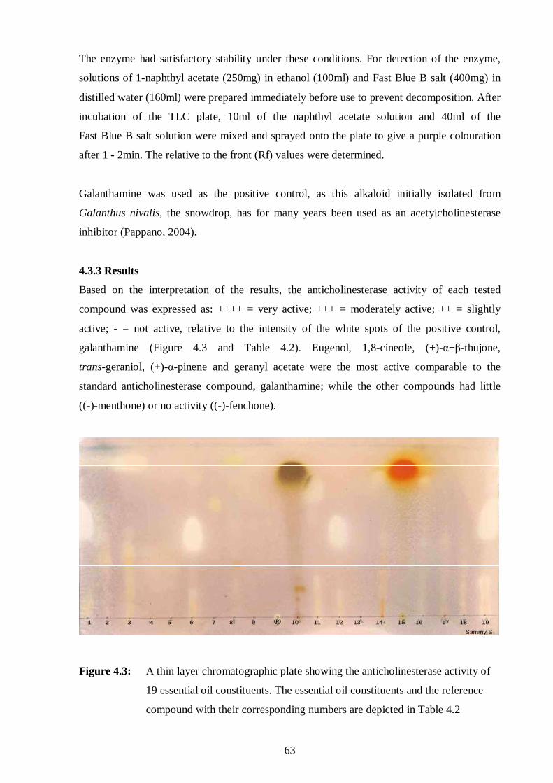
63
The enzyme had satisfactory stability under these conditions. For detection of the enzyme,
solutions of 1-naphthyl acetate (250mg) in ethanol (100ml) and Fast Blue B salt (400mg) in
distilled water (160ml) were prepared immediately before use to prevent decomposition. After
incubation of the TLC plate, 10ml of the naphthyl acetate solution and 40ml of the
Fast Blue B salt solution were mixed and sprayed onto the plate to give a purple colouration
after 1 - 2min. The relative to the front (Rf) values were determined.
Galanthamine was used as the positive control, as this alkaloid initially isolated from
Galanthus nivalis, the snowdrop, has for many years been used as an acetylcholinesterase
inhibitor (Pappano, 2004).
4.3.3 Results
Based on the interpretation of the results, the anticholinesterase activity of each tested
compound was expressed as: ++++ = very active; +++ = moderately active; ++ = slightly
active; - = not active, relative to the intensity of the white spots of the positive control,
galanthamine (Figure 4.3 and Table 4.2). Eugenol, 1,8-cineole, (±)-α+β-thujone,
trans-geraniol, (+)-α-pinene and geranyl acetate were the most active comparable to the
standard anticholinesterase compound, galanthamine; while the other compounds had little
((-)-menthone) or no activity ((-)-fenchone).
1 2 3 4 5 6 7 8 9 ® 10 11 12 13 14 15 16 17 18 19
Sammy S
1 2 3 4 5 6 7 8 9 ® 10 11 12 13 14 15 16 17 18 19
Sammy S
Figure 4.3: A thin layer chromatographic plate showing the anticholinesterase activity of
19 essential oil constituents. The essential oil constituents and the reference
compound with their corresponding numbers are depicted in Table 4.2

64
4.3.4 Discussion
Overall the acetylcholinesterase enzyme was found to be generally susceptible to the action of
the essential oil constituents (Table 4.2). Although this was the general trend for the tested
constituents, eugenol, 1,8-cineole, (±)-α+β-thujone, trans-geraniol, (+)-α-pinene and geranyl
acetate were comparable to galanthamine exhibiting promising anticholinesterase activity
(Figure 4.3). The oxide, 1,8-cineole has previously been reported to possess anticholinesterase
activity (Mills et al., 2004; Savelev et al., 2003), which is in agreement with the present
study. Although, Miyazawa and Yamafuji (2005) reported that essential oil constituents with
the pinane skeleton, like (+)-α-pinene and not (+)-β-pinene, showed a strong inhibitory effect
on acetylcholinesterase, as is the case with the α-isomer, α-pinene in the present study
(Table 4.2).
Lahlou (2004a) also reported that some monoterpene derivatives such as linalyl acetate,
geranyl acetate, eugenol and carvacrol inhibit acetylcholinesterase, which is congruent with
the results obtained in this present study (Table 4.2). The action of acetylcholinesterase in the
neuromuscular junction ensures muscle contraction, thus if the action of acetylcholinesterase
is inhibited there will be an increased accumulation of acetylcholine in the synaptic cleft
which will induce further muscle contraction or could antagonise the effects of pancuronium
(a neuromuscular blocker). However, this is not the only mechanism by which EOC’s are
proposed to cause muscle contraction. Lahlou (2004a) reported that EOC’s like eugenol are
able to induce muscle contraction (or blockade of excitation-contraction coupling) by
releasing calcium ions from the sarcoplasmic reticulum. Moreover, the spasmolytic effect of
(±)-linalool was thought to be mediated through cyclic adenosine monophosphate (cAMP).
Miyazawa and Yamafuji (2005) also reported that overall the hydrocarbon compounds
showed potent inhibition of acetylcholinesterase as compared with alcohols and ketones.
However, in our study most EOC’s like (R)-(+)-limonene, carvacrol, linalyl acetate and
E- and Z-(±)-nerolidol showed equipotent activity (Table 4.2 and Figure 4.3), irrespective of
the class each falls under. Whilst the alcohol, trans-geraniol showed moderate
anticholinesterase activity. The degree of saturation, including the position of the C=C
double bond, affected the inhibitory activity of acetylcholinesterase and is also related to the
strength or potency of inhibition of acetylcholinesterase (Miyazawa and Yamafuji, 2005).

65
Table 4.2: The anticholinesterase activity of 19 essential oil constituents. Where: ++++ = very active; +++ = moderately active; ++ = slightly active; - = not active.
Essential oil constituents Corresponding number in TLC
plate (Figure 4.3) Anticholinesterase activity
Alcohols
(-)-α-Bisabolol 13 -
trans-Geraniol 11 ++++
(±)-Linalool 5 -
E- and Z-(±)-Nerolidol 16 +++
Aldehyde
(+)-β-Citronellal 4 -
Esters
Linalyl acetate 7 +++
Geranyl acetate 19 ++++
Ketones
(±)-α+β-Thujone 6 ++++
(+)-Carvone 8 ++
(-)-Fenchone 15 -
(-)-Menthone 18 ++
(-)-Pulegone 10 -
Phenols
Carvacrol 2 +++
Eugenol 1 ++++
Terpene hydrocarbons
(+)-α-Pinene 12 ++++
(+)-β-Pinene 9 -
(R)-(+)-Limonene 14 +++
p-Cymene 17 ++
Oxide
1,8-Cineole 3 ++++
Control
Galanthamine ® ++++
66
EOC’s like (-)-α-bisabolol, (-)-fenchone, (-)-pulegone, and (-)-citronellal, (±)-linalool and
(+)-β-pinene did not show any anticholinesterase activity. However, (-)-α-bisabolol from the
Salvia species has been reported to possess anticholinestrase activity (Perry et al., 2003). The
difference between the activities of the EOC’s from study to study could be attributed to the
existence of infraspecific chemical differences in the essential oils of some aromatic plants
(or chemovarieties). It is important to pinpoint that the test method applied in this study may
not be the best when testing the anticholinesterase activity and might have contributed to the
poor activity of most EOC’s. Regardless, it is relevant to mention that acetylcholinesterase
inhibitors including most EOC’s mentioned above, have a broader spectrum of activity and
thus possess possible therapeutic potential other than just for Alzheimer’s disease. These
include:
1. Anthelmintic infections: drugs such as pyrantel pamoate used in the treatment of pinworm
(Enterobius vermicularis), ascariasis (Ascaris lumbricoides), hookworm (Ancylostoma
duodenale and Necator americanus) and Trichostrongylus orientalis infections are known to
be depolarising neuromuscular blockers, but also inhibit the activity of helmintic
acetylcholinesterase enzyme, resulting in paralysis and subsequent expulsion of the helminth
(Rosenthal and Goldsmith, 2004).
2. Insecticidal agents: organophosphates (parathion) and organocarbamates are irreversible
pesiticides and used to control infestations (Pappano, 2004).
3. Bilharzia infections: the agent metrifonate used in the treatment of Schistosoma
haematobium (bilharzia) and as an insecticide, is an organophosphate cholinesterase inhibitor;
where the active metabolite, 2,2-dimethyldichlorovinyl phosphate irreversibly inhibits
acetylcholinesterase enzyme. The pharmacokinetics of this metabolite is favourable for
long-term treatment of Alzheimer’s disease with the half-life of cholinesterase inhibition
being approximately 26 days; even though the plasma half-life of the drug is 2-3h. This drug
is currently under investigation to determine its safety and efficacy in the treatment of the
cognitive symptoms of Alzheimer's disease (Ringman and Cummings, 1999).
4. Antagonists to neuromuscular blockers: acetylcholinesterase inhibitors, such as
physostigmine and neostigmine are regularly used to reverse the effects of neuromuscular
blockers, such as pancuronium. Thus, if the safety profile of the essential oil constituents
(e.g. geranyl acetate, trans-geraniol, (±)-α+β-thujone, eugenol, (+)-α-pinene and 1,8-cineole)
will be acceptable then further investigations may be warranted in using the EOC´s in this
scenario.
5). Ectoparacidal agents: organophosphates (dichlorvos, diazinon) and organocarbamates
(carbaryl, propoxur) are regularly used in the veterinary treatment of ectoparasites such as
67
ticks, lice and fleas. The organophosphates irreversibly phosphorylate acetylcholinesterase,
while the organocarbamates reversibly inhibit enzyme activity such that the increased
endogenous acetylcholine causes muscle fasciculations with subsequent paralysis and death.
Dichlorvos is effective in the treatment of roundworm in pigs and horses (McKellar and
Jackson, 2004).
Although the inhibition of acetylcholinesterase activity may be beneficial in many diseases
and clinical situations, it should also be remembered that this enzyme is necessary for the
metabolism and breakdown of numerous drugs such as the ester-type local anaesthetic
(procaine) and neuromuscular blockers (succinylcholine, mivacurium). The consequence
being increased plasma concentrations of the drugs with increased risk of adverse drug
effects. These types of interactions should be taken into account when further investigating
the potential of using the EOC´s.
68
CHAPTER FIVE
CYTOTOXICITY PROFILE
5.1 Introduction
Toxicology is concerned with the deleterious effects of chemical agents on all living systems.
However, in the biomedical area toxicology is primarily concerned with unfavourable effects
in humans resulting from exposure to drugs and other chemicals, including plant extracts and
essential oils (Plaa, 2004). As with all drugs, plant essential oils can also exhibit unfavourable
or deleterious effects on mammalian cells such as: 1) topical effects including the skin,
mucous membranes, eye irritation, phototoxicity, skin sensitization and photosensitization; 2)
systemic effects including mutagenicity, carcinogenicity, embryotoxicity and reproductive
toxicity (Yoo et al., 2005). Furthermore, these adverse effects can involve specific organs like
the liver, heart, kidney and brain. The cytotoxicity of an essential oil may be changed by the
presence of other compounds, where the compounds may potentiate each other´s activity or
chemically modify each other or alter their respective bioavailability and overwhelm their
therapeutic range. For example, Tea Tree oil consists of many chemical constituents that are
also prominent in pine oil and because of their similar properties, simultaneous administration
could potentiate the expected adverse effects if their doses are not adjusted (Nakatsu et al.,
2000). Therefore, essential oil usage by people using other drugs may lead to complications of
essential oil-drug interactions, because some drugs have a narrow therapeutic range (e.g. an
anticoagulant warfarin), thus these can compromise the pharmacokinetic and
pharmacodynamic profiles of therapeutic agents. Several clinical cases have been documented
on the interaction and treatment failure between herbal remedies and prescribed medications
(Constable, 2006). The combined use of several herbal preparations (garlic, feverfew and
ginseng) can increase the risk of bleeding, as all three preparations inhibit platelet aggregation
(Constable, 2006).
Overdosing or incorrect usage of essential oils may result in serious side effects (Zhou et al.,
2004), since most essential oils should not be used in their pure (neat) form, but diluted with
carrier oils before used topically and on mucous membranes. Infants, children, the elderly,
pregnant and lactating women and epileptics are vulnerable, therefore, particular care should
be taken when using essential oils (Lawrence, 2000). The general rule of thumb is to avoid
prolonged use of oils; so it is advisable to take breaks from their usage (Lawrence, 2000).
This is due to the tendency of building up a residue in the body which may result in

69
deleterious effects (Zhou et al., 2004). There is well established evidence that the formation of
reactive metabolites of drugs is associated with drug toxicity (Zhou et al., 2004). Likewise,
there is data suggesting that the formation of reactive metabolites through bioactivation has a
significant role in herbal toxicity and carcinogenicity. Reports revealed that these resultant
reactive metabolites covalently bind to cellular proteins and DNA, leading to toxicity through
multiple mechanisms such as direct cytotoxicity, oncogene activation, and hypersensitivity
reactions (Zhou et al., 2004). For example, with aristolochic acids present in Aristolochia
species, the nitro group is reduced by hepatic cytochrome P450 (CYP1A1/2) or peroxidases in
extrahepatic tissues, this results in a reactive cyclic nitrenium ion. The nitrenium ion possibly
reacts with DNA and proteins, resulting in activation of H-ras oncogene, gene mutation and
ultimately carcinogenesis (Zhou et al., 2004). Other examples are pulegone present in
essential oils from many mint species; and teucrin A, a diterpenoid found in germander
(Chamaedrys) used as add on therapy to slimming tablets (Lawrence, 2000; Yoo et al., 2005).
An excessive metabolism of pulegone may generate p-cresol which depletes glutathione
(Zhou et al., 2004). The furan ring of the diterpenoids in germander is oxidized by CYP3A4
to a reactive epoxide which reacts with CYP3A and epoxide hydrolase (Yoo et al., 2005).
Zhou et al. (2004) reported that hepatic cytochrome P450 enzymes (CYPs) are known to
metabolize more than 95% of therapeutic drugs and activate a number of procarcinogens as
well. Therefore, mechanism-based inhibition of CYPs may explicitly explain some reported
herb-drug interactions and chemopreventive/chemotherapeutic herbal and essential oil
activities (Zhou et al., 2004).
Toxic reactions vary qualitatively depending on the duration of the exposure, with a single
exposure occurring over a day or two, presenting acute exposure which is reversible and tends
not to be severe. However, multiple exposures continuing over a longer period of time
represent a chronic exposure, which result in severe adverse effects (Plaa, 2004).
Overexposure to essential oils, topically or via inhalation can result in nausea, dizziness,
headache or a light-headed feeling (Yoo et al., 2005); but more serious topical and systemic
effects as mentioned below, have also been reported.
5.1.1 Topical effects
• Allergic reactions are common with essential oils and include possible symptoms like
nausea, dizziness, sweating, palpitations, stomach pains and irritation to mucous
membranes. If these problems are experienced, it is advisable to discontinue the use of
essential oils as the reactions are reversible.
70
• Skin irritation is an inflammatory reaction to certain essential oils and the severity
depends upon the concentration and duration of exposure to the oil. It may be partially
attributed to the terpene content in the oils, but it is more likely to be caused by the high
phenol content in certain oils (Yoo et al., 2005).
• Dermatitis is possible even when a diluted essential oil is applied to the skin, sometimes
even the vapours can cause a sensitization reaction. The effects can present as a rash,
blotches and itching, or blistering. Allergies involve the interaction of the immune system
forming antibodies to render the antigen/essential oil harmless. However, investigations
demonstrated that dermatitis is an immunological response not an antigen-antibody
reaction (Yoo et al., 2005). By far, only a few skin sensitizing agents have been identified,
namely: basil, cinnamon, lemon, lemongrass, melissa, peppermint, thyme and tea tree oils
(Nakatsu et al., 2000).
• Phototoxic reactions resulting in photosensitization, redness and trauma to the skin when
exposed to sunlight have been reported to occur with bergamot, lemon, lime, orange and
other citrus oil (Nakatsu et al., 2000).
• Essential oils with thujone or apiol are abortifacient and have been proposed to cause
blood accumulation in the pelvic-uterus area and liver damage (Zhou et al., 2004). These
oils include hyssop, mugwort, parsley, pennyroyal, sage and thuja. Thus, pregnant women
should avoid these essential oils and also others such as basil, camphor, clary sage,
cedarwood, juniper, marjoram, myrrh, rue, tansy and wormwood (Yoo et al., 2005).
• Overdosage of oils containing thujone and apiol can also have narcotic, analgesic or
paralyzing effects (Zhou et al., 2004).
• Essential oils that are excreted via the renal system such as juniper and sandalwood are
rendered nephrotoxic and their use must be discontinued after four weeks (Yoo et al.,
2005).
5.1.2 Systemic effects
Hypertensive patients must be cautious when using essential oil of hyssop, rosemary, sage
and thyme (Zhou et al., 2004), while those patients with epileptic seizures must be careful
when using oils from fennel, hyssop, rosemary, sage or wormwood (Halcon, 2002).
Toxicological cases caused by volatile oils such as eucalyptus, sassafras, turpentine,
wintergreen, chenopodium, citronella oil, pennyroyals and peppermint oils have also been
reported (Halcon, 2002). In infants these aforementioned oils may cause respiratory
depression or even death, when applied directly into the nose or close to the nose onto the
71
skin or mucous membranes (Halcon, 2002). These problems arise due to misidentification,
drug-herb interactions and lack of standardization. The mechanism of herbal or essential oil
toxicity remains elusive and the following systemic problems may also arise from abuse of
essential oils.
5.1.2.1 Hepatotoxicity
The liver has various functions and helps with carbohydrate and lipid metabolism, secretion
of bile as well as the synthesis of proteins, yet most people perceive the liver as only a
detoxification organ for blood. Metabolism of essential oil constituents is via human hepatic
cytochrome P450 enzymes to non-toxic metabolites, but the formation of toxic metabolites is
also possible (Zhou et al., 2004). The monooxygenases oxidizing metabolism of these
essential oil constituents require a haemoprotein P450, a flavoprotein NADPH-P450
reductase and phospholipids (Ueng et al., 2005).
In the human liver, CYP1A2, CYP2A6, CYP2C9, CYP2D6, CYP2E1, and CYP3A4 are the
major forms of the cytochrome P450 enzymes, which constitute about 13%, 4%, 18%, 2%,
7%, and 29% of total P450 contents, respectively (Ueng et al., 2005). Hepatotoxicity is
unlikely to occur following administration of the essential oil to the skin; but it is more likely
to occur following oral ingestion. No essential oil should be taken internally unless so
prescribed by a licensed medical practitioner. Essential oils which are hepatotoxic when
swallowed are aniseed, buchu, basil, clove and cinnamon bark (Ueng et al., 2005).
5.1.2.2 Carcinogenicity
Some essential oils are carcinogenic and have been banned (Table 5.1) in aromatherapy and
cannot be applied for internal usage. The oils which still raise doubts are calamus oil (contains
asarone), and those containing ample amounts of safrole like sassafras found in yellow and
brown camphor (Nakatsu et al., 2000). Other carcinogenic compounds in essential oils
include methyl chavicol found in basil oil. Notably, this statement does not make basil
dangerous, but care should be taken to select the basil oil containing ample amounts of
linalool and lowest methyl chavicol content (Ueng et al., 2005). It must however be pointed
out that studies showed that these oils can be carcinogenic when applied daily in high
concentration for many months (Lawrence, 2000; Ueng et al., 2005).
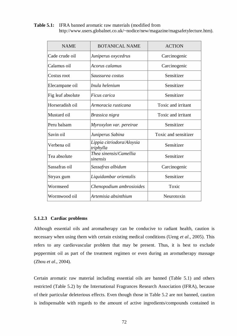
72
Table 5.1: IFRA banned aromatic raw materials (modified from http://www.users.globalnet.co.uk/~nodice/new/magazine/magsafetylecture.htm).
NAME BOTANICAL NAME ACTION
Cade crude oil Juniperus oxycedrus Carcinogenic
Calamus oil Acorus calamus Carcinogenic
Costus root Saussurea costus Sensitizer
Elecampane oil Inula helenium Sensitizer
Fig leaf absolute Ficus carica Sensitizer
Horseradish oil Armoracia rusticana Toxic and irritant
Mustard oil Brassica nigra Toxic and irritant
Peru balsam Myroxylon var. pereirae Sensitizer
Savin oil Juniperus Sabina Toxic and sensitizer
Verbena oil Lippia citriodora/Aloysia triphylla
Sensitizer
Tea absolute Thea sinensis/Camellia sinensis
Sensitizer
Sassafras oil Sassafras albidum Carcinogenic
Stryax gum Liquidambar orientalis Sensitizer
Wormseed Chenopodium ambrosioides Toxic
Wormwood oil Artemisia absinthium Neurotoxin
5.1.2.3 Cardiac problems Although essential oils and aromatherapy can be conducive to radiant health, caution is
necessary when using them with certain existing medical conditions (Ueng et al., 2005). This
refers to any cardiovascular problem that may be present. Thus, it is best to exclude
peppermint oil as part of the treatment regimen or even during an aromatherapy massage
(Zhou et al., 2004).
Certain aromatic raw material including essential oils are banned (Table 5.1) and others
restricted (Table 5.2) by the International Fragrances Research Association (IFRA), because
of their particular deleterious effects. Even though those in Table 5.2 are not banned, caution
is indispensable with regards to the amount of active ingredients/compounds contained in

73
them, as the existence of the side effects such as sensitization and phototoxicity is possible
when treating patients with various clinical conditions (Ueng et al., 2005).
Table 5.2: IFRA restricted aromatic raw materials (modified from
http://www.users.globalnet.co.uk/~nodice/new/magazine/magsafetylecture.htm).
NAME BOTANICAL NAME ACTION
Angelica root oil Angelica archangelica Phototoxic
Bergamot oil Citrus bergamia Phototoxic
Bitter orange oil Citrus aurantium Phototoxic
Cassia oil Cinnamomum cassia Sensitizer
Cinnamon bark Cinnamomum zeylanicum Sensitizer
Cumin oil Cuminum cyminum Phototoxic
Grapefruit oil Citrus paradisi Phototoxic
Lemon oil Citrus limon Phototoxic
Lime oil Citrus aurantifolia Phototoxic
Tagetes oil and absolute
Tagetes minuta Phototoxic
Oak moss absolute and resinoid
Evernia prunastri Sensitizer
Pinaceae oils Pinaceae mugo, P. nigra, P. pinaster, P. sylvatica
Sensitizer
Rue oil Ruta graveolens Sensitizer
Verbena absolute Lippia citriodora/Aloysia triphylla
Sensitizer
Tree moss absolute (Pseudo) Evernia furfuracea Sensitizer
Due to the wide use and easy availability of herbal medicines, there is increasing concern
about essential oil toxicity (Constantin et al., 1991). Despite this fact, there is still a great
necessity for more scientific evidence on the toxicity of volatile oils. Thus, this study took the
opportunity to meet this increasing demand by investigating the toxic profiles of selected
essential oil constituents when used individually and in combination.
74
5.2 Methodology
5.2.1 Principles of the method
The methods to measure the number of viable proliferating cells vary widely, thus a reliable,
versatile and quantitative method for detecting the viability is a prerequisite. Tetrazolium
compounds are an increasing popular non-radioactive option. Moreover, assays involving
tetrazolium salts such as the MTT [3-(4,5-dimethyl-2-thiazol-2-yl)-2,5-diphenyl-2H-
tetrazolium bromide] assay, can easily detect living cells, and the resultant colour formation
depends on the degree of functionality of the mitochondria. The main advantages of this assay
are: 1) no washing steps; 2) no radioisotope is required; and 3) results can be read with high
degree of accuracy, reproducibility and rapidity (Mosmann, 1983).
The MTT assay is reliant on succinate dehydrogenase which is an enzyme localised in the
mitochondria of living cells that cleaves the tetrazolium ring of a tetrazolium dye. The yellow,
water soluble MTT dye changes to a violet, insoluble product (MTT-formazan) (Mosmann,
1983). This chemical reaction can easily be measured by a spectrophotometer, and the amount
of formazan formed is directly proportional to cell density.
5.2.2 Culture maintenance
The transformed human kidney epithelial (Graham) cells were continuously maintained in
vitro at 37ºC in 5% CO2 according to the departmental protocol. The cells were maintained
with complete culture medium at least thrice weekly and trypsinized (Section 5.2.2.4) once
weekly to subculture the cells.
5.2.2.1 Preparation of complete culture medium
The culture medium was prepared in Milli-Q® water with the following ingredients: 9.8g/l
HAM F10 supplemented with 1.18g/l NaHCO3. The culture medium was sterilized through a
Sterivex-GS 0.22µm filter unit (Highveld Biologicals, South Africa) and stored at 4ºC. Before
use, the complete culture medium was prepared by supplementing the culture medium with
5% foetal calf serum (FCS, Section 5.2.2.2) and 0.1ml of sterile 50mg/ml gentamicin. The
gentamicin stock was prepared by dissolving 50mg gentamicin sulphate in 1ml PBS (Section
3.2.1.3), filtered through a Sterivex-GS 0.22µm filter unit and stored at 4ºC until required.

75
5.2.2.2 Preparation of foetal calf serum
FCS (Highveld Biologicals, South Africa) was heat inactivated at 56ºC in a water bath for 1h.
The inactivated FCS was centrifuged at 1500rpm for 10min. Thereafter, aliquots of the
supernatant were stored at -20ºC until required.
5.2.2.3 Assessment of cell growth
The cells were examined microscopically to assess their growth at least thrice weekly.
Following which the spent medium was discarded and 50ml of the fresh complete culture
medium was added and the cells were then incubated at 37ºC.
5.2.2.4 Trypsinization
Trypsinization was carried out to adjust the number of adherent proliferating cells in the flask
in order to optimize growing conditions. A volume of 4ml of 0.25% trypsin-0.1% Versene-
EDTA (Highveld Biologicals, South Africa) was added to the culture flask and allowed to
cover the bottom of the flask, left to stand at room temperature for 5 - 10min, thereafter the
flask was gently agitated to loosen the cells. Once the cells were in suspension, 6ml complete
culture medium was added to neutralise the trypsin. The FCS in the culture medium has the
ability to inactivate trypsin. Thereafter, the cells were gently agitated to ensure a single cell
suspension. Thereafter, 9ml of this suspension was either discarded or used for experimental
purposes. To the remaining cells, 50ml complete culture medium (Section 5.2.2.1) was added
and the cells were then incubated at 37°C in 5% CO2 such as to readhere and propagate.
5.2.3 MTT cytotoxicity assay
To investigate the toxicity profile of the EOC´s, the MTT assay was performed on the human
kidney (Graham) epithelial cells according to the method of Mosmann (1983). Various
dilutions of twenty essential oil constituents were incubated under optimal conditions with the
cells for 48h to determine their effect on the proliferation of the cells.
5.2.3.1 Preparation of the cells
From Section 5.2.2.4, the cell suspension was centrifuged at 1500rpm for 5min and the
supernatant discarded. A volume of 20ml experimental medium (i.e. the complete culture
medium (Section 5.2.2.1) but without gentamicin) was then added to the cells in a 50ml
centrifuge tube. The cells were once again gently agitated to ensure a single cell suspension.
The cells were then stained with 0.2% trypan blue in a 1:1 (v/v) ratio and two grids of cells
counted using a haemocytometer at 100x magnification. The trypan blue is taken up into

76
compromised or dead cell, whilst viable cells exclude the dye. At least, 95% cellular viability
was ensured before using the cells in the experiment. The number of cells/ml was determined
using Equation 5.1 before the cell suspension was adjusted with experimental medium to 0.25
million cells/ml.
Equation 5.1:
Cells/ml = number of counted cells/2 x dilution factor (2) x 104
5.2.4 Preparation of essential oil constituents and control drugs
Test compounds and a positive control drug, quinine, were prepared in eppendorfs as
previously described in Sections 3.2.3.3; 3.2.3.3.1; 3.2.3.3.2; 3.2.4 and 3.2.4.1.
5.2.5 Selection and preparation of essential oil constituents used in combination assays
Based on the IC50 values obtained from the individual EOC´s, various EOC combinations
were selected; namely: two of the most active EOC´s, one of the most active with one of the
least active, as well as two of the least active EOC´s. Eight fixed ratios of the two
predetermined EOC´s were then prepared such that the concentration of one EOC increased,
the concentration of the second EOC decreased. Both EOC were also prepared in the absence
of the other EOC to serve as a positive control. The eight dilutions for each individual EOC
were prepared in DMSO as described in Chapter 3 (Section 3.2.3.3.2). Further, at least six
dilutions from each of the abovementioned eight dilutions were prepared by 1:10 serial
dilution in eppendorfs using DMSO.
The dilutions were then combined in a 1:1 (v/v) ratio. A further dilution factor of 2 was taken
into account, so that the final dilution factor increased to 200x for the EOC´s. In the wells of
row A, 2µl of the first dilution of the combined EOC’s were plated out in triplicate and 18µ l
RPMI experimental medium (Section 3.2.1.3) was added. In rows B to G 18µl RPMI
experimental medium was added before 2µl of previous dilution was transferred from row to
row. To row H, only 20µl of 18µl RPMI experimental medium was added, while 180µl of the
adjusted cell suspension (Section 5.2.3.1) was added to all the wells of the plate
(Section 5.2.6), except in the first well of row H. This latter well served as a cell free blank,
and contained 180µl of HAM F10 experimental medium and 20µl of RPMI experimental
medium. The experimental runs were repeated in triplicate for reasons of consistency.

77
5.2.6 Preparation of microtitre plates
A volume of 180µl of the adjusted cell suspension was added to all wells except one. This
well served as the cell-free blank control, to which 180µl of experimental HAM F10 culture
medium containing FCS plus 20µl hypoxanthine-free medium (Section 3.2.1.3) were added.
The cells were incubated for 6h under humidified conditions in 5% CO2 at 37ºC so that they
could adhere to the bottom of the wells of the plate well. After this incubation period, 2µl of
the individual EOC or combined EOC´s and 18µl of hypoxanthine-free medium were added
to all wells, except the last row (row H). A total of 20µl of each dilution of the test EOC’s and
control, and quinine were added in triplicate to wells of rows A-G. To row H, 20µl of
hypoxanthine-free medium was added and served as the drug-free controls. Thereafter, the
cells were incubated under humidified conditions in 5% CO2 at 37ºC. After 44h of incubation,
40µl of 50mg/ml (= 0.05mM) MTT (Section 5.2.7) was added to all wells and the plate was
further incubated for 4h.
5.2.7 Addition of MTT and DMSO
A 0.05mM stock solution of MTT (USB™) prepared in PBS (Section 3.2.1.3) was sterile
filtered using a Sterivex-GS 0.22µm filter unit and stored at 4ºC. After 48h of incubation,
200µl of the contents from all wells was aspirated and discarded from all wells. Thereafter,
150µl of DMSO was added to all the wells to stop the reaction and dissolve the purple
formazan crystals.
5.2.8 Data analysis
The plate was shaken for 4min using a UV-VIS spectrophotometer to ensure complete
dissolution of the crystals and the absorbance was read at the test wavelength of 540nm and
reference wavelength of 690nm using the Ascent® software (Biosoft, UK) and log sigmoidal
dose response curves drawn. The percentage cell viability was calculated with the appropriate
controls taken into account according to Equation 5.2.
Equation 5.2:
Cellular viability = Drug treated absorbance - Mean absorbance of cell-free control x 100%
Mean absorbance of drug-free control - Mean cell-free control
Thereafter, the IC50 values were determined using the Enzfitter® software (Biosoft, UK). The
IC50 values from at least three independent experiments were used to calculate the mean and
standard deviation for each EOC. Data from the combination experiments were analysed and
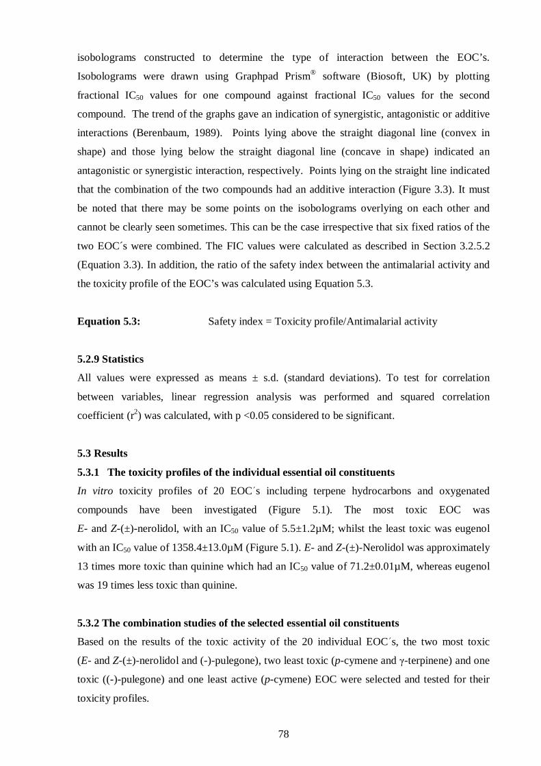
78
isobolograms constructed to determine the type of interaction between the EOC’s.
Isobolograms were drawn using Graphpad Prism® software (Biosoft, UK) by plotting
fractional IC50 values for one compound against fractional IC50 values for the second
compound. The trend of the graphs gave an indication of synergistic, antagonistic or additive
interactions (Berenbaum, 1989). Points lying above the straight diagonal line (convex in
shape) and those lying below the straight diagonal line (concave in shape) indicated an
antagonistic or synergistic interaction, respectively. Points lying on the straight line indicated
that the combination of the two compounds had an additive interaction (Figure 3.3). It must
be noted that there may be some points on the isobolograms overlying on each other and
cannot be clearly seen sometimes. This can be the case irrespective that six fixed ratios of the
two EOC´s were combined. The FIC values were calculated as described in Section 3.2.5.2
(Equation 3.3). In addition, the ratio of the safety index between the antimalarial activity and
the toxicity profile of the EOC’s was calculated using Equation 5.3.
Equation 5.3: Safety index = Toxicity profile/Antimalarial activity
5.2.9 Statistics
All values were expressed as means ± s.d. (standard deviations). To test for correlation
between variables, linear regression analysis was performed and squared correlation
coefficient (r2) was calculated, with p <0.05 considered to be significant.
5.3 Results
5.3.1 The toxicity profiles of the individual essential oil constituents
In vitro toxicity profiles of 20 EOCs including terpene hydrocarbons and oxygenated
compounds have been investigated (Figure 5.1). The most toxic EOC was
E- and Z-(±)-nerolidol, with an IC50 value of 5.5±1.2µM; whilst the least toxic was eugenol
with an IC50 value of 1358.4±13.0µM (Figure 5.1). E- and Z-(±)-Nerolidol was approximately
13 times more toxic than quinine which had an IC50 value of 71.2±0.01µM, whereas eugenol
was 19 times less toxic than quinine.
5.3.2 The combination studies of the selected essential oil constituents
Based on the results of the toxic activity of the 20 individual EOCs, the two most toxic
(E- and Z-(±)-nerolidol and (-)-pulegone), two least toxic (p-cymene and γ-terpinene) and one
toxic ((-)-pulegone) and one least active (p-cymene) EOC were selected and tested for their
toxicity profiles.
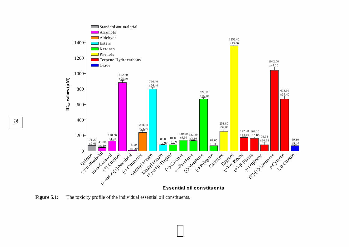
0
Quinin
e
(-)- α -B
isabo
lol
trans
-Ger
aniol
( ± )-Li
nalo
ol
E- and
Z-(±)-N
eroli
dol
(-)-C
itron
ellal
Gerany
l ace
tate
Linaly
l ace
tate
( ± )-α + β -
Thujon
e(+
)-Carv
one
(-)-F
ench
one
(-)-M
enth
one
(-)-P
ulego
neCarv
acro
lEug
enol
(+)- α -P
inene
(+)- β -
Pinen
eγ -T
erpine
ne
(R)-(
+)-L
imon
ene
p-Cym
ene
1, 8-
Cineo
le
0
200
400
600
800
1000
1200
1400
Standard antimalarialAlcoho lsAldehydeEsters
KetonesPhenolsTerpene HydrocarbonsOxide
71.20±0.01 41.80
±5.10
128.50±4.70
882.70±25.40
5.50±1.20
238.30±24.90
796.40±26.40
80.00±2.90
81.00±12.90
140.90±9.60
132.20±3.10
672.10±15.10
64.60±3.30
251.80±32.80
1358.40±13.00
172.20±13.40
164.10±15.86 79.33
±30.98
1042.00±41.10
673.60±35.40
69.10±9.40
Essential oil constituents
IC50
val
ues
(µM
)
Figure 5.1: The toxicity profile of the individual essential oil constituents.
79

80
From the isobolograms of the combinations tested in Table 5.3, it was evident that the toxicity
profiles between the combined EOC΄s (Figures 5.2 and 5.3) were all potentiated.
Table 5.3: The toxicity profile of the combined essential oil constituents (n = 3).
EOC1 EOC2 ∑FIC value Interaction
E- and Z-(±)-Nerolidol (-)-Pulegone 0.076 Synergism
p-Cymene γ-Terpinene 0.002 Synergism
p-Cymene (-)-Pulegone 0.306 Synergism
0.00 0.25 0.50 0.75 1.00 1.250.00
0.25
0.50
0.75
1.00
1.25
E- and Z-(±)-Nerolidol(IC50 combined EOC /IC50 individual EOC)
(-)-
Pu
lego
ne
(IC
50 c
ombi
ned
EO
C /
IC50
indi
vidu
al E
OC
)
Figure 5.2: The synergistic interaction between E- and Z-(±)-nerolidol and (-)-pulegone,
indicating an increase in their toxicity profile.
5.3.3 The toxicity profile of the essential oil constituent and quinine
Potentiation of the toxicity profile (∑FIC= 0.001) was observed when the most active EOC
(E- and Z-(±)-nerolidol) was combined with the standard antimalarial agent, quinine
(Figure 5.4).

81
0.00 0.25 0.50 0.75 1.00 1.250.00
0.25
0.50
0.75
1.00
1.25
p-Cymene(IC50 combined EOC /IC50 individual EOC)
gam
ma-
Ter
pin
ene
/ (-)
-Pul
egon
e(I
C50
com
bine
d E
OC
/IC
50 in
divi
dual
EO
C)
Figure 5.3: p-Cymene interacted in a synergistic manner with both γ-terpinene (▲)
and (-)-pulegone (▼) to potentiate the toxicity profile of the essential
oil constituents.
Figure 5.4: The synergistic interaction between quinine and E- and Z-(±)-nerolidol.
0.00 0.25 0.50 0.75 1.00 1.250.00
0.25
0.50
0.75
1.00
1.25
Quinine(IC50 combined / IC50 individual)
E-
and
Z-(±)
-Ner
olid
ol(I
C50
com
bine
d E
OC
/IC
50 in
divi
dual
EO
C)
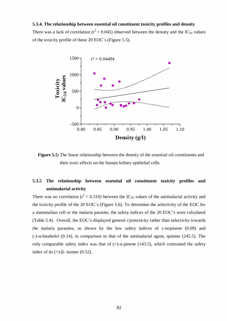
82
5.3.4. The relationship between essential oil constituent toxicity profiles and density
There was a lack of correlation (r2 = 0.045) observed between the density and the IC50 values
of the toxicity profile of these 20 EOC´s (Figure 5.5).
0.80 0.85 0.90 0.95 1.00 1.05 1.10-500
0
500
1000
1500 r² = 0.04494
Density (g/l)
Tox
icit
yIC
50
valu
es
Figure 5.5: The linear relationship between the density of the essential oil constituents and
their toxic effects on the human kidney epithelial cells.
5.3.5 The relationship between essential oil constituent toxicity profiles and
antimalarial activity
There was no correlation (r2 = 0.310) between the IC50 values of the antimalarial activity and
the toxicity profile of the 20 EOC´s (Figure 5.6). To determine the selectivity of the EOC for
a mammalian cell or the malaria parasite, the safety indices of the 20 EOC’s were calculated
(Table 5.4). Overall, the EOC’s displayed general cytotoxicity rather than selectivity towards
the malaria parasites, as shown by the low safety indices of γ-terpinene (0.09) and
(-)-α-bisabolol (0.14), in comparison to that of the antimalarial agent, quinine (245.5). The
only comparable safety index was that of (+)-α-pinene (143.5), which contrasted the safety
index of its (+)-β- isomer (0.52).
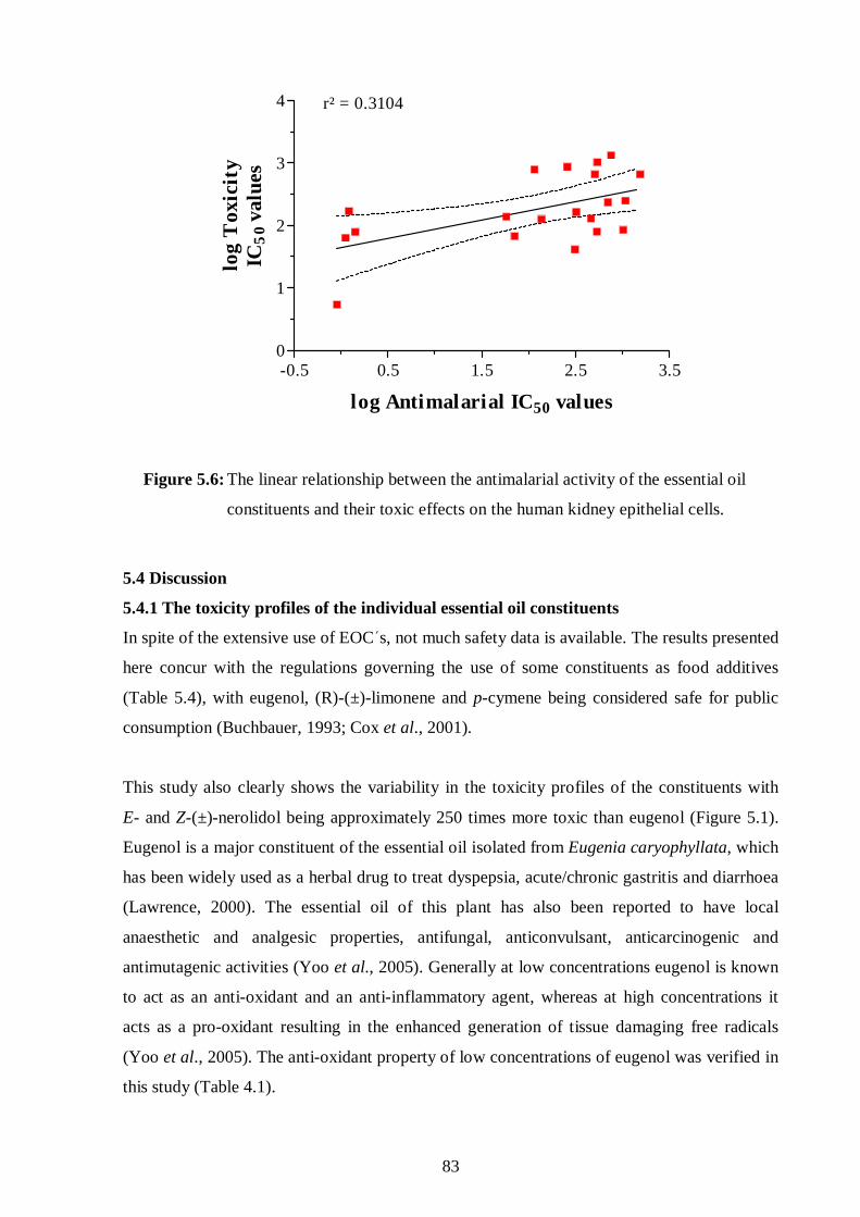
83
-0.5 0.5 1.5 2.5 3.50
1
2
3
4 r² = 0.3104
log Antimalarial IC50 values
log
Tox
icit
yIC
50
valu
es
Figure 5.6: The linear relationship between the antimalarial activity of the essential oil
constituents and their toxic effects on the human kidney epithelial cells.
5.4 Discussion
5.4.1 The toxicity profiles of the individual essential oil constituents
In spite of the extensive use of EOC΄s, not much safety data is available. The results presented
here concur with the regulations governing the use of some constituents as food additives
(Table 5.4), with eugenol, (R)-(±)-limonene and p-cymene being considered safe for public
consumption (Buchbauer, 1993; Cox et al., 2001).
This study also clearly shows the variability in the toxicity profiles of the constituents with
E- and Z-(±)-nerolidol being approximately 250 times more toxic than eugenol (Figure 5.1).
Eugenol is a major constituent of the essential oil isolated from Eugenia caryophyllata, which
has been widely used as a herbal drug to treat dyspepsia, acute/chronic gastritis and diarrhoea
(Lawrence, 2000). The essential oil of this plant has also been reported to have local
anaesthetic and analgesic properties, antifungal, anticonvulsant, anticarcinogenic and
antimutagenic activities (Yoo et al., 2005). Generally at low concentrations eugenol is known
to act as an anti-oxidant and an anti-inflammatory agent, whereas at high concentrations it
acts as a pro-oxidant resulting in the enhanced generation of tissue damaging free radicals
(Yoo et al., 2005). The anti-oxidant property of low concentrations of eugenol was verified in
this study (Table 4.1).
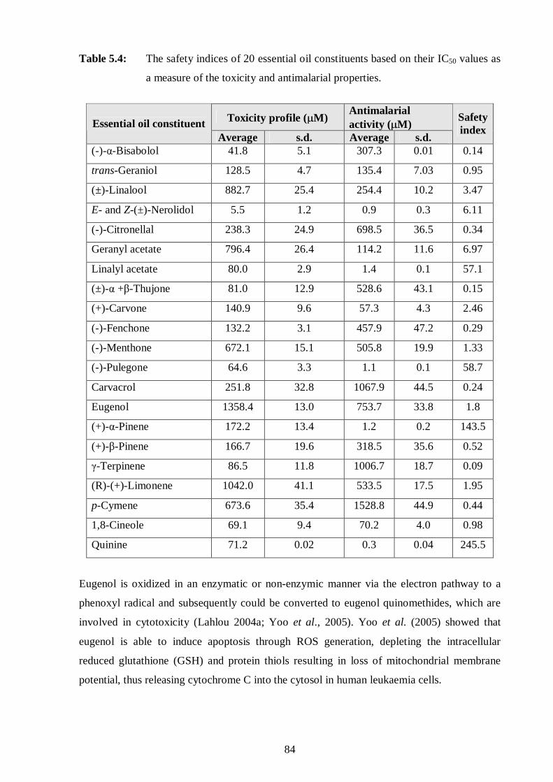
84
Table 5.4: The safety indices of 20 essential oil constituents based on their IC50 values as
a measure of the toxicity and antimalarial properties.
Toxicity profile (µM) Antimalarial activity (µM) Essential oil constituent
Average s.d. Average s.d.
Safety index
(-)-α-Bisabolol 41.8 5.1 307.3 0.01 0.14
trans-Geraniol 128.5 4.7 135.4 7.03 0.95
(±)-Linalool 882.7 25.4 254.4 10.2 3.47
E- and Z-(±)-Nerolidol 5.5 1.2 0.9 0.3 6.11
(-)-Citronellal 238.3 24.9 698.5 36.5 0.34
Geranyl acetate 796.4 26.4 114.2 11.6 6.97
Linalyl acetate 80.0 2.9 1.4 0.1 57.1
(±)-α +β-Thujone 81.0 12.9 528.6 43.1 0.15
(+)-Carvone 140.9 9.6 57.3 4.3 2.46
(-)-Fenchone 132.2 3.1 457.9 47.2 0.29
(-)-Menthone 672.1 15.1 505.8 19.9 1.33
(-)-Pulegone 64.6 3.3 1.1 0.1 58.7
Carvacrol 251.8 32.8 1067.9 44.5 0.24
Eugenol 1358.4 13.0 753.7 33.8 1.8
(+)-α-Pinene 172.2 13.4 1.2 0.2 143.5
(+)-β-Pinene 166.7 19.6 318.5 35.6 0.52
γ-Terpinene 86.5 11.8 1006.7 18.7 0.09
(R)-(+)-Limonene 1042.0 41.1 533.5 17.5 1.95
p-Cymene 673.6 35.4 1528.8 44.9 0.44
1,8-Cineole 69.1 9.4 70.2 4.0 0.98
Quinine 71.2 0.02 0.3 0.04 245.5
Eugenol is oxidized in an enzymatic or non-enzymic manner via the electron pathway to a
phenoxyl radical and subsequently could be converted to eugenol quinomethides, which are
involved in cytotoxicity (Lahlou 2004a; Yoo et al., 2005). Yoo et al. (2005) showed that
eugenol is able to induce apoptosis through ROS generation, depleting the intracellular
reduced glutathione (GSH) and protein thiols resulting in loss of mitochondrial membrane
potential, thus releasing cytochrome C into the cytosol in human leukaemia cells.
85
Terpenes have a number of common characteristics such as their highly lipophilic properties
and most of them readily interact with biomembranes. They can increase the fluidity of the
membranes, which leads to uncontrolled efflux of ions and metabolites, resulting in cell
leakage and ultimately in cell death (Griffin et al., 1999). A conformational change of
membrane proteins (e.g. ion channels and transporters) can occur when lipophilic compounds
enter biomembranes and accumulate close to the membrane proteins (Griffin et al., 1999). If
Na+, K+ or Ca2+ channels are affected, a disturbance of signal transduction could result. The
use of essential oils to treat spasms and light pain in the case of dyspeptic or gall disorders
could thus be plausibly explained. Since the lipophilic terpenes can cross the blood brain
barrier, sedative and analeptic effects ascribed to several complementary drugs could be
explained. Dormans and Deans (2000) reported that this ability of terpenes to cross the blood
brain barrier may be attributed to their size and densities (Figure 3.6). This membrane activity
is rather unspecific, therefore terpenes show antimicrobial, antimalarial, anti-oxidant and
cytotoxic activities against a wide range of organisms, ranging from bacteria, fungi, to insects
and vertebrates; such that they are used to treat bacterial and parasitic infections and
respiratory tract disorders (Halcon, 2002; Nakatsu et al., 2000). The lipophilicity of the
EOC’s enable them to readily cross the membranes, but this is not the sole determinant of
their biological or toxic activity as can be seen by a lack of correlation between the toxicity
profiles and density (Figure 5.5) or antimalarial activity (Figure 5.6) of the EOC’s. Thus,
further research is required to determine the precise cellular target(s) of the EOC’s.
The monoterpene ketone, pulegone constitutes more than 80% of the terpenes in the oils from
many mint species such as the pennyroyals, which is used as a flavourant/fragrant constituent
or to induce menstruation (Zhou et al., 2004). Many pennyroyals containing pulegone have
been reported to cause gastritis, induce abortion, and result in hepatic failure, central nervous
system, renal and pulmonary toxicity, and even death (Zhou et al., 2004). The toxic profile is
confirmed in this study where (-)-pulegone plus E- and Z-(±)-nerolidol inhibited the growth of
human kidney epithelial cells (Figure 5.2). Thus, when examining the efficacy of an essential
oil its toxicity profile needs to be considered especially since the composition and ratio of
EOC´s are influenced by seasonal and geographical variation (Buchbauer, 1993; Viljoen et
al., 2005). With all factors considered, the toxicity of the oil may be minimised and the
therapeutic benefit optimised.

86
5.4.2 The toxicity profile of the essential oil constituent and quinine
The combined effects of various EOC´s were assessed for their resultant toxicity properties
(Table 5.3; Figures 5.2 to 5.4). Essential oils consist of numerous constituents present in
variable concentrations. Thus, to examine the interaction between some of these EOC´s
various combinations were assessed to determine how they may contribute to the overall
toxicity profile of the plant. All tested EOC combinations potentiated each other´s toxicity
profile (Table 5.3). For example, the IC50 values of the individual oils E- and Z-(±)-nerolidol,
the most active EOC’s, and p-cymene, the least active EOC’s, were 5.5±1.2µM and
673.6±35.4µM respectively, but when combined the IC50 values decreased dramatically.
Similarly, for the other combined EOC’s, the combined toxic effect was more potent
compared to the effects of the individual EOC’s.
Thus, although the alcohol, E- and Z-(±)-nerolidol and the ketone, (-)-pulegone displayed
promising antimalarial activity when used individually (Figure 3.5) and interacted
antagonistically when used in combination (Table 3.2), they are not ideal candidates to be
used in combination therapy due to the increased toxicity profile of the two EOC’s when used
in combinations (Table 5.3). This possibly indicates that there is a more specific effect on the
malaria parasite rather than non-specific inhibitory or toxic effects on viable cells. Notably,
even if this is the case, E- and Z-(±)-nerolidol was the most active antimalarial and also the
most toxic. The combination of E- and Z-(±)-nerolidol and quinine showed promising
antimalarial activity (∑FIC = 0.01), but had a high toxicity profile (∑FIC = 0.001)
(Figure 5.4).
The combination of two monoterpenes, namely γ-terpinene and p-cymene (Figure 5.3) also
displayed a very potent toxicity profile (∑FIC = 0.002). Likewise when the ketone, such as
(-)-pulegone was combined with the monoterpene, p-cymene (Figure 5.3), the combination
had a potent toxic profile (∑FIC = 0.306) (Table 5.3). The cytotoxic effects of these EOC´s
are confirmed by their low safety indices, ranging from 0.09 to 143.5 (Table 5.4). Thus, based
on these observations of the in vitro effect of the EOC’s on human kidney cells, the route of
administration and the concentration used should be carefully monitored to avoid detrimental
and even fatal adverse effects. These cytotoxic effects in the kidney could be due to a
membrane or a membrane-protein interaction (Lawrence, 2000).
Although, this study of twenty EOC´s concentrated more on the cytotoxic effects of
individual EOC’s and some selected combinations, the profiles of many essential oil products

87
and other individual constituents are as yet not fully understood. As is the maximum dose that
can safely be administered (Cragg, 1997; Cassella et al., 2002). Infants, children, the elderly,
and people with severe illnesses should not use essential oils internally except under the strict
supervision of an experienced physician. Healthy adults should use well established products
(such as peppermint oil and its constituents) for which safe dosages have been determined
(Cassella et al., 2002). Topical or inhaled essential oils are considered safer than orally
administered oils. However, allergic reactions to inhaled or topical plant fragrances are
common. Furthermore, when applied to the skin, some essential oils might also promote
hypersensitivity reactions, photosensitization, increase the risk of skin cancer, or be absorbed
sufficiently to cause systemic toxic effects (Jirovertz et al., 1990; Carson and Riley, 1995;
Lis-Balchin et al., 1997; WHO, 2003).
In many chemotherapeutic regimens, drugs are used in combination to delay or overcome
factors of resistance. If EOC’s were to be combined with the standard antimalarial agent,
quinine, the combined antimalarial effect would be potentiated (∑FIC = 0.01; Section 3.3.4).
However, so would the likelihood of increased adverse effects (∑FIC = 0.001; Table 5.3).
But, therapeutically the administration of an EOC, such as E- and Z-(±)-nerolidol would not
compromise patient treatment or recovery.
Thus, it is clear from this study that extensive research still needs to be conducted to
determine the toxic profiles of the whole essential oils and the individual EOC’s before they
are widely administered and combined with other essential oils or “western” drugs. In
addition, it is known that the essential oil may exert a severe effect on an individual organism
or cell type, but may have no effect on other cell types. Thus, it is essential that the in vivo
effects be determined where the effects of metabolism and the resultant metabolites may alter
the profile observed in this study.

88
CHAPTER SIX
ANTIMICROBIAL ACTIVITY
6.1 Introduction
The antimicrobial properties of plant essential oils and their constituents from a wide variety
of plants have been assessed against a comprehensive range of organisms (Griffin et al., 1999;
Harris, 2002; Knobloch et al., 1989; Lis-Balchin et al., 1997; Dormans and Deans, 2000;
Kalemba and Kunicka, 2003; Kang et al., 1992; Nakatsu et al., 2000; Onawumni et al., 1984;
Ultee et al., 2002). It is clear from these studies that these plant volatile oils have potential
application in the cosmetic, food and pharmaceutical industries (Halcon, 2002; Ultee et al.,
2002). Investigations into the antimicrobial activities, mode of action and potential uses of
these essential oils have regained momentum (Griffin et al., 1999). There appears to be a
revival in the use of traditional approaches in the protection of livestock and food from
diseases, pests and spoilage (Dormans and Deans, 2000). Essential oils have been used
against food spoiling organisms (Ultee et al., 2002) and food poisoning organisms (Dormans
and Deans, 2000), spoilage by mycotoxigenic filamentous fungi (Knobloch et al., 1989) and
pathogenic or dimorphic yeasts (Griffin et al., 1999).
In addition to these, Kalemba and Kunicka (2003) reported on the usage of tea tree oil in
treating mucous membrane infections, including oral bacteria and oropharyngeal candidiasis.
Studies have also been conducted in the treatment of Tinea pedis and toenail onychomycosis,
acne, dandruff and headlice, whereby tea tree oil was found to be as effective or even better
than standard treatment and often presented with a lower side effect profile (Halcon, 2002;
Lis-Balchin et al., 1997). Since 1887, research has shown the effectiveness of volatile oils on
the major illnesses afflicting mankind, such as the antibacterial actions of volatile oils on
anthrax spores (Knobloch et al., 1989; Lis-Balchin et al., 1997). Aromatherapists and
caregivers also know that the inhalation of essential oils emitted from salve of menthol was
one of the best treatments for respiratory infections (Kang et al., 1992).
The antibacterial properties of volatile oils were well demonstrated in 1924 on a mixed
bacterial culture obtained from the human respiratory tract (Lis-Balchin et al., 1997). Later, in
1958 this was confirmed by using the direct contact method to test the antimicrobial activity
of these volatile oils against selected bacteria and fungi (Kalemba and Kunicka, 2003;

89
Lis-Balchin et al., 1997). This study showed that 90% or more of the oils were effective in
killing Aspergillus niger (Kalemba and Kunicka, 2003; Lis-Balchin et al., 1997). In 1959, it
was again shown that the same volatile oils were effective against Candida albicans and
A. niger, with 82 of the 100 volatile oils inhibiting the growth of A. niger (Lis-Balchin et al.,
1997). In 1960, 133 volatile oils were tested against a number of bacteria including
Escherichia coli, Staphylococcus aureus, Streptococcus faecalis, Salmonella species and
Mycobacterium avium (Knobloch et al., 1989). The documented data indicated that
Gram-positive bacteria where more vulnerable to essential oils than Gram-negative bacteria
(Knobloch et al., 1989). Inouye et al. (2001) reported that essential oils in their gaseous state
were effective against the respiratory tract pathogens.
As investigations in this field continued, analysis of the literature revealed that the results
were not convincing. It was concluded that the variability in the results was due to the
volatility, low water solubility and complexity of the essential oils (Janssen et al., 1987). The
following main factors which require consideration when testing essential oils are: extraction
procedures, dissolution and dispersion of the oils (the purity of the essential oils), the various
chemical and biological assay techniques (the growth medium, the microorganisms being
tested, the incubation periods) (Lahlou, 2004b; Nakatsu et al., 2000). On examination of the
literature it is difficult to compare the antimicrobial activity of essential oils (Nakatsu et al.,
2000). The methods used to assess the biological activities of the essential oils also differed
widely, such that it was difficult to reproduce and compare results (Inouye et al., 2001).
Knobloch et al. (1989) highlighted the problems with the solubility of the essential oils and
the assay procedures used and that these errors did not lend credibility to the reported results.
6.1.1 Microbial infections
Microorganisms are ubiquitous and can be transmitted from person to person in many ways;
direct contact, inhalation of infected air and consumption of contaminated water or food
(WHO, 2003). Not all microorganisms cause disease. Bacteria exist on the skin surface or in
the bowel without causing ill-effects and their presence is beneficial to the host
(Nakatsu et al., 2000; WHO, 2003). Infectious diseases occur when the body has little or no
natural immunity to the infection in question, or because the number of invading microbes is
too great for the immune system to overcome or because the organism is pathogenic
(Halcon, 2002; Burt, 2004). Microbial infections span a huge variety of different types of
life-threatening diseases, involving bacteria, fungi and viruses. These infections can

90
effectively be managed, however pathogens constantly adapt and develop resistance to
established therapies (Cowan, 1996).
Classic examples of such infectious bacteria include the Gram-positive S. aureus which is
found mainly on human skin and in skin glands (mucous membrane) and sometimes in the
mouth, blood, mammary glands, intestinal, genitourinary and upper respiratory tracts (Cox et
al, 2001; Jirovertz et al., 1990). Pathogenic strains are known to form toxins and their
antibacterial resistance has been reported towards β-lactam antibiotics (oxacillin and
ampicillin), aminoglycosides (gentamicin), macrolides (erythromycin), quinolones
(ciprofloxacin), clindamycin, chloramphenicol, tetracycline and trimethropim, to mention a
few (Cox et al, 2001; De Smet, 1997).
Another Gram-positive bacterium, Bacillus cereus is well known for food spoilage and its
pathogenic strains form toxins causing severe illness in the host (Ultee et al., 2002).
Antibacterial resistance has been reported for β-lactam antibiotics (oxacillin),
aminoglycosides (gentamicin), macrolides (erythromycin), clindamycin and chloramphenicol
(Gollin, 1999).
Resistance by Gram-negative bacteria, such as E. coli have also expanded against various
antibacterial drugs such as; β-lactam antibiotics (ampicillin), aminoglycosides, quinolones,
chloramphenicol, doxycycline and trimethoprim (Griffin et al., 1999; Inouye et al., 2001).
Escherichia coli belongs to the normal intestinal flora of humans, however the
enteropathogenic strains are capable of forming toxins that can cause severe diarrhoea
(Burt, 2004).
Historically, fungal infections have taken a low priority in the clinical, laboratory and
medicinal research settings (De Smet, 1997). However, these infections are now occurring in
ever-increasing numbers among neutropenic (low white blood cell count) cancer patients,
transplant recipients and patients with AIDS (Kalemba and Kunicka, 2003). These patients
are particularly more susceptible to infections, due to the administration of cancer
chemotherapeutic or immunosuppressive agents or corticosteroids (Halcon, 2002). Although,
many species of yeasts live commensally in and on the human body and lack clinical
significance, some of them like C. albicans can cause systemic or topical pathological
changes in susceptible persons (Tampieri et al., 2005).
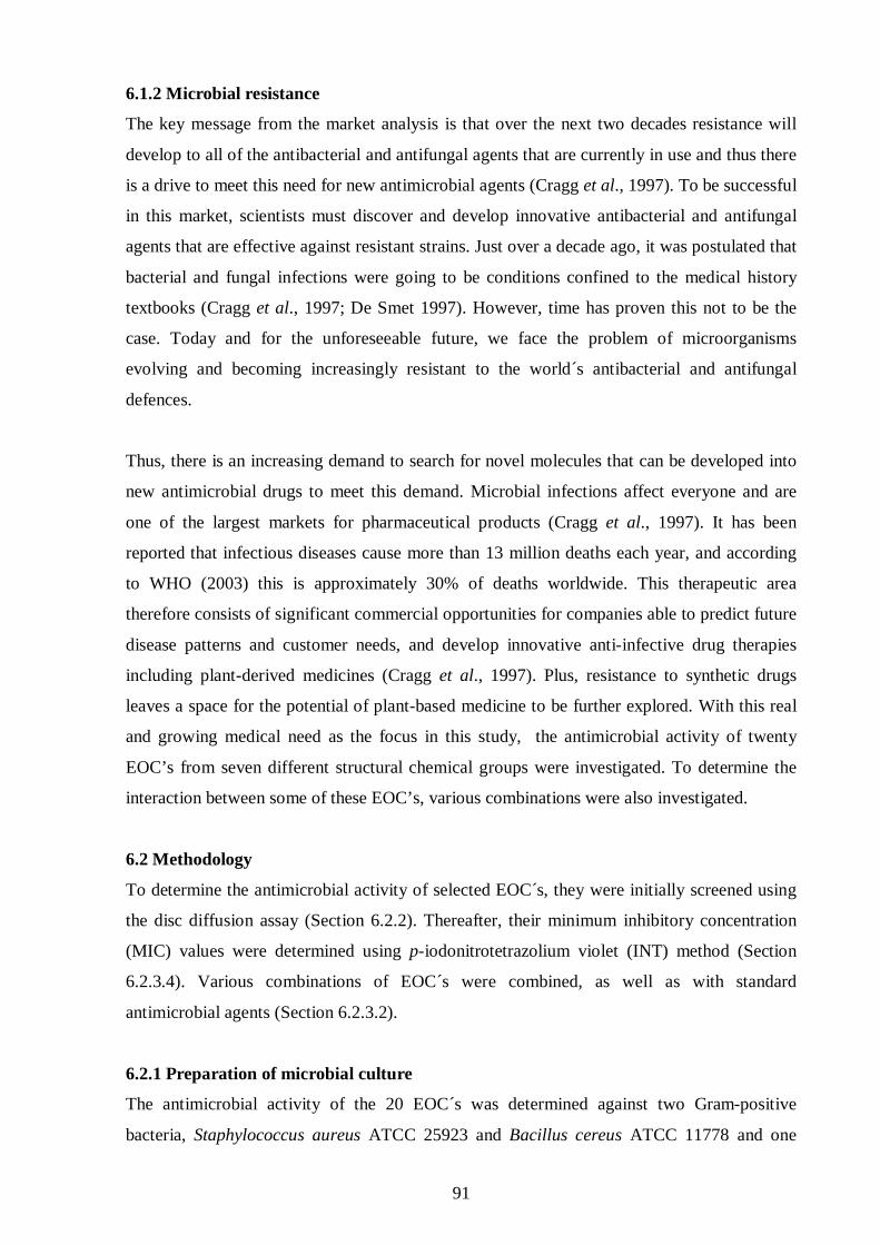
91
6.1.2 Microbial resistance
The key message from the market analysis is that over the next two decades resistance will
develop to all of the antibacterial and antifungal agents that are currently in use and thus there
is a drive to meet this need for new antimicrobial agents (Cragg et al., 1997). To be successful
in this market, scientists must discover and develop innovative antibacterial and antifungal
agents that are effective against resistant strains. Just over a decade ago, it was postulated that
bacterial and fungal infections were going to be conditions confined to the medical history
textbooks (Cragg et al., 1997; De Smet 1997). However, time has proven this not to be the
case. Today and for the unforeseeable future, we face the problem of microorganisms
evolving and becoming increasingly resistant to the world´s antibacterial and antifungal
defences.
Thus, there is an increasing demand to search for novel molecules that can be developed into
new antimicrobial drugs to meet this demand. Microbial infections affect everyone and are
one of the largest markets for pharmaceutical products (Cragg et al., 1997). It has been
reported that infectious diseases cause more than 13 million deaths each year, and according
to WHO (2003) this is approximately 30% of deaths worldwide. This therapeutic area
therefore consists of significant commercial opportunities for companies able to predict future
disease patterns and customer needs, and develop innovative anti-infective drug therapies
including plant-derived medicines (Cragg et al., 1997). Plus, resistance to synthetic drugs
leaves a space for the potential of plant-based medicine to be further explored. With this real
and growing medical need as the focus in this study, the antimicrobial activity of twenty
EOC’s from seven different structural chemical groups were investigated. To determine the
interaction between some of these EOC’s, various combinations were also investigated.
6.2 Methodology
To determine the antimicrobial activity of selected EOC´s, they were initially screened using
the disc diffusion assay (Section 6.2.2). Thereafter, their minimum inhibitory concentration
(MIC) values were determined using p-iodonitrotetrazolium violet (INT) method (Section
6.2.3.4). Various combinations of EOC´s were combined, as well as with standard
antimicrobial agents (Section 6.2.3.2).
6.2.1 Preparation of microbial culture
The antimicrobial activity of the 20 EOC´s was determined against two Gram-positive
bacteria, Staphylococcus aureus ATCC 25923 and Bacillus cereus ATCC 11778 and one

92
Gram-negative, Eschericia coli ATCC 11775, as well as the yeast Candida albicans ATCC
10231. These reference stock cultures were obtained from the South African Bureau of
Standards (SABS), Johannesburg. They were maintained viable in the Pharmaceutical
Microbiology Laboratory, University of the Witwatersrand, Johannesburg. For the disc
diffusion and MIC assays, the inoculum size of approximately 1 million colony forming units
(CFU)/ml, was prepared in Tryptone Soya broth (Oxoid) from the stock microbial culture.
6.2.2 Disc diffusion assay
6.2.2.1 Preparation of individual essential oil constituents
The individual EOC’s were prepared by aseptically transferring a 6mm paper disc into a petri
dish with a sterile needle and then the disc was saturated with 20µl of the pure (100%) EOC.
6.2.2.2 Preparation of the combinations of two or three essential oil constituents
Based on the values of the zones of inhibition obtained from the individual EOC´s, forty five
combinations of the EOC’s per each microbial strain mentioned above were performed. When
two EOC’s or three EOC’s were combined, 20µl of two or three EOC´s were combined in a
1:1 or 1:1:1 (v/v) ratio in eppendorfs, respectively. The oils were vigorously mixed before
20µl was applied to the disc paper.
6.2.2.3 Preparation of agar and bioassay agar plates
Agar was prepared by dissolving 30g of Tryptone Soya agar (Oxoid) into 750ml of distilled
water and autoclaved at 121ºC for 15min to sterilize the solution. The sterilized agar was
poured into bioassay plates in two layers: the first layer served as the base and was half the
total volume added to the plate. This was left to solidify before the second layer which was
seeded with the test organism was poured on top. Once this had solidified, paper discs
impregnated with EOC’s (Section 6.2.2.1) were transferred onto each of the four seeded agar
plates using a sterile needle. The discs were left to prediffuse for at least 1h at 4ºC before
being incubated at 37ºC for 24h (bacteria) and 48h (yeast). Prediffusion is of utmost
importance as it enhances diffusion of the EOC into the agar (Janssen et al., 1987; Onawumni
et al., 1984). Neomycin (30µg, Oxoid) and nystatin (100IU) served as positive controls for
the bacteria and yeast, respectively. The assay was repeated in duplicate to confirm
consistency.
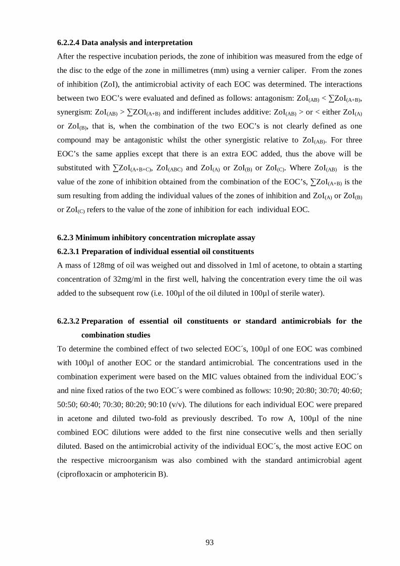
93
6.2.2.4 Data analysis and interpretation
After the respective incubation periods, the zone of inhibition was measured from the edge of
the disc to the edge of the zone in millimetres (mm) using a vernier caliper. From the zones
of inhibition (ZoI), the antimicrobial activity of each EOC was determined. The interactions
between two EOC’s were evaluated and defined as follows: antagonism: ZoI(AB) < ∑ZoI(A+B),
synergism: ZoI(AB) > ∑ZOI(A+B) and indifferent includes additive: ZoI(AB) > or < either ZoI(A)
or ZoI(B), that is, when the combination of the two EOC’s is not clearly defined as one
compound may be antagonistic whilst the other synergistic relative to ZoI(AB). For three
EOC’s the same applies except that there is an extra EOC added, thus the above will be
substituted with ∑ZoI(A+B+C), ZoI(ABC) and ZoI(A) or ZoI(B) or ZoI(C). Where ZoI(AB) is the
value of the zone of inhibition obtained from the combination of the EOC’s, ∑ZoI(A+B) is the
sum resulting from adding the individual values of the zones of inhibition and ZoI(A) or ZoI(B)
or ZoI(C) refers to the value of the zone of inhibition for each individual EOC.
6.2.3 Minimum inhibitory concentration microplate assay
6.2.3.1 Preparation of individual essential oil constituents
A mass of 128mg of oil was weighed out and dissolved in 1ml of acetone, to obtain a starting
concentration of 32mg/ml in the first well, halving the concentration every time the oil was
added to the subsequent row (i.e. 100µl of the oil diluted in 100µl of sterile water).
6.2.3.2 Preparation of essential oil constituents or standard antimicrobials for the
combination studies
To determine the combined effect of two selected EOC´s, 100µl of one EOC was combined
with 100µl of another EOC or the standard antimicrobial. The concentrations used in the
combination experiment were based on the MIC values obtained from the individual EOC´s
and nine fixed ratios of the two EOC´s were combined as follows: 10:90; 20:80; 30:70; 40:60;
50:50; 60:40; 70:30; 80:20; 90:10 (v/v). The dilutions for each individual EOC were prepared
in acetone and diluted two-fold as previously described. To row A, 100µl of the nine
combined EOC dilutions were added to the first nine consecutive wells and then serially
diluted. Based on the antimicrobial activity of the individual EOC´s, the most active EOC on
the respective microorganism was also combined with the standard antimicrobial agent
(ciprofloxacin or amphotericin B).
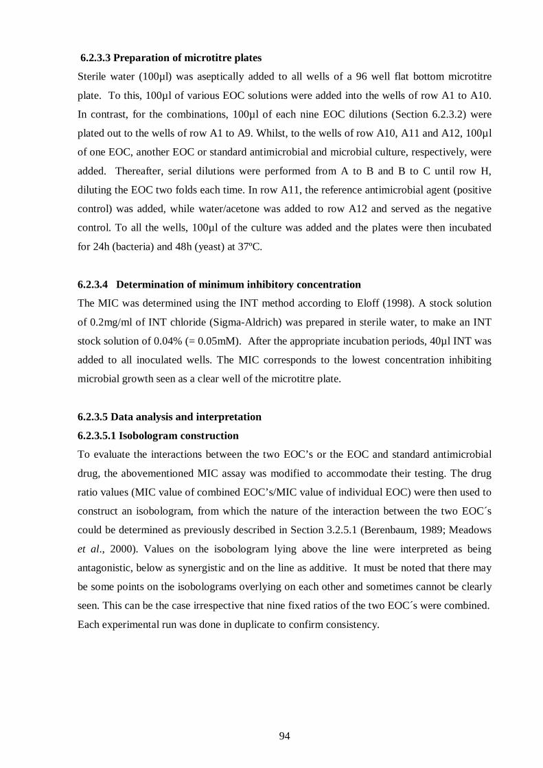
94
6.2.3.3 Preparation of microtitre plates
Sterile water (100µl) was aseptically added to all wells of a 96 well flat bottom microtitre
plate. To this, 100µl of various EOC solutions were added into the wells of row A1 to A10.
In contrast, for the combinations, 100µl of each nine EOC dilutions (Section 6.2.3.2) were
plated out to the wells of row A1 to A9. Whilst, to the wells of row A10, A11 and A12, 100µ l
of one EOC, another EOC or standard antimicrobial and microbial culture, respectively, were
added. Thereafter, serial dilutions were performed from A to B and B to C until row H,
diluting the EOC two folds each time. In row A11, the reference antimicrobial agent (positive
control) was added, while water/acetone was added to row A12 and served as the negative
control. To all the wells, 100µl of the culture was added and the plates were then incubated
for 24h (bacteria) and 48h (yeast) at 37ºC.
6.2.3.4 Determination of minimum inhibitory concentration
The MIC was determined using the INT method according to Eloff (1998). A stock solution
of 0.2mg/ml of INT chloride (Sigma-Aldrich) was prepared in sterile water, to make an INT
stock solution of 0.04% (= 0.05mM). After the appropriate incubation periods, 40µl INT was
added to all inoculated wells. The MIC corresponds to the lowest concentration inhibiting
microbial growth seen as a clear well of the microtitre plate.
6.2.3.5 Data analysis and interpretation
6.2.3.5.1 Isobologram construction
To evaluate the interactions between the two EOC’s or the EOC and standard antimicrobial
drug, the abovementioned MIC assay was modified to accommodate their testing. The drug
ratio values (MIC value of combined EOC’s/MIC value of individual EOC) were then used to
construct an isobologram, from which the nature of the interaction between the two EOC´s
could be determined as previously described in Section 3.2.5.1 (Berenbaum, 1989; Meadows
et al., 2000). Values on the isobologram lying above the line were interpreted as being
antagonistic, below as synergistic and on the line as additive. It must be noted that there may
be some points on the isobolograms overlying on each other and sometimes cannot be clearly
seen. This can be the case irrespective that nine fixed ratios of the two EOC´s were combined.
Each experimental run was done in duplicate to confirm consistency.

95
6.2.3.5.2 Fractional inhibitory concentration determination
To verify the interaction read from the isobolograms, the sum of fractional inhibitory
concentrations (∑FIC or FICAB) was calculated for each combination according to
Equation 6.1.
Equation 6.1:
FICAB = MICA in combination + MICB in combination
MICA alone MICB alone
Where A and B are the two selected EOC’s used in combination.
Values from these ∑FIC or FICAB calculations were interpreted as follows: the ∑FIC values
<0.5 is synergistic, 0.5-1.0 is additive, >1.0 is indifferent, while that >2.0 are antagonistic
(Berenbaum, 1989). Combinations were performed in duplicate for both the MIC and disc
diffusion assays to ensure consistency between the results.
6.3 Results
6.3.1 Disc diffusion assay
6.3.1.1 The disc diffusion assay: individual essential oil constituents
Table 6.1 summarizes the results of the disc diffusion assay of the 10 individual EOC´s. All
the bacterial strains and the yeast demonstrated various degrees of sensitivity to the EOC´s
tested. The EOC with the widest spectrum of antimicrobial activity was found to be eugenol
and had the highest zones of inhibition in most instances. Its values of the zones of inhibition
against S. aureus, E. coli, B. cereus and C. albicans were 3mm, 3.4mm, 10mm and 11.1mm,
respectively.
6.3.1.2 The disc diffusion assay: the combined effects of two essential oil constituents
When the selected EOC´s were combined, the combinations interacted in varying ways
(Table 6.2; Tables A.1 and A.2 in Appendix A). The raw data for the disc diffusion assay in
Tables A.1 and A.2 are interpreted and summarised in Tables 6.2 and 6.3. In order to
highlight some of the interactions between the individual EOC´s that could occur in the whole
essential oil mixture, the combined effects of six interactions on various organisms are shown
in Figures 6.1 to 6.4. The EOC´s interacted in synergistic, additive, indifferent and
antagonistic manners, with (R)-(+)-limonene and E- and Z-(±)-nerolidol displaying the most
synergistic interaction against all test organisms except B. cereus (Table 6.2; Figures 6.1, 6.2,
6.3 and 6.4).
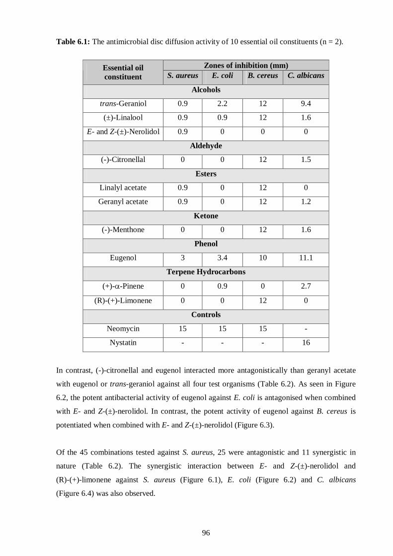
96
Table 6.1: The antimicrobial disc diffusion activity of 10 essential oil constituents (n = 2).
Zones of inhibition (mm) Essential oil
constituent S. aureus E. coli B. cereus C. albicans
Alcohols
trans-Geraniol 0.9 2.2 12 9.4
(±)-Linalool 0.9 0.9 12 1.6
E- and Z-(±)-Nerolidol 0.9 0 0 0
Aldehyde
(-)-Citronellal 0 0 12 1.5
Esters
Linalyl acetate 0.9 0 12 0
Geranyl acetate 0.9 0 12 1.2
Ketone
(-)-Menthone 0 0 12 1.6
Phenol
Eugenol 3 3.4 10 11.1
Terpene Hydrocarbons
(+)-α-Pinene 0 0.9 0 2.7
(R)-(+)-Limonene 0 0 12 0
Controls
Neomycin 15 15 15 -
Nystatin - - - 16
In contrast, (-)-citronellal and eugenol interacted more antagonistically than geranyl acetate
with eugenol or trans-geraniol against all four test organisms (Table 6.2). As seen in Figure
6.2, the potent antibacterial activity of eugenol against E. coli is antagonised when combined
with E- and Z-(±)-nerolidol. In contrast, the potent activity of eugenol against B. cereus is
potentiated when combined with E- and Z-(±)-nerolidol (Figure 6.3).
Of the 45 combinations tested against S. aureus, 25 were antagonistic and 11 synergistic in
nature (Table 6.2). The synergistic interaction between E- and Z-(±)-nerolidol and
(R)-(+)-limonene against S. aureus (Figure 6.1), E. coli (Figure 6.2) and C. albicans
(Figure 6.4) was also observed.

97
Table 6.2: The antimicrobial disc diffusion interactions between two selected EOC’s (An: antagonism, I: indifferent and S: synergism, Raw data seen on Table A.1, n = 2).
Zones of inhibition (mm) Essential oil constituent
S. aureus E. coli B. cereus C. albicans Linalyl acetate An I An An
Geranyl acetate S I An S
(±)-Linalool An I An S trans-Geraniol I An An S Eugenol An An An An (+)-α-Pinene I An I An E- and Z-(±)-Nerolidol S I An An (-)-Menthone S I An An
(-)-Citronellal
(R)-(+)-Limonene S S An An Geranyl acetate An I An S trans-Geraniol An An An An Eugenol S An An An (+)-α-Pinene An I I An E- and Z-(±)-Nerolidol An I I I (-)-Menthone An I An An (±)-Linalool An An An S
Linalyl acetate
(R)-(+)-Limonene I S An S (±)-Linalool An I An An trans-Geraniol An An An An Eugenol An An An An (+)-α-Pinene An I An An E- and Z-(±)-Nerolidol An I An An (-)-Menthone An I An An
Geranyl acetate
(R)-(+)-Limonene An I An An trans-Geraniol S An An An Eugenol S An An An (+)-α-Pinene An An I An E- and Z-(±)-Nerolidol An I I An (-)-Menthone An I An An
(±)-Linalool
(R)-(+)-Limonene I S An S Eugenol An An I An
(+)-α-Pinene S An I An E- and Z-(±)-Nerolidol An An An An (-)-Menthone An An An An
trans-Geraniol
(R)-(+)-Limonene S An An An (+)-α-Pinene An An S An E- and Z-(±)-Nerolidol An An S An (-)-Menthone An An An An
Eugenol
(R)-(+)-Limonene S An An An E- and Z-(±)-Nerolidol I I S An (-)-Menthone I An An An (+)-α-Pinene (R)-(+)-Limonene I S An S (-)-Menthone I I An An E- and Z-(±)-
Nerolidol (R)-(+)-Limonene S S I S (-)-Menthone (R)-(+)-Limonene I S An An

98
In addition, the combinations of EOC’s against E. coli showed 21 antagonistic interactions of
the 45 tested combinations (Table 6.2). While synergism was only observed six times when
(R)-(+)-limonene interacted with one of the following EOC’s: E- and Z-(±)-nerolidol,
(+)-α-pinene (Figure 6.2), (-)-citronellal, (-)-menthone, (±)-linalool and linalyl acetate
(Table 6.2).
Eugen
ol
(+)- α
-Pine
ne
Eugen
ol +
(+)-α
-Pine
ne
Eugen
ol
E- and
Z- (
±)-N
erolid
ol
Euge
nol +
E- a
nd Z
- (±)
-Nero
lidol
Eugen
ol
(R)-(
+)-L
imon
ene
Euge
nol +
(R)-(
+)-L
imon
ene
(+)-α
-Pine
ne
E- and
Z- (
±)-N
eroli
dol
(+)- α
-Pine
ne +
E- a
nd Z
- (±)
-Ner
olido
l
E- a
nd Z
- (±)
-Ner
olido
l
(R)-(
+)-L
imon
ene
E- and
Z- (
±)-N
erolid
ol +
(R)-(
+)-L
imon
ene
(+)- α
-Pine
ne
(R)-(
+)-L
imon
ene
(+)- α
-Pine
ne + (R
)-(+)
-Lim
onen
e0.0
0.5
1.0
1.5
2.0
2.5
3.0
3.5
0.0 0.0 0.00.00.00.00.0
3.0
2.5
3.0 3.0
0.90.90.9
2.1
0.9
3.5
3.1
Zon
es o
f in
hibi
tio
n(m
m)
Figure 6.1: The antimicrobial disc diffusion activity of six selected combinations of
essential oil constituents against Staphylococcus aureus.
The combination of (+)-α-pinene and E- and Z-(±)-nerolidol was extremely synergistic
against B. cereus (Figure 6.3), while individually both EOC’s did not show any antibacterial
activity (Table 6.1). Only three synergistic interactions were observed of the 45 combinations,
with eugenol interacting favourably with (+)-α-pinene or E- and Z-(±)-nerolidol (Figure 6.3).
Thirty-four antagonistic interactions were recorded with the more antagonistic interaction
observed between (+)-α-pinene and (R)-(+)-limonene (Figure 6.3), (-)-menthone along with
either (R)-(+)-limonene, geranyl acetate and linalyl acetate; as well as between (±)-linalool
and trans-geraniol (Table 6.2).
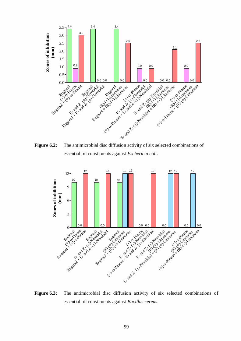
99
Eugen
ol
(+)-α
-Pine
ne
Eugen
ol + (
+)- α
-Pin
ene
Eugen
ol
E- and
Z- (
±)-N
eroli
dol
Eugen
ol + E
- and
Z- (±)-N
eroli
dol
Eugen
ol
(R)-(
+)-L
imon
ene
Eugen
ol +
(R)-(
+)-L
imon
ene
(+)-α
-Pine
ne
E- and
Z- (
±)-N
erolid
ol
(+)-α
-Pine
ne +
E- a
nd Z
- (±)
-Neroli
dol
E- and
Z- (
±)-N
erolid
ol
(R)-(
+)-Lim
onen
e
E- and
Z- (
±)-N
erolid
ol +
(R)-(
+)-L
imon
ene
(+)-α
-Pine
ne
(R)-(
+)-L
imon
ene
(+)-α
-Pine
ne +
(R)-(
+)-L
imon
ene
0.0
0.5
1.0
1.5
2.0
2.5
3.0
3.5
0.00.0 0.0 0.0 0.0 0.0 0.0
3.4 3.4 3.4
3.0
0.9 0.9 0.9 0.9
2.5 2.5
2.1Z
one
s o
f in
hib
itio
n(m
m)
Figure 6.2: The antimicrobial disc diffusion activity of six selected combinations of
essential oil constituents against Eschericia coli.
Eugen
ol
(+)-α
-Pine
ne
Eugen
ol + (+
)-α-P
inen
e
Eugen
ol
E- and
Z- (
±)-N
eroli
dol
Eugen
ol + E
- and
Z- (
±)-N
erolid
ol
Eugen
ol
(R)-(
+)-Lim
onen
e
Eugen
ol +
(R)-(
+)-L
imon
ene
(+)- α
-Pine
ne
E- and
Z- (
±)-N
eroli
dol
(+)- α
-Pine
ne + E
- and
Z- (
±)-N
eroli
dol
E- and
Z- (
±)-N
eroli
dol
(R)-(
+)-Lim
onen
e
E- and
Z- (
±)-N
eroli
dol +
(R)-(+
)-Lim
onen
e
(+)-α
-Pine
ne
(R)-(
+)-Lim
onen
e
(+)-α
-Pin
ene
+ (R)-(
+)-Lim
onen
e0
3
6
9
12
10
12
0.0
10
0.0
12
10
12 12
0.0 0.0
12
0.0
12 12
0.0
12
0.0
Zon
es o
f in
hib
ition
(mm
)
Figure 6.3: The antimicrobial disc diffusion activity of six selected combinations of
essential oil constituents against Bacillus cereus.

100
Furthermore, the combinations of EOC’s against C. albicans were dominated by mostly
antagonistic interactions with 35 of 45 combinations being antagonistic. While nine
synergistic interactions were observed when (R)-(+)-limonene interacted with either
E- and Z-(±)-nerolidol, (+)-α-pinene, (±)-linalool or linalyl acetate) (Table 6.2 and
Figure 6.4). Moreover, when geranyl acetate interacted synergistically with either
(-)-citronellal or trans-geraniol or linalyl acetate or (±)-linalool and linalyl acetate with the
latter EOC against C. albicans (Table 6.2).
Eugen
ol
(+)-α
-Pine
ne
Eugen
ol + (+
)-α-P
inene
Eugen
ol
E- and
Z- (±
)-Nero
lidol
Eugen
ol +
E- a
nd Z
- (±)
-Neroli
dol
Eugen
ol
(R)-(
+)-L
imon
ene
Eugen
ol + (R
)-(+)
-Lim
onen
e
(+)- α
-Pine
ne
E- and
Z- (±)
-Nero
lidol
(+)- α
-Pine
ne +
E- a
nd Z
- (±)-N
eroli
dol
E- and
Z- (±
)-Nero
lidol
(R)-(
+)-L
imon
ene
E- and
Z- (±)
-Nero
lidol
+ (R)-(
+)-L
imon
ene
(+)-α
-Pine
ne
(R)-(
+)-L
imon
ene
(+)-α
-Pin
ene +
(R)-(
+)-Lim
onen
e0
3
6
9
12 11.1 11.1 11.1
5.34.9
4.03.33.4
2.7 2.7 2.7
0.0 0.0 0.0 0.0 0.0 0.0
Zon
es o
f in
hibi
tion
(mm
)
0.0
Figure 6.4: The antimicrobial disc diffusion activity of six selected combinations of
essential oil constituents against Candida albicans.
6.3.1.3 The disc diffusion assay: the combined effects of three essential oil constituents
Due to the complexity of the essential oils, three EOC’s were combined and tested against the
four test microorganisms (Table 6.3; Table A.2 in Appendix A; Figure 6.5). The antagonistic
interactions among the following EOC’s were observed against all test organisms:
(-)-citronellal, trans-geraniol and geranyl acetate; linalyl acetate, (±)-linalool and
(R)-(+)-limonene; (R)-(+)-limonene, trans-geraniol and (-)-menthone; eugenol,
E- and Z-(±)-nerolidol and (±)-linalool. The only exception were indifferent interactions
among linalyl acetate, (±)-linalool and (R)-(+)-limonene against E. coli and (R)-(+)-limonene,
trans-geraniol and (-)-menthone against S. aureus.

101
Table 6.3: The antimicrobial disc diffusion interactions among three selected essential oil
constituents (Raw data seen on Table A.2 in Appendix A, n = 2).
Essential oil constituents Zones of inhibition (mm)
S. aureus E. coli B. cereus C. albicans (-)-Citronellal + trans-Geraniol + Geranyl acetate
An An An An
Linalyl acetate + (±)-Linalool + (R)-(+)-Limonene
An I An An
(R)-(+)-Limonene + trans-Geraniol + (-)-Menthone
I An An An
Eugenol + E- and Z-(±)-Nerolidol + (±)-Linalool An An An An
S. aure
us
E. coli
B. cere
us
C. albi
cans
0
3
6
9
12
(-)-Citronellaltrans-GeraniolGeranyl acetate
(-)-Citronellal +trans-Geraniol + Geranyl acetate
0.0 0.00.00.0 0.00.9 0.90.9
12 12 12
9.4
1.81.21.5
2.2Zon
es o
f in
hibi
tion
(mm
)
Figure 6.5: The antimicrobial disc diffusion activity of four selected combinations of
essential oil constituents against four test microorganisms.
6.3.2. The minimum inhibitory concentration micropate assay
6.3.2.1 The minimum inhibitory concentration assay: individual essential oil
constituents
The in vitro antimicrobial activity of the 20 EOC´s are shown in Table 6.4 along with the
antibacterial and antifungal controls. The EOC´s inhibited the growth of Gram-positive and
Gram-negative bacteria, with MIC values ranging from 12.2 to >238.4mM.

102
Table 6.4: In vitro antimicrobial minimum inhibitory concentration activity of 20
individual essential oil constituents and the standard antimicrobial agents (n = 2).
Antimicrobial activity (mM) Essential oil
constituent S. aureus E. coli B. cereus C. albicans
Alcohols
(-)-α-Bisabolol 143.9±0.0 143.9±0.0 143.9±0.0 36.0±0.0
trans-Geraniol 38.9±18.2 25.9±0.0 51.9±0.0 19.5±0.0
(±)-Linalool 77.8±37.0 51.9±29.8 103.7±29.8 38.9±0.0
E-and Z-(±)-Nerolidol
143.9±0.0 143.9±24.7 143.9±24.7 143.9±0.0
Aldehyde
(-)-Citronellal 129.7±55.1 207.5±45.4 207.5±64.8 77.8±0.0
Esters
Geranyl acetate 163.0±35.7 163.0±29.0 163.0±51.0 163.0±0.0
Linalyl acetate 163.0±0.0 163.0±0.0 163.0±51.0 163.0±0.0
Ketones (cis+trans)-(±)-α+β Thujone
210.1±0.0 210.2±0.0 210.2±0.0 157.7±0.0
(+)-Carvone 53.3±0.0 106.5±0.0 106.5±0.0 106.51±0.0
(-)-Fenchone 105.1±0.0 210.2±0.0 210.2±0.0 210.2±0.0
(-)-Menthone 207.5±59.6 207.5±0.0 207.5±0.0 6.48±0.0
(-)-Pulegone 157.7±36.1 157.7±36.1 105.1±0.0 26.3±0.0
Phenols
Carvacrol 13.3±0.0 20.0±9.3 13.3±0.0 1.66±0.0
Eugenol 194.9±6.7 12.2±0.0 12.2±0.0 79.2±0.0
Terpene Hydrocarbons
(+)-α-Pinene 234.9±0.0 234.9±0.0 234.9±0.0 88.1±0.0
(+)-β-Pinene 22.0±1.5 117.4±0.0 117.4±0.0 7.3±0.0
γ-Terpinene 234.9±0.0 234.9±0.0 234.9±0.0 44.0±0.0
(R)-(+)-Limonene 176.2±40.4 176.2±40.4 234.9±0.0 73.4±0.0
p-Cymene 238.4±0.0 238.4±0.0 238.4±0.0 238.4±0.0
Oxide
1,8-Cineole 207.5±0.0 207.5±0.0 207.5±0.0 207.5±0.0
Controls
Ciprofloxacin 0.91±0.0 1.8±0.0 0.3±0.0 -
Amphotericin B - - - 0.7±0.0
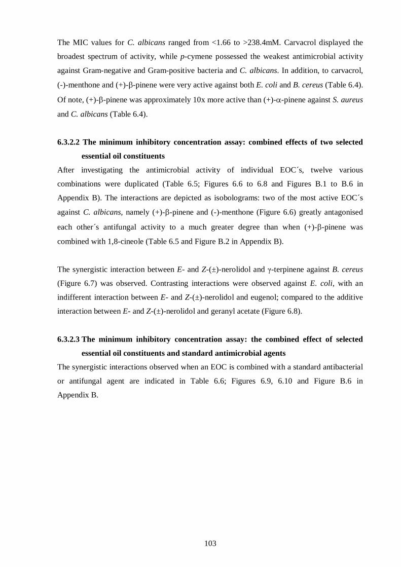
103
The MIC values for C. albicans ranged from <1.66 to >238.4mM. Carvacrol displayed the
broadest spectrum of activity, while p-cymene possessed the weakest antimicrobial activity
against Gram-negative and Gram-positive bacteria and C. albicans. In addition, to carvacrol,
(-)-menthone and (+)-β-pinene were very active against both E. coli and B. cereus (Table 6.4).
Of note, (+)-β-pinene was approximately 10x more active than (+)-α-pinene against S. aureus
and C. albicans (Table 6.4).
6.3.2.2 The minimum inhibitory concentration assay: combined effects of two selected
essential oil constituents
After investigating the antimicrobial activity of individual EOC´s, twelve various
combinations were duplicated (Table 6.5; Figures 6.6 to 6.8 and Figures B.1 to B.6 in
Appendix B). The interactions are depicted as isobolograms: two of the most active EOC´s
against C. albicans, namely (+)-β-pinene and (-)-menthone (Figure 6.6) greatly antagonised
each other´s antifungal activity to a much greater degree than when (+)-β-pinene was
combined with 1,8-cineole (Table 6.5 and Figure B.2 in Appendix B).
The synergistic interaction between E- and Z-(±)-nerolidol and γ-terpinene against B. cereus
(Figure 6.7) was observed. Contrasting interactions were observed against E. coli, with an
indifferent interaction between E- and Z-(±)-nerolidol and eugenol; compared to the additive
interaction between E- and Z-(±)-nerolidol and geranyl acetate (Figure 6.8).
6.3.2.3 The minimum inhibitory concentration assay: the combined effect of selected
essential oil constituents and standard antimicrobial agents
The synergistic interactions observed when an EOC is combined with a standard antibacterial
or antifungal agent are indicated in Table 6.6; Figures 6.9, 6.10 and Figure B.6 in
Appendix B.
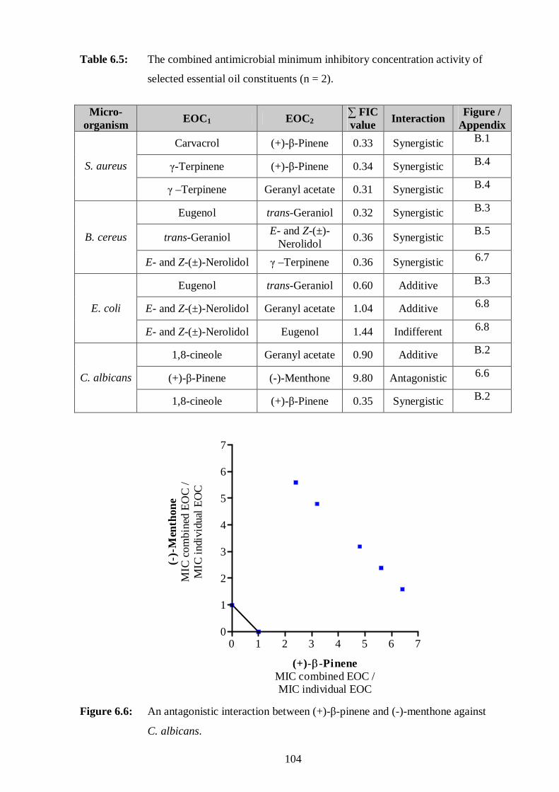
104
Table 6.5: The combined antimicrobial minimum inhibitory concentration activity of
selected essential oil constituents (n = 2).
Micro-
organism EOC1 EOC2
∑ FIC value
Interaction Figure /
Appendix
Carvacrol (+)-β-Pinene 0.33 Synergistic B.1
γ-Terpinene (+)-β-Pinene 0.34 Synergistic B.4 S. aureus
γ –Terpinene Geranyl acetate 0.31 Synergistic B.4
Eugenol trans-Geraniol 0.32 Synergistic B.3
trans-Geraniol E- and Z-(±)-
Nerolidol 0.36 Synergistic
B.5 B. cereus
E- and Z-(±)-Nerolidol γ –Terpinene 0.36 Synergistic 6.7
Eugenol trans-Geraniol 0.60 Additive B.3
E- and Z-(±)-Nerolidol Geranyl acetate 1.04 Additive 6.8 E. coli
E- and Z-(±)-Nerolidol Eugenol 1.44 Indifferent 6.8
1,8-cineole Geranyl acetate 0.90 Additive B.2
(+)-β-Pinene (-)-Menthone 9.80 Antagonistic 6.6 C. albicans
1,8-cineole (+)-β-Pinene 0.35 Synergistic B.2
0 1 2 3 4 5 6 70
1
2
3
4
5
6
7
(+)-β-PineneMIC combined EOC /MIC individual EOC
(-)-
Men
thon
eM
IC c
ombi
ned
EO
C /
MIC
indi
vidu
al E
OC
Figure 6.6: An antagonistic interaction between (+)-β-pinene and (-)-menthone against
C. albicans.

105
Figure 6.7: A synergistic interaction between E- and Z-(±)-nerolidol and γ-terpinene against
B. cereus.
0.00 0.25 0.50 0.75 1.00 1.250.00
0.25
0.50
0.75
1.00
1.25
E- and Z-(±)-Nerolidol(MIC combined EOC /MIC individual EOC)
Eu
geno
l / G
eran
yl a
ceta
te(M
IC c
ombi
ned
EO
C /
MIC
indi
vidu
al E
OC
)
Figure 6.8: Contrasting interactions against E. coli, with an indifferent interaction between
E- and Z-(±)-nerolidol and eugenol (�); compared to the additive interaction
between E- and Z-(±)-nerolidol and geranyl acetate (▲).
0.00 0.25 0.50 0.75 1.00 1.250.00
0.25
0.50
0.75
1.00
1.25
E- and Z-(±)-Nerolidol(MIC combined EOC /MIC individual EOC)
γ -T
erpi
nen
e(M
ICco
mbi
ned
EO
C /
MIC
indi
vidu
al E
OC
)
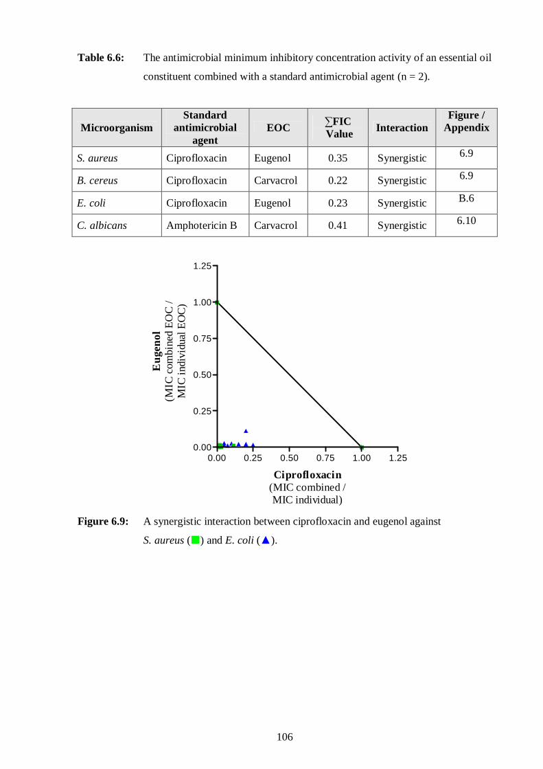
106
Table 6.6: The antimicrobial minimum inhibitory concentration activity of an essential oil
constituent combined with a standard antimicrobial agent (n = 2).
Microorganism Standard
antimicrobial agent
EOC ∑FIC Value Interaction
Figure / Appendix
S. aureus Ciprofloxacin Eugenol 0.35 Synergistic 6.9
B. cereus Ciprofloxacin Carvacrol 0.22 Synergistic 6.9
E. coli Ciprofloxacin Eugenol 0.23 Synergistic B.6
C. albicans Amphotericin B Carvacrol 0.41 Synergistic 6.10
0.00 0.25 0.50 0.75 1.00 1.250.00
0.25
0.50
0.75
1.00
1.25
Ciprofloxacin(MIC combined /MIC individual)
Eu
gen
ol(M
IC c
ombi
ned
EO
C /
MIC
indi
vidu
al E
OC
)
Figure 6.9: A synergistic interaction between ciprofloxacin and eugenol against
S. aureus (�) and E. coli (▲).
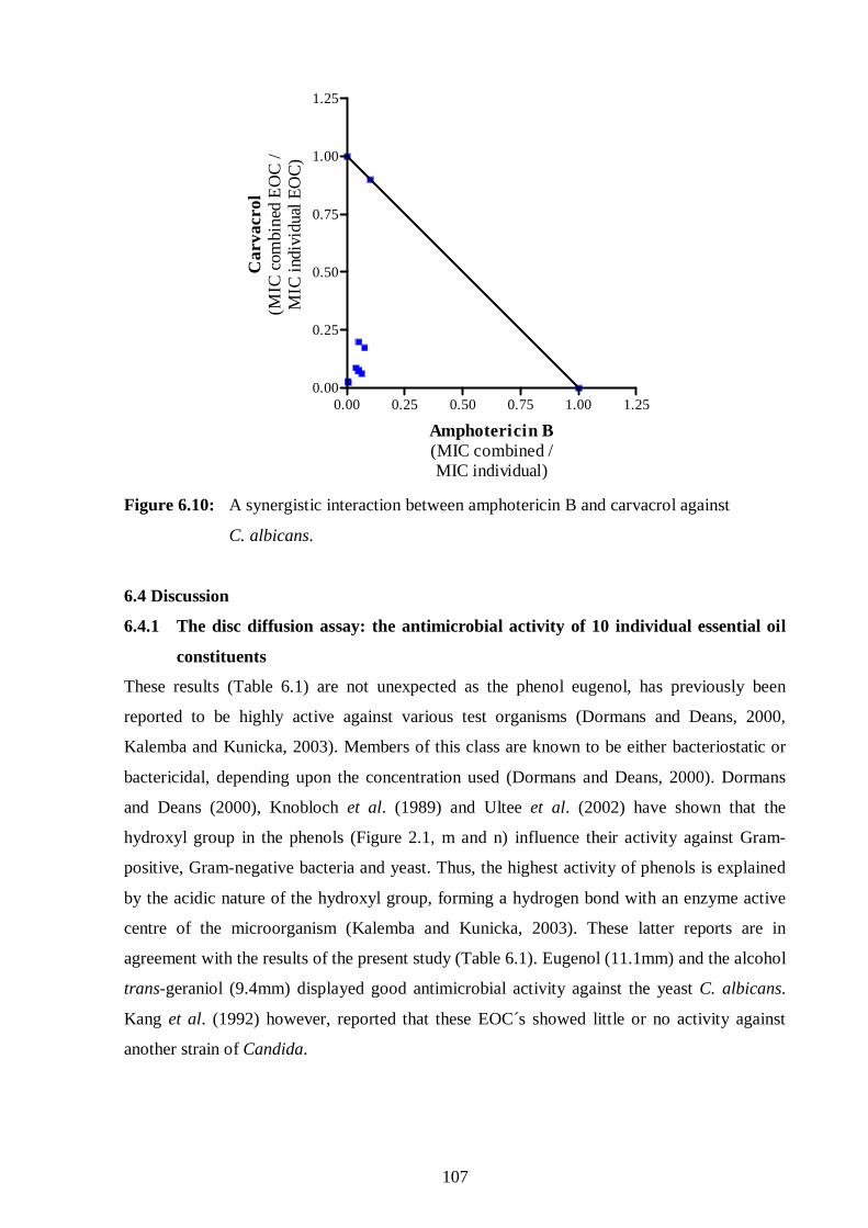
107
0.00 0.25 0.50 0.75 1.00 1.250.00
0.25
0.50
0.75
1.00
1.25
Amphotericin B(MIC combined /MIC individual)
Car
vacr
ol(M
IC c
ombi
ned
EO
C /
MIC
indi
vidu
al E
OC
)
Figure 6.10: A synergistic interaction between amphotericin B and carvacrol against
C. albicans.
6.4 Discussion
6.4.1 The disc diffusion assay: the antimicrobial activity of 10 individual essential oil
constituents
These results (Table 6.1) are not unexpected as the phenol eugenol, has previously been
reported to be highly active against various test organisms (Dormans and Deans, 2000,
Kalemba and Kunicka, 2003). Members of this class are known to be either bacteriostatic or
bactericidal, depending upon the concentration used (Dormans and Deans, 2000). Dormans
and Deans (2000), Knobloch et al. (1989) and Ultee et al. (2002) have shown that the
hydroxyl group in the phenols (Figure 2.1, m and n) influence their activity against Gram-
positive, Gram-negative bacteria and yeast. Thus, the highest activity of phenols is explained
by the acidic nature of the hydroxyl group, forming a hydrogen bond with an enzyme active
centre of the microorganism (Kalemba and Kunicka, 2003). These latter reports are in
agreement with the results of the present study (Table 6.1). Eugenol (11.1mm) and the alcohol
trans-geraniol (9.4mm) displayed good antimicrobial activity against the yeast C. albicans.
Kang et al. (1992) however, reported that these EOC´s showed little or no activity against
another strain of Candida.
108
The ketone, (-)-menthone has demonstrated that the presence of an oxygen function in its
framework (Figure 2.1k) increases its antimicrobial activity (Knobloch et al., 1989). This
study revealed that (-)-menthone (1.6mm) has the modest antimicrobial activity against C.
albicans, whilst B. cereus is the most significantly affected (Table 6.1). However, the results
in the disc diffusion assay of the study by Dormans and Deans (2000) revealed that among the
test organisms used, S. aureus was significantly affected, which is not the case in the present
study. Notably, it is not clear in the study by Dormans and Deans (2000) which strain they
used.
The terpene hydrocarbon, (R)-(+)-limonene demonstrated little or no activity against all the
test micoorganisms, when using the disc diffusion assay (Table 6.1). However, when the
MIC assay was used it showed some antimicrobial activity, suggesting that the method
employed in the study can and does influence the results. Individually, the EOC´s
demonstrate antimicrobial activity, although their combined properties are poorly studied and
understood.
6.4.2 The disc diffusion assay: the combined effects of two essential oil constituents
The interactions between the combined effects of two EOC´s were diverse and can be
categorised as discussed below (Figure 6.2 to 6.4; Table 6.2 and Table A.1 in Appendix A).
Antagonism was observed between the following EOC´s against all four test organisms:
(-)-citronellal and eugenol; (-)-menthone and eugenol; trans-geraniol and linalyl acetate;
trans-geraniol and E- and Z-(±)-nerolidol; trans-geraniol and (-)-menthone; trans-geraniol
and geranyl acetate. Notably, individually trans-geraniol was the second most active, while
eugenol was the most active against all test organisms, except B. cereus; where eugenol was
83% less active compared with trans-geraniol and other EOC´s, except (+)-α-pinene and
E- and Z-(±)-nerolidol (Table 6.1).
Upon selecting the most active EOC´s when used individually namely, trans-geraniol and
eugenol, one would have expected a synergistic interaction. However, their observed
interaction was antagonistic against all four test organisms, irrespective of their potent
individual antimicrobial activity (Table 6.2 and Table A.1 in Appendix A). It was also clearly
observed that against two Gram-positive bacteria, S. aureus and B. cereus the obtained ZoI(AB)
values (i.e. from the results) of the combined effect of these two EOC´s were 82.5% and
54.5% less than the expected ∑ZoI(A+B) values (i.e. the added sum of the values of their zones
of inhibition), respectively. In addition, against the Gram-negative E. coli bacteria and C.

109
albicans yeast, the obtained ZoI(AB) values were 46.4% and 22.4% less than the expected
∑ZoI(A+B) values, respectively (Table 6.1 and 6.2).
It has been mentioned (Section 6.4.1) that apart from the influence of the hydroxyl group in
the structure of eugenol, the high activity of the phenolic components may be further
explained in terms of the alkyl substitution into the phenol nucleus (Figure 2.1, m; for
example the presence of a methyl group), which is known to enhance the antimicrobial
activity of phenols (Dormans and Deans, 2000). Futhermore, the distribution of alkylation has
been proposed to alter the distribution ratio between the aqueous and the nonaqueous phases
(including bacterial phases) by reducing surface tension or altering the species selectivity
(Dormans and Deans, 2000). While alcohols including trans-geraniol are known to possess
bactericidal rather than bacteriostatic activity against vegetative cells (Griffin et al., 1999),
they act either as protein denaturating agents, solvents or dehydrating agents (Kalemba and
Kunicka, 2003). It is possible that these two EOC´s (trans-geraniol and eugenol) by attacking
the same target sites on the microbial cell membrane, decreased each other´s effect in an
antagonistic manner (Table 6.2 and Table A.1 in Appendix A).
While antagonism was observed for most combinations, some combinations such as linalyl
acetate and eugenol; trans-geraniol and (±)-linalool; (±)-linalool and eugenol; eugenol and
(R)-(+)-limonene, showed synergism against S. aureus and antagonism against E. coli, B.
cereus and C. albicans (Table 6.2). The advantageous synergistic effects of the EOC´s have
been observed before (Dormans and Deans, 2000; Griffin et al., 1999). However, if eugenol
and (R)-(+)-limonene were to be chosen as the most and least active EOC´s, respectively,
irrespective that (R)-(+)-limonene had potent activity against B. cereus, they interacted
antagonistically against all test organisms except S. aureus. When (R)-(+)-limonene was
combined with (±)-linalool or linalyl acetate (an ester enantiomer of (±)-linalool), synergism
against E. coli and C. albicans; indifference against S. aureus and antagonism against B.
cereus were seen. The combination of (±)-linalool and linalyl acetate interacted
synergistically against C. albicans and antagonistically against S. aureus, E. coli and B.
cereus. The interaction of these two enantiomers appeared to be more active against the yeast
than against Gram-positive and Gram-negative bacteria. It is quite interesting though, as it has
been reported before that Gram-positive bacteria are more susceptible to EOC’s than Gram-
negative bacteria (Burt, 2004), as both bacteria appeared to be less susceptible in this instance.
Linalyl acetate with E- and Z-(±)-nerolidol showed an indifferent interaction against E. coli
110
and C. albicans; and an antagonistic interaction against S. aureus (Table 6.2 and Table A.1 in
Appendix A). These interactions between an ester (linalyl acetate) and the alcohols
((±)-linalool or E- and Z-(±)-nerolidol), further show that when these two chemical groups are
combined they have no potent inhibitory effect on Gram-positive bacteria.
When the two alcohols, namely (±)-linalool and E- and Z-(±)-nerolidol were combined
antagonism was observed against S. aureus and C. albicans; and an indifferent manner
against E. coli and B. cereus (Table 6.2 and Table A.1 in Appendix A). In contrast, when
another set of alcohols, namely E- and Z-(±)-nerolidol and trans-geraniol were combined,
they interacted in an antagonistic manner against all four test organisms. Here the interactions
observed cannot be associated with the Gram-reaction of the microorganism, that is, whether
the combination affected Gram-positive bacteria more than Gram-negative bacteria or vice
versa. In contrast, when two esters (linalyl acetate and geranyl acetate) were combined,
antagonism, indifference and synergism were observed against S. aureus and B. cereus;
E. coli and C. albicans, respectively (Table 6.2). In this scenario, the reported observation
(Burt, 2004), that Gram-positive bacteria are more susceptible than Gram-negative bacteria
has been verified.
The combination between E- and Z-(±)-nerolidol with (+)-α-pinene resulted in synergism
against B. cereus, indifference against S. aureus or E. coli and antagonism against
C. albicans. When (+)-α-pinene and R-(+)-limonene were combined, synergism against
E. coli (Figure 6.2) and C. albicans; antagonism against B. cereus (Figure 6.3) and
indifference against and S. aureus. The indifferent interaction between (+)-α-pinene and
(R)-(+)-limonene against S. aureus (Figure 6.1 and Table 6.2) may be due to the fact that
these two terpene hydrocarbons appear to act at the same target site where they both access
and accumulate in the microbial membrane and exhibit their antimicrobial activity, but failed
to increase each other´s effect when combined. Both molecules have carbon to carbon double
bonds, where this feature of conjugation has been described as having an increase
electronegativity arrangement which increases the antibacterial activity (Dormans and Deans,
2000). Electronegative compounds may interfere in biological processes involving electron
transfer and react with vital nitrogen components, e.g. proteins and nucleic acids and therefore
inhibit microbial growth (Dormans and Deans, 2000; Griffin et al., 1999; Kang et al., 1992).
Moreover, the alkylation of (R)-(+)-limonene [1-methyl-4-(1-methylethenyl)-cyclohexene]
(Table 2.2 and Figure 2.1, r), could influence its antibacterial activity, as seen against E. coli
111
(Kang et al., 1992) especially when combined with (+)-α-pinene (Table 6.2 and Table A.1 in
Appendix A).
The combinations of the EOC´s in varying ratios produced synergistic, indifferent or
antagonistic interactions (Figures 6.1 to 6.4 and Table 6.2). However, the mechanism of
action of these combinations is not yet fully understood, and warrants further detailed
investigation.
6.4.3 The disc diffusion assay: the combined effects of three essential oil constituents
Essential oil constituents in triple combinations (Figure 6.5; Table 6.3 and Table A.2 in
Appendix A) failed to show potent antimicrobial activity. Antagonistic interactions were
observed when (-)-citronellal, trans-geraniol and geranyl acetate; linalyl acetate, (±)-linalool
and (R)-(+)-limonene; (R)-(+)-limonene, trans-geraniol and (-)-menthone; eugenol,
E- and Z-(±)-nerolidol and (±)-linalool were combined against all four test organisms. It is
unknown why such an observation occurred. It is proposed that there is an optimal ratio at
which the EOC´s interact such that there is no interference with their uptake and incorporation
into the bacteria or fungi. In these triple combination studies, the three EOC´s were combined
in 1:1:1 ratio, however, it should be noted that the ratio at which an optimal effect is achieved
can be quite variable and specific against a particular microorganism. However, indifferent
interactions were noted for linalyl acetate, (±)-linalool and (R)-(+)-limonene against E. coli
and (R)-(+)-limonene, trans-geraniol and (-)-menthone against S. aureus. In contrast, the
yeast C. albicans was relatively susceptible to all of the combinations tested (Table 6.3 and
Table A.1 in Appendix A), where the interaction with the cell wall structure and cellular
target(s) could have influenced this observation.
6.4.4 Minimum inhibitory concentration assay: the antimicrobial activity of the
individual essential oil constituents
Gram-negative bacteria are generally less susceptible to the action of essential oils due to the
presence of the outer membrane surrounding the cell wall, which restricts the diffusion of
lipophilic compounds through the lipopolysaccharide covering (Burt, 2004). While this was
the general trend for the tested constituents, eugenol, carvacrol, trans-geraniol and
(±)-linalool showed promising activity against E. coli and Gram-positive bacteria (Table 6.4).
Carvacrol is a major constituent in the essential oils of thyme and oregano (Burt, 2004;
Vardar-Ünlü et al., 2003) and displayed the broadest spectrum of activity against Gram-

112
negative and Gram-positive bacteria and C. albicans. It has been reported that the activity of
carvacrol against the food-borne pathogen, B. cereus is ascribed to the interaction of the
phenolic hydroxyl group with the bacterial cell membrane (Ultee et al., 2002). Where it aligns
between the fatty acid chains in the phospholipid bilayer increasing membrane permeability
and impairing the pH gradient that is required for essential processes in the cell, ultimately
resulting in cell death (Ultee et al., 2002). In contrast, p-cymene which is the biological
precursor of carvacrol possessed weak antimicrobial activity which is possibly linked to the
absence of the phenolic hydroxyl group (Figure 2.1, m and r) (Burt, 2004; Ultee et al., 2002;
Vardar-Ünlü et al., 2003). Interestingly, carvacrol (Figure 2.1, m) was only a tenth more
active than eugenol (Figure 2.1, n) against B. cereus. The activity of both these two phenols
compared favourably against this microorganism and may be attributed to the presence of the
hydroxyl groups and their lipophilic property (Knobloch et al., 1989; Ultee et al., 2002).
In addition, the high activity of phenolic components may also be explained in terms of the
alkyl substitution into the phenol nucleus, which is known to enhance the antimicrobial
activity of phenols (Dormans and Deans, 2000). Along with other studies, these results
support the fact that alone p-cymene is ineffective; whilst carvacrol or eugenol could be
applied therapeutically as an antibacterial or antifungal agent (Griffin et al., 1999; Ultee et al.,
2002).
The non-phenolic alcohols such as trans-geraniol (Figure 2.1, b) and (±)-linalool (Figure 2.1,
c) were less active than the above phenolic alcohols. Griffin et al. (1999) showed that in
alcohols the presence of a functional group rather than its location along the carbon chain, is
more crucial for the antimicrobial activity against E. coli, S. aureus and C. albicans. In the
present study, among the four alcohols tested in Table 6.4, trans-geraniol was the most active
against all four test microbes. While, (±)-linalool was half as active as trans-geraniol against
all these microbes. The antimicrobial activity of the acyclic monoterpenoid alcohol trans-
geraniol, found mainly in the oils of citrus fruits (Choi et al., 2000), concurs with that
reported for S. aureus and various fungi including C. albicans (Tampeiri et al., 2005) and
Aspergillus (Shin and Kang, 2003). Similarly, the activity of (±)-linalool, the major
constituent in the oils of Perilla frutescens, Lavandula angustifolia and Melaleuca alternifolia
correlates with that reported for S. aureus and E. coli (Carson and Riley, 1995; Kang et al.,
1992; Knobloch et al., 1989; Nakatsu et al., 2000). The oil of P. frutescens (Lamiaceae)
forms part of the traditional Chinese herbal medicine prescribed for colds, coughs and for
promoting digestion (Kang et al., 1992). However, this study does not confirm the activity of
γ-terpinene against E. coli and S. aureus (Kang et al., 1992).

113
Carvacrol was the most active against C. albicans with (-)-menthone, (+)-β-pinene, trans-
geraniol, (-)-pulegone, (-)-α-bisabolol, (±)-linalool and γ-terpinene also displaying favourable
activity, as previously reported (Constantin et al., 2001; Tampeiri et al., 2005). Interestingly,
of the two structural isomers, (+)-β-pinene (Figure 2.1, o) was approximately two to twelve
times more active than (+)-α-pinene (Figure 2.1, p) against both Gram-positive and Gram-
negative bacteria as well as C. albicans (Table 6.4). There is corroborative evidence that beta-
isomers are relatively more microbiologically active than alpha-isomers (Carson and Riley,
1995). These two isomers predominate in the oils of the leaves and flowers of Hedychium
gardnerianum and show antimicrobial activity against S. aureus and Pseudomonas
aeruginosa (Medeiros et al., 2003). Terpinene, found in tea tree (M. alternifolia) and citrus
fruit oils is active against C. albicans, but the results of this MIC assay unlike those of disc
diffusion assay, do not confirm this antifungal activity of eugenol (Griffin et al., 1999;
Tampeiri et al., 2005). Linalool has been shown to suppress spore formation, while linalyl
acetate inhibited spore germination and fungal growth (Kang et al., 1992).
The aldehyde, (-)-citronellal, most ketones including (±)-α+β-thujone, the hydrocarbons,
p-cymene and 1,8-cineole (Table 6.4) all showed minimal antimicrobial activity, although
citronellal has been reported to be active against A. niger (Shin and Kang, 2003). The relative
inactivity of (-)-citonellal, (±)-α+β-thujone, p-cymene and 1,8-cineole has been associated
with their low water solubility and hydrogen bonding capacity, thus limiting their entry into
the Gram-negative organisms that possess inefficient hydrophobic pathways in the outer
membrane (Griffin et al., 1999). 1,8-Cineole (eucalyptol), a major constituent (60%) in the oil
of Osmitopsis asteriscoides (Asteraceae) has been shown to be active against C. albicans
(Viljoen et al., 2003), E. coli and S. aureus (Nakatsu et al., 2000). A possible reason for this
disparity is that Tween™ was used as an oil-in-water emulsifier that could facilitate the
uptake of EOC´s which was not the case in this study.
Geranyl acetate (3,7-dimethyl-2,6-octadiene-1-ol acetate) a colourless liquid prepared by
acetylation of geraniol (Nakatsu et al., 2000) was five fold less active than trans-geraniol. The
addition of an acetate moiety to the molecule appeared to decrease the antibacterial activity.
This effect is mirrored with linalyl acetate and (±)-linalool (Table 6.4).
These antimicrobial results are not unexpected as it has been reported that terpene aldehydes,
terpene hydrocarbons, terpene ketones and terpene esters are generally less active in

114
comparison with the terpene alcohols (Burt; 2004; Griffin et al., 1999; Knobloch et al., 1989).
In contrast to the former compounds the terpene alcohols, trans-geraniol and (±)-linalool also
possessed a broad spectrum of activity against both Gram-positive, Gram-negative bacteria
and Candida.
6.4.5 Minimum inhibitory concentration assay: the antimicrobial activity of the two
combined essential oil constituents
This study investigated the antimicrobial activity of EOC′s in combinations against three
bacteria and a yeast (Table 6.5). The isobolograms of the combinations of the selected EOC´s
for each microorganism are given in Figure 6.6 to 6.8. The following isobolograms show
antagonism (Figure 6.6), synergism (Figure 6.7), indifference and additivity (Figure 6.8).
Based on previous studies (Griffin et al, 1999; Kalemba and Kunicka, 2003) the combined
antimicrobial action of essential oils and their constituents are prposed to take place through
two stages. Firstly, the transfer of the terpenoids into a bioavailable form, followed by
penetration into the lipid bilayer of the cytoplasmic membrane in sufficient amounts to
instigate membrane disruption (Griffin, et al, 1999). For Gram-negative bacteria, entry of the
terpenoids into the lipid bilayer depends on the protective cellular structures, molecular size
and hydrogen bonding parameters of these organisms (Kalemba and Kunicka, 2003).
In contrast, for other organisms like fungi and Gram-positive bacteria antimicrobial potency
depends on log kow (the solubility factor). Thus, terpenoids require properties that will allow
them to enter and accumulate in bioavailable forms to cause membrane disruption (Griffin et
al., 1999). Thus, terpene alcohols (trans-geraniol and E- and Z-(±)-nerolidol), ketones
((-)-methone) and oxides (1,8-cineole) possess the essential balance of properties to exhibit
promising antimicrobial activity.
For example, when (+)-β-pinene was combined with 1,8-cineole against C. albicans (Figure
B.2 in Appendix B and Table 6.5) and γ–terpinene with E- and Z-(±)-nerolidol against
B. cereus (Figure 6.7 and Table 6.5), synergistic interactions were observed. Likewise, when
geranyl acetate was combined with 1,8-cineole against C. albicans (Figure B.2 in Appendix B
and Table 6.5) an additive interaction was observed. While an indifferent interaction between
E- and Z-(±)-nerolidol and eugenol was observed against E. coli (Figure 6.8 and Table 6.5).

115
However, the combination of geranyl acetate with E- and Z-(±)-nerolidol against E. coli
(Figure 6.8) showed an additive interaction. It is possible that these two EOC´s might have
had simultaneous inhibitory effect at the same microbial cell targets or acting on different
targets in the microbial cell membrane (Halcon, 2002).
The other mechanism by which terpenoids destroy the microorganism is by accumulating in
the microbial cytoplasmic membrane, resulting in the inhibition of cell growth (Kalemba and
Kunicka, 2003). This is attributed to an increased bilayer disorder and direct bilayer damage,
as well as K+ leakage as shown with E. coli (Griffin et al., 1999). Once the osmotic balance
of the microbial cell due to loss of small ions is disturbed, the microbial cell energy stores
become depleted. Thus, the microbial structural integrity becomes compromised resulting in a
detrimental effect on the cytoplasm and nuclear material, eventually leading to cell death or
inhibition of cell growth (Halcon, 2002).
This study showed that if at any given dose the EOC is unable to reach the targeted site in
sufficient amounts, it cannot exert a desired effect of microbial cell death. When combining
two EOC´s targeting the same site or different sites, the chance of killing a microbe is
increased. This is due to the synergistic or additive interaction that occurs between them
(Figure 6.7 and Table 6.5). It is important to note however, that the activity of two active
EOC´s can be compromised when they are combined. For example the combination of a
ketone, (-)-menthone and a monterpene, (+)-β-pinene showed antagonism against S. aureus
(Table 6.4 and Figure 6.5). (+)-β-Pinene like other terpene hydrocarbons has been reported to
disrupt microbial membranes (Griffin et al., 1999; Knobloch et al., 1989). However, together
with (-)-menthone they are precluded from accessing the membrane in abundance to exhibit
antimicrobial activity, hence failure to eradicate the microbial cell.
6.4.6 Minimum inhibitory concentration assay: the antimicrobial activity of essential
oil constituents with the standard antimicrobial agents
Potential interactions of EOC´s with antibiotics is another area where essential oils hold a
great promise. In an age of increasing microbial resistance to drugs, there is a trend to look for
ways to minimise the development of resistance to existing drugs instead of seeking new and
stronger antibiotics. The benefit is that due to the chemical complexity of the combination, the
chance of developing resistance is significantly reduced. Harris (2002) reported that the
combination of chloramphenicol and essential oils from Korean aromatic plants, were
effective in a synergistic manner against S. aureus. The present study shows that the

116
combination of eugenol with ciprofloxacin is also effective against S. aureus and E. coli in a
synergistic way (Table 6.6 and Figure 6.9). Similarly, when another phenol namely, carvacrol
was combined with ciprofloxacin and amphotericin B against B. cereus (Figure B.6 in
Appendix A and Table 6.6) and C. albicans (Figure 6.10 and Table 6.6), respectively,
synergistic interactions were observed.
6.4.7 The correlation between the disc diffusion and minimum inhibitory
concentration assay
There was no correlation found between the results of the disc diffusion and MIC assays. The
more contrasting observation was when for example, trans-geraniol was combined with
eugenol or E- and Z-(±)-nerolidol against B. cereus, indifference and antagonism were
observed, respectively, in the disc diffusion assay (Table 6.2); whilst in the MIC assay (Table
6.5) synergistic interactions were observed. Generally these differences in the results could be
accounted for by the different methodological conditions used. Where the main contributing
factors have proposed to be: assay technique, the growth medium, the microorganism and the
essential oil (Nakatsu et al., 2000). However, under the experimental conditions and
environment of this study, the former three factors can be eliminated as the growth media was
the same in both assays, as was the source or batch for the microorganism and essential oil.
Lahlou (2004b) reported that laboratory conditions and the techniques used to evaluate the
pharmacological activities of essential oils should be standardized. The lipophilicity and
volatility of the EOC´s are a major concern when evaluating these compounds, but these too
can influence the observed results. Solvents such as Tween™ can be used to solubilise and
facilitate incorporation of the EOC. However, upon incubation at 37°C a certain percentage of
the EOC is inevitably going to escape into the atmosphere and may contribute to any
observed discrepancies.
In conclusion, the antimicrobial results of this study show that EOC´s in combination have the
ability to potentiate each others mechanisms of action, and can be an advantage when treating
microorganisms resistant to standard antimicrobial agents (Table 6.5). However, the dosage
and route of administration of the EOC together with a standard drug still require further
research to support their credibility as a plausible therapeutic combination to eradicate
resistant infectious microorganisms.
Essential oil constituents individually and in combinations clearly demonstrate promising
antimicrobial activity, but efforts to find a correlation between their structure, potency and the

117
mechanism by which they affect the microbial cell, has yet not been brought to a satisfactory
conclusion (van Zyl et al., 2006). In the antimicrobial action of EOC´s, the lipophilic
character of their hydrocarbon skeleton and the hydrophilic character of their functional
groups are of main importance (Kang et al., 1992). The potency rank of the antimicrobial
activity of the essential oil components is as follows: phenols > aldehydes > ketones >
alcohols > esters > hydrocarbons (Kalemba and Kunicka, 2003). However, the general trend
observed in the seven chemical group in this study is as follows phenols > alcohols > ketones
> aldehydes > esters > hydrocarbons > oxides. However, the antimicrobial activity of
essential oils is not only dependent on the EOC´s present, but on their percentage
composition, as well as the degree of resistance of the microbial strain (Burt, 2004; Kalemba
and Kunicka, 2003).
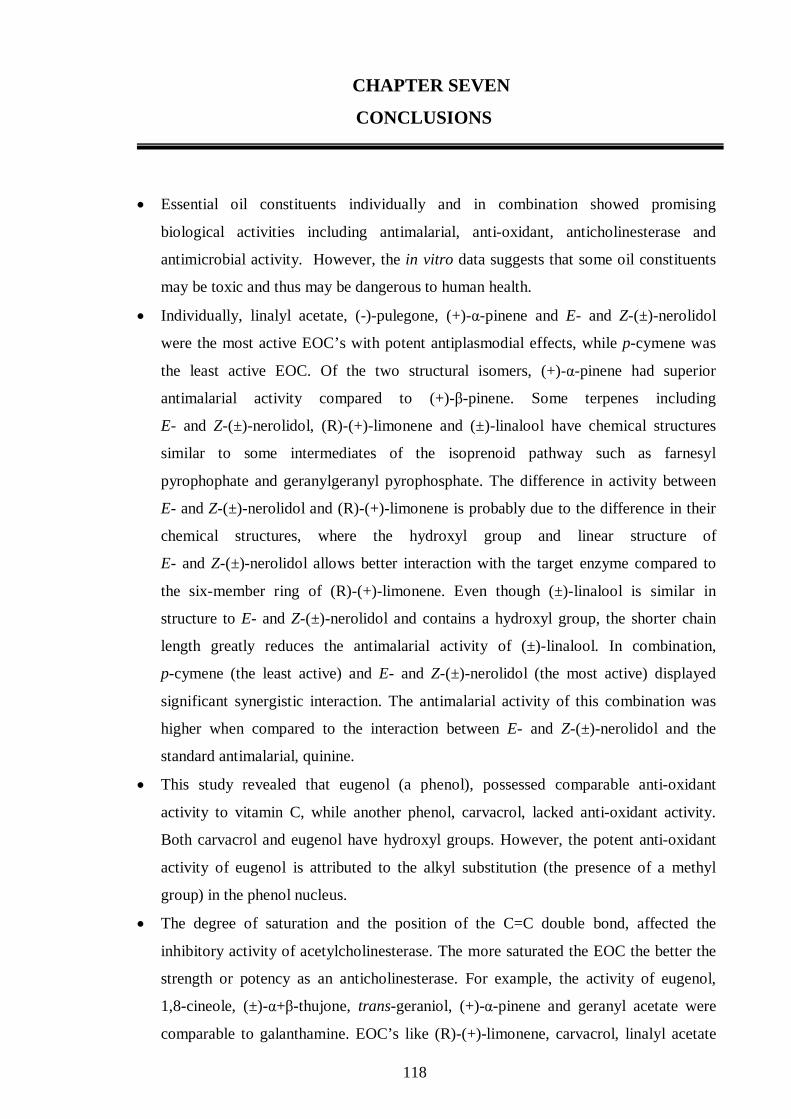
118
CHAPTER SEVEN
CONCLUSIONS
• Essential oil constituents individually and in combination showed promising
biological activities including antimalarial, anti-oxidant, anticholinesterase and
antimicrobial activity. However, the in vitro data suggests that some oil constituents
may be toxic and thus may be dangerous to human health.
• Individually, linalyl acetate, (-)-pulegone, (+)-α-pinene and E- and Z-(±)-nerolidol
were the most active EOC’s with potent antiplasmodial effects, while p-cymene was
the least active EOC. Of the two structural isomers, (+)-α-pinene had superior
antimalarial activity compared to (+)-β-pinene. Some terpenes including
E- and Z-(±)-nerolidol, (R)-(+)-limonene and (±)-linalool have chemical structures
similar to some intermediates of the isoprenoid pathway such as farnesyl
pyrophophate and geranylgeranyl pyrophosphate. The difference in activity between
E- and Z-(±)-nerolidol and (R)-(+)-limonene is probably due to the difference in their
chemical structures, where the hydroxyl group and linear structure of
E- and Z-(±)-nerolidol allows better interaction with the target enzyme compared to
the six-member ring of (R)-(+)-limonene. Even though (±)-linalool is similar in
structure to E- and Z-(±)-nerolidol and contains a hydroxyl group, the shorter chain
length greatly reduces the antimalarial activity of (±)-linalool. In combination,
p-cymene (the least active) and E- and Z-(±)-nerolidol (the most active) displayed
significant synergistic interaction. The antimalarial activity of this combination was
higher when compared to the interaction between E- and Z-(±)-nerolidol and the
standard antimalarial, quinine.
• This study revealed that eugenol (a phenol), possessed comparable anti-oxidant
activity to vitamin C, while another phenol, carvacrol, lacked anti-oxidant activity.
Both carvacrol and eugenol have hydroxyl groups. However, the potent anti-oxidant
activity of eugenol is attributed to the alkyl substitution (the presence of a methyl
group) in the phenol nucleus.
• The degree of saturation and the position of the C=C double bond, affected the
inhibitory activity of acetylcholinesterase. The more saturated the EOC the better the
strength or potency as an anticholinesterase. For example, the activity of eugenol,
1,8-cineole, (±)-α+β-thujone, trans-geraniol, (+)-α-pinene and geranyl acetate were
comparable to galanthamine. EOC’s like (R)-(+)-limonene, carvacrol, linalyl acetate
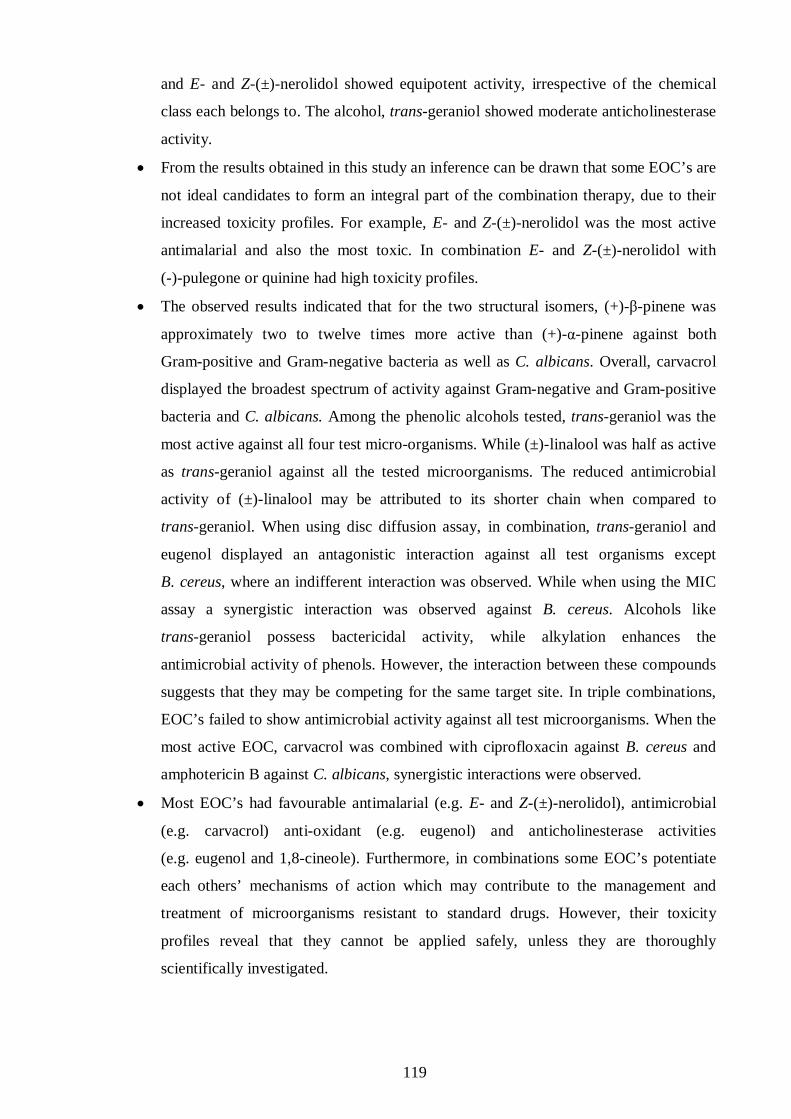
119
and E- and Z-(±)-nerolidol showed equipotent activity, irrespective of the chemical
class each belongs to. The alcohol, trans-geraniol showed moderate anticholinesterase
activity.
• From the results obtained in this study an inference can be drawn that some EOC’s are
not ideal candidates to form an integral part of the combination therapy, due to their
increased toxicity profiles. For example, E- and Z-(±)-nerolidol was the most active
antimalarial and also the most toxic. In combination E- and Z-(±)-nerolidol with
(-)-pulegone or quinine had high toxicity profiles.
• The observed results indicated that for the two structural isomers, (+)-β-pinene was
approximately two to twelve times more active than (+)-α-pinene against both
Gram-positive and Gram-negative bacteria as well as C. albicans. Overall, carvacrol
displayed the broadest spectrum of activity against Gram-negative and Gram-positive
bacteria and C. albicans. Among the phenolic alcohols tested, trans-geraniol was the
most active against all four test micro-organisms. While (±)-linalool was half as active
as trans-geraniol against all the tested microorganisms. The reduced antimicrobial
activity of (±)-linalool may be attributed to its shorter chain when compared to
trans-geraniol. When using disc diffusion assay, in combination, trans-geraniol and
eugenol displayed an antagonistic interaction against all test organisms except
B. cereus, where an indifferent interaction was observed. While when using the MIC
assay a synergistic interaction was observed against B. cereus. Alcohols like
trans-geraniol possess bactericidal activity, while alkylation enhances the
antimicrobial activity of phenols. However, the interaction between these compounds
suggests that they may be competing for the same target site. In triple combinations,
EOC’s failed to show antimicrobial activity against all test microorganisms. When the
most active EOC, carvacrol was combined with ciprofloxacin against B. cereus and
amphotericin B against C. albicans, synergistic interactions were observed.
• Most EOC’s had favourable antimalarial (e.g. E- and Z-(±)-nerolidol), antimicrobial
(e.g. carvacrol) anti-oxidant (e.g. eugenol) and anticholinesterase activities
(e.g. eugenol and 1,8-cineole). Furthermore, in combinations some EOC’s potentiate
each others’ mechanisms of action which may contribute to the management and
treatment of microorganisms resistant to standard drugs. However, their toxicity
profiles reveal that they cannot be applied safely, unless they are thoroughly
scientifically investigated.
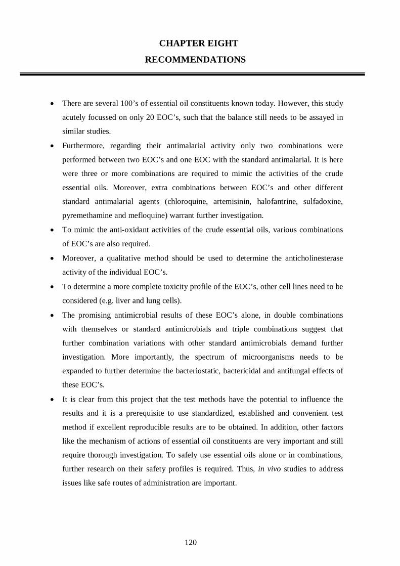
120
CHAPTER EIGHT
RECOMMENDATIONS
• There are several 100’s of essential oil constituents known today. However, this study
acutely focussed on only 20 EOC’s, such that the balance still needs to be assayed in
similar studies.
• Furthermore, regarding their antimalarial activity only two combinations were
performed between two EOC’s and one EOC with the standard antimalarial. It is here
were three or more combinations are required to mimic the activities of the crude
essential oils. Moreover, extra combinations between EOC’s and other different
standard antimalarial agents (chloroquine, artemisinin, halofantrine, sulfadoxine,
pyremethamine and mefloquine) warrant further investigation.
• To mimic the anti-oxidant activities of the crude essential oils, various combinations
of EOC’s are also required.
• Moreover, a qualitative method should be used to determine the anticholinesterase
activity of the individual EOC’s.
• To determine a more complete toxicity profile of the EOC’s, other cell lines need to be
considered (e.g. liver and lung cells).
• The promising antimicrobial results of these EOC’s alone, in double combinations
with themselves or standard antimicrobials and triple combinations suggest that
further combination variations with other standard antimicrobials demand further
investigation. More importantly, the spectrum of microorganisms needs to be
expanded to further determine the bacteriostatic, bactericidal and antifungal effects of
these EOC’s.
• It is clear from this project that the test methods have the potential to influence the
results and it is a prerequisite to use standardized, established and convenient test
method if excellent reproducible results are to be obtained. In addition, other factors
like the mechanism of actions of essential oil constituents are very important and still
require thorough investigation. To safely use essential oils alone or in combinations,
further research on their safety profiles is required. Thus, in vivo studies to address
issues like safe routes of administration are important.
121
REFERENCES
Anthony J-P, Fyfe L and Smith H. Plant active components – a resource for antiparasitic
agents? Trends in Parasitology 2005; 21(10): 462-465.
Atindehou KK, Shmid C, Brun R, Krone MW and Traore D. Antitrypanosomal activity of
medicinal plants from Côte d′ Ivore. Journal of Ethnopharmacology 2004; 90(2-3): 221-227.
Bell A. Antimalarial drug synergism and antagonism: Mechanistic and clinical significance.
FEMS Microbiology Letters 2005; 253: 171-184.
Benoit-Vical F, Valentin A, Mallie M, Bastide JM and Bessiere JM. In vitro antimalarial
activity and cytotoxicity of Cochlospermum tinctorium and C. planchonii leaf extracts and
essential oils. Planta Medica. 1999; 65(4): 378-81.
Berenbaum MC. A method for testing synergy with any number of agents. Journal of
Infectious Diseases 1989; 137(2): 122-130.
Boyom F, Ngouana V, Zollo A, Menut C, Bessiere J, Gut J and Rosenthal J. Composition and
antiplasmodial activities of essential oils from some Cameroonian medicinal plants.
Phytochemistry 2003; 64: 1269-1275.
Buchbauer G. Biological effects of fragrances and essential oils. Perfurmer and Florist 1993;
13: 19-24.
Burfield T and Reekie S-L. Mosquitoes, malaria and essential oils. The International Journal
of Aromatherapy 2005; 15: 30-41.
Burt S. Essential oils: their antibacterial properties and potential applications in food - a
review. International Journal of Food Microbiology 2004; 94: 223-253.
Campbell WE, Gammon DW, Smith P, Abrahams M and Purves TD. Composition and
antimalarial activity in vitro of the essential oil of Tetradenia riparia. Planta Medica 1997;
63(3): 270-272.
Carson CF and Riley TV. Antimicrobial activity of the major components of the essential oil
of Melaleuca alternifolia. Journal of Applied Bacteriology 1995; 78(3): 264-269.

122
Cassella JP, Cassella S and Smith I. Synergistic antifungal activity of tea tree (Melaleuca
alternifolia) and lavender (Lavandula angustifolia) essential oils against dermatophyte
infection. The International Journal of Aromatherapy 2002; 12(1): 1-15.
Choi H, Song H, Ukeda H and Sawamura M. Radical scavenging activities of citrus essential
oils and their components: Detection using 1,1-Diphenyl-2-picrylhydrazyl. Journal of
Agriculture and Food Chemistry 2000; 48: 4156-4161.
Clarkson C, Vinesh J, Maharaj B, Crouch N, Olwen M, Grace C, Pamisha P, Motlalepula G,
Matsabisa E, Niresh B, Smith P and FolB P. In vitro antiplasmodial activity of medicinal
plants native to or naturalised in South Africa. Journal of Ethnopharmacology 2004;
92: 177–191.
Constable S, Ham A and Pirmohamed M. Herbal medicines and acute medical emergency
admissions to hospital. British Journal of Clinical Pharmacology 2006; 63(2): 247-248.
Constantin J, Ishii-Iwamoto EL, Ferraresi-Filho O, Kelmer-Bracht AM and Bracht A.
Sensitivity of ketogenesis and citric acid cycle to stevioside inhibition of palmitate transport
across the cell membrane. Journal of Biological Research 1991; 24(8): 767-71.
Constantin M, Sartorelli P, Limberger R, Henriques T, Steppe M, Ferreira J, Ohara M,
Emerenciano V and Kato M. Essential oils from Piper cernuum and Piper regnellii:
antimicrobial activities and analysis by GC/MS and 13CNMR. Planta Medica 2001;
67: 771-773.
Cowan MM. Plant products as a resource for new drugs. Pharmaceutical Research 1996;
13: 1133-1141.
Cox SD, Mann CM, Markham JL, Gustafson JE Warmington JR and Wyllie G. Determining
the antimicrobial actions of tea tree oil. Molecules 2001; 6: 87-91.
Cragg MG, Newman DJ and Snader KM. Natural products in drug discovery and
development. Journal of Natural Products 1997; 60: 52-60.
De Macedo C, Uhring M, Kimura E and Katzin A. Characterization of the isoprenoid chain of
coenzyme Q in Plasmodium falciparum. FEMS Microbiology Letter 2002; 207(1): 13-20.
De Smet PAGM. The role of plant-derived drugs and herbal medicines in healthcare. Drugs
1997; 54(6): 801-840.
123
Department of Health and Human services, 2006. http://www.cdc.gov/malaria/biology/
life_cycle.htm. (accessed: February 15, 2006).
Department of Health, 2005. http://www.doh.gov.za/issues/malaria/2004-2005.html
(accessed: March 04, 2006).
Desjardins RE, Carfield CJ, Haynes DJ and Chulay JD. Quantitative assessment of foods – a
review. International Journal of Food Microbiology 1972; 94: 223-253.
Dormans HJ and Deans SD. Anti-oxidant and antimicrobial activity of Foeniculum vulgare
and Crithmum maritimum essential oils. Planta Medica 2000; 66(8): 687-93.
Ellis JM. Cholinesterase inhibitors in the treatment of dementia. Journal of American
Osteopathic Association 2005; 105(3): 145-158.
Eloff J.N. A sensitive and quick microplate method to determine the minimal inhibitory
concentration of plant extracts for bacteria. Planta Medica 1998; 64: 711-713.
Geron C, Rasmussen R, Arnts RR and Guenther A. A review and synthesis of monoterpenes
speciation from forests in the United States. Atmospheric environment 2000; 34: 1761-1781.
Gollin MA. New rules for natural products research: dwindling natural resources have spurred
tighter regulations for biodiversity prospecting. Nature Biotechnology 1999; 17: 921-922.
Goulart HR, Kimura AE, Peres JV, Conto SA, Aquino Duarte AF and Katzin AM. Terpenes
arrest parasite development and inhibit biosynthesis of isoprenoids in Plasmodium
falciparum. Antimicrobial Agents and Chemotherapy 2004; 48(7): 2502-2509.
Graβmann J, Hippeli S, Dornisch K, Rohnet U, Beuscher N and Elstner EF. Anti-oxidant
properties of essential oils. Drug Research 2000; 50(1): 135-139.
Gregson A and Plowe CV. Mechanisms of resistance of malaria parasites to antifolates.
Pharmacology Reviews 2005; 57:117-145.
Griffin SG, Wyllie SG, Markham JL and Leach DN. The role of structure and molecular
properties of terpenoids in determining their antimicrobial activity. Flavour and Fragrance
Journal 1999; 14: 322-332.
Halcon LL. Aromatherapy: Therapeutic applications of essential oils. Minnesota Medical
Association 2002; 85(11): 42-46.

124
Harris R. Synergism in the essential oil world. The International Journal of Aromatherapy
2002; 12(4): 178-186.
Hayward R, Sullivan D and Day K. Plasmodium falciparum: Histidine-rich protein II is
expressed during gametocyte development. Experimental parasitology 2000; 96: 139-146.
Holstein SA and Hohl RJ. Monoterpene regulation of Ras and Ras-related protein expression.
Journal of Lipid Research. 2003; 44:1209–1215.
Inouye S, Takizawa T and Yamaguchi H. Antibacterial activity of essential oils and their
major constituents against respiratory tract pathogens by gaseous contact. Journal of
Antimicrobial Chemotherapy 2001; 47: 565-573.
Jansen JB and Trager W. Human malaria parasites in continuous culture. Science 1976;
193: 675-676.
Janssen A, Scheffer J and Svendsen A. Antimicrobial activity of essential oils: A 1976-1986
literature review. Aspects of the test methods. Planta Medica 1987; 53: 395-398.
Jirovertz L, Jäger W, Buchbauer G, Raverdino V and Nikiforov A. Determination of lavender
oil fragrance compounds in blood samples. Journal of Analytical Chemistry 1990;
338: 922-923.
Kalemba D and Kunicka A. Antibacterial and antifungal properties of essential oils. Current
Medicinal chemistry 2003; 10: 813-829.
Kamatou GP, Viljoen AM, Gono-Bwalya AB, van Zyl RL, Vuuren SF, Lourens AC, Başer
KH, Demirci B, Lindsey KL, van Staden J and Steenkamp P. The in vitro pharmacological
activities and a chemical investigation of three South African Salvia species. Journal of
Ethnopharmacology 2005; 102(3): 382-90.
Kang R, Helms R, Stout M, Jaber J, Chen Z and Nakatsu T. Antimicrobial activity of the
volatile constituents of Perilla frutescens and its synergistic effects with polygodial. Journal
of Agriculture and Food Chemistry 1992; 40: 2328-2330.
Knobloch K, Pauli A, Iberl B, Weigand H and Weis N, Antibacterial and antifungal properties
of essential oil components. Journal of Essential Oil Research 1989; 1: 119-128.
Lahlou M. Biological and pharmacological approaches to the screening and evaluation of
natural products. Therapie 2004a; 58(6): 535-539.
125
Lahlou M. Methods to study the phytochemistry and bioactivity of essential oils.
Phytotherapy Research. 2004b; 18(6): 435-448.
Lambros C and Vanderberg P. Synchronization of Plasmodium falciparum erythrocytic stages
in culture. Journal of Parasitology 1979; 65: 418-420.
Lawrence BM. Essential oils: from agriculture to chemistry. The International Journal of
Aromatherapy 2000; 10(3): 82-98.
Lis-Balchin M. Essential oils and aromatherapy: their modern role in healing. Journal of
Royal Society Health 1997; 117(5): 324-329.
Lopes NP, Kato MJ, Andrade EH, Maia JG, Yoshida M, Planchart AR and Katzin AM.
Antimalarial use of volatile oil from leaves of Virola surinamensis (Rol.) Warb. by Waiapi
Amazon Indians. Journal of Ethnopharmacology 1999; 67(3): 313-319.
Lourens AC, Reddy D, Başer KH, Viljoen AM and van Vuuren SF. In vitro biological
activity and essential oil composition of four indigenous South African Helichrysum species.
Journal of Ethnopharmacology 2004; 95(2-3): 253-8.
Lukwa N. Do traditional mosquito repellent plants work as mosquito larvicides? Centre for
African Journal of Medicine 1994; 40(11): 306-309.
Mahmoud SS and Croteau RB. Strategies for transgenic manipulation of monoterpene
biosynthesis in plants. Trends in Plant Science 2002; 10: 1360-1367.
Marston A, Kissling J and Hostettmann K. A rapid TLC bioautographic method for the
detection of acetylcholinesterase and butyrylcholinesterase inhibitors in plants. Phytochemical
Analysis 2002; 13(1): 51-4.
McKellar QA and Jackson F. Veterinary anthelmintics: old and new. Trends in Parasitology
2004; 20(10): 456-61.
Meadows S, Gennings C, Carter H and Bae D. Experimental designs for mixtures along fixed
ratio rays. Environmental Health Perspective 2002; 110(6): 979-983.
Medeiros J, Campos B, Mendonca C, Davin B and Lewis G. Composition and antimicrobial
activity of the essential oils from invasive species of the Azores, Hedychium gardnerianum
and Pittosporum undulatum. Phytochemistry 2003; 64: 561-565.
126
Mills C, Cleary BJ, Gilmer JF and Walsh JJ. Inhibition of acetylcholinesterase by Tea Tree
oil. Journal of Pharmaceutics and Pharmacology 2004; 56(3): 375-9.
Miyazawa M and Yamafuji C. Inhibition of acetylcholinesterase activity by bicyclic
monoterpenoids. Journal of Agriculture, Food and Chemistry 2005; 53: 1765-1768.
Mi-Yeon K, Sang-Won C and Shin-Kyo C. Anti-oxidative flavonoids from the garlic (Allium
sativum L.) shoot. Food Science Biotechnology 2000; 9(4): 199-203.
Mosmann T. Rapid colorimetric assay for cellular growth and survival: application to
proliferation and cytotoxicity assays. Journal of Immunological Methods 1983; 65: 55-63.
Moura C, Wunderlich G, Uhrig M, Couto A, Peres S, Katzin A and Kimura E. Limonene
arrests parasite development and inhibits isoprenylation of proteins in Plasmodium
falciparum. Antimicrobial Agents Chemotherapy 2001; 45(9): 2553–2558.
Nakatsu T, Lupo A, Chinn J and Kang R. Biological activity of essential oils and their
constituents. Study of Natural Products and Chemistry 2000; 21: 571-631.
Nchinda TC. Malaria: A reemerging disease in Africa. Emerging Infectious Diseases 1998;
4(3): 398-402.
Noedl H, Wongsrichanalai C and Wernsdorfer H. Malaria drug-sensitivity testing: New
Perspectives. Trends in Parasitology 2003; 19(4): 175-181.
Onawumni GO, Yisak WAB and Ogunlana EO. Antibacterial constituents in the essential oil
of Cymbopogon citrates (DC) STAPF. Journal of Ethnopharmacology 1984; 12: 279-286.
Pappano AJ. Chapter 7: Cholinoceptor-activating and cholinesterase-inhibiting drugs.
In: Basic and Clinical Pharmacology. Eds: Katzung BG. 9th Edition. Lange Medical
Books/McGraw-Hill, New York. 2004. pp 94-108.
Pauli A and Schilcher H. Specific selection of essential oil compounds for treatment of
children′s infectious diseases. Pharmaceuticals 2004; 1: 1-30.
Peiwu L, Hopia A, Jari S, Teijo Yrjőnen T and Vuorela H. TLC method for evaluation of free
radical scavenging activity of Rapeseed meal by video scanning technology. The Regional
Institute Limited 1999; 10: 1-6.
127
Perry NSL, Bollen C and Perry EK, Ballard C. Salvia for dementia therapy: review of
pharmacological activity and pilot tolerability clinical trial. Pharmacology, Biochemistry and
Behaviour 2003; 75: 651–659.
Plaa GL. Chapter 57: Introduction to toxicology: occupational and environmental. In: Basic
and Clinical Pharmacology. Eds: Katzung BG. Lange Medical Books/McGraw-Hill. New
York 2004, 9th edition. pp 958-959.
Plant aromatics safety data manual, 2004. http://www.users.globalnet.co.uk/~nodice
/new/magazine/magsafetylecture.htm. - accessed June 02, 2005.
Prozesky EA, Meyer JJ and Louw AI. In vitro antiplasmodial activity and cytotoxicity of
ethnobotanically selected South African plants. Journal of Ethnopharmacology 2001;
76(3): 239-45.
Ridley RG. Medical need, scientific opportunity and the drive for antimalarial drugs. Nature
2002; 415:686-693.
Ringman JM and Cummings JL. Metrifonate: update on a new antidementia agent. Journal of
Clinical Psychiatry 1999; 60(11): 776-82.
Rosenthal and Goldsmith, Chapter 54: Clinical pharmacology of the anthelmintic drugs. In:
Basic and Clinical Pharmacology. Eds: Katzung BG. 9th Edition. Lange Medical
Books/McGraw-Hill, New York. 2004. pp 886-897.
Sanchez-Moreno C. Anti-oxidant activity of selected Spanish wines in corn oil emulsions.
Journal of Agriculture, Food and Chemistry 2002; 48(11): 5581-7.
Savelev S, Okello E, Perry NSL, Wilkins RM and Perry EK. Synergistic and antagonistic
interactions of anticholinesterase terpenoids in Salvia lavandulaefolia essential oil.
Pharmacology, Biochemistry and Behaviour 2003; 75: 661–668.
Schwikkard S and Van Heerden F. Antimalarial activity of plant metabolites. Natural
Products Representative 2002; 19:1-19.
Scott G, Springfield E and Coldrey N. A pharmacognostical study of 26 South African Plant
species used as traditional medicines. Pharmaceutical Biology 2004; 42(3): 186-213.
128
Shale TL, Strik WA and van Staden J. Screening of medicinal plants used in Lesotho for
antibacterial and anti-inflammatory activity. Journal of Ethnopharmacology 1999;
67(3): 347-354.
Shin S and Kang CA. Antifungal activity of the essential oil of Agastache rugosa Kuntze and
its synergism with ketoconazole. Letters in Applied Microbiology 2003; 36: 111-115.
Sramek JJ and Cutler NR. RBC Cholinesterase Inhibition: a useful surrogate marker for
cholinesterase inhibitor activity in Alzheimer´s Disease therapy? Alzheimer´s Disease and
Associated Disorders 2000; 14(4): 216-27.
Subramanyam VR, Pattnaik S and Kole CR. Antibacterial and antifungal activity of ten
essential oils in vitro. Microbiology 1996; 128: 151-153.
Svoboda KP, Svoboda TG, Syred A. A Closer Look: Secretory Structures of Aromatic and
Medicinal Plants. The Journal of the American Botanical Council (Herbal Gram) 2001;
53: 34-43.
Tampieri M, Galuppi R, Macchioni F, Carelle S, Falcioni L, Cioni P, Morelli I. The inhibition
of Candida albicans by selected essential oils and their major components. Mycopatholgia
2005; 159: 339-345.
Ueng Y-F, Hsieh C-H and Don M-J. Inhibition of human cytochrome P450 enzymes by the
natural safrole. Food and Chemical Toxicology 2005; 43: 707-712.
Ultee A, Bennik M and Moezelaar R. The phenolic hydroxyl group of carvacrol is essential
for action against the food-borne pathogen Bacillus cereus. Applied Environmental
Microbiology 2002; 68: 1561-1568.
Valentin A, Pélissier Y, Benoit F, Marion C, Kone D, Mallie M, Bastide J-M and Bessiére
J-M. Composition and antimalarial activity in vitro of volatile components of Lippia
multiflora. Phytochemistry 1995; 40: 1439-1442.
van Vuuren SF, Viljoen AM, van Zyl RL , van Heerden FR, Hǘşnǘ S and Başer C. The
antimicrobial, antimalarial and toxicity profiles of helihumulone, leaf essential oil and extracts
of Helichrysum cymosum (L.) D. Don subsp. cymosum. South African Journal of Botany
2006; 72: 287 – 290.

129
van Zyl RL, Seatlholo ST, van Vuuren SF and Viljoen AM. The biological activity of 20
nature identical essential oil constituents. Journal of Essential Oil Research 2006;
18: 129-133.
Vardar-Ünlü G, Candan F, Sökmen A, Daferera D, Polissiou M, Sökmen M, Dönmez E and
Bektaş T. Antimicrobial and antioxidant activity of the essential oils and methanol extracts of
Thymus pectinatus Fisch. et Mey. var. pectinatus (Lamiaceae). Journal of Agriculture Food
and Chemistry 2003; 51: 63-67.
Viljoen AM, Subramoney S, van Vuuren SF, Başer KH and Dermici B. The composition,
geographical variation and antimicrobial activity of Lippia javanica (Verbenaceae) leaf
essential oils. Journal of Ethnopharmacology 2005; 96(1-2): 271-277.
Viljoen AM, van Vuuren SF, Ernst E, Klepser M, Demirci B, Başer KH and van Wyk B-E.
Osmitopsis asteriscoides (Asteraceae) - the antimicrobial activity and essential oil
composition of a Cape-Dutch remedy. Journal of Ethnopharmacology 2003; 88: 137-143.
Viljoen AM, van Vuuren SF, Gwebu T, Dermici B and Başer C. The geographical variation
and antimicrobial activity of African wormwood (Artemisia afra Jacq.) essential oil. Journal
of Essential Oil Research 2006; 18: 19-25.
Waako P, Smith P and Folb P. In vitro interactions of Aspilia africana (Pers.) C.D. Adams, a
traditional antimalarial medicinal plant with artemisinin against Plasmodium falciparum.
Journal of Ethnopharmacology 2005; 102(2): 262-268.
Warhurst D C. A molecular marker for chloroquine-resistant falciparum malaria.
Phytochemistry, 2001; 12: 114-118.
Watt JM and Breyer-Brandwijk MG. (1962) The Medicinal and Poisonous Plants of Southern
and Eastern Africa. Livingstone.
WHO (2003) “Traditional medicines.” World Health Organisation media centre.
<http://www.who.int/mediacentre/factsheets/fs134 (accessed: March 20, 2006)
WHO Guidelines for the assessment of herbal medicines. World Health Organisation
Technical Representative Services 2005; 908(i-viii): 130 -136.
130
Yanpallewar SU, Rai S, Kumar M and Acharya SB. Evaluation of antioxidant and
neuroprotective effect of Ocimum sanctum on transient cerebral ischemia and long-term
cerebral hypoperfusion. Pharmacology, Biochemistry and Behaviour 2004; 79(1): 155-64.
Yoo CB, Ki-Tae H, Cho K, Ha J, Park H, Nam J, Kil U and Lee K. Eugenol isolated from the
essential oil of Eugenia caryophyllata induces a reactive oxygen species-mediated apoptosis
in HL-60 human promyelocytic leukemia cells. Cancer letters 2005; 225: 41- 42.
Yoshida N, Takagi A, Kitazawa H, Kawakami J and Adachi I. Inhibition of p-glycoprotein-
mediated transport by extracts of and monoterpenoids contained in Zanthoxyli fructus.
Toxicology and Applied Pharmacology 2005; 209: 167 – 173.
Zhou S, Koh HL, Gao Y, Gong ZY and Lee EJ. Herbal bioactivation: the good, the bad and
the ugly. Life Sciences 2004; 74(8): 935-968.
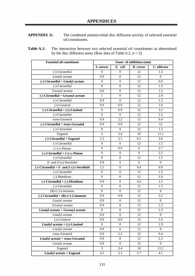
131
APPENDICES APPENDIX A: The combined antimicrobial disc diffusion activity of selected essential
oil constituents. Table A.1: The interaction between two selected essential oil constituents as determined
by the disc diffusion assay (Raw data of Table 6.2, n = 2).
Zones of inhibition (mm) Essential oil constituent
S. aureus E. coli B. cereus C. albicans
(-)-Citronellal 0 0 12 1.5
Linalyl acetate 0.9 0 12 0
(-)-Citronellal + Linalyl acetate 0 0 12 0.9
(-)-Citronellal 0 0 12 1.5
Geranyl acetate 0.9 0 12 1.2
(-)-Citronellal + Geranyl acetate 1 0 12 2.9
(-)-Citronellal 0.9 0 12 1.5
(±)-Linalool 0.9 0.9 12 1.6
(-)-Citronellal + (±)-Linalool 0 0.9 12 3.2
(-)-Citronellal 0 0 12 1.5
trans-Geraniol 0.9 2.2 12 9.4
(-)-Citronellal + trans-Geraniol 0.9 0.9 12 12
(-)-Citronellal 0 0 12 1.5
Eugenol 3 3.4 10 11.1
(-)-Citronellal + Eugenol 1.5 2.1 8.1 3.6
(-)-Citronellal 0 0 12 1.5
(+)-α-Pinene 0 0.9 0 2.7
(-)-Citronellal + (+)-α-Pinene 0 0 0 2.2
(-)-Citronellal 0 0 12 1.5
E- and Z-(±)-Nerolidol 0.9 0 0 0
(-)-Citronellal + E- and Z-(±)-Nerolidol 1.2 0 8.4 0.9
(-)-Citronellal 0 0 12 1.5
(-)-Menthone 0 0 12 1.6
(-)-Citronellal + (-)-Menthone 0.9 0 4.5 1.5
(-)-Citronellal 0 0 12 1.5
(R)-(+)-Limonene 0 0 12 0
(-)-Citronellal + (R)-(+)-Limonene 0.9 0.9 12 3.2
Linalyl acetate 0.9 0 12 0
Geranyl acetate 0.9 0 12 1.2
Linalyl acetate + Geranyl acetate 0 0 12 1.5
Linalyl acetate 0.9 0 12 0
(±)-Linalool 0.9 0.9 12 1.6
Linalyl acetate + (±)-Linalool 0 0 12 1.9
Linalyl acetate 0.9 0 12 0
trans-Geraniol 0.9 2.2 12 9.4
Linalyl acetate + trans-Geraniol 0.9 0 12 2.2
Linalyl acetate 0.9 0 12 0
Eugenol 3 3.4 10 11.1
Linalyl acetate + Eugenol 4.3 3.3 5.7 4.1
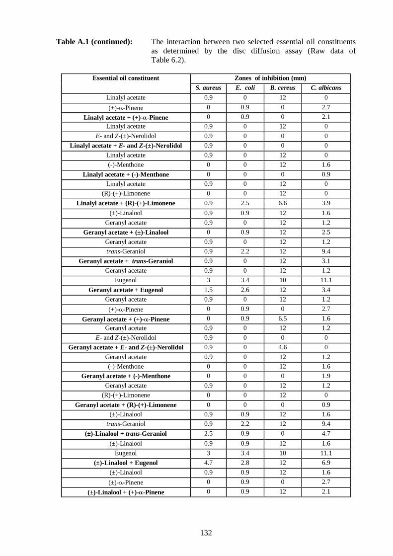
132
Table A.1 (continued): The interaction between two selected essential oil constituents as determined by the disc diffusion assay (Raw data of Table 6.2).
Zones of inhibition (mm) Essential oil constituent
S. aureus E. coli B. cereus C. albicans
Linalyl acetate 0.9 0 12 0
(+)-α-Pinene 0 0.9 0 2.7
Linalyl acetate + (+)-α-Pinene 0 0.9 0 2.1
Linalyl acetate 0.9 0 12 0
E- and Z-(±)-Nerolidol 0.9 0 0 0
Linalyl acetate + E- and Z-(±)-Nerolidol 0.9 0 0 0
Linalyl acetate 0.9 0 12 0
(-)-Menthone 0 0 12 1.6
Linalyl acetate + (-)-Menthone 0 0 0 0.9
Linalyl acetate 0.9 0 12 0
(R)-(+)-Limonene 0 0 12 0
Linalyl acetate + (R)-(+)-Limonene 0.9 2.5 6.6 3.9
(±)-Linalool 0.9 0.9 12 1.6
Geranyl acetate 0.9 0 12 1.2
Geranyl acetate + (±)-Linalool 0 0.9 12 2.5
Geranyl acetate 0.9 0 12 1.2
trans-Geraniol 0.9 2.2 12 9.4
Geranyl acetate + trans-Geraniol 0.9 0 12 3.1
Geranyl acetate 0.9 0 12 1.2
Eugenol 3 3.4 10 11.1
Geranyl acetate + Eugenol 1.5 2.6 12 3.4
Geranyl acetate 0.9 0 12 1.2
(+)-α-Pinene 0 0.9 0 2.7
Geranyl acetate + (+)-α-Pinene 0 0.9 6.5 1.6
Geranyl acetate 0.9 0 12 1.2
E- and Z-(±)-Nerolidol 0.9 0 0 0
Geranyl acetate + E- and Z-(±)-Nerolidol 0.9 0 4.6 0
Geranyl acetate 0.9 0 12 1.2
(-)-Menthone 0 0 12 1.6
Geranyl acetate + (-)-Menthone 0 0 0 1.9
Geranyl acetate 0.9 0 12 1.2
(R)-(+)-Limonene 0 0 12 0
Geranyl acetate + (R)-(+)-Limonene 0 0 0 0.9
(±)-Linalool 0.9 0.9 12 1.6
trans-Geraniol 0.9 2.2 12 9.4
(±)-Linalool + trans-Geraniol 2.5 0.9 0 4.7
(±)-Linalool 0.9 0.9 12 1.6
Eugenol 3 3.4 10 11.1
(±)-Linalool + Eugenol 4.7 2.8 12 6.9
(±)-Linalool 0.9 0.9 12 1.6
(±)-α-Pinene 0 0.9 0 2.7
(±)-Linalool + (+)-α-Pinene 0 0.9 12 2.1

133
Table A.1 (continued): The interaction between two selected essential oil constituents as determined by the disc diffusion assay (Raw data of Table 6.2).
Zones of inhibition (mm) Essential oil constituent
S. aureus E. coli B. cereus C. albicans
(±)-Linalool 0.9 0.9 12 1.6
E- and Z-(±)-Nerolidol 0.9 0 0 0
(±)-Linalool + E- and Z-(±)-Nerolidol 0.9 0.9 12 0.9
(±)-Linalool 0.9 0.9 12 1.6
(-)-Menthone 0 0 12 1.6
(±)-Linalool + (-)-Menthone 0 0.9 12 0
(±)-Linalool 0.9 0.9 12 1.6
(R)-(+)-Limonene 0 0 12 0
(±)-Linalool + (R)-(+)-Limonene 0.9 4.1 12 4.5
trans-Geraniol 0.9 2.2 12 9.4
Eugenol 3 3.4 10 11.1
trans-geraniol + Eugenol 3.2 2.6 12 4.6
trans-Geraniol 0.9 2.2 12 9.4
(+)-α-Pinene 0 0.9 0 2.7
trans-Geraniol + (+)-α-Pinene 2 0.9 12 3.4
trans-Geraniol 0.9 2.2 12 9.4
E- and Z-(±)-Nerolidol 0.9 0 0 0
trans-Geraniol + E- and Z-(±)-Nerolidol 0.9 0.9 0 2.6
trans-Geraniol 0.9 2.2 12 9.4
(-)-Menthone 0 0 12 1.6
trans-Geraniol + (-)-Menthone 0 0 6.6 4.6
trans-Geraniol 0.9 2.2 12 9.4
(R)-(+)-Limonene 0 0 12 0
trans-Geraniol + (R)-(+)-Limonene 2.4 0.9 12 3.1
Eugenol 3 3.4 10 11.1
(+)-α-Pinene 0 0.9 0 2.7
Eugenol + (+)-α-Pinene 2.5 3 12 4.9
Eugenol 3 3.4 10 11.1
E- and Z-(±)-Nerolidol 0.9 0 0 0
Eugenol + E- and Z-(±)-Nerolidol 3.1 0 12 3.4
Eugenol 3 3.4 10 11.1
(-)-Menthone 0 0 12 1.6
Eugenol + (-)-Menthone 2.3 0 12 4.1
Eugenol 3 3.4 10 11.1
(R)-(+)-Limonene 0 0 12 0
Eugenol + (R)-(+)-Limonene 3.5 2.5 12 5.3
(+)-α-Pinene 0 0.9 0 2.7
E- and Z-(±)-Nerolidol 0.9 0 0 0
(+)-α-Pinene + E- and Z- (±)-Nerolidol 0.9 0.9 12 0
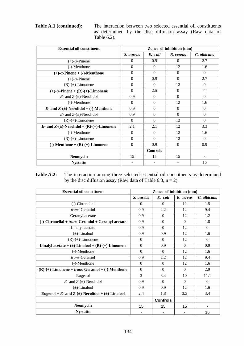
134
Table A.1 (continued): The interaction between two selected essential oil constituents as determined by the disc diffusion assay (Raw data of Table 6.2).
Zones of inhibition (mm) Essential oil constituent
S. aureus E. coli B. cereus C. albicans
(+)-α-Pinene 0 0.9 0 2.7
(-)-Menthone 0 0 12 1.6
(+)-α-Pinene + (-)-Menthone 0 0 0 0
(+)-α-Pinene 0 0.9 0 2.7
(R)-(+)-Limonene 0 0 12 0
(+)-α-Pinene + (R)-(+)-Limonene 0 2.5 0 4
E- and Z-(±)-Nerolidol 0.9 0 0 0
(-)-Menthone 0 0 12 1.6
E- and Z-(±)-Nerolidol + (-)-Menthone 0.9 0 0 0
E- and Z-(±)-Nerolidol 0.9 0 0 0
(R)-(+)-Limonene 0 0 12 0
E- and Z-(±)-Nerolidol + (R)-(+)-Limonene 2.1 2.1 12 3.3
(-)-Menthone 0 0 12 1.6
(R)-(+)-Limonene 0 0 12 0
(-)-Menthone + (R)-(+)-Limonene 0 0.9 0 0.9
Controls
Neomycin 15 15 15 -
Nystatin - - - 16
Table A.2: The interaction among three selected essential oil constituents as determined
by the disc diffusion assay (Raw data of Table 6.3, n = 2).
Essential oil constituent Zones of inhibition (mm)
S. aureus E. coli B. cereus C. albicans
(-)-Citronellal 0 0 12 1.5
trans-Geraniol 0.9 2.2 12 9.4
Geranyl acetate 0.9 0 12 1.2
(-)-Citronellal + trans-Geraniol + Geranyl acetate 0.9 0 0 1.8
Linalyl acetate 0.9 0 12 0
(±)-Linalool 0.9 0.9 12 1.6
(R)-(+)-Limonene 0 0 12 0
Linalyl acetate + (±)-Linalool + (R)-(+)-Limonene 0 0.9 0 0.9
(-)-Menthone 0 0 12 1.6
trans-Geraniol 0.9 2.2 12 9.4
(-)-Menthone 0 0 12 1.6
(R)-(+)-Limonene + trans-Geraniol + (-)-Menthone 0 0 0 2.9
Eugenol 3 3.4 10 11.1
E- and Z-(±)-Nerolidol 0.9 0 0 0
(±)-Linalool 0.9 0.9 12 1.6
Eugenol + E- and Z-(±)-Nerolidol + (±)-Linalool 2.4 1.8 3.3 3.4
Controls Neomycin 15 15 15 - Nystatin - - - 16
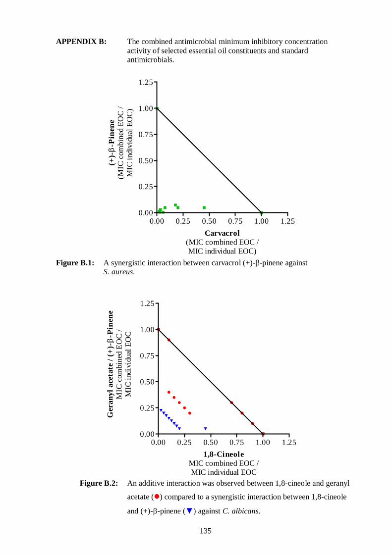
135
APPENDIX B: The combined antimicrobial minimum inhibitory concentration activity of selected essential oil constituents and standard antimicrobials.
Figure B.1: A synergistic interaction between carvacrol (+)-β-pinene against S. aureus.
0.00 0.25 0.50 0.75 1.00 1.250.00
0.25
0.50
0.75
1.00
1.25
1,8-CineoleMIC combined EOC /MIC individual EOC
Ger
anyl
ace
tate
/ (
+)-β
-Pin
ene
MIC
com
bine
d E
OC
/M
IC in
divi
dual
EO
C
Figure B.2: An additive interaction was observed between 1,8-cineole and geranyl
acetate (�) compared to a synergistic interaction between 1,8-cineole
and (+)-β-pinene (▼) against C. albicans.
0.00 0.25 0.50 0.75 1.00 1.250.00
0.25
0.50
0.75
1.00
1.25
Carvacrol(MIC combined EOC /MIC individual EOC)
(+)-β -
Pin
ene
(MIC
com
bine
d E
OC
/M
IC in
divi
dual
EO
C)

136
0.00 0.25 0.50 0.75 1.00 1.250.00
0.25
0.50
0.75
1.00
1.25
Eugenol(MIC combined EOC /MIC individual EOC)
tran
s-G
era
nio
l(M
IC c
ombi
ned
EO
C /
MIC
indi
vidu
al E
OC
)
Figure B.3: The synergistic and additive interactions were observed between eugenol
and trans-geraniol against B. cereus (�) and E. coli (▲), respectively.
0.00 0.25 0.50 0.75 1.00 1.250.00
0.25
0.50
0.75
1.00
1.25
γ-Terpinene(MIC combined EOC /MIC individual EOC)
(+)-β-
Pin
ene
/ G
eran
yl a
ceta
te(M
IC c
ombi
ned
EO
C /
MIC
indi
vidu
al E
OC
)
Figure B.4: γ-Terpinene interacted in a synergistic manner with both (+)-β-pinene (▲)
and geranyl acetate (▼) against S. aureus.

137
0.00 0.25 0.50 0.75 1.00 1.250.00
0.25
0.50
0.75
1.00
1.25
trans-Geraniol(MIC combined EOC /MIC individual EOC)
E-
and
Z-(± )
-Ner
olid
ol
(MIC
com
bine
d E
OC
/M
IC in
divi
dual
EO
C)
Figure B.5: A synergistic interaction between trans-geraniol and E- and Z-(±)-nerolidol
against B. cereus.
0.00 0.25 0.50 0.75 1.00 1.250.00
0.25
0.50
0.75
1.00
1.25
Ciprofloxacin(MIC combined /MIC individual)
Ca
rvac
rol
(MIC
com
bine
d E
OC
/M
IC in
divi
dual
EO
C)
Figure B.6: A synergistic interaction observed between ciprofloxacin and carvacrol
against B. cereus.