The Bacterial Actin-Like Cytoskeleton · The actin cytoskeleton is highly dynamic in most cells,...
23
MICROBIOLOGY AND MOLECULAR BIOLOGY REVIEWS, Dec. 2006, p. 888–909 Vol. 70, No. 4 1092-2172/06/$08.000 doi:10.1128/MMBR.00014-06 Copyright © 2006, American Society for Microbiology. All Rights Reserved. The Bacterial Actin-Like Cytoskeleton Rut Carballido-Lo ´pez* Ge ´ne ´tique Microbienne, Institut National de la Recherche Agronomique, 78352 Jouy-en-Josas Cedex, France INTRODUCTION .......................................................................................................................................................888 EUKARYOTIC ACTIN AND THE EUKARYOTIC ACTIN CYTOSKELETON................................................889 The Actin Superfamily ...........................................................................................................................................890 THE BACTERIAL ACTIN MreB..............................................................................................................................890 MreB Filaments Are Generated by Actin-Like Polymerization........................................................................890 Structure of MreB monomers and MreB protofilaments .............................................................................890 MreB assembly properties .................................................................................................................................891 Dynamics of MreB Filaments ...............................................................................................................................891 Ultrastructural Organization of MreB Filaments In Vivo................................................................................893 MreB-LIKE PROTEINS AND CELL MORPHOGENESIS .................................................................................893 MreB Filaments Govern Cell Morphogenesis by Actively Directing Lateral Wall Biogenesis ....................893 Mbl filaments direct lateral wall synthesis in Bacillus subtilis .....................................................................893 PBP localization and the MreB cytoskeleton..................................................................................................894 MreBH filaments direct lateral wall hydrolysis in Bacillus subtilis .............................................................895 Is Transmission of Shape Mediated by MreB-Directed Peptidoglycan Factories? .......................................896 The Essential MreBCD Complex and Lateral Wall Synthesis.........................................................................896 MreB Proteins and Spore Lateral Wall Formation in Actinomycetes ............................................................897 MreB Proteins and Cell Shape Determination in Wall-Less Prokaryotes .....................................................897 ACTIN-LIKE PROTEINS AND DNA SEGREGATION .......................................................................................897 The Actin-Like ParM Protein and Plasmid-DNA Segregation.........................................................................898 ParM.....................................................................................................................................................................898 Model for in vivo function of ParM filaments................................................................................................898 The Actin-Like MreB Protein and Chromosomal DNA Segregation ..............................................................899 MreB-LIKE PROTEINS AND CELL POLARITY .................................................................................................901 MreB-LIKE PROTEINS AND CELL DIVISION...................................................................................................903 OTHER PROKARYOTIC ACTIN-LIKE PROTEINS ...........................................................................................903 MamK.......................................................................................................................................................................903 Ta0583 ......................................................................................................................................................................903 MreB-ASSOCIATED PROTEINS: TOWARD AN UNDERSTANDING OF THE FUNCTIONS OF THE BACTERIAL ACTIN-LIKE CYTOSKELETON ........................................................................................................904 Trafficking of Proteins: Going Helical .................................................................................................................904 Are There MreB-Associated Proteins That Modulate Filament Organization? ............................................905 The MreB Hub: a Central Organizing Role for the Actin-Like Cytoskeleton ...............................................905 CONCLUSIONS .........................................................................................................................................................905 ACKNOWLEDGMENTS ...........................................................................................................................................906 REFERENCES ............................................................................................................................................................906 INTRODUCTION The cytoskeleton is a key regulator and central organizer of many eukaryotic cellular processes, including cell shape deter- mination (morphogenesis), division, segregation, polarity, phagocytosis, movement, and macromolecular trafficking. It is a complex and highly dynamic network of protein filaments composed of actin microfilaments, microtubules (MTs [poly- mers of tubulin]), and intermediate filaments (IFs). These pro- teins were traditionally thought to be absent in prokaryotes, and the eukaryotic origin of the cytoskeleton was a long-stand- ing dogma of cell biology. For decades, microscopic and bio- chemical studies failed to detect cytoskeletal elements in bac- teria. Moreover, the sequencing of an increasing number of bacterial genomes (352 to date [15 September 2006] [Genomes Online Database, release 2.0 {www.genomesonline.org}]) did not reveal any putative candidates displaying any significant primary sequence similarity to a cytoskeletal protein. How- ever, over the last 15 years, the dogma has been overturned completely, with the identification in bacteria of structural and functional homologues of all three main eukaryotic cytoskel- etal proteins: FtsZ and BtubA/B are tubulin orthologues; MreB, ParM, and the recently uncovered MamK (and the archaeal Ta0583) are actin orthologues; and crescentin is an intermediate filament protein. The first to be identified, in the early 1990s, was the tubulin- like protein FtsZ. FtsZ is a highly conserved cytosolic GTPase (23, 136), present in virtually all eubacteria (and archaea), which forms a ring (namely the Z ring) at the future site of cytokinesis and plays an essential role in cell division (4, 6). * Mailing address: Ge ´ne ´tique Microbienne, Institut National de la Recherche Agronomique, 78352 Jouy-en-Josas Cedex, France. Phone: 33 1 34652534. Fax: 33 1 34652521. E-mail: rut.carballido-lopez@jouy .inra.fr. 888 on June 25, 2020 by guest http://mmbr.asm.org/ Downloaded from on June 25, 2020 by guest http://mmbr.asm.org/ Downloaded from on June 25, 2020 by guest http://mmbr.asm.org/ Downloaded from
Transcript of The Bacterial Actin-Like Cytoskeleton · The actin cytoskeleton is highly dynamic in most cells,...

MICROBIOLOGY AND MOLECULAR BIOLOGY REVIEWS, Dec. 2006, p. 888–909 Vol. 70, No. 41092-2172/06/$08.00�0 doi:10.1128/MMBR.00014-06Copyright © 2006, American Society for Microbiology. All Rights Reserved.
The Bacterial Actin-Like CytoskeletonRut Carballido-Lopez*
Genetique Microbienne, Institut National de la Recherche Agronomique, 78352 Jouy-en-Josas Cedex, France
INTRODUCTION .......................................................................................................................................................888EUKARYOTIC ACTIN AND THE EUKARYOTIC ACTIN CYTOSKELETON................................................889
The Actin Superfamily ...........................................................................................................................................890THE BACTERIAL ACTIN MreB..............................................................................................................................890
MreB Filaments Are Generated by Actin-Like Polymerization........................................................................890Structure of MreB monomers and MreB protofilaments .............................................................................890MreB assembly properties.................................................................................................................................891
Dynamics of MreB Filaments ...............................................................................................................................891Ultrastructural Organization of MreB Filaments In Vivo................................................................................893
MreB-LIKE PROTEINS AND CELL MORPHOGENESIS .................................................................................893MreB Filaments Govern Cell Morphogenesis by Actively Directing Lateral Wall Biogenesis....................893
Mbl filaments direct lateral wall synthesis in Bacillus subtilis .....................................................................893PBP localization and the MreB cytoskeleton..................................................................................................894MreBH filaments direct lateral wall hydrolysis in Bacillus subtilis .............................................................895
Is Transmission of Shape Mediated by MreB-Directed Peptidoglycan Factories? .......................................896The Essential MreBCD Complex and Lateral Wall Synthesis.........................................................................896MreB Proteins and Spore Lateral Wall Formation in Actinomycetes ............................................................897MreB Proteins and Cell Shape Determination in Wall-Less Prokaryotes .....................................................897
ACTIN-LIKE PROTEINS AND DNA SEGREGATION .......................................................................................897The Actin-Like ParM Protein and Plasmid-DNA Segregation.........................................................................898
ParM.....................................................................................................................................................................898Model for in vivo function of ParM filaments................................................................................................898
The Actin-Like MreB Protein and Chromosomal DNA Segregation ..............................................................899MreB-LIKE PROTEINS AND CELL POLARITY.................................................................................................901MreB-LIKE PROTEINS AND CELL DIVISION...................................................................................................903OTHER PROKARYOTIC ACTIN-LIKE PROTEINS ...........................................................................................903
MamK.......................................................................................................................................................................903Ta0583 ......................................................................................................................................................................903
MreB-ASSOCIATED PROTEINS: TOWARD AN UNDERSTANDING OF THE FUNCTIONS OF THEBACTERIAL ACTIN-LIKE CYTOSKELETON ........................................................................................................904
Trafficking of Proteins: Going Helical .................................................................................................................904Are There MreB-Associated Proteins That Modulate Filament Organization? ............................................905The MreB Hub: a Central Organizing Role for the Actin-Like Cytoskeleton ...............................................905
CONCLUSIONS .........................................................................................................................................................905ACKNOWLEDGMENTS ...........................................................................................................................................906REFERENCES ............................................................................................................................................................906
INTRODUCTION
The cytoskeleton is a key regulator and central organizer ofmany eukaryotic cellular processes, including cell shape deter-mination (morphogenesis), division, segregation, polarity,phagocytosis, movement, and macromolecular trafficking. It isa complex and highly dynamic network of protein filamentscomposed of actin microfilaments, microtubules (MTs [poly-mers of tubulin]), and intermediate filaments (IFs). These pro-teins were traditionally thought to be absent in prokaryotes,and the eukaryotic origin of the cytoskeleton was a long-stand-ing dogma of cell biology. For decades, microscopic and bio-chemical studies failed to detect cytoskeletal elements in bac-
teria. Moreover, the sequencing of an increasing number ofbacterial genomes (352 to date [15 September 2006] [GenomesOnline Database, release 2.0 {www.genomesonline.org}]) didnot reveal any putative candidates displaying any significantprimary sequence similarity to a cytoskeletal protein. How-ever, over the last 15 years, the dogma has been overturnedcompletely, with the identification in bacteria of structural andfunctional homologues of all three main eukaryotic cytoskel-etal proteins: FtsZ and BtubA/B are tubulin orthologues;MreB, ParM, and the recently uncovered MamK (and thearchaeal Ta0583) are actin orthologues; and crescentin is anintermediate filament protein.
The first to be identified, in the early 1990s, was the tubulin-like protein FtsZ. FtsZ is a highly conserved cytosolic GTPase(23, 136), present in virtually all eubacteria (and archaea),which forms a ring (namely the Z ring) at the future site ofcytokinesis and plays an essential role in cell division (4, 6).
* Mailing address: Genetique Microbienne, Institut National de laRecherche Agronomique, 78352 Jouy-en-Josas Cedex, France. Phone:33 1 34652534. Fax: 33 1 34652521. E-mail: [email protected].
888
on June 25, 2020 by guesthttp://m
mbr.asm
.org/D
ownloaded from
on June 25, 2020 by guest
http://mm
br.asm.org/
Dow
nloaded from
on June 25, 2020 by guesthttp://m
mbr.asm
.org/D
ownloaded from

Although FtsZ’s primary amino acid sequence identity to tu-bulin is low (�17%), their three-dimensional (3D) structuresand assembly properties are remarkably similar (106, 124).Two other tubulin homologues, BtubA and BtubB, were re-cently identified in the bacterial genus Prosthecobacter (76).BtubA and BtubB display higher sequence identity (�35%)and closer structural homology to eukaryotic tubulin than toFtsZ (BtubA/B and FtsZ sequences share only 8 to 11% iden-tity) (76, 149). It is believed that FtsZ and tubulin divergedfrom a common ancestor early in evolution, whereas BtubA/Blikely split from eukaryotic tubulin more recently by horizontalgene transfer (76, 149, 157).
Many attempts had been made to isolate actin-like and ac-tinomyosin-like complexes from bacterial cells, but none ofthese studies was conclusive and correlated with a specificprotein. The breakthrough came in 2001, when the MreB andMbl (MreB-like) proteins of Bacillus subtilis were shown to berequired for different aspects of cell morphogenesis and toassemble into helical structures that run along the length of thecell (80). Shortly after, the nature of these helical filaments wasrevealed when purified MreB from Thermotoga maritima wasshown to undergo actin-like polymerization and to have athree-dimensional structure remarkably similar to that of actin(164). Two other actin homologues that form cytoskeletalstructures in bacterial cells (ParM and MamK) and one ar-chaeal actin (Ta0583) have since been identified.
Finally, in 2003, the Caulobacter crescentus coiled-coil-richprotein crescentin was shown to assemble into filaments thatplay a key role in determining the curved and helical cellshapes of this bacterium and to have biochemical propertiesand a domain structure similar to those of IFs (3). Further-more, in addition to homologues of eukaryotic cytoskeletalproteins (actin, tubulin, and IFs), a subclass of filament-form-ing Walker A ATPases (85) belonging to the large MinD/ParAsuperfamily was recently categorized as a new class of bacterialcytoskeletal proteins. These proteins, renamed Walker A cy-toskeletal ATPases (114), form ATP-induced dynamic fila-ments in vivo and play important organizing roles in cell divi-sion (MinD subgroup) and plasmid/chromosome DNAsegregation (ParA/Soj subgroup) in bacteria. Although WalkerA cytoskeletal ATPases display no homology to known eukary-otic cytoskeletal elements, they are now considered an addi-tional component of the prokaryotic cytoskeleton (for recentreviews, see references 114 and 142). The discovery of cy-toskeletal elements in bacteria opened up new and excitingfields of research, which have evolved rapidly over the last fewyears. Most of the advances made have arisen from develop-ments in imaging technology and analysis, in particular high-resolution fluorescence microscopy techniques, which previ-ously could not be applied to organisms as small as abacterium.
Among all prokaryotic cytoskeletal proteins, the field ofbacterial actins has developed the most in recent years. Thediscovery of MreB has led to a continuous flow of new andimportant findings from several organisms, and the MreB-likeproteins have become a major research focus in many labora-tories. It is now clear that prokaryotic cells possess actin andthat a dynamic actin-like cytoskeleton is involved in a variety ofessential cellular processes in bacteria. These functions, likethose of the eukaryotic actin cytoskeleton, require the target-
ing and accurate positioning of proteins and molecular com-plexes. A series of landmark papers investigating the roles ofthe actin-like proteins has provided tremendous insights intothe mechanisms of cell wall (CW) morphogenesis, DNA seg-regation, and cell polarity in bacteria.
In this review, I aim to draw together what is known aboutthe cellular, structural, and biochemical properties of MreB(and ParM and MamK) proteins (the bacterial actins). I ex-amine their known roles before considering other possiblefunctions for these cytoskeletal proteins. Currently, interactingproteins for MreB and its relatives remain largely unknown.The quest for them and for a few proteins known to associatewith the dynamic bacterial actin-like cytoskeleton is also dis-cussed. The bulk of these studies have been performed with themodel organisms Bacillus subtilis, Escherichia coli, and Cau-lobacter crescentus, although some findings have emerged fromother systems (e.g., Thermotoga, Rhodobacter, and Streptomy-ces), and they are generally thought to be conserved through-out eubacteria. Some perspectives on directions for futureresearch in the field are also provided.
EUKARYOTIC ACTIN AND THE EUKARYOTICACTIN CYTOSKELETON
Actin is one of the most abundant and highly conservedproteins found in all eukaryotic cells. The 43-kDa monomer ofconventional actin (globular actin, or G-actin) spontaneouslyassembles in vitro to form long linear or branched structures(filamentous actin, or F-actin) upon the addition of salt, pro-vided that ATP is present (86). The filaments polymerize non-covalently from both ends, with different affinities for the ad-dition of monomers at each end. This results in an intrinsicpolarity in the filament, in the form of a slow-growing end(minus end) and a fast-growing end (plus end). At steady state,the loss of subunits at the minus end and the equivalent gain atthe plus end give rise to an effect known as treadmilling (17).Actin microfilaments are thin (3 to 6 nm in diameter) andflexible, and they rarely occur in isolation within the cell but,rather, in cross-linked aggregates and bundles. In vivo, theycan form either stable or labile structures. Actin polymeriza-tion is a highly regulated process controlled both by nucleotidebinding and hydrolysis and by the action of a number of actin-binding proteins that can cross-link, nucleate, cleave, bundle,stabilize, or destabilize the filaments (18, 86, 150). The visual-ization of the eukaryotic actin cytoskeleton in an unperturbed,close-to-life state was achieved only recently, with the applica-tion of cryo-electron tomography to vitrified cells of Dictyo-stelium discoideum (92).
The actin cytoskeleton is highly dynamic in most cells, andF-actin populations continuously assemble and disassemble,with measured half-lives on the order of a few minutes. Thisturnover is a consequence of the ATPase activity of actin.Irreversible hydrolysis of the bound nucleotide occurs once themonomer is fully incorporated into the filament (86), and thus,like the case for GTP hydrolysis in tubulin polymerization, it isnot required to form the actin filaments. Instead, it destabilizesthe polymer and promotes depolymerization from its endssince ATP monomers prefer to associate and ADP monomersprefer to disassociate (111). A difference between the twocytoskeletal polymers, i.e., F-actin and MTs, is that GTP-GDP
VOL. 70, 2006 BACTERIAL ACTINS 889
on June 25, 2020 by guesthttp://m
mbr.asm
.org/D
ownloaded from
exchange is very rapid for free tubulin (half-time of seconds)while ATP-ADP exchange is relatively slow for free actin (half-time of minutes).
The Actin Superfamily
Actin amino acid sequences are extremely conserved acrosseukaryotes (e.g., there is 100% identity between human andchicken skeletal muscle actin proteins and 88% identity to theyeast Saccharomyces cerevisiae actin). The main functional cri-teria originally used to identify actins included the ability topolymerize spontaneously into thin filaments able to stimulatethe ATPase activity of myosin. Most conventional actins canalso bind DNase I and drugs such as cytochalasins, phalloidins,and macrolide toxins (e.g., latrunculins). However, there is anemerging subfamily of highly divergent actins that still sharesignificant sequence similarity but have limited functional ho-mology and ligand-binding specificity. In addition, in a land-mark publication in 1992, Bork et al. (7) reported on a largegroup of functionally very different proteins (which includedheat shock proteins, sugar kinases, and the following proteinsexpressed in prokaryotes: the bacterial chaperone DnaK[Hsp70] [176]; the cell division protein FtsA [161]; the plasmidstability protein ParM [StbA] [115, 129]; and the cell shapedeterminant MreB [36]) that share very limited amino acidsequence identity/similarity (e.g., only �11% similarity be-tween ParM and actin and �15% identity between MreB andactin [much less than the 20% identity generally used as abaseline to establish homologues]) but that contain five con-served sequence motifs that were predicted to determine athree-dimensional fold similar to that of actin. This fold con-sists of two alpha/beta domains (subdomains IA, IB, IIA, andIIB, which correspond to subdomains 1, 2, 3, and 4, respec-tively, in actin) folding around the central core of the structure,the nucleotide-binding pocket (see Fig. 1I and 5C) (7, 53, 81).Although the primary biological functions of these proteins arediverse and appeared to have little to do with the cytoskeleton,they were all predicted to share with actins the ability to bindand hydrolyze ATP at a structurally equivalent site, suggestingthat they were highly diversified groups of descendants from acommon ATP-binding ancestor (7). Ten years after Borket al.’s prediction (which is essentially correct), two of theseproteins, bacterial MreB and ParM, were shown to be struc-tural and functional homologues of actin. MreB proteins arecloser to actins in overall size and topology than any othersuperfamily member, while the plasmid-encoded ParM pro-teins are smaller and more divergent but are also true homo-logues of actin (see below).
THE BACTERIAL ACTIN MreB
Bacterial Mre (murein cluster e) proteins have been knownfor a long time to be cell shape determinants (171). MreB iswidespread in bacteria with complex (nonspherical) shapes butis absent from most bacteria displaying coccoid (spherical)morphologies (80). It is present in both gram-positive andgram-negative bacteria (and also in some mollicutes andarchaebacteria [see below]). However, multiple copies of themreB gene are conspicuously absent from gram-negative spe-cies but often present in gram-positive organisms. The rod-
shaped organism B. subtilis, for example, has three mreB-likegenes, namely, mreB, mbl (mreB-like), and mreBH. MreB ap-pears to be essential in all bacteria studied so far, including B.subtilis (80, 166), E. coli (88), C. crescentus (52), Rhodobactersphaeroides (156), Salmonella enterica serovar Typhimurium(19, 20), and Streptomyces coelicolor (9, 112). In E. coli, mreBmutants are spherical, and MreB was inferred not to be essen-tial for many years (170, 171), but a recent report showed thatMreB is indeed essential in this bacterium, too (88). In S.coelicolor, mreB mutants could not be created by conventionalgene disruption, and MreB was originally reported as beingessential (9). However, a null mutant was recently generatedusing a PCR-based targeting procedure (112). Although MreBappeared not to play a vital role during vegetative growth, itwas revealed to be essential in differentiation and spore for-mation, which are part of the complex developmental cycle ofthis organism (see below).
MreB depletion has been shown to induce the formation ofenlarged cells with gross morphological defects and, ultimately,cell lysis in B. subtilis (Fig. 1B), E. coli, and C. crescentus (52,80, 88). B. subtilis mbl mutants also display highly distortedmorphologies, with bent, twisted, and irregularly shaped cells,a proportion of which are also affected in cell width (Fig. 1C).In light of these findings, an actively determined, MreB-depen-dent cell shape system was suggested to be conserved acrossnonspherical microorganisms.
Subcellular localization studies using immunofluorescencemicroscopy (IFM) and green fluorescent protein (GFP) fu-sions in several bacteria have shown that MreB-like proteinsgenerally localize to helical filamentous structures that encirclethe cytoplasm, just under the cell membrane (see below). In B.subtilis, all three isoforms, i.e., MreB, Mbl, and MreBH, formsimilar helical structures (Fig. 1D to F, respectively) (15, 26,55, 80). Pioneer localization studies suggested that MreB, Mbl,and MreBH form distinct helical structures with different con-figurations (26, 80), but these studies were done with separatecell populations with different genotypes and in separate im-aging experiments. Colocalization studies (i.e., simultaneous,same-cell imaging) have recently shown that all three of the B.subtilis MreBs are in fact in close proximity, in a single appar-ently helical structure (15). Such filament-like helices couldresult from the interaction of monomeric MreBs with a pre-existing helical structure in the cell or, alternatively, from thenoncovalent association of the monomers (i.e., polymeriza-tion) into high-order helical forms.
MreB Filaments Are Generated by Actin-LikePolymerization
Structure of MreB monomers and MreB protofilaments. Toinvestigate whether MreB could self-assemble into actin-likefilaments to form the helical structures observed in vivo, vanden Ent et al. (164) cloned and purified MreB from Thermo-toga maritima. Biochemical and electron microscopy (EM)analyses showed that the protein polymerized into filamentswith a longitudinal repeat similar to that of actin (in MreB, 51Å; in F-actin, 55 Å). Elucidation of the crystal structure ofMreB showed that MreB and actin are very similar in threedimensions, allowing superimposition of the molecules withvery little deviation (Fig. 1I). Furthermore, close inspection of
890 CARBALLIDO-LOPEZ MICROBIOL. MOL. BIOL. REV.
on June 25, 2020 by guesthttp://m
mbr.asm
.org/D
ownloaded from

the crystal packing by high-resolution X-ray crystallographyrevealed that the protein had crystallized in its polymeric formand that the crystals contained protofilaments of MreB withexactly the same subunit spacing (51 Å). This allowed directcomparison of the MreB polymer to the atomic model con-structed for F-actin (67) and showed that they are in remark-ably good agreement, with nearly identical molecular orienta-tions and contacts between the monomers of the two proteins(Fig. 1J) (164).
MreB assembly properties. Despite their high structural ho-mology, MreB and actin display significantly different assemblyproperties and nucleotide-binding specificities. Light-scatter-ing and EM studies have been used to explore the basic as-sembly and mechanical properties of MreB from T. maritima(47, 48). As with F-actin, MreB assembly is triggered by ATPin vitro, and the filament ultrastructure and polymerization aretemperature and cation dependent (47, 164). Furthermore,MreB catalyzes ATP hydrolysis and releases phosphate (Pi) ata similar rate to that of F-actin (47). However, GTP (but notADP or GDP) can mediate MreB assembly as effectively asATP (whereas eukaryotic actin assembly is favored in the pres-ence of ATP over GTP), indicating that MreB is an equallyeffective ATPase and GTPase (48, 164). MreB polymerizesmuch more rapidly than actin, without nucleation (or nucle-ation is highly favorable and fast) and with little or no contri-bution from filament end-to-end annealing (i.e., joining offilaments through the direct association of filament ends [an-nealing], which contributes significantly to the assembly ofactin filaments) (47). MreB exhibits a critical concentration of�3 nM, which is �100-fold lower than that of actin. Finally,without the need for accessory proteins, MreB was shown toform predominantly filamentous bundles that display different
morphologies and have the ability to spontaneously form ring-like structures (Fig. 1G and H) (47). The presence of bothstraight and curved filaments was suggested to depend uponthe state of nucleotide hydrolysis within the filament (48), aphenomenon that has also been observed in filamentous pro-teins such as microtubules (125) and FtsZ (108). Using quan-titative rheometry, Esue et al. (48) recently showed that MreBfilaments possess significant elasticity and mechanical stiffness,also like MTs, and are much less labile than actin filaments innetworks. It should be noted that another difference betweenMreB and actin applies at the filament level, as MreB assem-bles into single straight protofilaments (164), not into double-helical protofilaments that twist around each other like thecase for F-actin (and ParM) (165; see below). Since thepolypeptide chain of actin (375 amino acids) is longer than thatof MreB (336 amino acids), there are a number of insertionsthat occur within the actin sequence, and these might accountfor the differences in the properties of the two proteins. Forexample, one insertion (arrowhead in Fig. 1I) forms a loop thathas been proposed to make an important interstrand interac-tion that holds the actin filament together, whereas two otherinsertions (arrows in Fig. 1I) are responsible for the binding ofactin to DNase I (41).
Dynamics of MreB Filaments
Time-lapse microscopy has shown that MreB-like filamentsare flexible and highly dynamic structures (like F-actin) thatmove continuously through the cell. Besides elongation anddivision, in parallel with cell cycle progression, the Mbl (Fig.2A) and MreBH helical structures of B. subtilis changed cur-vature and configuration during growth, suggesting that they
FIG. 1. Properties of MreB-like proteins. (A to C) Effects of mutations of Bacillus subtilis mreB and mbl on cell shape. Phase-contrast imagesare shown. Bar, 5 �m. (A) Wild-type cells showing the typical rod shape. (B) Lytic phenotype of cells depleted of MreB. (Panels A and B arereprinted from reference 55 with permission from Blackwell Publishing.) (C) Aberrant morphology of mbl mutant cells. (Reprinted from reference80 with permission from Elsevier.) (D to F) Helical filaments formed in vivo by the three MreB-like proteins of B. subtilis. (D) GFP-MreB (courtesyof A. Formstone [unpublished]); (E) GFP-Mbl; (F) GFP-MreBH. (G and H) Electron micrographs of negatively stained MreB filaments.(G) MreB forms filamentous bundles and ring-like structures. An enlarged ring structure is shown in panel H. Bars, 0.2 �m and 0.1 �m,respectively. (Reprinted from Journal of Biological Chemistry [47] with permission of the publisher.) (I and J) Comparison of the three-dimensionalstructures of actin and MreB. (I) Superimposition of uncomplexed actin (purple) and MreB from Thermotoga maritima (blue). (Reprinted, withpermission, from the Annual Review of Biophysics and Biomolecular Structure [107] volume 33, copyright 2004 by Annual Reviews.) Despite havingvery low amino acid sequence identity (�15%), the two proteins have essentially the same fold. The arrows point to insertions within the actinsequence responsible for binding to DNase I. The arrowhead indicates a loop proposed to make an important interstrand interaction. (J) MreBcrystals contain protofilaments that are similar to one strand (protofilament) of modeled F-actin. PDB entry codes are shown in parentheses.(Reprinted from reference 164 with permission from Macmillan Publishers Ltd.)
VOL. 70, 2006 BACTERIAL ACTINS 891
on June 25, 2020 by guesthttp://m
mbr.asm
.org/D
ownloaded from
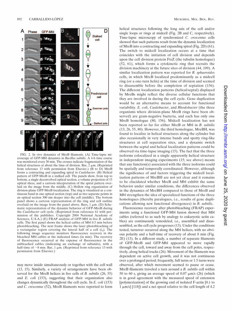
may move inside simultaneously or together with the cell wall(13, 15). Similarly, a variety of arrangements have been ob-served for the MreB helices in live cells of B. subtilis (26, 55)and E. coli (153), suggesting that their organization alsochanges dynamically throughout the cell cycle. In E. coli (153)and C. crescentus (52), MreB filaments were reported to form
helical structures following the long axis of the cell and/orsingle loops or rings at midcell (Fig. 2B and C, respectively).Time-lapse microscopy of synchronized C. crescentus cellsshowed that such patterns result from the dynamic localizationof MreB into a contracting and expanding spiral (Fig. 2D) (61).The switch to midcell localization occurs at a time thatcoincides with the initiation of cell division and dependsupon the cell division protein FtsZ (the tubulin homologue)(52, 61), which forms a cytokinetic ring that recruits thedivision machinery at the future sites of division (44, 109). Asimilar localization pattern was reported for R. sphaeroidescells, in which MreB localized predominantly as a midcellring (or a one-turn helix) at the time of division and seemedto disassemble before the completion of septation (156).The different localization patterns (helical/septal) displayedby MreBs might reflect the diverse cellular functions thatthey are involved in during the cell cycle. Gene duplicationwould be an alternative means to account for functionalvariability. E. coli, Caulobacter, and Rhodobacter (the threeorganisms where division-plane MreB rings have been ob-served) are gram-negative bacteria, and each has only oneMreB homologue (80, 156). Midcell localization has notbeen reported so far for either MreB or Mbl in B. subtilis(13, 26, 55, 80). However, the third homologue, MreBH, wasfound to localize in helical structures along the cylinder butalso occasionally in very intense bands and spotty ring-likestructures at cell separation sites, and a dynamic switchbetween the septal and helical localization patterns could beobserved via time-lapse imaging (15). The fact that the threeisoforms colocalized in a single apparently helical structurein independent imaging experiments (15; see above) meansthat any function(s) associated with the three isoforms couldbe spatially and temporally coordinated. However, althoughthe significance of and factors triggering the midcell local-ization patterns of MreBH are not yet clear and it remainsto be elucidated whether MreB and Mbl exhibit the samebehavior under similar conditions, the differences observedin the dynamics of MreBH compared to those of MreB andMbl strengthen the idea of specialized functions of the threehomologues (thereby paralogues, i.e., results of gene dupli-cations allowing new functional divergences) in B. subtilis.
Fluorescence recovery after photobleaching (FRAP) exper-iments using a functional GFP-Mbl fusion showed that Mblcables (referred to as such by analogy to eukaryotic actin ca-bles) are continuously remodeled, i.e., assembled and disas-sembled, as the cell cycle progresses (13). Under the conditionstested, turnover occurred along the Mbl helices, with no obvi-ous polarity and a half-time of recovery of about 8 min (Fig.2E) (13). In a different study, a number of separate filamentsof GFP-MreB and GFP-Mbl appeared to move rapidlythrough the cell, toward and away from the cell poles, respec-tively, along helical tracks (26). Movement of the filaments wasdependent on active cell growth, and it was not continuousover a prolonged period; frequently, full turns or 1.5 turns wereobserved, after which movement seemed to pause or cease.MreB filaments traveled a turn around a B. subtilis cell within50 to 60 s, giving an average speed of 0.07 �m/s (26) (whichis in good agreement with the measured speed of extension[polymerization] of the growing end of isolated F-actin [0.1 to1 �m/s] [118]) and a net speed relative to the cell length of 4.2
FIG. 2. In vivo dynamics of MreB filaments. (A) Time-lapse mi-croscopy of GFP-Mbl dynamics in Bacillus subtilis. A 4-h time coursewas monitored every 30 min. The crosses indicate fragmentation of thehelical structures at about the time of division. Bar, 2 �m. (Reprintedfrom reference 13 with permission from Elsevier.) (B to D) MreBforms a contracting and expanding spiral in Caulobacter. (B) Helicalpattern of GFP-MreB in a stalked cell. The panels show, from top tobottom, a single deconvolved optical section, a volume projection of 15optical slices, and a cartoon interpretation of the spiral pattern over-laid on the image from the middle. (C) Hollow ring organization ofdivision-plane GFP-MreB localization. The ring is visualized as a con-tinuous band in one optical section (top) and as two separate points inan optical section 300 nm deeper into the cell (middle). The bottompanel shows a cartoon representation of the ring and cell outlineoverlaid on the image from the panel above. Bars, 1 �m. (D) Sche-matic representation of the dynamic behavior of GFP-MreB duringthe Caulobacter cell cycle. (Reprinted from reference 61 with per-mission of the publisher. Copyright 2004 National Academy ofSciences, U.S.A.) (E) FRAP analysis of GFP-Mbl in live B. subtiliscells. The first panel shows the helical cables of Mbl 0.5 min beforephotobleaching. The next frame shows the laser photobleaching ofa rectangular region covering the lateral half of a cell (t0). Thefollowing image sequence monitors fluorescence recovery in thebleached Mbl cables at the indicated times (in min). The recoveryof fluorescence occurred at the expense of fluorescence in theunbleached cables (indicating an exchange of subunits), with ahalf-time of �8 min. Bar, 1 �m. (Reprinted from reference 13 withpermission from Elsevier.)
892 CARBALLIDO-LOPEZ MICROBIOL. MOL. BIOL. REV.
on June 25, 2020 by guesthttp://m
mbr.asm
.org/D
ownloaded from
nm/s (0.24 �m/min) (26). The general helical movement andthe speed of migration of the Mbl filaments were similar,although their general direction seemed to be opposite that ofMreB filaments. It was concluded that a potential poleward orcenterward pushing velocity of 0.24 �m/min was generated byMreB or Mbl, respectively, possibly through a treadmillingmechanism (26). The treadmilling behavior of MreB fila-ments in vivo was indeed confirmed recently, using quanti-tative imaging of single molecules of fluorescent MreB-yel-low fluorescent protein fusions in living Caulobacter cells(82). In this study, both polymerized MreB (filamentousMreB [fMreB]) and unpolymerized MreB (globular MreB[gMreB]) populations could be distinguished and wereshown to display different dynamics: gMreB moved rapidlyin a random fashion, whereas fMreB displayed slow, di-rected motion. The fast motion of unpolymerized monomers(gMreB) had a rate of diffusion that was restricted com-pared to that of cytoplasmic proteins but that appearedsimilar to that of membrane-bound proteins, suggesting thatgMreB may associate with an additional factor(s), possibly(in) the cytoplasmic membrane (82). By analyzing the rate,distance, and direction of labeled MreB in the polymers, itwas shown that the slow directional movement of fMreB didnot result from whole-filament translocation but from tread-milling of the MreB monomers through short MreB fila-ments with fixed ends, i.e., by preferential polymerization atone end and depolymerization at the other end of a filament(82). Therefore, MreB, like F-actin (17), exhibits treadmill-ing behavior in vivo and thus assembles in a polarizedmanner. From these treadmilling observations, it was alsoextracted that the steady-state rate of MreB monomer ad-dition was 1.2 s�1, that the average MreB filament lengthwas �400 nm (much shorter than the cell length of 3.5 �m),and that the polarized assembly of individual MreB fila-ments was random relative to the overall cell polarity (82).
Ultrastructural Organization of MreB Filaments In Vivo
The important implications of the dynamic behavior de-scribed above are still hampered by our lack of knowledge ofthe ultrastructure of the high-order filamentous forms dis-played by the MreB-like proteins and of the mechanisms thatregulate their dynamics in vivo. Nevertheless, in view of (i) theturnover detected by FRAP experiments (13), (ii) the fluores-cence intensity and low background of inducible GFP fusions(13, 15, 26), (iii) the average MreB filament length (signifi-cantly shorter than the overall MreB helices) (82), and (iv) therecent biochemical data on MreB assembly (ATP/GTP hydro-lysis, no or little filament end-to-end annealing, and filamen-tous bundles observed by EM) (47, 48), it is likely that in vivothe helical structures are composed of, and exchange subunitsin the form of, lateral bundles of protofilaments (rather thanmonomers). According to this model, the dynamics of MreBhelical cables involve the exchange of short polarized proto-filaments (which assemble randomly, i.e., with no uniformglobal polarity) to the sides of a multistranded polarized struc-ture. Like in the case of eukaryotic actin, the mechanism maybe controlled by both nucleotide binding and hydrolysis. Thestructural basis for the colocalization of the three MreB iso-forms of B. subtilis is still unknown, but it seems likely that they
form a triplex helical structure composed of some kind ofmixed heteropolymeric bundles (15). It still remains to beelucidated whether this model is correct and whether directinteractions between the MreB protofilaments or cross-linkingthrough accessory proteins occurs to form such bundled struc-tures.
MreB-LIKE PROTEINS AND CELL MORPHOGENESIS
Prokaryotic cells display a wide diversity of shapes. In alleukaryotic cells, shape is determined primarily by cytoskeletalstructures, particularly actin filaments. Since such structureswere traditionally thought to be absent from bacteria, thetough external peptidoglycan (PG) cell wall was assumed to bethe primary determinant of bacterial cell shape. Indeed, iso-lated PG sacculi comprise a single huge molecule that retainsthe shape associated with their original cell (69), and con-versely, removal of the wall results in spherical protoplasts (i.e.,loss of shape). Moreover, most genes identified to be involvedin cell shape (morphogenes) were associated with CW biosyn-thesis. For decades, research on cell shape in bacteria focusedon cell wall synthesis and structure (for a recent review, seereference 133). However, the discovery of a morphogeneticactin-like cytoskeleton brought about a radical change in thecontext in which bacterial cell shape is studied.
MreB Filaments Govern Cell Morphogenesis by ActivelyDirecting Lateral Wall Biogenesis
MreB proteins play an essential role in the control of cellmorphogenesis in nonspherical bacteria (see above). It seemedlikely that the MreB cytoskeletal structures would control cellshape by determining CW architecture (13, 80).
Mbl filaments direct lateral wall synthesis in Bacillus subti-lis. A range of studies with rod-shaped bacteria suggested thatPG insertion occurs at the nascent septum and randomly allover the surface of the lateral wall during growth (10, 27, 30,113, 117). However, the sensitivity and resolution of the meth-ods used in these studies were limited: they did not provideadequate spatial resolution to determine the underlying pat-terns of PG incorporation. A more sensitive, high-resolutionprobe for nascent PG insertion in nonfixed cells was recentlydeveloped by Daniel and Errington (22). These authors used afluorescent derivative of the antibiotic vancomycin (a cell wallsynthesis inhibitor that binds specifically to PG intermediates)to label nascent PG in gram-positive bacteria by fluorescencemicroscopy (note that gram-negative bacteria do not stain be-cause their outer membrane presents a permeability barrier tovancomycin) (169). This novel staining method revealed that,at least in B. subtilis, synthesis of the wall occurs in a helicalpattern over the cylindrical part of the cell and also specificallyat the septum in dividing cells (Fig. 3A). The lateral helicalpattern of fluorescein-labeled vancomycin (Van-FL) was rem-iniscent of the helical localization of MreB and Mbl, both ofwhich are required for cell shape determination in B. subtilis(1, 80). Strikingly, the helical staining was abolished in a strainlacking Mbl (Fig. 3C) and not in a strain lacking MreB (Fig.3D) (22, 55). The septal insertion was dependent on cell divi-sion (FtsZ), as expected (Fig. 3E) (22). Recently, in a similarindependent study that used fluorescent derivatives of vanco-
VOL. 70, 2006 BACTERIAL ACTINS 893
on June 25, 2020 by guesthttp://m
mbr.asm
.org/D
ownloaded from

mycin and ramoplanin (another PG-binding antibiotic), inser-tion of nascent PG along the lateral wall of B. subtilis wasconfirmed to be helical (160). However, in this study, a sidewallhelical staining pattern qualitatively similar to that observed inwild-type cells (although less regular) was observed in cellslacking Mbl (160); the authors of that study concluded thatMbl plays an indirect role in directing PG synthesis but that itis not essential for the incorporation of sidewall PG.
These staining methods cannot be applied to gram-negativebacteria, but it is interesting that murein deposition patterns inthe sacculi of growing E. coli cells (31) (by the incorporation ofD-cysteine [D-Cys] [30]) also suggest a helical pattern of PGinsertion into the lateral CW, although this remains to beconfirmed. In contrast, Van-FL staining of two spherical bac-teria (which do not possess MreB homologues) (Streptococcuspneumoniae [22] and Staphylococcus aureus [130]) and of arod-shaped bacterium with no MreB/Mbl system (Corynebac-terium glutamicum [22]) was highly specific for the divisionseptum (and its derived poles in C. glutamicum cells) and
absent in the lateral walls, indicating that PG insertion in theseorganisms occurs exclusively in zones specified by the FtsZ-dependent division machinery.
Taken together, these findings have several important im-plications. First, insertion of PG along the cylindrical walls ofB. subtilis (and possibly E. coli) cells occurs in a helical pattern.Second, there are two spatially specialized systems for PGsynthesis in B. subtilis (and probably in all rod-shaped bacteriawith an MreB system): dispersed helical insertion of PGthroughout the lateral wall during growth results in rapid cy-lindrical extension (elongation), and cell division-directed PGsynthesis allows septum formation (division) (Fig. 3B). Third,Mbl may be the MreB homologue required mainly, or exclu-sively, for lateral (helical) wall biosynthesis. This has raisedinteresting questions about the role of MreB in B. subtilismorphogenesis. MreB is essential under normal growth con-ditions and has an important role in the control of cell width(55, 80). Consistent with a role in CW integrity, the lethal mreBmutant phenotype could be ameliorated by high concentra-tions of magnesium (Mg2�) (55), like the phenotypes of sev-eral B. subtilis mutants thought to be required for differentaspects of PG synthesis (94, 95, 121, 138, 139; see below). Themechanism by which Mg2� is able to rescue the phenotypes ofthese mutants is currently unknown. It also remains unclearhow MreB controls cell width, but it might influence the syn-thesis or structure of the cylindrical and/or septal CW. Van-FLstaining showed that MreB was not primarily required forlateral PG synthesis (22, 55; see above), although a redundantrole in this process cannot be excluded. Alternatively, MreBcould be required for insertion of teichoic acids or of autolysinsinto the CW, but all of these hypotheses remain to be tested. Ithas been suggested that MreB might act either continuously torestrain the diameter during elongation or discontinuously toreset the correct diameter when the cell divides (55). Finally, itis exciting to mention that a new, distinct role in cell wallmorphogenesis has recently been uncovered for MreBH, thethird MreB isologue in B. subtilis (15; see below).
PBP localization and the MreB cytoskeleton. The Van-FLfindings provided strong support for the view that at least thecables of Mbl direct the synthesis of lateral PG in a spatiallycontrolled manner. Conceivably, this might involve the local-ization of PG-synthesizing enzymes, named penicillin-bindingproteins (PBPs), that incorporate the PG precursors into thegrowing CW sacculus. Two factors (or rather a combination ofthem) are thought to be critical for PBP localization, and theyare protein-protein interactions and substrate recognition(147). The PG-synthesizing septal machinery (i.e., septal PBPs)has been shown to be recruited by the FtsZ ring (44, 109).However, no factors for targeting of PBPs to the lateral wallhave yet been identified. Hence, the helical MreB scaffoldingstructures could direct PBP localization either by providing asubstrate(s) that can be recognized by elongation-specific PBPsor by providing a track for protein-protein interactions thattarget PBPs to their site(s) of action (see below).
A putative candidate for MreB-directed targeting is theproduct of pbpA, PBP2, a high-molecular-weight PBP thatdisplays transpeptidase activity (i.e., catalyzes PG cross-link-ing) and has classically been associated with sidewall synthesisduring elongation (158). In E. coli, a functional GFP-PBP2fusion localized preferentially in a spot-like pattern over the
FIG. 3. Control of lateral wall synthesis by MreB-like proteins.(A) Van-FL staining of nascent cell wall synthesis in wild-type cells ofBacillus subtilis. A compilation of images of typical cells during the cellcycle progression is shown from left to right. (B) Model for separatesystems for septal and cylindrical PG synthesis in B. subtilis. Gray linesand ovals show the sites of PG insertion during elongation and divi-sion, respectively. Arrows show the directions of elongation or divisiondriven by cell wall biosynthesis. (C to E) Effects of �mbl null mutation(C), �mreB null mutation (D), and FtsZ depletion (E) on Van-FLstaining. (Panels A, B, C, and E were reprinted from reference 22 withpermission from Elsevier. Panel D was reprinted from reference 55with permission from Blackwell Publishing.) (F) Schematic represen-tation of interactions within the MreBCD complex in E. coli and thepossible interaction of MreBCD with periplasmic proteins involved inpeptidoglycan synthesis (PBP2 and RodA). (Reprinted from reference88 with permission from Blackwell Publishing.)
894 CARBALLIDO-LOPEZ MICROBIOL. MOL. BIOL. REV.
on June 25, 2020 by guesthttp://m
mbr.asm
.org/D
ownloaded from
cylindrical part of the envelope, and also at midcell during celldivision (28). The localization of GFP-PBP2 over the lateralwall was suggestive of a helical pattern strikingly similar to thatof MreB. Consistent with this, PBP2 formed a banding patternreminiscent of that formed by MreB filaments in C. crescentuscells, as shown by both IFM (35, 52) and the use of a GFP-PBP2 fusion (40). Since the distinct banding pattern of PBP2was lost (although PBP2 foci were still present) in C. crescentuscells that had been depleted of MreB for 10 h, it was originallysuggested that PBP2 localization was dependent on MreB (52).Under such conditions, the aberrant localization pattern ob-served could indeed be attributed to the lack of MreB or,alternatively, to a secondary effect resulting from the severeshape defects resulting from the long-term absence of MreB.The latter hypothesis was supported by a recent IFM study,where rapid disruption of the MreB filaments by treatmentwith A22 did not affect the helical pattern of PBP2 (35). A22is a small molecule that specifically and rapidly (�1 min) de-localizes MreB in Caulobacter cells, allowing the effects of theabsence of MreB helices to be assessed prior to observabledeformations in shape (62; see below for more details). How-ever, in an independent study, GFP-PBP2 was reported tomislocalize to the division plane upon A22 treatment (40).Interestingly, only newly synthesized GFP-PBP2 seemed torelocalize to the septum, and thus it was suggested that (MreB-dependent) helical localization of PBP2 was regulated at thelevel of insertion and that established helical PBP2 patternsare stable in the absence of MreB (35). Although this findingmay explain the differences between the two reports, whetherPBP2 localization depends on MreB in C. crescentus remains tobe unambiguously demonstrated. (Indeed, in the GFP-PBP2study [40], cells were treated with 10 �g/ml A22 for 6 h,whereas in the IFM study [35] cells were treated with 50 �g/mlA22 for 2 h. After 2 h of A22 treatment at 50 �g/ml, growth isarrested but shape defects are not yet visible [62], and thus thepreviously inserted PBP2 patterns could be stable. On theother hand, C. crescentus cells grown in 10 �g/ml A22 for 6 hkeep growing [albeit slowly] but already display significant cellshape deformations [62], which could indirectly affect PBP2localization.)
IFM studies of R. sphaeroides cells showed that MreB colo-calized with PBP2 in a cell cycle-dependent manner (155).PBP2 localized in partial rings (which presumably representunresolved helices) at the middle of elongating cells and at theone-quarter and three-quarter positions in septating cells.MreB colocalized with PBP2 during elongation only; duringseptation, MreB remained at the septation site, whereas PBP2relocalized to the midcell sites of the forming daughter cells. Itwas concluded that MreB and PBP2 interact during elongationto synthesize PG at or near midcell but are involved in differentcellular roles during septation (155). The possible dependenceof PBP2 localization on MreB in R. sphaeroides (and in E. coli)remains to be tested, and this point needs to be resolved for C.crescentus (see above). Work with B. subtilis showed that somePBPs fused to GFP also localize specifically to the sidewall indistinct foci and bands around the cell periphery, again remi-niscent of the helical distribution of MreB and Mbl (146). Thisfurther suggested that PG synthesis occurs at distinct regionsof the lateral wall and that new PG is inserted in a helicalmanner during the elongation-specific phase. In this study,
however, the patterns of GFP-PBP localization were not de-tectably altered in the absence of either MreB or Mbl (146).Nevertheless, in light of the recent finding that MreB, Mbl, andMreBH colocalize in a single helical structure in B. subtilis(15), it remains plausible that PBP localization depends onmore than one MreB isoform in this organism. The link be-tween the MreB cytoskeleton and the cell wall synthesis ma-chinery undoubtedly remains a major challenge for futurework.
MreBH filaments direct lateral wall hydrolysis in Bacillussubtilis. It was recently shown that MreBH also has an impor-tant role in B. subtilis cell morphogenesis and that this functionis effected, at least in part, by controlling the autolytic activityover the lateral wall by direct interaction with LytE, a cell wallhydrolase (15). Depletion of MreBH led to a mild cell shapedefect, in contrast to the case for MreB- and Mbl-depletedcells (24). Similarly, under normal growth conditions, mreBHmutant cells displayed a mild phenotype; they were slightlyaffected in length and width and were bent at points thatappeared correlated with abnormal thickening of the CW (15).However, all aspects of the mreBH mutant phenotype werestrongly affected by the Mg2� concentration, and mreBH mu-tants required higher levels of Mg2� for viability than did thewild type (15). A similar dependence on Mg2� concentrationhas been observed for mreB (55; see above), mbl (15), andmreCD (95; see below) mutant cells and for several other B.subtilis mutants thought to be affected in different aspects ofCW synthesis and structure, as mentioned before (94, 121, 138,139). Strikingly, MreBH was found to specifically interact withLytE (previously thought to be a cell septum-specific autolysin[72, 127]) in a genome-wide two-hybrid screen, and the Mg2�
dependence and shape defetcts of lytE mutants appeared re-markably similar to those of mreBH mutants (15). MreBH, likeMbl and MreB, forms dynamic helical filaments (see above andFig. 1F) (15, 26). A functional LytE-GFP fusion localized toongoing division sites and to cell separation sites, as previouslyobserved by IFM (178), but surprisingly, it also localized aspunctate fluorescence over the external lateral wall (15). Tar-geting of LytE to the sidewall of the cell was dependent onMreBH (and not on MreB or Mbl), while targeting of LytE tothe sites of division was dependent on early (FtsZ) and late(PBP 2B) division proteins (15).
On the basis of the similar phenotypes, the direct protein-protein interaction in vivo and in vitro, and the MreBH-de-pendent localization of LytE along the lateral wall, it wasconcluded that MreBH influences lateral wall maturation bydirecting the localization of LytE (15). In light of these find-ings, together with the colocalization of all three MreB iso-forms, an MreBH-dependent helical mode of cell wall hydro-lysis that is coordinated with an Mbl-dependent helical modeof cell wall insertion has been suggested to control elongationof the rod-shaped B. subtilis cells during growth (15).
In summary, it is currently believed that the MreB helicalstructures are spatial regulators of cell wall biogenesis. HowMreB-like proteins direct the insertion and maturation of CWmaterial remains to be elucidated, but the current model is thatthey target/position cell wall enzymes (synthases and hydro-lases) and/or membrane-associated and extracellular proteins,such as MreC and MreD (see below), that are involved inmorphogenesis. It is interesting that the same genomic ar-
VOL. 70, 2006 BACTERIAL ACTINS 895
on June 25, 2020 by guesthttp://m
mbr.asm
.org/D
ownloaded from
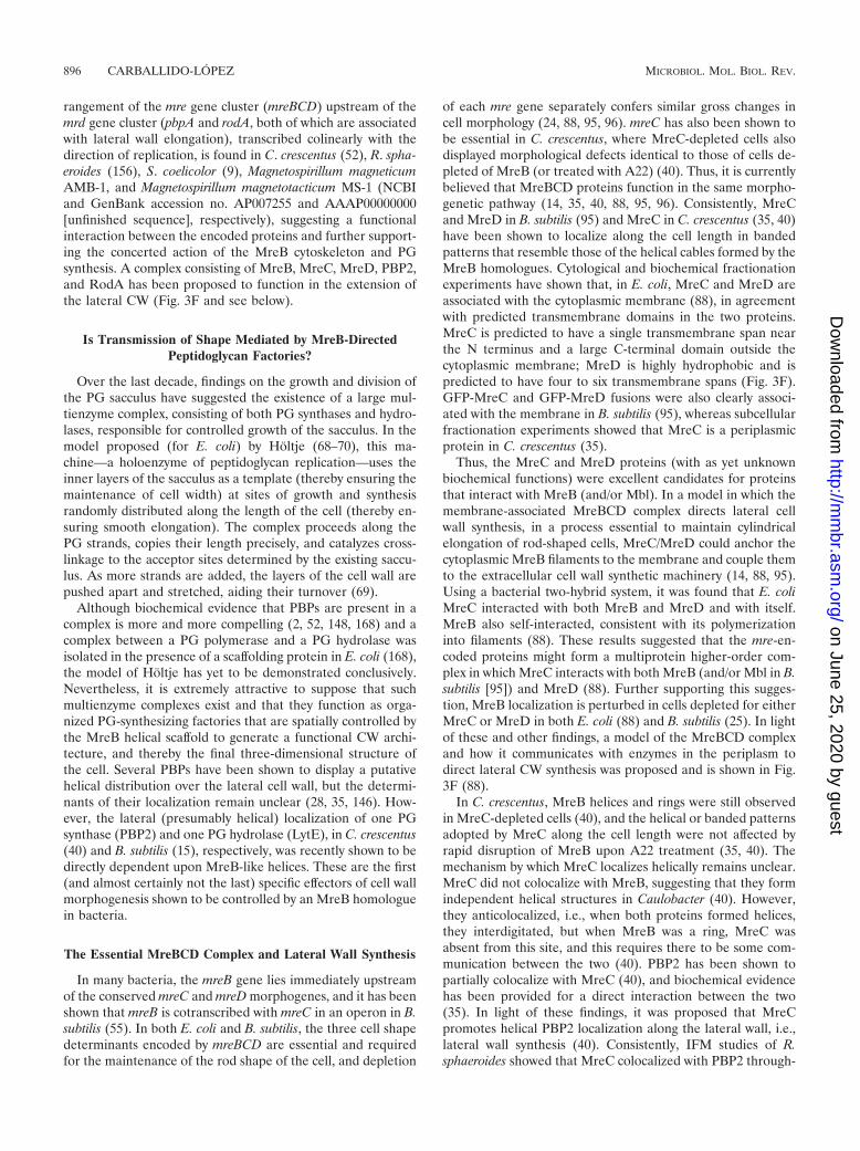
rangement of the mre gene cluster (mreBCD) upstream of themrd gene cluster (pbpA and rodA, both of which are associatedwith lateral wall elongation), transcribed colinearly with thedirection of replication, is found in C. crescentus (52), R. spha-eroides (156), S. coelicolor (9), Magnetospirillum magneticumAMB-1, and Magnetospirillum magnetotacticum MS-1 (NCBIand GenBank accession no. AP007255 and AAAP00000000[unfinished sequence], respectively), suggesting a functionalinteraction between the encoded proteins and further support-ing the concerted action of the MreB cytoskeleton and PGsynthesis. A complex consisting of MreB, MreC, MreD, PBP2,and RodA has been proposed to function in the extension ofthe lateral CW (Fig. 3F and see below).
Is Transmission of Shape Mediated by MreB-DirectedPeptidoglycan Factories?
Over the last decade, findings on the growth and division ofthe PG sacculus have suggested the existence of a large mul-tienzyme complex, consisting of both PG synthases and hydro-lases, responsible for controlled growth of the sacculus. In themodel proposed (for E. coli) by Holtje (68–70), this ma-chine—a holoenzyme of peptidoglycan replication—uses theinner layers of the sacculus as a template (thereby ensuring themaintenance of cell width) at sites of growth and synthesisrandomly distributed along the length of the cell (thereby en-suring smooth elongation). The complex proceeds along thePG strands, copies their length precisely, and catalyzes cross-linkage to the acceptor sites determined by the existing saccu-lus. As more strands are added, the layers of the cell wall arepushed apart and stretched, aiding their turnover (69).
Although biochemical evidence that PBPs are present in acomplex is more and more compelling (2, 52, 148, 168) and acomplex between a PG polymerase and a PG hydrolase wasisolated in the presence of a scaffolding protein in E. coli (168),the model of Holtje has yet to be demonstrated conclusively.Nevertheless, it is extremely attractive to suppose that suchmultienzyme complexes exist and that they function as orga-nized PG-synthesizing factories that are spatially controlled bythe MreB helical scaffold to generate a functional CW archi-tecture, and thereby the final three-dimensional structure ofthe cell. Several PBPs have been shown to display a putativehelical distribution over the lateral cell wall, but the determi-nants of their localization remain unclear (28, 35, 146). How-ever, the lateral (presumably helical) localization of one PGsynthase (PBP2) and one PG hydrolase (LytE), in C. crescentus(40) and B. subtilis (15), respectively, was recently shown to bedirectly dependent upon MreB-like helices. These are the first(and almost certainly not the last) specific effectors of cell wallmorphogenesis shown to be controlled by an MreB homologuein bacteria.
The Essential MreBCD Complex and Lateral Wall Synthesis
In many bacteria, the mreB gene lies immediately upstreamof the conserved mreC and mreD morphogenes, and it has beenshown that mreB is cotranscribed with mreC in an operon in B.subtilis (55). In both E. coli and B. subtilis, the three cell shapedeterminants encoded by mreBCD are essential and requiredfor the maintenance of the rod shape of the cell, and depletion
of each mre gene separately confers similar gross changes incell morphology (24, 88, 95, 96). mreC has also been shown tobe essential in C. crescentus, where MreC-depleted cells alsodisplayed morphological defects identical to those of cells de-pleted of MreB (or treated with A22) (40). Thus, it is currentlybelieved that MreBCD proteins function in the same morpho-genetic pathway (14, 35, 40, 88, 95, 96). Consistently, MreCand MreD in B. subtilis (95) and MreC in C. crescentus (35, 40)have been shown to localize along the cell length in bandedpatterns that resemble those of the helical cables formed by theMreB homologues. Cytological and biochemical fractionationexperiments have shown that, in E. coli, MreC and MreD areassociated with the cytoplasmic membrane (88), in agreementwith predicted transmembrane domains in the two proteins.MreC is predicted to have a single transmembrane span nearthe N terminus and a large C-terminal domain outside thecytoplasmic membrane; MreD is highly hydrophobic and ispredicted to have four to six transmembrane spans (Fig. 3F).GFP-MreC and GFP-MreD fusions were also clearly associ-ated with the membrane in B. subtilis (95), whereas subcellularfractionation experiments showed that MreC is a periplasmicprotein in C. crescentus (35).
Thus, the MreC and MreD proteins (with as yet unknownbiochemical functions) were excellent candidates for proteinsthat interact with MreB (and/or Mbl). In a model in which themembrane-associated MreBCD complex directs lateral cellwall synthesis, in a process essential to maintain cylindricalelongation of rod-shaped cells, MreC/MreD could anchor thecytoplasmic MreB filaments to the membrane and couple themto the extracellular cell wall synthetic machinery (14, 88, 95).Using a bacterial two-hybrid system, it was found that E. coliMreC interacted with both MreB and MreD and with itself.MreB also self-interacted, consistent with its polymerizationinto filaments (88). These results suggested that the mre-en-coded proteins might form a multiprotein higher-order com-plex in which MreC interacts with both MreB (and/or Mbl in B.subtilis [95]) and MreD (88). Further supporting this sugges-tion, MreB localization is perturbed in cells depleted for eitherMreC or MreD in both E. coli (88) and B. subtilis (25). In lightof these and other findings, a model of the MreBCD complexand how it communicates with enzymes in the periplasm todirect lateral CW synthesis was proposed and is shown in Fig.3F (88).
In C. crescentus, MreB helices and rings were still observedin MreC-depleted cells (40), and the helical or banded patternsadopted by MreC along the cell length were not affected byrapid disruption of MreB upon A22 treatment (35, 40). Themechanism by which MreC localizes helically remains unclear.MreC did not colocalize with MreB, suggesting that they formindependent helical structures in Caulobacter (40). However,they anticolocalized, i.e., when both proteins formed helices,they interdigitated, but when MreB was a ring, MreC wasabsent from this site, and this requires there to be some com-munication between the two (40). PBP2 has been shown topartially colocalize with MreC (40), and biochemical evidencehas been provided for a direct interaction between the two(35). In light of these findings, it was proposed that MreCpromotes helical PBP2 localization along the lateral wall, i.e.,lateral wall synthesis (40). Consistently, IFM studies of R.sphaeroides showed that MreC colocalized with PBP2 through-
896 CARBALLIDO-LOPEZ MICROBIOL. MOL. BIOL. REV.
on June 25, 2020 by guesthttp://m
mbr.asm
.org/D
ownloaded from
out the cell cycle (while MreB colocalized with PBP2 only atcertain stages of the cell cycle [see above]), again suggestingthat MreC and PBP2 function in concert in PG synthesis dur-ing elongation (155). Interestingly, several PBPs (includingPBP2 and PBP1) and outer membrane proteins were isolatedfrom C. crescentus cell extracts by affinity chromatography us-ing Sepharose-bound MreC (35). Undoubtedly, elucidation ofthe biochemical functions of the MreC and MreD proteinsremains a major question for future research.
MreB Proteins and Spore Lateral Wall Formationin Actinomycetes
In actinomycetes (Mycobacter, Actinomyces, Nocardia, andStreptomyces spp.), mreB homologues are present (andhighly conserved) only in genera that form aerial myceliaand sporulate (112). Streptomyces spp. are gram-positivefilamentous soil bacteria with a complex developmental cy-cle. This involves a phase of vegetative growth as branchinghyphal filaments followed by the development of aerial hy-phae, which undergo multiple septation and eventually dif-ferentiate into spores (for a review, see reference 54).Van-FL staining confirmed the previous finding that vege-tatively growing S. coelicolor grows specifically by CW ex-tension at the tips of the branching mycelia (22). However,S. coelicolor has two MreB isoforms, namely, MreB and Mbl,whose roles (in a bacterium that does not undergo cylindri-cal wall expansion) appeared particularly intriguing. In arecent study, Mazza et al. (112) addressed the role of MreBin the morphogenesis of S. coelicolor and demonstrated thatMreB is dispensable for vegetative growth but essential forspore formation and assembly of the spore lateral cell wall.Previous attempts to create mreB mutants of S. coelicolorhad been unsuccessful, leading to the suggestion that thegene is essential (9), but Mazza et al. (112) were able togenerate a viable mreB null mutant. Surprisingly, growthand morphology of the substrate mycelium of the mreBmutant were normal, but aerial hyphae were irregularlyshaped, swelled, and lysed, and spores doubled their volumeand lost their cell wall consistence (112). MreB specificallylocalized at the septa and their derived poles in presporechains, and it completely surrounded more mature spores(112). Although a role for MreB in vegetatively growing S.coelicolor cannot be excluded and although MreB and Mblcould display some functional redundancy in this organism,it was concluded that MreB primarily affects lateral wallformation, but exclusively during the sporulation process.The lateral CW of streptomycetes thickens and undergoesstructural changes during spore maturation (54). Thus, thePG biosynthetic activity may be reorganized in sporogenicaerial hyphae, and MreB might play a key role in thisprocess. Sporulating bacteria activate separate programsof gene expression and undergo different morphologicalchanges during vegetative growth and during sporulation.However, spatial and temporal control of cell morphogen-esis is required during the two independent developmentalcycles. The work by Mazza et al. (112) suggests that theprimary role of MreB in directing lateral CW formationmight be conserved during sporulation, at least in Strepto-myces. It will be interesting to see if MreBs are involved in
spore formation in other bacteria. Interestingly, in B. subti-lis, mbl lies immediately downstream of the sporulation genespoIIID. It has been reported that mbl is constitutively tran-scribed from its own promoter during vegetative growth butalso from a promoter located upstream of spoIIID and con-trolled by the sporulation-specific transcription factor �E
(43, 51). Whether MreBs play a role in sporulation in addi-tion to their roles during vegetative growth is an importantquestion for future work.
MreB Proteins and Cell Shape Determinationin Wall-Less Prokaryotes
As mentioned above, the cell wall is the major structure thatdefines the shape of most bacterial cells, and MreB proteinscontrol cell morphogenesis by actively directing the synthesisand maturation of PG. However, not all prokaryotic cells pos-sess a cell wall. Mollicutes (Acholeplasma, Mycoplasma, andSpiroplasma spp.) are enveloped only by a cholesterol-contain-ing cell membrane, and even so (despite lacking cell walls),these cells display different morphologies. How is shape deter-mined in these organisms? Interestingly, cells of Spiroplasmacitri encode five MreB homologues, and MreB has been de-tected in cells of Spiroplasma melliferum, too (91). Are MreBproteins also involved in the morphogenesis of the wall-lessSpiroplasma cells? Structurally, Spiroplasma cells are unique inhaving a well-defined, dynamic, helical cell geometry (Fig. 4A)and a membrane-bound, cytoskeleton-like ribbon, which fol-lows the inner, shortest helical path of the cell (Fig. 4B) (163).The cytoskeleton from Spiroplasma was isolated a long timeago, and it was thought to consist of a single protein, the55-kDa fibril protein, currently exclusive to Spiroplasma (162).Recently, Kurner et al. (91) used cryo-electron tomography tostudy the 3D structure of the S. melliferum cytoskeleton in aclose-to-native state. Tomograms showed that this cytoskele-ton possesses two types of filaments (thick and thin) arrangedin three parallel ribbons that are anchored to each other and tothe cell membrane and that span the length of the cell (Fig. 4Ato E). The two outer ribbons consist of thick filaments of fibril,as expected. The inner ribbon appears to be composed of ninethin filaments with a spacing of �4 nm (the same width as thatreported for T. maritima MreB protofilaments [164]). Kurneret al. suggested that these were composed of MreB, and theyassumed that MreB filaments give the Spiroplasma cell a rod-like shape. Although no proof that these ribbons are composedof MreB protein or that MreB is involved in cell shape deter-mination in Spiroplasma was provided, the approach is ex-tremely challenging and promises a bright future for the inves-tigation of the ultrastructure and spatial organization of thecytoskeleton in both prokaryotic and eukaryotic cells.
ACTIN-LIKE PROTEINS AND DNA SEGREGATION
Evidence has emerged during the last few years for both aforce-generating mechanism of chromosome segregation inbacteria and a centromere-like locus in the bacterial chromo-some (63, 79, 97–99, 145). It has been shown that the complexof replication proteins (the replisome) is more or less station-ary at midcell throughout most of the cell cycle (99), whereasthe origin of replication, as a first step (followed by other
VOL. 70, 2006 BACTERIAL ACTINS 897
on June 25, 2020 by guesthttp://m
mbr.asm
.org/D
ownloaded from

duplicated regions), rapidly moves toward opposite cell poles(177). Several genes involved in chromosome and plasmid seg-regation and their subcellular localization have been identified,but the mechanisms underlying prokaryotic DNA movement
and positioning remained unknown. By analogy to the cy-toskeleton-based eukaryotic spindle apparatus (83), the dis-covery of cytoskeletal filaments in bacteria made them goodcandidates for orchestrating chromosome segregation. Thebacterial tubulin homologue FtsZ was an unlikely candidate;ftsZ mutants do not display segregation defects (109), and thelocalization of FtsZ at midcell is inconsistent with a role insegregating sister chromosomes toward opposite poles. How-ever, an alternative and attractive possibility was put forwardwith the identification of bacterial actin homologues that formfilamentous structures running the length of the cell.
The Actin-Like ParM Protein and Plasmid-DNA Segregation
The first evidence of actin-like filament-forming proteinsrequired for DNA segregation was obtained from the parti-tioning system of the low-copy-number plasmid R1 of E. coli. TheR1 partitioning locus (par) carries or encodes three elements,namely, parC, ParR, and ParM, which together form a nucleo-protein complex that secures the faithful distribution of plas-mid copies to the daughter cells before cell division (for areview, see reference 58). Two plasmid molecules are pairedby the binding of ParR to 10 direct repeats of parC, acis-acting centromere-like DNA sequence (78). ParM (par-titioning motor) is an ATPase that interacts with the ParR-parC complex (77).
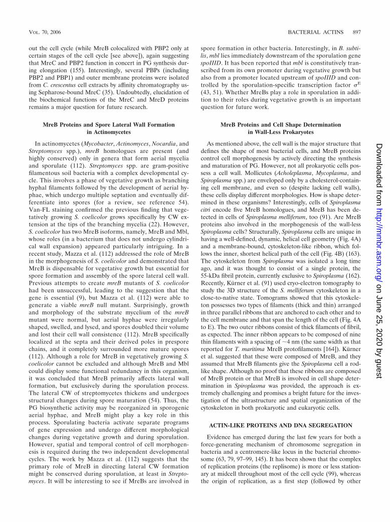
ParM. Immunofluorescence microscopy revealed that ParMformed highly dynamic filaments along the longitudinal axis ofthe cell (Fig. 5A), which were essential for the DNA partition-ing process and displayed the properties expected for a force-generating or -directing cytoskeletal element (120). ParM (alsocalled StbA) was one of the four bacterial proteins (togetherwith Hsp70, FtsA, and MreB) reported to belong to the actinsuperfamily by Bork et al. (7; see above). Like the case forMreB, structural homology was uncovered behind the func-tional homology, and ParM was shown to have an atomicstructure closely related to that of eukaryotic actin (and MreB)(Fig. 5C) and to undergo ATP-dependent polymerization/de-polymerization into double-helical filaments similar to actinfilaments (Fig. 5B) (helical repeat sizes are as follows: forF-actin, 55 Å; for MreB, 51 Å; and for ParM, 49 Å) (165).Importantly, the structure of ParM was solved in two states,i.e., in the absence of nucleotide and with a bound nucleotide(ADP) (165). The transition between these two states (un-bound and ADP bound, called closed and open conformations,respectively) was shown to involve a conformational change inwhich the two major domains (I and II) undergo a rigid twist of�25° with respect to each other upon nucleotide binding. Thisfinding was extremely significant because actin (and any actin-like molecule) subunits are expected to undergo a similar do-main rotation upon nucleotide binding and/or hydrolysis, butso far only the closed state (relative to the ParM conforma-tions) of G-actin (and MreB) has been trapped in crystals andobserved at a high resolution.
Model for in vivo function of ParM filaments. Cytologicaland biochemical studies revealed that segregating plasmidslocalize to the ends of the dynamic ParM filaments and thatParM interacts specifically with the plasmid DNA-bound pro-tein ParR in an ATP-dependent manner (119). It was pro-posed that ParM nucleates via the interaction with the parC-
FIG. 4. Cytoskeleton structures organized by actin-like proteins inSpiroplasma and Magnetospirillum cells. (A to E) Cryo-electron tomog-raphy reveals the cytoskeleton structure of Spiroplasma melliferum.(A) Volume-rendering representation of a whole cell of S. melliferumwith a tomogram slice cutting through it longitudinally. (B) 3D simu-lation of an S. melliferum cell indicating the localization of the threeparallel ribbons of the cytoskeleton (red, purple, and green), which areanchored to the cell membrane (blue) and span the length of the cell.(Panels A and B were reprinted from reference 92 with permissionfrom Elsevier.) (C to E) Superimposed slices of a tomogram (z sectionsare indicated by a bounding box) (C) and two corresponding 3Dvisualizations (D and E) of part of an S. melliferum cell showing thearrangement of the three ribbons. (C) The cytoskeleton comprises twoouter ribbons of thicker filaments and a region of thinner filamentssandwiched between them. (D) Simplified 3D representation of thefilament ribbons (green, purple, and red) that wind in parallel helicallyaround the cell just underneath the cell membrane (blue). (E) Ideal-ized visualization of the filaments, with a smooth transition to theoriginal data (yellow). (Reprinted from reference 91 with permissionof the publisher. Copyright 2005 American Association for the Ad-vancement of Science.) (F to I) MamK, a homologue of MreB, orga-nizes the magnetosome chain and forms filaments in vivo in Magneto-spirillum magneticum sp. strain AMB-1. (F and G) 3D reconstructionsof a wild-type AMB-1 cell (F) and a �mamK mutant cell (G). Thecell membrane (gray), magnetosome membrane (yellow), magnetite(orange), and magnetosome-associated filaments (green) are ren-dered. (H) MamK fused to GFP (green) forms filaments that localizeto the inner curvature of the cell (cell membrane is stained red withFM4-64). (I) Phylogenetic relationship between MamK and other bac-terial actins, demonstrated by an unrooted tree. These proteins sepa-rate into three distinct groups, i.e., MamK (green), ParM/StbA (red),and MreB (blue). (Reprinted from reference 84 with permission of thepublisher. Copyright 2006 American Association for the Advancementof Science.)
898 CARBALLIDO-LOPEZ MICROBIOL. MOL. BIOL. REV.
on June 25, 2020 by guesthttp://m
mbr.asm
.org/D
ownloaded from

ParR complex at midcell and that the subsequent filamentpolymerization provides the mechanical force to propel theassociated molecules apart before cell division occurs (120).No other factors are known to be required for the partitioningprocess, making the par system the simplest and best-under-stood mechanism for DNA segregation in bacteria. Recently,Garner et al. (57) used total internal reflection fluorescencemicroscopy and fluorescence resonance energy transfer to pro-vide spectacular evidence that the actin-like ParM filamentsdisplay dynamic instability and bidirectional polymerization(rather than polarized polymerization, like in F-actin [131, 132]and MreB [82; see above]). Dynamic instability (i.e., cata-strophic decay) has usually been associated with microtubules(33) rather than with actin filaments and consists of periods ofsteady polymerization (elongation) followed by rapid disas-sembly (catastrophe) which are regulated by nucleotide hydro-lysis. Interestingly, regulation of ParM filaments by dynamicinstability appears to be an important component of the plas-mid-DNA segregation process. These striking findings led tothe current model, which is shown in Fig. 5D. According to thismodel, ParM spontaneously nucleates and polymerizes in thecell, and the filaments spontaneously depolymerize unless theyare stabilized by interaction with ParR-parC-paired plasmids.Only ParM filaments with plasmid bound at both ends arestabilized against catastrophic disassembly, and bidirectionalpolymerization at the ParM-ParR interface drives plasmid seg-regation (57). Such an insertional polymerization merchanismhas been proposed for elongating MT ends attached to kinet-ochores (8, 71, 116) and for various examples of actin-basedmotility (21, 34, 90, 128). Also, as mentioned above, MTs, butnot F-actin, display dynamic instability. Thus, the function ofthe par system actually seems to be the direct equivalent ofmitosis in eukaryotes. Do the bacterial actin-like ParM fila-ments use a force-generating strategy similar to that of theeukaryotic microtubule-based mitotic spindle? This apparentinversion of function is also extended to FtsZ, the tubulinhomologue, which drives cytokinesis in bacteria, in a reversalof the actin-based contractile ring in eukaryotic cells. Is therea general inversion of actin and tubulin functions in theprokaryotic and eukaryotic lineages (59)? These findings areraising important questions regarding the evolution of thecytoskeleton. Furthermore, the identification of the ParM-dependent plasmid segregation mechanism raised the intrigu-ing possibility of a similar, MreB-dependent, mitotic-like chro-mosome segregation mechanism in bacteria.
The Actin-Like MreB Protein and ChromosomalDNA Segregation
Evidence from three divergent model organisms suggestedthat the morphogenetic MreB proteins might be involved insegregation of the bacterial chromosome. In E. coli, deletion ofMreB resulted in segregation of the chromosomes in pairs(89), and overexpression of an ATPase mutant allele of mreBprevented nucleoid separation and simultaneously resulted insevere mislocalization of the origin region (oriC, the site ofinitiation of DNA replication) (Fig. 6A). Similarly, depletionof MreB in both B. subtilis and C. crescentus was reported toperturb DNA segregation before a defect in shape becamevisible, and MreB appeared to be required for polar localiza-
FIG. 5. Actin-like filaments formed by plasmid segregation proteinParM. (A) ParM axial filaments in Escherichia coli visualized by com-bined phase-contrast and immunofluorescence microscopy. (B) Elec-tron micrograph of negatively stained ParM filaments. Polymers havea cross-sectional diameter of �7 nm. Bar, 50 nm. (Panels A and B werereprinted from reference 120 with permission from Macmillan Pub-lishers Ltd.) (C) Ribbon representation of the three-dimensionalmonomer structures of proteins of the actin superfamily, includingeukaryotic actin and prokaryotic MreB, ParM, and Ta0583 (MreB–AMPPNP {adenosine 5-[-�-imido]triphosphate}; ADP-bound formfor the others) (PDB accession numbers are 1J6Z, 1JCG, 1MWM, and2FSN, respectively). Subdomains IA, IB, IIA, and IIB correspond tosubdomains 1, 2, 3, and 4 in actin. The ADP molecule bound to actinand to ParM is shown in ball-and-stick representation. (Reprintedfrom reference 137 with permission from Elsevier.) (D) Model fordynamic instability and in vivo function of ParM filaments during thecell cycle. ParM filaments (red) spontaneously assemble/disassemble,displaying dynamic instability in the cell. Upon plasmid (green) repli-cation, plasmid pairs are held together by ParR-bound protein (yellowsquares). Both ends of a set of ParM filaments are captured andstabilized by the ParR nucleoprotein complex. As more copies ofParM-ATP (blue) are added at the ParM-ParR interface, the filament/spindle elongates, pulling apart the plasmid copies toward either endof the cell before cell division occurs at midcell. (Reprinted fromreference 57 with some modifications, with permission of the pub-lisher. Copyright 2004 American Association for the Advancement ofScience.)
VOL. 70, 2006 BACTERIAL ACTINS 899
on June 25, 2020 by guesthttp://m
mbr.asm
.org/D
ownloaded from
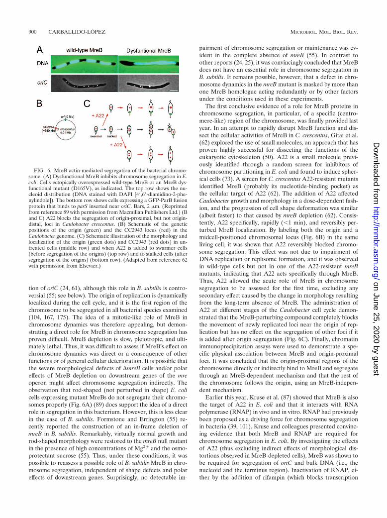
tion of oriC (24, 61), although this role in B. subtilis is contro-versial (55; see below). The origin of replication is dynamicallylocalized during the cell cycle, and it is the first region of thechromosome to be segregated in all bacterial species examined(104, 167, 175). The idea of a mitotic-like role of MreB inchromosome dynamics was therefore appealing, but demon-strating a direct role for MreB in chromosome segregation hasproven difficult. MreB depletion is slow, pleiotropic, and ulti-mately lethal. Thus, it was difficult to assess if MreB’s effect onchromosome dynamics was direct or a consequence of otherfunctions or of general cellular deterioration. It is possible thatthe severe morphological defects of �mreB cells and/or polareffects of MreB depletion on downstream genes of the mreoperon might affect chromosome segregation indirectly. Theobservation that rod-shaped (not perturbed in shape) E. colicells expressing mutant MreBs do not segregate their chromo-somes properly (Fig. 6A) (89) does support the idea of a directrole in segregation in this bacterium. However, this is less clearin the case of B. subtilis. Formstone and Errington (55) re-cently reported the construction of an in-frame deletion ofmreB in B. subtilis. Remarkably, virtually normal growth androd-shaped morphology were restored to the mreB null mutantin the presence of high concentrations of Mg2� and the osmo-protectant sucrose (55). Thus, under these conditions, it waspossible to reassess a possible role of B. subtilis MreB in chro-mosome segregation, independent of shape defects and polareffects of downstream genes. Surprisingly, no detectable im-
pairment of chromosome segregation or maintenance was ev-ident in the complete absence of mreB (55). In contrast toother reports (24, 25), it was convincingly concluded that MreBdoes not have an essential role in chromosome segregation inB. subtilis. It remains possible, however, that a defect in chro-mosome dynamics in the mreB mutant is masked by more thanone MreB homologue acting redundantly or by other factorsunder the conditions used in these experiments.
The first conclusive evidence of a role for MreB proteins inchromosome segregation, in particular, of a specific (centro-mere-like) region of the chromosome, was finally provided lastyear. In an attempt to rapidly disrupt MreB function and dis-sect the cellular activities of MreB in C. crescentus, Gitai et al.(62) explored the use of small molecules, an approach that hasproven highly successful for dissecting the functions of theeukaryotic cytoskeleton (50). A22 is a small molecule previ-ously identified through a random screen for inhibitors ofchromosome partitioning in E. coli and found to induce spher-ical cells (73). A screen for C. crescentus A22-resistant mutantsidentified MreB (probably its nucleotide-binding pocket) asthe cellular target of A22 (62). The addition of A22 affectedCaulobacter growth and morphology in a dose-dependent fash-ion, and the progression of cell shape deformation was similar(albeit faster) to that caused by mreB depletion (62). Consis-tently, A22 specifically, rapidly (�1 min), and reversibly per-turbed MreB localization. By labeling both the origin and amidcell-positioned chromosomal locus (Fig. 6B) in the sameliving cell, it was shown that A22 reversibly blocked chromo-some segregation. This effect was not due to impairment ofDNA replication or replisome formation, and it was observedin wild-type cells but not in one of the A22-resistant mreBmutants, indicating that A22 acts specifically through MreB.Thus, A22 allowed the acute role of MreB in chromosomesegregation to be assessed for the first time, excluding anysecondary effect caused by the change in morphology resultingfrom the long-term absence of MreB. The administration ofA22 at different stages of the Caulobacter cell cycle demon-strated that the MreB-perturbing compound completely blocksthe movement of newly replicated loci near the origin of rep-lication but has no effect on the segregation of other foci if itis added after origin segregation (Fig. 6C). Finally, chromatinimmunoprecipitation assays were used to demonstrate a spe-cific physical association between MreB and origin-proximalfoci. It was concluded that the origin-proximal regions of thechromosome directly or indirectly bind to MreB and segregatethrough an MreB-dependent mechanism and that the rest ofthe chromosome follows the origin, using an MreB-indepen-dent mechanism.
Earlier this year, Kruse et al. (87) showed that MreB is alsothe target of A22 in E. coli and that it interacts with RNApolymerase (RNAP) in vivo and in vitro. RNAP had previouslybeen proposed as a driving force for chromosome segregationin bacteria (39, 101). Kruse and colleagues presented convinc-ing evidence that both MreB and RNAP are required forchromosome segregation in E. coli. By investigating the effectsof A22 (thus excluding indirect effects of morphological dis-tortions observed in MreB-depleted cells), MreB was shown tobe required for segregation of oriC and bulk DNA (i.e., thenucleoid and the terminus region). Inactivation of RNAP, ei-ther by the addition of rifampin (which blocks transcription
FIG. 6. MreB actin-mediated segregation of the bacterial chromo-some. (A) Dysfunctional MreB inhibits chromosome segregation in E.coli. Cells ectopically overexpressed wild-type MreB or an MreB dys-functional mutant (D165V), as indicated. The top row shows the nu-cleoid distribution (DNA stained with DAPI [4,6-diamidino-2-phe-nylindole]). The bottom row shows cells expressing a GFP-ParB fusionprotein that binds to parS inserted near oriC. Bars, 2 �m. (Reprintedfrom reference 89 with permission from Macmillan Publishers Ltd.) (Band C) A22 blocks the segregation of origin-proximal, but not origin-distal, loci in Caulobacter crescentus. (B) Schematic of the geneticpositions of the origin (green) and the CC2943 locus (red) in theCaulobacter genome. (C) Schematic illustration of the morphology andlocalization of the origin (green dots) and CC2943 (red dots) in un-treated cells (middle row) and when A22 is added to swarmer cells(before segregation of the origins) (top row) and to stalked cells (aftersegregation of the origins) (bottom row). (Adapted from reference 62with permission from Elsevier.)
900 CARBALLIDO-LOPEZ MICROBIOL. MOL. BIOL. REV.
on June 25, 2020 by guesthttp://m
mbr.asm
.org/D
ownloaded from
initiation in bacteria) or by using temperature-sensitive RNAPalleles, prevented nucleoid and terminus segregation (87). Fi-nally, flow cytometry analyses showed that MreB depletion andinactivation of RNAP conferred virtually identical and unusualchromosome segregation defects. These findings raised theobvious question of the function of the MreB-RNAP inter-action. Kruse et al. (87) suggested that MreB and RNAPfunction together in chromosome segregation, and they pro-vided two possible interpretations of their data. One possi-bility is that the MreB cytoskeleton immobilizes the tran-scription machinery in such a way that the motor power ofRNAP would drive chromosome segregation. Alternatively,and consistent with other findings that make the dynamicMreB cytoskeleton another possible candidate for the gen-eration of the force needed for chromosome segregation(24, 26, 62, 89), chromosomal DNA bound by RNAP couldbe actively driven toward opposite cell poles by interactionwith the MreB cables (87).
In summary, in light of the above stunning findings and thework with ParM, it is now believed that MreB filaments, in C.crescentus (and likely in E. coli) at least, actively direct themigration of the origins toward the poles, probably in coordi-nation with lateral cell wall growth. However, the mechanismto achieve this remains to be elucidated. Whether the helicalMreB filaments are used as “rails” for the ori regions to travelalong (propelled by motor proteins that remain undiscovered),whether the treadmilling behavior of the MreB monomersthrough polarized MreB filaments is the driving force thatgenerates movement (26, 82), or whether MreB filaments actas a ParM-type mitotic apparatus that pushes apart the oriregions awaits experimental demonstration. Another majorchallenge is to identify the putative centromere region ofthe bacterial chromosome and the putative protein complex
(kinetochore complex) that interacts with this region to drivechromosome segregation.
MreB-LIKE PROTEINS AND CELL POLARITY
The establishment and maintenance of a polarity axis anddifferentiation of the cell poles are fundamental aspects of thedevelopment of most cells. Indeed, essential processes such asgrowth, divison site placement, and movement rely on a de-fined axis of cell polarity. In eukaryotic cells, cytoskeletal sys-tems provide the structural basis for cell polarization (38, 49,134, 174), but the origin and the mechanisms responsible forcell polarization and asymmetry in prokaryotic cells remainpoorly understood. Recently, a new function of the actin-likecytoskeleton was uncovered when MreB-like proteins wereshown to be involved in generating cell polarity in C. crescentusand E. coli (61, 123, 152, 173). Gitai et al. (61) described therequirement for the MreB filaments in the establishment ofglobal C. crescentus cell polarity, through MreB-directed asym-metric localization of developmental regulators at the cellpoles. C. crescentus divides asymmetrically to generate twofunctionally different progeny cells, known as the swarmer cell(with a flagellum at one pole) and the stalked cell (with a stalkstructure at one pole) (Fig. 2D). Prior to cell division, proteinsthat specify cell fate, such as the integral membrane histidinekinases DivJ, PleC, and CckA, are segregated asymmetricallyto opposite poles of the cell (Fig. 7, wild type) (74, 103). Asecond molecular marker of cell polarity is the origin of rep-lication, which dynamically localizes to one or both poles dur-ing the cell cycle (untreated cells [Fig. 6C]) (79). By examiningMreB depletion and overexpression strains, Gitai et al. (61)demonstrated that MreB at the appropriate concentration isrequired for both the polar localization of the oriC sequence
FIG. 7. MreB-directed polarity in Caulobacter crescentus. (A) MreB affects the localization of the PleC, DivJ, and CckA developmentalregulators. Representative images of cells expressing PleC-GFP, DivJ-GFP, and CckA-GFP are shown. Shown are the localization in wild-typecells, in cells with mreB depleted for 24 h, and in cells overexpressing mreB for 6 h. For each cell, images shown were taken with differentialinterference contrast light microscopy (top image) and GFP fluorescence (bottom image). Bar, 1 �m. (Reprinted from reference 61 withpermission of the publisher. Copyright 2004 National Academy of Sciences, U.S.A.) (B) Schematic representing the MreB-dependent localizationof molecular markers of cell polarity. In the wild type (top row), the origin of replication (red ovals) and the histidine kinases PleC (blue ovals),DivJ (orange ovals), and CckA (purple ovals) localize to specific poles during the cell cycle. Depletion or overexpression of MreB (middle row)causes the origin to localize to multiple foci and PleC, DivJ, and CckA to delocalize and diffuse throughout the cell. Reinduction of MreBexpression (bottom row) repolarizes PleC and DivJ, but the localization of the polar markers is randomized to either pole. (Reprinted, with somemodifications, from reference 103 with permission from Elsevier.)
VOL. 70, 2006 BACTERIAL ACTINS 901
on June 25, 2020 by guesthttp://m
mbr.asm
.org/D
ownloaded from
and the dynamic localization of regulatory proteins to thecorrect cell pole (Fig. 7). The mechanism by which MreBmediates polar localization, however, remains to be elucidated.Analogous to the cytoskeleton-directed establishment of po-larity in eukaryotic cells (38, 134, 135, 174), polarized MreBfilaments could directly traffic the movement of polarity mark-ers (75). Alternatively (or simultaneously), given the role ofMreB in cell wall biosynthesis, it could affect the positioning oflandmarks necessary for protein localization. The rapid rees-tablishment of polar colocalization of mislocalized polar pro-teins when mreB expression was reinduced in an MreB-de-pleted strain (Fig. 7B) favored the former possibility (61).Furthermore, the polar localization of asymmetric polar mark-ers was restablished but randomized when MreB helices werereconstituted after depletion (Fig. 7B), suggesting a loss ofmemory and a general randomization of the cell’s polarity. Ithas been proposed that MreB contains polarity information todetermine subcellular localization, translated either from themolecular polarity inherent in an actin-like filament (61) orfrom signaling complexes recruited by landmark proteins atcell polarity sites (93). Indeed, TipN, a cell polarity determi-nant (landmark protein) recently identified in C. crescentus,has been suggested to be upstream of MreB in regulating cellpolarity (93). TipN was shown to localize to the new polethrough most of the cell cycle and to be essential for orientingthe polarity axis of the cell. The MreB cytoskeleton was notresponsible for the polar localization of TipN. However, TipNaffected the dynamic organization of MreB during the cellcycle (93). In light of these findings, it was proposed that TipNrecruits or organizes signaling proteins at the new pole andthat this polarized signaling complex might polarize and trans-duce the positional information to the MreB filaments, which,in turn, might propagate polarization inside the cell and reg-ulate the localization of polar markers (93).
Another report, however, favors the idea of a role for MreBin defining cell polarity through its ability to direct depositionof cell wall material. Wagner et al. (173) showed that C. cres-centus requires MreB for stalk synthesis (suggested to be aspecialized form of cell elongation) and also for stalk place-ment and prevention of ectopic pole formation, therefore de-fining polarity. MreB depletion led to a stalk elongation defect,and cells recovering from MreB depletion were unable to re-cover proper polarity and developed bumps and branches,often tipped by stalks, a strong indication that these sites werebehaving as ectopic poles (173). Similar budding and branchingphenotypes have been observed in E. coli mutants lackingmultiple PBPs (29, 32, 179). It was concluded that MreB iscritical to the maintenance of cell polarity and stalk placementin C. crescentus, probably through its role in PG synthesis(173). Interestingly, overproduction of Mbl in B. subtilis alsoled to branching and shape abnormalities: cells were branchedat different angles and displayed Y shapes and deformed polarcaps (13). Branches are unrelated to the division process andare generally understood as the consequence of local asymme-tries or disturbances in the metabolism of the sacculus (66).The shape abnormalities resulting from the overproduction ofMbl were associated with an altered helical pitch of the fila-ments, and Mbl was consistently clustered at the bases ofbumps and bud-like morphologies but not at the tips of devel-oped branches (12, 13). For E. coli cells, it has been shown that
PG (and outer membrane proteins) is stable at the poles andthat the tips of branches behave as polar caps and are corre-lated with areas of inert PG (30, 32, 122). Thus, a strongcorrelation between the occurrence of cell wall active growthand Mbl clustering was suggested. Taken together, these ob-servations support the idea that MreB proteins play a role ingeneral cell polarity through their ability to direct the localiza-tion of proteins and deposition of new cell wall material.
Recently, two independent studies showed that the MreBcytoskeleton is required for polar targeting of several polarcomponents in E. coli as well. Proteins displaying MreB-de-pendent polar localization in E. coli included the aspartatechemoreceptor Tar (inner membrane), the chemotaxis proteinCheY (cytoplasmic), the Shigella sp. virulence factor IcsA(outer membrane), and the Vibrio cholerae type II secretionprotein EpsM (inner membrane) (123, 152). Although IcsAand EpsM are polar constituents exclusive to other species,when introduced into E. coli they also localized to the poles(16, 144, 151), indicating that their mechanism of polar local-ization is conserved. In one of these two studies, mreB wasidentified in a screen of a transposon mutant library for factorsrequired for the polar localization of IcsA in E. coli (123). Incells lacking mreB, IcsA localized to multiple faint patches overthe entire surfaces of the cells. Similarly, EpsM and two dis-tinct cytoplasmic domains of IcsA that are independently suf-ficient for its polar targeting (16) (co)localized in multipleperipheral foci within the cytoplasm of mreB mutant cells(123). It was concluded that MreB is required, either directlyor indirectly, for the restriction of certain polar material con-taining positional information to defined sites in the cytoplas-mic membrane and that, in the absence of MreB, multipleectopic pole-like sites that contain such material (and that areat least partially competent for the secretion and insertion ofpolar outer membrane proteins) are formed (123).
The second study showed that targeting of Tar and IcsA tothe poles is dependent on MreB and independent of the Minsystem (152). The Min system (MinCDE) is also involved incell polarization in E. coli and other organisms, as it is requiredfor the establishment of the division site at midcell (reviewedin references 141 and 143). The Min proteins oscillate repeat-edly between polar zones by redistribution within a cytoskele-ton-like helical structure that coils around the cell from pole topole and that is similar to that formed by MreB (153). How-ever, the two membrane-associated helical structures havebeen shown to be independent (152, 153; see below). Thus, theMreB and Min cytoskeleton-like systems appear to act inde-pendently in the establishment of an axis of polarity and in thedifferentiation of the cell poles in E. coli (152). Interestingly, itwas recently reported that besides its (MreB-dependent)polar localization, Tar (and the histidine kinase CheA) alsodisplays a helical arrangement in the lateral membrane(154). This was deduced to reflect the sites of membraneinsertion of Tar via the general protein secretory (Sec) ma-chinery, which was also shown to be arranged into a helicalarray in E. coli (154; see below). Since the Tar coils did notcolocalize with those of MreB, it was concluded that theSec/Tar helices are distinct from those of MreB and that Taremploys an indirect mechanism for polar localization: it israndomly inserted into lateral cytoplasmic membrane re-
902 CARBALLIDO-LOPEZ MICROBIOL. MOL. BIOL. REV.
on June 25, 2020 by guesthttp://m
mbr.asm
.org/D
ownloaded from

gions, which are organized in a helical array independentlyof MreB, and then it migrates to the cell poles (154).
MreB-LIKE PROTEINS AND CELL DIVISION
As described previously, the subcellular localization of MreBchanges dynamically throughout the cell cycle, and MreB he-lices condense into a tight ring at the future plane of celldivision in E. coli, C. crescentus, and R. sphaeroides cells (52, 61,153, 156). In B. subtilis, MreBH at least also localizes in ring-like structures at future cell separation sites (15; see above).Such midcell localization patterns are reminiscent of those ofseveral division proteins, which assemble in a ring-like struc-ture at sites of imminent division (for a review, see reference44). In dividing eukaryotic cells, F-actin localizes to midcell toform the actin-myosin contractile ring that drives cytokinesis(102). In bacteria, this role is performed by the tubulin-likeFtsZ protein, which assembles into the filamentous Z ring thatmediates cell division (109). The helical localization of MreB-like proteins was not affected in the absence of FtsZ (13, 15).However, the formation of division-plane MreB rings de-pended on FtsZ in C. crescentus (52, 61), suggesting that MreBproteins might also play a role in cell division. This idea issupported by several observations. First, cells of C. crescentustreated with A22 failed to divide, suggesting either the exis-tence of a DNA segregation defect-induced cell cycle check-point or an independent role of MreBs in cell division (62).Second, cell division and the placement of the Z ring wereperturbed in B. subtilis mreB and mbl mutants (55, 80). Thesedefects were thought to be secondary to the propagation of theZ ring around the wider and irregular sidewalls of the mreBand mbl mutant cells. However, they could also result from acooperative role of MreBs with FtsZ during cell division. MreBproteins could be directly responsible for orienting the FtsZring or could be part of a mechanism to localize the properdivision plane. It has also been suggested that MreB could actdiscontinuously at the division site to reset the correct diame-ter each time the cell divides (55).
Alternatively, MreB may coordinate the cell division-elon-gation on-and-off control system believed to control growth inrod-shaped bacteria. According to the two-competing-sitesmodel (105), there are two morphogenetic, spatially special-ized, and presumably mutually exclusive pathways for PG syn-thesis, one for elongation (along the long axis) and the otherfor division (septum specific). The shape of the cell is deter-mined by the balance between these two competing reactions(sites) (105). Indeed, an attractive hypothesis is that MreBcontrols the switch between the division and elongation CWgrowth modes. This is consistent with early studies of E. colithat suggested that MreB functions as a negative regulator ofthe septum-specific division protein FtsI (172) and with theobservation that, also in E. coli, overexpression of wild-typemreB inhibits cell division (but has no segregation or shapedefects) (Fig. 6A) (89). Furthermore, in C. crescentus, in theabsence of MreB (or MreC), the elongation-specific PBP2 lostits helical pattern over the lateral wall and mislocalized to thedivision plane in an FtsZ-dependent manner (40). In wild-typecells, MreB transitioned between a spiral and a ring, whereasPBP2 (and MreC) always remained helical (40). Thus, it wasproposed that MreC promotes helical PBP2 localization along
the lateral wall (see above), whereas MreB inhibits divisionplane localization of PBP2 (40). The midcell (mis)localizationof PBP2 in C. crescentus cells lacking MreB is consistent withthe observation that, in E. coli, a GFP-PBP2 fusion localized inthe bacterial envelope in a spot-like pattern and also at midcellduring cell division (28). The unrelated PBP2 of S. aureus(coccoid, lacking MreB homologues) also localizes to midcell(where all CW synthesis occurs in this organism [see above]),and this targeting has been suggested to depend upon substraterecognition (130, 147).
It remains unclear whether MreB proteins play a primaryrole in cell division or if the effects on division are a secondaryconsequence of other functions. Whatever the mechanism, thefunction of MreB in this process remains an important ques-tion for future work.
OTHER PROKARYOTIC ACTIN-LIKE PROTEINS
MamK
Recently, homologues of MreB have been identified in mag-netotactic bacteria. Magnetosomes are intracellular membra-nous organelles that contain magnetite (iron) crystals and arehighly organized in linear chains to orient magnetotactic bac-teria in geomagnetic fields, thereby causing the cells to alignand to swim with respect to local or induced magnetic fieldlines. Using electron cryotomography, Komeili et al. (84) im-aged M. magneticum sp. strain AMB-1 cells and found that themagnetosomes were positioned and organized by a network ofcytoskeletal filaments (Fig. 4F). These ran parallel to four orfive individual magnetosomes along a chain, with up to sevenfilaments flanking each magnetosome. The filaments, whichwere 200 to 250 nm in length and had a thickness of �6 nm(just as expected for an F-actin filament), appeared to becomposed of MamK, a homologue of MreB (84). A functionalMamK-GFP fusion localized to straight filaments extendingacross most of the cell along its inner curvature (Fig. 4H),consistent with the magnetosome-associated filaments ob-served by electron cryotomography. (It is interesting that thislocalization is similar to that of the membrane-bound ribbon-like cytoskeleton of Spiroplasma, which is presumably com-posed of MreB filaments and also follows the inner curvatureof the cell [see above and Fig. 4B].) In a mamK mutant,magnetosomes appeared to be dispersed throughout the cell,and no cytoskeletal filaments were associated with them (Fig.4G). Although the possibility that MamK is required for thefilaments to form without being part of their structure cannotbe excluded, it was concluded that the MreB-like MamK pro-tein is the most likely candidate for the cytoskeletal networkthat organizes magnetosome membranes into a chain roughlyparallel to the long axis of the cell. MamK proteins of differentspecies are more similar to each other than to their respectiveMreB paralogues. Thus, MreB, ParM, and MamK were pre-dicted to form three phylogenically and functionally distinctgroups of prokaryotic actin-like proteins (Fig. 4I).
Ta0583
mreB genes are found in archaeal genomes, too (80). How-ever, in contrast to their bacterial counterparts, almost nothing
VOL. 70, 2006 BACTERIAL ACTINS 903
on June 25, 2020 by guesthttp://m
mbr.asm
.org/D
ownloaded from

is known about the archaeal actins. Very recently, the crystalstructure of a predicted actin homologue, Ta0583, of the ther-mophilic bacterium Thermoplasma acidophilum was deter-mined at a 2.1-Å resolution (137). The structure of Ta0583showed that it clearly belongs to the actin superfamily. It con-tains the core structure of actin and subdomains IA, IB, IIA,and IIB of actin homologues (Fig. 5C). Ta0583 appears ap-proximately equidistant from actin and MreB on the structurallevel and combines features of both bacterial actin homo-logues, i.e., MreB and the plasmid-encoded ParM protein.However, on both the sequence level and the structural level,Ta0583 is slightly closer to ParM than to MreB and actin, andthe conformation of the nucleotide (ADP) cocrystallized withTa0583 closely resembles that of ParM-bound ADP (i.e., in theopen conformation of ParM) (165; see above). In vitro, Ta0583displayed ATPase activity that was inhibited by A22 andformed crystalline sheets with a longitudinal repeat of 51 Å(F-actin repeat, 55 Å; MreB repeat, 51 Å; and ParM repeat, 49Å). Although polymerization of Ta0583 was not induced byATP (or GTP or ADP), its crystal packing indicated an inher-ent propensity to form filamentous structures (probably nucle-ated by as yet unknown factors). Sequence analysis showedthat archaeal actin homologues are very divergent, and homo-logues of Ta0583 were found only in the order Thermoplama-tales. On the basis of these findings, it was proposed thatTa0583 orthologues derive from a ParM-like actin homologuethat was once encoded by a plasmid and was transferred into acommon ancestor of the Thermoplasmatales (137). A peculiar-ity of the members of this order is that the cells are pleiomor-phic and lack a cell wall. It was suggested that Ta0583 might beinvolved in cell organization (137). Establishing the function ofTa0583 is an important topic for future work.
MreB-ASSOCIATED PROTEINS: TOWARD ANUNDERSTANDING OF THE FUNCTIONS OF THE
BACTERIAL ACTIN-LIKE CYTOSKELETON
Trafficking of Proteins: Going Helical
The subcellular localization of an increasing number of pro-teins, as detected by fluorescence microscopy, has been re-ported in the last few years. Since the discovery of the MreBhelical filaments in 2001 (80), other proteins have been shownto localize in a helical pattern. Dynamic spiral-like structuresare formed by FtsZ (and FtsZ-associated proteins) in B. subtilisand S. coelicolor cells undergoing sporulation (5, 64) and, un-der certain conditions, in E. coli cells (110, 159). Similarly,spirals that coil along the long axis of the cell are formed in E.coli by the MinC and MinD cell division inhibitors (152, 153;see above); the LamB outer membrane protein, which is in-volved in nutrient uptake (60); the Tar aspartate chemorecep-tor and the CheA histidine kinase, which are members of thechemotaxis family (152); and the SetC sugar efflux transporter(46). They are also formed by some components of the highlyconserved general secretory (Sec) machinery in both B. subtilis(11) and E. coli (154). However, all these helix-like structuresseem to be independent of MreB proteins and their determi-nants remain unclear, indicating that there is no simple rela-tionship between a spiral or helix-like organization and theactin-like cytoskeleton. The most striking example is probably
that of the Sec machinery. In B. subtilis, the helix-like localiza-tion patterns of Sec components were also observed in eithermbl or mreBH mutants and in MreB-depleted cells (11, 15). InE. coli, the helical arrays of Tar (presumably reflecting Sec-dependent sites of membrane insertion) and of MreB did notoverlap (154; see above). It was concluded that the helicaldistribution of the Sec machinery is independent of the MreBcytoskeleton in these two organisms (11, 15, 154). However,this point has not been determined unambiguously and re-mains particularly intriguing. Indeed, the presence of multiplesites of protein export distributed in helical arrays in the rod-shaped B. subtilis and E. coli organisms is in marked contrastwith the presence of a unique export site (namely, the ExPor-tal) in the coccoid organism Streptococcus pyogenes. In S. pyo-genes, the Sec translocons are clustered in a single microdo-main, which is in an asymmetric hemispherical location in thecellular membrane (140). Since the Sec machinery localizes toa single site in a spherical organism, which lack an MreBsystem, and localizes in a helical pattern in rod-shaped organ-isms that contain an MreB system, it is tempting to envisage arelationship between the two, which the data available atpresent cannot completely rule out. In B. subtilis, positioning ofthe essential Sec components might depend on, or communi-cate with, more than one MreB homologue (acting redun-dantly). For E. coli, it remains to be confirmed (i) that Tar isinserted in the membrane via a Sec-dependent process and (ii)that the noncolocalization of Tar and MreB helices does notresult from the different dynamics of membrane proteins ver-sus that of MreB cables in the cytosol. It is also possible thatthe MreB-directed helical PG (i.e., the rigid CW) anchors theSec machinery (or certain kinds of membrane lipids) to form ahelical array. Again, any perfect colocalization would be lostduring growth because of the different dynamics of MreB ca-bles versus expansion and rotation of the external CW.
SetB, an integral membrane protein of E. coli that is closelyrelated to SetC (i.e., most likely part of a sugar transportercomplex), is one of the three proteins (together with MreC andLytE [see above]) known so far to localize in a helix and todirectly interact with an MreB homologue. SetB-GFP formsregular helical structures similar to the MreB helices, and SetBinteracted with MreB in a two-hybrid assay (46). Interestingly,SetB was identified through a genetic screen for suppressors ofthe chromosome segregation defect exhibited by mutants ofParC, a subunit of topoisomerase IV responsible for topolog-ical unlinking of the chromosomes during segregation (45, 46).Deletion of SetB caused a delay in chromosome segregation,whereas its overproduction caused chromosome disintegrationand stretching (46). It was suggested, although not proved, thatthe SetB and MreB helices are associated and that they worktogether in chromosome segregation in E. coli. It should benoted, though, that the helical structures formed by SetC andMreB did not colocalize. The SetB and MreB helices seemedspatially coordinated, but rather than overlapping, theyseemed to be interwound and to describe parallel paths (sim-ilar to the MreB and MreC helices in C. crescentus) (40). Thus,the precise role of SetB in segregation, the determinant of itslocalization, and whether its two-hybrid interaction with MreBis biologically significant remain to be elucidated.
MreC (and MreD in B. subtilis) has also been shown tolocalize in a helical configuration that resembles that of MreB
904 CARBALLIDO-LOPEZ MICROBIOL. MOL. BIOL. REV.
on June 25, 2020 by guesthttp://m
mbr.asm
.org/D
ownloaded from

(35, 40, 95; see above). In C. crescentus, GFP fusions to outermembrane proteins found to interact with MreC in vitro (byaffinity chromatography coupled to mass spectrometry-basedprotein identification) displayed spiral, punctate, or bandedpatterns of localization similar to those adopted by MreC (andPBP2) (35). As in the case of MreC (35, 40), the localization ofthese outer membrane protein-GFP fusions was unperturbedby rapid disruption of MreB spirals by A22, but it was lost incells with shape defects resulting from MreB depletion for anextended time period (35). It was proposed that a helical spa-tial organization might be a property shared by many periplas-mic proteins and associated with regions of active PG assem-bly. MreB cables might be ultimately responsible for theaccurate tracking of MreC and the PG-synthesizing complexesin a helical pattern. By these means, an intrinsic helical patternin the PG layer is established, which can persist and propagatefor a certain period of time in the absence of an internal MreBstructure (35).
Are There MreB-Associated Proteins That ModulateFilament Organization?
By analogy to eukaryotic actins, it is anticipated that a myr-iad of MreB-associated proteins exist in the cell (at least 170distinct actin-binding proteins have been identified in eukary-otic cells) and that some of them modulate filament dynamics.Several proteins that interact with the tubulin-like FtsZ proteinand play a role in the dynamics of FtsZ assembly in vivo havebeen identified. ZapA is an FtsZ-polymerizing factor (65),while EzrA, MinC, and SulA inhibit FtsZ assembly in B. subtilis(44, 100). The actin-like DNA-segregating ParM protein isstabilized via the interaction with the ParR-parC nucleoproteincomplex (119; see above). However, no interacting proteins areyet known for MreB-like proteins that spatially or temporallyregulate their assembly/disassembly and/or that are involved inmonomer or protofilament interactions. EM showed thatMreB, unlike F-actin and ParM, spontaneously forms ringswithout auxiliary proteins (47). Nevertheless, the diameter ofthe rings formed by MreB in vitro was smaller (approximatelyone-fifth) than that observed in vivo (80). Cross-linking factorsand/or associated proteins may be required to anchor theMreB bundles to the membrane and to generate a larger spiraldiameter around the periphery of the cell. Clearly, future re-search will prompt the identification of binding partners andinteracting modulators that would help us to understand howMreB proteins function and are organized in the bacterial cell.
The MreB Hub: a Central Organizing Role for theActin-Like Cytoskeleton
Like the case for MreB/ParM and actin (or FtsZ and tubu-lin), no homologues of actin-binding proteins can be identifiedin bacteria by amino acid sequence comparison. A combina-tion of both genetic and biochemical screening methods istherefore needed to uncover MreB-interacting proteins. In apioneer work, Butland et al. (10a) recently reported the firstlarge-scale analysis of protein complexes in E. coli. In thisstudy, 648 affinity-tagged proteins (about 16% of the E. coliproteome), expressed at endogenous levels, were purified tohomogeneity, and their interacting protein partners were iden-
tified by mass spectrometry. An interaction network containingconserved and essential protein complexes involved in diversebiological processes was uncovered. Most of the proteins hadfew interacting partners, whereas a subset of hubs formed agreat number of connections. These hubs are predicted to behighly conserved. MreB appeared to be one of the 20 mostconnected nodes (�15 interactions), and it was included in theset of 71 proteins that potentially fulfill critical roles across allbacteria.
Similarly, an actin cytoskeleton protein interaction network,composed at present of �65 interactions, has been establishedin B. subtilis by genome-wide yeast two-hybrid screens (R.Carballido-Lopez, unpublished data). The proteins shown tospecifically interact with MreBCD, Mbl, and MreBH belong tovery different functional categories, anticipating that MreB-like proteins might be involved in a variety of cellular pro-cesses. Furthermore, many of these interactions link the actincytoskeleton network to the replication network (126), thedivision network, and the preliminary stress response network(P. Noirot, unpublished data) of B. subtilis, further suggestingthe centrality of the cytoskeleton in coordinating key cellularfunctions.
In summary, large-scale approaches have identified putativeMreB-interacting partners and provided insights into the func-tions of previously uncharacterized bacterial proteins whileoutlining the overall topology of a microbial interactomewhose core components (such as MreB) are broadly conservedacross prokaryotes. The biological significance of each proteininteraction identified by these approaches, however, needs tobe demonstrated.
CONCLUSIONS
The discovery of the prokaryotic homologues of actin (aswell as of other cytoskeletal elements in bacteria) has forced aradical rethink about the origin of the cytoskeleton and revo-lutionized our view of bacterial cell architecture. The currentview is that of a complex, highly structured, and dynamic bac-terial subcellular organization, where the actin cytoskeleton,like the case in eukaryotic cells, plays a central organizing role.Although the mechanisms by which MreB-like proteins medi-ate insertion/degradation of CW material, segregation, andpolarity remain to be elucidated, it has been shown that theyare involved in these (and probably other) essential cellularprocesses. It is thought that MreB filaments serve as an orga-nizer or tracking device for the movement of chromosomesand for the targeting of many proteins to their sites of biolog-ical function, thus playing a role analogous to the eukaryoticcytoskeleton in macromolecular trafficking. A major challengenow is to identify the targets that might move along and/or thatare positioned by the actin-like filaments. The mechanisticdetails of how MreB proteins function and how they are spa-tially and temporally organized using other cellular factors arealso main directions for future work.
Although we now know that actin filaments have almostcertainly existed for more than 2 billion years, eukaryotic andprokaryotic actins seem to have developed into genuine cy-toskeletal systems with very different and highly extended evo-lutionary histories (37). However, despite different functional-ities and probably uniquely associated proteins, new insights
VOL. 70, 2006 BACTERIAL ACTINS 905
on June 25, 2020 by guesthttp://m
mbr.asm
.org/D
ownloaded from

into our understanding of eukaryotic actin might be obtainedby studying its bacterial orthologues. Despite decades of effort,the atomic structure of F-actin has not yet been resolved andwas deduced only from the atomic structure of the actin mono-mer by modeling (67). MreB crystals (164) have provided thefirst ultrastructural view of actin-like strands at atomic resolu-tion. ParM crystals (165), on the other hand, have provided thefirst approach at atomic resolution to the multiple conforma-tional states presumed to exist in F-actin and all actin homo-logues (42, 56).
ACKNOWLEDGMENTS
The emerging field of the bacterial cytoskeleton has undergone areal explosion in the last few years. Some publications about actin-likeproteins could not be cited here because of length constraints. I apol-ogize to those authors whose work contributed to the field but couldnot be included or was cited only indirectly in this review.
I thank Paul Hoskisson, Dirk-Jan Scheffers, Jeff Errington, PhilippeNoirot, and particularly Alex Formstone for critical reading and valu-able comments in the preparation of the manuscript.
My work during the writing of this review was supported by theInstitut National de la Recherche Agronomique (INRA) and by along-term fellowship from the European Molecular Biology Organi-zation (EMBO).
REFERENCES
1. Abhayawardhane, Y., and G. C. Stewart. 1995. Bacillus subtilis possesses asecond determinant with extensive sequence similarity to the Escherichiacoli mreB morphogene. J. Bacteriol. 177:765–773.
2. Alaedini, A., and R. A. Day. 1999. Identification of two penicillin-bindingmultienzyme complexes in Haemophilus influenzae. Biochem. Biophys. Res.Commun. 264:191–195.
3. Ausmees, N., J. R. Kuhn, and C. Jacobs-Wagner. 2003. The bacterialcytoskeleton: an intermediate filament-like function in cell shape. Cell115:705–713.
4. Beall, B., and J. Lutkenhaus. 1991. FtsZ in Bacillus subtilis is required forvegetative septation and for asymmetric septation during sporulation.Genes Dev. 5:447–455.
5. Ben-Yehuda, S., and R. Losick. 2002. Asymmetric cell division in B. subtilisinvolves a spiral-like intermediate of the cytokinetic protein FtsZ. Cell109:257–266.
6. Bi, E. F., and J. Lutkenhaus. 1991. FtsZ ring structure associated withdivision in Escherichia coli. Nature 354:161–164.
7. Bork, P., C. Sander, and A. Valencia. 1992. An ATPase domain common toprokaryotic cell cycle proteins, sugar kinases, actin, and hsp70 heat shockproteins. Proc. Natl. Acad. Sci. USA 89:7290–7294.
8. Brouhard, G. J., and A. J. Hunt. 2005. Microtubule movements on the armsof mitotic chromosomes: polar ejection forces quantified in vitro. Proc.Natl. Acad. Sci. USA 102:13903–13908.
9. Burger, A., K. Sichler, G. Kelemen, M. Buttner, and W. Wohlleben. 2000.Identification and characterization of the mre gene region of Streptomycescoelicolor A3(2). Mol. Gen. Genet. 263:1053–1060.
10. Burman, L. G., J. Raichler, and J. T. Park. 1983. Evidence for diffusegrowth of the cylindrical portion of the Escherichia coli murein sacculus. J.Bacteriol. 155:983–988.
10a.Butland, G., J. M. Peregrin-Alvarez, J. Li, W. Yang, X. Yang, V. Canadien,A. Starostine, D. Richards, B. Beattie, N. Krogan, M. Davey, J. Parkinson,J. Greenblatt, and A. Emili. 2005. Interaction network containing con-served and essential protein complexes in Escherichia coli. Nature 433:531–537.
11. Campo, N., H. Tjalsma, G. Buist, D. Stepniak, M. Meijer, M. Veenhuis, M.Westermann, J. P. Muller, S. Bron, J. Kok, O. P. Kuipers, and J. D.Jongbloed. 2004. Subcellular sites for bacterial protein export. Mol. Micro-biol. 53:1583–1599.
12. Carballido-Lopez, R. 2002. Bacterial cytoskeleton: cell shape determinationin Bacillus subtilis.Ph.D. thesis. University of Oxford, Oxford, UnitedKingdom.
13. Carballido-Lopez, R., and J. Errington. 2003. The bacterial cytoskeleton. Invivo dynamics of the actin-like protein Mbl of Bacillus subtilis. Dev. Cell4:19–28.
14. Carballido-Lopez, R., and J. Errington. 2003. A dynamic bacterial cytoskel-eton. Trends Cell Biol. 13:577–583.
15. Carballido-Lopez, R., A. Formstone, Y. Li, S. D. Ehrlich, P. Noirot, and J.Errington. 2006. Actin homolog MreBH governs cell morphogenesis bylocalization of the cell wall hydrolase LytE. Dev. Cell 11:399–409.
16. Charles, M., M. Perez, J. H. Kobil, and M. B. Goldberg. 2001. Polartargeting of Shigella virulence factor IcsA in Enterobacteriacae and Vibrio.Proc. Natl. Acad. Sci. USA 98:9871–9876.
17. Cleveland, D. W. 1982. Treadmilling of tubulin and actin. Cell 28:689–691.18. Cooper, J. A., and D. A. Schafer. 2000. Control of actin assembly and
disassembly at filament ends. Curr. Opin. Cell Biol. 12:97–103.19. Costa, C. S., and D. N. Anton. 1999. Conditional lethality of cell shape
mutations of Salmonella typhimurium: rodA and mre mutants are lethal onsolid but not in liquid medium. Curr. Microbiol. 38:137–142.
20. Costa, C. S., and D. N. Anton. 1993. Round-cell mutants of Salmonellatyphimurium produced by transposition mutagenesis: lethality of rodA andmre mutations. Mol. Gen. Genet. 236:387–394.
21. Dabiri, G. A., J. M. Sanger, D. A. Portnoy, and F. S. Southwick. 1990.Listeria monocytogenes moves rapidly through the host-cell cytoplasm byinducing directional actin assembly. Proc. Natl. Acad. Sci. USA 87:6068–6072.
22. Daniel, R. A., and J. Errington. 2003. Control of cell morphogenesis inbacteria: two distinct ways to make a rod-shaped cell. Cell 113:767–776.
23. de Boer, P., R. Crossley, and L. Rothfield. 1992. The essential bacterialcell-division protein FtsZ is a GTPase. Nature 359:254–256.
24. Defeu Soufo, H. J., and P. L. Graumann. 2003. Actin-like proteins MreBand Mbl from Bacillus subtilis are required for bipolar positioning of rep-lication origins. Curr. Biol. 13:1916–1920.
25. Defeu Soufo, H. J., and P. L. Graumann. 2005. Bacillus subtilis actin-likeprotein MreB influences the positioning of the replication machinery andrequires membrane proteins MreC/D and other actin-like proteins forproper localization. BMC Cell Biol. 6:10.
26. Defeu Soufo, H. J., and P. L. Graumann. 2004. Dynamic movement ofactin-like proteins within bacterial cells. EMBO Rep. 5:789–794.
27. de Jonge, B. L., F. B. Wientjes, I. Jurida, F. Driehuis, J. T. Wouters, and N.Nanninga. 1989. Peptidoglycan synthesis during the cell cycle of Escherichiacoli: composition and mode of insertion. J. Bacteriol. 171:5783–5794.
28. Den Blaauwen, T., M. E. Aarsman, N. O. Vischer, and N. Nanninga. 2003.Penicillin-binding protein PBP2 of Escherichia coli localizes preferentiallyin the lateral wall and at mid-cell in comparison with the old cell pole. Mol.Microbiol. 47:539–547.
29. Denome, S. A., P. K. Elf, T. A. Henderson, D. E. Nelson, and K. D. Young.1999. Escherichia coli mutants lacking all possible combinations of eightpenicillin binding proteins: viability, characteristics, and implications forpeptidoglycan synthesis. J. Bacteriol. 181:3981–3993.
30. de Pedro, M. A., J. C. Quintela, J. V. Holtje, and H. Schwarz. 1997. Mureinsegregation in Escherichia coli. J. Bacteriol. 179:2823–2834.
31. de Pedro, M. A., H. Schwarz, and A. L. Koch. 2003. Patchiness of mureininsertion into the sidewall of Escherichia coli. Microbiology 149:1753–1761.
32. de Pedro, M. A., K. D. Young, J. V. Holtje, and H. Schwarz. 2003. Branchingof Escherichia coli cells arises from multiple sites of inert peptidoglycan.J. Bacteriol. 185:1147–1152.
33. Desai, A., and T. J. Mitchison. 1997. Microtubule polymerization dynamics.Annu. Rev. Cell Dev. Biol. 13:83–117.
34. Dickinson, R. B., and D. L. Purich. 2002. Clamped-filament elongationmodel for actin-based motors. Biophys. J. 82:605–617.
35. Divakaruni, A. V., R. R. Loo, Y. Xie, J. A. Loo, and J. W. Gober. 2005. Thecell-shape protein MreC interacts with extracytoplasmic proteins includingcell wall assembly complexes in Caulobacter crescentus. Proc. Natl. Acad.Sci. USA 102:18602–18607.
36. Doi, M., M. Wachi, F. Ishino, S. Tomioka, M. Ito, Y. Sakagami, A. Suzuki,and M. Matsuhashi. 1988. Determinations of the DNA sequence of themreB gene and of the gene products of the mre region that function information of the rod shape of Escherichia coli cells. J. Bacteriol. 170:4619–4624.
37. Doolittle, R. F., and A. L. York. 2002. Bacterial actins? An evolutionaryperspective. Bioessays 24:293–296.
38. Drubin, D. G., and W. J. Nelson. 1996. Origins of cell polarity. Cell 84:335–344.
39. Dworkin, J., and R. Losick. 2002. Does RNA polymerase help drive chro-mosome segregation in bacteria? Proc. Natl. Acad. Sci. USA 99:14089–14094.
40. Dye, N. A., Z. Pincus, J. A. Theriot, L. Shapiro, and Z. Gitai. 2005. Twoindependent spiral structures control cell shape in Caulobacter. Proc. Natl.Acad. Sci. USA 102:18608–18613.
41. Egelman, E. H. 2001. Molecular evolution: actin’s long lost relative found.Curr. Biol. 11:R1022–R1024.
42. Egelman, E. H., N. Francis, and D. J. DeRosier. 1982. F-actin is a helix witha random variable twist. Nature 298:131–135.
43. Eichenberger, P., S. T. Jensen, E. M. Conlon, C. van Ooij, J. Silvaggi, J. E.Gonzalez-Pastor, M. Fujita, S. Ben-Yehuda, P. Stragier, J. S. Liu, and R.Losick. 2003. The sigmaE regulon and the identification of additionalsporulation genes in Bacillus subtilis. J. Mol. Biol. 327:945–972.
44. Errington, J., R. A. Daniel, and D. J. Scheffers. 2003. Cytokinesis in bac-teria. Microbiol. Mol. Biol. Rev. 67:52–65.
45. Espeli, O., C. Levine, H. Hassing, and K. J. Marians. 2003. Temporalregulation of topoisomerase IV activity in E. coli. Mol. Cell 11:189–201.
906 CARBALLIDO-LOPEZ MICROBIOL. MOL. BIOL. REV.
on June 25, 2020 by guesthttp://m
mbr.asm
.org/D
ownloaded from
46. Espeli, O., P. Nurse, C. Levine, C. Lee, and K. J. Marians. 2003. SetB: anintegral membrane protein that affects chromosome segregation in Esche-richia coli. Mol. Microbiol. 50:495–509.
47. Esue, O., M. Cordero, D. Wirtz, and Y. Tseng. 2005. The assembly of MreB,a prokaryotic homolog of actin. J. Biol. Chem. 280:2628–2635.
48. Esue, O., D. Wirtz, and Y. Tseng. 2006. GTPase activity, structure, andmechanical properties of filaments assembled from bacterial cytoskeletonprotein MreB. J. Bacteriol. 188:968–976.
49. Etienne-Manneville, S., and A. Hall. 2002. Rho GTPases in cell biology.Nature 420:629–635.
50. Fenteany, G., and S. Zhu. 2003. Small-molecule inhibitors of actin dynamicsand cell motility. Curr. Top. Med. Chem. 3:593–616.
51. Feucht, A., L. Evans, and J. Errington. 2003. Identification of sporulationgenes by genome-wide analysis of the sigmaE regulon of Bacillus subtilis.Microbiology 149:3023–3034.
52. Figge, R. M., A. V. Divakaruni, and J. W. Gober. 2004. MreB, the cellshape-determining bacterial actin homologue, coordinates cell wall mor-phogenesis in Caulobacter crescentus. Mol. Microbiol. 51:1321–1332.
53. Flaherty, K. M., D. B. McKay, W. Kabsch, and K. C. Holmes. 1991. Sim-ilarity of the three-dimensional structures of actin and the ATPase frag-ment of a 70-kDa heat shock cognate protein. Proc. Natl. Acad. Sci. USA88:5041–5045.
54. Flardh, K. 2003. Growth polarity and cell division in Streptomyces. Curr.Opin. Microbiol. 6:564–571.
55. Formstone, A., and J. Errington. 2005. A magnesium-dependent mreB nullmutant: implications for the role of mreB in Bacillus subtilis. Mol. Micro-biol. 55:1646–1657.
56. Galkin, V. E., A. Orlova, N. Lukoyanova, W. Wriggers, and E. H. Egelman.2001. Actin depolymerizing factor stabilizes an existing state of F-actin andcan change the tilt of F-actin subunits. J. Cell Biol. 153:75–86.
57. Garner, E. C., C. S. Campbell, and R. D. Mullins. 2004. Dynamic instabilityin a DNA-segregating prokaryotic actin homolog. Science 306:1021–1025.
58. Gerdes, K., J. Møller-Jensen, and R. Bugge Jensen. 2000. Plasmid andchromosome partitioning: surprises from phylogeny. Mol. Microbiol. 37:455–466.
59. Gerdes, K., J. Møller-Jensen, G. Ebersbach, T. Kruse, and K. Nordstrom.2004. Bacterial mitotic machineries. Cell 116:359–366.
60. Gibbs, K. A., D. D. Isaac, J. Xu, R. W. Hendrix, T. J. Silhavy, and J. A.Theriot. 2004. Complex spatial distribution and dynamics of an abundantEscherichia coli outer membrane protein, LamB. Mol. Microbiol. 53:1771–1783.
61. Gitai, Z., N. Dye, and L. Shapiro. 2004. An actin-like gene can determinecell polarity in bacteria. Proc. Natl. Acad. Sci. USA 101:8643–8648.
62. Gitai, Z., N. A. Dye, A. Reisenauer, M. Wachi, and L. Shapiro. 2005. MreBactin-mediated segregation of a specific region of a bacterial chromosome.Cell 120:329–341.
63. Gordon, G. S., D. Sitnikov, C. D. Webb, A. Teleman, A. Straight, R. Losick,A. W. Murray, and A. Wright. 1997. Chromosome and low copy plasmidsegregation in E. coli: visual evidence for distinct mechanisms. Cell 90:1113–1121.
64. Grantcharova, N., U. Lustig, and K. Flardh. 2005. Dynamics of FtsZ as-sembly during sporulation in Streptomyces coelicolor A3(2). J. Bacteriol.187:3227–3237.
65. Gueiros-Filho, F. J., and R. Losick. 2002. A widely conserved bacterial celldivision protein that promotes assembly of the tubulin-like protein FtsZ.Genes Dev. 16:2544–2556.
66. Gullbrand, B., T. Akerlund, and K. Nordstrom. 1999. On the origin ofbranches in Escherichia coli. J. Bacteriol. 181:6607–6614.
67. Holmes, K. C., D. Popp, W. Gebhard, and W. Kabsch. 1990. Atomic modelof the actin filament. Nature 347:44–49.
68. Holtje, J. V. 1995. From growth to autolysis: the murein hydrolases inEscherichia coli. Arch. Microbiol. 164:243–254.
69. Holtje, J. V. 1998. Growth of the stress-bearing and shape-maintainingmurein sacculus of Escherichia coli. Microbiol. Mol. Biol. Rev. 62:181–203.
70. Holtje, J. V. 1996. A hypothetical holoenzyme involved in the replication ofthe murein sacculus of Escherichia coli. Microbiology 142:1911–1918.
71. Hunt, A. J., and J. R. McIntosh. 1998. The dynamic behavior of individualmicrotubules associated with chromosomes in vitro. Mol. Biol. Cell 9:2857–2871.
72. Ishikawa, S., Y. Hara, R. Ohnishi, and J. Sekiguchi. 1998. Regulation of anew cell wall hydrolase gene, cwlF, which affects cell separation in Bacillussubtilis. J. Bacteriol. 180:2549–2555.
73. Iwai, N., K. Nagai, and M. Wachi. 2002. Novel S-benzylisothiourea com-pound that induces spherical cells in Escherichia coli probably by acting ona rod-shape-determining protein(s) other than penicillin-binding protein 2.Biosci. Biotechnol. Biochem. 66:2658–2662.
74. Jacobs-Wagner, C. 2004. Regulatory proteins with a sense of direction: cellcycle signalling network in Caulobacter. Mol. Microbiol. 51:7–13.
75. Janakiraman, A., and M. B. Goldberg. 2004. Recent advances on thedevelopment of bacterial poles. Trends Microbiol. 12:518–525.
76. Jenkins, C., R. Samudrala, I. Anderson, B. P. Hedlund, G. Petroni, N.Michailova, N. Pinel, R. Overbeek, G. Rosati, and J. T. Staley. 2002. Genes
for the cytoskeletal protein tubulin in the bacterial genus Prosthecobacter.Proc. Natl. Acad. Sci. USA 99:17049–17054.
77. Jensen, R. B., and K. Gerdes. 1997. Partitioning of plasmid R1. The ParMprotein exhibits ATPase activity and interacts with the centromere-likeParR-parC complex. J. Mol. Biol. 269:505–513.
78. Jensen, R. B., R. Lurz, and K. Gerdes. 1998. Mechanism of DNA segrega-tion in prokaryotes: replicon pairing by parC of plasmid R1. Proc. Natl.Acad. Sci. USA 95:8550–8555.
79. Jensen, R. B., and L. Shapiro. 1999. The Caulobacter crescentus smc gene isrequired for cell cycle progression and chromosome segregation. Proc. Natl.Acad. Sci. USA 96:10661–10666.
80. Jones, L. J., R. Carballido-Lopez, and J. Errington. 2001. Control of cellshape in bacteria: helical, actin-like filaments in Bacillus subtilis. Cell 104:913–922.
81. Kabsch, W., H. G. Mannherz, D. Suck, E. F. Pai, and K. C. Holmes. 1990.Atomic structure of the actin:DNase I complex. Nature 347:37–44.
82. Kim, S. Y., Z. Gitai, A. Kinkhabwala, L. Shapiro, and W. E. Moerner. 2006.Single molecules of the bacterial actin MreB undergo directed treadmillingmotion in Caulobacter crescentus. Proc. Natl. Acad. Sci. USA 103:10929–10934.
83. Kline-Smith, S. L., and C. E. Walczak. 2004. Mitotic spindle assembly andchromosome segregation: refocusing on microtubule dynamics. Mol. Cell15:317–327.
84. Komeili, A., Z. Li, D. K. Newman, and G. J. Jensen. 2006. Magnetosomesare cell membrane invaginations organized by the actin-like protein MamK.Science 311:242–245.
85. Koonin, E. V. 1993. A superfamily of ATPases with diverse functions con-taining either classical or deviant ATP-binding motif. J. Mol. Biol. 229:1165–1174.
86. Korn, E. D., M. F. Carlier, and D. Pantaloni. 1987. Actin polymerizationand ATP hydrolysis. Science 238:638–644.
87. Kruse, T., B. Blagoev, A. Lobner-Olesen, M. Wachi, K. Sasaki, N. Iwai, M.Mann, and K. Gerdes. 2006. Actin homolog MreB and RNA polymeraseinteract and are both required for chromosome segregation in Escherichiacoli. Genes Dev. 20:113–124.
88. Kruse, T., J. Bork-Jensen, and K. Gerdes. 2005. The morphogeneticMreBCD proteins of Escherichia coli form an essential membrane-boundcomplex. Mol. Microbiol. 55:78–89.
89. Kruse, T., J. Møller-Jensen, A. Lobner-Olesen, and K. Gerdes. 2003. Dys-functional MreB inhibits chromosome segregation in Escherichia coli.EMBO J. 22:5283–5292.
90. Kuo, S. C., and J. L. McGrath. 2000. Steps and fluctuations of Listeriamonocytogenes during actin-based motility. Nature 407:1026–1029.
91. Kurner, J., A. S. Frangakis, and W. Baumeister. 2005. Cryo-electrontomography reveals the cytoskeletal structure of Spiroplasma melliferum.Science 307:436–438.
92. Kurner, J., O. Medalia, A. A. Linaroudis, and W. Baumeister. 2004. Newinsights into the structural organization of eukaryotic and prokaryoticcytoskeletons using cryo-electron tomography. Exp. Cell Res. 301:38–42.
93. Lam, H., W. B. Schofield, and C. Jacobs-Wagner. 2006. A landmark proteinessential for establishing and perpetuating the polarity of a bacterial cell.Cell 124:1011–1023.
94. Lazarevic, V., B. Soldo, N. Medico, H. Pooley, S. Bron, and D. Karamata.2005. Bacillus subtilis alpha-phosphoglucomutase is required for normal cellmorphology and biofilm formation. Appl. Environ. Microbiol. 71:39–45.
95. Leaver, M., and J. Errington. 2005. Roles for MreC and MreD proteins inhelical growth of the cylindrical cell wall in Bacillus subtilis. Mol. Microbiol.57:1196–1209.
96. Lee, J. C., and G. C. Stewart. 2003. Essential nature of the mreC determi-nant of Bacillus subtilis. J. Bacteriol. 185:4490–4498.
97. Lemon, K. P., and A. D. Grossman. 2001. The extrusion-capture model forchromosome partitioning in bacteria. Genes Dev. 15:2031–2041.
98. Lemon, K. P., and A. D. Grossman. 1998. Localization of bacterial DNApolymerase: evidence for a factory model of replication. Science 282:1516–1519.
99. Lemon, K. P., and A. D. Grossman. 2000. Movement of replicating DNAthrough a stationary replisome. Mol. Cell 6:1321–1330.
100. Levin, P. A., I. G. Kurtser, and A. D. Grossman. 1999. Identification andcharacterization of a negative regulator of FtsZ ring formation in Bacillussubtilis. Proc. Natl. Acad. Sci. USA 96:9642–9647.
101. Lewis, P. J., S. D. Thaker, and J. Errington. 2000. Compartmentalization oftranscription and translation in Bacillus subtilis. EMBO J. 19:710–718.
102. Li, F., X. Wang, P. C. Bunger, and A. M. Gerdes. 1997. Formation ofbinucleated cardiac myocytes in rat heart. I. Role of actin-myosin contrac-tile ring. J. Mol. Cell Cardiol. 29:1541–1551.
103. Li, R., and S. C. Wai. 2004. Bacterial cell polarity: a “swarmer-stalked” taleof actin. Trends Cell Biol. 14:532–536.
104. Li, Y., K. Sergueev, and S. Austin. 2002. The segregation of the Escherichiacoli origin and terminus of replication. Mol. Microbiol. 46:985–996.
105. Lleo, M. M., P. Canepari, and G. Satta. 1990. Bacterial cell shape regula-tion: testing of additional predictions unique to the two-competing-sites
VOL. 70, 2006 BACTERIAL ACTINS 907
on June 25, 2020 by guesthttp://m
mbr.asm
.org/D
ownloaded from
model for peptidoglycan assembly and isolation of conditional rod-shapedmutants from some wild-type cocci. J. Bacteriol. 172:3758–3771.
106. Lowe, J., and L. A. Amos. 1998. Crystal structure of the bacterial cell-division protein FtsZ. Nature 391:203–206.
107. Lowe, J., F. van den Ent, and L. A. Amos. 2004. Molecules of the bacterialcytoskeleton. Annu. Rev. Biophys. Biomol. Struct. 33:177–198.
108. Lu, C., M. Reedy, and H. P. Erickson. 2000. Straight and curved confor-mations of FtsZ are regulated by GTP hydrolysis. J. Bacteriol. 182:164–170.
109. Lutkenhaus, J., and S. G. Addinall. 1997. Bacterial cell division and the Zring. Annu. Rev. Biochem. 66:93–116.
110. Ma, X., D. W. Ehrhardt, and W. Margolin. 1996. Colocalization of celldivision proteins FtsZ and FtsA to cytoskeletal structures in living Esche-richia coli cells by using green fluorescent protein. Proc. Natl. Acad. Sci.USA 93:12998–13003.
111. Maciver, S. K., H. G. Zot, and T. D. Pollard. 1991. Characterization of actinfilament severing by actophorin from Acanthamoeba castellanii. J. Cell Biol.115:1611–1620.
112. Mazza, P., E. E. Noens, K. Schirner, N. Grantcharova, A. M. Mommaas,H. K. Koerten, G. Muth, K. Flardh, G. P. van Wezel, and W. Wohlleben.2006. MreB of Streptomyces coelicolor is not essential for vegetative growthbut is required for the integrity of aerial hyphae and spores. Mol. Microbiol.60:838–852.
113. Merad, T., A. R. Archibald, I. C. Hancock, C. R. Harwood, and J. A. Hobot.1989. Cell wall assembly in Bacillus subtilis: visualization of old and new wallmaterial by electron microscopic examination of samples stained selectivelyfor teichoic acid and teichuronic acid. J. Gen. Microbiol. 135:645–655.
114. Michie, K. A., and J. Lowe. 2006. Dynamic filaments of the bacterialcytoskeleton. Annu. Rev. Biochem. 75:467–492.
115. Min, Y. N., A. Tabuchi, Y. L. Fan, D. D. Womble, and R. H. Rownd. 1988.Complementation of mutants of the stability locus of IncFII plasmid NR1.Essential functions of the trans-acting stbA and stbB gene products. J. Mol.Biol. 204:345–356.
116. Mitchison, T., L. Evans, E. Schulze, and M. Kirschner. 1986. Sites ofmicrotubule assembly and disassembly in the mitotic spindle. Cell 45:515–527.
117. Mobley, H. L., A. L. Koch, R. J. Doyle, and U. N. Streips. 1984. Insertionand fate of the cell wall in Bacillus subtilis. J. Bacteriol. 158:169–179.
118. Mogilner, A., and G. Oster. 2003. Polymer motors: pushing out the frontand pulling up the back. Curr. Biol. 13:R721–R733.
119. Møller-Jensen, J., J. Borch, M. Dam, R. B. Jensen, P. Roepstorff, and K.Gerdes. 2003. Bacterial mitosis: ParM of plasmid R1 moves plasmid DNAby an actin-like insertional polymerization mechanism. Mol. Cell 12:1477–1487.
120. Møller-Jensen, J., R. B. Jensen, J. Lowe, and K. Gerdes. 2002. ProkaryoticDNA segregation by an actin-like filament. EMBO J. 21:3119–3127.
121. Murray, T., D. L. Popham, and P. Setlow. 1998. Bacillus subtilis cells lackingpenicillin-binding protein 1 require increased levels of divalent cations forgrowth. J. Bacteriol. 180:4555–4563.
122. Nilsen, T., A. S. Ghosh, M. B. Goldberg, and K. D. Young. 2004. Branchingsites and morphological abnormalities behave as ectopic poles in shape-defective Escherichia coli. Mol. Microbiol. 52:1045–1054.
123. Nilsen, T., A. W. Yan, G. Gale, and M. B. Goldberg. 2005. Presence ofmultiple sites containing polar material in spherical Escherichia coli cellsthat lack MreB. J. Bacteriol. 187:6187–6196.
124. Nogales, E., K. H. Downing, L. A. Amos, and J. Lowe. 1998. Tubulin andFtsZ form a distinct family of GTPases. Nat. Struct. Biol. 5:451–458.
125. Nogales, E., H. W. Wang, and H. Niederstrasser. 2003. Tubulin rings: whichway do they curve? Curr. Opin. Struct. Biol. 13:256–261.
126. Noirot-Gros, M. F., E. Dervyn, L. J. Wu, P. Mervelet, J. Errington, S. D.Ehrlich, and P. Noirot. 2002. An expanded view of bacterial DNA replica-tion. Proc. Natl. Acad. Sci. USA 99:8342–8347.
127. Ohnishi, R., S. Ishikawa, and J. Sekiguchi. 1999. Peptidoglycan hydrolaseLytF plays a role in cell separation with CwlF during vegetative growth ofBacillus subtilis. J. Bacteriol. 181:3178–3184.
128. Pantaloni, D., C. Le Clainche, and M. F. Carlier. 2001. Mechanism ofactin-based motility. Science 292:1502–1506.
129. Paterson, E. S., M. I. More, G. Pillay, C. Cellini, R. Woodgate, G. C.Walker, V. N. Iyer, and S. C. Winans. 1999. Genetic analysis of the mobi-lization and leading regions of the IncN plasmids pKM101 and pCU1. J.Bacteriol. 181:2572–2583.
130. Pinho, M. G., and J. Errington. 2005. Recruitment of penicillin-bindingprotein PBP2 to the division site of Staphylococcus aureus is dependent onits transpeptidation substrates. Mol. Microbiol. 55:799–807.
131. Pollard, T. D. 1986. Rate constants for the reactions of ATP- and ADP-actin with the ends of actin filaments. J. Cell Biol. 103:2747–2754.
132. Pollard, T. D., and M. S. Mooseker. 1981. Direct measurement of actinpolymerization rate constants by electron microscopy of actin filamentsnucleated by isolated microvillus cores. J. Cell Biol. 88:654–659.
133. Popham, D. L., and K. D. Young. 2003. Role of penicillin-binding proteinsin bacterial cell morphogenesis. Curr. Opin. Microbiol. 6:594–599.
134. Pruyne, D., and A. Bretscher. 2000. Polarization of cell growth in yeast.J. Cell Sci. 113:571–585.
135. Pruyne, D., and A. Bretscher. 2000. Polarization of cell growth in yeast. I.Establishment and maintenance of polarity states. J. Cell Sci. 113:365–375.
136. RayChaudhuri, D., and J. T. Park. 1992. Escherichia coli cell-division geneftsZ encodes a novel GTP-binding protein. Nature 359:251–254.
137. Roeben, A., C. Kofler, I. Nagy, S. Nickell, F. U. Hartl, and A. Bracher. 2006.Crystal structure of an archaeal actin homolog. J. Mol. Biol. 358:145–156.
138. Rogers, H. J., and P. F. Thurman. 1978. Temperature-sensitive nature ofthe rodB maturation in Bacillus subtilis. J. Bacteriol. 133:298–305.
139. Rogers, H. J., P. F. Thurman, and R. S. Buxton. 1976. Magnesium andanion requirements of rodB mutants of Bacillus subtilis. J. Bacteriol. 125:556–564.
140. Rosch, J., and M. Caparon. 2004. A microdomain for protein secretion ingram-positive bacteria. Science 304:1513–1515.
141. Rothfield, L., S. Justice, and J. Garcia-Lara. 1999. Bacterial cell division.Annu. Rev. Genet. 33:423–448.
142. Rothfield, L., A. Taghbalout, and Y. L. Shih. 2005. Spatial control ofbacterial division-site placement. Nat. Rev. Microbiol. 3:959–968.
143. Rothfield, L. I., Y. L. Shih, and G. King. 2001. Polar explorers: membraneproteins that determine division site placement. Cell 106:13–16.
144. Sandlin, R. C., and A. T. Maurelli. 1999. Establishment of unipolar local-ization of IcsA in Shigella flexneri 2a is not dependent on virulence plasmiddeterminants. Infect. Immun. 67:350–356.
145. Sawitzke, J., and S. Austin. 2001. An analysis of the factory model forchromosome replication and segregation in bacteria. Mol. Microbiol. 40:786–794.
146. Scheffers, D. J., L. J. Jones, and J. Errington. 2004. Several distinct local-ization patterns for penicillin-binding proteins in Bacillus subtilis. Mol.Microbiol. 51:749–764.
147. Scheffers, D. J., and M. G. Pinho. 2005. Bacterial cell wall synthesis: newinsights from localization studies. Microbiol. Mol. Biol. Rev. 69:585–607.
148. Schiffer, G., and J. V. Holtje. 1999. Cloning and characterization of PBP 1C,a third member of the multimodular class A penicillin-binding proteins ofEscherichia coli. J. Biol. Chem. 274:32031–32039.
149. Schlieper, D., M. A. Oliva, J. M. Andreu, and J. Lowe. 2005. Structure ofbacterial tubulin BtubA/B: evidence for horizontal gene transfer. Proc.Natl. Acad. Sci. USA 102:9170–9175.
150. Schmidt, A., and M. N. Hall. 1998. Signaling to the actin cytoskeleton.Annu. Rev. Cell Dev. Biol. 14:305–338.
151. Scott, M. E., Z. Y. Dossani, and M. Sandkvist. 2001. Directed polar secre-tion of protease from single cells of Vibrio cholerae via the type II secretionpathway. Proc. Natl. Acad. Sci. USA 98:13978–13983.
152. Shih, Y. L., I. Kawagishi, and L. Rothfield. 2005. The MreB and Mincytoskeletal-like systems play independent roles in prokaryotic polar dif-ferentiation. Mol. Microbiol. 58:917–928.
153. Shih, Y. L., T. Le, and L. Rothfield. 2003. Division site selection in Esche-richia coli involves dynamic redistribution of Min proteins within coiledstructures that extend between the two cell poles. Proc. Natl. Acad. Sci.USA 100:7865–7870.
154. Shiomi, D., M. Yoshimoto, M. Homma, and I. Kawagishi. 2006. Helicaldistribution of the bacterial chemoreceptor via colocalization with the Secprotein translocation machinery. Mol. Microbiol. 60:894–906.
155. Slovak, P. M., S. L. Porter, and J. P. Armitage. 2006. Differential localiza-tion of Mre proteins with PBP2 in Rhodobacter sphaeroides. J. Bacteriol.188:1691–1700.
156. Slovak, P. M., G. H. Wadhams, and J. P. Armitage. 2005. Localization ofMreB in Rhodobacter sphaeroides under conditions causing changes in cellshape and membrane structure. J. Bacteriol. 187:54–64.
157. Sontag, C. A., J. T. Staley, and H. P. Erickson. 2005. In vitro assembly andGTP hydrolysis by bacterial tubulins BtubA and BtubB. J. Cell Biol. 169:233–238.
158. Spratt, B. G. 1975. Distinct penicillin binding proteins involved in thedivision, elongation, and shape of Escherichia coli K12. Proc. Natl. Acad.Sci. USA 72:2999–3003.
159. Thanedar, S., and W. Margolin. 2004. FtsZ exhibits rapid movement andoscillation waves in helix-like patterns in Escherichia coli. Curr. Biol. 14:1167–1173.
160. Tiyanont, K., T. Doan, M. B. Lazarus, X. Fang, D. Z. Rudner, and S.Walker. 2006. Imaging peptidoglycan biosynthesis in Bacillus subtilis withfluorescent antibiotics. Proc. Natl. Acad. Sci. USA 103:11033–11038.
161. Tormo, A., and M. Vicente. 1984. The ftsA gene product participates information of the Escherichia coli septum structure. J. Bacteriol. 157:779–784.
162. Townsend, R., and D. B. Archer. 1983. A fibril protein antigen specific toSpiroplasma. J. Gen. Microbiol. 129:199–206.
163. Trachtenberg, S., and R. Gilad. 2001. A bacterial linear motor: cellular andmolecular organization of the contractile cytoskeleton of the helical bacte-rium Spiroplasma melliferum BC3. Mol. Microbiol. 41:827–848.
164. van den Ent, F., L. A. Amos, and J. Lowe. 2001. Prokaryotic origin of theactin cytoskeleton. Nature 413:39–44.
165. van den Ent, F., J. Møller-Jensen, L. A. Amos, K. Gerdes, and J. Lowe.2002. F-actin-like filaments formed by plasmid segregation protein ParM.EMBO J. 21:6935–6943.
908 CARBALLIDO-LOPEZ MICROBIOL. MOL. BIOL. REV.
on June 25, 2020 by guesthttp://m
mbr.asm
.org/D
ownloaded from
166. Varley, A. W., and G. C. Stewart. 1992. The divIVB region of the Bacillussubtilis chromosome encodes homologs of Escherichia coli septum place-ment (MinCD) and cell shape (MreBCD) determinants. J. Bacteriol. 174:6729–6742.
167. Viollier, P. H., M. Thanbichler, P. T. McGrath, L. West, M. Meewan, H. H.McAdams, and L. Shapiro. 2004. Rapid and sequential movement of indi-vidual chromosomal loci to specific subcellular locations during bacterialDNA replication. Proc. Natl. Acad. Sci. USA 101:9257–9262.
168. Vollmer, W., M. von Rechenberg, and J. V. Holtje. 1999. Demonstration ofmolecular interactions between the murein polymerase PBP1B, the lytictransglycosylase MltA, and the scaffolding protein MipA of Escherichia coli.J. Biol. Chem. 274:6726–6734.
169. Vuorio, R., and M. Vaara. 1992. Mutants carrying conditionally lethalmutations in outer membrane genes omsA and firA (ssc) are phenotypicallysimilar, and omsA is allelic to firA. J. Bacteriol. 174:7090–7097.
170. Wachi, M., M. Doi, Y. Okada, and M. Matsuhashi. 1989. New mre genesmreC and mreD, responsible for formation of the rod shape of Escherichiacoli cells. J. Bacteriol. 171:6511–6516.
171. Wachi, M., M. Doi, S. Tamaki, W. Park, S. Nakajima-Iijima, and M.Matsuhashi. 1987. Mutant isolation and molecular cloning of mre genes,which determine cell shape, sensitivity to mecillinam, and amount ofpenicillin-binding proteins in Escherichia coli. J. Bacteriol. 169:4935–4940.
172. Wachi, M., and M. Matsuhashi. 1989. Negative control of cell division by
mreB, a gene that functions in determining the rod shape of Escherichia colicells. J. Bacteriol. 171:3123–3127.
173. Wagner, J. K., C. D. Galvani, and Y. V. Brun. 2005. Caulobacter crescentusrequires RodA and MreB for stalk synthesis and prevention of ectopic poleformation. J. Bacteriol. 187:544–553.
174. Wasteneys, G. O. 2000. The cytoskeleton and growth polarity. Curr. Opin.Plant Biol. 3:503–511.
175. Webb, C. D., P. L. Graumann, J. A. Kahana, A. A. Teleman, P. A. Silver,and R. Losick. 1998. Use of time-lapse microscopy to visualize rapid move-ment of the replication origin region of the chromosome during the cellcycle in Bacillus subtilis. Mol. Microbiol. 28:883–892.
176. Wetzstein, M., U. Volker, J. Dedio, S. Lobau, U. Zuber, M. Schiesswohl, C.Herget, M. Hecker, and W. Schumann. 1992. Cloning, sequencing, andmolecular analysis of the dnaK locus from Bacillus subtilis. J. Bacteriol.174:3300–3310.
177. Wu, L. J. 2004. Structure and segregation of the bacterial nucleoid. Curr.Opin. Genet. Dev. 14:126–132.
178. Yamamoto, H., S. Kurosawa, and J. Sekiguchi. 2003. Localization of thevegetative cell wall hydrolases LytC, LytE, and LytF on the Bacillus subtiliscell surface and stability of these enzymes to cell wall-bound or extracellularproteases. J. Bacteriol. 185:6666–6677.
179. Young, K. D. 2003. Bacterial shape. Mol. Microbiol. 49:571–580.
VOL. 70, 2006 BACTERIAL ACTINS 909
on June 25, 2020 by guesthttp://m
mbr.asm
.org/D
ownloaded from
MICROBIOLOGY AND MOLECULAR BIOLOGY REVIEWS, Mar. 2007, p. 254 Vol. 71, No. 11092-2172/07/$08.00�0 doi:10.1128/MMBR.00002-07
AUTHOR’S CORRECTION
The Bacterial Actin-Like CytoskeletonRut Carballido-Lopez
Genetique Microbienne, Institut National de la Recherche Agronomique, 78352 Jouy-en-Josas Cedex, France
Volume 70, no. 4, p. 888–909, 2006. Page 888, column 2, line 12: “. . . filament protein” should read “. . . filament protein (fora recent review, see reference 151a).”
Page 889, column 1, lines 43 and 44: “. . . (for recent reviews, see references 114 and 142)” should read “. . . (for recent reviews,see references 114, 142, and 151a).”
Page 889, column 2, line 40: “(59)” should read “(59, 151a).”Page 908: The following reference was inadvertently omitted.
151a.Shih, Y.-L., and L. Rothfield. 2006. The bacterial cytoskeleton. Microbiol.Mol. Biol. Rev. 70:729–754.
254
![CYTOSKELETON NEWS - fnkprddata.blob.core.windows.net · Dynamic remodeling of the actin cytoskeleton [i.e., rapid cycling between filamentous actin (F-actin) and monomer actin (G-actin)]](https://static.fdocuments.us/doc/165x107/609edd2b88630103265d18ee/cytoskeleton-news-dynamic-remodeling-of-the-actin-cytoskeleton-ie-rapid-cycling.jpg)
![Actin cytoskeleton and cell motility - Indico [Home]indico.ictp.it/event/a10138/session/33/contribution/22/material/0/... · Actin cytoskeleton and cell motility Julie Plastino, UMR](https://static.fdocuments.us/doc/165x107/5bcc339f09d3f232618dcbfd/actin-cytoskeleton-and-cell-motility-indico-home-actin-cytoskeleton-and.jpg)