The Arabidopsis Phospholipase D Family. Characterization ... · The Arabidopsis Phospholipase D...
12
The Arabidopsis Phospholipase D Family. Characterization of a Calcium-Independent and Phosphatidylcholine-Selective PLD1 with Distinct Regulatory Domains 1 Chunbo Qin and Xuemin Wang* Department of Biochemistry, Kansas State University, Manhattan, Kansas 66506 Four types of phospholipase D (PLD), PLD, , , and , have been characterized in Arabidopsis, and they display different requirements for Ca 2 , phosphatidylinositol 4,5-bisphosphate (PIP 2 ), substrate vesicle composition, and/or free fatty acids. However, all previously cloned plant PLDs contain a Ca 2 -dependent phospholipid-binding C2 domain and require Ca 2 for activity. This study documents a new type of PLD, PLD1, which is distinctively different from previously characterized PLDs. It contains at the N terminus a Phox homology domain and a pleckstrin homology domain, but not the C2 domain. A full-length cDNA for Arabidopsis PLD1 has been identified and used to express catalytically active PLD in Escherichia coli. PLD1 does not require Ca 2 or any other divalent cation for activity. In addition, it selectively hydrolyzes phosphati- dylcholine, whereas the other Arabidopsis PLDs use several phospholipids as substrates. PLD1 requires PIP 2 for activity, but unlike the PIP 2 -requiring PLD or , phosphatidylethanolamine is not needed in substrate vesicles. These differences are described, together with a genomic analysis of 12 putative Arabidopsis PLD genes that are grouped into , , , , and based on their gene architectures, sequence similarities, domain structures, and biochemical properties. Phospholipase D (PLD) is a prevalent family of phospholipases in plant tissues, and it cleaves phos- pholipids, producing phosphatidic acid and a free head group such as choline (Wang, 2000). PLD was first identified in plants more than 50 years ago (Hanahan and Chaikoff, 1947). It did not receive widespread attention in other organisms until the 1980s when PLD was revealed to be activated by external stimuli and was later recognized as a lipid- signaling enzyme together with phospholipase A 2 , phospholipase C, and sphingomyelinase (Cockcroft, 1984; Bocckino et al., 1987). Since the first cloning of a PLD cDNA from castor bean (Ricinus communis; Wang et al., 1994), understanding of PLDs at the molecular, biochemical, and cellular levels has since advanced greatly in plants, animals, and fungi (Froh- man et al., 1999; Liscovitch et al., 2000; Wang, 2000; Munnik, 2001). PLD has been proposed to play a pivotal role in many cellular processes such as signal transduction, membrane trafficking, cytoskeletal re- arrangements, and membrane degradation. Multiple PLDs have been identified in plants. Four types of Arabidopsis PLDs, PLD, , 1, and , have been characterized at the molecular biological and biochemical levels (Pappan et al., 1997a, 1997b, 1998; Qin et al., 1997; Wang and Wang, 2001). PLD rep- resents the conventional plant PLD, which does not require phosphoinositides for activity when assayed at millimolar levels of Ca 2 (Pappan and Wang, 1999). In contrast, PLD and 1 are phosphatidyl- inositol 4,5-bisphosphate (PIP 2 ) dependent and have maximum activity at micromolar levels of Ca 2 (Pappan et al., 1997a; Qin et al., 1997). Recently identified PLD displays unique biochemical prop- erties; it is activated by free oleic acid and is tightly associated with the plasma membrane (Wang and Wang, 2001). Despite many differences in the biochemical prop- erties, all of the previously cloned plant PLDs contain a Ca 2 -dependent phospholipid-binding C2 domain (protein kinase C-conserved 2 domain) and require Ca 2 for activity (Wang, 2000). In addition, they have broad substrate specificity, hydrolyzing several com- mon membrane phospholipids, phosphatidylcholine (PC), phosphatidylethanolamine (PE), phosphatidyl- glycerol (PG), and phosphatidyl-Ser (PS; Pappan et al., 1998). This study has identified a new type of PLD, PLD1, which is independent of Ca 2 and se- lectively hydrolyzes PC. PLD1 contains a Phox ho- mology (PX) domain and a pleckstrin homology (PH) domain, but not the C2 domain. The different bio- chemical properties and domain structures are de- scribed, together with a genomic analysis of the evo- lutionary relationships, sequence similarities, and gene architectures of 12 PLD genes in Arabidopsis. 1 This work was supported by the National Science Foundation (grant no. IBN–9808729) and the U.S. Department of Agriculture (2001–35304 –10087). This is contribution 02–142–J of the Kansas Agricultural Experiment Station. * Corresponding author; e-mail [email protected]; fax 785–532– 7278. Article, publication date, and citation information can be found at www.plantphysiol.org/cgi/doi/10.1104/pp.010928. Plant Physiology, March 2002, Vol. 128, pp. 1057–1068, www.plantphysiol.org © 2002 American Society of Plant Biologists 1057 www.plantphysiol.org on May 28, 2018 - Published by Downloaded from Copyright © 2002 American Society of Plant Biologists. All rights reserved.
Transcript of The Arabidopsis Phospholipase D Family. Characterization ... · The Arabidopsis Phospholipase D...
The Arabidopsis Phospholipase D Family.Characterization of a Calcium-Independent andPhosphatidylcholine-Selective PLD�1 with DistinctRegulatory Domains1
Chunbo Qin and Xuemin Wang*
Department of Biochemistry, Kansas State University, Manhattan, Kansas 66506
Four types of phospholipase D (PLD), PLD�, �, �, and �, have been characterized in Arabidopsis, and they display differentrequirements for Ca2�, phosphatidylinositol 4,5-bisphosphate (PIP2), substrate vesicle composition, and/or free fatty acids.However, all previously cloned plant PLDs contain a Ca2�-dependent phospholipid-binding C2 domain and require Ca2�
for activity. This study documents a new type of PLD, PLD�1, which is distinctively different from previously characterizedPLDs. It contains at the N terminus a Phox homology domain and a pleckstrin homology domain, but not the C2 domain.A full-length cDNA for Arabidopsis PLD�1 has been identified and used to express catalytically active PLD in Escherichiacoli. PLD�1 does not require Ca2� or any other divalent cation for activity. In addition, it selectively hydrolyzes phosphati-dylcholine, whereas the other Arabidopsis PLDs use several phospholipids as substrates. PLD�1 requires PIP2 for activity,but unlike the PIP2-requiring PLD� or �, phosphatidylethanolamine is not needed in substrate vesicles. These differences aredescribed, together with a genomic analysis of 12 putative Arabidopsis PLD genes that are grouped into �, �, �, �, and �based on their gene architectures, sequence similarities, domain structures, and biochemical properties.
Phospholipase D (PLD) is a prevalent family ofphospholipases in plant tissues, and it cleaves phos-pholipids, producing phosphatidic acid and a freehead group such as choline (Wang, 2000). PLD wasfirst identified in plants more than 50 years ago(Hanahan and Chaikoff, 1947). It did not receivewidespread attention in other organisms until the1980s when PLD was revealed to be activated byexternal stimuli and was later recognized as a lipid-signaling enzyme together with phospholipase A2,phospholipase C, and sphingomyelinase (Cockcroft,1984; Bocckino et al., 1987). Since the first cloning ofa PLD cDNA from castor bean (Ricinus communis;Wang et al., 1994), understanding of PLDs at themolecular, biochemical, and cellular levels has sinceadvanced greatly in plants, animals, and fungi (Froh-man et al., 1999; Liscovitch et al., 2000; Wang, 2000;Munnik, 2001). PLD has been proposed to play apivotal role in many cellular processes such as signaltransduction, membrane trafficking, cytoskeletal re-arrangements, and membrane degradation.
Multiple PLDs have been identified in plants. Fourtypes of Arabidopsis PLDs, PLD�, �, �1, and �, have
been characterized at the molecular biological andbiochemical levels (Pappan et al., 1997a, 1997b, 1998;Qin et al., 1997; Wang and Wang, 2001). PLD� rep-resents the conventional plant PLD, which does notrequire phosphoinositides for activity when assayedat millimolar levels of Ca2� (Pappan and Wang,1999). In contrast, PLD � and �1 are phosphatidyl-inositol 4,5-bisphosphate (PIP2) dependent and havemaximum activity at micromolar levels of Ca2�
(Pappan et al., 1997a; Qin et al., 1997). Recentlyidentified PLD� displays unique biochemical prop-erties; it is activated by free oleic acid and is tightlyassociated with the plasma membrane (Wang andWang, 2001).
Despite many differences in the biochemical prop-erties, all of the previously cloned plant PLDs containa Ca2�-dependent phospholipid-binding C2 domain(protein kinase C-conserved 2 domain) and requireCa2� for activity (Wang, 2000). In addition, they havebroad substrate specificity, hydrolyzing several com-mon membrane phospholipids, phosphatidylcholine(PC), phosphatidylethanolamine (PE), phosphatidyl-glycerol (PG), and phosphatidyl-Ser (PS; Pappan etal., 1998). This study has identified a new type ofPLD, PLD�1, which is independent of Ca2� and se-lectively hydrolyzes PC. PLD�1 contains a Phox ho-mology (PX) domain and a pleckstrin homology (PH)domain, but not the C2 domain. The different bio-chemical properties and domain structures are de-scribed, together with a genomic analysis of the evo-lutionary relationships, sequence similarities, andgene architectures of 12 PLD genes in Arabidopsis.
1 This work was supported by the National Science Foundation(grant no. IBN–9808729) and the U.S. Department of Agriculture(2001–35304 –10087). This is contribution 02–142–J of the KansasAgricultural Experiment Station.
* Corresponding author; e-mail [email protected]; fax 785–532–7278.
Article, publication date, and citation information can be foundat www.plantphysiol.org/cgi/doi/10.1104/pp.010928.
Plant Physiology, March 2002, Vol. 128, pp. 1057–1068, www.plantphysiol.org © 2002 American Society of Plant Biologists 1057 www.plantphysiol.orgon May 28, 2018 - Published by Downloaded from Copyright © 2002 American Society of Plant Biologists. All rights reserved.
RESULTS AND DISCUSSION
Genomic Organization and Grouping of theArabidopsis PLD Gene Family
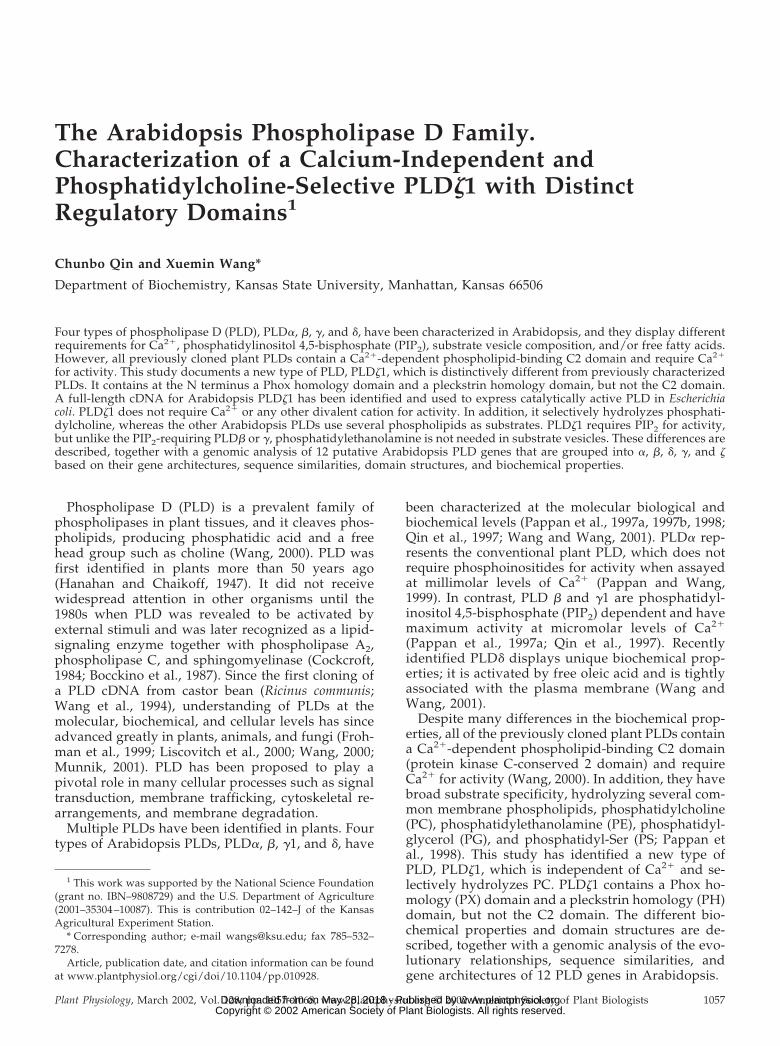
Twelve PLD genes were identified in the Arabi-dopsis genome by BLAST searching against thecloned cDNAs. Two of these genes are located onchromosome I, one on chromosome II, three on chro-mosome III, five on chromosome IV, and one onchromosome V (Fig. 1; Table I). They are groupedinto five classes, PLD�(1, 2, 3, 4), �(1, 2), �(1, 2, 3), �,and �(1, 2), based on the gene architectures (Fig. 1),sequence similarities (Fig. 2; Table II), domain struc-tures (Figs. 3 and 4), and biochemical properties (Ta-ble III). This classification updates an earlier one(Wang, 2000) in light of the new sequence informa-tion and analysis. In particular, the previously des-ignated �1 has been regrouped into the � class as �2,and the previous �2 is not included at this time, as itsputative genomic sequence is not annotated in theGenBank. The current PLD� encodes a newly char-acterized PLD that exhibits unique properties (Kata-giri et al., 2001; Wang and Wang, 2001).
Of the four � genes, PLD�1 and �2 are very similarin terms of gene structure and sequence similarity.Their deduced amino acid sequences share about92% similarity (Table II). Comparison of the clonedPLD cDNAs of castor bean and Arabidopsis PLD�1with their genomic sequences has shown the pres-ence of an intron in the 5�-untranslated region ofPLD�1 (Xu et al., 1997). Arabidopsis EST databasesearches against these two genes revealed a numberof EST clones corresponding to �1, but none for �2(Table I), indicating that they might differ greatly inabundance despite their high degree of sequencesimilarity. The complete cDNA for PLD�1 has beenisolated (Dyer et al., 1995), and its nucleotide anddeduced amino acid sequences show some discrep-ancies from those based on the genomic exons. Ver-ification of this and other PLD cDNA sequences isunder way to determine which differences resultfrom polymorphism or sequencing inaccuracies.
The PLD�3 and �4 genes contain four exons intheir coding region (Fig. 1). PLD�3 shares about 70%protein sequence similarity to �1 and �2. However,to date, no EST clone is yet identified for this gene.
Figure 1. Arabidopsis PLD gene structures and genomic organization. Boxes with numbers mark exons, and bars betweenexons represent introns. The exon and intron junctions are defined based on the comparison between PLD cDNAs and genesequences and/or predicted intron-exon splicing sites annotated in the GenBank database. The PLD gene sequences wereobtained by BLAST searching in GenBank using PLD cDNAs.
Qin and Wang
1058 Plant Physiol. Vol. 128, 2002 www.plantphysiol.orgon May 28, 2018 - Published by Downloaded from Copyright © 2002 American Society of Plant Biologists. All rights reserved.

The deduced amino acid sequence of PLD�4 isshorter than PLD�1, 2, and 3 (762 versus 810 or 820amino acids). It shows no more than 55% similarity tothe other three �s, or to any other member of Arabi-dopsis PLDs. EST clones for PLD�4 are present in thedatabase, so it is expressed. PLD�3 and �4 aregrouped into the � class because they have fourexons, whereas other PLDs have 10 or more exons(Fig. 1). In addition, their overall deduced amino acidsequences are evolutionarily closer to those ofPLD�1and �2 than to other PLDs (Fig. 2). Further-more, their domain structures involved in catalysis(Fig. 4), Ca2� binding (Fig. 5), and PIP2 interaction(Fig. 6) are more closely related to those of PLD�1and �2 than to the other PLDs. Thus, they may havesimilar biochemical properties to PLD�. However,the amino acid sequence of PLD�4 is quite distantfrom all PLDs (Table II), and it might possess uniqueregulatory and catalytic properties. Therefore, itsdesignation to the PLD� group is tentative, and itsappropriate position in the Arabidopsis PLD genefamily awaits further experimental characterizationof its domain structures and biochemical properties.
PLD�, �, and � genes all consist of 10 exons in thecoding region. The previously cloned and character-ized PLD� is now designated PLD�1, and the aminoacid sequences of PLD�1 and �2 are about 89% sim-ilar (Table II). The annotated gene structure of �2 inthe GenBank contains 11 exons, which is why it waspreviously designated as a new class (Wang, 2000).However, a close examination of the GenBank anno-tation indicates that the original prediction for thepresence of the first intron is somewhat ambiguous.Inclusion of this intron removes part of an openreading frame that encodes the beginning part of thePLD C2 domain. Changing this region to be part ofthe first exon would increase the overall similarity
between �2 and �1 from 85% to 89%. Thus, the firstintron is removed in Figure 1, although ultimatedetermination of the exon-intron structures of PLD�2awaits the cloning of its cDNA. EST database search-ing against the �2 gene has not identified any ex-pressed sequence. However, northern blottingprobed with a �2-specific fragment showed a weakbut positive result (C. Qin and X. Wang, unpublisheddata), suggesting at least that the �2 gene is ex-pressed in low abundance under certaincircumstances.
The three PLD� genes cluster in tandem on chro-mosome IV with a similar pattern of exon-intronspacing. The three PLD� genes are very closely re-lated, sharing almost 95% sequence similarities in thededuced amino acid level (Table II). The PLD� genewhose cDNA was first cloned and characterized wasdesignated �1. A complete cDNA for the secondPLD� gene, designated �2, was cloned recently, andsome of its biochemical properties are the same asthose of �1 (C. Qin and X. Wang, unpublished data).The third gene in the PLD� gene cluster is designatedas PLD�3. EST clones corresponding to the PLD�sequences have been identified, indicating that theyall are expressed.
PLD� is a single gene class (Fig. 1). The gene an-notated in the GenBank is actually much longer, withsix more exons on the 3� end that encodes a nuclearlocalization signal and an RNA-binding region.However, experimental evidence from cDNA cloningand transcript size has shown that the authenticPLD� gene is shorter than what was annotated andthat does not contain the six extra exons at the 3� end(Wang and Wang, 2001). Two full-length cDNAs ofPLD� have been cloned recently; they differ by 33nucleotides near the end of the second exon (Katagiriet al., 2001; Wang and Wang, 2001). The cDNA that is
Table I. Arabidopsis PLD genes, predicted proteins, cDNAs, and expressed sequence tag (EST)clones
Information on the gene locus is from The Institute for Genomic Research database (http://www.tigr.org/). Some of the predicted PLD protein sequences have more than one accession no.
Name Gene LocusProtein
Accession No.cDNA
EST CloneIdentified
PLD�1 At3g15730 BAB02304 U36381 YesPLD�2 At1g52570 AAD55607 NoPLD�3 At5g25370 NP_197919 NoPLD�4 At1g55180 AAG51567 YesPLD�1 At2g42010 AAB63542 U84568 YesPLD�2 At4g00240 AAF02803 NoPLD�1 At4g11850 CAB78228 AF027408 YesPLD�2 At4g11830 CAB78226 AF138281 YesPLD�3 At4g11840 CAB78227 YesPLD�aa At4g35790b CAB81488 AB031047 YesPLD�ba AF322228 YesPLD�1 At3g16790(5�) BAA95772 AF411833 Yes
At3g16785(3�)PLD�2 At3g05630 AAF26134 Yes
a Both are products of PLD� gene; see text for details. b Contains six extra exons at the C terminusthat need to be removed.
The Arabidopsis Phospholipase D Family
Plant Physiol. Vol. 128, 2002 1059 www.plantphysiol.orgon May 28, 2018 - Published by Downloaded from Copyright © 2002 American Society of Plant Biologists. All rights reserved.
33 nucleotides shorter encodes a catalytically activePLD (Wang and Wang, 2001). It is possible that thetwo cDNAs come from two transcripts resulting fromalternative splicing of the same PLD� gene. Thenames PLD�a and PLD�b are proposed to distin-guish the two PLD� transcripts and proteins (Wang
and Wang, 2001). The alphabetic affixes distinguishdifferent products of the same gene, whereas theArabic numerals represent separate PLD genes. A90-kD Arabidopsis PLD (accession no. AF306345) hasrecently been shown to be associated with microtu-bules and has been implicated in regulating cell di-vision (Gardiner et al., 2001). Its cDNA and deducedamino acid sequences exactly match those of PLD�b(accession no. AF322228).
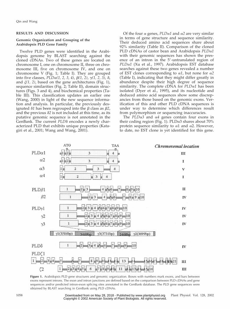
The last two members of putative PLD genes in theArabidopsis genome are grouped together as PLD�1and �2 (Fig. 1) because of their similar protein do-main structures (Fig. 3B). Both genes are located onchromosome III. The two PLDs share about 74% se-quence similarity in deduced amino acids, and bothare quite distant to all the other Arabidopsis PLDs,with no more than 46% sequence similarity (Table II).In addition, their deduced amino acid sequences are1,096 and 1,039 amino acids in length, respectively,which are longer than the other plant PLD proteins,but in the same range as the mammalian PLD1 (hu-man PLD1 is 1,074 amino acids). Sequence compari-son also suggests that PLD�s are closer to the mam-malian PLDs than to the other plant ones in terms ofamino acid sequence similarity (Table II) and theprotein domain structures (Figs. 3–5). EST cloneshave been identified for both PLD� genes.
Domain Structures of the Arabidopsis PLD Proteins
HKD Motif
The highly conserved domain in the PLD family isthe HKD motif, which is used to define the PLDsuperfamily. It was termed “HKD” because the do-main contains the motif HxKxxxxD/E, which isfound twice without exception in all cloned PLDs(Hammond et al., 1995; Koonin, 1996; Ponting andKerr, 1996). The two HKD motifs are far from eachother in the primary structure (Fig. 3), but they in-
Figure 2. Phylogenetic relationship of Arabidopsis PLDs. The treewas constructed from amino acid sequences deduced from genomicPLD sequences, with some modifications being made on PLD� andPLD�2 as described in the text. Accession numbers for these se-quences are listed in Table I. This was generated using the GrowTreeprogram from the University of Wisconsin Genetics Computer Group(GCG).
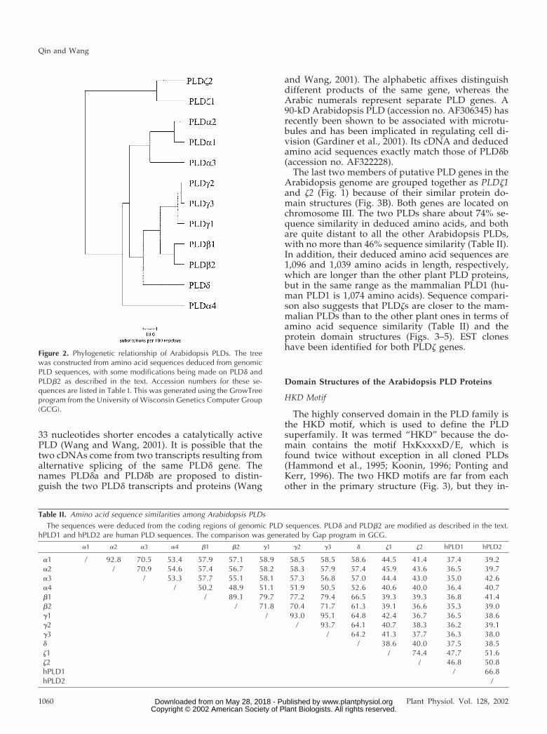
Table II. Amino acid sequence similarities among Arabidopsis PLDs
The sequences were deduced from the coding regions of genomic PLD sequences. PLD� and PLD�2 are modified as described in the text.hPLD1 and hPLD2 are human PLD sequences. The comparison was generated by Gap program in GCG.
�1 �2 �3 �4 �1 �2 �1 �2 �3 � �1 �2 hPLD1 hPLD2
�1 / 92.8 70.5 53.4 57.9 57.1 58.9 58.5 58.5 58.6 44.5 41.4 37.4 39.2�2 / 70.9 54.6 57.4 56.7 58.2 58.3 57.9 57.4 45.9 43.6 36.5 39.7�3 / 53.3 57.7 55.1 58.1 57.3 56.8 57.0 44.4 43.0 35.0 42.6�4 / 50.2 48.9 51.1 51.9 50.5 52.6 40.6 40.0 36.4 40.7�1 / 89.1 79.7 77.2 79.4 66.5 39.3 39.3 36.8 41.4�2 / 71.8 70.4 71.7 61.3 39.1 36.6 35.3 39.0�1 / 93.0 95.1 64.8 42.4 36.7 36.5 38.6�2 / 93.7 64.1 40.7 38.3 36.2 39.1�3 / 64.2 41.3 37.7 36.3 38.0� / 38.6 40.0 37.5 38.5�1 / 74.4 47.7 51.6�2 / 46.8 50.8hPLD1 / 66.8hPLD2 /
Qin and Wang
1060 Plant Physiol. Vol. 128, 2002 www.plantphysiol.orgon May 28, 2018 - Published by Downloaded from Copyright © 2002 American Society of Plant Biologists. All rights reserved.
teract with each other to form the active site. Pointmutagenesis of PLD from several species has re-vealed that these amino acids are critical for catalysisin vitro and for PLD function in vivo (Sung et al.,1997; Gottlin et al., 1998). This has led to a model forthe catalytic cycle using the conserved His for nu-cleophilic attack on the substrate phosphorous andinvolving a covalent phosphatidyl enzyme interme-diate. The crystal structure of the bacterial endonu-
clease member Nuc of the PLD superfamily, whichencodes a single and very divergent HKD motif, hasbeen determined (Stuckey and Dixon, 1999). Recentstructural studies have also led to expansion of thesecond HKD motif of the PLD family to HxKxxxx-DxxxxxxGSxN. However, definition of the preciserole that each amino acid plays will await determi-nation from structural analyses of an eukaryotic PLDcrystallized with its normal substrate.
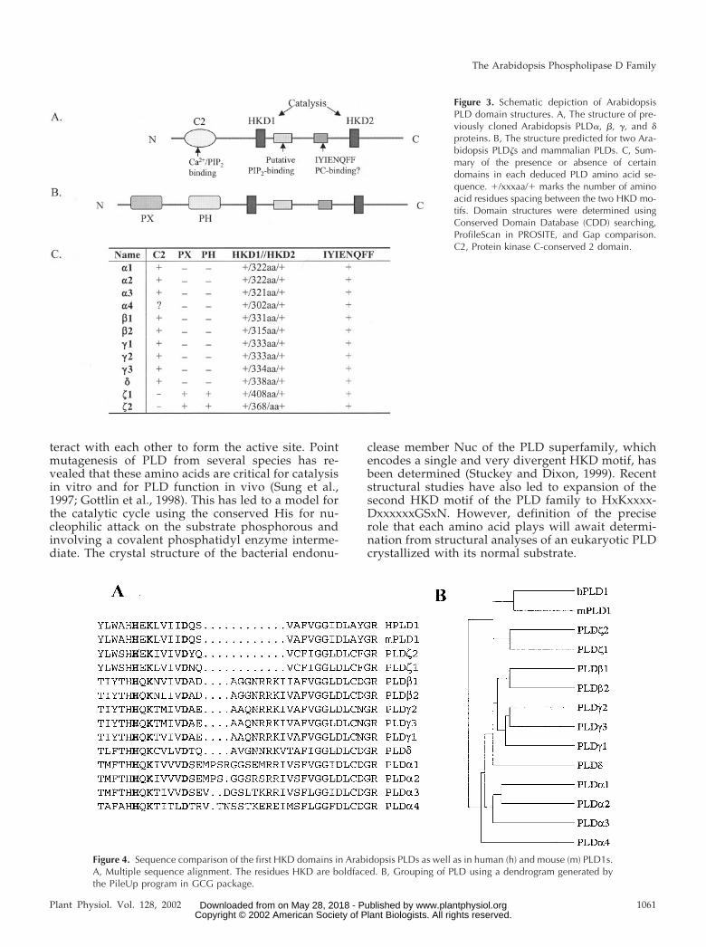
Figure 3. Schematic depiction of ArabidopsisPLD domain structures. A, The structure of pre-viously cloned Arabidopsis PLD�, �, �, and �proteins. B, The structure predicted for two Ara-bidopsis PLD�s and mammalian PLDs. C, Sum-mary of the presence or absence of certaindomains in each deduced PLD amino acid se-quence. �/xxxaa/� marks the number of aminoacid residues spacing between the two HKD mo-tifs. Domain structures were determined usingConserved Domain Database (CDD) searching,ProfileScan in PROSITE, and Gap comparison.C2, Protein kinase C-conserved 2 domain.
Figure 4. Sequence comparison of the first HKD domains in Arabidopsis PLDs as well as in human (h) and mouse (m) PLD1s.A, Multiple sequence alignment. The residues HKD are boldfaced. B, Grouping of PLD using a dendrogram generated bythe PileUp program in GCG package.
The Arabidopsis Phospholipase D Family
Plant Physiol. Vol. 128, 2002 1061 www.plantphysiol.orgon May 28, 2018 - Published by Downloaded from Copyright © 2002 American Society of Plant Biologists. All rights reserved.
These duplicated HKD motifs are conserved indeduced amino acid sequences of all the 12 Arabi-dopsis PLD genes. They are separated by approxi-mately 320 amino acids, except for �1 and �2, whichhave longer spacing sequences (Fig. 3C). Sequencealignment reveals that although all the second HKDmotif structures look highly identical, the first HKDsand flanking regions display more diversity (Fig.4A). The 10 exon-containing PLDs, four exon-containing PLD�s, and PLD�s form three distinctgroups (Fig. 4B). The first HKD domains in the �group are more similar to that of hPLD1, again indi-cating a closer relationship between PLD� genes andthe mammalian PLDs. The dendrogram of the clus-tering relationship of the HKD1 domains (Fig. 4B) isconsistent with the phylogenetic tree of the wholeproteins (Fig. 2).
“IYIENQFF” Motif
The “IYIENQFF” region between the two HKDmotifs is also called conserved region III in mamma-lian PLD structures and is found only in family mem-bers that exhibit bona fide PLD activity (Frohman etal., 1999). Mutagenesis studies have demonstratedthat it is almost as critical as the HKD domains (Sunget al., 1997), but the precise role has yet to be deter-
mined. This region may be responsible for hydropho-bic interactions with the methyl groups of the cholinehead group, due to the enrichment of aromatic aminoacids. An alternative hypothesis is that it might en-code the targeting signal for caveolae (Frohman et al.,1999). This “IYIENQFF” motif is the most conserveddomain with the least diversity among the 12 Arabi-dopsis PLDs. This sequence in the PLD� group isexactly the same as that in mammalian PLDs. For theother PLDs, only the seventh residue, Phe(F), is sub-stituted by a Tyr(Y), which should not alter the prop-erty of this region.
Ca2�/Phospholipid-Binding C2 Domain
All previously cloned plant PLDs have a C2 do-main near the N terminus, which is unique to plantPLDs and is not present in animal or fungal PLDs.The C2 domains are approximately 130 residues inlength and can bind Ca2� and other effectors, includ-ing phospholipids, inositol phosphates, and proteins(Nalefski and Falke, 1996; Rizo and Sudhof, 1998;Zheng et al., 2000). The three-dimensional structuresand the Ca2�-binding properties of C2 domains insynatagmin (Shao et al., 1996), cPLA2 (Perisic et al.,1998; Xu et al., 1998), PLC�1 (Essen et al., 1997), andPKC� (Sutton and Sprang, 1998) have been well stud-
Figure 5. Sequence alignment of the Arabidopsis PLD C2 domains with PLC�1 and cPLA2. The conserved amino acidresidues involved in Ca2� binding are boldfaced. The non-Ca2� ligand residues that occur in positions corresponding to theCa2� ligands are underlined. Only the portion involved in Ca2� binding is shown, and the �-sheet strands and Ca2�-bindingloops are defined according to a previous study (Perisic et al., 1998). CBL, Ca2�-binding loops.
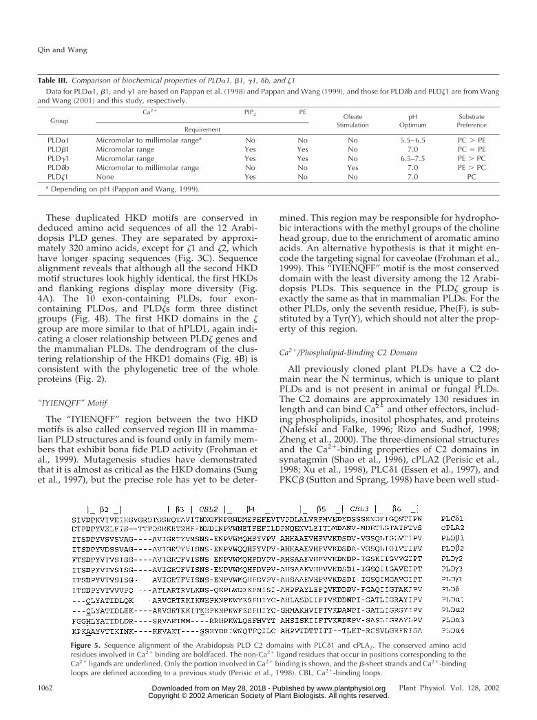
Table III. Comparison of biochemical properties of PLD�1, �1, �1, �b, and �1
Data for PLD�1, �1, and �1 are based on Pappan et al. (1998) and Pappan and Wang (1999), and those for PLD�b and PLD�1 are from Wangand Wang (2001) and this study, respectively.
GroupCa2� PIP2 PE
OleateStimulation
pHOptimum
SubstratePreference
Requirement
PLD�1 Micromolar to millimolar rangea No No No 5.5–6.5 PC � PEPLD�1 Micromolar range Yes Yes No 7.0 PC � PEPLD�1 Micromolar range Yes Yes No 6.5–7.5 PE � PCPLD�b Micromolar to millimolar range No No Yes 7.0 PE � PCPLD�1 None Yes No No 7.0 PC
a Depending on pH (Pappan and Wang, 1999).
Qin and Wang
1062 Plant Physiol. Vol. 128, 2002 www.plantphysiol.orgon May 28, 2018 - Published by Downloaded from Copyright © 2002 American Society of Plant Biologists. All rights reserved.

ied by x-ray crystallography and/or NMR. Althoughthe C2 domains have two topological variants, theirthree-dimensional structures are strikingly con-served. Ca2� binding is coordinated by four to fiveamino acid residues in bipartite loops within the C2domain.
Arabidopsis PLD�s, �s, and � all have the Ca2�-coordinating acidic residues, whereas the PLD� C2domains lack one or more of these potential Ca2�
ligands (Fig. 5). Compared with cPLA2, PLD�1 hasone, and �2 and �3 have two of them substituted,whereas �4 contains none of the Ca2�-binding resi-dues. The presence of C2 domain in PLD�4 is notclear, but several reserved hydrophobic amino acids,which have been proposed to maintain the structuralintegrity of the C2 fold, are present in its correspond-ing region. On the other hand, Conserved DomainDatabase (CDD) searching and Gap comparison didnot reveal the existence of the C2 domain in the twoPLD�s.
The differences in the Ca2�-binding residues sug-gests that Ca2� affinity of PLD�s could be lower thanthat of �, �, and �. This lower Ca2� affinity ofPLD�-C2 than PLD�-C2 domain has been demon-strated experimentally (Zheng et al., 2000). Ca2�- andPC-binding properties of PLD� C2 and PLD� C2follow a trend similar to the Ca2� requirements of thewhole enzymes, PLD�1 and PLD�1, for PC hydroly-sis (Table III). Thus, it has been suggested that the C2domains of PLD� and PLD� serve as handles bywhich Ca2� differentially regulates these PLD activ-ities (Zheng et al., 2000).
PIP2-Binding Motifs
PIP2 plays a critical role in PLD activation in mam-mals and plants. PIP2 is required for activities ofArabidopsis PLD� and �; replacement of PIP2 byother phospholipids such as PC, PS, PG, PE, andphosphatidylinositol (PI) resulted in loss of PLD ac-tivity (Qin et al., 1997). Two putative PIP2-bindingmotifs were also predicted, flanking the second HKDdomain of PLD� (Pappan et al., 1997b), but the PIP2-
binding ability of these motifs in PLD has not beenverified experimentally. Recent studies on mousePLD2 have suggested that a small region containingmany conserved basic and hydrophobic residues lo-cated between the two HKD motifs is responsible forthe PIP2 requirement (Fig. 6; Sciorra et al., 1999).Mutagenesis experiments revealed that this region isrequired for activation of mammalian and yeastPLDs by PIP2.
Sequence alignments of PLDs from plants, animals,and yeast reveals conservation and variation of thebasic residues of this PIP2-binding motif in plantPLDs (Fig. 6). The clustering relationship is quitesimilar to that revealed by comparison of HKD1 do-mains. For the five conserved basic amino acids orig-inally identified in mouse PLD2, human PLD1, andyeast PLD (Sciorra et al., 1999), only the last one issubstituted by an acidic residue in the two PLD�s(Fig. 6). PLD�s and �s all conserved the last threeresidues, but the first two are replaced by negativelycharged residues or by neutral ones. PLD� retainstwo of the conserved residues, whereas PLD�s haveonly one of them (Fig. 6). A recent study usingPLD�1 has provided experimental evidence for thismotif as a PIP2-binding region, and this binding isstimulated by Ca2� (L. Zheng, R. Krishnamoorthi,and X. Wang, unpublished data). It should be notedthat the N-terminal C2 domain of PLDs also bindsPIP2, but unlike the PIP2-binding region, the C2-PIP2interaction is inhibited by Ca2� (Zheng et al., 2000).
PX Domain
A conserved PX domain is present in ArabidopsisPLD �1 and �2, as well as in mammalian PLDs, but isnot found in the other Arabidopsis PLDs (Fig. 3B).The PX domain was originally identified in the pro-tein p47phox, a component of phagocyte NADPH ox-idase (Ponting, 1996). It is a novel protein modulecontaining a conserved Pro-rich motif. Recent struc-tural and cell biological studies show that the PXdomain can bind phosphoinositides and SH3 domain(Cheever et al., 2001; Hiroaki et al., 2001; Kanai et al.,
Figure 6. Alignment of a putative PIP2-bindingmotif in Arabidopsis PLDs, as well as in mouse(m) PLD2, human (h) PLD1, and yeast PLD(Spo14). The conserved basic amino acids orig-inally identified in mouse PLD2 are in bold-face, and some additional basic residues areunderlined.
The Arabidopsis Phospholipase D Family
Plant Physiol. Vol. 128, 2002 1063 www.plantphysiol.orgon May 28, 2018 - Published by Downloaded from Copyright © 2002 American Society of Plant Biologists. All rights reserved.
2001). Therefore, it may play a critical role in coordi-nating membrane localization and protein complexassembly during cell signaling. The PX domain iscritical for activities of mammalian PLDs (Sung et al.,1999a, 1999b). How it functions is unknown, al-though potential roles include regulating interactionswith factors that promote translocation or activation.
PH Domain
PH domain is another potential regulatory modulepresent in Arabidopsis PLD �1 and �2, as well as inmammalian PLDs, but not in any other ArabidopsisPLDs (Fig. 3B). PH domains are composed of approx-imately 120 amino acids found in more than 100proteins involved in cell signaling, cytoskeletal rear-rangement, and other processes (Lemmon and Fer-guson, 2000). All PH domains studied to date appearto bind to phosphoinositides, but most bind weaklyand nonspecifically. Only a small subclass of PHdomains show strong and specific binding to mem-brane phosphoinositides (Lemmon and Ferguson,2000). Deletion analysis of mammalian PLD1 andPLD2 revealed that the PH domain is not required forenzymatic activity nor is the dependence of the en-zymatic activity on PIP2 affected by deletion of thePH domain (Sung et al., 1999a, 1999b). PH domaincould be important in membrane targeting and asso-ciation with other cellular components. However, itsfunction in PLDs awaits further investigation.
PLD�1 Is a Calcium-Independent, PC-Selective PLD
To characterize the PX/PH-containing, putativePLD�s, the Arabidopsis EST database was searchedfor putative full-length cDNA clones of PLD�s usingthe PLD� coding sequences annotated in the Gen-Bank database. Several putative PLD�1 EST cloneswere identified, and one (EST no. AV529766) wassequenced completely and found to be a full-lengthPLD�1 cDNA. The cDNA is composed of 3,785 nu-cleotides, and the nucleotide sequence of the cDNAmatches that annotated from the genomic sequenc-ing. The coding region of the PLD�1 cDNA starts atnucleotide 248 and ends at 3,538 (GenBank accessionno. AF411833). It encodes a protein of 1,096 aminoacids with the domain structures depicted in Figure3B. The calculated molecular mass and pI are 124 kDand 6.27, respectively.
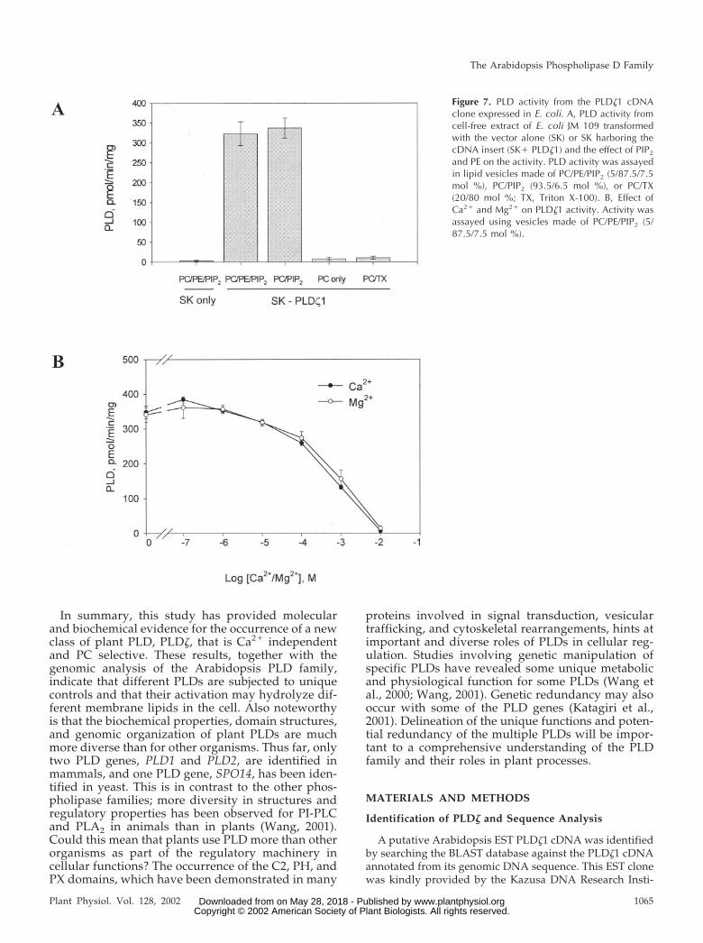
To validate that this cDNA encodes a PLD, proteinfrom the cDNA was expressed in Escherichia coli us-ing pBluescript SK(-) as expression vector, which hasbeen used successfully to express catalytically activePLD�, �, and �1 (Wang et al., 1994; Pappan et al.,1997b; Qin et al., 1997). After isopropyl-1-thio-�-d-galactopyranoside induction, protein extracts from E.coli JM109 harboring the SK alone exhibited negligi-ble PLD activity (Fig. 7A). Proteins from E. coli har-boring the vector with the PLD�1 cDNA insert had
significant PLD activity. PLD�1 required PIP2 foractivity; it displayed no activity when PC-only vesi-cles and PC/Triton X-100 vesicles were used as sub-strates (Fig. 7A). The PIP2 requirement is a propertyshared by PLD� and �, as well as mammalian andyeast PLDs, but the activity of PLD� and � also needsPE (Table III; Pappan et al., 1998). In contrast, PLD�1requires only PIP2, but not PE (Fig. 7A).
Previous analysis of PLD� and PLD� indicates thatthe C2 domain is a major determinant for the require-ment of different levels of Ca2� for their activities(Zheng et al., 2000). Accordingly, the absence of theC2 domain in PLD�1 (Fig. 3B) could mean that thisPLD might not require Ca2� for activity. To test thishypothesis, 2 mm EGTA and 2 mm EDTA wereadded to the reaction to chelate Ca2� and any otherdivalent cation. In addition, varied concentrations ofCaCl2 or MgCl2 were used to examine the cationeffect on PLD�1. The highest PLD�1 activity occurredin the zero to nanomolar concentrations of Ca2� andMg2� (Fig. 7B). The activity decreased gradually atthe micromolar range and dropped rapidly whencation concentrations approached millimolar levels(Fig. 7B). These results show that PLD�1 is indepen-dent of Ca2� or any other cation for activity. Thisproperty is in contrast to all the other cloned plantPLDs that require micromolar or millimolar ranges ofCa2� for activity (Wang, 2000; Table III).
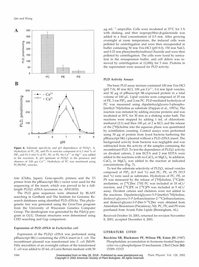
The substrate specificity was examined next.PLD�1 hydrolyzed PC well, but had negligible activ-ity toward PE or PS (Fig. 8A). No PG-hydrolyzingactivity was observed (data not shown). This PC-selective activity is distinctively different from othercharacterized Arabidopsis PLDs, but similar to thecloned mammalian ones. PLD� hydrolyzes PC, PE,and PG equally well, and PLD� and � hydrolyze PC,PE, and PS (Pappan et al., 1998; Table III), whereasPLD� uses PE better than PC (C. Qin, C. Wang, andX. Wang, unpublished data). The mammalian PLD1is PC specific and has no activity toward PE or PI(Hammond et al., 1995), and activity of the clonedmammalian PLD2 toward lipids other than PC hasnot been reported.
PLD�1 functioned at a rather narrow range of pHwith a pH optimum at 7. Its activity decreased con-siderably at pH 6.5 and 7.5, and was virtually abol-ished at pH 6 and 8 (Fig. 8B). PLD� and �1 are alsomost active at pH 7, but their functional pH isbroader than that of PLD�1 (Pappan and Wang, 1999;Table III). PLD� has a comparable activity from pH5.5 to 8.5. PLD� has a similar activity at pH 6.5 to 7.5,and has still approximately 35% activity at pH 5.PLD� has acidic pH optima that are influenced bythe Ca2� concentrations. The pH optimum is 4.5 to 5when assayed in the presence of micromolar levels ofCa2� and PIP2, but it increases to 5.5 to 6.5 at milli-molar levels of Ca2� (Pappan and Wang, 1999). How-ever, the presence or absence of Ca2� did not alter thepH optimum of PLD�1 (Fig. 8B).
Qin and Wang
1064 Plant Physiol. Vol. 128, 2002 www.plantphysiol.orgon May 28, 2018 - Published by Downloaded from Copyright © 2002 American Society of Plant Biologists. All rights reserved.
In summary, this study has provided molecularand biochemical evidence for the occurrence of a newclass of plant PLD, PLD�, that is Ca2� independentand PC selective. These results, together with thegenomic analysis of the Arabidopsis PLD family,indicate that different PLDs are subjected to uniquecontrols and that their activation may hydrolyze dif-ferent membrane lipids in the cell. Also noteworthyis that the biochemical properties, domain structures,and genomic organization of plant PLDs are muchmore diverse than for other organisms. Thus far, onlytwo PLD genes, PLD1 and PLD2, are identified inmammals, and one PLD gene, SPO14, has been iden-tified in yeast. This is in contrast to the other phos-pholipase families; more diversity in structures andregulatory properties has been observed for PI-PLCand PLA2 in animals than in plants (Wang, 2001).Could this mean that plants use PLD more than otherorganisms as part of the regulatory machinery incellular functions? The occurrence of the C2, PH, andPX domains, which have been demonstrated in many
proteins involved in signal transduction, vesiculartrafficking, and cytoskeletal rearrangements, hints atimportant and diverse roles of PLDs in cellular reg-ulation. Studies involving genetic manipulation ofspecific PLDs have revealed some unique metabolicand physiological function for some PLDs (Wang etal., 2000; Wang, 2001). Genetic redundancy may alsooccur with some of the PLD genes (Katagiri et al.,2001). Delineation of the unique functions and poten-tial redundancy of the multiple PLDs will be impor-tant to a comprehensive understanding of the PLDfamily and their roles in plant processes.
MATERIALS AND METHODS
Identification of PLD� and Sequence Analysis
A putative Arabidopsis EST PLD�1 cDNA was identifiedby searching the BLAST database against the PLD�1 cDNAannotated from its genomic DNA sequence. This EST clonewas kindly provided by the Kazusa DNA Research Insti-
Figure 7. PLD activity from the PLD�1 cDNAclone expressed in E. coli. A, PLD activity fromcell-free extract of E. coli JM 109 transformedwith the vector alone (SK) or SK harboring thecDNA insert (SK� PLD�1) and the effect of PIP2
and PE on the activity. PLD activity was assayedin lipid vesicles made of PC/PE/PIP2 (5/87.5/7.5mol %), PC/PIP2 (93.5/6.5 mol %), or PC/TX(20/80 mol %; TX, Triton X-100). B, Effect ofCa2� and Mg2� on PLD�1 activity. Activity wasassayed using vesicles made of PC/PE/PIP2 (5/87.5/7.5 mol %).
The Arabidopsis Phospholipase D Family
Plant Physiol. Vol. 128, 2002 1065 www.plantphysiol.orgon May 28, 2018 - Published by Downloaded from Copyright © 2002 American Society of Plant Biologists. All rights reserved.
tute (Chiba, Japan). Gene-specific primers and the T3primer from the pBluescript SK(-) vector were used for thesequencing of the insert, which was proved to be a full-length PLD�1 cDNA (accession no. AF411833).
The PLD gene sequences were obtained by BLASTsearching in GenBank and The Institute for Genomic Re-search databases using identified PLD cDNAs. This phylo-genetic tree was generated using the GrowTree programfrom the University of Wisconsin Genetics ComputerGroup. The dendrogram was generated by the PileUp pro-gram in GCG. Domain structures were determined usingCDD searching and Gap comparison.
Expression of PLD cDNA in Escherichia coli
Expression of the PLD�1 cDNA was performed usingpBluescript SK(-) containing the cDNA insert in E. coli. Therecombinant plasmid was transformed into E. coli JM109.Fifty microliters of an overnight culture of the transformedE. coli was added to 25 mL of Luria Bertani medium with 50
�g mL�1 ampicillin. Cells were incubated at 37°C for 3 hwith shaking, and then isopropylthio-�-galactoside wasadded to a final concentration of 0.5 mm. After growingovernight at room temperature, the induced cells werepelleted by centrifugation and were then resuspended inbuffer containing 50 mm Tris-HCl (pH 8.0), 150 mm NaCl,and 0.25 mm phenylmethylsulfonyl fluoride and were thenpelleted by centrifugation. The cells were lysed by sonica-tion in the resuspension buffer, and cell debris was re-moved by centrifugation at 12,000g for 5 min. Proteins inthe supernatant were assayed for PLD activity.
PLD Activity Assays
The basic PLD assay mixture contained 100 mm Tris-HCl(pH 7.0), 80 mm KCl, 100 �m Ca2�, 0.4 mm lipid vesicles,and 30 �g of pBluescript SK-expressed protein in a totalvolume of 100 �L. Lipid vesicles were composed of 35 nmof PE, 3 nm PIP2, and 2 nm PC. PLD-mediated hydrolysis ofPC was measured using dipalmitoylglycero-3-phospho-[methyl-3H]choline as substrate (Pappan et al., 1997a). Thereaction was initiated by adding enzyme proteins and wasincubated at 30°C for 30 min in a shaking water bath. Thereactions were stopped by adding 1 mL of chloroform:methanol (2:1) and then 100 �L of 2 m KCl, and the releaseof the [3H]choline into the aqueous phase was quantitatedby scintillation counting. Control assays were performedusing 30 �g of protein from lysed bacteria harboring thepBluescript SK(-) plasmid without a PLD cDNA insert. Thebackground activity from bacteria was negligible and wassubtracted from the activity of the samples containing therecombinant PLD. To test the dependence of PLD�1 activityon divalent cations, 2 mm EGTA and 2 mm EDTA wereadded to the reactions with no CaCl2 or MgCl2. In addition,CaCl2 or MgCl2 was added to the reaction at indicatedconcentrations (Fig. 7).
To test the substrate selectivity of PLD�1, mixed vesiclescomposed of PIP2 (6.5 mol %) and PC, PE, or PS (93.5mol %) were used as substrates. Hydrolysis of PC, PE, orPS was measured by the release of [3H]choline, [14C]eth-anolamine, or [14C]Ser. [3H] PC was included at 18 nCi/reaction, and [14C]PE or [14C]PS was included at 9 nCi/assay. Divalent cations and chelators were not added tothe reactions. Dipalmitoylglycero-3-P-[methyl-3H]choline,dioleoyl-glycero-3-P-[ethanolamine-2-14C]ethanolamine,and dioleoyl-glycero-3-P-[Ser-3-14C]Ser were obtained fromAmersham Biosciences (Piscataway, NJ). PC, PE, and PS werepurchased from Avanti Polar Lipids (Birmingham, AL).
Received October 10, 2001; returned for revision November8, 2001; accepted December 4, 2001.
LITERATURE CITED
Bocckino SB, Blackmore PF, Wilson PB, Exton JH (1987)Phosphatidate accumulation in hormone-treated hepato-cytes via a phospholipase D mechanism. J Biol Chem 262:15309–15315
Figure 8. Substrate specificity and pH dependence of PLD�1. A,Hydrolysis of PC, PE, and PS in vesicles composed of 6.5 mol % ofPIP2 and 93.5 mol % of PC, PE, or PS. No Ca2� or Mg2� was addedto the reactions. B, pH optimum of PLD�1 in the presence andabsence of 100 �M Ca2�. Hydrolysis of PC was monitored usingPC/PE/PIP2 vesicles.
Qin and Wang
1066 Plant Physiol. Vol. 128, 2002 www.plantphysiol.orgon May 28, 2018 - Published by Downloaded from Copyright © 2002 American Society of Plant Biologists. All rights reserved.
Cheever ML, Sato TK, de Beer T, Kutateladze TG, EmrSD, Overduin M (2001) Phox domain interaction with-PtdIns(3) P targets the Vam7 t-SNARE to vacuole mem-branes. Nat Cell Biol 3: 613–618
Cockcroft S (1984) Ca2�-dependent conversion of phos-phatidylinositol to phosphatidate in neutrophils stimu-lated with fMet-Leu-Phe or ionophore A23187. BiochimBiophys Acta 795: 37–46
Dyer JH, Zheng L, Wang X (1995) Cloning and nucleotidesequence of a cDNA encoding phospholipase D fromArabidopsis (Accession No. U36381) (PGR 95-096).Plant Physiol 109: 1497
Essen LO, Perisic O, Lynch DE, Katan M, Williams RL(1997) A ternary metal binding site in the C2 domain ofphosphoinositide-specific phospholipase C-d1. Bio-chemistry 36: 2753–2762
Frohman MA, Sung TC, Morris AJ (1999) Mammalianphospholipase D structure and regulation. Biochim Bio-phys Acta 1439: 175–186
Gardiner JC, Harper JDI, Weerakoon ND, Collings DA,Ritchie S, Gilroy S, Cyr RJ, Marc J (2001) A 90-kDphospholipase D from tobacco binds to microtubulesand the plasma membrane. Plant Cell 13: 2143–2158
Gottlin EB, Rudolph AE, Zhao Y, Matthews HR (1998)Catalytic mechanism of the phospholipase D superfam-ily proceeds via a covalent phosphohistidine interme-diate. Proc Natl Acad Sci USA 95: 9202–9207
Hammond SM, Altshuller YM, Sung TC, Rudge SA,Rose K, Engebrecht J, Morris AJ, Frohman MA (1995)Human ADP-ribosylation factor-activatedphosphatidylcholine-specific phospholipase D defines anew and highly conserved gene family. J Biol Chem 270:29640–29643
Hanahan DJ, Chaikoff IL (1947) A new phospholipid-splitting enzyme specific for the ester linkage betweenthe nitrogenous base and the phosphoric acid grouping.J Biol Chem 169: 699–705
Hiroaki H, Ago T, Ito T, Sumimoto H, Kohda D (2001)Solution structure of the PX domain, a target of the SH3domain. Nat Struct Biol 8: 526–530
Kanai F, Liu H, Field SJ, Akbary H, Matsuo T, BrownGE, Cantley LC, Yaffe MB (2001) The PX domains ofp47phox and p40phox bind to lipid products of PI(3) K.Nat Cell Biol 3: 675–678
Katagiri T, Takahashi S, Shinozaki K (2001) Involvementof a novel Arabidopsis phospholipase D, AtPLD�, indehydration-inducible accumulation of phosphatidicacid in stress signaling. Plant J 26: 595–605
Koonin EV (1996) A duplicated catalytic motif in a newsuperfamily of phosphohydrolases and phospholipidsynthases that includes poxvirus envelope proteins.Trends Biochem Sci 21: 242–243
Lemmon MA, Ferguson KM (2000) Signal-dependentmembrane targeting by pleckstrin homology (PH) do-mains. Biochem J 350: 1–18
Liscovitch M, Czarny M, Fiucci G, Tang X (2000) Phos-pholipase D: molecular and cell biology of a novel genefamily. Biochem J 345: 401–415
Munnik T (2001) Phosphatidic acid: an emerging plantlipid second messenger. Trends Plant Sci 6: 227–233
Nalefski EA, Falke JJ (1996) The C2 domain calcium-binding motif: structural and functional diversity. Pro-tein Sci 5: 2375–2390
Pappan K, Austin-Brown S, Chapman KD, Wang X(1998) Substrate selectivities and lipid modulation ofplant phospholipase D�, -�, and -�. Arch Biochem Bio-phys 353: 131–140
Pappan K, Qin W, Dyer JH, Zheng L, Wang X (1997a)Molecular cloning and functional analysis ofpolyphosphoinositide-dependent phospholipase D,PLD�, from Arabidopsis. J Biol Chem 272: 7055–7061
Pappan K, Wang X (1999) Plant phospholipase D� is anacidic phospholipase active at near-physiological Ca2�
concentrations. Arch Biochem Biophys 368: 347–353Pappan K, Zheng S, Wang X (1997b) Identification and
characterization of a novel plant phospholipase D thatrequires polyphosphoinositides and submicromolar cal-cium for activity in Arabidopsis. J Biol Chem 272:7048–7054
Perisic O, Fong S, Lynch DE, Bycroft M, Williams RL(1998) Crystal structures of a calcium-phospholipidbinding domain from cytosolic phospholipase A2. J BiolChem 273: 1596–1604
Ponting CP (1996) Novel domains in NADPH oxidasesubunits, sorting nexins, and PtdIns 3-kinases: bindingpartners of SH3 domain? Protein Sci 5: 2353–2357
Ponting CP, Kerr ID (1996) A novel family of phospho-lipase D homologous that includes phospholipid syn-thases and putative endonucleases: identification of du-plicated repeats and potential active site residues.Protein Sci 5: 914–922
Qin W, Pappan K, Wang X (1997) Molecular heterogene-ity of phospholipase D(PLD): cloning of PLD� and reg-ulation of plant PLD�, -�, and -� by polyphosphoi-nositides and calcium. J Biol Chem 272: 28267–28273
Rizo J, Sudhof TC (1998) C2-domains, structure and func-tion of a universal Ca2�-binding domain. J Biol Chem273: 15879–15882
Sciorra VA, Rudge SA, Prestwich GD, Frohman MA,Engebrecht J, Morris AJ (1999) Identification of a phos-phoinositide binding motif that mediates activation ofmammalian and yeast phospholipase D isoenzymes.EMBO J 18: 5911–5921
Shao X, Davletov BA, Sutton RB, Sudhof TC, Rizo J(1996) Bipartite Ca2�-binding motif in C2 domains ofsynaptotagmin and protein kinase C. Science 273:248–251
Stuckey JA, Dixon JE (1999) Crystal structure of a phos-pholipase D family member. Nat Struct Biol 6: 278–284
Sung TC, Altshuller YM, Morris AJ, Frohman MA(1999a) Molecular analysis of mammalian phospho-lipase D2. J Biol Chem 274: 494–502
Sung TC, Roper RL, Zhang Y, Rudge SA, Temel R, MossB, Engebrecht J, Frohman MA (1997) Mutagenesis ofphospholipase D defines a superfamily including atrans-Golgi, viral protein required for poxvirus patho-genicity. EMBO J 16: 4519–4530
Sung TC, Zhang Y, Morris AJ, Frohman MA (1999b)Structural analysis of human phospholipase D1. J BiolChem 274: 3659–3666
The Arabidopsis Phospholipase D Family
Plant Physiol. Vol. 128, 2002 1067 www.plantphysiol.orgon May 28, 2018 - Published by Downloaded from Copyright © 2002 American Society of Plant Biologists. All rights reserved.
Sutton RB, Sprang SR (1998) Structure of the proteinkinase C� phospholipid-binding C2 domain complexwith Ca2�. Structure 6: 1395–1405
Wang C, Wang X (2001) A novel Arabidopsis PLD that isactivated by oleate and associated with the plasmamembrane. Plant Physiol 127: 1102–1112
Wang C, Zien C, Afitlhile M, Welti R, Hildebrand DF,Wang X (2000) Involvement of phospholipase D inwound-induced accumulation of jasmonic acid in Ara-bidopsis. Plant Cell 12: 2237–2246
Wang X (2000) Multiple forms of phospholipase D inplants: the gene family, catalytic and regulatory proper-ties, and cellular functions. Prog Lipid Res 39: 109–149
Wang X (2001) Plant phospholipases. Annu Rev PlantPhysiol Plant Mol Biol 52: 211–231
Wang X, Xu L, Zheng L (1994) Cloning and expressionof phosphatidylcholine hydrolyzing phospholipaseD from Ricinus communis. J Biol Chem 269: 20312–20317
Xu G, McDonagh T, Yu H, Nalefski EA, Clark JD, Cum-ming DA (1998) Solution structure and membrane in-teractions of the C2 domain of cytosolic phospholipaseA2. J Mol Biol 280: 485–500
Xu L, Zheng S, Zheng L, Wang X (1997) Promoter anal-ysis and expression of a phospholipase D gene fromcastor bean. Plant Physiol 115: 387–395
Zheng L, Krishnamoorthi R, Zolkiewski M, Wang X(2000) Distinct Ca2� binding properties of novel C2domains of plant phospholipase D � and �. J Biol Chem275: 19700–19706
Qin and Wang
1068 Plant Physiol. Vol. 128, 2002 www.plantphysiol.orgon May 28, 2018 - Published by Downloaded from Copyright © 2002 American Society of Plant Biologists. All rights reserved.