THE APPLICATION OF HIGH SENSITIVITY LASER METHODS FOR ...library/doktorati/okolje/8Boskin.pdf ·...
104
UNIVERSITY OF NOVA GORICA GRADUATE SCHOOL THE APPLICATION OF HIGH SENSITIVITY LASER METHODS FOR DETECTION OF ORGANOPHOSPHORUS PESTICIDES AND CHOLINESTERASE ACTIVITY Aleš BOŠKIN Dissertation Mentor: prof. dr. Mladen Franko Nova Gorica, 2008
Transcript of THE APPLICATION OF HIGH SENSITIVITY LASER METHODS FOR ...library/doktorati/okolje/8Boskin.pdf ·...

UNIVERSITY OF NOVA GORICA GRADUATE SCHOOL
THE APPLICATION OF HIGH SENSITIVITY LASER METHODS FOR DETECTION OF ORGANOPHOSPHORUS
PESTICIDES AND CHOLINESTERASE ACTIVITY
Aleš BOŠKIN
Dissertation
Mentor: prof. dr. Mladen Franko
Nova Gorica, 2008

Filipu in njegovi mami, Jasni .........
II

ZAHVALA
ŽIVLJENJE JE ZAPRAVLJENO IMETJE, ČE GA NE ŽIVIŠ, KOT SI GA ŽELIŠ Vsakdo izmed VAS je delček tega mozaika. In ta ne bi bil popoln, če še tako majhen in navidezno nepomemben košček manjka. Zato iskrena HVALA vsem, ki ste tako ali drugače „krivi“, da zdaj lahko to berete, vsem, ki ste začrtali mojo tukajšnjo pot in poskrbeli, da mi ni bilo ob tem nikoli dolgčas. Hvala za spoznanja, dragocene izkušnje, bodritve in spodbude takrat, ko jih človek najbolj rabi. Lepo je bilo .......

IV
POVZETEK
Organofosfatne (OP) spojine so zelo raznolika skupina kemikalij, z zelo različnimi nameni uporabe. Pretežno se jih uporablja kot insekticide za zaščito agrikulturnih produktov. Zaradi hitrejše razgradnje v primerjavi z organoklornimi pesticidi so OP pesticidi bili množično uporabljani širom sveta, kar je vodilo k težavam, povezanim z okoljsko in prehrambeno varnostjo. Čeprav se trend uporabe OP pesticidov v EU (samo še klorpirifos ter klorpirifos-metil sta dovoljena do leta 2016) zadnja leta zmanjšuje na račun drugih manj nevarnih biocidov, pa v ZDA ter v državah v razvoju njihova uporaba narašča. Rezultati monitoringa pesticidnih ostankov v državah EU, Norveške in Islandije so pokazali, da je 45 % preiskovanih vzorcev vsebovalo pesticide. Takšno spoznanje zahteva redno preverjanje kvalitete in neoporečnosti živil, za kar pa je potrebna analiza velikega števila vzorcev dnevno. Čeprav so obstoječe metode detekcije pesticidov (predvsem različne vrste kromatografij) zelo občutljive, pa je pri njihovi aplikaciji poraba topil in časa velika. Torej, njihova izbira za analizo velikega števila vzorcev ni smiselna. Potreba po hitrih in zanesljivih alternativnih tehnikah detekcije pesticidov nas je zatorej vodila v smeri pretočne injekcijske analize (flow injection analyses - FIA), kombinirane z inhibicijo encima acetilholinesteraze (AChE) in detekcije s spektroskopijo temelječe na principu termične leče (thermal lens spectroscopy – TLS). Dokazano je namreč že bilo, da se pri uporabi omenjene TLS tehnike (v primerjavi s klasično UV/VIS spektroskopijo) občutljivost detekcije pesticidov zviša za faktor 5 do 10. Tio-OP spojine (s P=S vezjo), katere se pretežno uporablja v komercialnih pesticidnih pripravkih imajo zelo nizko in vitro toksičnost za encim AChE. Posledično to pomeni visok prag detekcije pri uporabi AChE bioanaliznega sistema. Pred analizo te vrste spojin morajo biti le te pretvorjene (aktivirane) v spojine, ki inhibirajo AChE encim, podobno, kot se to zgodi po vnosu v telo. Pri tej aktivaciji v telesu sodeljujejo različni encimski sitemi. Primarni cilj naših raziskav je bila implementacija on-line sistema aktivacije v obstoječo FIA-AChE-TLS metodo. Eden izmed možnih načinov aktivacije pesticidov je oksidacija teh z encimom kloroperoksidazo (CPO), izoliranim iz morske glive vrste Caldariomyces fumago. Prvi korak k obstoječemu cilju je bilo preizkušanje šaržne oksidacije. Pokazali smo, da je po optimizaciji koncentracij CPO encima in vodikovega peroksida transformacija malationa, paration-metila in klorpirifosa v njihove okso-analoge kompletna. Sledili so poskusi opravljeni v FIA-TLS sistemu z imobiliziranim CPO encimom. Z optimizacijo nekaterih reakcijskih parametrov (pretok mobilnih faz, koncentracija CPO in vodikovega peroksida) smo dosegli kvantitativno pretvorbo treh izmed štirih testiranih tio-pesticidov (malation, paration-metil in klorpirifos) v njihove okso-analoge. Pretvorba diazinona v diazokson ni bila popolna, je pa njegova oksidacija vseeno izboljšala doseženo mejo detekcije. Vsi poskusi so bili preverjani z vzporedno opravljenimi analizami vzorcev na GC-MS inštrumentu. Z opisano metodo oksidacije v pretočnem sistemu se čas potreben za analizo enega vzorca skrajša na vsega 7 do 10 minut v primerjavi s predhodnim, iz literature podanim časom 2 ur (metoda inhibicije AChE s šaržno CPO oksidacijo). Z namenom nadaljnje karakterizacije in optimizacije sistema smo nato preizkusili vpliv količine polnjenja imobiliziranega AChE encima na občutljivost metode. Testiranja so bila izvedena tako na FIA sistemu, ki je vključeval samo AChE encim, kot tudi na sistemu z integriranim CPO oksidacijskim korakom. V obeh primerih je bilo ugotovljeno, da občutljivost, izražena kot stopnja z enoto pesticida povzročene inhibicije ni odvisna od količine AChE encima v bioanalitski koloni. Občutljivost omenjene metode lahko izboljšamo tudi z uporabo ionskih tekočin pri TLS meritvah. To je posledica boljših termooptičnih lastnosti ionskih tekočin, le-te imajo namreč višji temperaturni koeficient lomnega količnika ter nižjo toplotno prevodnost. Nekatere reprezentativne ionske tekočine smo zato dodajali reakcijskemu pufru ter tako študirali njihov vpliv na AChE in CPO encimsko aktivnost. Rezultati so pokazali, da imajo različne ionske tekočine zelo različen vpliv na encimsko aktivnost. Tako je pri aplikaciji ionskih tekočin pri AChE-TLS meritvah nujno potrebno izbrati ustrezno ionsko tekočino in določiti njeno optimalno koncentracijo. V splošnem pa velja, da se z naraščajočo koncentracijo ionske tekočine aktivnost CPO encima zmanjšuje. V nekaterih primerih (vzorca z EtPyPF6 – 2 %, BMIMBr – 30 %) je bila opažena kompletna inhibicija CPO encima, oksidacija pesticida ni potekla. V poskusih opravljenih v FIA pretočnem sistemu, kjer je bila določena ionska tekočina injicirana preko kolone z AChE encimom je bila opažena reverzibilna inhibicija tega encima. Študij interakcije med pesticidom in AChE encimom v FIA-TLS sistemu je pokazal, da le majhen delež
V
injiciranega pesticida (manj kot tretjina) dejansko interagira (se veže) z encimom. Z namenom izboljšanja te interakcije in posledično povišane občutljivosti metode smo kot alternativne encimske nosilce testirali tudi CIM monolitne diske. V poskusih smo uporabljali diske z etilendiaminskimi (EDA) oz. epoksi reaktivnimi skupinami na katere je bil predhodno imobiliziran AChE encim. EDA CIM diski delujejo kot šibak ionski izmenjevalec in so se kot taki pokazali za neprimerno izbiro, saj prihaja do adsorpcije spojine TNB2-, kolorimetričnega produkta Ellmanove reakcije, ki absorbira svetlobo določene valovne dolžine. Rezultati dobljeni z uporabo epoksi CIM diskov pa ne kažejo izboljšav v interakciji med pesticidom ter encimom. Inhibicija opažena z uporabo epoksi monolitnih nosilcev je enaka inhibiciji dobljeni z uporabo encima vezanega na aktivirano steklo (CPG steklo). Ker aktivacija nekaterih pesticidov (npr. dimefoksa) z oksidacijo ni mogoča, smo v ta namen preizkusili uporabo HepG2 celične linije (celična linija jetrnega karcinoma), ki ima ohranjen specifičen jetrni metabolizem. Z namenom razjasnitve aktivacijskega mehanizma smo HepG2 celice inkubirali skupaj z paraokson-metilom ali dimefoksom in tekom eksperimenta spremljali njihovo koncentracijo s plinsko kromatografijo (GC-MS). Opaženo je tudi bilo, da je prisotnost obeh pesticidov v celicah zelo nizka (pod 1.0 % izhodne množine za paraokson-metil ter pod 0.6 % za dimefoks). Seveda je lahko nizka opažena prisotnost v celicah lahko tudi posledica intenzivnih celičnih metabolnih procesov. Drugih razpadnih produktov pri nobenem analiziranem vzorcu nismo detektirali. Z namenom aktivacije dimefoksa so bili preizkušani tudi različni kemijski postopki (NBS oksidacija, FeSO4 + EDTA) ter uporaba HepG2 celičnega solubilizata, vendar se in vitro inhibicija dimefoksa po nobeni izmed testiranih procedur ni povečala. V poskusih merjenja celične holinesterazne aktivnosti s pomočjo UV/VIS spektrofotometra je bila meja detekcije ocenjena na 0.003 U mL-1. Slednjo lahko sicer z uporabo TLS spektrofotometrične tehnike merjenja še znižamo, vendar je zaradi velike standardne deviacije naših meritev prednost TLS tehnike bila izničena. Težave z opaženo nestabilnostjo lahko pripišemo nepopolnemu mešanju različnih medijev (celični solubilizat, dodani surfaktant, Ellmanov reagent), z različnimi ∂n/∂T in različnimi toplotnimi prevodnostimi, kar končno rezultira v osciliranju merjenega signala.
VI
SUMMARY
Organophosphorus (OP) compounds are a wide group of chemicals with very broad application purposes. However, these compounds have been mostly used as insecticides for crop protection in agricultural practice. Due to their relatively low environmental persistence in comparison to organochlorine pesticides, OP pesticides were applied in vast amounts in many countries around the world. Such an extensive use led to several problems in regards to environmental and food safety. Although in recent years the trend of the use of organophosphates as insecticides in EU is moving toward other less harmful biocide compounds (with only chlorpyrifos and chlorpyrifos-methyl allowed till 2016), their use in the USA and developing countries is not diminishing. The results of pesticide residue monitoring program performed in EU countries, Norway and Iceland showed that 45 % of screened food samples contained pesticide residues. Such a finding clearly calls for continuous monitoring of food quality and safety, which requires large number of analyses daily. Although the existing methods (mainly chromatographic techniques) of pesticide detection are very sensitive, they are also time- and solvent-consuming, thus being an inappropriate choice, when a large number of samples has to be analyzed. These needs led us to explore alternative techniques of pesticide detection that would be both reliable and fast. One of the options that meet our criteria is the FIA (flow injection analysis) method, based on the inhibition of acetylcholinesterase (AChE) enzyme and combined with thermal lens spectrometric (TLS) detection. Using the later detection system, the sensitivity for pesticide detection was already shown to increase by a factor of five to ten in comparison to classical UV/VIS spectrophotometric detection. Thio-OP compounds (with P=S bond), which are the predominant form of insecticides applied in agriculture, are characterized by low in vitro AChE toxicity, which in turn results in high LODs achived with AChE bioanalytical system. Such compounds need to be activated into AChE inhibiting species before the analysis, like it is also done by various enzymatic systems in the bodies of organisms after the intake of thio-OPs. It was therefore our objective to implement the on-line pesticide activation step into the existing FIA-AChE-TLS system. One of the possible modes of pesticide activation is oxidation by chloroperoxidase (CPO) enzyme from Caldariomyces fumago. In order to accomplish the integration of CPO oxidation, batch enzyme reaction experiments were done first. We achieved complete transformation of malathion, parathion-methyl and chlorpyrifos to their oxo-analogs, when CPO and hydrogen peroxide concentration were optimized. Next, CPO enzyme was immobilized and oxidation reaction was studied on FIA-TLS system. Several reaction parameters (flow rate, hydrogen peroxide concentration, CPO concentration) were optimized. We were able to quantitatively transform three out of four chosen thio-pesticides (malathion, parathion-methyl and chlorpyrifos) to their oxo-analogs, while the conversion of diazinon was not complete, but still enabled improvement in sensitivity of its detection. All oxidation experiments were also verified by performing GC-MS analysis of oxidized samples. Efficient on-line pesticide oxidation enables reduction of the time needed for one analysis to 7 to 10 minutes in comparison to the reported time of more than 2 hours when AChE bioassay with batch CPO oxidation is used. In the context of further method characterization and optimization the influence of the amount of the immobilized AChE enzyme in the bioanalytical column on the sensitivity of the method was studied. The experiments were performed using FIA system containing only the AChE column and also with integrated CPO oxidation step. In either case, the sensitivity, expressed as the degree of inhibition caused by the unit concentration of pesticide was shown not to be dependent on the amount of AChE enzyme on bioanalytical column. Since ionic liquids (ILs) were demonstrated to significantly increase the sensitivity of TLS measurements through altering thermal conductivity and the temperature coefficient of refractive index of the sample, several ionic liquids were tested as additives to carrier buffers, and their effect on CPO and AChE enzyme activity was investigated. New important data were obtained considering the enzymatic activity in solvents containing ILs. The results indicate that various ILs have different effects on enzyme activity and that careful selection of appropriate IL and its concentration should be made for the application in AChE-TLS measurement. In general, when concentration of IL is increased the CPO oxidation yield decreases. In some cases (EtPyPF6 – 2 %, BMIMBr – 30 %) the oxidation of pesticide was completely inhibited. Furthermore, reversible AChE inhibition was observed when ILs samples were injected through the AChE column.
VII
In studying the AChE-pesticide interaction in FIA-TLS, the observed results indicated low pesticide-enzyme interaction (less than a third of all the pesticide binds to AChE). With the aim to improve this interaction, and thus increase the sensitivity of the method, we tested convective interaction media (CIM) disks as alternative enzyme supports. AChE enzyme was immobilized on ethylenediamine (EDA) and epoxy CIM disks. The former was shown to function as weak ionic exchanger, thus being unsuitable for used Ellman`s reaction, due to the retention of TNB2- (light absorbing species from the reaction of thiocholine and the colouring reagent). Results obtained on epoxy CIM supports showed no improvement of enzyme-pesticide interaction. It was shown that enzyme inhibition on epoxy CIM disks was similar to inhibition obtained when AChE was immobilized on CPG glass. As certain pesticides (for example dimefox) can not be activated by oxidation, we conducted experiments involving hepatic (HepG2) cell line, which has preserved specific hepatic metabolism. In order to elucidate possible pesticide activation mechanism paraoxon-methyl and dimefox were incubated with HepG2 cells. The pesticide concentrations were monitored by GC-MS. It was observed that just a minute fraction of the present pesticide (paraoxon-methyl or dimefox) is detected in the cells (below 1.0 % and 0.6 %, respectively). However, low concentration of parent pesticide compounds in the cells can be also due to the metabolic processes occurring in the cells. No other pesticide degradation products were confirmed in any of the analyzed samples. For the purpose of dimefox activation the solubilizate from the HepG2 cell line and various chemical (NBS oxidation, FeSO4 + EDTA) activation procedures were also used, but none resulted in increased in vitro inhibition potency of dimefox. In cellular cholinesterase activity measurements the LOD of the UV/VIS spectrophotometric method was estimated to be 0.003 U mL-1. Although one could expect improved sensitivity with using TLS detection principle, the large standard deviations obtained in our experiments impaired the LODs of the TLS method. This observation was attributed to incomplete solvent mixing and thus inhomogenities of ∂n/∂T and thermal conductivity of the sample, which resulted from the differences between the cell solubilizate, added surfactants and Ellman’s reagents.
VIII
TABLE OF CONTENTS
1 INTRODUCTION ............................................................................................................1
1.1 Objectives ......................................................................................................................2
2 THEORETICAL BACKGROUND ................................................................................3
2.1 Organophosphorus compounds ..................................................................................3 2.1.1 ORGANOPHOSPHORUS PESTICIDE INTERACTION WITH ACETYLCHOLINESTERASE ENZYME............................................................................5 2.1.2 TOXICITY OF ORGANOPHOSPHORUS COMPOUNDS.................................8
2.2 Detection of pesticide residues.....................................................................................9 2.2.1 CHROMATOGRAPHIC TECHNIQUES FOR PESTICIDE RESIDUES DETERMINATION .............................................................................................................10 2.2.2 BIOANALYTICAL SYSTEMS FOR PESTICIDE DETECTION .....................10
2.2.2.1 Bioanalytical systems based on Organophosphorus hydrolase enzyme...........11 2.2.2.2 Bioanalytical systems based on Tyrosinase and Acid phosphatase enzymes ..12 2.2.2.3 Bioanalytical systems based on Cutinase enzyme............................................13 2.2.2.4 Immunosensors.................................................................................................13 2.2.2.5 Bioanalytical systems based on inhibition of Cholinesterases (ChE) ..............13
2.2.3 ACTIVATION OF ORGANOPHOSPHORUS PESTICIDES ............................21 2.2.3.1 Chemical oxidation of organophosphorus pesticides .......................................21 2.2.3.2 Enzymatical activation of organophosphorus pesticides..................................21
2.2.3.2.1 P450 MONOOXYGENASE................................................................................................ 21 2.2.3.2.2 CHLOROPEROXIDASE .................................................................................................... 22 2.2.3.2.3 OTHER MODES OF OP PESTICIDE ACTIVATION....................................................... 24
2.3 Flow injection analysis (FIA) coupled with thermal lens spectrometry ................26 2.3.1 THERMAL LENS SPECTROMETRY ...............................................................26 2.3.2 IONIC LIQUIDS ..................................................................................................27 2.3.3 CONVECTIVE INTERACTION MEDIA (CIM) AS ENZYME SUPPORT .....29
3 EXPERIMENTAL .........................................................................................................30
3.1 Materials......................................................................................................................30
3.2 Preparation of solutions .............................................................................................31 3.2.1 PESTICIDE SOLUTION .....................................................................................31 3.2.2 BUFFER SOLUTIONS........................................................................................31
3.3 Extraction of pesticides ..............................................................................................32
3.4 Pesticide GC-μECD and GC-MS analysis................................................................32
3.5 Batch CPO oxidations ................................................................................................32 3.5.1 CPO OXIDATION IN CITRATE BUFFER OR DILUTED APPLE JUICE MEDIUM .............................................................................................................................32
IX
3.5.2 CPO OXIDATION IN THE PRESENCE OF IONIC LIQUID A CO-SOLVENT 33
3.6 Bioanalytical system................................................................................................... 33 3.6.1 ENZYME IMMOBILIZATION.......................................................................... 33
3.6.1.1 Enzyme immobilization on glass beads ........................................................... 33 3.6.1.2 Enzyme immobilization on CIM disks ............................................................ 34
3.6.2 BIOANALYTICAL FIA WITH TLS DETECTION........................................... 35 3.6.3 BIOANALYTICAL FIA-TLS WITH ON-LINE CHLOROPEROXIDASE OXIDATION STEP............................................................................................................. 36
3.7 Dimefox activation and HepG2 cell studies -determination of ChE activity........ 37 3.7.1 DIMEFOX ACTIVATION.................................................................................. 37 3.7.2 PARAOXON-METHYL AND DIMEFOX IN HEPATIC CELL MEDIUM AND DETERMINATION OF CELL CHOLINESTERASE ACTIVITY.................................... 38
4 RESULTS AND DISCUSSION .................................................................................... 39
4.1 Optimization of solid phase extraction (SPE) of pesticides.................................... 39
4.2 FIA-TLS bioanalytical system characterization ..................................................... 39 4.2.1 FIA-TLS WITH HUMAN ACHE ENZYME...................................................... 39
4.2.1.1 Influence of the amount of enzyme on inhibition effect.................................. 39 4.2.1.2 Efficiency of pesticide binding ........................................................................ 42 4.2.1.3 Regeneration of inhibited AChE enzyme ........................................................ 43 4.2.1.4 Detection of pesticides in complex samples .................................................... 45
4.3 Improving the sensitivity of FIA-TLS bioassay ...................................................... 46 4.3.1 ACHE ENZYME ACTIVITY AND INHIBITION IN THE PRESENCE OF IONIC LIQUID AS CO-SOLVENT ................................................................................... 46 4.3.2 CPO OXIDATION OF OP PESTICIDES IN THE PRESENCE OF IONIC LIQUID AS CO-SOLVENT................................................................................................ 49 4.3.3 FIA-TLS BIOASSAY USING CIM DISKS AS ACHE IMMOBILIZATION SUPPORT ............................................................................................................................ 51
4.4 Chloroperoxidase oxidation of organophosphorus pesticides ............................... 54 4.4.1 BATCH CPO OXIDATION EXPERIMENTS ................................................... 54
4.4.1.1 Oxidation reaction pH optimum ...................................................................... 54 4.4.1.2 Optimum hydrogen peroxide concentration .................................................... 55 4.4.1.3 Optimum CPO enzyme concentration ............................................................. 56 4.4.1.4 The detection of oxidized pesticides with AChE-TLS bioanalytical system... 57
4.4.2 CPO OXIDATION COUPLED WITH FIA-TLS SYSTEM............................... 58 4.4.2.1 Oxidation with immobilized CPO enzyme ...................................................... 58 4.4.2.2 Coupling on-line CPO oxidation with FIA-TLS bioanalytical system............ 60
4.5 Dimefox activation and HepG2 cell studies - determination of ChE activity....... 65 4.5.1 DIMEFOX ACTIVATION.................................................................................. 65 4.5.2 PARAOXON-METHYL AND DIMEFOX IN HEPATIC CELL MEDIUM AND DETERMINATION OF CELL CHOLINESTERASE ACTIVITY.................................... 67
5 CONCLUSIONS ............................................................................................................ 71
X

6 REFERENCES ...............................................................................................................73
XI
LIST OF TABLES
Table 1: In vivo LD50 values for selected organophosphorus pesticides in mouse.............. 8 Table 2: Comparison of bioanalytical systems for OP (and some carbamate) compounds
detection........................................................................................................................... 17 Table 3: Dimefox extraction efficiency (%) with various solvents obtained using gas
chromatography with MS detector................................................................................... 39 Table 4: Signal peak areas for chemical compounds detecetd after batch-oxidation of
diazinon with CPO enzyme.............................................................................................. 63 Table 5: Signal peak areas for dimefox are shown and dimefox remaining after activation
was calculated. ................................................................................................................. 66 Table 6: Calculated slopes of regression lines for solubilized HepG2 samples measured on
spectrophotometer at 412 nm wavelenght. ...................................................................... 69 Table 7: Calculated slopes of regression lines for solubilized HepG2 samples, obtained by
TLS detection at 478 nm wavelenght.. ............................................................................ 70
XII

LIST OF FIGURES
Figure 1: General structure of organophosphorus compounds ..............................................3 Figure 2: Chemical structure of OP pesticides (and their respective oxons) used in our
experiments.........................................................................................................................4 Figure 3: Schematic representation of the cholinergic synapse and nerve impulse
transmission........................................................................................................................5 Figure 4: Mechanism of organophosphate pesticide AChE enzyme inhibition.....................6 Figure 5: Schematic representation of the active site gorge of the AChE enzyme with
catalytic triad Ser203, His447 and Glu334.........................................................................7 Figure 6: Schematic representation of the conversion of dimefox into AChE inhibitor .......9 Figure 7: Three basic components of bioanalytical system are: specific receiver, transducer
of the signal and electronics for signal processing and recording....................................11 Figure 8: Reaction scheme of OP hydrolysis with the enzyme OPH; X- oxygen or sulhur,
R-alkoxy group, R`- alkoxy or phenyl group, Z- phenoxy group, a thiol moiety, a cyanide or a fluorine group...............................................................................................11
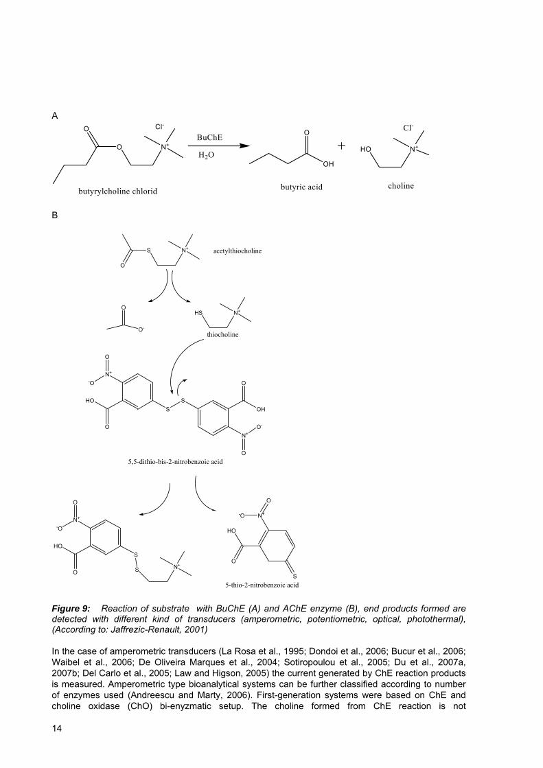
Figure 9: Reaction of substrate with BuChE (A) and AChE enzyme (B), end products formed are detected with different kind of transducers (amperometric, potentiometric, optical, photothermal).......................................................................................................14
Figure 10: Mechanistic cycle of peroxidases .....................................................................23 Figure 11: Proposed activation mechanism of pesticide prothiophos, which already
contains P=O bond but instead show weak in vitro AChE inhibition effect....................25 Figure 12: Chemical structure of imidazolium ring (left) and pyridinium ring (right) IL.28 Figure 13: Bioanalytical FIA setup without integrated on-line oxidation step. Setup
consisting of HPLC pump, two injector valves, bioanalytical column with AChE enzyme and detector system is shown. ..........................................................................................35
Figure 14: Schematic representation of a dual beam TLS setup........................................36 Figure 15: Bioanalytical FIA setup used in experiments including on-line CPO oxidation
step. Two HPLC pumps were used, with different flow rates..........................................37 Figure 16: Signal and enzyme inhibition obtained with 26 μg L-1 (left column) and 52 μg
L-1 (right column) chlorpyrifos oxon using different column glass beads fillings with the AChE enzyme immobilized in three different concentrations. ........................................40
Figure 17: Signal and enzyme inhibition obtained with 142 μg L-1 (left column) and 284 μg L-1 (right column) chlorpyrifos oxon using different column glass beads fillings with the ellectric eel AChE enzyme immobilized in two different concentrations..................42
Figure 18: AChE enzyme inhibition obtained by chlorpyrifos oxon (26 μg L-1) with different flow-rates and two stop-flow experiments.........................................................43
Figure 19: Human AChE regeneration using three commercially available regenerators (in 1 mM concentration), following the inhibition by 40 µg L-1 chlorpyrifos oxon..............44
Figure 20: Human AChE enzyme activity whith sequential injection of various concentrations of 2-PAM chloride (left) and obidoxime chloride (right) ........................45
Figure 21: Pesticide recovery in different kind of natural juice and fruit pulp samples spiked with chlorpyrifos oxon (50 μg L-1)........................................................................46
Figure 22: Peaks representing substrate injection and observed inhibition of human AChE initial activity after injection of BMIMPyBF4 (1 and 5 %, left) and QATf2N (both isomers and racemic mixture, 1 %, right) samples...........................................................47
Figure 23: Peaks representing substrate injection and observed inhibition of the AChE enzyme with chlorpyrifos oxon (40 μg L-1) in PBS buffer or ionic liquid medium (BMIMPyBF4, 1 and 5 % concentration).. .......................................................................48
XIII
Figure 24: Peaks representing substrate injection and observed inhibition of human AChE enzyme achieved with chlorpyrifos oxon (40 μg L-1) with or without precedent IL injection (BMIMPyBF4,, 1 and 5 % concentration, left and QATf2N, both isomers and racemic mixture, 1 %, right).. .......................................................................................... 48
Figure 25: Oxidation yield (paraoxon-methyl formed) and parathion-methyl remained in CPO enzyme oxidation reaction in the presence of various tested ILs............................ 49
Figure 26: Sequential inhibition of human AChE performed with chlorpyrifos oxon...... 52 Figure 27: TLS signal obtained using AChE immobilized on CIM disk (blue) and CPG
glass (red)......................................................................................................................... 53 Figure 28: GC-ECD chromatograms of malathion solutions oxidized by CPO in buffer at
different pH values........................................................................................................... 54 Figure 29: GC-MS chromatograms of malathion in diluted juice solutions oxidized by
CPO at various hydrogen peroxide concentrations.......................................................... 55 Figure 30: Depletion of thio-analog of OP pesticide (malathion) and formation of oxo-
analog (malaoxon) during CPO oxidation reaction at various H2O2 concentrations.. ..... 56 Figure 31: Depletion of pesticide thio-analog (malathion) and formation of its oxo-analog
(malaoxon) during CPO oxidation of malathion in 0.1 M citrate buffer, pH=2.90 (left) and in 10x diluted apple juice concentrate, pH=2.90 (right) at various concentrations of the CPO enzyme............................................................................................................... 56
Figure 32: Remaining enzyme activity after injection of unspiked or pesticide-spiked apple juice samples.. ........................................................................................................ 57
Figure 33: GC-MS chromatograms of juice sample spiked with malathion (blue), the same sample after its oxidation with CPO enzyme (black) and juice sample spiked with malaoxon (red). ................................................................................................................ 58
Figure 34: CPO oxidation of sequentially injected samples (1 mL) containing 8.1 µM parathion-methyl with 0.05 mM H2O2 (A), 0.25 mM H2O2 (B), 1 mM H2O2 (C) and no H2O2 added (D). Experiments were also performed by injecting paraoxon-methyl with 0.25 mM H2O2 (E) and using 19 µM parathion-methyl with 1 mM H2O2 (F). .............. 59
Figure 35: TLS signals corresponding to AChE activity in some of the flow conditions tested (A) and degree of inhibition obtained with 1 mg L-1 paraoxon-methyl (B) at various mixing ratios of two buffers and different flow rates.......................................... 61
Figure 36: Chloroperoxidase oxidation and inhibition of thio-analog of OP pesticides (malathion, 263 μg L-1; parathion-methyl, 798 μg L-1; chlorpyrifos, 149 μg L-1 and diazion, 1.06 mg L-1) depending on H2O2 concentration.. ............................................... 62
Figure 37: GC-MS chromatograms (ion mode) of representative samples of diazinon (5.28 mg L-1, black line), equimolar concentration of diazoxon (5 mg L-1, red) and diazinon oxidized with CPO enzyme (5.28 mg L-1, blue).. ............................................. 63
Figure 38: Inhibition of AChE by consecutive injection of identical samples with malathion (126 μgL-1) with various concentrations of H2O2 and two immobilized CPO concentrations.. ................................................................................................................ 64
Figure 39: GC-MS chromatograms of activated dimefox solution (red) and control without activation (black) ................................................................................................ 65
Figure 40: GC-MS spectra of activated product (left) and dimefox (right) ...................... 66 Figure 41: TLS signals after consecutive injections of substrate (left) and monitoring of
the remaining human AChE enzyme activity after injection of dimefox sample following the activation reaction. ..................................................................................................... 67
Figure 42: Paraoxon-methyl (1 mg L-1 and 10 mg L-1 concentration) and dimefox (10 mg L-1) relative concentration in the HepG2 cell medium with or without (control) cells.. . 68
Figure 43: GC chromatograms of cell extract screened for paraoxon-methyl .................. 68
XIV

Figure 44: ChE assay performed using UV-VIS spectrophotometer at 412 nm. On the right graph insert from left is shown using smaller scale. ................................................69
Figure 45: AChE assay performed using TLS detection at 478 nm. .................................70
XV

Abbreviations and symbols ACh – Acetylcholine AChE - Acetylcholinesterase ASChI – Acetylthiocholine iodide BuChE – Butyrylcholinesterase ChE – Cholinesterase ChO – Cholineoxidase CIM – Convective interaction media CNS – Central nervous system CPO - Chloroperoxidase CYP - Cytochrome DAD – Diode-array detection DTNB - 5,5'-dithio-bis(2-nitrobenzoic acid) EC50 - Half maximal effective concentration μECD – Micro-electron capture detector EDA – Ethylenediamine EDTA - Ethylenediaminetetraacetic acid ELISA - Enzyme linked immuno sorbent assay FIA – Flow injection analysis FPD – Flame photometric detector GC – Gas chromatography GluOX – Glucose oxidase HPLC – High pressure liquid chromatography IL – Ionic liquid ISE - Ion selective eletrodes ISFET - Ion sensitive field effect transistor IMP - 2-isopropyl-6-methyl-4-pyrimidinol ki – enzyme-inhibitor constant Km - Michaelis-Menten constant LAPS - light-addressable potentiometric sensors LC-MS – liquid chromatography-mass spectrometry LD50 – Lethal dose 50 i.e. the dosage that is lethal to 50 % of tested animals LOD – Limit od detection MFO - Mxed function oxidase MRL – Maximum residue limit MS – Mass spectrometry NBS - N-bromosuccinimide NPD – Nitrogen-phosphorus detector OP - Organophosphate OPH – Organophosphorus hydrolase OPIDN - Organophosphate-induced delayed neuropathy PAS – Peripheral anionic site PBS – Phosphate buffer saline PNS – Peripheral nervous system rpm – rotations per minute SEM – Scanning electron microscope SPE – Solid phase extraction SPR – Surface plasmon resonance TLS – Thermal lens spectrometry
XVI

TNB2- - 5-thio-2-nitro-benzoic acid Tyr - Tyrosinase UV/VIS – Ultraviolet-visible Vmax – Maximum velocity of the enzyme
XVII

XVIII

1 INTRODUCTION
The history of organophosphate (OP) compounds began in 1854, when Phillipe the Clermont synthesized tetraethyl pyrophosphate. Nearly 80 years latter Gerhard Schrader, led by observations of Lange and Kruger, who described the synthesis of two OP compounds and noted that their vapor inhalation produced certain health effects, engaged in the exploration of this type of compounds. Their work resulted in the synthesis of parathion, one of the most frequently used OP pesticide in recent decades. After World War II thousand of OP compounds were synthesized worldwide for various purposes (pesticides, nerve agents in medicine and in chemical warfare, flame retardants and parasiticides in veterinary medicine). Due to the lack of persistence in the environment and in exposed individuals and due to lesser insect resistance development in comparison to organochlorine pesticides, the OP pesticides are today the most commonly used group of pesticides throughout the world. It should be emphasized, that from several points of view (public health and intensive agriculture) their use today is a must and not an option. Although their persistence in environment is relatively low, the extensive use of OP pesticides in modern agriculture has raised several problems regarding environmental and food safety issues (Chambers and Levi, Eds., 1992; Abdel-Halim et al., 2006; Konstantinou et al., 2006). For example, in the European Union countries, Norway and Iceland a monitoring program of pesticide residues in products of plant origin showed that in 39.7 % of fruit, vegetable, processed food and cereal samples residues of pesticides at or below maximum residual limit (MRL) were found. In 4.7 % of samples, residues above the MRL were detected and 55.6 % of analyzed samples contained no pesticide residues (Monitoring of pesticide residues…., 2006). The most frequently detected pesticides were organophosphate and carbamate pesticides. Even more concerning is the observation that the proportion of samples with multiple pesticide residues has increased since 1998, from 13.7 % to 23.4 % in the year 2004. To meet the demands of strict food quality control regulations, large number of samples have to be collected daily and analyzed throughout the world. Existing methods for OP pesticide monitoring employ various well established chromatographic techniques (mainly gas chromatography- GC and high performance liquid chromatography- HPLC, coupled with different kinds of detectors), (Štajnbaher and Zupančič-Kralj, 2003; Tse et al., 2004; Ballesteros and Parrado, 2004; Vidal et al., 2000; Sosa et al., 2003; Di Corcia and Marchetti, 1991; Schenck and Howard-King, 1999; Lambropoulou et al., 2002; Lacorte et al., 1997; Hernando et al., 2005). These techniques have reasonably low limits of detection (LOD) and are very selective, but they are often time- and solvent-consuming, the number of residues, which can be identified by each multiresidue method is limited and they are quite expensive, as the analysis of one food sample can cost several hundreds of euros. Therefore, they are not very appropriate for screening large number of samples for possible pesticide traces. As a good alternative and with the advantage of being fast and reliable, several bioanalytical techniques based on inhibition of enzyme acetylcholinesterase (AChE) and coupled with simple detectors (UV/Vis spectrometers, amperometric electrodes) were developed recently (Andreescu et al., 2002a; Schulze et al., 2002a; Kok et al., 2002; Bucur et al., 2006; Dondoi et al., 2006; Bachmann et al., 2000; Marty and Jeanty, 1998; Walz and Schwack, 2007a, 2007b). Another example of such a bioanalytical assay for determination of organophosphate pesticides, which exploits thermal lens spectrometry (TLS) (Franko and Tran, 1996) to improve the LODs and sample throughput, was presented by Pogačnik and Franko (Pogačnik and Franko, 1999). However, bioassays based on AChE inhibition principle are severely hindered by the fact, that thio-analogs of organophosphate pesticides (P=S group), which are predominantely used in agricultural applications, exhibit low in vitro inhibition potency towards AChE. Consequently, LOD for their determination is relatively high, in many cases being in the several mg L-1 range (Pogačnik and Franko, 1999; Jeanty et al., 2001; Dondoi et al., 2006). To achieve satisfactory LOD, thio-OP pesticides need to be activated (oxidized) to their oxo-forms (P=O bond), thus mimicking the process occuring in organisms with the action of cytochrome P450 enzymatic system. In vitro activation can be performed chemically, with various oxidants (Dondoi et al., 2006; Lee et al., 2002) or enzymatically, using cytochrome P450 system (Buratti et al., 2003; Waibel et al., 2006) or chloroperoxidase (CPO) enzyme from marine fungi Caldariomyces fumago (Hernandez et al., 1998; Walz and Schwack, 2007c). With the application of
1

ionic liquids (ILs) in TLS, the sensitivity of the measurements can be further enhanced (Tran et al., 2005) through the improvement of the thermooptical properties of the flowing medium. Ionic liquids as green solvents have found many interesting applications in various fields including biocatalysis and chemical analysis (Tran et al., 2005; Zhang and Malhotra, 2005; Zhao et al., 2002; Yang and Pan, 2005; Park and Kazlauskas, 2003; Rantwijk et al., 2003). With some of the pesticides the activation reaction occurring in vivo can be different than a simple oxidation. This is the case with dimefox, which is a model substance for toxic nerve gases. Despite being an oxo-analog (already contains P=O bond), dimefox has very low in vitro toxicity (Fenwick et al., 1957; Fenwick, 1958), but after activation, which occurs in the liver with the P450 enzymatic system, the formed product(s) have strong AChE inhibition effect. This demonstrates that additional activation is required to enable detection of non-active oxo-analogs of OP pesticides, such as dimefox.
1.1 Objectives
As discussed in the Introduction, fast and reliable methods for daily screening of organophosphate pesticides in large number of environmental and food samples are still needed. To contribute to the progress in this field the following objectives of the research conducted as part of this dissertation were defined: 1. Investigation and integration of on-line CPO oxidation step in FIA-AChE bioassay (Pogačnik and Franko, 1999). In order to achieve this objective the designed experiments included:
a.) testing of batch CPO oxidation of selected representative pesticides, optimization of several reaction conditions (pH, hydrogen peroxide concentration, CPO concentration).
b.) integration of on-line oxidation, by immobilization of CPO enzyme. c.) optimization of various FIA conditions (mixing of buffers, flow rates, hydrogen peroxide
concentration, AChE and CPO enzyme column fillings) with the aim of achieving complete thio-OP to oxo-OP conversion. 2. Improvement of sensitivity by the addition of ionic liquids into the FIA carrier buffer and investigation of the effect of selected ILs on CPO and AChE enzyme activity. 3. Investigate the possibility of in vivo activation processes for inactive oxo-OPs, such as dimefox by human hepatoma HepG2 cell line. 4. Development of the TLS method for measurements of cellular ChE activity and elucidation of the relation between pesticide concentration and cellular ChE expression.
2
2 THEORETICAL BACKGROUND
2.1 Organophosphorus compounds
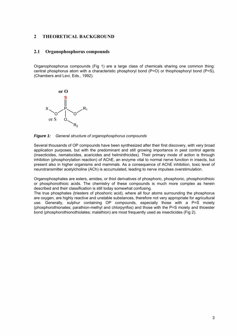
Organophosphorus compounds (Fig 1) are a large class of chemicals sharing one common thing: central phosphorus atom with a characteristic phosphoryl bond (P=O) or thiophosphoryl bond (P=S), (Chambers and Levi, Eds., 1992).
S
PO O
R1
OR2
or O
or S
X
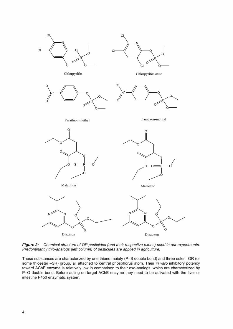
Figure 1: General structure of organophosphorus compounds Several thousands of OP compounds have been synthesized after their first discovery, with very broad application purposes, but with the predominant and still growing importance in pest control agents (insecticides, nematocides, acaricides and helminthicides). Their primary mode of action is through inhibition (phosphorylation reaction) of AChE, an enzyme vital to normal nerve function in insects, but present also in higher organisms and mammals. As a consequence of AChE inhibition, toxic level of neurotransmitter acetylcholine (ACh) is accumulated, leading to nerve impulses overstimulation. Organophosphates are esters, amides, or thiol derivatives of phosphoric, phosphonic, phosphorothioic or phosphonothioic acids. The chemistry of these compounds is much more complex as herein described and their classification is still today somewhat confusing. The true phosphates (triesters of phoshoric acid), where all four atoms surrounding the phosphorus are oxygen, are highly reactive and unstable substances, therefore not very appropriate for agricultural use. Generally, sulphur containing OP compounds, especially those with a P=S moiety (phosphorothionates; parathion-methyl and chlorpyrifos) and those with the P=S moiety and thioester bond (phosphorothionothiolates; malathion) are most frequently used as insecticides (Fig 2).
3
PO
O
O
S
N
Cl
Cl
Cl
Chlorpyrifos
PO
O
O
O
N
Cl
Cl
Cl
Chlorpyrifos oxon
PO
O
O
S
N+
O
-O
PO
O
O
O
N+
O
-O
Parathion-methyl Paraoxon-methyl
O
O
O
O
S
PS O
O
Malathion
O
O
O
O
S
PO O
O
Malaoxon
PO
O
OS
NN
Diazinon Diazoxon
PO
O
OO
NN
Figure 2: Chemical structure of OP pesticides (and their respective oxons) used in our experiments. Predominantly thio-analogs (left column) of pesticides are applied in agriculture. These substances are characterized by one thiono moiety (P=S double bond) and three ester –OR (or some thioester –SR) group, all attached to central phosphorus atom. Their in vitro inhibitory potency toward AChE enzyme is relatively low in comparison to their oxo-analogs, which are characterized by P=O double bond. Before acting on target AChE enzyme they need to be activated with the liver or intestine P450 enzymatic system.
4
2.1.1 ORGANOPHOSPHORUS PESTICIDE INTERACTION WITH ACETYLCHOLINESTERASE ENZYME
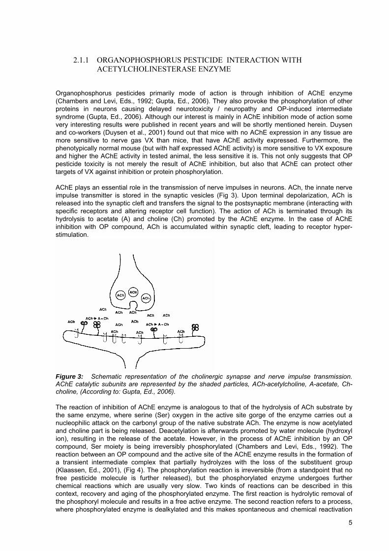
Organophosphorus pesticides primarily mode of action is through inhibition of AChE enzyme (Chambers and Levi, Eds., 1992; Gupta, Ed., 2006). They also provoke the phosphorylation of other proteins in neurons causing delayed neurotoxicity / neuropathy and OP-induced intermediate syndrome (Gupta, Ed., 2006). Although our interest is mainly in AChE inhibition mode of action some very interesting results were published in recent years and will be shortly mentioned herein. Duysen and co-workers (Duysen et al., 2001) found out that mice with no AChE expression in any tissue are more sensitive to nerve gas VX than mice, that have AChE activity expressed. Furthermore, the phenotypically normal mouse (but with half expressed AChE activity) is more sensitive to VX exposure and higher the AChE activity in tested animal, the less sensitive it is. This not only suggests that OP pesticide toxicity is not merely the result of AChE inhibition, but also that AChE can protect other targets of VX against inhibition or protein phosphorylation. AChE plays an essential role in the transmission of nerve impulses in neurons. ACh, the innate nerve impulse transmitter is stored in the synaptic vesicles (Fig 3). Upon terminal depolarization, ACh is released into the synaptic cleft and transfers the signal to the postsynaptic membrane (interacting with specific receptors and altering receptor cell function). The action of ACh is terminated through its hydrolysis to acetate (A) and choline (Ch) promoted by the AChE enzyme. In the case of AChE inhibition with OP compound, ACh is accumulated within synaptic cleft, leading to receptor hyper-stimulation.
Figure 3: Schematic representation of the cholinergic synapse and nerve impulse transmission. AChE catalytic subunits are represented by the shaded particles, ACh-acetylcholine, A-acetate, Ch-choline, (According to: Gupta, Ed., 2006). The reaction of inhibition of AChE enzyme is analogous to that of the hydrolysis of ACh substrate by the same enzyme, where serine (Ser) oxygen in the active site gorge of the enzyme carries out a nucleophilic attack on the carbonyl group of the native substrate ACh. The enzyme is now acetylated and choline part is being released. Deacetylation is afterwards promoted by water molecule (hydroxyl ion), resulting in the release of the acetate. However, in the process of AChE inhibition by an OP compound, Ser moiety is being irreversibly phosphorylated (Chambers and Levi, Eds., 1992). The reaction between an OP compound and the active site of the AChE enzyme results in the formation of a transient intermediate complex that partially hydrolyzes with the loss of the substituent group (Klaassen, Ed., 2001), (Fig 4). The phosphorylation reaction is irreversible (from a standpoint that no free pesticide molecule is further released), but the phosphorylated enzyme undergoes further chemical reactions which are usually very slow. Two kinds of reactions can be described in this context, recovery and aging of the phosphorylated enzyme. The first reaction is hydrolytic removal of the phosphoryl molecule and results in a free active enzyme. The second reaction refers to a process, where phosphorylated enzyme is dealkylated and this makes spontaneous and chemical reactivation
5
impossible (Chambers and Levi, Eds., 1992).
SER
SER
OH
P X
O
OR1
OR2
P X
O
OR1
OR2
OHSER
O
kaP
O
OR1
OR2
SER
O
kp (XH)
P
O
OR1
-O
SER
O
(ROH)
P OH
O
OR1
OR2
Organophosphorus ester +Free Acetylcholinesterase
Reversible complex Phosphorylated enzyme
Aged enzyme(irrevesible)Free enzyme
Figure 4: Mechanism of organophosphate pesticide AChE enzyme inhibition; Ser203 is phosphorylated, with the cleavage of the substituent group. Reaction can further proceed with the hydrolytic removal of the phosphoryl group (slow) or with dealkylation, leading to aged enzyme (According to: Gupta, Ed., 2006). To fully understand the OP compound - AChE interaction and to get an insight into interaction specifity of various OP compounds in this vast and heterogeneous group, deeper knowledge is needed. The active center of AChE enzyme lies in the 20 Å deep and narrow gorge (Axelsen et al., 1994), lined by 14 aromatic amino acid residues (representing almost 40 % of total amino acid residues). At the base of the gorge three residues, Ser, His and Glu, constitute the catalytic triad (enzyme active center), which is directly related to phosphorylation of the serine by the OP molecule (Fig 5, amino acid sequence number can slightly alter in respect to AChE origin). Several other subsites are also important for interactions of the enzyme with structurally different ligands. Peripheral anionic binding site (PAS) is located at the rim of the gorge and is the first site of interaction with various ligands. Through steric hindrance and allosteric modulation the interaction of the ligand with active center is being controled, promoting or inhibiting its interaction (Radić and Taylor, 1999; Kousba et al., 2004). Large cationic and aromatic molecules (D-tubocurarine, coumarin, snake toxins fasciculins, Triton® X-100), that can not reach the bottom of the gorge, can bind here and control the activity of the AChE enzyme (Radić and Taylor, 1999; Bourne et al., 1995; Marcel et al., 2000). Next, acyl binding site or acyl pocket (Fig 5), named after interaction with the acyl part of the ACh molecule is likely important in positioning the inhibitor for nucleophilic attack from Ser203 (Ordentlich et al., 1996). The third site of notable importance regarding catalytic process is hydrophobic oxyanion hole, which has the capacity
6

to attract carbonyl oxygen of ACh as well as phosphyl oxygen of oxo-analogs of OP compounds. Furthermore, this site polarizes P=O bond, thus facilitating Ser nucleophilic attack (Ordentlich et al., 1998). Proton transfer from Ser203 through imidazole ring of the His447 and further to carboxyl group of Glu334 enable nucleophilic attack of the phosphorus atom, resulting in the phosphorylation of the enzyme (Gupta, Ed., 2006).
O-
Ser203
N
HN
His447
C
HO O
Glu334
PO
OR2R1O
R3O
Peripheral binding site
Acyl binding site
Oxyanion hole
Figure 5: Schematic representation of the active site gorge of the AChE enzyme with catalytic triad Ser203, His447 and Glu334 (amino acid sequence number can slightly alter in respect to AChE enzyme origin). Proton transfer from Ser203 through imidazole ring of the His447 and further to carboxyl group of Glu334 enable nucleophilic attack of the phosphorus atom, resulting in the phosphorylation of the enzyme (According to: Gupta, Ed., 2006). Since many interactions of OP molecule and amino acid residues are possible, the nature of the OP substituent groups plays an important role in the potency and specifity of OP compounds as anti-AChE agents (Chambers and Levi, Eds., 1992; Klaassen, Ed., 2001). One of the prevalent factors responsible for structure-inhibition potency relation is electrophilicity of the central P atom (Chambers and Levi, Eds., 1992), as the transphosphorylation reaction relies on interaction between P atom and an unshared pair of electrons of the oxygen of the serine-hydroxyl group of the AChE active site. The presence of S atom in P=S bond (phosphorotionates or phosphorothionotiolates), with the S atom being sufficiently electron donating, yields poor in vitro AChE inhibition molecule. Also, the alkyl substituents of the OP molecule have some influence on AChE toxicity (Kasagami et al., 2002). Di-n-propyl and di-n-butyl phosphates are equally effective against mammalian AChE, as are the methyl and ethyl homologs, but less effective against insect AChE. Furthermore, branched chain alkyl substituents decrease the toxic anti-AChE effect, this is probably the consequence of steric effects (Chambers and Levi, Eds., 1992). Steric effects also play an important role in selectivity of different enantiomers for AChE inhibition, as different functional group of the ligand (phosphoryl, quarternary nitrogen) can occupy different locations within the active center gorge depending on the chirality of the phosphorus atom (Bernard et al., 1998). However, there is no one straightforward rule to describe stereoselectivity toward different enantiomers. Ordentlich and his group (Ordentlich et al., 2005) cite charged interactions (with the Asp74 at the peripheral anionic site) of the VX gas leaving group being the most responsible for stereoselectivity of different VX enantiomers. Strenght of interaction of the OP molecule with the Ser203 moiety, depending on the chemical properties and sterical configuration of the relevant OP molecule, on electronegativity of the central P
7

atom (P=S or P=O bond) and on AChE variety (origin) results in various in vitro toxicity of the relevant OP molecule toward specific AChE. It is noteworthy to stress that in vitro toxicity can significantly differ from in vivo toxicity, since several pharmacokinetic processes (like adsorption, distribution, metabolism and activation) of OP compound can take place in different tissues in the body. This problematic will be discussed in the following chapter.
2.1.2 TOXICITY OF ORGANOPHOSPHORUS COMPOUNDS
Exposure to OP compounds produces two distinct toxic effects in target organisms: direct cholinergic toxicity, with anti-AChE mechanism and neuropathic response, termed organophosphate-induced delayed neuropathy (OPIDN) (Chambers and Levi, Eds., 1992; Klaassen, Ed., 2001). Target sites of action (synaptic clefts) for anti-AChE compounds are present in both central nervous sytem (CNS) and peripheral nervous system (PNS). We already described the mechanism of action of OP molecule in the synaptic cleft in previous chapter. The initial direct consequences of increased ACh neurotransmitter levels are expressed as bradycardia, diarrhea, urination and lacrimation, followed by muscle twitching and paralysis (Gupta, Ed., 2006) and are the result of hyperstimulation of PNS. CNS symptoms include tremors, headache and finally death occurring from depression of respiratory centers in the brain. In vivo acute toxicity of OP insecticides (expressed as the LD50 i.e. the dosage that is lethal to 50 % of tested animals) can significantly differ in regards to selected OP molecule and intake route (Table 1) or species of target organism (owing to different pharmacokinetics parameters and AChE structure variation). From Table 1 it could also be seen, that minor changes in in vivo toxicity exist between thio- and oxo-analogs of selected OP pesticides. In the case of malathion there is even an inversely relation, in vivo toxicity of malathion (thio-analog) is higher than the toxicity of malaoxon (oxo-analog). The reason for such a difference between in vitro and in vivo toxicity is in OP pesticide pharmacokinetic, which regulates compound’s action within the body over a period of time.
Table 1: In vivo LD50 values for selected organophosphorus pesticides in mouse Pesticide Route LD50 Referencesa
dermal 19 mg kg-1 1
intramuscular 7.2 mg kg-1 1
oral 5.0 mg kg-1 1
Parathion-methyl
intraperitoneal 3.0 mg kg-1 1
intramuscular 710 μg kg-1 1
intravenous 520 μg kg-1 1
intraperitoneal 330 μg kg-1 1
Paraoxon-methyl
oral 760 μg kg-1 1
intraperitoneal 193 mg kg-1 1
intravenous 184 mg kg-1 1 Malathion
oral 190 mg kg-1 1
intraperitoneal 75 mg kg-1 1 Malaoxon oral 215 mg kg-1 1
Dimefox intraperitoneal 1.4 mg kg-1 3
oral 60 mg kg-1 1 intraperitoneal 192 mg kg-1 1
Chlorpyrifos
oral 60 mg kg-1 2 oral 85 mg kg-1 1 Diazinon
intraperitoneal 65 mg kg-1 1
a1, US National Toxicology Program acute toxicity studies for, 2000; 2, Cometa et al., 2007 3, Dimefox, Profile from hazardous substances database, 1986.
After ingestion or some other way of OP insecticide uptake, the molecules can be activated (via
8

oxidative desulfuration) within the liver, intestine and other tissues with cytochorme P450 monooxygenase enzyme system and FAD-containing monooxygenases (Kulkarni and Hodgson, 1984; Guengerich, 2005). The product being generated within the reaction is pesticide oxo-analog, which has increased anti-AChE effect in comparison to its thio-analog (Chambers and Levi, Eds., 1992). However, the reactions taking place in the liver tisue are numerous and beside mentioned desulphurization, also dearylation, oxidative disruption of the acid-anhydride bond, epoxidation, aliphatic hydroxylation and dealkylation reactions are present (Kulkarni and Hodgson, 1984). Such a number of possible reactions also give a wide spectrum of formated products, which can play a meaningful role in activation or deactivation processes. Included in the latter, A- and B-esterases, that are present in plasma and tissue have a meaningful role in protecting against toxicity. The A-esterases, where also oxonases and arylesterases are included, hydrolyse the thio- or oxo- compounds, producing inactive metabolites (Karanth and Pope, 2000) and are not inhibited in the reaction. In contrast, B-esterases (carboxylesterases, aliesterase, AChE, BuChE) bind OP molecule steichiometrically and are irreversibly inhibited in the reaction (Karanth and Pope, 2000; Moser et al., 1998). In the case of pesticide dimefox, which is a strong AChE inhibitor in vivo (Lethotzky, 1982; Fenwick et al., 1957), no direct in vitro inhibition of rat and human erytrocytes and plasma AChE acitivity was observed until 10-2 M concentration used (Fenwick et al., 1957). The results suggest that dimefox is metabolized aerobically by a liver enzyme system (Fig 6, process 1 ) and the product, which is a strong AChE inhibitor undergoes either an irreversible bimolecular reaction with AChE (2), resulting in an inhibited enzyme or a rapid spontaneous (particularly in vivo), enzymatic or chemical breakdown (3).
Figure 6: Schematic representation of the conversion of dimefox into AChE inhibitor; 1- aerobic metabolism of dimefox, 2- inhibition of enzyme AChE, 3- enzymatic or chemical breakdown
2.2 Detection of pesticide residues
According to the status list of all active pesticide substances (Status of active substances......, 2007), 1140 pesticide active ingredients are currently registered in EU countries. Therefore, it is not surprising, that the detection of pesticide residues in environmental and food samples has acquired a lot of attention in recent years. Among most frequently detected pesticide group are OP and carbamate pesticides (Monitoring of pesticide residues…., 2006), which use greatly increased in last two decades on behalf of the organochlorine pesticides. Their widespread use is mainly due to their relatively low mammalian toxicity, wide-ranging biological activity, the possibility of easy molecular modification and relatively low-persistence in comparison to previously used organochlorine pesticides. The most disseminated approaches in the determination of pesticide residues in natural samples are still various chromatographic methods, with extensive work devoted to development of numerous multiresidue detection methods (Tse et al., 2004; Huang et al., 2007; Braga et al., 2007; Ballesteros and Parrado, 2004; Štajnbaher and Zupančič-Kralj, 2003; Vidal et al., 2000; Hernando et al., 2005).
9
2.2.1 CHROMATOGRAPHIC TECHNIQUES FOR PESTICIDE RESIDUES DETERMINATION
Nowadays, large number of methods are used in the determination of different pesticide residues in natural samples. Most frequently applied methods use gas chromatography (GC) with electron capture detector (ECD), flame photometric detection (FPD), nitrogen-phosphorus detection (NPD) and mass spectrometric (MS) detection, or high-performance liquid chromatography (HPLC) with diode-array detection (DAD) and fluorescence detection (Guardia-Rubio et al., 2007; Karamfilov et al., 1996; Soleas et al., 2000; Fenoll et al., 2007, Rial-Otero et al., 2007). Without any doubt, the main advantage of these methods over the bioanalytical ones, described in continuation is their sensitivity and selectivity, as their LOD is usually in the sub µg kg-1 to several µg kg-1 range (Fenoll et al., 2007, Guardia-Rubio et al., 2007, Huang et al., 2007). However, chromatographic analysis of samples characterized by complex matrices requires several steps in sample pretreatment to be included: matrix modification, extraction and clean-up (Ahmed, 2001; Hernando et al., 2005). These steps are often time- and solvent-consuming, and when choosing the optimal procedure for sample preparation, there exist a risk of loosing some substance in the process. Also, because of great diversity in structures and related physical-chemical properties of pesticides (e.g. polarity, vapor pressure), a suitable method for sample preparation and analysis should be chosen for particular application, thus limiting the number of residues which can be identified by each multiresidue method. And finally, a fact that must not be overlooked is the price of the laboratory equipment and operational costs. Consequently, these methods are not very suitable for monitoring large number of samples for pesticide residues in the food, beverage or other environmental samples. Another important drawback of using chromatographic methods in screening for pesticide residues is the fact, that toxicity of the tested sample is not being measured. Pesticide degradation products, which can be spontaneously formed in the environment or in the commercial product, can be even more toxic than active compounds in pesticide formulations (Cacares et al., 2007; Bavcon Kralj et al., 2007). Increased toxicity can also result from the interaction of various compounds in complex mixture samples, even if each individual chemical is below the threshold concentration. Combined toxicity of various chemicals can be the sum of additive effects of individual compounds or from synergistic interaction, thus producing even greater toxicity effect.
2.2.2 BIOANALYTICAL SYSTEMS FOR PESTICIDE DETECTION
An alternative to solve the problems associated with the generally used methods in routine environmental and food analysis is the application of different types of bioanalytical systems. Three basic elements of every bioanalytical system (receiver, transducer and electronics component) are depicted in Fig 7.
10

Figure 7: Three basic components of bioanalytical system are: specific receiver, transducer of the signal and electronics for signal processing and recording Receiver is a sort of biological recognition element and can incorporate different enzymes, antibodies, microbial cells or some other receptors. The receiver has to be specific for selected analyte(s) or group of analytes, thus ensuring the molecular recognition or transformation of the analyte. Next, the transducing element (e.g. optical, amperometric, accoustic, thermal or electrochemical) recognizes the biochemical modification of the analyte and transforms it into electrical (or other kind of) signal, which is processed and stored by data acquisition electronics (Patel, 2002; D`Souza, 2001). In the field of OP insecticide detection, bioanalytical systems are mainly based on different enzymes, mainly AChE and BuChE and antibodies for specific pesticides isolated from different sources. However, in recent years, several alternative methods were developed, that do not involve biological component, but have other advantages in comparison to chromatographic methods. Further details and reviews can be found elsewhere (Liu and Lin, 2005; Cao et al., 2007; Hu et al., 2005; Huang et al., 2004). In the following paragraphs recently published research work, covering bioanalytical systems based on various sorts of biological components (recognition elements) will be presented. A more detailed informations regarding OP pesticide detection in samples with various matrices, achieved LODs and transducer element employed are summarized in Table 2. 2.2.2.1 Bioanalytical systems based on Organophosphorus hydrolase enzyme
Organophosphorus hydrolase (OPH) is an organophosphotriester hydrolysing enzyme, which has been first discovered in soil microorganisms Pseudomonas diminuta MG and Flavobacterium spp.. It catalyse the hydrolysis of pesticides such as parathion, diazinon, dursban, coumaphos, acephate and nerve gases soman, sarin and tabun (Jaffrezic-Renault, 2001). The hydrolysis of each molecule leads to the production of two protons (and usually chromophoric alcohol) from cleavage of P-O, P-F, P-S or P-CN bond according to the reaction, schematically represented in Fig 8.
P
X
Z
R´
R H20OPH
P
X
OH
R´
R ZH
Figure 8: Reaction scheme of OP hydrolysis with the enzyme OPH; X- oxygen or sulhur, R-alkoxy group, R`- alkoxy or phenyl group, Z- phenoxy group, a thiol moiety, a cyanide or a fluorine group Different transducers can be employed to construct an easy to use system for rapid determination of organophosphorus compounds, for instance amperometric (Wang et al., 2003; Mulchandani et al.,
11
2006), carbon nanotube based amperometric (Deo et al., 2005), optical (Kumar et al., 2006a), potentiometric (Mulchandani et al., 1999; Schöning et al., 2003; Ristori et al., 1996) and microcantilever transducer (Karnati et al., 2007). In Table 2, LODs achived with OPH bioanalytical ystems and other techniques are compared.
ifferent pesticides in the same group (paraoxon > parathion > diazinon), (Mulchandani et al., 1999).
2.2.2.2 Bioanalytical systems based on Tyrosinase and Acid phosphatase enzymes
f monophenols, to form o-diphenols and oxidation of o-diphenols to o-quinones
cryo gel (Deng et al., 1996) and
s compounds, this severely limits the use of Tyr iosensors for other than the water-based samples.
. The later solution showed better results in terms of enzyme tability and biosensor performance.
s However, bioanalytical systems using OPH enzyme have a limit in expressing much higher selectivity towards phosphotriester (e.g. paraoxon) and phosphothiolester (e.g. malathion) pesticides than to phosphorothiolate neurotoxins (e.g. nerve gas VX) (Di Sioudi et al., 1999). Some improvement can be achieved through protein engineering strategies to improve sensitivity towards the later group (Di Sioudi et al., 1999). Wild-type OPH has also higher selectivity in the range of several orders towards d
Tyrosinase (Tyr) is a binuclear copper monooxygenase enzyme containg metalloprotein and catalyses the o-hydroxylation o(Vidal et al., 2006). Bioanalytical systems using Tyr enzyme (in context of OP pesticide monitoring) are mainly based on pesticide inhibition of its activity, during a reaction with the substrate in the presence of molecular oxygen (Vidal et al., 2006; De Albuquerque and Ferreira, 2007; Campanella et al., 2007). In the process either oxygen depletion or quinone formation are monitored. As Tyr enzyme has at least two binding sites, with one of them having the affinity for aromatic compounds and the other one for metal binding agents, the activity of the enzyme could be affected by a large number of inihibtors (Albuquerque and Ferreira, 2007). Inhibition of Tyr enzyme can thus be induced by the following agents: carbamate, organophosphorus and dithiocarbamate pesticides, atrazines, thioureas, aromatic carboxylic acids, triazine and phenyl-urea herbicides, chlorophenols and copper chelating agents (Nistor and Emneus, 1999). Several Tyr immobilization protocols have been proposed so far, electrode surface cross-linking with glutaraldehyde and bovine serum albumine (Albuquerque and Ferreira, 2007), incorporation into carbon paste (Rogers et al., 2000),polypyrrole, using amphiphilic pyrrole (Cosnier and Popescu, 1996). Sensitivity of bioanalytical systems using Tyr enzyme (in this case defined as pesticide concentration that cause 30 % inhibition of amperometric signal) reached 30.4 µg L-1 for diazinon and 68.4 µg L-1 for parathion-methyl (Albuquerque and Ferreira, 2007). Vidal and co-workers (Vidal et al., 2006) report LOD for dichlorvos of 13.2 µg L-1. The main advantage of this type of sensors is their sensitivity toward thio-OP pesticides as Tyr enzyme does not discriminate between thio- and oxo-analogs. However, Tyr enzyme is quite unstable, also when physical methods of immobilization are used, operational stability is low, due to enzyme loss in surrounding environment. Albuquerque and Ferreira (Albuquerque and Ferreira, 2007) report their sensor can be used during 10 day period without problems due to enzyme activity loss. After 15 days, the sensor could be used with 20 % initial value and significant loss of amperometric signal was observed thereafter. Another, already mentioned drawback is nonspecificity in Tyr activity inhibition caused by various compounds, thus cathehins from green tea are reported as mushroom Tyr inhibitors (No et al., 1999), seven isoflavones extracted from fermented soygerm koji also exhibit anti-Tyr activity (Chang et al., 2007) and phenylethylaminoalanine and cysteine found in food samples are acting in the same mode (Friedman et al., 1986) As natural samples with complex matrices usually contain unknown numbers of varioub Reported bioanalytical systems for OP pesticide detection based on acid phosphatase inhibition are very rare. The main advantage cited in regards to ChE sensors are their reversible inhibition, thus eliminating the need for enzyme regeneration after inhibition. Mazzei and co-workers (Mazzei et al., 1996) developed two different variants of bienzymatic bioelectrodes. The first is a classical one, using immobilized acid phosphatase (catalyzing glucose formation from glucose-6-phosphate) and glucose oxidase (catalyzing H2O2 formation from glucose) on the tip of H2O2 electrode. The second is a hybrid biosensor, in which acid phosphatase has been employed in the thin layer of potato tissue, endowed with high content of enzyme activitys
12
2.2.2.3 Bioanalytical systems based on Cutinase enzyme
sed, as the assay could be easily applicable to 10 % methanol with 40 % diethylene glycol medium.
2.2.2.4 Immunosensors
et al. (Kumar et al., 2006b). The later method is based
to 13 regeneration cycles (Kumar et al., 2006b), while thereafter teeply decline was observable.
2.2.2.5 Bioanalytical systems based on inhibition of Cholinesterases (ChE)
phenyl acetate, indophenyl cetate, indoxyl acetate) can be detected by several kinds of transducers.
Recently developed bioanalytical system based on new kind of receiver component is described by Walz and Schwack (Walz and Schwack, 2007a). The enzyme cutinase from yeast Fusarium solani pisi is dissolved in buffer solution and p-Nitrophenyl butyrate substrate is used as cromophor agent, with spectrophotometric kind of detection. Authors have reported LOD (based on 10 % inhibition criteria) of 0.04 and 2.6 mg L-1 for paraoxon and chlorpyrifos, respectively. In their subsequent work (Walz and Schwack, 2007b) LODs reported for other OP compounds (see Table 2) are somewhat higher in comparison to µg L-1 range for AChE assay (Schulze et al., 2002b; Pogačnik and Franko, 2001; Andreescu et al., 2002b). Surprisingly, malaoxon and demeton-S-methyl, both strong AChE inhibitors could not inhibit cutinase, thus making their detection impossible. Moreover, thio-analogs of tested pesticides were showed to have minor enzyme inhibition effect. In their later work (Walz and Schwack, 2007c) this problem was solved by performing batch chloroperoxidase oxidation step before inhibition assay. The advantage of their cutinase assay seems to be in the choice of possible reaction matrixu
Immunosensors combine the power of antibodies as recognition element and an appropriate physicochemical transduction mechanism to convert the recognition event into detectable signal. In the field of OP pesticide immunodetection, mainly surface plasmon resonance (SPR) and optical detection with the use of enzyme linked immuno sorbent assay (ELISA) as tranducer element are used (Kumar et al., 2006b; Mauriz et al., 2006a; 2006b). Some common attributes of immunosensors are their high sensitivity and specificity for selected compound. Mauriz et al. (Mauriz et al., 2006a) have reached LOD as low as 55-60 ng L-1 for chlorpyrifos in different environmental water samples with their SPR based immunosensor. Similarly sensitive method, with the achieved LOD of 50 ng L-1 for parathion-methyl was developed by Kumar on ELISA priciple coupled with FIA technique. Unfortunately, immunosensor`s high specificity is at the same time also their main limitation in the environmental sample analysis field. Antibodies are usually highly specific merely to its antigen (in this case being a specific type of pesticide molecule). Therefore, with the application of one type of antibodies, the detection of a group or class of chemicals (e.g. OP pesticides) is not possible. This problem could be somewhat mitigated by multiple and combined immobilization of several analyte recognition elements on the sensing surface (Mauriz et al., 2006b). Further difficulty connected with immunosensors is antibody-antigen or antigen-enzyme dissociation from immobilized antibodies after the analysis (Kandimalla et al., 2004), as repeated use of the same sensor requires the antibody to be free of antigens. Repeating dissociation step after each use and a retention of activity and specificity are crucial requirements for practical use. Previously described immunosensors show less than 10 % change in response after 190 regeneration cycles (Mauriz et al., 2006a) and more than 95 % initial binding capacity was retained ups
There are two different types of cholinesterase enzymes, acetylcholinesterases, also called true cholinesterases and butyrylcholinesterases, each of them having different biological role and function. A common attribute of both groups is their inhibition effect caused by OP pesticide molecule. This principle can be exploited in designing bioanalytical system with various possible transducing elements used. The type of transducer and detection method is dictated by the choice of substrate, enzyme system (mono- or multiple- enzymes) and by the end application. The enzyme, AChE or BuChE is immobilized on the transducer or on the inert support through different possible procedures (Pogačnik and Franko, 2001; Bartolini et al., 2005). The products formed (Fig 9) from various substrates (which can be choline, acetylcholine, acetylthiocholine, 4-aminoa
13
A
O N+
O Cl-
butyrylcholine chlorid
O
OH
butyric acid
HO N+
choline
BuChE
H2O
Cl-
B
S N+
O
acetylthiocholine
HS N+
thiocholine
O
O-
SS
O
OH
N+
O
O-O
HO
N+
O
-O
5,5-dithio-bis-2-nitrobenzoic acid
O
HO
N+
O
-O
S5-thio-2-nitrobenzoic acid
S
O
HO
N+
O
-O
S N+
ucers (amperometric, potentiometric, optical, photothermal), ccording to: Jaffrezic-Renault, 2001)
Figure 9: Reaction of substrate with BuChE (A) and AChE enzyme (B), end products formed are detected with different kind of transd(A In the case of amperometric transducers (La Rosa et al., 1995; Dondoi et al., 2006; Bucur et al., 2006; Waibel et al., 2006; De Oliveira Marques et al., 2004; Sotiropoulou et al., 2005; Du et al., 2007a, 2007b; Del Carlo et al., 2005; Law and Higson, 2005) the current generated by ChE reaction products is measured. Amperometric type bioanalytical systems can be further classified according to number of enzymes used (Andreescu and Marty, 2006). First-generation systems were based on ChE and choline oxidase (ChO) bi-enyzmatic setup. The choline formed from ChE reaction is not
14
electrochemically active, thus it needs to be further oxidised to hydrogen peroxide with ChO. The later can be easily detected amperometrically at +650 mV versus Ag/AgCl. Another procedure involves measuring the consumption of O2 (with Clark electrode) in ChO reaction instead of hydrogen peroxide detection. A significant drawback in bi-enyzmatic sensor design is that due to high applied potential, the sensor is susceptible to interferences from other electroactive species (ascorbic acid, glutathion, L-cysteine) possibly present in the reaction medium of real samples (Andreescu and Marty, 2006). The second-generation amperometric sensors use mono-enzyme design and non specific ChE substrates (as acetylthiocholine in the case of AChE). The principle is based on direct amperometric detection of the enzymatic reaction of thiocholine (Andreescu and Marty, 2006). In comparison to first-generation systems, there are two major advantages. The use of single enzyme simplifies the design of the sensor and secondly, the detection potential is lower (+410 mV), thus decreasing possible interferences from other electroactive species. The effect can be further reduced and the selectivity and sensitivity improved by applying electronic mediators, such as tetracyanoquinodimethane or cobalt phtalocyanine (Li et al., 1999; Schulze et al., 2002b; Andreescu et al., 2002b). However, mediator addition also involves interferences caused by added chemicals. Recently, Shi and co-workers (Shi et al., 2006) developed mediator-free bioanalytical system by entrapping AChE in Al2O3 sol-gel and the possibility of thiocholine detection at +250 mV (versus Ag/AgCl), hundreds mV lower than other comparable mediator-free systems. Sensitivity and selectivity have been increased with the simultaneous reduction of possible interferences. A novel approach was introduced by Andreescu et al. (Andreescu et al., 2002a) by constructing bi-enzyme sensor using AChE and Try enzyme (with phenyl acetate as a substrate) and comparing that to mono-enzyme AChE sensor, using p-aminophenyl acetate substrate. The main reason for Try enzyme inclusion was in p-aminophenyl acetate inaccessibility, as it is not commercially available. The sensitivity of both sensors are comparable and are in the range of several µg L-1 (Table 2), with improved functionality of the bi-nzymatic technical solution.
eir size is reduced in omparison to traditional pH glass electrodes (Andreescu and Marty, 2006).
aline phosphatase enzyme catalysis of macrocyclic
ad of spectrophotometric detection, the LOD for paraoxon can be lowered by almost a ctor of five.
e Second kind of transducers are potentiometric transducers. They are measuring the pH variation due to the acetic acid formed in enzymatic hydrolysis reaction (Fig 9). The sensors for measuring pH shift due to AChE inhibition are either pH glass electrode (with immobilized AChE), ion selective electrodes (ISE), ion sensitive field effect transistor (ISFET; Jaffrezic-Renault, 2001) or light-addressable potentiometric sensors (LAPS; Andreescu and Marty, 2006). ISFET and LAPS are fabricated using microelectronic technologies, are more sensitive, less expensive and thc Another variety of transducers rely on optical detection, e.g. absorbance, reflectance, luminescence and fluorescence of the products formed in the enzymatic reaction (Andres and Narayanaswamy, 1997; Lui et al., 1997; Diaz et al., 1997; Jin et al., 2004; No et al., 2007; Kuswandi et al., 2007) or photothermal effects (Pogačnik and Franko, 1999, 2003; Franko and Tran, 1996). The simplest strategy is based on colour change of a pH indicator (thymol blue and chlorphenol red) as a result of acetic acid formed (Andres and Narayanaswamy, 1997; Xavier et al., 2000) and spectrophotometric detection. Change of pH can also be monitored using pH-sensitive fluorescence indicator. This strategy was used by Doong and Tsai (Doong and Tsai, 2001), which encapsulated AChE and various pH-sensitive fluorophores in sol-gel network on a glass cap, that was fixed on optical fiber and integrated in FIA system. Florescence effect can also be monitored by using AChE substrates indoxyl-acetate and 2-naphthyl acetate, that are converted to highly flourescent products (Diaz et al., 1997). Another method using optical transducing element was developed by Ayyagari et al. (Ayyagari et al., 1995). Although the method is not based on inhibition of AChE enzyme, we describe it here briefly as an illustration within optical transducer context. Ayyagari and his group took advantage of chemiluminescence effect, produced by alkcompound and simultaneous release of light. Photothermal spectrometry is based on indirect absorbance measurements of photothermal effect (Franko and Tran, 1996). Details regarding the fundamentals of TLS will be presented in separate chapter. Pogačnik and Franko (Pogačnik and Franko, 1999) showed, that by using photothermal technique instefa Other transducing principle worth mentioning is monitoring the heat released during the enzymatic reaction with a thermopile sensor (Zheng et al., 2006). As a bio-recognition element, chicken liver-
15

esterase instead of AChE is used, a cheap and sensitive enzyme. As pesticide inhibit the esterase, the heat developed is reduced when the substrate for the enzyme is injected in the FIA system. Although authors used relatively high concentration of dichlorvos (1 and 10 mg L-1) in their experiments, the results demonstrate the feasibility of the approach for rapid detection of OP pesticide
sidues.
ined pesticide recovery rate was 83.4 %, indicating very low interferences of the matrix onstituents.
ing this goal. etailed review of pesticide activation problematic is carried out in the following chapter.
re In view of practical applications, ChE and other sensors were developed with the aim of using them for the detection of pesticides in real environmental and food samples. However, the majority of described sensors were tested on pesticide solutions, prepared in buffer medium and not on real samples. Some examples of practical applications however exist, Kumar et al. (Kumar et al., 2006a) measured parathion-methyl (after methanol extraction) in soil, several authors (Mulchandani et al., 2006; De Albuquerque and Ferreira, 2007; Mauriz et al., 2006a; Bucur et al., 2005; Bucur et al., 2006) operated with ground, surface or drinking water, while there are several reports of measuring AChE inhibitors (after buffer or solvent extraction) in vegetables or baby food (Jin et al., 2004; Lui et al., 1997; Xavier et al., 2006; Pogačnik and Franko, 2003; Schulze et al., 2002a; No et al., 2007). Some outstanding results in the field of practical application of ChE sensor were reported by Schulze et al. (Schulze et al., 2002b), with their attempt to measure pesticide residues in untreated juice or baby food samples. With treating their AChE sensor electrode with 1 vol.% Tween-20 solution, they completely avoided matrix effects, expressing as atypical AChE inhibition and long equlibration times (time until constant current is reached). The need of pesticide extraction or pre-concentration step was completely avoided. Even more promising is in situ measurement of carbamate pesticide carbaryl in tomato pulp, performed by Caetano and Machado (Caetano and Machado, 2008). They immersed their carbon paste electrode directly into tomato pulp of a tomato cut in two halves and previously spiked with pesticide. Obtac Common problem associated with techniques based on ChE (AChE, BuChE) enzyme inhibition is low in vitro toxicity of some OP pesticides e.g. all phosphorothionates and certain S-alkyl phosphorothiolates (RS-P(O)) (Chambers and Levi, Eds., 1992; Pogačnik and Franko, 1999; Jeanty et al., 2001; Dondoi et al., 2006) and some phosphoroamidates, for example dimefox (Fenwick et al., 1957; Kasagami et al., 2002). One method of assuring a reasonably low-limit of detection of this group of pesticides in food and water samples is employing more sensitive AChE enzyme. Generally, AChE isolated from insects are more sensitive to OP pesticides than other AChE. Recently, mutant enzymes were designed and produced with genetic engineering, which have improved sensitivity and selectivity for OP pesticides (De Oliveira Marques et al., 2004; Bucur et al., 2006; Sotiropoulou et al., 2005; Istamboulie et al., 2007). Only by taking the advantage of using more sensitive enzymes, LOD can be lowered for a factor of 70 (Istamboulie et al., 2007). Another elegant way is pesticide activation, thus increasing its in vitro toxicity. Chemical or enzymatic oxidation of P=S to P=O bond for the phosphorothionates and phosphorothionothiolates is a preferential approach for reachD
16
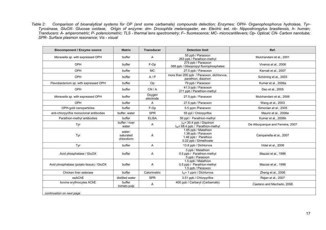
Table 2: Comparison of bioanalytical systems for OP (and some carbamate) compounds detection; Enzymes: OPH- Organophosphorus hydrolase, Tyr-
Tyrosinase, GluOX- Glucose oxidase, Origin of enzyme: dm- Drosophila melanogaster, ee- Electric eel, nb- Nippostrongylus brasiliensis, h- human; Tranducers: A- amperometric; P- potenciometric; TLS - thermal lens spectrometry; F– fluorescence; MC- microcantilevers; Op- Optical; CN- Carbon nanotube; SPR- Surface plasmon resonance; Vis - visual
Biocomponent / Enzyme source Matrix Transducer Detection limit Ref.
Moraxella sp. with expressed OPH buffer A 55 ppb / Paraoxon 263 ppb / Parathion-methyl Mulchandani et al., 2001
OPH buffer F-Op 275 ppb / Paraoxon 368 ppb / Diisopropyl fluorophosphates Viveros et al., 2006
OPH buffer MC 27.5 ppb / Paraoxon Karnati et al., 2007
OPH buffer A / P more than 200 ppb / Paraoxon, dichlorvos, parathion, diazinon Schöning et al., 2003
Flavobacterium sp. with expressed OPH buffer Op 79 ppb / Paraoxon Kumar et al., 2006a
OPH buffer CN / A 41.3 ppb / Paraoxon 211 ppb / Parathion-methyl Deo et al., 2005
Moraxella sp. with expressed OPH buffer Oxygen electrode 27.5 ppb / Paraoxon Mulchandani et al., 2006
OPH buffer A 27.5 ppb / Paraoxon Wang et al., 2003 OPH-gold nanoparticles buffer F-Op 5.5 ppm /Paraoxon Simonian et al., 2005
anti-chlorpyrifos monoclonal antibodies buffer, water SPR 55 ppt / Chlorpyrifos Mauriz et al., 2006a Parathion-methyl antibodies buffer ELISA 50 ppt / Parathion-methyl Kumar et al., 2006b
Tyr buffer / river water A I30= 30.4 ppb / Diazinon
I30= 68.4 ppb / Parathion-methyl De Albuquerque and Ferreira, 2007
Tyr water-
saturated chloroform
A
1.65 ppb / Malathion 1.38 ppb / Paraoxon 1.46 ppb / Parathion
0.22 ppb / Dimethoate
Campanella et al., 2007
Tyr buffer A 13.8 ppb / Dichlorvos Vidal et al., 2006
Acid phosphatase / GluOX buffer A 3 ppb / Malathion
0.5 ppb / Parathion-methyl 5 ppb / Paraoxon
Mazzei et al., 1996
Acid phosphatase (potato tissue) / GluOX buffer A 1.5 ppb / Malathion
0.5 ppb / Parathion-methyl 1.5 ppb / Paraoxon
Mazzei et al., 1996
Chicken liver esterase buffer Calorimetric I30= 1 ppm / Dichlorvos Zheng et al., 2006
eeAChE distilled water SPR 3.51 ppb / Chlorpyrifos Rajan et al., 2007 bovine erythrocytes AChE
buffer
tomato pulp A 400 ppb / Carbaryl (Carbamate) Caetano and Machado, 2008
continuation on next page
17
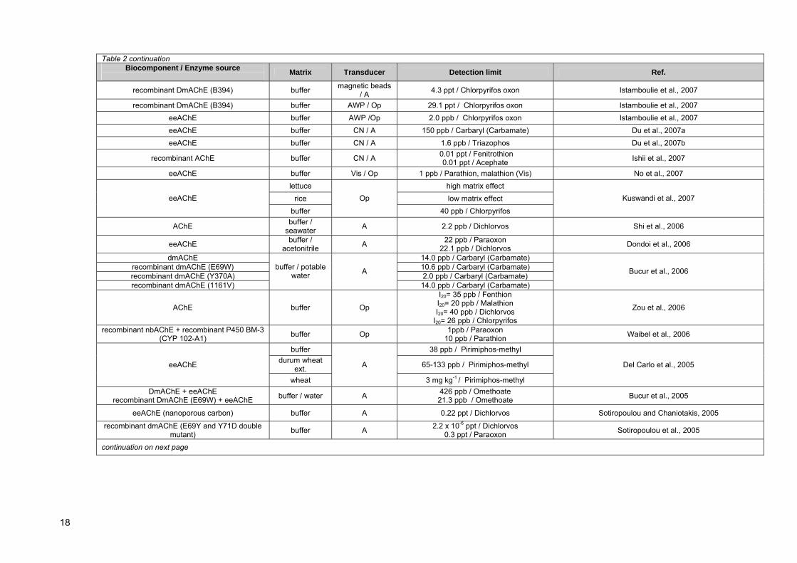
Table 2 continuation Biocomponent / Enzyme source Matrix Transducer Detection limit Ref.
recombinant DmAChE (B394) buffer magnetic beads / A 4.3 ppt / Chlorpyrifos oxon Istamboulie et al., 2007
recombinant DmAChE (B394) buffer AWP / Op 29.1 ppt / Chlorpyrifos oxon Istamboulie et al., 2007 eeAChE buffer AWP /Op 2.0 ppb / Chlorpyrifos oxon Istamboulie et al., 2007 eeAChE buffer CN / A 150 ppb / Carbaryl (Carbamate) Du et al., 2007a eeAChE buffer CN / A 1.6 ppb / Triazophos Du et al., 2007b
recombinant AChE buffer CN / A 0.01 ppt / Fenitrothion 0.01 ppt / Acephate Ishii et al., 2007
eeAChE buffer Vis / Op 1 ppb / Parathion, malathion (Vis) No et al., 2007 lettuce high matrix effect
rice low matrix effect eeAChE
buffer
Op
40 ppb / Chlorpyrifos
Kuswandi et al., 2007
AChE buffer / seawater A 2.2 ppb / Dichlorvos Shi et al., 2006
eeAChE buffer / acetonitrile A 22 ppb / Paraoxon
22.1 ppb / Dichlorvos Dondoi et al., 2006
dmAChE 14.0 ppb / Carbaryl (Carbamate) recombinant dmAChE (E69W) 10.6 ppb / Carbaryl (Carbamate) recombinant dmAChE (Y370A) 2.0 ppb / Carbaryl (Carbamate) recombinant dmAChE (1161V)
buffer / potable water A
14.0 ppb / Carbaryl (Carbamate)
Bucur et al., 2006
AChE buffer Op
I20= 35 ppb / Fenthion I20= 20 ppb / Malathion I20= 40 ppb / Dichlorvos
I20= 26 ppb / Chlorpyrifos
Zou et al., 2006
recombinant nbAChE + recombinant P450 BM-3 (CYP 102-A1) buffer Op 1ppb / Paraoxon
10 ppb / Parathion Waibel et al., 2006
buffer 38 ppb / Pirimiphos-methyl durum wheat
ext. 65-133 ppb / Pirimiphos-methyl eeAChE
wheat
A
3 mg kg-1 / Pirimiphos-methyl
Del Carlo et al., 2005
DmAChE + eeAChE recombinant DmAChE (E69W) + eeAChE buffer / water A 426 ppb / Omethoate
21.3 ppb / Omethoate Bucur et al., 2005
eeAChE (nanoporous carbon) buffer A 0.22 ppt / Dichlorvos Sotiropoulou and Chaniotakis, 2005
recombinant dmAChE (E69Y and Y71D double mutant) buffer A 2.2 x 10⁻⁶ ppt / Dichlorvos
0.3 ppt / Paraoxon Sotiropoulou et al., 2005
continuation on next page
18

Table 2 continuation
Biocomponent / Enzyme source Matrix Transducer Detection limit Ref.
recombinant dmAChE
(B4-21, B4-27 and B3 mutant) buffer A 2.2 x 10⁻⁶ ppt / Dichlorvos 2.9 x 10⁻⁵ ppt / Parathion
3.2 x 10⁻⁵ ppt / Azinphos-methyl Law and Higson, 2005
eeAChE
pure water / Chinese
cabbage and cole
Op 12 ppb / Paraoxon 25 ppb / Dichlorvos Jin et al., 2004
eeAChE 90 ppb / Methamidophos recombinant dm AChE (various)
buffer A 1 ppb / Metamidophos
De Oliveira Marques et al., 2004
dmAChE buffer A
I20= 4.7 ppb / Paraoxon I20= 0.9 ppb / Dichlorvos
I20= 0.02 ppb / Chlorpyrifos oxon (for metal-chelate aff. immobilizat.)
Andreescu et al., 2002b
eeAChE / hAChE juice and infant food A 5 ppb / Paraoxon-methyl Schulze et al., 2002b
dmAChE 5.2 ppb / Paraoxon 0.56 ppb / Chlorpyrifos oxon
dmAChE + Tyr buffer A 5 ppb / Paraoxon
0.95 ppb / Chlorpyrifos oxon
Andreescu et al., 2002a
eeAChE buffer / juices A 2 ppb / Parathion Schulze et al., 2002a eeAChE 0.32 ppb / Paraoxon
bovine AChE 0.46 ppb / Paraoxon hAChE 1.4 ppb / Paraoxon
horse AChE
buffer TLS
1.6 ppb / Paraoxon
Pogačnik and Franko, 2001
eeAChE buffer Op
> 15 ppm / Chlorpyrifos > 6.8 ppm Chlorpyrifos-methyl
24 ppb / Chlorpyrifos oxon 270 ppb / Chlorpyrifos-methyl oxon
Jeanty et al., 2001
dmAChE buffer A 4.8 ppb / Methamidophos Nunes et al., 2001
recombinant dm AChE (B03 mutant) 1.4 ppb / Methamidophos eeAChE 53 ppb / Methamidophos
eeAChE
buffer Op
I30= 152 ppb / Paraoxon
Doong and Tsai, 2001
recombinant dmAChE (Y408F, F368L, F368H and F368W)
buffer / surface water A 0.1 ppb / Paraoxon Bachmann et al., 2000
Cholinesterase deionized water Op
17.8 ppm / Fenitrothion 17.9 ppm / Azinphos-methyl
57.6 ppm / Methidation Diaz and Peinado, 1997
eeAChE buffer Op 24.7 ppb / Paraoxon Andres and Narayanaswamy, 1997
continuation on next page
19

Table 2 continuation
Biocomponent / Enzyme source Matrix Transducer Detection limit Ref.
tap water 1.5 ppb / Paraoxon
33 ppm / Chlorpyrifos 6.8 ppm / Diazinon
orange juice 2.8 ppb / Paraoxon bovine AChE
apple juice
TLS
4.0 ppb / Paraoxon
Pogačnik and Franko, 1999
eeAChE 1.7 – 5.4 ppm / Fenitrothion Cholinesterase
buffer Op 1.5 – 4.9 ppm / Fenitrothion
Diaz et al., 1997
lettuce 12 ppm / Methamidophos bovine AChE chinese
cabbage Op
3 ppm / Methamidophos Lui et al., 1997
buffer P 100 ppb / Malathion eeAChE
water > 100 ppb / Malathion Ristori et al., 1996
eeAChE buffer A 1.3 ppb / Paraoxon Cremisini et al., 1995 eeAChE lagoon water A 27.5 ppb / Paraoxon La Rosa et al., 1995
eeAChE buffer A 0.02 ppb / Paraoxon 26.3 ppm / Parathion-methyl Marty et al., 1993
eeAChE buffer F IC50= 102 ppb / Paraoxon
IC50= 78 ppm / Dicrotophos > 200 ppm / Malathion, Parathion
Rogers et al., 1991
OPH 143 µg m-3 / Paraoxon 6.9 mg m-3 / Malathion
5.4 mg m-3 / Demethon-S BuChE
aerosol Op
143 µmol m-3 / Paraoxon
Luckarift et al., 2007
horse BuChE buffer A 90 ppb / Trichlorfon 54 ppb / Coumaphos Gogol et al., 2000
buffer 5 ppb / Paraoxon
12 ppb / Sarin 14 ppb / Vx
Arduini et al., 2007 horse BuChE
gas phase
A
0.1 mg m-3 / Sarin
Cutinase / Fusarium solani pisi buffer Op
2.2 ppb / Chlorpyrifos oxon 2.4 ppm / Chlorpyrifos
320 ppb / Chlorpyrifos-methyl oxon 6.8 ppm / Chlorpyrifos-methyl
40 ppb / Paraoxon 101 ppm / Parathion
160 ppb / Paraoxon-methyl Not detected / Parathion-methyl
Not detected / Malaoxon Not detected / Malathion
Walz and Schwack, 2007b
20

2.2.3 ACTIVATION OF ORGANOPHOSPHORUS PESTICIDES
One possible approach of increasing the sensitivity of OP pesticide detection using bioanalytical systems based on ChE inhibition is pesticide molecule activation, thus mimicking the process that occurs in liver tissue with cytochrome P450 monooxygenase enzyme complex (Poet et al., 2003; Kulkarni and Hodgson, 1984). A common way of pesticide activation is its oxidation, which can be performed chemically or enzymatically. 2.2.3.1 Chemical oxidation of organophosphorus pesticides
Numerous authors performed activation step using chemical oxidants, thus Kumaran et al. (Kumaran et al., 1992) obtained good detection sensitivities (0.5 to 275 µg L-1) using bromine water to oxidize some organophosphate pesticides in synthetic sea water. Kim and co-workers (Kim et al., 2000) used 10 fold molar excess bromine in organic solvent for the same purpose. The oxidation was completed within few seconds, with a 82-100 % yield for 9 out of 10 tested OP pesticides (fenthion, which contains a sulphide group was further oxidised to yield a sulfoxide product). However, chemical oxidation of compounds with a sulphide group is problematic and yields also other types of end products beside respective oxons, with the result of loosing AChE inhibition potency and sensitivity (Lee et al., 2002). Schulze et al. (Schulze et al., 2002a) oxidised samples of juice and baby food with N-bromosuccinimide (NBS) and studied influence of complex matrix. A quantitative oxidation of phosphorothionates was assured using 4 mg L-1 NBS concentration. The used biosensor screening test was very fast in comparison to chromatographic methods (90 minutes vs. 1 day). NBS was also used for oxidation of pesticide parathion-methyl retained on the solid phase extraction column in the research work of Dondoi et al. (Dondoi et al., 2006). The researchers demonstrated the feasibility of their oxidation procedure by obtaining similar degree of AChE inhibition with on-column oxidated parathion-methyl in comparison to paraoxon-ethyl control. No further oxidation of the later substance was determined. To our knowledge, only bromine and NBS oxidations are used today for the purpose of increasing OP pesticide limit of detection using bioanalytical systems based on AChE inhibition principle. Other chemical oxidants were used for OP pesticide oxidation purpose (Bellet and Casida, 1974; Wozniak and Stec, 1999; Jackson et al., 1992), but drastical experimental conditions required (e.g. extreme temperatures and pH values, organic solvent medium) are not applicable to previously described bioanalytical systems. To summarize, only bromine and NBS oxidation of phoshorothionates (only P=S bond, no thioester bond) yields good results in terms of reaction products and sensitivity improvement for subsequent detection on AChE bioanalytical systems. Chemical oxidation of other compounds (which contain other S-bonds, e.g. phosphorothionothiolates, phosphoramidothiolates) yields also other end products, beside their oxons. 2.2.3.2 Enzymatical activation of organophosphorus pesticides
Several studies concerning OP pesticide bioactivation have been so far performed, mainly with P450 monooxygenases or chloroperoxidase enzyme. Cytochrome P450 enzymes are a primary catalytic promoters of activation processes occuring in vivo after pesticide uptake in the body, thus using these molecules in vitro seems to be a brilliant way for outpassing the problematic of low in vitro toxicity of thio-analogs of pesticides.
2.2.3.2.1 P450 MONOOXYGENASE Buratti et al. (Buratti et al., 2002, 2003) investigated desulphurization (oxidation) of four OP pesticides by several cytochrome (CYP) P450 isoforms. Their results are indicating that various CYP isoforms
21
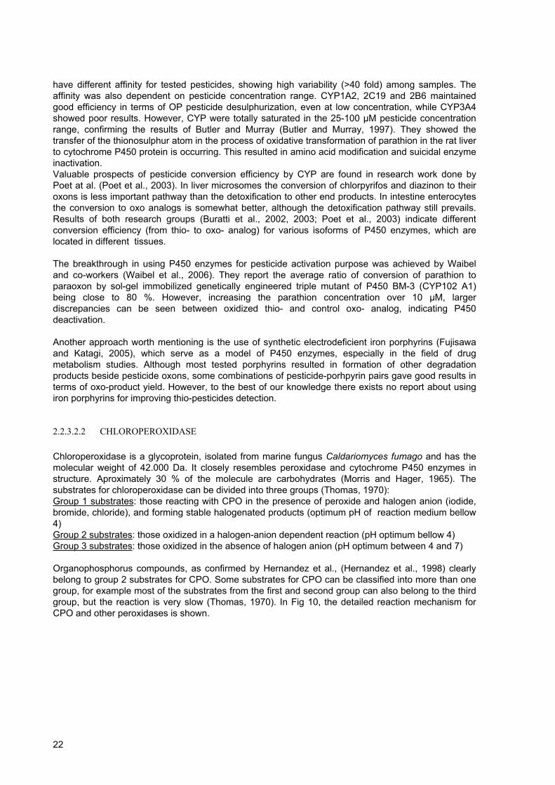
have different affinity for tested pesticides, showing high variability (>40 fold) among samples. The affinity was also dependent on pesticide concentration range. CYP1A2, 2C19 and 2B6 maintained good efficiency in terms of OP pesticide desulphurization, even at low concentration, while CYP3A4 showed poor results. However, CYP were totally saturated in the 25-100 µM pesticide concentration range, confirming the results of Butler and Murray (Butler and Murray, 1997). They showed the transfer of the thionosulphur atom in the process of oxidative transformation of parathion in the rat liver to cytochrome P450 protein is occurring. This resulted in amino acid modification and suicidal enzyme inactivation. Valuable prospects of pesticide conversion efficiency by CYP are found in research work done by Poet at al. (Poet et al., 2003). In liver microsomes the conversion of chlorpyrifos and diazinon to their oxons is less important pathway than the detoxification to other end products. In intestine enterocytes the conversion to oxo analogs is somewhat better, although the detoxification pathway still prevails. Results of both research groups (Buratti et al., 2002, 2003; Poet et al., 2003) indicate different conversion efficiency (from thio- to oxo- analog) for various isoforms of P450 enzymes, which are located in different tissues. The breakthrough in using P450 enzymes for pesticide activation purpose was achieved by Waibel and co-workers (Waibel et al., 2006). They report the average ratio of conversion of parathion to paraoxon by sol-gel immobilized genetically engineered triple mutant of P450 BM-3 (CYP102 A1) being close to 80 %. However, increasing the parathion concentration over 10 µM, larger discrepancies can be seen between oxidized thio- and control oxo- analog, indicating P450 deactivation. Another approach worth mentioning is the use of synthetic electrodeficient iron porphyrins (Fujisawa and Katagi, 2005), which serve as a model of P450 enzymes, especially in the field of drug metabolism studies. Although most tested porphyrins resulted in formation of other degradation products beside pesticide oxons, some combinations of pesticide-porhpyrin pairs gave good results in terms of oxo-product yield. However, to the best of our knowledge there exists no report about using iron porphyrins for improving thio-pesticides detection.
2.2.3.2.2 CHLOROPEROXIDASE Chloroperoxidase is a glycoprotein, isolated from marine fungus Caldariomyces fumago and has the molecular weight of 42.000 Da. It closely resembles peroxidase and cytochrome P450 enzymes in structure. Aproximately 30 % of the molecule are carbohydrates (Morris and Hager, 1965). The substrates for chloroperoxidase can be divided into three groups (Thomas, 1970): Group 1 substrates: those reacting with CPO in the presence of peroxide and halogen anion (iodide, bromide, chloride), and forming stable halogenated products (optimum pH of reaction medium bellow 4) Group 2 substrates: those oxidized in a halogen-anion dependent reaction (pH optimum bellow 4) Group 3 substrates: those oxidized in the absence of halogen anion (pH optimum between 4 and 7) Organophosphorus compounds, as confirmed by Hernandez et al., (Hernandez et al., 1998) clearly belong to group 2 substrates for CPO. Some substrates for CPO can be classified into more than one group, for example most of the substrates from the first and second group can also belong to the third group, but the reaction is very slow (Thomas, 1970). In Fig 10, the detailed reaction mechanism for CPO and other peroxidases is shown.
22
Figure 10: Mechanistic cycle of peroxidases; hydrogen peroxide transforms the iron(III) porphyrin group to an oxo-iron(IV)porphyrin π free radical (pathway 1). Subsequently, compound 1 can follow three alternative pathways (2, 4, 9); CPO shows a significant catalactic activity (pathway 8); pathways 9 and 10 show peroxidase-like catalytic activity, pathway 9 and 10 represent C-H bond oxidation (According to: Valderrama et al., 2002) CPO is a versatile enzyme with the capability of peroxidative, catalactic, halogenating, and chlorinating activity (Thomas, 1970), acting on large specter of substrates. However, from the perspective of our research work, CPO ability of performing oxygen transfer reactions that resembles those performed by P450 monooxygenases (Valderrama et al., 2002) is much more interesting. Such oxygen transfer reactions include hetero-atom oxidation (S-oxidation and N-oxidation), epoxidation and C-H bond oxidation (Valderrama et al., 2002). The source of oxygen has to be added to reaction medium in the form of hydrogen peroxide (H2O2) or some other hydroperoxide (methyl- or ethyl- hydroperoxides, tert-butyl hydroperoxide) (Manoj and Hager, 2001; Park and Clark, 2006). However, as shown by Shevelkova and Ryabov (Shevelkova and Ryabov, 1996) CPO enzyme is susceptible to high H2O2 concentration in reaction medium, what rapidly leads to enzyme suicide inactivation. The loss of CPO activity is accompanied by heme degradation, rather than protein aggregation or denaturation (Park and Clark, 2006), a process promoted by free radicals formation (HO2•, HO• and superoxide radical) in pathway 2 and 3 (Fig 10). It was observed by the same authors, that higher CPO activity at pH 6 is a result of less or no radical formation in comparison to reaction medium with pH 4.1, and that radical formation during H2O2 dissmutation is dependent on reaction pH. On the contrary, Manoj and Hager (Manoj and Hager, 2001) claim that significant deactivation of CPO by hydrogen peroxide occurs only above pH 6 and that the sulfoxidation and catalase reaction are in competition, favouring one or another in dependance of H2O2 concentration and reaction pH. There exist several solutions to beforementioned H2O2-promoted inactivation problem. A simple one would be, to constantly feed small amount of H2O2 to reaction medium, thus preventing rapid CPO inactivation (Shevelkova and Ryabov, 1996). Next, some radical scavenger (e.g. tert-butyl alcohol) could be added or pH of the reaction should be optimized (Park and Clark, 2006). One of the solution is also the application of hydroperoxide that has smaller utilization rate in catalase dismutation reaction, e.g. tert-butyl hydroperoxide (Manoj and Hager, 2001), thus minimazing free radicals formation. More elaborative solution would be to employ random mutagenesis in C. fumago CPO locus, with the aim of producing more resitant mutants. This principle was successfully employed for
23
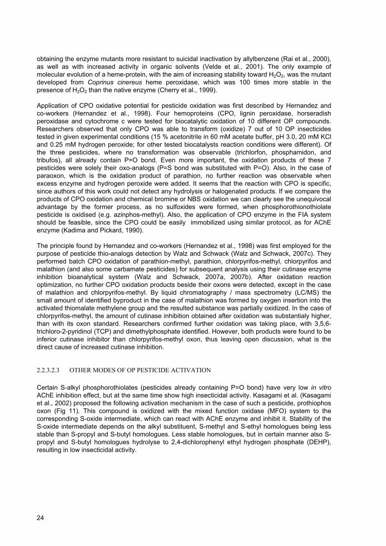
obtaining the enzyme mutants more resistant to suicidal inactivation by allylbenzene (Rai et al., 2000), as well as with increased activity in organic solvents (Velde et al., 2001). The only example of molecular evolution of a heme-protein, with the aim of increasing stability toward H2O2, was the mutant developed from Coprinus cinereus heme peroxidase, which was 100 times more stable in the presence of H2O2 than the native enzyme (Cherry et al., 1999). Application of CPO oxidative potential for pesticide oxidation was first described by Hernandez and co-workers (Hernandez et al., 1998). Four hemoproteins (CPO, lignin peroxidase, horseradish peroxidase and cytochrome c were tested for biocatalytic oxidation of 10 different OP compounds. Researchers observed that only CPO was able to transform (oxidize) 7 out of 10 OP insecticides tested in given experimental conditions (15 % acetonitrile in 60 mM acetate buffer, pH 3.0, 20 mM KCl and 0.25 mM hydrogen peroxide; for other tested biocatalysts reaction conditions were different). Of the three pesticides, where no transformation was observable (trichlorfon, phosphamidon, and tribufos), all already contain P=O bond. Even more important, the oxidation products of these 7 pesticides were solely their oxo-analogs (P=S bond was substituted with P=O). Also, in the case of paraoxon, which is the oxidation product of parathion, no further reaction was observable when excess enzyme and hydrogen peroxide were added. It seems that the reaction with CPO is specific, since authors of this work could not detect any hydrolysis or halogenated products. If we compare the products of CPO oxidation and chemical bromine or NBS oxidation we can clearly see the unequivocal advantage by the former process, as no sulfoxides were formed, when phosphorothionothiolate pesticide is oxidised (e.g. azinphos-methyl). Also, the application of CPO enzyme in the FIA system should be feasible, since the CPO could be easily immobilized using similar protocol, as for AChE enzyme (Kadima and Pickard, 1990). The principle found by Hernandez and co-workers (Hernandez et al., 1998) was first employed for the purpose of pesticide thio-analogs detection by Walz and Schwack (Walz and Schwack, 2007c). They performed batch CPO oxidation of parathion-methyl, parathion, chlorpyrifos-methyl, chlorpyrifos and malathion (and also some carbamate pesticides) for subsequent analysis using their cutinase enzyme inhibition bioanalytical system (Walz and Schwack, 2007a, 2007b). After oxidation reaction optimization, no further CPO oxidation products beside their oxons were detected, except in the case of malathion and chlorpyrifos-methyl. By liquid chromatography / mass spectrometry (LC/MS) the small amount of identified byproduct in the case of malathion was formed by oxygen insertion into the activated thiomalate methylene group and the resulted substance was partially oxidized. In the case of chlorpyrifos-methyl, the amount of cutinase inhibition obtained after oxidation was substantialy higher, than with its oxon standard. Researchers confirmed further oxidation was taking place, with 3,5,6-trichloro-2-pyridinol (TCP) and dimethylphosphate identified. However, both products were found to be inferior cutinase inhibitor than chlorpyrifos-methyl oxon, thus leaving open discussion, what is the direct cause of increased cutinase inhibition.
2.2.3.2.3 OTHER MODES OF OP PESTICIDE ACTIVATION Certain S-alkyl phosphorothiolates (pesticides already containing P=O bond) have very low in vitro AChE inhibition effect, but at the same time show high insecticidal activity. Kasagami et al. (Kasagami et al., 2002) proposed the following activation mechanism in the case of such a pesticide, prothiophos oxon (Fig 11). This compound is oxidized with the mixed function oxidase (MFO) system to the corresponding S-oxide intermediate, which can react with AChE enzyme and inhibit it. Stability of the S-oxide intermediate depends on the alkyl substituent, S-methyl and S-ethyl homologues being less stable than S-propyl and S-butyl homologues. Less stable homologues, but in certain manner also S-propyl and S-butyl homologues hydrolyse to 2,4-dichlorophenyl ethyl hydrogen phosphate (DEHP), resulting in low insecticidal activity.
24
PO
O
S
SCl
ClS-alkyl 2,4-dichlorophenyl ethyl phosphorodithioate
R
MFO
PO
O
S
OCl
Cl
R
S
PO
O
S
OCl
Cl
R
PO
O
S
OCl
Cl
RO
S-Oxide
PO
O
AChE
OCl
Cl
PO
O
OH
OCl
Cl 2,4-dichlorophenyl ethyl hydrogen phosphate
Hydrolysis
Figure 11: Proposed activation mechanism of pesticide prothiophos, which already contains P=O bond but instead show weak in vitro AChE inhibition effect (According to: Kasagami et al., 2002) The basic mode of dimefox activation was already explained before (see Fig 6). Here we will give a deeper knowledge about dimefox problematic and review some important research work referring to it. Dimefox is a weak in vitro AChE inhibitor, so to improve the detection limit when using AChE bioanalytical system some sort of activation should be used. Because of its very short residual time after application and high insecticidal activity, dimefox use is banned (as insecticide) in the USA and in great deal of other countries. Dimefox exerts chemical similarity with some of the nerve gases, therefore it is rather convenient to use it as a model substance in nerve gas studies. Fenwick et al. (Fenwick et al., 1957) proposed that anticholinesterase product formed from dimefox is either amine oxide or hydroxymethilamide. Unfortunatelly, both compounds are very unstable and could not be determined. Kasagami et al. (Kasagami et al., 2002) tried to clarify the activation mechanism of phosphoramidate (P-N bond) insectides, such as dimefox by studying anticholinesterase effect by the transformation of the phosphoramide site. They proposed, that the corresponding N-desalkyl and N-oxide in the form of O-aminophosphate have the principal role in in vivo AChE inhibition. Measuring direct anti-ChE effect on a selected cell line would be a good solution to study toxicity and an in vitro alternative for screening of antiesterase compounds (e.g. dimefox and related compounds) which have low anticholinesterase effect and complex activation mechanisms. Barber and Ehrich (Barber and Ehrich, 2001) have noticed weak conversion of protoxicants (thio-analogs of OP pesticide) to active toxicants (oxo-analogs), when SH-SY5Y neuroblastoma cell line was exposed to chlorpyrifos and parathion. Although this technique has some deficencies, authors think, that their
25
study indicates promise for developing this cell system as an alternative to the 28-day hen test for screening OP compounds on esterase inhibition. A number of benefits, like easy cell growing and maintainance, possible application of multiple doses over a period of weeks and the use of serum-free media (no degradation of tested OP compound) makes this test practical for in vitro procedures. Using human hepatic cell line HepG2, which has preserved specific hepatic function (cytochrome P450 dependent mixed function oxidases activity, Roe et al., 1993) should enable simultaneous investigation of direct anti-ChE effect in connection with activated metabolites formed. To the best of our knowledge, there exists no previous work on measuring AChE activity in HepG2 cell line. The method developed by Ellman and co-workers (Ellman et al., 1961) alows measuring cholinestrase activity in tissue extracts, homogenates and cell suspensions. A lot of literature exists on measuring AChE activity in different nerve tissues and neuroblastoma cell lines and also in red blood cells; in tissue and cells where AChE enzyme is present because of its primary physiological role. However, AChE activity was confirmed also in other human and nonhuman tissues. It has been found in newt retina (Cheon and Saito, 1999), rat pancreatic acinar cells (Bendayan and Gisiger, 2001), in rabbit intrapulmonary arteries (Altiere et al., 1994), in mammalian kidneys (Suda, 1986) and human pulmonary arteries and veins (Kotelevets et al., 2005). Usually, AChE activity in these tissues is very weak. Beside neuroblastoma cell lines, AChE activity is also expressed in PC12 pheochromocytoma cell line (noncholinergic adrenal medulla cells). Grisaru et al. (Grisaru et al., 1999) reported three different post-transcriptional classes of AChE, originating from one AChE pre-mRNA, which is subjected to alternative splicing. The end isoforms carry different peptides in their C-terminus. Beside AChE-S (synaptic form) and AChE-E (erythrocytic form), monomeric ”readthrough” AChE-R is produced. It is found in embryonic and tumor cells and its expression is induced under physiological, chemical and physical stress. Predominating form of cholinesterase in liver, plasma and other peripheral tissues is BuChE. Based on the results of Pogačnik and Franko (Pogačnik and Franko, 2001), tested horse serum BuChE is less sensitive to inhibition by paraoxon. In order to obtain observable ChE inhibition in specified cell line with low pesticide dose the native ChE activity should not be highly expressed (as thus less pesticide is needed to inhibite lower amount of the enzyme).
2.3 Flow injection analysis (FIA) coupled with thermal lens spectrometry
Since the mid-seventies, when FIA was demonstrated for the first time (Ruzicka and Hansen, 1975), the outstanding features of FIA for analytical applications have gained enormous importance in the field of environmental sample analysis. The most important characteristics are easy and safe operation, low cost with low sample and reagent consumption and compatibility with almost any detection principle. These advantages in comparison to off-line analysis were applied in FIA system in combination with AChE biological receiver and TLS detection component (Pogačnik and Franko, 1999, 2001). In the following paragraphs selected components of FIA (regarding improvement of pesticide detection) and basics of TLS theory will be presented. For the sake of better comprehension TLS is firstly reviewed.
2.3.1 THERMAL LENS SPECTROMETRY
Thermal lens spectrometry and other photothermal techniques rely on absorption of optical radiation (laser beam) by the sample and subsequent non-radiative relaxation of absorbed energy. The consequence of this phenomena is the increase of temperature of the sample, which results in changes of physical properties of the sample (e.g. refractive index). To monitor these changes, originating from absorption of optical radiation, different concepts have been applied, TLS being one of them.
26

During deexcitation process the absorbed energy is transformed to heat and the amount of heat released is equal to the energy absorbed by the sample (assuming no fluorescence of the sample). Released heat is proportional to the absorbance of the sample and to the power of the excitation light. When using excitation laser beam with a Gaussian profile, the absorbed energy and the deposited heat result in a lens like element (thermal lens) forming, due to the change of refractive index of the medium. Thermal lens is a direct consequence of radial temperature distribution of the medium with the maximum temperature on the axis of the laser beam. Temperature change are however almost insignificant, being in the order of 10-4 K (Piepmeier, Ed., 1986). The dependence of thermal lens signal on several parameters could be shown with next equation (Bialkowski, Ed., 1996):
kTnAPS
λ)/(303.2 ∂−∂
= (Equation 1)
where S is thermal lens signal, i.e. relative change (∆I/I) of beam intensity on its axis, A is absorbance, λ is probe beam wavelenght, P is excitation laser power, k is thermal conductivity of the medium and ∂n/∂T is the dependance of the refractive index on temperature of the medium. The relation between relative change of the beam centre intensity and the absorbance (concentration) is linear only when changes in the beam centre intensity are adequately low (∆I/I < 0.1). The described technique of indirect measurement of the absorbance through the changes of temperature-dependent refractive index of the sample is more sensitive method than conventional spectrometry, based on light transmission measurements (Franko and Tran, 1996; Franko, 2001). According to Eq. 1, by increasing the power of excitation laser beam (P) we can increase the effect of thermal lens, what finally results in better sensitivity. Pogačnik and Franko (Pogačnik and Franko, 1999) showed, that by using photothermal technique instead of spectrophotometric detection the limit of detection for paraoxon can be lowered by almost a factor of five. Furthermore, TLS is much less sensitive to errors resulting from frequently present light scattering in environmental and food samples. With changing the properties of the medium (thus altering thermal conductivity and dependance of refractive index on temperature) we can further increase the sensitivity of the method. In the optimization experiments performed on FIA system with immobilized AChE enzyme and TLS detection, acetylthiocholine iodide (ASChI) was found to be the optimal substrate (Pogačnik and Franko, 2003). Among the buffers as flowing medium, 0.05 M Tris pH 7.4 and 0.05 M phosphate buffer saline (PBS) pH 8 were tested, the later was found as a better option, with approx. 25 % higher signal.
2.3.2 IONIC LIQUIDS
Ionic liquids (IL) are salts consisting of ions, which exist in the liquid state at ambient temperature. Typically, they consist of organic nitrogen-containing heterocyclic cations and inorganic anions (Fig 12, Zhao et al., 2002). Ionic liquids have gained much attention as »green« solvents due to their outstanding features, such as chemical and thermal stability, low vapour pressure, non-flammability and non-toxicity.
27

A-
N+R1R2
A-
N+
N
R2
R1
Figure 12: Chemical structure of imidazolium ring (left) and pyridinium ring (right) IL; R1 and R2 can be various alkyl chains, A- can be various inorganic anions (Br-, Cl-,I-, BF4
-, CH3COO-, MeSO4-, PF6
-, SCN-....). Despite their low volatility and little impact on the atmosphere, their effects on water quality and aquatic life should be considered (Romero et al., 2007; Latala et al., 2005). From this point of view the effects of ionic liquids on the activity and function of various enzymatic systems is of particular interest. Such information is also relevant for bioanalytical methods relying on enzymes in media containing ionic liquids, which affect the activity of the enzyme and/or other parameters contributing to higher sensitivity or selectivity of the methods. An example of such a technique is the already described bioanalytical assay for determination of OP pesticides based on inhibition of AChE which is determined in a FIA and detected by TLS (Franko and Tran, 1996; Pogačnik and Franko, 2001). Although there are a lot of reports on activity and stability of various enzymes used in industrial processes and chemical synthesis in pure ionic liquid or as co-solvent (Zhao et al., 2002; Yang and Pan, 2005; Park and Kazlauskas, 2003; Rantwijk et al., 2003), studying AChE-IL interaction is rather obscured thematic. Results obtained by Stock et al. (Stock et al., 2004) indicate, that imidazolium and pyridinium ILs inhibit the electric eel AChE enzyme at relatively low concentration and that inhibitory potency depends on the alkyl chain lenght at position R1 (Fig 12). Longer chains caused stronger AChE enzyme inhibition. Also, there was a difference between pyridinium and imidazolium ILs, the former being slightly stronger enzyme inhibitors. The tested phosphonium IL were less potent inhibitors. The influence of the anion component was minimal, with anions alone not capable of AChE inhibition. For the illustration of ILs inhibition potency, the EC50 (half maximal effective concentration) varied between 13 and >2000 µM in comparison to aldicarb (oxime carbamate pesticide) EC50 of 5 µM (Stock et al., 2004). Interestingly, Zhang and Malhotra (Zhang and Malhotra, 2005) found little AChE inhibition (14 %) with 125 µM [EtPy]+[PF6]-, IL not tested by previous research group. Even more important, when comparing the inhibition of 10-7 M paraoxon, the relative inhibition effect in 125 µM IL medium was 22-fold more efficient than in buffer medium. These results clearly show, that for taking full advantage of the benefits ILs can offer, the type and IL concentration should be accurately chosen. Due to demonstrated low in vitro inhibition potency towards AChE by thio-analogs of OP pesticides (see previous chapters), the later have to be activated (their toxicity increased). This can be done using CPO enzyme. Some research work regarding the activity of CPO in the IL reaction medium has already been done (Sanfilippo et al., 2004; Chiappe et al., 2006). The results are showing enhanced activity, stability and selectivity in the presence of IL as co-solvent in comparison to organic solvents. Also here, for maximizing reaction yield adequate IL should be used and its concentration optimized. The primary benefit of using IL rather than water-based medium in bioanalytical system used in our experiments is sensitivity increase. This advantage is possible, because the sensitivity of TLS technique greatly depends on the physical properties (temperature coefficient of refractive index ∂n/∂T and thermal conductivity (k) of the medium in which the measurements are carried out (see Eq. 1). The reason for this is because the thermal lens signal S is directly proportional to ∂n/∂T and inversely proportional to k. Both values depend on the chosen solvent. Unfortunately, water, which is one of the most suitable and often used solvent, especially for bioanalytical enzymatic reactions, has relatively low ∂n/∂T and high k values in comparison to organic solvents, which are known to provide high sensitivity for TLS measurements. But organic solvents are not desirable from environmental standpoint. Furthermore, they are not suitable for enzymatic reactions, as they greatly reduce the activity of enzymes. It has been shown, that enhancements in sensitivity of TLS measurements by 20 to almost 40 times compared to water can be achieved in ILs (Tran et al., 2005).
28

The information presented here prompted us to include the segment dealing with IL in our work. The investigation aims to elucidate the effects of various ionic liquids on the AChE enzyme and to test the efficiency of CPO oxidation of organophosphorus compounds in order to determine the most suitable IL and experimental conditions (IL concentration) needed to achieve, not only efficient oxidation of thio OP compounds, but also highest possible sensitivity of the FIA-AChE bioassay. Furthermore, it is expected, that the investigations will also provide valuable fundamental information on the effects of various ILs as well as their cations or anions on the activity of CPO.
2.3.3 CONVECTIVE INTERACTION MEDIA (CIM) AS ENZYME SUPPORT
To fully exploit the advantages offered by the FIA, the enzymes used have to be immobilized on some inert solid support. A wide range of supports have been utilized for enzyme immobilization through different kinds of applications and it is beyond the scope of this work to review all the existing literature. However, in recent years polymer-based monolithic support (convective interaction media) was developed, that according to literature (Barut et al., 1999; Mihelič et al., 2005) has better characteristics in terms of mass transfer and specific surface area in comparison to controlled pored glass and other porous material. Usually, very porous and small particles (packed bed columns) are used to obtain as high specific area as possible. When mobile phase is pumped through the column, it runs freely around the glass beads, while the fluid within the beads is stagnant. Molecules from the mobile phase are carried to the active sites within the pores mainly by difussion process, therefore, to assure a reasonably high level of exchange from mobile phase to pore interior, low levels of flow rate should be used (Barut et al., 1999). Contrary, due to predominately convective mass transport between mobile and stationary phase in CIM (Barut et al., 1999), these limitations can be factored out. In the case of enzymes bounded to the stationary phase, the diffusion is commonly much slower proces than the kinetic process at the enzyme active site, thus the overall kinetic behavior of the enzyme immobilized on packed bed column is governed by the former process. There are few reports of studying enzymes immobilized on CIM supports. Vodopivec and co-workers (Vodopivec et al., 2003) characterized four different enzymes immobilized on CIM epoxy monoliths. They claim the used immobilization support exhibit good characteristics in terms of reproducibility, linear responses and long-term stability. Enzymes kinetic parameters were flow-unaffected, thus confirming that mass transfer in CIM monoliths is not diffusional limited. Bartolini et al. (Bartolini et al., 2004) immobilized human AChE on ethylenediamine (EDA) CIM and connected it in HPLC system studying various AChE inhibitors. The results shown are somewhat advantageous in comparison to their results reported before (Andrisano et al., 2001) from experiments using AChE immobilized on silica supports. The advantages are mainly in terms of lower specific matrix interaction, lower backpressure and in reduction of analysis time for one substrate injection (2 min), although the time needed is still much longer in comparison to results obtained using FIA-AChE bioassay, developed and described by Pogačnik and Franko (Pogačnik and Franko, 1999). In their subsequent work (Bartolini et al., 2005) they compared human AChE bounded on different support in terms of rate of immobilization, stability and aspecific interaction. The enzyme was immobilized on two CIM disks with different reactive groups (epoxy and EDA) and Glut-P silica matrix. Immobilization yield was the highest in the immobilization performed on Glut-P matrix; however, catalytic efficiency (defined as Km/Vmax) was in this case lower than in monolithic material support. It was hypothesized, that this effect was observable due to the lack of diffusion resistance, when monoliths were used. Other advantages of monolithic supports described were very short conditioning time in respect to silica porous material and fast recovery of enzymatic activity. However, tested monoliths had also several drawbacks, the first being their cost. Moreover, EDA CIM contains protonated amine groups, thus it functions also as weak ionic exchanger. Therefore, the system necessarily demands some anion exchanger competitor in the flowing mobile phase to displace 5-thio-2-nitro-benzoic acid (TNB2-) when Ellman`s colorimetric reaction is used. (Ellman et al., 1961). Regarding epoxy CIM monoliths, their stability was reported to be somewhat lower in comparison to EDA and Glut-P supports, with only 30 % of enzyme activity remained after 2 months of use in contrast to 80 % of remained activity with EDA CIM and Glut-P. The same authors report also stronger
29
side interactions of some of the tested inhibitor compounds with epoxy CIM disk, citing their hydrophobicity as the primary reason. 3 EXPERIMENTAL
3.1 Materials
The pesticides used in our studies (parathion-methyl and paraoxon-methyl, malathion and malaoxon, chlorpyrifos and chlorpyrifos oxon, diazinon and diazoxon, dimefox) were obtained from Pestanal (except for diazoxon, which was from Ultra Scientific and in 100 mg mL-1 concentration in acetonitrile) and were at least 93.7 % pure (most of them above 99 % pure), so no further purification of the chemicals was performed. All of the enzymes were obtained from the following producers: electric eel AChE (1100 U mg-1 protein), human erythrocyte AChE (0.39 U mg-1) and human recombinant AChE (2929 and 2692 U mg-1) from Sigma and chloroperoxidase (CPO) from C. fumago was from Fluka (26776 U mL-1). In the extraction optimization studies solid phase extraction (SPE) cartridges Phenomenex Strata C-8E and C-18E were used (100 mg cartridges). Sodium sulfate aqueous solution (5 %, Riedel-de Haën, RdH) was used for cartridge equilibration. Organic solvents used in liquid-liquid extraction were from Fluka (ethyl acetate, 2-propanol, cyclohexane, iso-octane, acetone, dimethylsulfoxide), Carlo Erba (ethanol, tetrahydrofuran and hexane), J. T. Baker (toluene, dichloromethane, acetonitrile) and Merck (benzene). For the bioanalytical measurements, acetylthiocholine iodide (ASChI), 5,5'-dithio-bis(2-nitrobenzoic acid) (DTNB), pyridine-2-aldoxime methiodide (2-PAM), controlled-pore glass (CPG 240, 80-120 mesh), monoethanolamine and sodium azide were purchased from Sigma Chemicals, obidoxime chloride was from Fluka, 25 % or 50 % glutaraldehyde solution and 3-aminopropyltriethoxysilane were obtained from Merck, while cyanoborohydride was purchased from Acros. Convective interaction media (CIM) disks (12 mm diameter, 0.34 mL bed volume) were kindly donated by Bia Separations, with EDA and Epoxy disk chemistry. Reagents used in oxidation and activation studies were 30 % hydrogen peroxide (Carlo Erba), potassium chloride, D-(+)-glucose and N-bromosuccinimide (NBS, Fluka), ethylenediaminetetraacetic acid (EDTA) and ascorbic acid (Sigma), magnesium chloride hexahydrate- MgCl2 x 7H2O (Merck) and iron(II) sulfate heptahydrate- FeSO4 x 7H2O (Aldrich). Apple juice concentrate and other juices were provided by food processing company Fructal and were tested for the presence of OP pesticides prior the experiments. All experiments were done with water-diluted apple juice concentrates or in juice pulps (as received from the producer). Ionic liquids tested were: N-ethylpyridinium tetrafluoroborate (EtPyBF4, solid), N-ethylpyridinium trifluoroacetate (EtPyTFA, liquid), N-ethylpyridinium hexafluorophosphate (EtPyPF6, solid), N-butylpyridinium bromide (N-BPyBr, solid), 1-butyl-3-methylimidazolium hexafluorophosphate (BMIMPF6, liquid), 1-butyl-3-methylimidazolium bromide (BMIMBr, solid), 1-butyl-3-methylimidazolium methylsulfate (BMIMmetSO4, liquid), ethylammonium nitrate (EAN, liquid), N-butyl-3-methylpyridinium tetrafluoroborate (BMPyBF4, liquid) and (3-chloro-2-hydroxypropyl) trimethylammonium- bis((trifluoromethyl)sulfonyl)amide (QATf2N, racemic mixture, R- and S- isomer, liquid). Some ionic liquids (N-BPyBr, BMIMPF6, BMIMBr, BMIMmetSO4, BMPyBF4) were purchased from Merck, while EtPyBF4, EtPyPF6, EtPyTFA, EAN and QATf2N (all isomers) were kindly provided by prof. Tran (Marquette University, Department of Chemistry).
30

3.2 Preparation of solutions
3.2.1 PESTICIDE SOLUTION
Pesticide stock solutions were prepared by diluting 5-8 mg of pesticide in approx. 5 mL of absolute ethanol or acetonitrile. Standard pesticide stock solutions were held in refrigerator until used. Ten- and fifty-times diluted solutions in absolute ethanol (or acetonitrile) were prepared daily if not otherwise specified. Solvents, due to their presence in pesticide stock solution never exceeded 1.5 % vol. in injected samples.
3.2.2 BUFFER SOLUTIONS
0.05 M phosphate buffer saline (PBS) containing 45 mM NaCl and 12 μM MgCl2 (pH 8.0) was prepared by dissolving 8.7 g of K2HPO4, 2.6 g of NaCl and 6.4 mg MgCl2 x 7H2O in 1 L of deionised water. The pH was adjusted to 8.0 with 37 % HCl. 0.2 M PBS (pH 9.1) was also prepared in experiments involving on-line CPO oxidation. The 0.05 M PBS buffer was also used when CIM epoxy group disks were tested. However, with EDA CIM supports 0.1 M TRIS-HCl buffer, pH 8 (12.114 g L-1 Trizma base) with 0.1 M KClO3 (12.255 g L-1) and 10 mM MgSO4 (1.204 g L-1 ) was used. In CPO oxidation experiments the starting 0.2 M citrate buffer was prepared by dissolving 58.8 g sodium citrate and 38.4 g citric acid separately in 1 L of deionised water. Citric acid and sodium citrate solutions were mixed with each other to give the desired pH. Dilution with deionised water to desired osmolarity (0.1 M) was performed. 0.025 M citrate buffer (pH 2.9) was also prepared in experiments involving on-line CPO oxidation and 0.1 M citrate buffer (pH 2.9) was used in experiments involving ionic liquids. DTNB solution was prepared by dissolving 4 mg of DTNB in 10 mL of PBS. The acetylthiocholine iodide (ASChI) solution (substrate) was prepared by dissolving 5 mg of ASChI in 3 ml of 0.05 M PBS and mixing with 1 mL of DTNB solution. The solution was prepared prior analysis and kept on ice to avoid non-enzymatic hydrolysis. All FIA and batch experiments with AChE enzymes were performed using 0.05 M PBS as reaction media. In on-line CPO experiments performed 2:1 mix of 0.2 M PBS and 0.025 M citrate buffer served as final medium for all reagents, respectively. For batch experiments involving ChE activity in cells determination, 75 mM ASChI (21.7 mg for 1 mL) and 10 mM DTNB 39.6 mg for 10 mL) were prepared in 0.05 M PBS. Pyridine-2-aldoxime (2-PAM) or obidoxime chloride was used as a regenerator of inhibited enzyme and was prepared in PBS or in appropriate medium (in several mM concentration) In experiments involving ChE activity measurements, phosphate buffer (1xPBS, pH 7.4) was used in cell preparation protocol. It was prepared by dissolving 8 g of NaCl, 0.2 g KCl, 1.44 g Na2HPO4 and 0.24 KH2PO4 in 0.8 L of deionised water. pH was corrected with 37 % HCl and volume was adjusted to 1 L. Solubilization buffer (10 mM TRIS-HCl, pH 7.2) containing 1 M NaCl, 50 mM MgCl2 and 1 % Triton X-100 was prepared by dissolving 121.1 mg of Trizma base, 1.02 g MgCl2 x 6H20 and 5.844 g NaCl in 90 mL of deionised water, 1 mL of Triton X-100 was added, then pH was corrected and volume was adjusted to 100 mL. Phosphate buffer used in AChE immobilization on CIM disks was prepared by mixing the 0.1 M Na2HPO4 (28.4 g L-1) and 0.1 M NaH2PO4 (31.21 g L-1) to give desired pH. The buffer was then diluted with deionized water to give desired osmolarity and pH was checked and corrected with concentrated HCl where needed.
31

3.3 Extraction of pesticides
To extract pesticides for GC-MS analyses two different extraction techniques were used, liquid-liquid extraction (LLE) and solid phase extraction (SPE). Due to simplicity of the method and low consumption of organic solvents, the later was preferred. Some problems were observed with extraction of pesticides from other kinds of juices with complex matrices (diluted apple juice was an exception, as good extraction efficiency and reproducibility was achieved). SPE method was used for extraction of pesticides from water, aqueous IL and juice matrix. General procedure, according to producer`s instruction and following general protocol described by Martinez Vidal and co-workers (Martinez Vidal et al., 2000) was as follows:
1. Cartridge conditioning step: 2 x 1 mL of solvent + 2 x 1 mL 5 % Na2SO4 aqueous solution 2. Sample load: - 1mL (or specified) volume of sample
3. Washing the unretained compounds with 200 µL 5 % Na2SO4 4. Analyte elution:
- 1 mL of solvent
Due to foaming effect when using SPE extraction and the need for vacuum extractor, LLE was preferred during the initial test for optimization of extraction method for dimefox from HepG2 cell medium. But due to emulsification effect SPE extraction was used in further work.
3.4 Pesticide GC-μECD and GC-MS analysis
Pesticide solvent extracts were analyzed by gas chromatography coupled with micro electron capture detector GC-μECD (HP 6890) and gas chromatography – mass spectrometry (Varian, Saturn 2100T). For separation, a non-polar HP 5 column (5 % Phenyl Methyl Siloxane; 30 m x 0.25 mm; film thickness 0.25 μm) was used in GC-μECD system, whereas a CP-Sil 8 CB low bleed/MS column (5 % phenyl – 95 % methylpolysiloxane, 30 m x 0.25 mm; film thickness 0.25 μm) in GC-MS system. Temperature programs and analysis parameters on both machines were optimized, according to analytes and solvent used, with the aim of achieving best signal in terms of sensitivity and selectivity. The injection volume was in all cases 1 µL. Where needed, calibration curves for pesticides were obtained with at least triplicate determinations of four to five pesticide concentrations in selected medium. Signal peak areas for OP pesticide analyzed with the use of GC-MS were obtained with peak integration in the “ion-mode” using the typical MS fragment ions.
3.5 Batch CPO oxidations
3.5.1 CPO OXIDATION IN CITRATE BUFFER OR DILUTED APPLE JUICE MEDIUM
All of the batch CPO oxidations were performed in 0.1 M citrate buffer at pH optimum 2.9 or 10x water-diluted apple juice medium at corrected pH of 2.9. KCl water solution (100 mg mL-1) and CPO enzyme were added in 20 mM and 2 U mL-1 final concentration (0.5 U mL-1 for buffer medium), respectively. Pesticide concentrations were from 500 µg L-1 to several mg L-1 and were daily prepared by dilution of their stock solution. Added ethanol or acetonitrile in final sample never exceeded 1.5 % vol. concentration. With the addition of the 10x or 100x diluted 30 % hydrogen peroxide water solution, oxidation reaction was initiated. Reaction was allowed to proceed for approx. five minutes with manual shaking. Before the oxidized sample was extracted or injected in the FIA-TLS bioanalytical system, pH
32

of the medium was corrected to about 8. Where analytical standards were available, oxo-analog of pesticide was added in equimolar concentration and was handled in the same manner (positive controls). Negative controls without added enzyme were also prepared in order to avoid possible effects of hydrogen peroxide or other interactions.
3.5.2 CPO OXIDATION IN THE PRESENCE OF IONIC LIQUID A CO-SOLVENT
Reaction mixture was prepared by adding appropriate amount of IL, dissolved in 0.1 M citrate buffer (pH 2.9). KCl and hydrogen peroxide were added to yield 20 mM and 2.5 mM final concentration, respectively, in a final volume of 21 mL. When IL was in a solid state, an approximate mass of IL was added to yield 2.3, 10.45 and 30.2 % final concentration. Part of the reaction mixture (1/3) was taken and paraoxon-methyl was added (positive control). Parathion-methyl was added to the rest of reaction mixture to yield the same molar concentration as paraoxon-methyl (8.1 x 10⁻⁶ M). The concentration of solvent in final reaction mixture was held under 1 vol. %. From this mixture a negative control was taken (1/3 initial final volume). For oxidation reactions the CPO enzyme, 100 times dilluted in 0.1 M citrate buffer (26.78 U mL-1) was added, to obtain the final enzyme concentration of 2 U mL-1. All samples were shaken from time to time for five minutes, the oxidation reaction was then stopped by adjusting the sample's pH to about 5 with the addition of 5 M NaOH. Next, the samples were extracted in twice into 1 mL ethyl acetate, following the procedure described before and analyzed by gas chromatography. Oxidation yield was obtained by dividing the concentration of paraoxon-methyl determined after oxidation with concentration of paraoxon-methyl determined in positive controls.
3.6 Bioanalytical system
3.6.1 ENZYME IMMOBILIZATION
3.6.1.1 Enzyme immobilization on glass beads
AChE enzyme was immobilized using the method based on cross-linking with glutaraldehyde and binding to the activated controlled porosity glass, CPG (Pogačnik and Franko, 2001). The method was slightly modified in a sense, that the bubbling of nitrogen in the cross-linking and AChE enzyme binding step was replaced by puting the immobilization medium under the reduced atmosphere with the aim of removing the air from the pores of the carrier (Kadima and Pickard, 1990). The procedure is as follows: Pre-cleaning step: 0.5 g of CPG-240 was boiled in 5 % nitric acid for 30 min. The CPG-240 was filtered on a sintered glass filter, washed with deionised water and dried in an oven at 95 °C. Aminoalkylating step: the pH of 10-times water diluted 3-aminopropyltriethoxysilane (98 %) was adjusted to 3.5 with HCl acid. The dried CPG-240 glass was added, and the mixture was kept at 75 °C on a water bath for 150 min, the flask being swirled every 15 min. The glass was filtered, washed with deionized water and dried at 95 °C until the moisture was completely absent. The alkylamination process was repeated to ensure complete activation of the glass. Cross-linking: the cross-linking agent, a solution of glutaraldehyde was prepared in phosphate buffer. Alkylamino glass was added to glutaraldehyde-buffer solution in a vessel, which was put under reduced air pressure for 15 min in order to remove air from CPG-240. Nitrogen gas was then added and left for another 45 min. The treated glass was thereafter washed well with deionised water. Enzyme immobilization: various amount of AChE (depending on enzyme type) was dissolved in a cold (4 °C) phosphate buffer (0.1 M, pH 6.0) and added to the pre-treated glass (to reach final concentration of AChE from 11.7 U g-1 to 58.5 U g-1 of oven-dried controlled-pore glass). Solution was put under reduced atmosphere for the first 15 min and then under nitrogen atmosphere for the remaining 45 min. The immobilized enzyme was washed with cold phosphate buffer to ensure the
33
removal of any unlinked enzyme. The glass beads with linked enzyme were stored at 4 °C in phosphate buffer (pH 6.0). No substantial decrease in activity of the stock immobilized enzyme was observed after storage for 3 months. The immobilized enzyme was packed into a bioanalytical column as needed. Chloroperoxidase enzyme was immobilized following a slightly modified procedure described by Kadima and Pickard (Kadima and Pickard, 1990). The immobilization procedure follows previously described steps for AChE enzyme, but instead of cross-linking step, the enzyme dissolved in 2 mM phosphate buffer was added to aminoalkylated glass beads. Following incubation for 2 hours at 25 °C, the beads were washed three times with 2 mM phosphate buffer. At this point 2.5 % of cross-linking agent in 2 mM phosphate buffer pH 6 was added and incubated at the same temperature for 30 minutes under reduced air pressure. Washing with 2 mM phosphate buffer was followed by washing with 0.5 M phosphate buffer (both pH 6) and storage in 100 mM phosphate buffer (pH 5.5) at 4 °C. Also in this case, two different concentrations of CPO enzyme were used in immobilization procedure: 1 U mg-1 and 5 U mg-1 of oven-dried controlled-pore glass. 3.6.1.2 Enzyme immobilization on CIM disks
Human AChE immobilization on EDA CIM disks was performed according to the protocol described by Bartolini et al. (Bartolini et al., 2004), but was slightly modified, with considering some of the producer’s immobilization protocol guidelines. The following procedure was applied: The EDA CIM disk was inserted into the FIA system and conditioned for 20 min with a mobile phase (phosphate buffer, 20 mM, pH 7) at 5 mL min-1. After the conditioning step, the disk was put in glass beaker in 10 mL of 10 % glutaraldehyde solution in phosphate buffer (50 mM, pH 6) and shaken for 6 h in the dark. The glutaraldehyde solution was firstly pumped through the disk (with manual injection), to assure the removal of the air in disk pores. After 6 h derivatization step, the disk was washed with phosphate buffer (50 mM, pH 6) and 7.4 µL (2 or 20 U, according to the dilution) of human AChE in cold phosphate buffer (0.1 M, pH 7) was added to 1.5 mL phosphate buffer (50 mM, pH 6). The mixture was again pumped once through the disk and left to react overnight at 4 ˚C. After immobilization step, the enzyme solution was analyzed for the remaining AChE activity with the Elmann`s assay (Elmann et al., 1961). The next day, the Schiff bases were reduced by shaking the disk in 10 mL of 0.1 M cyanoborohydride solution in phosphate buffer (50 mM, pH 6) for 2 h at 25 ˚C. The disk was again washed with phosphate buffer (50 mM, pH 6) and shaken for 3 h with 0.2 M monoethanolamine solution in phosphate buffer (50 mM, pH 6) at room temperature. The EDA CIM disk was then washed with phosphate buffer (100 mM, pH 7.4) and stored in the same medium, containing 0.1 % sodium azide at 4 ˚C. The immobilization of human AChE enzyme on epoxy CIM matrix described by Bartolini et al. (Bartolini et al., 2005) was used as a general procedure. Small modification was applied in terms of injecting the enzyme solution once through the disk, before leaving for overnight immobilization. The disk was first washed in a glass beaker with 10 mL phosphate buffer (100 mM, pH 8) and afterwards with phosphate buffer (100 mM, pH 8) containing ammonium sulphate (1.25 M). 7.4 µL (2 or 20 U in total, according to the dilution) of human AChE in cold phosphate buffer (0.1 M, pH 7) was added to 1.5 mL phosphate buffer (100 mM, pH 8) containing ammonium sulphate (1.25 M). The solution was injected through the disk and left to react overnight at 4 ˚C. After immobilization, the enzyme solution was analyzed for the remaining AChE activity with the Elmann`s assay (Elmann et al., 1961). The disk was then washed with phosphate buffer (20 mM, pH 8) and shaken for 3 h with 0.2 M monoethanolamine solution in phosphate buffer (20 mM, pH 8). The next step was washing the disk with phosphate buffer (20 mM, pH 8), while afterwards it was stored in phosphate buffer (20 mM, pH 7.4), containing 0.1 % sodium azide at 4 ˚C.
34
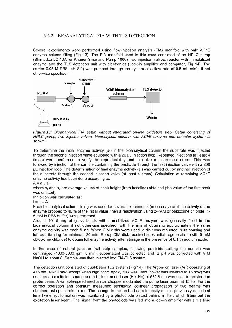
3.6.2 BIOANALYTICAL FIA WITH TLS DETECTION
Several experiments were performed using flow-injection analysis (FIA) manifold with only AChE enzyme column filling (Fig 13). The FIA manifold used in this case consisted of an HPLC pump (Shimadzu LC-10Ai or Knauer Smartline Pump 1000), two injection valves, reactor with immobilized enzyme and the TLS detection unit with electronics (Lock-in amplifier and computer, Fig 14). The carrier 0.05 M PBS (pH 8.0) was pumped through the system at a flow rate of 0.5 mL min-1, if not otherwise specified.
Figure 13: Bioanalytical FIA setup without integrated on-line oxidation step. Setup consisting of HPLC pump, two injector valves, bioanalytical column with AChE enzyme and detector system is shown. To determine the initial enzyme activity (a0) in the bioanalytical column the substrate was injected through the second injection valve equipped with a 20 μL injection loop. Repeated injections (at least 4 times) were performed to verify the reproducibility and minimize measurement errors. This was followed by injection of the sample containing the pesticide through the first injection valve with a 200 μL injection loop. The determination of final enzyme activity (ar) was carried out by another injection of the substrate through the second injection valve (at least 4 times). Calculation of remaining AChE enzyme activity has been done according to: A = ar / a0where ar and a0 are average values of peak height (from baseline) obtained (the value of the first peak was omitted). Inhibition was calculated as: I = 1 – A Each bioanalytical column filling was used for several experiments (in one day) until the activity of the enzyme dropped to 40 % of the initial value, then a reactivation using 2-PAM or obidoxime chloride (1-5 mM in PBS buffer) was performed. Around 10-15 mg of glass beads with immobilized AChE enzyme was generally filled in the bioanalytical column if not otherwise specified, with the aim of obtaining approximately the same enzyme activity with each filling. When CIM disks were used, a disk was mounted in its housing and left equilibrating for minimum 20 min. Epoxy CIM disk required substantial regeneration (with 5 mM obidoxime chloride) to obtain full enzyme activity after storage in the presence of 0.1 % sodium azide. In the case of natural juice or fruit pulp samples, following pesticide spiking the sample was centrifuged (4000-5000 rpm, 5 min), supernatant was collected and its pH was corrected with 5 M NaOH to about 8. Sample was then injected into FIA-TLS system. The detection unit consisted of dual-beam TLS system (Fig 14). The Argon-ion laser (Ar+) operating at 476 nm (40-60 mW, except when high conc. epoxy disk was used, power was lowered to 15 mW) was used as an excitation source and a helium–neon laser (He–Ne) at 632.8 nm was used to provide the probe beam. A variable-speed mechanical chopper modulated the pump laser beam at 15 Hz. For the correct operation and optimum measuring sensitivity, collinear propagation of two beams was obtained using dichroic mirror. The change in the probe beam intensity due to previously described lens like effect formation was monitored by a photodiode placed behind a filter, which filters out the excitation laser beam. The signal from the photodiode was fed into a lock-in amplifier with a 1 s time
35
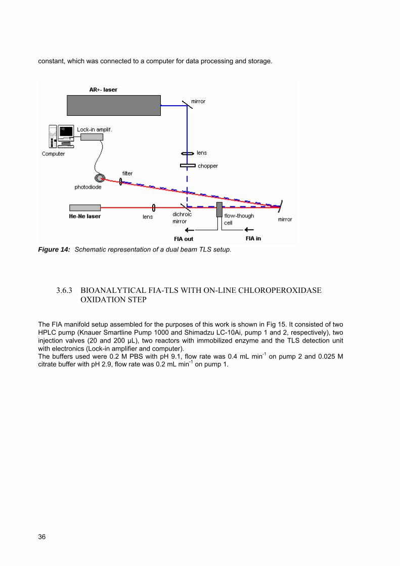
constant, which was connected to a computer for data processing and storage.
Figure 14: Schematic representation of a dual beam TLS setup.
3.6.3 BIOANALYTICAL FIA-TLS WITH ON-LINE CHLOROPEROXIDASE OXIDATION STEP
The FIA manifold setup assembled for the purposes of this work is shown in Fig 15. It consisted of two HPLC pump (Knauer Smartline Pump 1000 and Shimadzu LC-10Ai, pump 1 and 2, respectively), two injection valves (20 and 200 μL), two reactors with immobilized enzyme and the TLS detection unit with electronics (Lock-in amplifier and computer). The buffers used were 0.2 M PBS with pH 9.1, flow rate was 0.4 mL min-1 on pump 2 and 0.025 M citrate buffer with pH 2.9, flow rate was 0.2 mL min-1 on pump 1.
36

Figure 15: Bioanalytical FIA setup used in experiments including on-line CPO oxidation step. Two HPLC pumps were used, with different flow rates (see text for details). Columns were filled with approx. 5-15 mg and 30-40 mg of glass beads with immobilized AChE and CPO enzyme, respectively (if not otherwise specified). The initial experiments involving oxidation with the immobilized CPO enzyme, where the injected samples were collected at the outlet and then analyzed by GC-MS, were performed in the following mode. Samples (1 mL) containing specified amount of parathion-methyl or paraoxon-methyl, H2O2 (0-1.0 mM) and 20 mM KCl were injected into the FIA system with 0.1 M citrate buffer, pH 2.9 as mobile phase. The flow rate was 0.5 mL min-1. After passing the CPO column (filled with approx. 30 mg glass beads with the enzyme) 1 mL of medium fraction was collected at the outlet (between CPO column and Valve 1 on Fig 15). The collected samples were then extracted according to the procedure described before using ethyl acetate as a solvent and analyzed with GC-MS system. All oxidation experiments were performed in duplicate.
3.7 Dimefox activation and HepG2 cell studies -determination of ChE activity
3.7.1 DIMEFOX ACTIVATION
Reactions of dimefox activation were performed using four possible activators, NBS, FeSO4 / EDTA, solubilizate of HepG2 cells and CPO enzyme. The CPO enzymatic reaction was performed in the same manner as previously described batch oxidation reaction (0.1 M citrate buffer, pH 2.9, dimefox in 5 mg L-1 concentration), dimefox was then extracted using described SPE technique for subsequent GC-MS analysis. NBS activation experiment was performed in the following mode: reaction medium and dimefox concentration were the same as in previous activation experiments (PBS, pH 7, 10 mg L-1 dimefox). Two NBS concentrations were tested by addition of 100 µL and 500 µL NBS solution (4 g L-1) to 10 mL of reaction media. Samples were put on ultrasonic bath for 10 min and then an equal volume of ascorbic acid solution (40 g L-1) was added, to reduce the remaining oxidant. The mixture was further shaken on ultrasonic bath for 10 min. Also, control was prepared without added oxidant or reducent. Possible activation with HepG2 cells solubilizate was tested by adding 10-100 µL of cell solubilizate to 5 mL final volume of PBS solution pH 8 with 5 mg L-1 dimefox. Samples were injected on bioanalytical system with recombinant human AChE and inhibition effect was studied. FeSO4 / EDTA activation test was carried out in the following mode: due to low EDTA solubility in PBS buffer, 10 mM EDTA solution was prepared, pH was checked and corrected to 7. Final EDTA concentration (5mM) was obtained by dilution with PBS. Dimefox stock solution and FeSO4 were
37

added simultaneously, to 10 mg L-1 and 5 mM end concentration, respectively. Samples were manually shaken for eight minutes and then extracted using C8 and C18 cartridges. Controls were also prepared, with only FeSO4 or only EDTA. All samples were prepared in duplicate. Samples were also tested on FIA bioanalytical system for AChE inhibition effect. Samples intended to be analyzed using GC coupled with MS detector were extracted according to standard procedure with 2-propanol as a solvent.
3.7.2 PARAOXON-METHYL AND DIMEFOX IN HEPATIC CELL MEDIUM AND DETERMINATION OF CELL CHOLINESTERASE ACTIVITY
Cell cultures were kindly provided and treated with dimefox or paraoxon-methyl by National Institute of Biology, Ljubljana. First, cell medium (Williams` medium E) was removed (or collected) and adhering cells were washed two times with 15 mL of 1xPBS buffer. Cells were removed enzymatically, by adding 5 mL trypsine or mechanically, by scraping. With the addition of trypsine (heated to 37 oC) the culture vessels were transferred to water bath at 37 oC and left there for 5 to 10 minutes. From time to time culture vessels were shaken, to assure all cells were detached from the surface. Then, 5 mL of fresh medium was added to inactivate trypsine. Detached cells were collected and centrifugated at 1000 rpm for 5 min. Supernatant was removed and cells were resuspended in 1xPBS buffer and transferred to microcentrifuge tubes. Once again, cells were centrifugated, supernatant was removed and cold solubilization buffer was added (500-1000 µL). The solution was now vigorously triturated with the aim to completely solubilize cell pellet. Enzymatic activity was measured in the same day cells were prepared, meanwhile, cells were kept on ice. When smaller plates were provided, volumes were proportionally adapted. Paraoxon-methyl and dimefox were extracted from the cell medium following standard procedure and using ethyl acetate or 2-propanol, respectively. Screening for pesticides in the cells and their extraction was performed after repeated cell freezing and thawing, following by the same standard extraction procedure. Measuring ChE activity was done using already described TLS detection unit and spectrophotometer (Hewlet Packard 8453) at 412 and 478 nm. In contrast to AChE inhibition FIA measurements, here the measurements were done in batch system in quartz cuvette. Total volume of solution in the cuvette was 2 mL; 100 µL of 10 mM DTNB solution was added to specified buffer volume, different sample volume 20-300 µL was mixed with the same amount of PBS buffer in separated microcentrifuge tube, triturated and transferred to cuvette. This was overlaid with parafilm, turned upside down for a couple of times and left to stabilize for 5 minutes. Then 20 µL of 75 mM ASChI was added and immediately, the reaction kinetic was measured (Lock-in parameters: time constant 3 s, time of measurement 300 s, sampling frequency 0.2 Hz) Measurements on the spectrophotometer were done with 3 mL total volume of solution in the cuvette, all volumes were proportionally modified. The absorbance of single sample was set to zero before substrate addition. Enzyme activity of the samples was obtained from the slope of linear regression lines.
38

4 RESULTS AND DISCUSSION
4.1 Optimization of solid phase extraction (SPE) of pesticides
Pesticide extraction from aqueous samples was performed using the procedure of SPE extraction described previously (see Experimental section). In the case of dimefox extraction from HepG2 cell culture medium (Williams` medium E), the extraction yields were relatively low and irreproducible. Therefore, SPE extraction procedure for dimefox was optimized by using different extraction solvents. By choosing the solvent that is suitable, both for extraction and for direct application on GC-MS, we wanted to avoid evaporation step of the extraction solvent (as dimefox is a volatile compound). Acetone, acetonitrile, tetrahydrofuran and iso-propyl alcohol (2-propanol) were tested for extraction efficiency. The best results in terms of extraction efficiency (signal peak area) and chromatographic separation (peak shape) were obtained using 2-propanol as extraction solvent (Table 3). The concentration of Na2SO4 aqueous solution and the substitution of the later with phosphate buffer as equilibration medium had no effect on extraction efficiency.
Table 3: Dimefox extraction efficiency (%) with various solvents obtained using gas chromatography with MS detector. Standard deviation of three replicate samples is shown
Extraction solvent/ Equlibration
medium
Acetone Tetrahydrofuran Acetonitril 2-propanol
5 % Na2SO4 61.2 ± 3.8 % 78.9 ± 10.6 % 68.0 ± 10.4 % 81.4 ± 3.5 %
4.2 FIA-TLS bioanalytical system characterization
Our aim was to characterize the bioanalytical system in regards to the amount of the AChE enzyme column filling and subsequent degree of inhibition. Due to some minor changes in immobilization protocol of the enzymes (application of reduced atmosphere to suck the air out of the pores) in comparison to procedures used by other authors (Pogačnik and Franko, 2001), the inhibition dynamics in our experiments could be different. In the following part the inhibition effect in dependence of the amount of glass beads filled in the column and the AChE enzyme immobilization concentration was studied. Furthermore, we attempted to estimate the amount of the injected pesticide that in fact reacts (and inhibits) with the enzyme and compare different commercially available regenerators for restoring inhibited AChE activity. Finally, the detection of pesticide in natural juice samples was tested and compared to buffer samples. Human AChE enzyme was used throughout the experiments described in this chapter.
4.2.1 FIA-TLS WITH HUMAN ACHE ENZYME
4.2.1.1 Influence of the amount of enzyme on inhibition effect
First, the relation between the amount of immobilized AChE and its inhibition at a given pesticide concentration was verified on the experimental setup depicted in Fig 13. Human AChE enzyme was immobilized with three immobilization enzyme concentrations, 11.9 U g-1, 60 U g-1 and 120 U g-1 of oven-dried glass beads. The column was filled with various amounts of glass beads and two pesticide concentrations (of chlorpyrifos oxon) were tested (Fig 16).
39

15 20 25 30 35 40 45
0.0012
0.0014
0.0016
0.0018
0.0020
0.0022
0.0024
0.0026
0.0028
0.0030
0.0032
A11.9 U g-1
Sign
al [V
]
Immobilized hAChE [mg glass beads]
Signal
16
18
20
22
24
26
28
30
32
34
blank corrected inhibition
Inhi
bitio
n [%
]
15 20 25 30 35 40 45
0.0012
0.0014
0.0016
0.0018
0.0020
0.0022
0.0024
0.0026
0.0028
0.0030
0.0032
Sig
nal [
V]
Immobilized hAChE [mg glass beads]
Signal
36
38
40
42
44
46
48
50
52B11.9 U g-1 blank corrected inhibition
Inhi
bitio
n [%
]
5 10 15 20 25 30 35 40 45 50
0.002
0.004
0.006
0.008
0.010
0.012
Sign
al [V
]
Immobilized hAChE [mg glass beads]
Signal
16
18
20
22
24
26
28
30
32
34C60 U g-1
Inhi
bitio
n [%
]
blank corrected inhibition
5 10 15 20 25 30 35 40 45 50
0.002
0.004
0.006
0.008
0.010
0.012
D
60 U g-1Si
gnal
[V]
Immobilized hAChE [mg glass beads]
Signal
36
38
40
42
44
46
48
50
52
Inih
ibito
n [%
]
blank corrected inhibition
3 4 5 6 7 8 9 10 11
0.006
0.008
0.010
0.012
0.014
0.016
0.018
E
Sign
al [V
]
Immobilized hAChE [mg glass beads]
Signal
16
18
20
22
24
26
28
30
32
34
120 U g-1 blank corrected inhibition
Inhi
bitio
n [%
]
3 4 5 6 7 8 9 10 11
0.006
0.008
0.010
0.012
0.014
0.016
0.018
F
Sign
al [V
]
Immobilized hAChE [mg glass beads]
Signal
36
38
40
42
44
46
48
50
52
120 U g-1
Inih
ibiti
on [%
]
blank corrected inhibition
Figure 16: Signal and enzyme inhibition obtained with 26 μg L-1 (left column) and 52 μg L-1 (right column) chlorpyrifos oxon using different column glass beads fillings with the AChE enzyme immobilized in three different concentrations (A,B- 11.9 U g-1 ; C,D- 60 U g-1 ; E,F- 120 U g-1 of oven-dried glass beads). Blank signal (from unreacted substrate) was subtracted and blank corrected inhibition calculated. Filling the column with more glass beads with immobilized AChE enzyme gives linearly higher TLS signal (A to F). Also, increasing the concentration from 26 µg L-1 to 52 µg L-1 chlorpyrifos oxon (comparison of left and right column) gave increased inhibition effect in all of studied cases. What was
40

not expected is that in some cases, by increasing the amount of glass beads with immobilized AChE enzyme, thus increasing possible sites for pesticide interaction the inhibition remains quite stable (designated on the graphs with red dots). The signal from unreacted substrate was approx. 4 x 10⁻⁴ V (measured at the begining and end of the day experiments). By comparing the signal expressed for 1 mg of column filling (AChE on CPG) in the three cases where different enzyme concentration was used in the immobilization procedure, we get the values of 5.47 x 10⁻⁵, 2.18 x 10⁻⁴ and 1.56 x 10⁻³ V mg-1 glass beads filling (begining from lower to higher enzyme immobilization concentration) or approximately the 1 : 4 : 28 ratio. This is a clear evidence that higher amounts of enzyme were in fact bounded to the porous CPG glass. Using the same pesticide concentration in all cases one would expect obtaining the higher inhibition effect, when lower amount of enzyme in the column is used (in this case pesticide/enzyme active sites ratio is higher). However, as can be seen by comparing A and B to C and D or E and F in Fig 16, this was not the case. Obtained results are unexpected, when one considers the inhibtion of AChE with OP compounds as a straightforward phosphorylation of the active center Ser203 by a mechanism that follows simple Michealis-Menton kinetics (Chambers and Levi, 1992). However, recent reports (Kaushik et al., 2007) show that inhibitory potency of OP oxons toward human recombinant AChE appears to change as a function of oxon concentration. Relative AChE inhibitory potential of oxon molecules increase at lower oxon concentration, probably through mechanism of binding of oxons to a second site (peripheral anionic site- PAS), that modulates the inhibitory potential of other oxon molecule at the catalytic site. Results obtained in our experiments can thus be explained using the theory provided by Kaushik et al. (Kaushik et al., 2007); with the increasing amount of glass beads with bounded AChE or the increasing amount of AChE bounded to glass beads, thus lowering OP pesticide to AChE concentration ratio, the relative inhibition potential of pesticide increases. As our main goal was the integration of CPO oxidation into the FIA-AChE-TLS system, the above experiment was repeated on the set up presented in Fig 15. As in such a parallel two-column system (CPO and AChE) the mixing of citrate and phosphate buffer is occuring before AChE bioanalytical column, we investigated the possible effect of this factor on the degree of inhibition in dependence of the amount of AChE enzyme on the column. In contrast to the previous experiments, immobilized electric eel AChE enzyme was used and only two AChE immobilization concentrations were tested (Fig 17). By using chlorpyrifos oxon (oxo-analog) we wanted to exclude possible impacts of oxidation reaction (low reaction yield or degradation of the oxon).
41
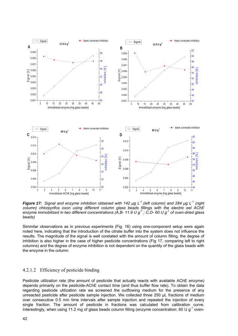
5 10 15 20 25 30 35 40 45 500.001
0.002
0.003
0.004
0.005
0.006
0.007
0.008
0.009
Sign
al [V
]
Immobilized enzyme [mg glass beads]
Signal
26
28
30
32
34
36
38
A11.9 U g-1
blank corrected inhibition
Inih
ibito
n [%
]
5 10 15 20 25 30 35 40 45 500.001
0.002
0.003
0.004
0.005
0.006
0.007
0.008
0.009
11.9 U g-1
Sig
nal [
V]
Immobilized enzyme [mg glass beads]
Signal
44
46
48
50
52
54
56
58
60
62B
Inhi
bitio
n [%
]
blank corrected inhibition
2 3 4 5 6 7 8 9 10 110.002
0.004
0.006
0.008
0.010
0.012
0.014
C60 U g-1
Signal
Sig
nal [
V]
Immobilized AChE [mg glass beads]
26
28
30
32
34
36
38
blank corrected inhibition
Inhi
bitio
n [%
]
2 3 4 5 6 7 8 9 10 110.002
0.004
0.006
0.008
0.010
0.012
Sign
al [V
]
Immobilized enzyme [mg glass beads]
Signal
44
46
48
50
52
54
56
58
60
62D60 U g-1
Inhi
bitio
n [%
]
blank corrected inhibition
Figure 17: Signal and enzyme inhibition obtained with 142 μg L-1 (left column) and 284 μg L-1 (right column) chlorpyrifos oxon using different column glass beads fillings with the electric eel AChE enzyme immobilized in two different concentrations (A,B- 11.9 U g-1 ; C,D- 60 U g-1 of oven-dried glass beads) Simmilar observations as in previous experiments (Fig. 16) using one-component setup were again noted here, indicating that the introduction of the citrate buffer into the system does not influence the results. The magnitude of the signal is well corelated with the amount of column filling, the degree of inhibition is also higher in the case of higher pesticide concentrations (Fig 17, comparing left to right columns) and the degree of enzyme inhibition is not dependent on the quantity of the glass beads with the enzyme in the column. 4.2.1.2 Efficiency of pesticide binding
Pesticide utilization rate (the amount of pesticide that actually reacts with available AChE enzyme) depends primarily on the pesticide-AChE contact time (and thus buffer flow rate). To obtain the data regarding pesticide utilization rate we screened the outflowing medium for the presence of any unreacted pesticide after pesticide sample injection. We collected three 250 µL fractions of medium over consecutive 0.5 min time intervals after sample injection and repeated the injection of every single fraction. The amount of pesticide in fractions was calculated from calibration curve. Interestingly, when using 11.2 mg of glass beads column filling (enzyme concentration: 60 U g-1 oven-
42

dried glass) with 52 μg L-1 chlorpyrifos oxon the resulted pesticide utilization rate was 21.7 ± 6.7 % (at 0.5 mL min-1 flow, duplicate experiment performed). Due to the unexpectedly low pesticide utilization rate, the obtained results were verified with the experiment, where the inhibition effect was studied in dependence of the buffer flow rate. Two stop-flow injections with 20 s and 35 s stop time were performed as well (Fig 18).
0.0 0.1 0.2 0.3 0.4 0.5
30
40
50
60
70
80
90
100
Inhi
bitio
n [%
]
Flow [mL min-1]
Figure 18: AChE enzyme inhibition obtained by chlorpyrifos oxon (26 μg L-1) with different flow-rates and two stop-flow experiments (20 s, 35 s; showed as two points at x=0). By using maximum obtained inhibition of 90.6 % (presupposing all the pesticide is bound to the AChE enzyme at these conditions) and inhibition obtained when 0.5 mL min-1 flow was used, the calculated utilization rate of 26 μg L-1 chlorpyrifos oxon is 34.3 ± 1.2 %, somewhat higher than previously obtained number. 4.2.1.3 Regeneration of inhibited AChE enzyme
Three commercially available enzyme regenerators were tested for their effectiveness: Obidoxime chloride, pyridine-2-aldoxime methiodide (2-PAM iodide) and pyridine-2-aldoxime methochloride (2-PAM chloride). Human AChE enzyme (60 U g-1 oven-dried glass) was first inhibited by 40 µg L-1 chlorpyrifos oxon and regeneration was performed using 1 mM solution of selected regenerator (Fig 19).
43
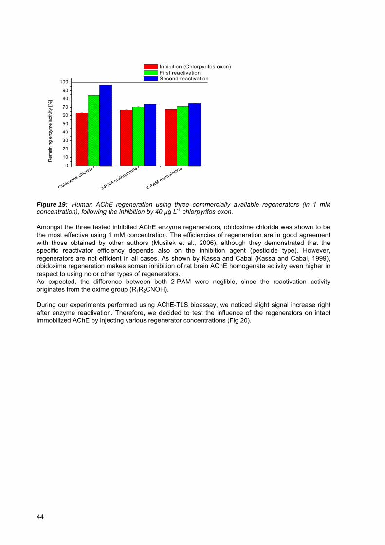
Obidoxime chloride
2-PAM methochlorid
2-PAM methoiodide0
10
20
30
40
50
60
70
80
90
100
Inhibition (Chlorpyrifos oxon) First reactivation Second reactivation
Rem
aini
ng e
nzym
e ac
tivity
[%]
Figure 19: Human AChE regeneration using three commercially available regenerators (in 1 mM concentration), following the inhibition by 40 µg L-1 chlorpyrifos oxon. Amongst the three tested inhibited AChE enzyme regenerators, obidoxime chloride was shown to be the most effective using 1 mM concentration. The efficiencies of regeneration are in good agreement with those obtained by other authors (Musilek et al., 2006), although they demonstrated that the specific reactivator efficiency depends also on the inhibition agent (pesticide type). However, regenerators are not efficient in all cases. As shown by Kassa and Cabal (Kassa and Cabal, 1999), obidoxime regeneration makes soman inhibition of rat brain AChE homogenate activity even higher in respect to using no or other types of regenerators. As expected, the difference between both 2-PAM were neglible, since the reactivation activity originates from the oxime group (R1R2CNOH). During our experiments performed using AChE-TLS bioassay, we noticed slight signal increase right after enzyme reactivation. Therefore, we decided to test the influence of the regenerators on intact immobilized AChE by injecting various regenerator concentrations (Fig 20).
44

1 2 3 4 5 6 7 8 90.000
0.001
0.002
0.003
0.004
0.005
0.006
0.007
0.008
1 2 3 4 5 6 7 8 90.000
0.001
0.002
0.003
0.004
0.005
0.006
0.007
0.008
Flow rate increase to 2mL/min for 2 min
50mM 2-PAM
10mM 2-PAM
5mM 2-PAM
3mM 2-PAM
3mM 2-PAM
1mM 2-PAM
1mM 2-PAM
Sign
al [m
V]
Substrate Injection
Flow rate increase to 2mL/min for 2 min
50mMObidoxCl
10mM ObidoxCl
5mM ObidoxCl
3mM ObidoxCl
3mM ObidoxCl
1mM ObidoxCl
1mM ObidoxCl
Substrate Injection
Figure 20: Human AChE enzyme activity with sequential injection of various concentrations of 2-PAM chloride (left) and obidoxime chloride (right) shown with the arrows. Standard deviations for at least four injections of substrate after every sample are shown. The mechanism involved in the signal increase following the regenerator injection could be explained by the findings of Sakurada et al. (Sakurada et al., 2006). It was shown that oximes express esterase-like activity, thus hydrolysing acetylthiocholine iodide, the substrate for AChE enzyme in Ellman`s colorimetric reaction (Ellman et al.,1961). The TLS signal incerase can thus be a direct consequence of a small amount of regenerator remained in the system (bound to the enzyme or glass beads) promoting the hydrolysis of the used substrate. However, enhanced rinsing of the FIA system by increasing the flow to 2 mL min-1 for 2 min (Fig 20) had no significant effect on the decrease of signal. Regarding the enzyme regeneration, structural similarities of several ionic liquids with some known AChE regenerators inspired us to test the ILs for the regeneration capabilities on chlorpyrifos oxon inhibited human AChE enzyme. The representative ionic liquids chosen were: imidazolium IL (BMIMmetSO4, see also Fig 12), pyridinium IL (BMPyBF4) and quaternary ammonium IL (QATf2N - both optical isomers and racemic mixture). However, no regeneration effect could be observed when 1 % or 5 % IL solution was injected through the inhibited AChE enzyme column (due to low water solubility QATf2N was tested only at 1 % concentration). 4.2.1.4 Detection of pesticides in complex samples
Practical applications of newly developed methods should allways involve the analysis of real samples with complex matrices to confirm their reliability. Therefore, we examined the described AChE-TLS bioanalytical system in the case of real sample analysis. Spiked samples of three kind of juices or fruit pulps were analyzed and the results were compared to those from buffer pesticide solutions (Fig 21).
45

Buffer
Apple juice
Peach pulp
Pear pulp0
20
40
60
80
100
120
Pes
ticid
e re
cove
ry [%
]
Figure 21: Pesticide recovery in different kind of natural juice and fruit pulp samples spiked with chlorpyrifos oxon (50 μg L-1). Results were normalized to buffer sample. Standard deviation of multiple substrate injection is shown. Pesticide recoveries were in all cases higher than 70 %. In case of apple juice concentrate, where the sample did not need further treatment (beside pH adjustment), the recovery rate was 100 % (inhibition similar as in buffer sample), thus indicating very low matrix interferences. Lower recovery in the peach and pear pulp sample is probably due to pesticide adsorption on solids present in the juice, which were later in the procedure of sample preparation removed by centrifugation (see the Experimental section). Possible explanation for this effect can also be found in differences of pesticide adsorption on laboratory glassware as a function of different pH values of various juices. Another factor that contributes to lower recovery is the effect of signal increase after injection of negative control (unspiked) samples. Approx. 6.4 % and 3.1 % higher signals were observed, when supernatants of unspiked peach and pear samples were injected, respectively. Such matrix effects should be seriously taken into consideration, when real samples are analysed. Calibration in the same matrix should be done or the method of standard addition used. Unspiked samples were also controlled with GC-MS analyses, and no pesticide residues were detected. Achieved pesticide recoveries are comparable to those obtained by Caetano and Machado (Caetano and Machado, 2008), who report 83 % recovery for measurements done in tomato pulp using the amperometric technique based on AChE inhibition. Considering high dry matter content, the obtained results are quite promising. The method allows achieving high sensitivity of pesticide detection in the juice concentrate already in the juice production stage, before dilution to commercial product.
4.3 Improving the sensitivity of FIA-TLS bioassay
Due to the possible enhancement in sensitivity of TLS measurements when medium other than water is used, we tested AChE and CPO enzyme activity in the presence of several ionic liquids (BMIMmetSO4, BMPyBF4 and QATf2N, both optical isomers and racemic mixture).
4.3.1 ACHE ENZYME ACTIVITY AND INHIBITION IN THE PRESENCE OF IONIC LIQUID AS CO-SOLVENT
We investigated the effect of IL type and concentration on the AChE activity by injecting 200 μL of chosen IL into the FIA-TLS system represented in Fig 13. The results shown in Fig 22 for BMPyBF4 (1 and 5 % concentration) and QATf2N (both isomers and racemic mixture, 1 % concentration) show different degree of inhibition expressed by tested ILs. Also, the inhibition effect was reversible; this is in strong contrast to the type of inhibition expressed by OP compounds, with the inhibition being
46

irreversible. The reversible interaction with AChE enzyme is presumably due to the ILs similarity to the choline part of the native enzyme substrate in regards to positive charged nitrogen (only imidazolium and pyridinium ring IL). The binding of IL to the enzyme occurs probably at the PAS (peripheral anionic site) of the enzyme. However, ILs are missing the molecular interaction potential of the ester group, which would enable them binding to the catalytic enzyme site.
0 50 100 150 200 250 300 3500.000
0.001
0.002
0.003
0.004
0.005
0.006
0.007
0.008
Sig
nal [
V]
Time [s]
BMPyBF4- 1 % BMPyBF4- 5 % Initial enzyme activity
0 50 100 150 200 250 300 350 400 4500.0000
0.0005
0.0010
0.0015
0.0020
0.0025
0.0030
0.0035
0.0040
0.0045
QATf2N-rac QATf2N-S QATf2N-R Initial enzyme activity
Sign
al [V
]
Time [s]
Figure 22: Peaks representing substrate injection and observed inhibition of human AChE initial activity after injection of BMPyBF4 (1 and 5 %, left) and QATf2N (both isomers and racemic mixture, 1 %, right) samples. With dotted line initial enzyme activity is shown. The obtained results are in good agreement with those reported by Stock and co-workers (Stock et al., 2004), who described AChE inhibition by relatively low concentration of imidazolium and pyridinium ILs. However, their experiments were performed in batch mode, thus reversible type of inhibition could not be observed. In contrast, by performing the experiments in FIA mode, a deeper insight into IL – AChE interaction could be obtained. Next, we investigated the AChE inhibition with the pesticide (chlorpyrifos oxon, 40 μg L-1) in the presence of various ILs. The pesticide solution was prepared in the PBS buffer (control) or IL medium and injected into the system. The results obtained with BMPyBF4 (1 and 5 % concentration) are shown in the next graph (Fig 23).
47

0 50 100 150 200 250 300 3500.000
0.001
0.002
0.003
0.004
0.005
0.006
0.007
Sign
al [V
]
Time [s]
PBS buffer BMPyBF4 - 1 % BMPyBF4 - 5 %
Figure 23: Peaks representing substrate injection and observed inhibition of the AChE enzyme with chlorpyrifos oxon (40 μg L-1) in PBS buffer or ionic liquid medium (BMPyBF4, 1 and 5 % concentration). With dotted line initial enzyme activity is shown. As can be seen, the observed enzyme inhibition in the presence of IL (blue and red curve) is initially higher than in the PBS buffer (black curve). However, with the increase of time it could be seen that almost all enzyme inhibition is caused by IL rather than pesticide. Very similar results were obtained with BMIMmetSO4, but not with QATf2N. Considering results obtained previously, there is an indication, that some competition between pesticide and IL molecules exist for the enzyme active site in the gorge (or active site phosphorylation performed by the pesticide molecule is sterically hindered, due to IL bounding to surrounding subsites). To exclude any possible IL-pesticide interaction in the sample the experiment was modified in a sense, that pesticide in the PBS buffer was injected right after IL sample injection. We expected lower degree of pesticide inhibition when AChE enzyme is in some way “protected” by IL bounded to it. Results of these experiments are shown in Fig 24.
0 50 100 150 200 250 300 350 400 4500.000
0.001
0.002
0.003
0.004
0.005
0.006
0.007
Sig
nal [
V]
Time [s]
BMPyBF4 - 1%
BMPyBF4 - 5% Initial enzyme activity no IL
0 50 100 150 200 2500.0000
0.0005
0.0010
0.0015
0.0020
0.0025
0.0030
0.0035
0.0040
Sign
al [V
]
Time [s]
QATf2N-S QATf2N-Rac QATf2N-R No IL Initial enzyme activity
Figure 24: Peaks representing substrate injection and observed inhibition of human AChE enzyme achieved with chlorpyrifos oxon (40 μg L-1) with or without precedent IL injection (BMPyBF4,, 1 and 5 % concentration, left and QATf2N, both isomers and racemic mixture, 1 %, right). With dotted line initial enzyme activity is shown. As can be seen from Fig 24, the inhibition effect of the pesticide was greatly diminished if IL BMPyBF4
48
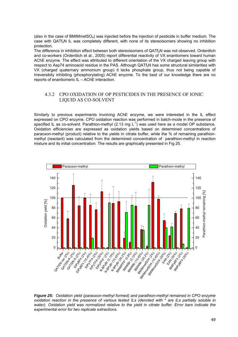
(also in the case of BMIMmetSO4) was injected before the injection of pesticide in buffer medium. The case with QATf2N IL was completely different, with none of its stereoisomers showing no inhibition protection. The difference in inhibition effect between both stereoisomers of QATf2N was not observed. Ordentlich and co-workers (Ordentlich et al., 2005) report differential reactivity of VX enantiomers toward human AChE enzyme. The effect was attributed to different orientation of the VX charged leaving group with respect to Asp74 aminoacid residue in the PAS. Although QATf2N has some structural similarities with VX (charged quaternary ammonium group) it lacks phosphate group, thus not being capable of irreversibily inhibiting (phosphorylating) AChE enyzme. To the best of our knowledge there are no reports of enantiomeric IL – AChE interaction.
4.3.2 CPO OXIDATION OF OP PESTICIDES IN THE PRESENCE OF IONIC LIQUID AS CO-SOLVENT
Similarly to previous experiments involving AChE enzyme, we were interested in the IL effect expressed on CPO enzyme. CPO oxidation reaction was performed in batch-mode in the presence of specified IL as co-solvent. Parathion-methyl (2.13 mg L-1) was used here as a model OP substance. Oxidation efficiencies are expressed as oxidation yields based on determined concentrations of paraoxon-methyl (product) relative to the yields in citrate buffer, while the % of remaining parathion-methyl (reactant) was calculated from the determined concentration of parathion-methyl in reaction mixture and its initial concentration. The results are graphically presented in Fig 25.
Buffe
r
QATf2
N-rac (
2%)
QATf2
N-R (2
%)
QATf2
N-S (2
%)
EtPy
BF4
(2%
)
EtPy
BF4
(10,
45%
)Et
PyTF
A (2
%)
EtPy
TFA
(30%
)
EtPy
PF6*
(2%
)
N-BPy
Br (2
,15%
)
N-BPy
Br (1
0,45
%)
N-BPy
Br (3
0,2%
)
BMIM
PF6*
(0,9
%)
BMIM
Br (2
,15%
)
BMIM
Br (1
0,45
%)
BMIM
Br (3
0,2%
)
BMIM
met
SO4
(2%
)
BMIM
met
SO4
(10,
45%
)
BMIM
met
SO4
(30%
)EA
N (2%
)EA
N (30%
)
BMPy
BF4
(2%
)
BMPy
BF4
(30%
)
0
20
40
60
80
100
120
140
0
20
40
60
80
100
120
140
Par
athi
on-m
ethy
l rem
aini
ng [%
]
Oxi
datio
n yi
eld
[%]
Paraoxon-methyl Parathion-methyl
Figure 25: Oxidation yield (paraoxon-methyl formed) and parathion-methyl remained in CPO enzyme oxidation reaction in the presence of various tested ILs (denoted with * are ILs partially soluble in water). Oxidation yield was normalized relative to the yield in citrate buffer. Error bars indicate the experimental error for two replicate extractions.
49

When the oxidation of parathion-methyl by CPO was carried out in the presence of ionic liquids, it was revealed, that the effects of ionic liquids are very much different (Fig 25). Compared to oxidation in citrate buffer, oxidation efficiency was actually 100 % in the presence of ionic liquids such as BPyBr, EtPyBF4, BMPyBF4, BMIMBr, or BMIMmetSO4 at 2 % and 10 % level and for EtPyTFA even at 30 % level. Some ionic liquids, however, significantly affected the capacity of CPO to oxidize parathion-methyl. Specifically, the oxidation efficiency was reduced to only 10 – 60 % of the efficiency in citrate buffer at 2 % of either EAN, BMIMPF6, EtPyTFA or QATf2N (recemic), at 10 % of BMIMBr or at 30 % of either BMIMmetSO4 or BPyBr. In some cases the oxidation of parathion-methyl was completely inhibited (EtPyPF6 – 2 %, BMIMBr – 30 %). In general, when concentration of IL is increased, the oxidation reaction yield decreases. The exemption was EAN, which demonstrated an increase in the efficiency of CPO oxidation with increased concentration of IL. The oxidation yield in 30 % EAN however, reached only 70 % of the oxidation yield in citrate buffer and since no remaining parathion-methyl was detected in the reaction mixture, no improvements in oxidation efficiency are expected at higher concentrations of this ionic liquid. From the results reported for ionic liquids based on [EtPy]+ (2 % concentration) it can be concluded, that the effect of anions increases in the series BF4
- < TFA- < PF6-. Similarly, the results for [BMIM]+ (10
% and 30 % concentration) demonstrate, that the effect on the activity of CPO is stronger in case of Br
- than in the case of metSO4- as counter anions. It is also interesting to observe, that both ionic liquids
containing hexafluorophosphate anion (EtPyPF6 and BMIMPF6) gave oxidation yields lower than 10 % even at just 2 % and 0.9 % concentrations, respectively. The results are in agreement with the observations of Sanfilippo et al. (Sanfilippo et al., 2004), who reported inhibition of CPO enzyme in pure ILs (BMIMBF4, BMIMPF6, BMIMmetSO4), when the oxidation of 1,2-dihydronaphthalene was studied. They also tested BMIMmetSO4 and MMIMmetSO4 in 10 % and 30 % IL concentration in citrate buffer medium, however, rapid decrease of conversion efficiency was observed at higher IL concentration. Due to unfavorable pH shift, higher concentrations of ILs were not used in their experiments. It is also important to stress the fact, that better CPO activity was observed in some of the tested ILs compared to non-ionic solvents, such as t-butyl hydroperoxide or acetone. For practical application in TLS measurements, concentration of ionic liquid should be as high as possible, but at the same time with the lowest possible inhibition of CPO activity. From this point of view the EtPyTFA, EAN and BMIMmetSO4 were found to be most suitable for such purpose. The enhancement in sensitivity of TLS measurements due to the addition of investigated ionic liquids can be readily estimated by assuming linear dependence of the increase in the absolute value of ∂n/∂T and decrease in k with the concentration of ionic liquid. Since no ∂n/∂T and k values are available for the investigated ionic liquids, the mean values for ionic liquids studied earlier (Tran et al., 2005) will be used for calculation, i.e. ∂n/∂T = 6×10⁻⁴ K-1, and k = 1.4 mW cm-1 K-1 and compared to values of ∂n/∂T = 8.8×10⁻⁵ K-1, and k = 6.04 mW cm-1 K-1 for water. In a 30 % solution of an ionic liquid the contribution to the enhancement of sensitivity due to increase of ∂n/∂T would be 2.7 fold and the contribution from the decrease in thermal conductivity 1.3 fold, thereby resulting in about 3.5 fold enhancement in the sensitivity. Similar enhancements in sensitivity can be expected for ionic liquids such as EtPyTFA, while for ionic liquids such as EAN the reduction of the CPO oxidation efficiency has to be taken into account for determination of parathion-methyl. Due to reversible AChE inhibition of some ILs described in previous section some attention should be devoted to carefully choosing the most appropriate IL (and its concentration) for both enzymes when FIA mode is used. Furthermore, one of the drawbacks of using IL in AChE-TLS bioanalytical system is also already described reduced AChE inhibition effect by pesticide in the presence of IL as co-solvent. Some other technical solution of FIA setup can be explored to solve this problem. For instance, measurement of AChE enzyme activity could be performed in IL-buffer medium (with better thermooptical properties), while sample could be injected when only buffer medium is pumped through the system, thus eliminating IL protective effect for pesticide AChE inhibition.
50

4.3.3 FIA-TLS BIOASSAY USING CIM DISKS AS ACHE IMMOBILIZATION SUPPORT
Due to reported advantages of using CIM disks as enzyme support in comparison to other inert supports (Barut et al., 1999; Vodopivec et al., 2003; Bartolini et al., 2004; Bartolini et al., 2005), the former were tested in our FIA-TLS system using the setup shown in Fig 13. The immobilization of human AChE was performed using two CIM disk chemistries, EDA and epoxy reactive groups. The former type of disk contains protonated amine groups, thus it functions also as weak ionic exchanger. The inclusion of 0.1 M KClO3 in our mobile phase as an anion competitor, with the aim of displacing yellow TNB2- enzyme product (product of Ellman`s reaction) did not give good results in terms of signal peak shape and displacement time. Although Bartolini et al. (Bartolini et al., 2004) found this competitor to be suitable for their bioanalytical system, the time needed for yellow anion displacement from the support was too long. This in turn leads to unwanted extension of the analysis time or in the other case, the number of sequential substrate injection has to be reduced. Therefore, further research was concentrated on epoxy CIM disks. The flow rate used was 0.5 mL min-1. Increasing the flow rate, thus reducing enzyme contact time, reduced the enzyme signal and also pesticide inhibition effect (data not shown here). To characterize epoxy CIM support, human AChE was immobilized in two different enzyme concentrations (2 U and 20 U per disk, see Experimental section, for easier representation we are referring to low and high disk concentration in the text below). Sequential inhibition with pesticide chlorpyrifos oxon (24.1 μg L-1 for CPG glass and low conc. disk and 100 μg L-1 for high conc. disk) was performed without intermediate regeneration of the enzyme activity (Fig 26).
51

0 1 2 3 4 5 6 7 8 90.000
0.002
0.004
0.006
0.008
0.010
Sig
nal [
V]
Sample number
CIM epoxy signal
60
65
70
75
80
85
90
95
100
A
Relative remaining activity
Rel
ativ
e R
emai
ning
Enz
yme
Activ
ity [%
]
0 1 2 3 4 5 6 7 8 90.000
0.001
0.002
0.003
0.004
0.005
0.006
0.007
0.008
CIM epoxy signal
Sig
nal [
V]
Sample number
60
65
70
75
80
85
90
95
100
B
Relative remaining activity
Rel
ativ
e R
emai
ning
Enz
yme
Activ
ity [%
]
0 1 2 3 4 5 6 7 8 90.000
0.001
0.002
0.003
0.004
0.005
0.006
0.007
0.008
Sign
al [V
]
Sample number
CPG glass signal
60
65
70
75
80
85
90
95
100
C
Rel
ativ
e R
emai
ning
Enz
yme
Act
ivity
[%]
Relative remaining activity
Figure 26: Sequential inhibition of human AChE performed with chlorpyrifos oxon. AChE enzyme was bound to epoxy CIM disk in 20 U per disk (A), 2 U per disk (B) and CPG glass in 120 U g-1 of oven-dried glass (C). In the case of CPG glass the column was fillled with approx. 10 to 15 mg of glass beads in buffer medium (1.2 to 1.8 U AChE). On the left axes of the graphs enzyme signal is depicted, while on the right remaining enzyme activity is shown. The later was corrected by subtracting out the value obtained with the unreacted substrate (blue horizontal line). Standard deviation of multiple substrate injection is also shown. The results represented in Fig 26 (B and C) are similar in context of inhibition dynamics. Based on these results, the advantages of using CIM monoliths in comparison to other porous support material reported by Barut et al. (Barut et al., 1999 1999) could not be confirmed. However, it is likely that the effects of convective mass transport (in CIM disks case) in comparison to diffusion processes (porous material) are more expressed at higher flow rates, when the contact time within the column is shorter. Sequential injections of the sample with the pesticide are expressed as progressively lower enzyme inhibition (higher relative remaining activity). The explanation of this effect could originate from the theory given by several researchers (Kousba et al., 2004; Kaushik et al., 2007; Stojan et al., 2004). According to the theory, the interaction of the substrate and pesticide molecule with AChE enzyme does not follow simple Michealis-Menton kinetics. High substrate concentration inhibits the enzyme activity through binding to the peripheral active site of the enzyme and related steric effects (blockage of choline exit, prevention of water molecule needed for substrate deacetylation, Stojan et al., 2004). The same principle can be applied in the case of pesticide molecule, where one molecule interaction reduces (or increases, depending on the nature of substrate and inhibitor) the capacity of other, subsequent molecules to inhibit the catalytic site (Kaushik et al., 2007). In the sequential enzyme inhibition experiment the ratio of pesticide molecules to free enzyme is constantly changing
52

(increasing), and the observed relative inhibition effect is decreasing (Fig 26, B and C), thus being in good agreement with the previous theory. Moreover, these results also coincide well with the results obtained, when inhibition was measured with different amount of glass beads column filling. The situation was quite different in the case of CIM support with higher enzyme immobilization concentration (Fig 26, A). In these experiments the power of the excitation laser beam was reduced to 15 mW, to avoid the saturation of the thermal lens signal. From the amount of the signals obtained from epoxy CIM disks with both enzyme concentration and taking into account the linear dependence of the signal on the excitation laser power (Eq. 1), we estimated the amount of the active enzyme on the support was approx. 3.5 times higher in the case of the higher concentration variant. The increased inhibition effect in the fist part of the sequential pesticide injection can be easily explained, as the active enzyme / pesticide ratio diminishes, thus proportionally higher inhibition effect could be seen. The second part of the experiment (after 4th sample injection) shows quite similar results in comparison to experiment depicted in Fig 26, B. At the point of 4th injection the signal from remaining enzyme activity (left axis) is approximately 0.003 mV. By taking into account the correction owing to excitation laser power by a factor of 2.66 (40 mW / 15 mW) we are at exactly the same amount of the active AChE enzyme, as at the beginning of the experiment involving CIM disk with lower immobilization concentration (Fig 26, B). The relative remaining activity curves (right axes) in the A and B case are in good agreement. Due to relatively large internal volume (0.34 mL) of the disk, the lag phase after substrate injection and before signal registration was somewhat extended (30 s in comparison to 10 s when CPG glass column filling was used). Due to increased flow-through time of the injected substrate (increased diffusion effect) peak broadening in the case of CIM disks was noticed (Fig 27). This in turn leads to unwanted total analysis time prolongation.
0 50 100 150 200 250 3000.000
0.001
0.002
0.003
0.004
0.005
0.006
0.007
0.008
Sig
nal [
V]
Time [s]
CIM disk CPG glass
Figure 27: TLS signal obtained using AChE immobilized on CIM disk (blue) and CPG glass (red). The large internal volume of the disk also severely limits the possibility of using up to 3 disks in one housing, thus reducing possible disks combination. However, the producer offers custom-made disks with smaller internal volumes (34 and 3.4 μL) that in our opinion would be a more appropriate choice in regards to the flow rates used in our experiments.
53

4.4 Chloroperoxidase oxidation of organophosphorus pesticides
The main objective of our work was to integrate the oxidation step into a FIA-AChE bioanalytical system. Limited publications exist that deal with CPO oxidation of OP pesticides (Hernandez et al., 1998; Walz and Schwack, 2007c) and to the best of our knowledge, there are no reports of using immobilized CPO enzyme in FIA bioanalytical systems. Therefore, several optimization experiments in batch mode were performed first, to verify reported results and to optimize the reaction parameters.
4.4.1 BATCH CPO OXIDATION EXPERIMENTS
For most of the batch oxidation experiments three selected model OP pesticides were used: malathion, parathion-methyl and diazinon. For malathion and parathion-methyl, their oxo-analogues (malaoxon, and paraoxon-methyl) were commercially available, thus allowing us to compare the results obtained in oxidation experiments with the results obtained with specific oxo-analog. First, results obtained by Hernandez and co-workers (Hernandez et al., 1998), which oxidized various OP pesticides in 15 mM acetonitrile - 60 mM acetate buffer (pH 3) were verified in citrate buffer (pH 2.9). As the authors did not explain the reason for chosen reaction conditions, we further explored also other reaction parameters (pH of the medium, H2O2 concentration, CPO enzyme concentration). The results are presented in the following section. 4.4.1.1 Oxidation reaction pH optimum
CPO has broad range of pH optimums, dependent on the type of substrate and reaction performed, with the optimum pH value for “group 2” substrates (such as thiourea and hydrogen peroxide) being under pH 4 (Thomas, 1970). As previous research work was done using medium with pH of 3 (Hernandez et al., 1998) and 4.8 (Walz and Schwack, 2007c), we decided to test the oxidation reaction yield at various pH values first. When pH value of the citrate buffer was increased from pH 2.9 to pH 6 (Fig 28), the oxidation reaction was slowed down and almost no malaoxon (oxidation product) could be detected already at pH 5. Although the fraction of remaining malathion in the case of pH 4 was around 50 %, the yield of malaoxon formation was quite the same (95 %) in comparison to reaction performed at pH 3, In the later case the starting compound malathion could not be detected. The results are indicating that further transformation of oxidation product (malaoxon) is occuring.
12.0 12.5 13.0 13.5 14.0 14.5 15.0 15.5 16.0 16.5 17.0 17.5 18.0
0
1000
2000
3000
4000
5000
6000
7000
8000
9000
Malaoxon
Malathion
pH 2.9 pH 4 pH 5 pH 6
Res
pons
e [H
z]
Time [min]
Figure 28: GC-ECD chromatograms of malathion solutions oxidized by CPO in buffer at different pH values. Chromatograms are shifted vertically and horizontally for clarity.
54

These findings are in agreement with the classification of OP compounds in “group 2” CPO substrates. Next experiment was performed to find out if OP pesticides (malathion) can participate in the oxidation reaction as a “group 3” substrates (oxidized in the absence of halogen anion, pH optimum between 4 and 7) for CPO. The same experiment as described above was repeated in the absence of halogen anions. No CPO oxidation reaction occured in any of the tested reaction conditions (pH), thus meaning that pH optimum is not dependent on the presence of halide anions. Therefore, OP compounds can not be classified as “group 3” substrates for CPO enzyme. 4.4.1.2 Optimum hydrogen peroxide concentration
For chloroperoxidase enzyme to be active, hydrogen peroxide (H2O2) and potassium chloride (KCl) need to be present in the reaction medium (Thomas et al., 1970; Walz and Schwack, 2007c). However, high H2O2 concentration in reaction medium has a deleterious effect on the CPO activity, as CPO can be irreversibly inactivated by amino acid modification and heme degradation (Park and Clark, 2006). In our batch experiments we tested different H2O2 concentration in citrate buffer and also in natural apple juice medium. Optimal H2O2 concentration in 0.1 M citrate buffer at pH 2.9 was 0.25 mM (results not shown here). At 0.05 mM H2O2 concentration malathion was still present in the reaction medium after 10 minutes of shaking. The optimal hydrogen peroxide concentration was chosen as concentration, where no starting compound (malathion) can be detected and where the yield of oxo-analog malaoxon is the largest. The same experiment was performed also in 10x diluted apple juice concentrate medium (Fig 29 and Fig 30). We can see from Fig 30 that malathion is completely consumed in the reaction already at 1 mM H2O2 concentration, while the highest malaoxon yield was achieved at 2.5 mM H2O2 concentration. The optimal hydrogen peroxide concentration was thus found to be approximately ten times higher (2.5 mM), compared to concentration needed in the buffer. Higher H2O2 concentration needed is probably due to complex juice matrix, which includes compounds like reducing sugars, proteins and various organic acids.
13.0 13.5 14.0 14.5 15.0 15.5 16.0
0
1000
2000
3000
4000
MalaoxonMalathion
0.25 mM 0.5 mM 1 mM 2.5 mM 10 mM 25 mM 0 mM
Res
onse
[Hz]
Time [min]
Figure 29: GC-MS chromatograms of malathion in diluted juice solutions oxidized by CPO at various hydrogen peroxide concentrations. Chromatograms are shifted vertically and horizontally for clarity. Malathion depletion and concurrent malaoxon formation are in anticorrelation (Fig 30). No malathion or malaoxon degradation products were detected in reaction mixture.
55

0 5 10 15 20 25
0.0
0.2
0.4
0.6
0.8
1.0
0 5 10 15 20 25
0.0
0.2
0.4
0.6
0.8
1.0
Mal
athi
on re
lativ
e am
ount
Hydrogen peroxide concentration [mM]
Mal
aoxo
n re
lativ
e am
ount
Figure 30: Depletion of thio-analog of OP pesticide (malathion) and formation of oxo-analog (malaoxon) during CPO oxidation reaction at various H2O2 concentrations. Error bars represent standard deviation of 3 samples oxidized at one H2O2 concentration. 4.4.1.3 Optimum CPO enzyme concentration
In the first set of experiments, different CPO enzyme concentrations in 0.1 M citrate buffer, pH 2.90 were tested (Fig 31, left). Using 0.5 U mL-1 CPO concentration all malathion was oxidized. Increasing the CPO concentration over this value had no positive or negative effect in terms of oxo-analog yield and further transformation of oxidation product was not observed.
0.0 0.2 0.4 0.6 0.8 1.0 1.2 1.4 1.6 1.8 2.0
0.0
0.2
0.4
0.6
0.8
1.0
1.2
Rel
ativ
e co
ncen
tratio
n
CPO concentration [U mL-1]
Malathion Malaoxon
0.0 0.5 1.0 1.5 2.0 2.5 3.0
0.0
0.2
0.4
0.6
0.8
1.0
1.2
1.4
Rel
ativ
e co
ncen
tratio
n
CPO concentration [U mL-1]
Malathion Malaoxon
Figure 31: Depletion of pesticide thio-analog (malathion) and formation of its oxo-analog (malaoxon) during CPO oxidation of malathion in 0.1 M citrate buffer, pH=2.90 (left) and in 10x diluted apple juice concentrate, pH=2.90 (right) at various concentrations of the CPO enzyme; Error bars represent standard deviation of duplicate samples. The same experiment was performed in 10x diluted apple juice concentrate (Fig 31, right) at the same pH value of the reaction medium. Based on previous results, the hydrogen peroxide concentration was
56

increased, when juice matrix was used. Very similar results were achieved in juice matrix as in citrate buffer. 4.4.1.4 The detection of oxidized pesticides with AChE-TLS bioanalytical system
To confirm previous results and to obtain the yield of oxidation reaction, we compared inhibition of batch oxidized thio-analog of pesticides (malathion, parathion-methyl) with inhibition obtained with their oxo-analogs (malaoxon, paraoxon-methyl), (Fig 32). After the oxidation reaction, pH value was adjusted to pH 7.4.
Unspike
d
Malathion
Malaoxon
Malathion oxidize
d
Malaoxon oxid
ized
0
20
40
60
80
100
120
Rem
aini
ng e
nzym
e ac
tivity
[%]
Figure 32: Remaining enzyme activity after injection of unspiked or pesticide-spiked apple juice samples. Standard deviation of multiple substrate injections is shown. No inhibition (100 % initial enzyme activity) was obtained, when juice spiked with malathion (1.51 x 10⁻⁶ M concentration) was injected, thus rendering its detection impossible. Almost the same AChE remaining activity was observed injecting juice sample spiked with malaoxon and batch-oxidized juice sample spiked with malathion. Malaoxon and malathion were added in equal molar concentrations. Equally important is the fact, that oxidation had no effect on oxo-analog malaoxon in terms of its further oxidation, as the toxicity of the oxidized oxo-analog sample is not reduced. Results obtained on bioanalytical system were verified and confirmed using GC-MS analysis, following C18 solid-phase extraction of OP insecticides from apple juice samples (Fig 33). All malathion was depleted from the oxidized sample and malaoxon was detected in the same concentration range, as in juice spiked with malaoxon (no oxidation performed). .
57
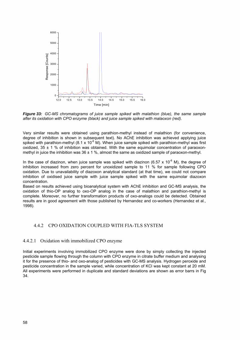
12.0 12.5 13.0 13.5 14.0 14.5 15.0 15.5 16.0
0
1000
2000
3000
4000
5000
6000
Res
pons
e [C
ount
s]
Time [min]
Figure 33: GC-MS chromatograms of juice sample spiked with malathion (blue), the same sample after its oxidation with CPO enzyme (black) and juice sample spiked with malaoxon (red). Very similar results were obtained using parathion-methyl instead of malathion (for convenience, degree of inhibition is shown in subsequent text). No AChE inhibition was achieved applying juice spiked with parathion-methyl (8.1 x 10⁻⁶ M). When juice sample spiked with parathion-methyl was first oxidized, 35 ± 1 % of inhibition was obtained. With the same equimolar concentration of paraoxon-methyl in juice the inhibition was 36 ± 1 %, almost the same as oxidized sample of paraoxon-methyl. In the case of diazinon, when juice sample was spiked with diazinon (6.57 x 10⁻⁶ M), the degree of inhibition increased from zero percent for unoxidized sample to 11 % for sample following CPO oxidation. Due to unavailability of diazoxon analytical standard (at that time), we could not compare inhibition of oxidised juice sample with juice sample spiked with the same equimolar diazoxon concentration. Based on results achieved using bioanalytical system with AChE inhibition and GC-MS analysis, the oxidation of thio-OP analog to oxo-OP analog in the case of malathion and parathion-methyl is complete. Moreover, no further transformation products of oxo-analogs could be detected. Obtained results are in good agreement with those published by Hernandez and co-workers (Hernandez et al., 1998).
4.4.2 CPO OXIDATION COUPLED WITH FIA-TLS SYSTEM
4.4.2.1 Oxidation with immobilized CPO enzyme
Initial experiments involving immobilized CPO enzyme were done by simply collecting the injected pesticide sample flowing through the column with CPO enzyme in citrate buffer medium and analysing it for the presence of thio- and oxo-analog of pesticides with GC-MS analysis. Hydrogen peroxide and pesticide concentration in the sample varied, while concentration of KCl was kept constant at 20 mM. All experiments were performed in duplicate and standard deviations are shown as error barrs in Fig 34.
58

0 1 2 3 4 5 6 70.000
0.001
0.002
0.003
0.004
0.005
0.006
0.007
0.008A Parathion
Paraoxon
Pes
ticid
e co
ncen
tratio
n [m
M]
Sample number
0 1 2 3 4 5 6 7 80.000
0.001
0.002
0.003
0.004
0.005
0.006
0.007
0.008B
Pest
icid
e co
ncen
tratio
n [m
M]
Sample number
Paraoxon Parathion
0 1 2 3 4 5 6 70.000
0.001
0.002
0.003
0.004
0.005
0.006
0.007
0.008C Paraoxon
Parathion
Pes
ticid
e co
ncen
tratio
n [m
M]
Sample number
0 1 2 3 4 5 6 70.000
0.001
0.002
0.003
0.004
0.005
0.006
0.007
0.008D Parathion
Paraoxon
Pes
ticid
e co
ncen
tratio
n [m
M]
Sample number
0 1 2 3 4 5 6 7 80.000
0.001
0.002
0.003
0.004
0.005
0.006
0.007
0.008
0.009E
Pes
ticid
e co
ncen
tratio
n [m
M]
Sample Number
Paraoxon
0 1 2 3 4 5 60.000
0.002
0.004
0.006
0.008
0.010
0.012
0.014
0.016
0.018F Paraoxon
Parathion
Pest
icid
e co
ncen
tratio
n [m
M]
Sample number
Figure 34: CPO oxidation of sequentially injected samples (1 mL) containing 8.1 µM parathion-methyl with 0.05 mM H2O2 (A), 0.25 mM H2O2 (B), 1 mM H2O2 (C) and no H2O2 added (D). Experiments were also performed by injecting paraoxon-methyl with 0.25 mM H2O2 (E) and using 19 µM parathion-methyl with 1 mM H2O2 (F). Standard deviation of duplicate experiments is shown. Sample numbered 0 was not injected through the CPO column. Due to difficulties in collecting the entire sample at the exit from the CPO column, there was some obvious loss of pesticide observed (Fig 34, D and E). Since there was no H2O2 present (Fig 34, D) we expected that parathion-methyl concentration would not change much during the series of consecutive sample injections (samples 1-6), as compared to pesticide concentration in non-injected sample
59

(sample 0). As CPO does not convert the oxo-analog, the same expectation should be valid in the case of paraoxon-methyl injection (Fig 34, E). The sample numbered 0 is control sample that was not injected through the column. Nonetheless, the dynamics of thio-analog depletion and oxo-analog production can be clearly seen. The yield of the oxidation reaction depends on the H2O2 concentration (Fig 34; A, B, C). No oxo-product was formed without H2O2 addition, thus confirming our previously obtained results. There is also no effect on oxo-form of pesticide in terms of further oxidation or modification (E). As already reported by Park and Clark (Park and Clark, 2006), rapid inactivation of immobilized CPO could be seen in case of 1 mM H2O2 concentration (Fig 34, C). Some minor effect of CPO inactivation after consecutive sample injections could also be seen in the case of lower H2O2 concentrations. However, this was due to large volume of injected sample (1 mL) containing H2O2. In the case of FIA-TLS bioanalytical system used in other experiments reported herein, the volumes of injected samples were 100 or 200 µL, thus meaning that more samples could be injected, before noticeable CPO enzyme inactivation takes place. The next stage of our research work was to implement obtained results in a FIA-TLS bioanalytical system with on-line oxidation step, as schematically represented in Fig 15. 4.4.2.2 Coupling on-line CPO oxidation with FIA-TLS bioanalytical system
In contrast to performing batch CPO oxidation before sample injection, the incorporation of the CPO enzyme into the FIA bioanalytical system brings additional restrictions regarding the optimal operation conditions. This is due to the fact, that the two enzymes (CPO and AChE), used in the FIA system have a different pH optimum: CPO in acidic, pH = 3 (Thomas, 1970) and AChE in neutral, pH = 7 to 9 (Kaplay and Jagannathan, 1970; Ngo and Laidler, 1978) pH range. Therefore, two important operational parameters were optimized initially: the pH of the two carrier buffers (see Fig 15) and pesticide contact time in the AChE bioreactor (through increasing or decreasing the flow-rate). Both parameters are directly related to the magnitude of the inhibition effect and therefore to the sensitivity of the FIA system. As the two carrier buffers were pumped through the FIA system and the sample flows through the CPO and AChE columns connected in series, buffer capacities of the PBS and citrate buffer and different flow rates of each buffer were tested, with the aim of achieving highest possible AChE activity and inhibition effect. Optimization experiments were performed using oxo-OP pesticide (paraoxon-methyl), to exclude any possible influence from incomplete oxidation reaction at this stage. Combination of 0.2 M phosphate buffer with pH 9.1 and 0.025 M citrate buffer with pH 2.9 in 2:1 flow-rate ratio, resulting in 0.6 mL min-1 cummulative flow-rate on the AChE column, and final pH value of 7.4 was selected for further experiments, since it provided conditions for the highest AChE activity and the highest inhibition effect (Fig 35).
60
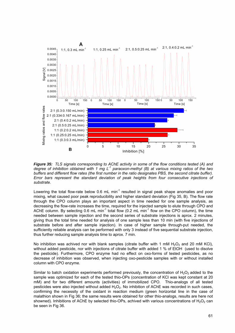
0 50 100 1500.0000
0.0005
0.0010
0.0015
0.0020
0.0025
0.0030
0.0035
0.0040
0.0045
0 50 100 150
0 50 100 150
0 50 100 150
B
A1:1, 0.3 mL min-1
Sign
al [V
]
Time [s]
1:1, 0.25 mL min-1
Time [s] Time [s]
2:1, 0.5:0.25 mL min-1
Time [s]
2:1, 0.4:0.2 mL min-1
1:1 (0.3:0.3 mL/min)1:1 (0.25:0.25 mL/min)
1:1 (0.2:0.2 mL/min)2:1 (0.5:0.25 mL/min)
2:1 (0.4:0.2 mL/min)2:1 (0.334:0.167 mL/min)
2:1 (0.3:0.150 mL/min)
0 5 10 15 20 25 30 35Inhibition [%]
Mix
ing
ratio
s an
d flo
w ra
tes
Figure 35: TLS signals corresponding to AChE activity in some of the flow conditions tested (A) and degree of inhibition obtained with 1 mg L-1 paraoxon-methyl (B) at various mixing ratios of the two buffers and different flow rates (the first number in the ratio designates PBS, the second citrate buffer). Error bars represent the standard deviation of peak heights from four consecutive injections of substrate. Lowering the total flow-rate below 0.6 mL min-1 resulted in signal peak shape anomalies and poor mixing, what caused poor peak reproducibility and higher standard deviation (Fig 35, B). The flow rate through the CPO column plays an important aspect in time needed for one sample analysis, as decreasing the flow-rate increases the time, required for the injected sample to elute through CPO and AChE column. By selecting 0.6 mL min-1 total flow (0.2 mL min-1 flow on the CPO column), the time needed between sample injection and the second series of substrate injections is aprox. 2 minutes, giving thus the total time needed for analysis of one sample less than 10 min (with five injections of substrate before and after sample injection). In case of higher sample through-put needed, the sufficiently reliable analysis can be performed with only 3 instead of five sequential substrate injection, thus further reducing sample analysis time to aprox. 7 min. No inhibition was achived nor with blank samples (citrate buffer with 1 mM H2O2 and 20 mM KCl), without added pesticide, nor with injections of citrate buffer with added 1 % of EtOH (used to disolve the pesticide). Furthermore, CPO enzyme had no effect on oxo-forms of tested pesticides, as no decrease of inhibition was observed, when injecting oxo-pesticide samples with or without installed column with CPO enzyme. Similar to batch oxidation experiments performed previously, the concentration of H2O2 added to the sample was optimized for each of the tested thio-OPs (concentration of KCl was kept constant at 20 mM) and for two different amounts (activities) of immobilized CPO. Thio-analogs of all tested pesticides were also injected without added H2O2. No inhibition of AChE was recorded in such cases, confirming the necessity of the oxidant in reaction medium (green horizontal line in the case of malathion shown in Fig 36; the same results were obtained for other thio-analogs, results are here not showned). Inhibitions of AChE by selected thio-OPs, achived with various concentrations of H2O2 can be seen in Fig 36.
61

0.0 0.1 0.2 0.3 0.4 0.50
10
20
30
40
50
60
70Malathion High CPO concentration
Low CPO concentration
Inhi
bitio
n [%
]
Hydrogen peroxide [mM]
0.0 0.2 0.4 0.6 0.8 1.00
5
10
15
20
25
30Parathion-methyl
Inhi
bitio
n [%
]
Hydrogen peroxide [mM]
High CPO concentration Low CPO concentration
0.0 0.5 1.0 1.5 2.00
5
10
15
20
25
30
35
Chlorpyrifos High CPO concentration Low CPO concentration
Inih
ibiti
on [%
]
Hydrogen peroxide [mM]
0 1 2 3 4 50
5
10
15
20
25
30Diazinon High CPO concentration
Low CPO concentrationIn
hibi
tion
[%]
Hydrogen peroxide [mM]
Figure 36: Chloroperoxidase oxidation and inhibition of thio-analog of OP pesticides (malathion, 263 μg L-1; parathion-methyl, 798 μg L-1; chlorpyrifos, 149 μg L-1 and diazion, 1.06 mg L-1) depending on H2O2 concentration. Error bars correspond to standard deviation of consecutive substrate injections. Horizontal red lines show inhibition achieved with same molar concentration of pesticide oxo-analog (standard deviation is shown with dashed lines), while horizontal green line in the case of malathion corresponds to thio-analog inhibition in the absence of H2O2. When the degree of inhibition by thio- and oxo-analog of the same pesticide was compared, almost complete conversion of malathion and parathion-methyl to malaoxon and paraoxon-methyl, respectively, was noticed already at 0.1 mM H2O2 concentration. At the same time, no difference in oxidation efficiency between high and low CPO activity of immobilized enzyme was observed. In the case of chlorpyrifos, higher H2O2 concentration is needed to achieve comparable inhibition, as obtained with the same molar concentration of chlorpyrifos-oxon. It can also be clearly seen, that in case of chlorpyrifos, the oxidation efficiency depends on activity of immobilized CPO. As expected, higher oxidation efficiency is achieved using column filled with glass beads with higher activity of immobilized CPO. The phenomenon is more pronounced at higher H2O2 concentration. H2O2 is known to have irreversible CPO inactivation effect (Park and Clark, 2006; Shevelkova and Ryabov, 1996), thus low CPO activity / H2O2 concentration ratio means faster CPO enzyme inactivation. In the case of diazinon inhibition of AChE enzyme, the degree of its oxidation corresponding to oxo-standard (diazoxon) could not be reached in tested experimental conditions, even when using column filled with the glass beads with higher activity of immobilzed CPO (Fig 36, lower right). The highest obtained inhibition with oxidized diazinon sample was in the 10-13 % range in comparison to 26 % achieved with the same equimolar concentration of diazoxon sample, thus indicating only 40-50 % oxidation reaction yield. Lowering citrate buffer flow right after diazinon injection from 0.2 mL min-1 to 0.1 mL min-1 (thus increasing contact time with CPO enzyme) resulted in somewhat increased reaction yield (raising from 46 % to 55 %). When testing weather the increased inhibition is the consequence of increased contact time between AChE and the pesticide (by injection of diazoxon), no increase in
62
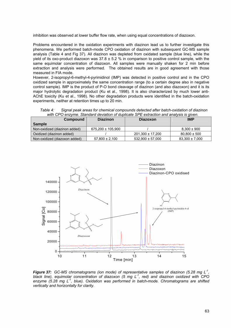
inhibition was observed at lower buffer flow rate, when using equal concentrations of diazoxon. Problems encountered in the oxidation experiments with diazinon lead us to further investigate this phenomena. We performed batch-mode CPO oxidation of diazinon with subsequent GC-MS sample analysis (Table 4 and Fig 37). All diazinon was depleted from oxidated sample (blue line), while the yield of its oxo-product diazoxon was 37.8 ± 5.2 % in comparison to positive control sample, with the same equimolar concentration of diazoxon. All samples were manually shaken for 2 min before extraction and analysis were performed. The obtained results are in good agreement with those measured in FIA mode. However, 2-isopropyl-6-methyl-4-pyrimidinol (IMP) was detected in positive control and in the CPO oxidized sample in approximately the same concentration range (to a certain degree also in negative control sample). IMP is the product of P-O bond cleavage of diazinon (and also diazoxon) and it is its major hydrolytic degradation product (Ku et al., 1998). It is also characterized by much lower anti-AChE toxicity (Ku et al., 1998). No other degradation products were identified in the batch-oxidation experiments, neither at retention times up to 20 min.
Table 4: Signal peak areas for chemical compounds detected after batch-oxidation of diazinon with CPO enzyme. Standard deviation of duplicate SPE extraction and analysis is given.
Compound Sample
Diazinon Diazoxon IMP
Non-oxidized (diazinon added) 675,200 ± 105,900 / 8,300 ± 900 Oxidized (diazinon added) / 201,300 ± 17,200 80,800 ± 500 Non-oxidized (diazoxon added) 57,800 ± 2,100 532,900 ± 57,000 83,300 ± 7,000
10 11 12 13 14 150
20000
40000
60000
80000
100000
120000
140000
N
N
HO
2-isopropyl-6-methyl-pyrimidin-4-ol (IMP)
PO
O
OS
NN
Diazoxon
PO
O
OS
NN
Diazinon
Diazinon Diazoxon Diazinon-CPO oxidised
Sign
al [C
o]
Time [min]
Figure 37: GC-MS chromatograms (ion mode) of representative samples of diazinon (5.28 mg L-1, black line), equimolar concentration of diazoxon (5 mg L-1, red) and diazinon oxidized with CPO enzyme (5.28 mg L-1, blue). Oxidation was performed in batch-mode. Chromatograms are shifted vertically and horizontally for clarity.
63
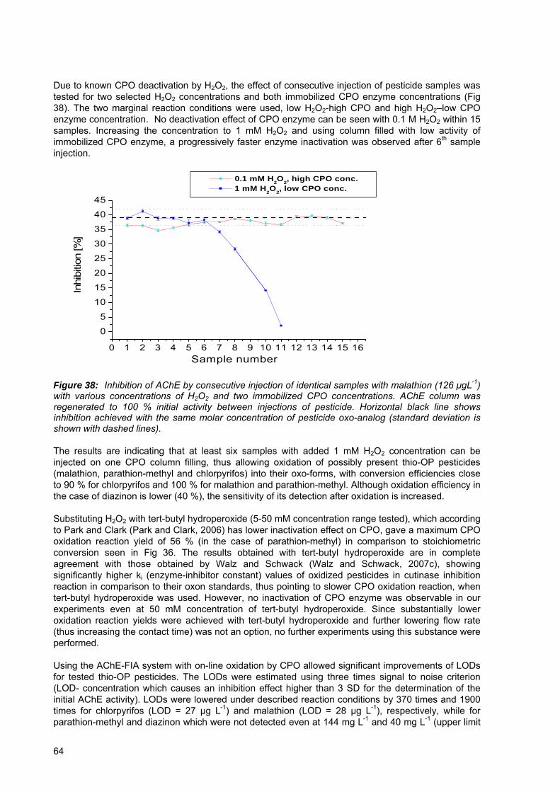
Due to known CPO deactivation by H2O2, the effect of consecutive injection of pesticide samples was tested for two selected H2O2 concentrations and both immobilized CPO enzyme concentrations (Fig 38). The two marginal reaction conditions were used, low H2O2-high CPO and high H2O2–low CPO enzyme concentration. No deactivation effect of CPO enzyme can be seen with 0.1 M H2O2 within 15 samples. Increasing the concentration to 1 mM H2O2 and using column filled with low activity of immobilized CPO enzyme, a progressively faster enzyme inactivation was observed after 6th sample injection.
0 1 2 3 4 5 6 7 8 9 10 11 12 13 14 15 16
0
5
10
15
20
25
30
35
40
45
0.1 mM H2O2, high CPO conc. 1 mM H2O2, low CPO conc.
Inhi
bitio
n [%
]
Sample number
Figure 38: Inhibition of AChE by consecutive injection of identical samples with malathion (126 μgL-1) with various concentrations of H2O2 and two immobilized CPO concentrations. AChE column was regenerated to 100 % initial activity between injections of pesticide. Horizontal black line shows inhibition achieved with the same molar concentration of pesticide oxo-analog (standard deviation is shown with dashed lines). The results are indicating that at least six samples with added 1 mM H2O2 concentration can be injected on one CPO column filling, thus allowing oxidation of possibly present thio-OP pesticides (malathion, parathion-methyl and chlorpyrifos) into their oxo-forms, with conversion efficiencies close to 90 % for chlorpyrifos and 100 % for malathion and parathion-methyl. Although oxidation efficiency in the case of diazinon is lower (40 %), the sensitivity of its detection after oxidation is increased. Substituting H2O2 with tert-butyl hydroperoxide (5-50 mM concentration range tested), which according to Park and Clark (Park and Clark, 2006) has lower inactivation effect on CPO, gave a maximum CPO oxidation reaction yield of 56 % (in the case of parathion-methyl) in comparison to stoichiometric conversion seen in Fig 36. The results obtained with tert-butyl hydroperoxide are in complete agreement with those obtained by Walz and Schwack (Walz and Schwack, 2007c), showing significantly higher ki (enzyme-inhibitor constant) values of oxidized pesticides in cutinase inhibition reaction in comparison to their oxon standards, thus pointing to slower CPO oxidation reaction, when tert-butyl hydroperoxide was used. However, no inactivation of CPO enzyme was observable in our experiments even at 50 mM concentration of tert-butyl hydroperoxide. Since substantially lower oxidation reaction yields were achieved with tert-butyl hydroperoxide and further lowering flow rate (thus increasing the contact time) was not an option, no further experiments using this substance were performed. Using the AChE-FIA system with on-line oxidation by CPO allowed significant improvements of LODs for tested thio-OP pesticides. The LODs were estimated using three times signal to noise criterion (LOD- concentration which causes an inhibition effect higher than 3 SD for the determination of the initial AChE activity). LODs were lowered under described reaction conditions by 370 times and 1900 times for chlorpyrifos (LOD = 27 µg L-1) and malathion (LOD = 28 µg L-1), respectively, while for parathion-methyl and diazinon which were not detected even at 144 mg L-1 and 40 mg L-1 (upper limit
64
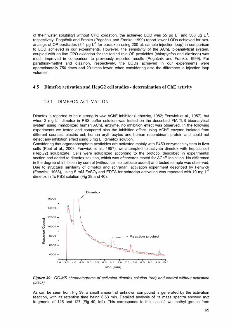
of their water solubility) without CPO oxidation, the achieved LOD was 55 µg L-1 and 500 µg L-1, respectively. Pogačnik and Franko (Pogačnik and Franko, 1999) report lower LODs achieved for oxo-analogs of OP pesticides (3.1 μg L-1 for paraoxon using 200 μL sample injection loop) in comparison to LOD achieved in our experiments. However, the sensitivity of the AChE bioanalytical system, coupled with on-line CPO oxidation for the tested thio-OP pesticides (chlorpyrifos and diazinon) was much improved in comparison to previously reported results (Pogačnik and Franko, 1999). For parathion-methyl and diazinon, respectively, the LODs achieved in our experiments were approximatelly 750 times and 20 times lower, when considering also the difference in injection loop volumes.
4.5 Dimefox activation and HepG2 cell studies - determination of ChE activity
4.5.1 DIMEFOX ACTIVATION
Dimefox is reported to be a strong in vivo AChE inhibitor (Lehotzky, 1982; Fenwick et al., 1957), but when 3 mg L-1 dimefox in PBS buffer solution was tested on the described FIA-TLS bioanalytical system using immobilized human AChE enzyme, no inhibition effect was observed. In the following experiments we tested and compared also the inhibition effect using AChE enzyme isolated from different sources, electric eel, human erythrocytes and human recombinant protein and could not detect any inhibition effect using 5 mg L-1 dimefox solution. Considering that organophosphate pesticides are activated mainly with P450 enzymatic system in liver cells (Poet et al., 2003, Fenwick et al., 1957), we attempted to activate dimefox with hepatic cell (HepG2) solubilizate. Cells were solubilized according to the protocol described in experimental section and added to dimefox solution, which was afterwards tested for AChE inhibition. No difference in the degree of inhibition by control (without cell solubilizate added) and tested sample was observed. Due to structural similarity of dimefox and schradan, activation experiment described by Fenwick (Fenwick, 1958), using 5 mM FeSO4 and EDTA for schradan activation was repeated with 10 mg L-1 dimefox in 1x PBS solution (Fig 39 and 40).
3.0 3.5 4.0 4.5 5.0 5.5 6.0 6.5 7.0 7.5 8.0 8.5 9.0 9.5 10.0
4000
6000
8000
10000
12000
14000
16000
Reaction product
Dimefox
Res
pons
e [C
ount
s]
Time [min]
Figure 39: GC-MS chromatograms of activated dimefox solution (red) and control without activation (black) As can be seen from Fig 39, a small amount of unknown compound is generated by the activation reaction, with its retention time being 6.53 min. Detailed analysis of its mass spectra showed m/z fragments of 126 and 127 (Fig 40, left). This corresponds to the loss of two methyl groups from
65

dimefox molecule. Dimefox depletion from FeSO4 / EDTA activated sample was neglible in comparison to control sample without added FeSO4 / EDTA (Table 5).
Table 5: Signal peak areas for dimefox are shown and dimefox remaining after activation was calculated. C8 and C18 extraction cartridges were used. Standard deviation of duplicate samples is shown.
Dimefox peak signal areas
Control FeSO4 / EDTA sample Remaining dimefox [%]
C8 extraction 1.97 x 10⁶ ± 2.8 x 10⁵ 1.88 x 10⁶ ± 2.3 x 10⁵ 95.4 ± 17.8 % C18 extraction 2.08 x 10⁶ ± 9.9 x 10⁴ 2.06 x 10⁶ ± 2.7 x 10⁵ 99.0 ± 13.8 %
According to Casida et al. (Casida et al., 1954), schradan activation product is its phosphoramide N-oxide and by analogy, Fenwick et al (Fenwick et al., 1957) infered, that dimefox activation product is either amine oxide or hydroxymethylamide. However, in the case of dimefox activation no clear explanation of reaction mechanism is given by the authors.
40 50 60 70 80 90 100 110 120 130 140 150 160 1700
100
200
300
400
500
600
139
127110
57
44
acquired range [m/z]
40 50 60 70 80 90 100 110 120 130 140 150 160 1700
10000
20000
30000
40000
50000
60000
70000
154
110
44
aquired range [m/z]
Figure 40: GC-MS spectra of activated product (left) and dimefox (right) To elucidate the situation, the same experiment with FeSO4 and EDTA was performed again and samples were analyzed for possible AChE inhibition effect (Fig 41). First, the series of substrate to check the initial enzyme activity was injected. Afterwards, the reaction sample was injected multiple times with intermediate substrate injection to check for any inhibition effect. Although there is some minor inhibition effect observable at the beginning, it is not consistent through the experiment. The reason may as well be in reaction products instability. However, as can be seen from Fig 39 and Table 5, the yield of the performed reaction is low and has no practical application in dimefox detection using AChE-TLS bioassay, even if the detected substance is in fact the AChE-inhibiting activation product.
66
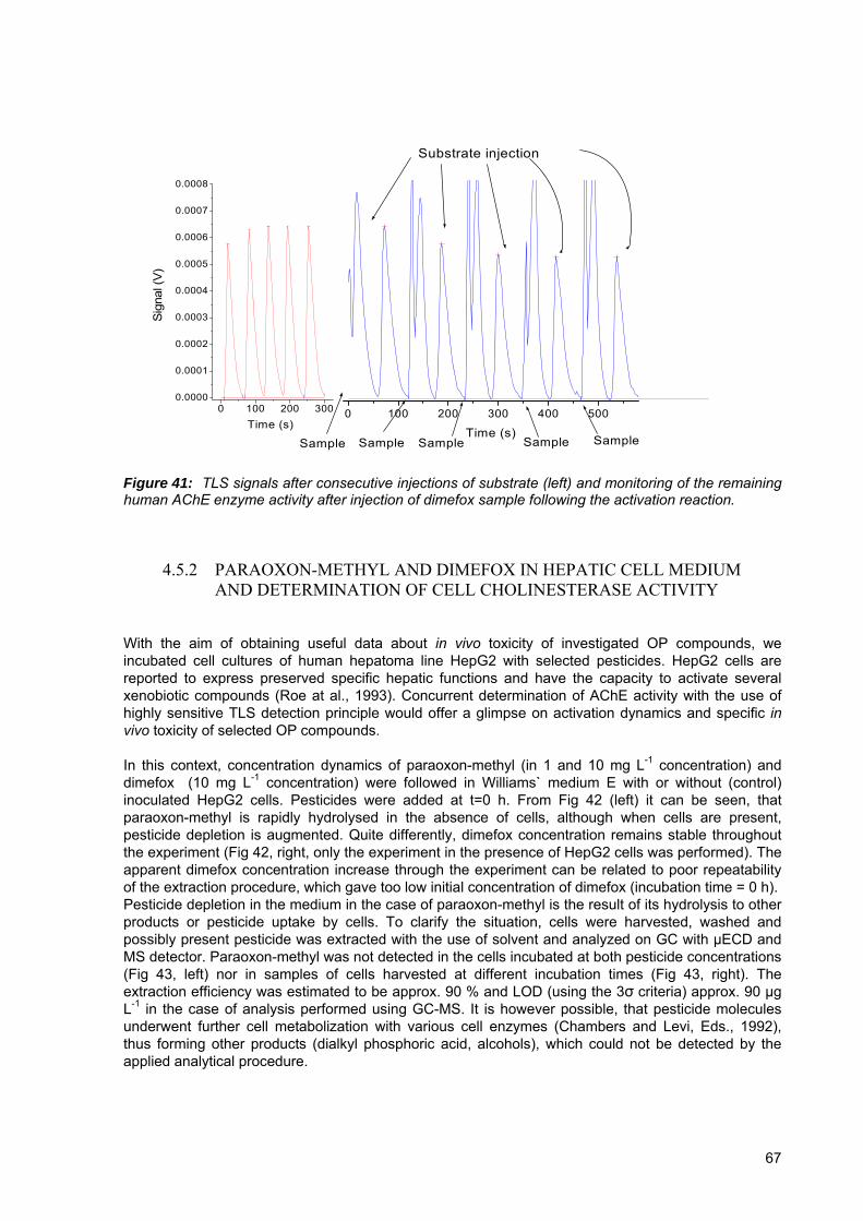
0 100 200 300 400 500
0 100 200 3000.0000
0.0001
0.0002
0.0003
0.0004
0.0005
0.0006
0.0007
0.0008
SampleSampleSample SampleSample
Substrate injection
Time (s)
Sig
nal (
V)
Time (s)
Figure 41: TLS signals after consecutive injections of substrate (left) and monitoring of the remaining human AChE enzyme activity after injection of dimefox sample following the activation reaction.
4.5.2 PARAOXON-METHYL AND DIMEFOX IN HEPATIC CELL MEDIUM AND DETERMINATION OF CELL CHOLINESTERASE ACTIVITY
With the aim of obtaining useful data about in vivo toxicity of investigated OP compounds, we incubated cell cultures of human hepatoma line HepG2 with selected pesticides. HepG2 cells are reported to express preserved specific hepatic functions and have the capacity to activate several xenobiotic compounds (Roe at al., 1993). Concurrent determination of AChE activity with the use of highly sensitive TLS detection principle would offer a glimpse on activation dynamics and specific in vivo toxicity of selected OP compounds. In this context, concentration dynamics of paraoxon-methyl (in 1 and 10 mg L-1 concentration) and dimefox (10 mg L-1 concentration) were followed in Williams` medium E with or without (control) inoculated HepG2 cells. Pesticides were added at t=0 h. From Fig 42 (left) it can be seen, that paraoxon-methyl is rapidly hydrolysed in the absence of cells, although when cells are present, pesticide depletion is augmented. Quite differently, dimefox concentration remains stable throughout the experiment (Fig 42, right, only the experiment in the presence of HepG2 cells was performed). The apparent dimefox concentration increase through the experiment can be related to poor repeatability of the extraction procedure, which gave too low initial concentration of dimefox (incubation time = 0 h). Pesticide depletion in the medium in the case of paraoxon-methyl is the result of its hydrolysis to other products or pesticide uptake by cells. To clarify the situation, cells were harvested, washed and possibly present pesticide was extracted with the use of solvent and analyzed on GC with μECD and MS detector. Paraoxon-methyl was not detected in the cells incubated at both pesticide concentrations (Fig 43, left) nor in samples of cells harvested at different incubation times (Fig 43, right). The extraction efficiency was estimated to be approx. 90 % and LOD (using the 3σ criteria) approx. 90 μg L-1 in the case of analysis performed using GC-MS. It is however possible, that pesticide molecules underwent further cell metabolization with various cell enzymes (Chambers and Levi, Eds., 1992), thus forming other products (dialkyl phosphoric acid, alcohols), which could not be detected by the applied analytical procedure.
67
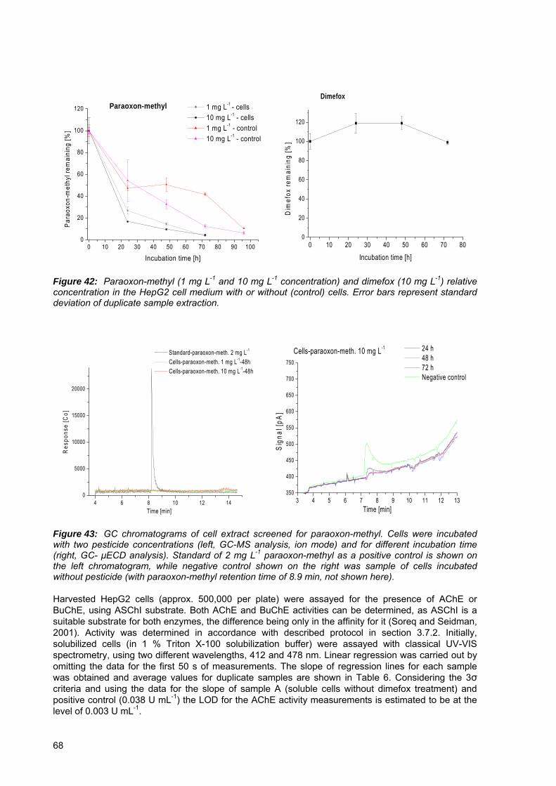
0 10 20 30 40 50 60 70 80 90 1000
20
40
60
80
100
120 Paraoxon-methyl
Par
aoxo
n-m
ethy
l rem
aini
ng [%
]
Incubation time [h]
1 mg L-1 - cells 10 mg L-1 - cells 1 mg L-1 - control 10 mg L-1 - control
0 10 20 30 40 50 60 70 800
20
40
60
80
100
120
Dimefox
Dim
efox
rem
aini
ng [%
]
Incubation time [h]
Figure 42: Paraoxon-methyl (1 mg L-1 and 10 mg L-1 concentration) and dimefox (10 mg L-1) relative concentration in the HepG2 cell medium with or without (control) cells. Error bars represent standard deviation of duplicate sample extraction.
4 6 8 10 12 140
5000
10000
15000
20000
Res
pons
e [C
o]
Time [min]
Standard-paraoxon-meth. 2 mg L-1
Cells-paraoxon-meth. 1 mg L-1-48h Cells-paraoxon-meth. 10 mg L-1-48h
3 4 5 6 7 8 9 10 11 12 13350
400
450
500
550
600
650
700
750
Cells-paraoxon-meth. 10 mg L-1 24 h 48 h 72 h Negative control
Sig
nal [
pA]
Time [min]
Figure 43: GC chromatograms of cell extract screened for paraoxon-methyl. Cells were incubated with two pesticide concentrations (left, GC-MS analysis, ion mode) and for different incubation time (right, GC- μECD analysis). Standard of 2 mg L-1 paraoxon-methyl as a positive control is shown on the left chromatogram, while negative control shown on the right was sample of cells incubated without pesticide (with paraoxon-methyl retention time of 8.9 min, not shown here). Harvested HepG2 cells (approx. 500,000 per plate) were assayed for the presence of AChE or BuChE, using ASChI substrate. Both AChE and BuChE activities can be determined, as ASChI is a suitable substrate for both enzymes, the difference being only in the affinity for it (Soreq and Seidman, 2001). Activity was determined in accordance with described protocol in section 3.7.2. Initially, solubilized cells (in 1 % Triton X-100 solubilization buffer) were assayed with classical UV-VIS spectrometry, using two different wavelengths, 412 and 478 nm. Linear regression was carried out by omitting the data for the first 50 s of measurements. The slope of regression lines for each sample was obtained and average values for duplicate samples are shown in Table 6. Considering the 3σ criteria and using the data for the slope of sample A (soluble cells without dimefox treatment) and positive control (0.038 U mL-1) the LOD for the AChE activity measurements is estimated to be at the level of 0.003 U mL-1.
68

0 50 100 150 200 250 300
0.00
0.05
0.10
0.15
0.20
Kpositive Knegative A1 A2 B1 B2 C1 C2 Kpositive2 Kpositive3
Abs
orba
nce
Time [s]
0 50 100 150 200 250 300-0.007
-0.006
-0.005
-0.004
-0.003
-0.002
-0.001
0.000
0.001
0.002
0.003
0.004
0.005
0.006
Knegative A1 A2 B1 B2 C1 C2
Abs
orba
nce
Time [s]
Figure 44:
Table 6:
ChE assay performed using UV-VIS spectrophotometer at 412 nm. On the right graph insert from left is shown using smaller scale. Samples were in parallels (A- cells without dimefox treatment, B- cells treated with 1 mg L-1 dimefox, C- cells treated with 0.1 mg L-1 dimefox, Kpositive- added AChE enzyme, 0.114 U mL-1, Kpositive 2 and 3- added AChE enzyme, 0.038 U mL-1, Knegative- solubilization buffer instead of sample)
Calculated slopes of regression lines for solubilized HepG2 samples measured on spectrophotometer at 412 nm wavelenght. Standard error of the mean for duplicate samples is shown.
Sample Regresion coefficient (with standard error of the mean)
A (without dimefox treatment) 1.93 x 10⁻⁵ ± 6.1 x 10⁻⁶
B (1 mg L-1 dimefox treatment) 1.20 x 10⁻⁵ ± 4.8 x 10⁻⁸
C (0.1 mg L-1 dimefox treatment) 8.18 x 10⁻⁶ ± 2.5 x 10⁻⁶
Kpositive (0.114 U mL-1 AChE) 6.55 x 10⁻⁴ Kpositive (0.038 U mL-1 AChE) 2.31 x 10⁻⁴ ± 1.3 x 10⁻⁵
Knegative (no cell sample) 8.76 x 10⁻⁶ In Fig 45 measurements performed with TLS detection at 478 nm are presented. Also in this case, linear regression of data for each sample was made and the slopes of regression lines were then calculated. Average values for duplicate samples are shown in Table 7. However, the difference obtained in measuring AChE activity in cell samples with or without pesticide treatment lies inside the range of experimental error. The large fluctuations, that can be observed in the signal recorded (Fig 45) and large standard error between duplicate samples can be explained by findings of Kožar Logar and co-workers (Kožar Logar et al., 2002). The large baseline noise in their gradient HPLC-TLS measurement was assigned to incomplete solvent mixing and thus ∂n/∂T and thermal conductivity changes (see Eq. 1). Similar effect of poor mixing and multiphase layers existence was observed in our sample preparation when cell solubilizate was added to reaction mixture within the cuvette (in spite of vigorous mixing of the cuvette contents before collecting the data).
69
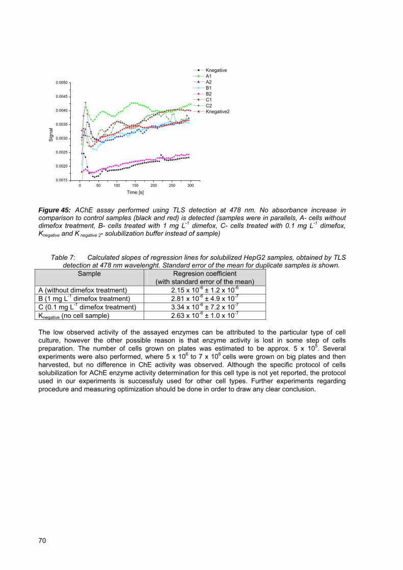
0 50 100 150 200 250 3000.0015
0.0020
0.0025
0.0030
0.0035
0.0040
0.0045
0.0050
Knegative A1 A2 B1 B2 C1 C2 Knegative2
Sign
al
Time [s]
Figure 45:
Table 7:
AChE assay performed using TLS detection at 478 nm. No absorbance increase in comparison to control samples (black and red) is detected (samples were in parallels, A- cells without dimefox treatment, B- cells treated with 1 mg L-1 dimefox, C- cells treated with 0.1 mg L-1 dimefox, Knegative and K negative 2- solubilization buffer instead of sample)
Calculated slopes of regression lines for solubilized HepG2 samples, obtained by TLS detection at 478 nm wavelenght. Standard error of the mean for duplicate samples is shown.
Sample Regresion coefficient (with standard error of the mean)
A (without dimefox treatment) 2.15 x 10⁻⁶ ± 1.2 x 10⁻⁶
B (1 mg L-1 dimefox treatment) 2.81 x 10⁻⁶ ± 4.9 x 10⁻⁷
C (0.1 mg L-1 dimefox treatment) 3.34 x 10⁻⁶ ± 7.2 x 10⁻⁷
Knegative (no cell sample) 2.63 x 10⁻⁶ ± 1.0 x 10⁻⁷
The low observed activity of the assayed enzymes can be attributed to the particular type of cell culture, however the other possible reason is that enzyme activity is lost in some step of cells preparation. The number of cells grown on plates was estimated to be approx. 5 x 10⁵. Several experiments were also performed, where 5 x 10⁶ to 7 x 10⁶ cells were grown on big plates and then harvested, but no difference in ChE activity was observed. Although the specific protocol of cells solubilization for AChE enzyme activity determination for this cell type is not yet reported, the protocol used in our experiments is successfuly used for other cell types. Further experiments regarding procedure and measuring optimization should be done in order to draw any clear conclusion.
70
5 CONCLUSIONS
Initially, the AChE-FIA analytical system was characterized in terms of AChE column filling, efficiency of pesticide binding and detection of pesticides in samples with complex matrices. In contrast to the generally accepted opinion and reported results (Pogačnik and Franko, 1999), the sensitivity of the method does not depend on the ratio between the pesticide concentration and the amount of immobilized AChE (corresponding to the number of free AChE active sites). The observed inhibition was more or less constant and lowering the amount of bounded active enzyme did not result in linearly increased inhibition effect. The efficiency of pesticide binding to AChE enzyme was shown to be very low, ranging from one quarter to one third of all the pesticide passing through the column. This observation leaves an open way for further optimizing the pesticide – AChE interaction through modification of FIA parameters, thus improving the general sensitivity of the method. The applicability of the AChE bioanalytical method for determination of oxo-OP pesticide content in samples with complex matrices such as fruit juices was confirmed. The achieved recovery for investigated pesticide (chlorpyrifos oxon) was always higher than 70 % in pulp juices and close to 100 % in clear juices. Besides taking into account the possible matrix effects of real samples on the activity of AChE by standard addition or matrix matched calibration, no additional labor-intensive sample pretreatment is needed for liquid samples. Experiments performed with ionic liquids have shown that ionic liquids inhibit the AChE reversibly, and by this they protect the enzyme from irreversible inhibition by pesticides. The effect of ILs was studied also on CPO enzyme. The results are pointing towards the need of careful selection and concentration optimization of IL. The most suitable ILs among those tested were shown to be EtPyTFA, EAN and BMIMmetSO4, which enabled high CPO activities and efficient oxidation of thio-OPs (60-100 %), even at 30 % concentration of ionic liquid. Despite better thermooptical properties of carrier buffers with added ILs the reversible AChE inhibition and protective effect from inhibition by pesticides seriously limits the application of ILs in AChE-FIA-TLS Monolithic CIM disks with epoxy and EDA reactive groups were tested as novel enzyme supports for determination of OP pesticides. Due to the weak ionic exchange character, the EDA CIM disk was found not suitable for the tested bioanalytical system and was not used further. No improvements in terms of sensitivity or inhibition dynamics were however achieved also by using CIM disk with epoxy reactive groups. It is however important to stress, that large internal volume (340 µL) of the available disks was a serious drawback, resulting in peak broadening and consequent increase in total time of analysis. Batch oxidation of thio-OP pesticides with CPO enzyme resulted in complete transformation of malathion, parathion-methyl and chlorpyrifos to their oxo-analogs. Oxidation reaction pH optimum was below pH 3, with optimal hydrogen peroxide concentration of 0.25 mM and 2.5 mM for 0.1 M citrate buffer and 10x diluted apple juice concentrate medium, respectively. By integrating the on-line oxidation step performed with immobilized CPO enzyme in a FIA system we improved the detection sensitivity and LODs for thio-OP analogs tested (malathion, parathion-methyl, chlorpyrifos and diazinon). Obtained LODs were 27 μg L-1, 28 μg L-1, 55 μg L-1 and 500 μg L-1 for chlorpyrifos, malathion, parathion-methyl and diazinon, respectively. Detection of unoxidized parathion-methyl and diazinon was not possible even at 144 mg L-1 and 40 mg L-1 concentration (upper limit of their water solubility). Equally important is the observation that with the incorporation of the CPO oxidation step, the time needed for one sample analysis was not increased in comparison to AChE-FIA-TLS system described earlier (Pogačnik and Franko, 1999, 2001) and is in the range of 7 to 10 minutes per sample. This is by a factor of 12 or more shorter in comparison to AChE bioassay with chemical or enzymatic batch oxidation reported by Schulze et al. (Schulze et al., 2002b) and Walz and Schwack (Walz and Schwack, 2007c). It can be concluded that the integration of on-line CPO oxidation reaction into the FIA-AChE bioassay represents an important progress towards fulfilling the demand for fast and reliable methods for daily screening of organophosphate pesticides in large number of environmental and food samples. The FIA manifold furthermore offers the possibility of the automation of the system.
71

Activation of dimefox with solubilizate from the HepG2 cell line and employing various chemical (NBS oxidation, FeSO4 + EDTA) activation procedures was shown to be inefficient. No procedure yielded increased inhibition effect and had any practical application in dimefox detection using AChE-TLS bioassay. The low observed activity of the assayed ChE enzymes in HepG2 cells can be attributed to low expressed ChE activity in this particular type of cells. The other possible reason is that there is some loss of enzyme activity in cell preparation protocol. Using highly sensitive TLS detection principle in measuring ChE activity did not yield improved results due to large signal fluctuations and large standard error between duplicate samples. This was attributed to incomplete solvent mixing and multiphase layers existence in sample cuvette.
72

6 REFERENCES
Abdel-Halim K. Y., Salama A. K., El-Khateeb E. N., Bakry N. M. 2006. Organophosphorus pollutants (OPP) in aquatic environment at Damietta Governorate, Egypt: Implications for monitoring and biomarker responses, Chemosfere. 63: 1491-1498. Ahmed F. E. 2001. Analyses of pesticides and their metabolites in foods and drinks, Trends in Analytical Chemistry, 20, 11: 649-661. Altiere R. J., Travis D. C., Thompson D. C. 1994. Characterization of acetylcholinesterase in rabbit intrapulmonary arteries. American Journal of Physiology- Lung Cellular and Molecular Physiology. 267, 6: 6-11. Andreescu A., Avramescu A., Bala C., Magearu V., Marty J.L., 2002a. Detection of organophosphorous insecticides with immobilized acetylcholinesterase-comparative study of two enzyme sensors. Analytical and Bioanalytical Chemistry. 374: 39-45 Andreescu S., Barthelmebs L., Marty J. L. 2002b. Immobilization of acetylcholinesterase on screen-printed electrodes: comparative study between three immobilization methods and applications to the detection of organophosphorus insecticides, Analytica Chimica Acta, 464: 171-180. Andreescu S., Marty J.-L. 2006. Twenty years research in choline biosensors: From basic research to practical applications, Biomolecular Engineering, 23: 1-15. Andres R. T., Narayanaswamy R. 1997. Fiber-optic pesticide biosensor based on covalently immobilized acetylcholinesterase and thymol blue, Talanta, 44: 1335-1352. Andrisano V., Bartolini M., Gotti R., Cavrini V., Felix G. 2001. Determination of inhibitors` potency (IC50) by a direct high-performance liquid chromatographic method on an immobilized acetylcholinesterase column, Journal of Chromatography B, 753: 375-383. Arduini F., Amine A., Moscone D., Ricci F., Palleschi G. 2007. Fast, sensitive and cost-effective detection of nerve agents in the gas phase using a portable instrument and an electrochemical biosensor, Analytical and Bioanalytical Chemistry, 388: 1049-1057. Axelsen P. H., Harel M., Silman I., Sussman J. L. 1994. Structure and dynamics of the active site gorge of acetylcholinesterase: Synergistic use of molecular dynamics simulation and X-ray crystallography. Protein Science, 3: 188-197. Ayyagari M. S., Kamtekar S., Pande R., Marx K. A., Kumar J., Tripathy S. K., Kaplan D. L. 1995. Biosensor for pesticide detection based on alkaline phosphatase-catalyzed chemiluminescence, Materials Science and Engineering: C, 2, 4: 191-196. Bachmann T. T., Leca B., Villate F., Marty J. L., Fournier D., Schmid R. D. 2000. Improved multianalyte detection of organophosphates and carbamates with disposable multielectrode biosensor using recombinant mutants of Drosophila acetylcholinesterase and artificial neural networks. Biosensor & Bioelectronics, 15: 193-201. Ballesteros E., Parrado M. J. 2004. Continuous solid-phase extraction and gas chromatographic determination of organophosphorus pesticides in natural and drinking waters. Journal of Chromatography A, 1029: 267-273 Barber D. S., Ehrich M. 2001. Esterase inhibition in SH-SY5Y human neuroblastoma cells following exposure to organophosphorus compounds for 28 days. In Vitro & Molecular Toxicology. 14, 2: 129-135. Bartolini M., Cavrini V., Andrisano V. 2004. Monolithic micro-immobilized-enzyme reactor with human
73

recombinant acetylcholinesterase for on-line inhibition studies, Journal of Chromatography A, 1031: 27-34. Bartolini M., Cavrini V., Andrisano V. 2005. Choosing the right chromatographic support in making a new acetylcholinesterase-micro-immobilized enzyme reactor for drug discovery, Journal of Chromatography A, 1065: 135-144. Barut M., Podgornik A., Podgornik H., Štrancar A., Josić D., Mac Farlane J. D. 1999. American Biotechnology Laboratory, 17: 48-51. Bavcon Kralj M., Franko M., Trebse P. Photodegradation of organophosphorus pesticides-Investigations of product and their toxicity using gas-chromatography mass-spectrometry and AChE- thermal lens spectrometric bioassay, Chemosphere, 67: 99-107. Bellet E. M., Casida J. E. 1974. Products of peracid oxidation of organothiophosphorus compounds, Journal of Agricultural and Food Chemistry, 22, 2: 207-211. Bendayan M., Gisiger V. 2001. Demonstration of acetylcholinesterase molecular forms in a continuous tubular lysosomal system of rat pancreatic acinar cells. The Journal of Histochemistry and Cytochemistry. 49, 1: 29-39. Bernard P. P., Kireev D. B., Chretien J. R., Fortier P. L., Coppet L., 1998. 3D model of the acetylcholinesterase catalytic cavity probed by stereospecific organophosphorus inhibitors, Journal of Molecular Modeling, 4: 323-334. Bialkowski S. E. (Ed.), Photothermal spectroscopy methods for chemical analysis, John Willey & Sons, Inc., New York, 1996. Bourne Y., Taylor P., Marchot P. 1995. Acetylcholinesterase inhibition by fasciculin: Crystal structure of the complex, Cell, 83: 503-512. Braga J. W. B., Bottoli C. B. G., Jardim I. C. S. F., Goicoechea H. C., Olivieri A. C., Poppi R. J. 2007. Determination of pesticides and metabolites in wine by high performance liquid chromatography and second-order calibration methods, Journal of Chromatography A, 1148: 200-210. Bucur B., Dondoi M., Danet A., Marty J.-L. 2005. Insecticide identification using a flow injection analysis system with biosensor based on various cholinesterases. Analytica Chimica Acta. 539: 195-201. Bucur B., Fournier D., Danet A., Marty J.-L. 2006. Biosensors based on highly sensitive acetylcholinesterase for enhanced carbamate insecticide detection, Analytica Chimica Acta, 562: 115-121. Buratti F. M., Volpe M. T., Fabrizi L., Meneguz A., Vitozzi L., Testai E. 2002. Kinetic parameters of OPT pesticide desulfuration by c-DNA expressed human CYPs, Environmental Toxicology and Pharmacology, 11: 181-190. Buratti F. M., Volpe M. T., Meneguz A., Vittozzi L., Testai E. 2003. CYP-specific bioactivation of four organophosphorotioate pesticides by human liver microsomes. Toxicology and Applied Pharmacology, 186: 143-154. Butler A. M., Murray M., 1997. Biotransformation of parathion in human liver: Participation of CYP3A4 and its inactivation during microsomal parathion oxidation, The Journal of Pharmacology and Experimental Therapeutics, 280, 2: 966-973. Cacares T., He W., Naidu R., Megharaj M., 2007. Toxicity of chlorpyrifos and TCP alone and in combination to Daphnia carinata: The influence of microbial degradation in natural water, Water research, doi:10.1016/j.watres.2007.06.025
74
Caetano J., Machado S. A. S. 2008. Determination of carbaryl in tomato “in natura” using an amperometric biosensor based on the inhibition of acetycholinesterase activity, Sensors and Actuators B: Chemical, 129, 1: 40-46. Campanella L., Lelo D., Martini E., Tomassetti M. 2007. Organophosphorus and carbamate pesticide analysis using an inhibition tyrosinase organic phase enzyme sensor; Comparision by butyrylcholinesterase + choline oxidase opee and application to natural waters, Analytica Chimica Acta, 587: 22-32. Cao H., Nam J., Harmon H. J., Branson D. H. 2007. Spectrophotometric detection of organophosphate diazinon by porphyrin solution and porphyrin-dyed cotton fabric, Dyes and Pigments, 74: 176-180. Casida J. E., Allen T. C., Stahmann M. A. 1954. Mammalian conversion of octamethylpyrophos-phoramide to a toxic phosphoramide N-oxide, The Journal of Biological Chemistry, 210, 2: 607-616. Chambers J.E., Levi P.E. (Eds.), Organophosphates, Chemistry, Fate, and Effects. Academic Press, San Diego, 1992. Chang T.-S., Ding H. Y., Tai S. S.-K., Wu C.-Y. 2007. Mushroom tyrosinase inhibitory effects of isoflavones isolated from soygerm koji fermented with Aspergillus oryzae BCRC 32288, Food Chemistry, 105: 1430-1438. Cheon E. W., Saito T. 1999. Choline acetyltranferase and acetylcholinesterase in the normal, developing and regenerating newt retinas. Developmental Brain Research. 116: 97-109. Cherry J. R., Lamsa M. H., Schneider P., Vind J., Svendsen A., Jones A., Pedersen A. H. 1999. Directed evolution of a fungal peroxidase, Nature Biotechnology, 17, 379-384. Chiappe C., Neri L., Pieraccini D. 2006. Application of hydrophilic ionic liquids as co-solvents in chloroperoxidase catalyzed oxidations, Tetrahedron Letters, 47: 5089-5093. Cometa M. F., Buratti F. M., Fortuna S., Lorenzini P., Volpe M. T., Parisi L., Testai E., Meneguz A. 2007. Cholinesterase inhibitions and alterations of hepatic metabolism by oral acute and repeated chlorpyrifos administration to mice, Toxicology, 234: 90-102. Cosnier P., Popescu I. C. 1996. Poly(amphiphilic pyrrole)-tyrosinase-peroxidase electrode for amplified flow injection-amperometric detection of phenol, Analytica Chimica Acta, 319, 1-2: 145-151. Cremisini C., Di Sario S., Mela J., Pilloton R., Palleschi G. 1995. Evaluation of the use of free and immobilized acetylcholinesterase for paraoxon detection with an amperometric choline oxidase biosensor, Analytica Chimica Acta, 311: 273-280. De Albuquerque Y. D. T., Ferreira L. F. 2007. Amperometric biosensing of carbamate and organophosphate pesticides utilizing screen-printed tyrosinase-modified electrodes, Analytica Chimica Acta, 596: 210-221. Del Carlo M., Pepe A., Mascini M., De Gregorio M., Visconti A. 2005. Determining pirimiphos-methyl in durum wheat samples using an acetylcholinesterase inhibition assay, Analytical and Bioanalytical Chemistry, 381: 1367-1372. Deng Q., Guo Y., Ding S. 1996. Cyro-hydrogel for the construction of a tyrosinase-based biosensor, Analytica Chimica Acta, 319, 1-2: 71-77. Deo R. P., Wang J., Block I., Mulchandani A., Joshi K. A., Trojanowicz M., Scholz F., Chen W., Lin Y. 2005. Determination of organophosphate pesticides at carbon nanotube /organophosphorus hydrolase electrochemical biosensor, Analytica Chimica Acta, 530: 185-189.
75

De Oliveira Marques P. R. B., Nunes G. S., Dos Santos T. C. R., Andreescu S., Marty J.-L. 2004. Comparative investigation between acetylcholinesterase obtained from commercial sources and genetically modified Drosophila Melanogaster. Application in amperometric biosensor for methamidophos pesticide detection, Biosensors and Bioengineering, 20: 825-832. Diaz A. N., Sanchez F. G., Bracho V., Lovillo J., Aguilar A. 1997. Enzymatic determination of fenitrothion by cholinesterase and acetylcholinesterase on fluorogenic substrates, Fresenius Journal of analytical chemistry, 357: 958-961. Diaz A. N., Peinado M. C. R. 1997. Sol-gel cholinesterase biosensor for organophosphorus pesticide fluorimetric analysis, Sensors and Actuators B, 38-39: 426-431. Di Corcia A., Marchetti M. 1991. Multiresidue method for pesticides in drinking water using a graphitized carbon black cartridge extraction and liquid chromatographic analysis. Analytical Chemistry 63, 6: 580-585 Dimefox, Profile from hazardous substances database. 1986. Fluoride Action Network Pesticide Project (8. sept. 2001). http://www.fluoridealert.org/pesticides/dimefox.toxnet.hsdb.html (9. oct. 2007). Di Sioudi B. D., Miller C. E., Lai K., Grimsley J. K., Wild J. R. 1999. Rational design of organophosphorus hydrolase for altered substrate specifities, Chemico-Biological Interactions, 119-120: 211-223. Dondoi P. D., Bucur B., Danet A. F. Toader C. N., Barthelmebs L., Marty J. L. 2006. Organophosphorus insecticide extraction and heterogeneous oxidation on column for analysis with an acetylcholinesterase (AChE) biosensor. Analytica Chimica Acta, 578: 162-169. Doong R.-A., Tsai H.-C. 2001. Immobilization and characterization of sol-gel encapsulated acetylcholinesterase fiber-optic biosensor, Analytica Chimica Acta, 434: 239-246. D`Souza S. F. 2001. Microbial biosensor: a review, Biosensors & Bioelectronics, 16: 337-353. Du D., Huang X., Cai J., Zhang A., Ging J., Chen S. 2007a. An amperometric acetylcholine sensor based on immobilization of acetylcholinesterase on a multiwall carbon nanotube-cross linked chitosan composite, Analytical and Bioanalytical Chemistry, 387: 1059-1065. Du D., Cai J., Song D., Zhang A., 2007b. Rapid determination of triazophos using acetylcholinesterase biosensor based on sol-gel interface assembling multiwall carbon nanotubes, Journal of Applied Electrochemistry, 37: 893-898. Duysen E.G., Li B., Xie W., Schopfer L. M., Andreson R. S., Broomfiled C. A., Lockridge O. 2001. Evidence of nonacetylcholinesterase targetsof organophosphorus nerve agents: Supersensitivity of acetylcholinesterase knockout mouse to VX lethality. The Journal of Pharmacology and Experimental Therapeutic. 299, 2: 528-535. Ellman G. L., Courtney K. D., Andres Jr. V., Featherstone R. M. 1961. A new and rapid colorimetric determination of acetylcholinesterase activity. Biochemical Pharmacology. 7: 88-95. Fenoll J., Hellin P., Martinez C. M., Miguel M., Flores P. 2007. Multiresidue method for analysis of pesticides in pepper and tomato by gas chromatography with nitrogen-phosphorus detection, Food Chemistry, 105: 711-719. Fenwick M. L., Barron J. R., Watson W. A. 1957. The conversion of dimefox into an anticholinesterase by rat liver in vitro. The Biochemical Journal, 65, 1: 58-67. Fenwick M. L. 1958. The production of an esterase inhibitor from schradan in the fat body of the desert locust. Biochemical Journal 70, 3: 373-381.
76
Franko M., Tran C.D. 1996. Analytical thermal lens instrumentation, Review of Scientific Instruments, 67, 1: 1-18. Franko M. 2001. Recent application of thermal lens spectrometry in food analysis and environmental research. Talanta. 54: 1-13. Friedman M., Grosjean O. K., Zahnley J. C. 1986. Inactivation of matalloenyzmes by food constituents, Food and Chemical Toxicilogy, 24, 9: 897-902. Fujisawa T., Katagi T. 2005. Oxidative activation and degradation of organophosphorus pesticides mediated by iron porphyrins, Journal of Pesticide Science, 30, 2: 103-110. Gogol E. V., Evtugyn G. A., Marty J.-L., Budnikov H. C., Winter V. G. 2000. Amperometric biosensor based on nafion coated screen-printed electrodes for the determination of cholinesterase inhibitors, Talanta, 53: 379-389. Grisaru D., Sternfeld M., Eldor A., Glick D., Soreq H. 1999. Structural role of acetylcholinesterase variants in biology and pathology. European Journal of Biochemistry. 264: 672-686. Guardia-Rubio M., Marchal-Lopez R. M., Ayora-Canada M. J., Ruiz-Medina A. 2007. Determination of pesticides in olives by gas chromatography using different detection systems, Journal of Chromatography A, 1145: 195-203. Guengerich F. P. 2005. Human cytochrome P450 enzymes, In: Cytochrome P450; Structure, Mechanism and Biochemistry, de Montellano P. R. O. (edt.), Kluwer Academic/ Plenum Publishers, New York. Gupta R. (Ed.), Toxicology of Organophosphate and Carbamate compounds. Academic Press, San Diego, 2006. Hernandez J., Robledo N.R., Velasco L., Quintero R., Pickard M.A., Vazquez-Duhalt R. 1998. Chloroperoxidase-mediated oxidation of organophosphorus pesticides. Pesticide Biochemistry and Physiology, 61:87-9. Hernando M. D., Ferrer I., Agüera A., Fernandez-Alba A. R. 2005. Evaluation of pesticides in wastewaters. A combined (chemical and biological) analytical approach, The Handbook of Environmental Chemistry, Vol 5, Part O, Water Pollution, Springer-Verlag, Berlin Heidelberg, 53-77. Hu Y., Pinkham W., French Jr. L. A., Frankel D., Vetelino J. F. 2005. Pesticide detection using a lateral field excited acoustic wave sensor, Sensors and Actuators B, 108: 910-916. Huang X., Sun Y., Meng F., Liu J. 2004. New approach for the detection of organophosphorus pesticide in cabbage using SPME/SnO2 gas sensor: principle and preliminary experiment, Sensors and Actuators B, 102: 235-240. Huang Z., LI Y., Chen B., Yao S. 2007. Simultaneous determination of 102 pesticide residues in Chinese teas by gas chromatography-mass spectrometry, Journal of Chromatography B, 853: 154-162. Ishii A., Takeda S., Hattori S., Sueoka K., Mukasa K. 2007. Ultrasensitive detection of organophosphate insecticides by carbon nanotube filed-effect transistor, Colloids and Surfaces A: Physicochemical and Engineering Aspects, doi:10.1016/j.colsurfa.2007.05.071 Istamboulie G., Andreescu S., Marty J.-L., Noguer T. 2007. Highly sensitive detection of organophosphorus insecticides using magnetic microbeads and genetically engineered acetylcholinesterase, Biosensors and Bioelectronics, 23: 506-512.
77
Jackson J. A., Berkman C. E., Thompson C E. 1992. Stereoselective and chemoselective oxidation of phosphorothionates using MMPP, Tetrahedron Letters, 33, 41: 6061-6064. Jaffrezic-Renault N. 2001. New trends in biosensors for organophosphate pesticide, Sensors, 1: 60-74. Jeanty G., Ghommidh Ch., Marty J.-L. 2001. Automated detection of chlorpyrifos and its metabolites by a continuous flow system-based enzyme sensor. Analytica Chimica Acta, 436: 119-128. Jin S., Xu Z., Chen J., Liang X., Wu Y., Qian X. 2004. Determination of organophosphate and carbamate pesticides based on enzyme inhibition using a pH-sensitive fluorescence probe, Analytica Chimica Acta, 523: 117-123. Kadima T. A., Pickard M. A. 1990. Immobilization of chloroperoxidase on aminopropyl-glass. Applied and Environmental Microbiology, 56: 3473-3477. Kandimalla V. B., Neeta N. S., Karanth N. G., Thakur M. S., Roshini K. R., Rani B. E. A., Pasha A., Karanth N. G. K. 2004. regeneration of ethyl parathion antibodies for repeated use in immunosensor: a study on dissociation of antigens from antibodies, Biosensors and Bioelectronics, 20: 903-906. Kaplay S. S., Jagannathan V. 1970. Purification and properties of ox brain acetylcholinesterase, Archives of Biochemistry and Biophysics, 138, 1: 48-57. Karamfilov V. K., Fileman T. W., Evans K. M., Mantoura R. F. C. 1996. Determination od dimethoate and fenitrothion in estuarine samples by C-18 solid-phase extraction and high-resolution gas-chromatography with nitrogen-phosphorus detection, Analytica Chimica Acta, 335: 51-61. Karanth S., Pope C., 2000. Carboxylesterase and A-esterase activities during maturation and aging: Relationship to the toxicity of chlorpyrifos and parathion in rats, Toxicological Sciences, 58, 2: 282-289. Karnati C., Du H., Ji H-F., Xu X., Lvov Y., Mulchandani A., Mulchandani P., Chen W. 2007. Organophosphorus hydrolase multilayer modified microcantilevers for organophosphorus detection, Biosensors and Bioelectronics, 22: 2636-2642. Kasagami T., Miyamoto T., Yamamoto I. 2002. Activated transformations of organophosphorus insecticides in the case of non-AChE inhibitory oxons. Pest Management Science. 58: 1107-1117. Kassa J., Cabal J. 1999. A comparison of the efficacy of a new asymmetric bispyridinium oxime BI-6 with currently available oximes and H oximes against soman by in vitro and in vivo methods, Toxicology, 132: 111-118. Kaushik R., Rosenfeld C. A., Sultatos L. G. 2007. Concentration-dependent interactions of the organophosphates chlorpyrifos oxon and methyl paraoxon with human recombinant acetylcholinesterase, Toxicology and Applied Pharmacology, 221: 243-250. Kim Y. A., Lee H. S., Park Y. C., Lee Y. T. 2000. A convenient method for oxidation of organophosphorus pesticides in organic solvents. Environmental Research Section A. 84: 303-309. Klaassen C. D. (Edt.). Casarett & Doull's Toxicology: The Basic Science of Poisons, 6th edition, McGraw-Hill Professional, 2001. Kok F.N., Bozoglu F., Hasirci V., 2002. Construction of acetylcholinesterase-choline oxidase biosensor for aldicarb determination. Biosensor and Bioeletronics. 17: 531-539 Konstantinou I. K., Hela D. G., Albanis T. A. 2006. The status of pesticide pollution in surface waters (rivers and lakes) of Greece. Part I. Review on occurrence and levels, Environmental pollution, 141: 555-570.
78

Kotelevets L., Walch L., Chastre E., Chatonnet A., Dulmet E., Brink C., Norel X. 2005. Cholinestrase activity in human pulmonary arteries and veins: Correlation with mRNA levels. Life Sciences. 76, 19: 2211-2220. Kousba A. A., Sultatos L. G., Poet T. S., Timchalk C. 2004. Comparision of chlorpyrifos-oxon and paraoxon acetylcholinesterase inhibition dynamics: Potential role of a peripheral binding site, Toxicological sciences, 80: 239-248. Kožar Logar J., Šikovec M., Malej A., Franko M. 2002. The effects of eluent mixing on TLS detection in gradient elution HPLC, Analytical and Bioanalytical Chemistry, 374: 323-328. Ku Y., Chang J - L., Cheng S - C. 1998. Effect of solution pH on the hydrolysis and photolysis of diazinon in aqueous solution, Water, Air and Soil Pollution, 108: 445-456. Kulkarni A. P., Hodgson E. 1984. The metabolism of insecticides: The role of monooxygenase enzymes. Annual Review of Pharmacology and Toxicology. 24: 19-42. Kumar J., Jha S. K., D`Souza S. F. 2006a. Optical microbial biosensor for detection of methyl parathion pesticide using Flavobacterium sp. whole cell adsorbed on glass filters as disposable biocomponent, Biosensors and Bioelectronics, 21: 2100-2105. Kumar M. A., Chouchan R. S., Thakur M. S., Rani B. E. A., Mattiasson B., Karanth N. G. 2006b. Automated flow enzyme-linked immunosorbent assay (ELISA) system for analysis of methyl parathion, Analytica Chimica Acta, 30-34. Kumaran S., Tran-Minh C. 1992. Determination of organophosphorus and carbamate Insecticides by flow injection analysis. Analytical Biochemistry. 200: 187-194. Kuswandi B., Fikriyah C. I., Gani A. A. 2007. An optical fiber biosensor for chlorpyrifos using a single sol-gel film containing acetylcholinesterase and bromothymol blue, Talanta, doi: 10.1016/j.talanta.2007.06.042 Lacorte S., Jeanty G., Marty J. L., Barcelo D. 1997. Identification of fenthion and temephos and their transformation products in eater by high-performance liquid chromatography with diode array detection and atmospheric pressure chemical ionization mass spectrometric detection. Journal of Chromatography A. 777: 99-114 Lambropoulou D. A., Sakkas V. A., Albanis T. A. 2002. Validation of an SPME method, using PDMS, PA, PDMS-DVB and CW-DVB SPME fibers coating, for analysis of organophosphorus insecticides in natural waters. Analytical and Bioanalytical Chemistry. 374: 932-941. La Rosa C., Pariente F., Hernandez L., Lorenzo E. 1995. Amperomtric flow-through biosensor for determination of pesticides. Analytica Chimica Acta. 308: 129-136. Latala A., Stepnowski P., Nedzi M., Mrozik W. 2005. Marine toxicity of imidazolium ionic liquids: Acute effects on the Baltic agae Oocystis submarina and Cyclotella meneghiniana, Aquatic Toxicology, 73: 91-98. Law K., Higson S. P. J. 2005. Sonochemically fabricated acetycholinesterase micro-electrode arrays within flow injection analyser for the determination of organophosphate pesticides, Biosensors and Bioelectronics, 20: 1914-1924. Lee H. S, Kim Y. A., Cho Y. A., Lee Y. T. 2002. Oxidation of organophosphorus pesticides for the sensitive detection of cholinesterase-based biosensor. Chemosphere. 46: 571-576. Lehotzky K. 1982. Effects of pesticides on central and peripheral nervous system function in rats. Neurobehavioral Toxicology and Teratology. 4: 665-669.
79

Li Y.-G., Zhou Y.-X., Feng J.-L., Jiang Z.-H., Ma L.-R. 1999. Immobilization of enzyme on screen-printed electrode by exposure to glutaraldehyde vapour for the construction of amperometric acetylcholinesterase electrodes, Analytica Chimica Acta, 382: 277-282. Liu G., Lin Y. 2005. Electrochemical stripping analysis of organophosphate pesticides and nerve agents, Electrochemistry Communications, 7: 339-343. Luckarift H. R., Greenwald R., Bergin M. H., Spain J. C., Johnson G. R. 2007. Biosensor for continuous monitoring of organophosphate aerosols, Biosensors and Bioelectronics, 23: 400-406. Lui J., Günther A., Bilitewski U., 1997. Detection of methamidophos in vegetables using a photometric flow injection system, Environmental Monitoring and Assessment, 44: 375-382. Manoj K. M., Hager L. P. 2001. Utilization of peroxide and its relevance in oxygen insertion reactions catalyzed by chloroperoxidase, Biochimica et Biophysica Acta, 1547, 408-417. Marcel V., Estrada-Mondaca S., Magne F., Stojan J., Klaebe A., Fournier D. 2000. Exploration of Drosophila acetylcholinesterase substrate activation site using a reversible inhibitor (Triton X-100) and mutated enzymes, The Journal of Biological Chemistry, 275, 16: 11603-11609. Martinez Vidal J. L., Pablos Espada M. C., Garrido Frenich A., Arrebola F. J., 2000. Pesticide trace analysis using solid-phase extraction and gas chromatography with electron-capture and tandem mass-spectrometric detection in water samples, Journal of Chromatography A, 867: 235-245. Marty J.-L., Mionetto N., Noguer T., Ortega F., Roux C., 1993. Enzyme sensor for the detection of pesticides, Biosensors and Bioelectronics, 8: 273-280. Marty J.-L., Jeanty G., 1998. Detection of paraoxon by continuous flow system based enzyme sensor. Biosensor and Bioeletronics. 13, 2: 213-218. Mauriz E., Calle A., Lechuga L. M., Quintana J., Montoya A., Manclus J. J. 2006a. Real-time detection of chlorpyrifos at part per trillion levels in ground, surface and drinking water samples by a portable surface plasmon resonance immunosensor, Analytica Chimica Acta, 561: 50-47. Mauriz E., Calle A., Manclus J. J., Montoya A., Escuela A. M. Sendra J. R., Lechuga L. M. 2006b. Single and multi-analyte surface plasmon resonance assays for simultaneous detection of cholinesterase inhibiting pesticides, Sensors and Actuators B, 118: 399-407. Mazzei F., Botre F., Botre C. 1996. Acid phosphatase/glucose oxidase-based biosensor for the determination of pesticides, Analytica Chimica Acta, 336: 67-75. Mihelič I., Nemec D., Podgornik A., Koloini T. 2005. Pressure drop in CIM disk monolithic column, Journal of Chromatography A, 1065: 59-67. Monitoring of pesticide residue in products of plant origin in the European Union, Norway, Iceland and Liechtenstein, 2004 report. 2006. European commission, 72 p. Morris D. R., Hager L. P. 1965. Chloroperoxidase: Isolation and properties of the crystalline glycoptotein. The Journal of Biological Chemistry. 241, 8: 1763-1768. Moser C. V., Chanda S. M., Mortensen S. P., Padilla S. 1998. Age- and gender-related differences in sensitivity to chlorpyrifos in the rat reflect developmental profiles of esterase activities. Toxicological Sciences, 46, 2: 211-222. Mulchandani P., Mulchandani A., Kaneva I., Chen W., 1999. Biosensor for direct determination of organophopshate nerve agents. 1. Potentiometric enzyme electrode, Biosensors and Bioelectronics,
80

14: 77-85. Mulchandani P., Chen W., Mulchandani A., Wang J., Chen L. 2001. Amperometric microbial biosensor for direct determination of organophosphate pesticides using recombinant microorganism with surface expressed organophosphorus hydrolase, Biosensors and Bioelectronics, 16: 433-437. Mulchandani P., Chen W., Mulchandani A. 2006. Microbial biosensor for direct determination of nitrophenyl-substituted organophosphate nerve agents using genetically engineered Moraxella sp., Analytica Chimica Acta, 568: 217-221. Musilek K., Kuca K., Jun D., Dohnal V., Dolezal M. 2006. Synthesis of the novel series of bispyridinium compounds bearing (E)-but-2-ene linker and evaluation of their reactivation activity against chlorpyrifos-inhibited acetylcholinesterase, Bioorganic & Medicinal Chemistry Letters, 16: 622-627. Ngo T. T., Laidler K. J. 1978. Temperature and pH effects with immobilized electric eel acetylcholinesterase, Biochimica et Biophysica Acta- Enzymology, 525, 1: 93-102. Nistor C., Emneus J. 1999. Bioanalytical tools for monitoring polar pollutants, Waste management, 19: 147-170. No J. K., Soung D. Y., Kim Y. J., Shim K. H., Jun Y. S., Rhee S. H., Yokozawa T., Chung H. Y. 1999. Inhibition of tyrosinase by green tea components, Life Scineces, 65, 21: 241-246. No H.-Y., Kim Y. A., Lee Y. T., Lee H.-S. 2007. Cholinesterase-based dipstick assay for detection of organophosphate and carbamate pesticides, Analytica Chimica Acta, 594: 37-43. Nunes G. S., Montesinos T., Marques P. B. O., Fournier D., Marty J.-L. 2001. Acetylcholine enzyme sensor for determining methamidophos insecticide. Evaluation of some genetically modified acetylcholinesterase from Drosophila melanogaster, Analytica Chimica Acta, 434: 1-8. Ordentlich A., Barak D., Kronman C., Ariel N., Segall Y., Velan B., Shafferman A. 1996. The architecture of human acetylcholinesterase active center probed by interactions with selected organophosphate inhibitors, The Journal of biological chemistry, 271, 20: 11953-11962. Ordentlich A., Barak D., Kronman C., Ariel N., Segall Y., Velan B., Shafferman A. 1998. Functional characteristics of the oxyanion hole in human acetylcholinesterase, The Journal of biological chemistry, 273, 31: 19509-19517. Ordentlich A., Barak D., Sod-Moriah G., Kaplan D., Mizrahi D., Segall Y., Kronman C., Karton Y., Lazar A., Marcus D., Velan B., Shafferman A. 2005. The role of AChE active site gorge in determining stereoselectivity of charged and noncharged VX enantiomers. Chemico-Biological Interactions, 157-158: 191-198. Park J-B., Clark D. S. 2006. Deactivation mechanism of chloroperoxidase during biotransformations, Biotechnology and Bioengineering, 93, 6: 1190-1195. Park S., Kazlauskas R.J. 2003. Biocatalysis in ionic liquids-advantages beyond green technology. Current opinion in Biotechnology, 14: 432-437. Patel P.D. 2002. (Bio)sensors for measurement of analytes implicated in food safety: a review, Trends in Analytical Chemistry, 21, 2: 96-115. Piepmeier E H. (Ed.). Analytical applications of lasers, John Wiley & Sons, Inc, New York, 1986. Poet T. S., Wu H., Kousba A. A., Timchalk C. 2003. In vitro rat hepatic and intestinal metabolism of the organophosphate pesticides chlorpyrifos and diazinon, Toxicological Sciences, 72: 193-200. Pogačnik L., Franko M., 1999. Determination of organophosphate and carbamate pesticides in spiked
81
samples of tap water and fruit juices by a biosensor with photothermal detection. Biosensor and Bioeletronics, 14: 569-578. Pogačnik L., Franko M., 2001. Optimization of FIA system for detection of organophosphorous and carbamate pesticides based on cholinesterase inhibition. Talanta, 54, 4: 631-641. Pogačnik L., Franko M. 2003. Detection of organophosphate and carbamate pesticides in vegetable samples by a photothermal biosensor, Biosensors and Bioelectronics, 18: 1-9. Radić Z., Taylor P. 1999. The influence of the peripheral site ligands on the reaction of symetric and chiral organophosphates with wildtype and mutant acetylcholinesterases. Chemico-Biological interactions, 119-120: 111-117. Rai G. P., Zong Q., Hager L. P. 2000. Isolation of directed evolution mutants of chloroperoxidase resistant to suicide inactivation by primary olefins, Israel Journal of Chemistry, 40: 63-70. Rajan, Chand S., Gupta B. D. 2007. Surface plasmon resonance based fiber-optic sensor for detection of pesticide, Sensors and Actuators B, 123: 661-661. Rantwijk van F., Lau R. M., Sheldon R. A. 2003. Biocatalytic transformations in ionic liquids. TRENDS in Biotechnology, 21, 3: 131-138. Rial-Otero R., Gaspar E. M., Moura I., Capelo J. L. 2007. Chromatographic-based methods for pesticide determination in honey: An overview, Talanta, 71: 503-514. Ristori C., Del Carlo C., Martini M., Barbaro A., Ancarani A. 1996. Potentiometric detection of pesticides in water samples, Analytica Chimica Acta, 325: 151-160. Roe A. L., Snawder J. E., Benson R. W., Roberts D.W., Casciano D.A. 1993. HepG2 cells: An in-vitro model for P450-dependent metabolism of acetaminophen. Biochemical and Biophysical Research Communications, 190, 1: 15-19. Rogers K. R., Cao C. J., Valdes J. J., Eldefrawi A. T., Eldefrawi M. E. 1991. Acetylcholinesterase fiber-optic biosensor for detection of anticholinesterases, Fundamental and Applied Toxicology, 16: 810-820. Rogers K. R., Becker J. Y., Cemrano J. 2000. Improved selective electrocatalytic oxidation of phenols by tyrosinase-based carbon paste electrode biosensor, Electrochimica Acta, 45: 4373-4379. Romero A., Santos A., Tojo J., Rodriguez A. 2007. Toxicity and biodegradability of imidazolium ionic liquids, Journal of Hazardous materials, 151, 1: 268-273. Ruzicka J., Hansen E. H. 1975. Flow injection analyses Part I. A new concept of fast continuous flow analysis, Analytica Chimica Acta, 78, 1: 145-157. Sakurada K., Ikegaya H., Ohta H., Akutsu T., Takatori T. 2006. Hydrolysis of an acetylcholine by pralidoxime iodide (2-PAM), Toxicology Letters, 166: 255-260. Sanfilippo C., D`Antona N., Nicolosi G. 2004. Chloroperoxidase from Caldariomyces fumago is active in the presence on an ionic liquid as co-solvent. Biotechnology Letters. 26: 1815-1819. Schenck F. J., Howard-King V. 1999. Rapid solid phase extraction cleanup for pesticide residues in fresh fruits and vegetables. Bulletin of Environmental Contamination and Toxicology. 63: 277-281. Schöning M. J., Krause R., Block K., Musahmeh M., Mulchandani A., Wang J. 2003. A dual amperometric/potentiometric FIA-based biosensor for the distinctive detection of organophosphorus pesticides, Sensors and Actuators B, 95: 291-296.
82

Schulze H., Schmid R. D., Bachmann T. T., 2002a. Rapid detection of neurotoxic insecticides in food using disposable acetylcholinesterase-biosensor and simple solvent extraction. Analytical and Bioanalytical Chemistry. 372: 268-272. Schulze H., Scherbaum E., Anastassiades M., Vorlova S., Schmid R. D., Bacmann T. T. 2002b. Development, validation and application of an acetylcholinesterase-biosensor test for the direct detection of insecticide residues in infant food, Biosensors and Bioelectronics, 17: 1095-1105. Shevelkova A. N., Ryabov A. D. 1996. Irreversible inactivation of Caldariomyces fumago chloroperoxidase by hydrogen peroxide. A Kinetic study in chloride and bromide systems, Biochemistry and Molecular Biology, 39, 4: 665-670. Shi M., Xu J., Zhang S., Liu B., Kong J. 2006. A mediator-free screen-printed amperometric biosensor for screening of organophosphorus pesticides with flow-injection analysis (FIA) system, Talanta, 68: 1089-1095. Simonian A. L., Good T. A., Wang S.-S., Wild J. R. 2005. Nanoparticle-based optical biosensor for the direct detection of organophosphate chemical warfare agents and pesticides, Analytica Chimica Acta, 534: 69-77. Soleas G. J., Yan J., Hom K., Goldberg D. M. 2000. Multiresidue analysis of seventeen pesticides in wine by gas chromatography with mass-selective detection, Journal of Chromatography A, 882: 205-212. Soreq H., Seidman S. 2001. Acetylcholinesterase- new roles for an old factor, Nature Neuroscience Reviews, 2: 2-17. Sosa A., Lenardon A., Mattassini M. L. 2003. Screening of pesticides via solid-phase extraction and gas chromatography. Journal of Chromatography Science. 41: 92-95 Sotiropoulou S., Chaniotakis N. A. 2005. Lowering the detection limit of the acetylcholineesterase biosensor using a nanoporous carbon mix, Analytica Chimica Acta, 530: 199-204. Sotiropoulou S., Fournier D., Chaniotakis N. A. 2005. Genetically engineered acetylcholinesterase-based biosensor for attomolar detection of dichlorvos, Biosensors and Bioelectronics, 20: 2347-2352. Status of the active substances under EU review (doc.3010). 2007. European commission, 2.10.2007, http://ec.europa.eu/food/plant/protection/evaluation/stat_active_subs_3010_en.xls, (12.10.2007) Stock F., Hoffmann J., Ranke J., Störmann R., Ondruschka B., Jastorff B. 2004. Effects of ionic liquids on the acetylcholinesterase – a structure-activity relationship consideration, Green Chemistry, 6: 286-290. Stojan J., Brochier L., Alies C., Colletier J. P., Fournier D. 2004. Inhibition of Drosophila melanogaster acetylcholinesterase by high concentration of substrate, European Journal of Biochemistry, 271: 1364-1371. Suda A. 1986. A histochemical localization of acetylcholinesterase and cholinesterase activities in mammalian kidneys. Acta Histochemica. 79, 1: 107-114. Štajnbaher D., Zupančič-Kralj L. 2003. Multiresidue method for determination of 90 pesticides in fresh fruit and vegetables using solid-phase extraction and gas chromatography-mass spectrometry. Journal of Chromatography A, 1015: 185-198. Thomas J. A. 1970. Chloroperoxidase: Classical peroxidatic, catalactic and halogenating forms of the enzyme. The Journal of Biological Chemistry. 245, 12: 3129-3134. Tran C.D., Challa S., Franko M. 2005. Ionic liquids as an attractive solvent for thermal lens
83
measurements. Analytical Chemistry, 77: 7442-7447. Tse H., Comba M., Alaee M. 2004. Method for the determination of organophosphate insecticides in water, sediment and biota. Chemosphere. 54: 41-47. Valderrama B., Ayala M., Vazqez-Duhalt R. 2002. Suicide inactivation of peroxidases and the challenge of engineering more robust enzymes, Chemistry & Biology, 9: 555-565. Velde van de F., Bakker M., Rantwijk van F., Rai G. P., Hager L. P., Sheldon R. A. 2001. Engineering chloroperoxidase for activity and stability, Journal of Molecular Catalysis B: Enzymatic, 11: 765-769. Vidal J. L. M., Espada M. C. P., Frenich A. G., Arrebola F. J. 2000. Pesticide trace analysis using solid-phase extraction and gas chromatography with electron-capture and tandem mass spectrometric detection in water samples, Journal of chromatography A. 867: 235-245. Vidal J. C., Esteban S., Gil J., Castillo J. R. 2006. A comparative study of immobilization methods of a tyrosinase enzyme on electrodes and their application to the detection of dichlorvos organophosphorus insecticide, Talanta, 68: 791-799. Viveros L., Paliwal S., McCrae D., Wild J., Simonian A. 2006. A fluorescence based biosensor for the detection of organophosphate pesticides and chemical warfare agents, Sensors and Actuators B, 115: 150-157. Vodopivec M., Podgornik A., Berovič M., Štrancar A. 2003. Characterization of CIM monoliths as enzyme reactors, Journal of Chromatography B., 795: 105-113. Waibel M., Schulze H., Huber N., Bachmann T. T. 2006. Screen-printed bienzymatic sensor based on sol-gel immobilized Nippostrongylus brasiliensis acetylcholinesterase and a cytochrome P450 BM-3 (CYP102-A1) mutant. Biosensors and Bioelectronics, 21: 1132-1140. Walz I., Schwack W. 2007a. Cutinase inhibition by means of insecticidal organophosphates and carbamates. Part 1: Basics in development of a new enzyme assay, European Food Research and Technology, 225, 3-4: 593-601. Walz I., Schwack W. 2007b. Cutinase inhibition by means of insecticidal organophosphates and carbamates. Part 2: Screening of representative insecticides on cutinase activity, European Food Research and Technology, 10.1007/s00217-007-0642-8. Walz I., Schwack W. 2007c. Cutinase inhibition by means of insecticidal organophosphates and carbamates. 3. Oxidation of phosphorothionates by chloroperoxidase from Caldariomyces fumago, Journal of Agricultural and Food Chemistry, 55: 8177-8186. Wang J., Krause R., Block K., Musameh M., Mulchandani A., Schöning M. J. 2003. Flow injection amperometric detection of OP nerve agents based on an organophosphorus-hydrolase biosensor detector, Biosensor & Bioelectronics, 18: 255-260. Wozniak L. A., Stec W. J. 1999. Oxidation in organophosphorus chemistry: Potassium peroxymonosulphate, Tetrahedron Letters, 40: 2637-2640. US National Toxicology Program acute toxicity studies for. 2000. Pesticide Action Network (PAN) Pesticide Database. http://www.pesticideinfo.org/Search_Chemicals.jsp#ChemSearch (9. oct. 2007) Xavier M. P., Vallejo B., Marazuela M. D., Moreno-Bondi M. C., Baldini F., Falai A. 2000. Fiber optic monitoring of carbamate pesticides using porous glass with covalently bound chlorphenol red, Biosensors and Bioelectronics, 14: 895-905. Yang Z., Pan W. 2005. Ionic liquids: Green solvents for nonaqueous biocatalysis. Enzyme and Microbial Technology, 37: 19-28.
84
Zhang C., Malhotra S.V. 2005. Increased paraoxon detection by acetylcholinesterase inactivation with ionic liquid additives. Talanta, 76, 3: 560-563. Zhao D., Wu M., Kou Y., Min E. 2002. Ionic liquids: application in catalysis, Catalysis Today, 74: 157-189. Zheng Y.-H., Hua T.-C., Sun D.-W., Xiao J.-J., Xu F., Wang F.-F. 2006. Detection of dichlorvos residue by flow injection calorimetric biosensor based on immobilized chicken liver esterase, Journal of Food Engineering, 74: 24-29. Zou M.-Q., Yang R., Wang D.-N., Li J.-F., Jin Q.-H. 2006. A novel immobilized cholinesterase for on-site screening of organophosphate and carbamate compounds, Pesticide Biochemistry and Physiology, 86: 162-166.
85

86
86