THE AMYOTROPHIC LATERAL SCLEROSIS 8 MUTANT … · THE AMYOTROPHIC LATERAL SCLEROSIS 8 MUTANT...
106
THE AMYOTROPHIC LATERAL SCLEROSIS 8 MUTANT VAPB-P56S CAUSES A NUCLEAR ENVELOPE AND NUCLEAR PORE DEFECT Antonious Chalhoub Thesis Supervisor Dr. Johnny K. Ngsee This Thesis is Submitted as a Partial Fulfilment of the M.Sc. Program in Neuroscience August 14, 2012 Faculty of Medicine Department of Cellular & Molecular Medicine Neuroscience Program University of Ottawa Ottawa, Ontario, Canada © Antonious Chalhoub, Ottawa, Canada, 2012
Transcript of THE AMYOTROPHIC LATERAL SCLEROSIS 8 MUTANT … · THE AMYOTROPHIC LATERAL SCLEROSIS 8 MUTANT...
THE AMYOTROPHIC LATERAL SCLEROSIS 8 MUTANT VAPB-P56S CAUSES A
NUCLEAR ENVELOPE AND NUCLEAR PORE DEFECT
Antonious Chalhoub
Thesis Supervisor
Dr. Johnny K. Ngsee
This Thesis is Submitted as a Partial Fulfilment of the M.Sc. Program in Neuroscience
August 14, 2012
Faculty of Medicine
Department of Cellular & Molecular Medicine
Neuroscience Program
University of Ottawa
Ottawa, Ontario, Canada
© Antonious Chalhoub, Ottawa, Canada, 2012
ii
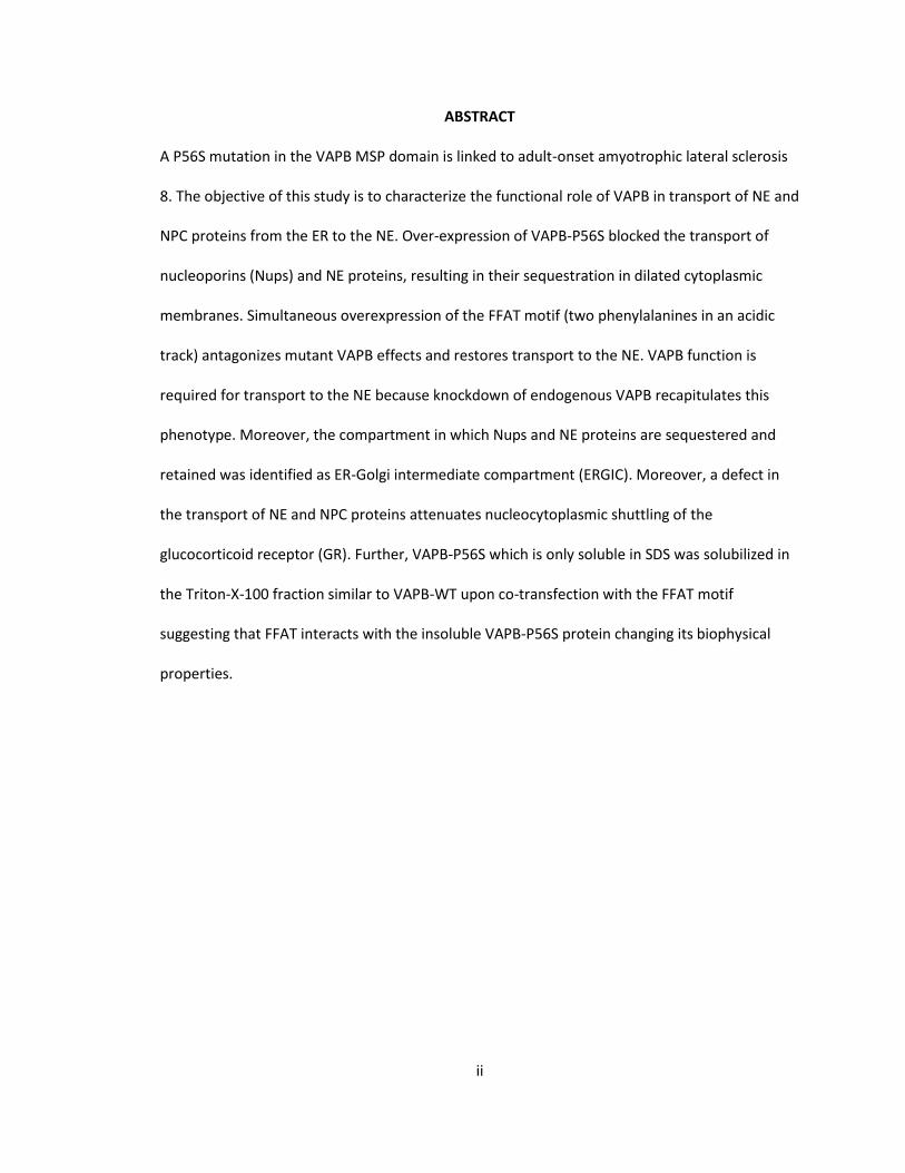
ABSTRACT
A P56S mutation in the VAPB MSP domain is linked to adult-onset amyotrophic lateral sclerosis
8. The objective of this study is to characterize the functional role of VAPB in transport of NE and
NPC proteins from the ER to the NE. Over-expression of VAPB-P56S blocked the transport of
nucleoporins (Nups) and NE proteins, resulting in their sequestration in dilated cytoplasmic
membranes. Simultaneous overexpression of the FFAT motif (two phenylalanines in an acidic
track) antagonizes mutant VAPB effects and restores transport to the NE. VAPB function is
required for transport to the NE because knockdown of endogenous VAPB recapitulates this
phenotype. Moreover, the compartment in which Nups and NE proteins are sequestered and
retained was identified as ER-Golgi intermediate compartment (ERGIC). Moreover, a defect in
the transport of NE and NPC proteins attenuates nucleocytoplasmic shuttling of the
glucocorticoid receptor (GR). Further, VAPB-P56S which is only soluble in SDS was solubilized in
the Triton-X-100 fraction similar to VAPB-WT upon co-transfection with the FFAT motif
suggesting that FFAT interacts with the insoluble VAPB-P56S protein changing its biophysical
properties.
iii
TABLE OF CONTENTS
ABSTRACT .………………………………………………………………………………………....……………………………….. ii LIST OF FIGURES ..……………………………………………………………………………………….………………...……… v LIST OF ABBREVIATIONS ………………………………………………………………………………………………………. vi ACKNOWLEDGEMENTS .…………………....………………………………………………………………………..……… vii
INTRODUCTION ………………………………………….………………………………………………………………………… 1
Amyotrophic Lateral Sclerosis ………………………………………………………………………………….. 1
Amyotrophic Lateral Sclerosis 8 (ALS8) …………………………………………………………………….. 1
Structure of VAPB …………………………………………………………………………………………………….. 3
The Consequences of VAPB-P56S ……………………………………………………………………………… 6
The Highly Conserved MSP Domain of VAPB ……………………………………………………………..7
Possible VAP function ………………………………………………………………………………………………. 8
Intracellular Trafficking ………………………………………………………………………………… 9
Phospholipid Metabolism ………………………………………………………………………….. 12
Lipid Binding or Transfer Protein Function ……………………………………… 12
Lipid Binding or Transfer Protein Families .…………………………………….. 12
Initial Characterization of the VAP-FFAT Interaction …………………………………….………….13
Crystallography of the VAP-FFAT Interaction ……………………………………………………………14
Consequences of Defective VAP-FFAT Binding ………………………………………………………… 16
The ER is Continuous with the NE …………………………………………………………………………….17
Nucleoporins and Inner Nuclear Membrane Proteins ……………………………………………… 20
Amyotrophic Lateral Sclerosis Disrupts the Nucleus …………………………………………………21
Hypothesis and Objective …………………………………………………………………………………………………….24
MATERIALS AND METHODS ……………………………………………………….…………………………………………26
DNA Plasmid Constructs ……………………………………………….………………………………………….26
Primary Antibodies ……………………………………………….…………………………………………………27
Secondary Antibodies ……………………………………………….……………………………………………..27
Cell Culture ……………………………………………….……………………………………………………………. 28
DNA Transfection ……………………………………………….……………………………………………………28
Immunocytochemistry …………………………………………………………………………………………….28
Image Collection ………………………………………………………………………………………………………29
Characterizing the Functional role of VAPB in Transport of INM and NPC Proteins …..29
Gp210, Nup214, and Emerin are mis-localized in VAPB-P56S transfected cells ..29
The FFAT Motif Rescues Mis-localized NE and NPC Proteins of VAPB-P56S .......30
Emerin, Pom-121, and Nup-214 are mis-localized upon siVAPB knockdown ……30
Characterizing the compartment in which INM and NPC proteins are retained ………..31
VAPB is localized to the ERGIC ………………………..………………………..…………………..31
VAPB-P56S and siVAPB Compromises the ERGIC ………………………..………………….32
The ERGIC distortion is not due to over-expression of ERGIC-53 ………..…..……….34
iv
Emerin, Mab414 and Nup214-RFP Co-localize with the ERGIC …..……………………35
Emerin, Mab414 and Nup-214 are Retained at the ERGIC upon shVAPB ………...36
ERGIC-53 Accumulates at the ERGIC at 15°C and Retains Emerin ….………………..38
Consequence of a Defective NE and NPC Assembly …….……………………….…..……………..39
VAPB-P56S causes a delay in nuclear translocation of activated GR and the FFAT
motif rescues the delay ………………………..……………………………..……………………….39
VAPB knockdown with siVAPB demonstrates a delay in GR nuclear entry ……… 40
VAPB knockdown with shVAPB increases GR nuclear translocation time ………..41
Addressing the Biophysical Properties of the P56S Mutation in VAPB …….……..……..…42
FFAT Changes the Solubility Properties of VAPB-P56S ………..………………………….42
RESULTS ……………………………………………………………………………………….………………………………………44
Characterizing the functional role of VAPB in Transport of INM and NPC Proteins ……44
Gp210, Nup214, and Emerin are mis-localized in VAPB-P56S transfected cells ..44
The FFAT motif rescues mis-localized NE and NPC proteins of VAPB-P56S ........45
Emerin, Pom-121, and Nup-214 are mis-localized upon siVAPB knockdown ……50
Characterizing the compartment in which INM and NPC proteins are retained ………..52
VAPB is localized to the ERGIC ………………………..………………………..…………………..52
VAPB-P56S and siVAPB compromises the ERGIC ………………………..………………….55
The ERGIC distortion is not due to over-expression of ERGIC-53 …..………..……….59
Emerin, Mab414 and Nup214-RFP Co-localize with the ERGIC …..……………………62
Emerin, Mab414 and Nup-214 are retained at the ERGIC upon shVAPB ………….63
ERGIC-53 Accumulates at the ERGIC at 15°C and Retains Emerin …………...………69
Consequence of a Defective NE and NPC Assembly …….……………………….…..………………71
VAPB-P56S causes a delay in nuclear translocation of activated GR and the FFAT
motif rescues the delay ………………………..……………………………..……………………….71
VAPB knockdown with siVAPB demonstrates a delay in Nuclear Entry of GR …..74
VAPB knockdown with shVAPB Increases GR Nuclear Translocation Time ……… 76
Addressing the Biophysical Properties of the P56S mutation in VAPB …….……..…………78
FFAT Changes the Solubility Properties of VAPB-P56S ………..………………………….78
DISCUSSION …………………………………………………………………………………………………………………………80
Characterizing the Functional Role of VAPB in Transport of INM and NPC Proteins ....80
Characterizing the Compartment in which INM and NPC proteins are retained ………..86
Consequence of a Defective NE and NPC assembly …….…………………………….……………..90
Addressing the Biophysical Properties of the P56S Mutation in VAPB …...……..…………93
REFERENCES …………………………………………………………………………………………………………………………95
v
LIST OF FIGURES
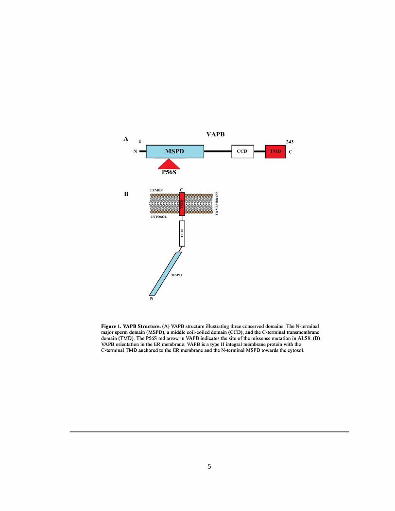
Figure 1. VAPB Structure ……………………………………….………………………………………………………………..5
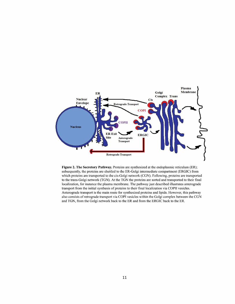
Figure 2. The secretory Pathway …………………………….……………………………………………………………..11
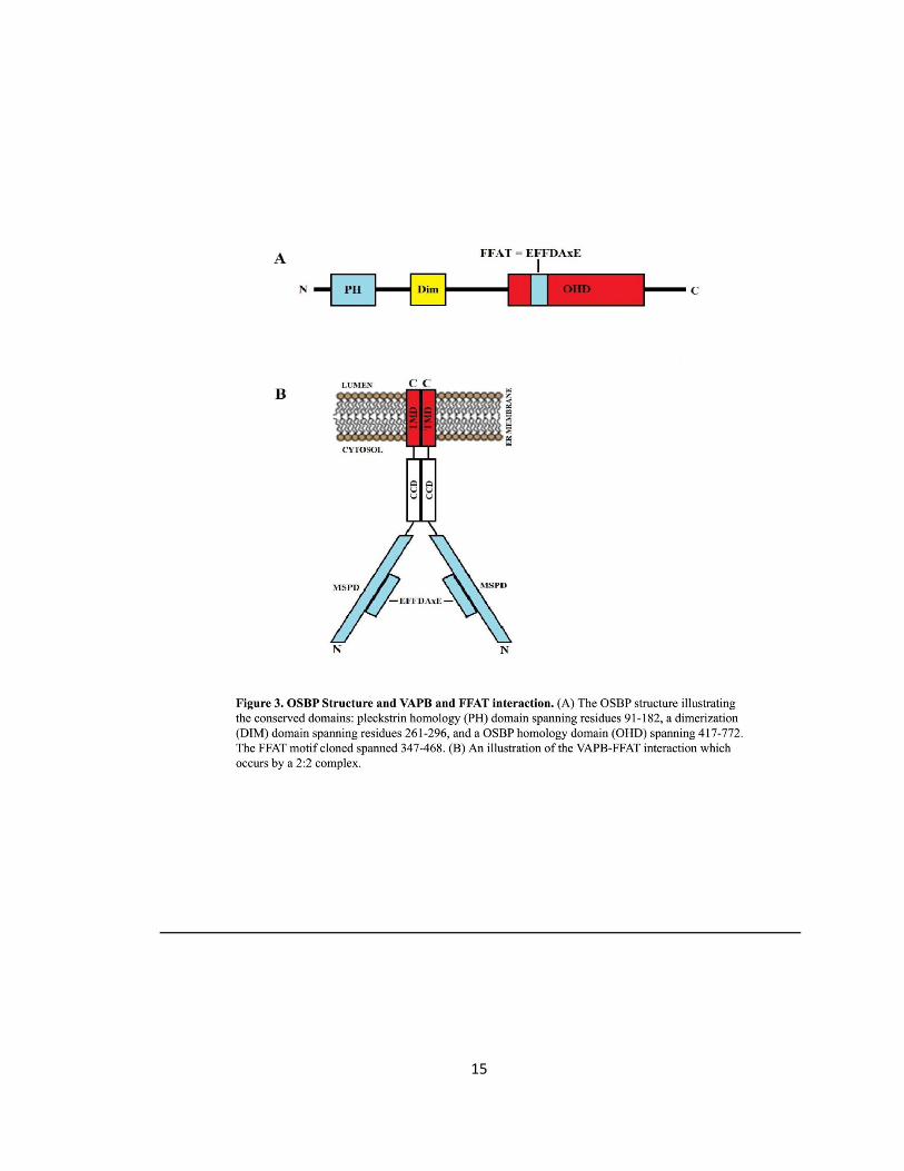
Figure 3. OSBP Structure and VAPB and FFAT interaction ….……………………………………………………15
Figure 4. The ER is Continuous with the NE and the NPC Structure …………………………………………19
Figure 5. Over-expression of VAPB-P56S induces a NE defect ……….………………………………………..23
Figure 6. NE proteins are retained in mutant VAPB-containing membranes and restored by FFAT
motif ………………………………………………………………………………………………………………………..46
Figure 7. NE proteins are retained in mutant VAPB-containing membranes and restored by FFAT
motif ………………………………………………………………………………………………………………………..47
Figure 8. NE proteins are retained in mutant VAPB-containing membranes and restored by FFAT
motif ………………………………………………………………………………………………………………………..48
Figure 9. NE proteins are retained in mutant VAPB-containing membranes …….….….….….….……49
Figure 10. Cytoplasmic retention of Nup214-GFP, Emerin, and Pom121-GFP upon siVAPB
knockdown ………………………………………………………………………………………………………………51
Figure 11. VAPB is localized to the ERGIC ….….….….….….….…..….….….….….….…..….….….….….……53
Figure 12. Immunocytochemistry of HeLa cells stained with anti-Golgin-97 towards the Golgi ..54
Figure 13. The ERGIC is compromised in the presence of VAPB-P56S and with shVAPB ….….……56
Figure 14. P56S induces an expanded and disrupted ERGIC ….….….….….….….….….….….…..….……57
Figure 15. The ERGIC is expanded in the presence of VAPB-P56S and shVAPB ….….….….….….…..58
Figure 16. The ERGIC distortion is not an over-expression effect ….….….….….….….….…..….….…..60
Figure 17. Endogenous ERGIC is expanded in the presence of VAPB-P56S and shVAPB ….….……61
Figure 18. NE Proteins and NPC Proteins are localized to the ERGIC ….….….….….….….…..….….….65
Figure 19. NE Proteins and NPC Proteins are localized to the ERGIC ….….….….….….….….….….…..66
Figure 20. NE Proteins and NPC Proteins are localized to the ERGIC ….….….….….….….….….….…..67
Figure 21. Endogenous Emerin and Mab414 are retained in expanded ERGIC upon knockdown of
VAPB ………………………………………………………………………………………………………………………..68
Figure 22. ERGIC-53 accumulates at ERGIC at 15°C and retains Emerin in this compartment…….70
Figure 23. VAPB-P56S causes a time delay in nuclear translocation of activated GR ….….….….…72
Figure 24. The FFAT motif restores proper nuclear translocation of GR ….….….….….….….….….…73
Figure 25. Knockdown of endogenous VAPB causes a time delay in nuclear translocation of
activated GR …………………………………………………………………………………………………………….75
Figure 26. Knockdown of endogenous VAPB increase nuclear translocation time of GR ….….…..77
Figure 27. The effect of the FFAT motif on the solubility of VAPB-P56S ….….….….….….….….….….79
vi
LIST OF ABBREVIATIONS
(ALS) Amyotrophic Lateral Sclerosis
(BSA) Bovine Serum Albumin
(CCD) Coiled-Coil Domain
(cDNA) Complimentary Deoxyribonucleic Acid
(CHO) Chinese Hamster Ovary
(CNS) Central Nervous System
(DIM) Dimerization
(EM) Electron Microscopy
(EPSPs) Excitatory Postsynaptic Potentials
(ER) Endoplasmic Reticulum
(ERG30) Endoplasmic Reticulum and Golgi 30-kDa protein
(ERGIC) ER-Golgi Intermediate Compartment
(FALS) Familial ALS
(FFAT) Two phenylalanines in an acidic tract
(GFP) Green Fluorescent Protein
(MEM) Minimum Essential Medium
(MSP) Major Sperm Protein
(NE) Nuclear Envelope
(NLS) Nuclear Localization Sequence
(NPCs) Nuclear Pore Complexes
(Nups) Nucleoporins
(OHD) OSBP Homology Domain
(ORD) OSBP-Related ligand-binding Domain
(ORPs) OSBP-Related Proteins
(OSBP) Oxysterol-Binding Proteins
(PH) Pleckstrin homology
(PM) Plasma Membrane
(PNS) Perinuclear Space
(rdgB) Retinal Degeneration Type B protein
(RFP) Red Fluorescent Protein
(SALS) Sporadic ALS
(SCS2) Suppressor of Choline Sensitivity 2
(SNARE) Soluble N-ethylmaleimide-sensitive factor attachment protein Receptor
(TMD) Transmembrane domain
(VAMP) Vesicle-Associated-Membrane-Protein
(VAP33) VAP of 33 kDa
(VAPB) Vesicle-Associated-membrane protein-associated protein B
vii
ACKNOWLEDGEMENTS
Finding your passion comes with many obstacles. You initially set a goal and then work to
achieve it. Along the way, experiences make you change your goal or try harder to obtain it. In
my Master’s I have learnt that even though you may not have obtained your final goal there is
great knowledge gained if you appreciate your situation, your surroundings and the people in
your life during that moment. Embarking on a Master’s of neuroscience is not an easy road, and
along the way I have encountered doubt about what I was doing. However, by appreciating the
people that gambled on my success and the community that has funded my research, I see how
privileged I am. Today, through a publication and the collaboration of myself and my lab
partners, we were able to make a change in the way researchers will examine ALS8.
I appreciate the guidance and patience of my supervisor, Dr. Johnny K. Ngsee. His support and
strength has helped me get through the program. I am lucky to have been given the privilege to
work with Dr. Ngsee. Initially, I was mentored by Duvinh Tran a Master’s graduate. He is
extremely educated in his field and was always there when needed. Kalina Abrol, was my
emotional support during the program and she always pushed me to do better. By working with
Angie Darbyson, a Master’s student, many of my doubts as to why I was doing ALS research
were answered. She suffered from the loss of a family member affected by ALS, and through
Angie I remembered what the research was about. Angie has a great amount of determination
and love in her research and I am lucky to have met her. Further, I thank the members of my
advisory committee for their guidance on my project: Dr. Antonio Colavita and Dr. Jean-Claude
Beique. Lastly, I thank Dr. Thompson for helping me on the confocal microscope.
I thank everyone that has been there during the past years and I have grown to admire their
love for the field of neuroscience.
INTRODUCTION
Amyotrophic Lateral Sclerosis
Amyotrophic lateral sclerosis (ALS) is a neurological disorder defined by the progressive loss of
upper and lower motor neurons in the cerebral cortex, brainstem and spinal cord. The loss of
motor neurons in these regions results in weakness, muscular atrophy and loss of voluntary
movement. Further, patients exhibit difficulty in speech and swallowing eventually leading to
full paralysis. Typically, death is followed approximately 1-5 years after a patient is diagnosed
due to respiratory failure (Cozzolino et al., 2008). A devastating property of patients affected by
ALS is that they tend to exhibit full cognitive functions allowing the patient to be fully aware of
their deterioration and loss of their voluntary movement (Cozzolino et al., 2008). The average
age of disease onset is 50 years of age (Cozzolino et al., 2008) with an incidence of 1-2 persons
out of 100,000 and prevalence of 4-6 persons out of 100,000 (Kanekura et al., 2009; Cozzolino et
al., 2008). Incidences of sporadic ALS (SALS) are 90-95% and familial ALS (FALS) cases make up 5-
10% (Shaw, 2005). Research has shown that whether the disease is SALS or FALS, the disease is
indistinguishable which allows researchers to apply current therapeutic techniques to a wide
spectrum of ALS cases.
Amyotrophic Lateral Sclerosis 8 (ALS8)
ALS8 is a late-onset adult neurodegenerative disorder manifested as a movement disorder. The
progressive loss of neurons and the degeneration of their synaptic connections is a common
characteristic of neurodegenerative disease (Meyer and Quenezer, 2005). Specifically, if
neurodegeneration of upper motor neurons and lower motor neurons occurs then the cardinal
clinical features become evident in ALS8 patients. The cardinal features are as follows:
2
weakness, muscular atrophy, loss of voluntary muscle movement and paralysis (Boillee et al.,
2006). There are currently eleven types of familial ALS and familial ALS8 is caused by a mutation
in the Vesicle-Associated Membrane Protein (VAMP)-associated protein B (VAPB) gene
(Nishimura et al., 2004). The VAPB gene encodes a Type II integral membrane protein
ubiquitously expressed in the CNS, and abundant in motor neurons (Teuling et al., 2007).
Interestingly, VAPB was found to be expressed throughout the CNS in the olfactory bulb, cortex,
mesencephelon, hippocampus, cerebellum, medulla oblongata and the spinal cord and yet the
genetic P56S mutation in the conserved major sperm protein domain of VAPB causing ALS8
specifically results in motor neuron degeneration (Teuling et al., 2007). Further, VAPB was
expressed in the kidney, heart, skeletal muscle, liver, spleen and lung (Teuling et al., 2007). Since
VAPB was found to be abundant in motor neurons, a closer examination revealed that VAPB in
neurons was concentrated to the soma and proximal dendrites (Teuling et al., 2007). The
specificity to motor neuronal death in VAPB-P56S is not well understood; however, studies have
shown that the mutation in VAPB forms cytosolic aggregates (Nishimura et al., 2004). Research
has been currently emphasizing on the functional deficits that are caused by the P56S
aggregates in order to explain the susceptibility to motor neurons.
It has been shown that VAPB-P56S induces a change in ER morphology and leads to a collapse of
the ER microtubules resulting in cytoplasmic aggregates (Prosser et al., 2008). Since motor
neurons are large cells and have extremely complex morphologies, the selective vulnerability of
motor neurons could be due to their great need of ER cellular processes (Boillee et al., 2006).
Interestingly, motor neurons have been shown to have an extensive ER network and VAPB is
localized to the ER. Taken together, motor neurons may be selectively damaged due to the
greater size of their ER, thus a greater abundance of the VAPB protein.
3
A hallmark study towards understanding ALS produced by Nishimura in 2004 identified the locus
for an atypical form of ALS now known as ALS8. The locus was mapped to chromosome 20q13.3
for vesicle-associated-membrane protein-associated protein B (VAPB) (Nishimura et al., 2004).
In 2005, a study identified seven families of Portuguese-Brazilian ancestry and one family of
African-Brazilian ancestry compromising more than 1,500 individuals and from those families
200 individuals suffered from ALS symptoms (Nishimura et al., 2005). Genetic analysis of the
family pedigree identified a common cytosine to thymine substitution at position 166 of the
open reading frame (Nishimura et al., 2005). This resulted in substitution of the proline residue
at position 56 to serine (P56S) (Nishimura et al., 2005).
Structure of VAPB
VAPs are ER-resident proteins initially discovered due to their interaction with v-SNARE VAMP2
(Skehel et al., 1995). The first VAP was identified in Aplysia californica as VAP of 33kDA (VAP33)
(Skehel et al., 1995). In humans, there are only two VAP genes encoding for the VAP proteins
VAPA, VAPB and the VAPB spliced variant VAPC. VAPA and VAPB are similar isoforms sharing
63% identical amino acids (Nishimura et al., 2004). Both VAPA and VAPB contain an N-terminal
major-sperm protein (MSP) domain, a central coiled-coil region, and a C-terminal
transmembrane domain (Nishimura et al., 1999) (Fig. 1 A and B). VAPC lacks the coiled-coil and
transmembrane regions. Although these VAPs are grouped in the same family, they hold unique
properties. VAPA gene is located on chromosome 18 and encodes a 249 amino acids long
protein with a predicted molecular mass of 27.3kDa (Nishimura et al., 1999). VAPB gene is
located on chromosome 20 and encodes a 243 amino acids long protein with a predicted
molecular mass of 27 kDa (Nishimura et al., 1999) (Fig. 1B). VAPC is the same gene as VAPB but
is an mRNA splice variant that leads to a truncated protein (Kukihara et al., 2009). The major
4
sperm protein (MSP) domain of VAPB consists of 150 amino acids organized into a seven-
stranded immunoglobin-like-β-sandwich with S-type topology (Baker et al., 2002). The MSP
domain is essential for the VAP-FFAT interaction because the MSP domain is the region that
directly interacts with the FFAT motif of lipid binding proteins (Loewen and Levine, 2005). The
coil-coiled domain is comprised of 40 amino acids and is suggested to function in protein-
protein interaction (Hamamoto et al., 2005). VAPB’s transmembrane domain acts as an anchor
anchoring VAP to the ER membrane which plays a crucial role in VAPBs ability to form homo-
and heterodimers via its dimerization motif (GxxxG) (Amarilio et al., 2005). VAPA and VAPB
localize to the ER and research suggests their primary function is within this compartment
(Soussan et al., 1999) involved in the early or late secretory pathway of protein transport from
the ER.
5
6
The Consequences of VAPB-P56S
The P56S mutation in ALS8 resides in the conserved MSP domain of VAPB (Nishimura et al.,
2004). The hallmark of the P56S missense substitution is ER membrane aggregates that are
resistant to degradation (Kanekura et al., 2006; Nishimura et al., 2004; Teuling et al., 2007).
Studies have shown that the mutant form of VAPB has the ability to recruit wild-type VAPA and
VAPB into insoluble aggregates leading to a loss of function of the wild-type protein (Teuling et
al., 2007). Since VAPB has the ability to form dimers, it is possible that this recruitment could be
due to a mutated form of VAPB forming dimers with wild-type VAPB. In addition, this
recruitment could also be due to the aggregation of the MSP domain simply aggregating nearby
wild-type VAPB. Although, it is not clear which mechanism is responsible for mediating neuronal
cell death, aggregation and recruitment of the wild-type form of VAPB by the mutant form of
VAPB could additively over time trigger cell death.
A mutation in copper-zinc superoxide dismutase (SOD1) has been linked to a form of familial ALS
known as ALS1. This form of ALS is the most common genetic cause of ALS (Tsuda et al., 1994). A
mouse model of SOD1 for ALS1 has shown that both VAPA and VAPB levels were down-
regulated (Teuling et al., 2007). What this study suggests is that ALS1 may also cause effects
similar to ALS8. The loss of VAP from SOD1 mice in ALS1 and the loss of VAP in VAP-P56S
transfected cells in ALS8 can be a leading factor in the pathogenesis of the disease.
Collectively, studies examining VAPB-P56S in cellular models have consistently shown mutant
VAPB to be aggregates prone and recruiting endogenous VAPB to insoluble aggregates resulting
in a dominant negative effect (Teuling et al., 2007). Animal models such as Drosophila
melanogaster and transgenic mice have been able to display these aggregates. Unfortunately, at
the present time there is no confirmation that these aggregates are present in human tissue;
7
therefore, most of the information gathered about these aggregates and the loss of proper
function due to VAPB-P56S mutation has to be generalized with existing models. In a model
utilizing Drosophila melanogaster the aggregates seen in cells have been reproduced (Chai et al.,
2008). The transgenic expression of mutant dVAPP56S in Drosophila reproduced these
aggregate structures (Chai et al., 2008). Further, transgenic mice expressing the human P56S
mutation of VAPB also reproduced these cytoplasmic aggregates (Tudor et al., 2010).
The Highly Conserved MSP Domain of VAPB
The MSP domain of VAPB is homologous to the major sperm protein of the nematodes A. suum
and Caenorhabditis elegans (Skehel et al., 2000). The crystal structure of VAPBs MSP domain
confirms that the MSP domain consists of 150 amino acids organized into a seven-stranded
immunoglobin-like-β-sandwich with S-type topology (Baker et al., 2002). Uncovering the crystal
structure revealed that the P56S mutation of VAPB is in the MSP domain of VAPB at a kink
within the extended β-sheet (Loewen and Levine, 2005). The S-shape loop is maintained by the
proline residue at codon 56 which connects the d1 and d2 strands within the MSP domain
resulting in an S-shape pattern (Nishimura et al., 2004). The proline stabilizes the S-shape loop
by holding it in a less energetically favourable cis-peptide bond conformation (Teuling et al.,
2007). The MSP domain has hydrophobic core residues, and the S-shape loop formed by the
proline residue shields these core residues (Teuling et al., 2007). Since VAPB-P56S forms
insoluble cytoplasmic aggregates (Kanekura et al., 2006), it is suggested that upon substitution
of proline to a serine residue the S-shape loop destabilizes into a more energetically favourable
trans-peptide bond conformation (Teuling et al., 2007).This change in protein conformation
exposes the hydrophobic core residues and formed aggregates may be the result of
8
accumulating neighbouring mutant VAPB proteins attempting to shield the exposed
hydrophobic core residues (Teuling et al., 2007).
In 2006 it was found that the loss of the proline residue is the main cause of the formed
aggregates and change in ER morphology (Kanekura et al., 2006). VAPB was mutated by
substituting the proline residue with amino acids such as alanine, lysine or aspartic acid. In all
cases VAPB increased insolubility similar to the P56S mutation in ALS8 (Kanekura et al., 2006).
Proline’s ability to maintain the S-shape loop in the MSP domain shields the hydrophobic core
residues and maintains the proper protein conformation of VAPB. The loss of the proline residue
results in the mis-folding of VAPB and In vitro studies have shown that over-expression of VAPB-
P56S consistently produces polyubiquitinated, detergent-insoluble, cytoplasmic aggregates
(Kanekura et al., 2006). It is certain that the mutation in VAPB prevents the proper folding of the
protein interfering with its function (Kanekura et al., 2006).
Possible VAP function
Earlier studies have addressed the possible functions of VAPs. Initially, VAPA was identified as
VAP of 33 kDa or VAP33 (Nishimura et al., 2004). Although VAPA holds unique properties and
functions, VAPA and VAPBs sequence and protein structure similarity may suggest that VAPA
function may be applicable to VAPB (Nishimura et al., 2004). Research has implemented VAPA
and VAPB in intracellular trafficking, the unfolded protein response (UPR) and with phospholipid
metabolism. A disruption in one of these pathways may explain why motor neurons are
targeted in ALS8 and may define the mechanism by which the disease is caused.
9
Intracellular Trafficking
The transport of proteins between organelle compartments in the protein secretory pathway
requires a great deal of proteins to mediate vesicle budding and fusion events. For the survival
of the cell, this pathway must be followed and disruption to this pathway can diminish cellular
functions. Currently, VAP has been linked to intracellular trafficking; however, the specific role
of VAPB is still in debate and is not yet fully understood. Further, the negative effects caused by
VAPB-P56S cytoplasmic aggregates are also still underway. The protein secretory pathway
consists of the early and late secretory pathways (Fig. 2). Specifically, VAPB is implicated in the
early secretory pathway. Proteins are synthesized at the endoplasmic reticulum (ER);
subsequently, the proteins are shuttled to the ER-Golgi intermediate compartment (ERGIC) from
which proteins are transported to the cis-Golgi network (CGN). Following, proteins are
transported to the trans-Golgi network (TGN). At the TGN the proteins are sorted and
transported to their final localization. The pathway described illustrates the anterograde
transport route from the initial synthesis of proteins to their final localization. Since the ER is the
site of lipid production, anterograde transport is the main route for synthesized proteins and
lipids (Prosser, 2008). However, this pathway also consists of retrograde transport located
within the Golgi complex – between the CGN and TGN – and from the Golgi network back to the
ER (Prosser, 2008).
Research suggests that VAPB is implicated in ER to Golgi protein trafficking. In 1999, the ER and
Golgi 30-kDa protein (ERG30), known as VAPB, was implicated in protein transport between the
ER and Golgi (Soussan et al., 1999). They showed that the use of an ERG30 antibody in vitro
resulted in COPI-coated vesicles accumulation at the Golgi complex (Soussan et al., 1999) linking
VAPB to the early secretory pathway between the ER and the Golgi. There are anterograde and
retrograde pathways in the protein secretory system. COPI-coatomers are involved in the
10
retrograde transport of proteins from the cis-Golgi to the ER (Soussan et al., 1999). The
accumulation of COPI vesicles linked VAPB with protein retrograde transport between the ER
and Golgi. Thus, VAPB is likely involved in intracellular trafficking of proteins in the early
secretory pathway between the ER and the Golgi complex. In the current study, an objective is
to understand how VAPB-P56S-induced disruption of protein trafficking between the ER and
Golgi may lead to the pathogenesis of ALS8.
11
12
Phospholipid Metabolism
The MSP domain of VAPs directly interacts with the FFAT motif of lipid binding proteins. The
FFAT motif, two phenylalanines in an acidic tract, was found to be the domain for interaction
between FFAT containing proteins and VAPs (Loewen et al., 2003). The FFAT motif has seven
amino acids with the consensus sequence EFFDAxE (Kaiser et al., 2005) (Fig. 3 A). Upon binding
of VAPs MSP domain to FFAT-motif containing proteins, lipid binding or transfer proteins are
recruited to the ER membrane to regulate lipid composition at the ER membrane (Lev et al.,
2008; Prosser et al., 2008). Presently, how mutant VAPB affects binding of FFAT with VAP is not
fully understood.
Lipid Binding or Transfer Protein Function
Prior to ER membrane recruitment, FFAT motif-containing lipid binding or transfer proteins are
localized to the cytosol (Perry and Ridgway, 2006).The cytosol is an aqueous environment with
numerous hydrophobic lipids molecules. Lipid binding or transfer proteins function by
solubilizing the hydrophobic lipids as they transport through the cytoplasm (Loewen et al.,
2003). Arrival at the target membrane induces the FFAT motif-containing lipid binding or
transfer proteins to bind to MSP domain of VAP and releases the lipids from lipid binding or
transfer proteins to insert them into the membrane (Olkkonen and Levine, 2004). This process
regulates the lipid composition at the ER membrane.
Lipid Binding or Transfer Protein Families
VAPs interact with FFAT motif –containing proteins involved in synthesis, transport and
metabolism of lipids. Oxysterol-binding proteins (OSBP) contain the FFAT sequence and are
cytosolic receptors that bind to oxysterols such as 25-hydrocholoesterol (Wyles et al., 2002).
OSBP-related proteins (ORP) are also FFAT motif containing lipid binding proteins consisting of
13
ORP1L, ORP2, ORP3, ORP4L, ORP6, ORP7, and ORP9. ORPs and OSBP sharer the same OSBP-
related ligand-binding domain (ORD) yet differ by protein sequence (Olkkonen et al., 2006).
Retinal degeneration type B proteins (rdgB) are also FFAT motif lipid binding proteins consisting
of Nir1, Nir2, and Nir3 (Olkkonen and Levine, 2004). This group is involved in membrane
trafficking and phospholipid metabolism (Amarilio et al., 2005).
Initial Characterization of the VAP-FFAT interaction
Lipid binding or transfer proteins facilitate intracellular lipid trafficking. To effectively execute
their function, lipid binding or transfer proteins must be guided to specific target membranes.
The initial finding illustrating the VAP-FFAT interaction was executed in Saccharomyces
cerevisiae (Loewen et al., 2003) where the FFAT motif acted as a membrane-targeting signal
directing proteins to the surface of the ER to bind with VAP (Loewen et al., 2003). Further, the
FFAT-motif is found in Opi1p, a transcriptional repressor of genes involved in phospholipid
synthesis in yeast (Loewen et al., 2003). Opi1 directly interacts by its FFAT motif with the MSP
domain of SCS2, the yeast homologue of VAP, in order to target the ER (Loewen et al., 2003).
Upon binding of Opi1 FFAT-motif to SCS2 MSP domain, Opi1 was retained in the cytoplasm
preventing Opi1s translocation to the nucleus in order to enhance the transcription of lipid-
synthesizing genes (Brickner and Walter, 2004). Thus, binding to VAP is executed by FFAT motif
containing proteins to promote the synthesis of new ER membranes and to regulate lipid
composition. A disruption in this process can lead to a collapse in ER morphology due to
unregulated lipid composition at the ER (Prosser et al., 2008).
14
Crystallography of the VAP-FFAT Interaction
In 2005, the VAP-FFAT interaction was clarified by crystallography (Kaiser et al., 2005). The
crystal structure resolved that the VAPA-FFAT interaction occurs by a 2:2 complex (Fig. 3 B). The
MSP domains of the two VAP proteins would each consist of a bound FFAT motif (Kaiser et al.,
2005). Further, the residues of VAPA-MSP domain crucial for binding FFAT-containing proteins
were resolved. In FFAT containing proteins, the phenylalanine at the 476 residue binds to
VAPAs-MSP domain at residue Met89. Binding occurs in a hydrophobic pocket of VAPAs-MSP
domain created by aliphatic side chains: Lys45, Thr47, Ly87, and Lys118 (Kaiser et al., 2005).
15
16
Consequences of Defective VAP-FFAT Binding
Brunger’s group (2005) mutated the key residues of the VAP-FFAT interaction in the
hydrophobic pocket and aliphatic side chains of Scs2p, VAP homologue in yeast, K87D/M89D
(Kaiser et al., 2005).This mutation disrupted the interaction between Scs2 and the FFAT
containing protein Opi1; thus, blocking the function of Scs2p (Kaiser et al., 2005). The defective
binding between FFAT containing proteins and VAP has shown to produce severe abnormalities
to ER morphology and cellular processes (Prosser et al., 2008; Kaiser et al., 2005). Upon
expressing the K87D/M89D mutation in COS7 cells, Brunger’s group showed severely altered ER
morphology (Kaiser et al., 2005). Untreated cells exhibited normal reticular ER pattern whereas
over-expression of VAPA K87D/M89D produced spaced patches that co-localized with ER
markers (Kaiser et al., 2005). The VAPB-P56S mutation may exhibit similar loss of cellular
processes and deterioration of ER morphology in ALS8. VAPB-P56S may block VAPBs binding to
FFAT containing lipid binding or transfer proteins leading to loss of function and ER
deterioration. An objective of the current study is to resolve if VAPB-P56S-induced deterioration
of ER morphology inhibits vesicle trafficking of nucleoporins (Nups) and nuclear envelope (NE)
proteins to their proper localization.
The VAP-FFAT interaction promotes synthesis of the ER membrane by regulating the lipid
composition at the ER. A disruption of this process can lead to a change or collapse in ER
morphology (Prosser et al., 2008). In 2005, a study addressing the severity of the FFAT-VAP
binding on ER structure demonstrated that exogenous expression of FFAT-motifs from ORP3 and
Nir2 causes formation of ER membrane stacks or whorls, while other proteins such as Nir3
rearranged ER structure (Amarilio et al., 2005). These results suggest that FFAT-containing
proteins initiate dynamic changes in ER morphology. In ALS8, VAPB-P56S has been shown to
produce insoluble cytoplasmic aggregates (Kanekura et al., 2006; Moumen et al., 2011)
17
Overexpression of VAPB-P56S in CHO cells has shown to form large ER aggregates (Prosser et al.,
2008). Moreover, co-overexpression of VAPB-P56S with the FFAT motif from rat OSBP in CHO
cells has been shown to resolve these large ER aggregates (Prosser et al., 2008). Although, it is
not clear as to why these aggregates form, a study in 2007 demonstrated that mutant VAPB-
P56S cannot interact with a green fluorescent protein (GFP)-tagged FFAT motif (Teuling et al.,
2007). Although it is possible that the FFAT motif cannot interact with the VAPB protein because
aggregation blocks FFAT from reaching VAPs binding site, it is clear that the VAP-FFAT
interaction is crucial for ER morphology and regulating lipid synthesis at the ER (Teuling et al.,
2007; Prosser et al., 2008; Amarilio et al., 2005).
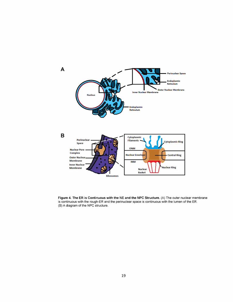
The ER is Continuous with the NE
The outer nuclear membrane (ONM) is continuous with the rough-ER (Fig. 4A). Since mutant
VAPB induces ER morphology defects and affects ER trafficking, the continuity between the ER
and NE raises the possibility that transport of NE proteins might also be affected and contribute
to the ALS8 disease pathogenesis. The NE divides eukaryotic cells into a nuclear and cytoplasmic
compartment providing a selective permeable barrier between the compartments (Starvu et al.,
2006). The nuclear envelope (NE) is comprised of two lipid bilayers. The inner nuclear
membrane (INM) is attached to lamins, a family of structural support proteins in the nuclear
arena and the ONM is continuous with the rough ER (Starvu et al., 2006). The INM and ONM are
separated by a perinuclear space approximately 30nm wide that is continuous with the lumen of
the ER (Starvu et al., 2006) (Fig. 4A). Local fusion between both membranes creates the aqueous
nuclear pore complexes (NPCs) spanning both membranes for nucleocytoplasmic transport
(Starvu et al., 2006). NPCs are around 125 MDa in size formed by approximately 30 Nups
(Antonin et al., 2005). The NPC has an eight-fold rotational symmetry with three concentric
rings: The cytoplasmic ring, the central ring and the nuclear ring. The nuclear and central rings
18
compose the nuclear basket (Fig. 4B). Shuttling between the cytoplasm and nucleus is a tightly
regulated process with more than one million molecules passing through approximately 2-3
thousand nuclear pores per minute (Antonin et al., 2005). This process requires the interaction
between Nups and nuclear localization signals in order to facilitate translocation. All proteins
that are imported into the nucleus bear a nuclear localization signal (NLS) and all proteins that
exported require a nuclear export sequence (NES) (Antonin et al., 2005).
19
20
Nucleoporins and Inner Nuclear Membrane Proteins
Each Nup of the NPCs holds specific functions. Pom121 is an integral membrane protein
localized to the central ring of the NPC (Antonin et al., 2005). Pom121 functions in anchoring the
NPC to the NE (Antonin et al., 2005). Gp210 is located at the central ring of the NPC (Antonin et
al., 2005). Gp210 also anchors the NPC to the NE. To date, Gp210, Pom121 and Ndc1 are the
three integral membrane proteins of the NPC anchoring the NPC to the NE (Antonin et al.,
2005). Nup214 is localized at the cytoplasmic ring of the NPC (Xu et al., 2009). Lamin-B1 is part
of a family of intermediate filaments which stabilize the NE and anchors nuclear membrane
proteins to the scaffold of the INM (Holaska et al., 2006). Emerin is an integral protein of the
INM expressed in almost all human cells belonging to the ‘LEM domain’ (LAP2/Emerin/MAN1)
family of nuclear proteins (Holaska et al., 2006). Emerin binds to and co-localizes with A and B-
type lamins in the INM which anchor Emerin for architectural, gene regulator and nuclear
assembly roles (Holaska et al., 2006).
The NE is disassembled during open mitosis, and nuclear pores are equally divided to the two
newly formed nuclei. Heightened NPC assembly occurs shortly thereafter to double the number
of pores in the daughter cells. The rate of de novo synthesis progressively decreases during
interphase. In post-mitotic cells, such as neurons, this mitotic renewal process is not followed.
Research has not yet fully established what occurs to NPCs in post-mitotic cells; however, it has
been shown that NPCs do not turn over appreciably in differentiated cells (D’Angelo et al.,
2009). Some NPC proteins, however, can be exchanged at the NPC, others are extremely long
lived and some are incorporated in the NPC the entire lifespan of a cell (D’Angelo et al., 2009).
The current thesis focuses on interphase cells and the described NPC and NE proteins will be
examined to illustrate the effects of VAPB-P56S on NPC and NE proteins.
21
Amyotrophic Lateral Sclerosis Disrupts the Nucleus
D’Angelo in 2009 showed that nuclear leakiness is accelerated during aging (D’Angelo et al.,
2009). The effect of oxidative damage and age-related deterioration on the NPC was examined.
Utilizing the environmental toxin paraquat induced oxidative damage by increasing the amount
of reactive oxygen species (ROS) in Caenorhabditis elegans for six days (D’Angelo et al., 2009).
The worms treated with paraquat exhibited a higher percentage and earlier onset of leaky nuclei
by nuclear entry of the 70 kDa dextran when compared to controls not treated with paraquat.
Also, older worms had increased nuclear permeability compared to young worms and addition
of paraquat treatment increased nuclear leakiness (D’Angelo et al., 2009). Thus, age-related
deterioration of NPCs leads to loss of the NPC selective permeability barrier (D’Angelo et al.,
2009). This consequently results in the leak of cytoplasmic proteins into the nucleus. ALS is an
adult-late onset disease; therefore, VAPB-P56S may increase nuclear permeability resulting from
damage to NPCs and loss of nuclear integrity (D’Angelo et al., 2009).
In 2009, a study examined Nup distribution in anterior horn cells of patients with ALS (Kinoshita
et al., 2009). The Nups examined were nup88, nup62, and nup153 (Kinoshita et al., 2009). They
examined Nup distribution by antibody staining of the spinal cord of SALS and FALS patients. In
control individuals, the Nups formed entirely smooth nuclear contours (Kinoshita et al., 2009).
However, in cells of both SALS and FALS patients the nucleus was irregular and twisted with
irregular nuclear contours (Kinoshita et al., 2009).
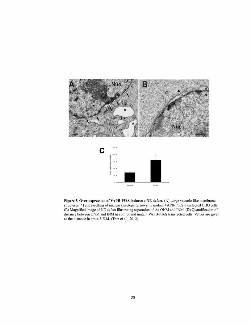
Ngsee’s group in 2012 linked ALS8 to a dysfunction in NE structure (Tran et al., 2012). The NE of
CHO fibroblasts overexpressing VAPB-P56S in the absence or present of the FFAT motif was
analyzed by transmission electron microscopy (EM). In the absence of FFAT, CHO cells contained
large empty vacuoles resembling expanded ER not observed in FFAT transfected cells. The NE
22
displayed a significant separation in distance between the ONM and INM in approximately 75%
of non-FFAT transfected cells compared to control or FFAT transfected cells (Fig. 5) (Tran et al.,
2012). The distance between the INM and ONM in VAPB-P56S transfected cells were as grand as
500 nm with an average of 160 ± 32 nm whereas VAPB-WT mean distance between the ONM
and INM was 70 ± 5 nm (Tran et al., 2012). The results illustrated a defect in NE structure and
are the foundation of the current thesis. A current objective is to examine VAPBs role in the
transport of NPC and NE proteins. If VAPB plays a role, then VAPB-P56S may induce the
separation of the ONM and INM by inhibiting the proper assembly of the NPC and transport of
NE proteins which may contribute to the pathogenesis of ALS.
23
24
Hypothesis and Objective
The main objective of this study is to identify and characterize the relationship between VAPB
and the trafficking of NPC and NE proteins. Since VAPB-P56S causes a NE defect characterize by
separation of the ONM and INM, I hypothesize that this could be caused by deterioration in NPC
assemble. Some Nups and NE proteins are synthesized at the ER and then transit to the NE, I
hypothesize that VAPB regulates this process. VAPB-P56S may disrupt Nup and NE protein
transport from the ER resulting in loss of proper Nup and NE protein localization thus inducing a
separation between the NEs INM and ONM. This may cause a defect in nucleocytoplasmic
shuttling. Since binding to FFAT-motif containing lipid binding proteins is crucial for VAPB
function, co-expressing the FFAT motif may counteract the mutations adverse effects.
In order to test this hypothesis, in vitro experiments were performed to examine the effects of
VAPB-P56S on Nup and NE protein trafficking. The major method employed to address this issue
involved transfecting VAPB-P56S DNA and subsequently analysing the cells morphology and
protein localization by confocal microscope. The effect of P56S on Nup transport was examined
using the Nups Gp210, Nup214, Pom121 and the INM proteins Emerin and Lamin-b1. Further, to
address the function of VAPB on NPC and NE protein transport, we knocked down synthesis of
VAPB using shRNA. If it is shown that Nups and NE proteins are retained in cytoplasmic VAPB-
P56S-induced aggregates and are shown to be retained in the cytoplasm upon knockdown of
VAPB, then the compartment in which NE and NPC proteins are retained must be characterized.
Bu utilizing ERGIC-53-GFP, a marker for the ERGIC which displays both anterograde and
retrograde transport, we can determine if the NPC and NE proteins are retained in this
compartment. Thus, this will demonstrate if VAPB is implicated in the transport of proteins from
the ERGIC. If the previous experiments indicate that NPC and NE proteins are in fact retained in
the cytoplasm resulting in an altered NPC composition, the consequences of a defective NE and
25
NPC can be examined by utilizing the ligand-activated glucocorticoid receptor (GR). This will
indicate if a defect in nucleocytoplasmic shuttling results due to VAPB-P56S or due to loss of
VAPB function by knockdown of endogenous VAPB. A further objective of this study is to
examine the biophysical properties of VAPB-P56S. It has been shown that the VAPB mutation
induces a biophysical change resulting in insolubility of the protein. Upon co-transfection of the
FFAT-motif, the adverse effects of VAPB-P56S are resolved. Therefore, I hypothesize that the co-
overexpression of the FFAT-motif with VAPB-P56S may induce a change in VAPB-P56S
biophysical properties manifested by proper VAPB function. Chinese hamster ovary (CHO)
fibroblasts and HeLa cells were used throughout this study.
26
MATERIALS AND METHODS
DNA PLASMID CONSTRUCTS
Human VAPB cDNA
Human VAPB complimentary DNA (cDNA) coding for human VAPB-wild type and the P56S
mutation of VAPB was previously produced by Dr. Derek C. Prosser (Prosser et al., 2008).
shRNA: pLKO.1 and shVAPB
Lentiviral pLKO.1 based plasmids were from Open Biosystems (Hunstville, AL, USA).
TRCN0000152888 matched both human and mouse VAPB sequences. Empty pLKO.1 vector was
used as a control. In some cases, the shRNA plasmids were co-transfected with monomeric RFP
(mRFP) to identify the transfected cells.
The FFAT Motif and the AAAT motif
The FFAT motif of rabbit OSBP (residue 347-468) and the AAAT motif of rabbit OSBP was
produced by Dr. Derek C. Prosser (Prosser et al., 2008).
Emerin-GFP
The Emerin cDNA was obtained from Open Biosystems (Hunstville, AL, USA) and then
subsequently cloned into pEGFP-C1 at the BspE1 and BamH1 sites by Duvinh Tran.
Nucleoporins and INM Protein Plasmids
The Gp210-GFP DNA plasmid was purchased as prGp210(s)-EGFP3-(TM+CT) with the triple GFP
tag on the C-terminus of Gp210. Nup214-GFP DNA plasmid was obtained as pNup214-EGFP with
the GFP tag on the C-terminus of Nup214. Subsequently, the Nup214-GFP DNA plasmid was
cloned into an mRFP-N1 plasmid at the EcoR1 and Age1 sites. Pom121-GFP DNA plasmid was
obtained as pPom121-EGFP3 with the GFP tag on the C-terminus of Pom121. Lamin-B1-GFP DNA
27
plasmid was obtained as pEGFP-LaminB1 with the GFP tag on the N-terminus of Lamin-B1. All
these plasmids were obtained from EUROSCARF (Frankfurt, Germany).
ERGIC-53-GFP
The ERGIC-53-GFP DNA plasmid was obtained from the lab of Dr. Hans-Peter Hauri.
GR-GFP
The Glucocorticoid Receptor (GR) tagged to GFP was obtained from the lab of Dr. Robert J.G.
Hache from the University of Ottawa, Ottawa Health Research Institute
PRIMARY ANTIBODIES
ERGIC-53 primary antibody was obtained from SIGMA-Aldrich (St. Louis, MO, USA) as Anti-
ERGIC-53/p58 polyclonal antibody produced in rabbit and was utilized at a dilution of 1:50.
Golgin-97 primary antibody was obtained from ABCAM as rabbit polyclonal antibody to Golgin
97 and was diluted at 1:50. Mab414 primary antibody was obtained from ABCAM (Covance,
Princeton, NJ, USA) as mouse monoclonal antibody to Nuclear Pore Complex Proteins and was
diluted at 1:100. Emerin primary antibody was obtained from Developmental Studies
Hybridoma Bank (Iowa City, IA, USA) as MANEM1 and was diluted at 1:50. VAPB, either WT or
mutant P56S, constructs were Flag-epitope tagged and were visualized with mouse anti-FLAG
primary antibody (Applied Biological Materials, Richmond, BC, Canada) diluted at 1:1000.
SECONDARY ANTIBODIES
Goat anti-mouse or goat anti-rabbit 488(green) or 594 (red) was obtained from Invitrogen
(Carlsbad, CA, USA) and diluted at 1:250.
28
CELL CULTURE
CHO and Hela
Chinese Hamster Ovary (CHO-K1) fibroblasts were maintained in minimum essential medium
(MEM) α (Invitrogen) and HeLa cells were maintained in Dulbecco’s modified Eagle’s medium
(DMEM) (Invitrogen). Both cell lines were supplemented with 5% fetal bovine serum (FBS)
(Invitrogen), 100 U/ml penicillin and 100µg/ml streptomycin (Invitrogen) and maintained at 37°C
with 5% CO2.
DNA TRANSFECTION
CHO fibroblasts were seeded at 0.8x10⁵ cells per well on 12mm glass coverslips (Fisher) in a 24-
well plate 16 hours prior to transfection. CHO fibroblasts were transfected with 500 ng of DNA
plasmid and 1 µl LipofectAMINE reagent (Invitrogen) in 200 µl Opti-MEM (Invitrogen) for 4
hours. After 4 hours, the transfection mix was removed and replaced with complete MEM α
described earlier. Similarly, HeLa cells were seeded and transfected equivalently; however, after
4 hours the transfection mix was removed and replaced with complete DMEM medium.
IMMUNOCYTOCHEMISTRY
Forty-eight hours after the cells were transfeced, the cells were washed with (1X) 0.0067 M
phosphate-buffered saline (PBS) (Fisher). To fix the cells, 4% paraformaldehyde from Cedarlane
in PBS was placed on the cells for 30 minutes. Following, the cells were washed 3 times each
time for 3 minute with PBS wash solution (PBS containing 0.1 M glycine (Roche) and 0.01 %
sodium azide/NaN3 (JT Baker)). The cells were then permeabilized (PBS containing 0.4% saponin
(Sigma), 1% bovine serum albumin (Sigma), 2% normal goat serum (Invitrogen) and 0.01%
sodium azide/NaN3 (JT Baker)) for 30 minutes followed by 3 washes for 3 minutes with PBS
wash. The cells were then incubated with the appropriate primary antibody required for the
29
experiment for 1 hour at room temperature or overnight at 4°C. Subsequently, the cells were
washed with PBS wash solution 3 times, 3 minutes each time and then incubated with the
appropriate secondary antibody required for the experiment for 1 hour followed by washing
with PBS wash solution 3 times. Further, the cells were equilibrated using equilibration buffer
(1X PBS, 30% glycerol and 0.01% NaN3) for 10 minutes. Lastly, the coverslips were mounted in
SlowFade Gold reagent or SlowFade Gold reagent with DAPI depending on the experiment
(Invitrogen) on 3x1x1 mm glass microscope slides (Fisher) and fixed with nail polish.
IMAGE COLLECTION
Images were captured using a Zeiss LSM510 META laser scanning confocal microscope equipped
with a 60x oil-immersion objective and LSM510 image acquisition software.
EXPERIMENTS
(A) Characterizing the Functional Role of VAPB in Transport of INM and NPC Proteins
Gp-210, Nup-214 and Emerin are Mis-localized in VAPB-P56S Transfected Cells
DNA Transfection
To determine if VAPB is required for transport of INM and NPC proteins and VAPB-P56Ss effect
on INM and NPC localization, CHO and HeLa were co-transfected with Flag-epitope tagged
VAPB-WT or VAPB-P56S and with Emerin-GFP or Gp210-GFP or Nup214-GFP or Lamin-B1-GFP.
Immunocytochemistry
The cells were incubated with mouse anti-FLAG (1:1000) (Applied Biological Materials) for VAPB
primary antibody and the protein was visualized with goat anti-mouse Alexa Fluor 594-(1:250)
secondary antibody (Invitrogen). Emerin-GFP, Gp210-GFP, Nup214-GFP and Lamin-B1-GFP were
visualized by their green-fluorescent protein tag (GFP).
30
The FFAT Motif Rescues Mis-localized NE and NPC Proteins of VAPB-P56S
DNA Transfection
It has been shown that simultaneous over-expression of the FFAT-motif with mutant VAPB
resolves the abnormal ER morphology and restores ER-Golgi trafficking (Prosser et al., 2008).
Thus, here I address if this could resolve the effects of VAPB-P56S on Nups and NE proteins. CHO
and HeLa cells were co-transfected with Emerin-GFP or Gp210-GFP or Nup214-GFP and with
either Flag-epitope tagged VAPB-WT or VAPB-P56S and with pcDNA3.1 (+)-Myc empty vector or
pcDNA3.1 (+)-Myc/OSBP-FFAT for conditions that were in the absence or presence of FFAT,
respectively.
Immunocytochemistry
The cells were incubated with mouse anti-FLAG (1:1000) (Applied Biological Materials) for VAPB
primary antibody and the protein was visualized with goat anti-mouse Alexa Fluor 594-(1:250)
secondary antibody (Invitrogen). Emerin-GFP, Gp210-GFP, Nup214-GFP and Lamin-B1-GFP were
visualized by their green-fluorescent protein tag (GFP).
Emerin, Pom-121- and Nup-214 are mis-localized upon siVAPB knockdown
DNA Transfection
To determine whether transport defect is due to loss of VAPB function and to exclude non-
specific sequestration of NE proteins with aggregated mutant VAPB, I examined the distribution
of Nups and NE proteins upon siRNA knockdown of endogenous VAPB. If similar phenotypes are
observed in the siRNA cells compared to the P56S cells then I can address if the P56S mutation
causes a loss of function of the WT protein. CHO and HeLa cells were co-transfected with either
Pom121-GFP or Nup214-GFP and with either empty lentiviral pLKO.1 vector control or with
31
siRNA. For the localization of Emerin, cells were co-transfected with either pLKO.1 or shRNA and
with monomeric RFP (MRFP) to identify the transfected cells.
Immunocytochemistry
Pom121 + siVAPB and Nup214 + siVAPB
There was no primary or secondary antibody used because Pom121-GFP or Nup214-GFP was
visualized by its green-fluorescent protein tag (GFP).
Emerin + siVAPB
Cells were incubated with mouse anti-Emerin (1:1000) primary antibody (Applied Biological
Materials) for endogenous Emerin and the protein was visualized with goat anti-mouse Alexa
Fluor 488-(1:250) secondary antibody (Invitrogen). Emerin was visualized by green fluorescents
and mRFP was red.
(B) Characterizing the compartment in which INM and NPC proteins are retained
VAPB is Localized to the ERGIC
DNA Transfection
To determine whether VAPB co-localized with the ERGIC, CHO and HeLa cells were co-
transfected with Flag-epitope tagged VAPB-WT and with ERGIC-53-GFP.
Immunocytochemistry
The cells were incubated with mouse anti-FLAG (1:1000) (Applied Biological Materials) for VAPB
primary antibody and the protein was visualized with goat anti-mouse Alexa Fluor 594-(1:250)
secondary antibody (Invitrogen). ERGIC-53 was visualized by its green-fluorescent protein tag
(GFP).
32
Cell Phenotype Quantification and Statistical Analysis
Cells exhibiting punctate spots in the cytoplasm where quantified as ERGIC containing cells. Only
low level of VAPB-WT expressing cells were examined to avoid over-expression artifacts and to
examine if VAPB co-localizes with the ERGIC puncta. After images were capture, statistical
analysis was examined utilizing ImageJ software’s intensity correlation analysis which provides a
Pearson’s correlation coefficient that quantifies the correlation between two channels. The
values for Pearson’s will range from 1 to -1. A value of 1 represents perfect positive correlation, -
1 represents perfect negative correlation and zero represents random localisation. Further,
Mander’s overlap coefficient was also calculated to quantify for co-localisation. This method
generates a figure between zero and 1, with 1 representing a high co-localisation between two
channels. These two methods of quantification were utilized to find co-localisation between
VAPB-WT and ERGIC-53. In order to precisely quantify a correlation between the VAPB signal
and the ERGIC-53 signal, the Golgi was excluded by examining, through ImageJ software, a
Region of Interest (ROI). This was performed because the Golgi was seen to be devoid of VAPB
and my interest is on examining VAPB containing compartments such as the ERGIC. This method
was carried on for every subsequent experiment involving a correlation analysis with the ERGIC.
VAPB-P56S and siVAPB Compromises the ERGIC
DNA Transfection
To determine the effects of VAPB-P56S on the ERGIC HeLa cells were co-transfected with either
Flag-epitope tagged VAPB-WT or VAPB-P56S and with ERGIC-53-GFP. For the siVAPB condition,
HeLa cells were co-transfected with ERGIC-53-GFP and empty pLKO.1 or siVAPB.
33
Immunocytochemistry
VAPB-P56S and ERGIC-53-GFP
The cells were incubated with mouse anti-FLAG (1:1000) (Applied Biological Materials) for VAPB
primary antibody and the protein was visualized with goat anti-mouse Alexa Fluor 594-(1:250)
secondary antibody (Invitrogen). ERGIC-53 was visualized by its green-fluorescent protein tag
(GFP).
siVAPB and ERGIC-53-GFP
No primary or secondary antibodies were utilized. ERGIC-53 was visualized by its green-
fluorescent protein tag (GFP).
Cell Phenotype Quantification and Statistical Analysis
Mutant VAPB-P56S expressing cells were examined to identify any change in correlation
between VAPB and the ERGIC. Firstly, after images were capture, statistical analysis was
performed to calculate the Pearson’s correlation coefficient and Mander’s overlap coefficient
for co-localisation as previously described. These two methods of quantification were utilized to
find co-localisation between VAPB-P56S and ERGIC-53.
Secondly, VAPB-P56S and siVAPB knockdown cells exhibited expanded punctate inclusions
throughout the cytoplasm. The siVAPB induced expansion and disruption of the ERGIC was
quantified by counting 200 cells for each condition. Values obtained represent the percentage of
HeLa cells containing ERGIC expanded aggregates ± S.E.M., n=3. Statistical analysis comparing
pLKO.1 to shVAPB expressing cells was performed using a Student t-test and a p-value <0.05 was
considered significant.
Lastly, the expanded ERGIC inclusions evident in HeLa cells were measured by their Feret’s
diameter in nm for pLKO.1 control cells and for shVAPB cells and for VAPB-WT and VAPB-P56S
34
overexpressing cells. Values are represented as the frequency of HeLa cells containing ERGIC of
a given diameter in nm ± S.E.M., n=3. Statistical analysis comparing pLKO.1 to shVAPB and
empty to VAPB-WT and VAPB-P56S overexpressing cells was performed using a Student t-test
and a p-value <0.05 was considered significant.
The ERGIC Distortion is not due to Over-Expression of ERGIC-53
DNA Transfection
In order to eliminate the possibility that the expansion seen in the ERGIC may be due to over-
expression, I repeated the previous experiments with an antibody towards the ERGIC. For the
VAPB condition, HeLa cells were transfected with either Flag-epitope tagged VAPB-WT or VAPB-
P56S. For the siVAPB condition, HeLa cells were co-transfected with ERGIC-53-GFP and empty
pLKO.1 or siVAPB.
Immunocytochemistry
VAPB and ERGIC-53 Antibody
The cells were incubated with mouse anti-FLAG (1:1000) (Applied Biological Materials) for VAPB
and with anti-ERGIC-53/p58 a polyclonal antibody produced in rabbit primary antibodies.
Subsequently, the VAPB protein was visualized with goat anti-mouse Alexa Fluor 594(red)-
(1:250) secondary antibody (Invitrogen) and the ERGIC was visualized with goat anti-rabbit Alexa
Fluor 488 (green)-(1:250) secondary antibody (Invitrogen).
siVAPB and ERGIC-53 Antibody
The cells were incubated with anti-ERGIC-53/p58 primary antibody and the ERGIC was visualized
with goat anti-rabbit Alexa Fluor 488 (green)-(1:250) secondary antibody (Invitrogen). The
coverslips were mounted in SlowFade Gold reagent with DAPI (Invitrogen).
35
Cell Phenotype Quantification and Statistical Analysis
After images were capture, statistical analysis performed calculated the Pearson’s correlation
coefficient and Mander’s overlap coefficient to quantify for co-localisation as described
previously. These two methods of quantification were utilized to find co-localisation between
VAPB-WT and ERGIC-53 and between VAPB-P56S and ERGIC-53.
Further, the expanded ERGIC inclusions evident in HeLa cells were measured by their Feret’s
diameter in nm as described previously.
Emerin, Mab414, and Nup214-RFP Co-localize with the ERGIC
DNA Transfection
To address if Nups and NE proteins are transported to the ERGIC, Hela cells were co-transfected
with pLKO.1 empty vector and ERGIC-53-GFP to examine Emerin and Mab414 distribution.
Subsequently, HeLa cells were co-transfected with pLKO.1, ERGIC-53-GFP, and Nup214-RFP to
examine Nup214 distribution.
Immunocytochemistry
Emerin + ERGIC-53-GFP
The cells were incubated with mouse anti-Emerin(MANEM1) (1:50) (Developmental Studies
Hybridoma Bank) primary antibody towards endogenous Emerin and the Emerin protein was
visualized with goat anti-mouse Alexa Fluor 594(red)-(1:250) secondary antibody (Invitrogen).
The coverslips were mounted in SlowFade Gold reagent with DAPI (Invitrogen). The ERGIC was
visualized by its GFP tag(green).
Mab414 + ERGIC-53-GFP
The cells were incubated with mouse anti-Mab414 (1:50), a monoclonal antibody that
recognizes several FG repeat Nup, primary antibody and Mab414 was visualized with goat anti-
36
mouse Alexa Fluor 594(red)-(1:250) secondary antibody (Invitrogen). The coverslips were
mounted in SlowFade Gold reagent with DAPI (Invitrogen). The ERGIC was visualized by its GFP
tag(green).
Nup214-RFP + ERGIC-53-GFP
No primary or secondary antibody was required and the ERGIC was visualized by its GFP tag
(green) and Nup214 was visualized by its RFP tag (red).
Cell Phenotype Quantification and Statistical Analysis
After images were capture, statistical analysis performed calculated the Pearson’s correlation
coefficient and Mander’s overlap coefficient to quantify for co-localisation as described
previously. These two methods of quantification were utilized to find co-localisation between
Emerin and ERGIC-53, Mab414 and ERGIC-53 and Nup214 and ERGIC-53.
Further, the size of the ERGIC-53-GFP, Mab414 and Emerin punctate inclusions in the cytoplasm
of HeLa cells were measured by their Feret’s diameter (nm) as described previously.
Emerin, Mab414, and Nup214 are Retained at the ERGIC upon shVAPB
DNA Transfection
To verify that Nups are retained at the ERGIC in a VAPB-dependent manner. Hela cells were co-
transfected with shVAPB and ERGIC-53-GFP and stained for endogenous Emerin or Mab414 to
examine Emerin and Mab414 distribution. Subsequently, HeLa cells were co-transfected with
shVAPB, ERGIC-53-GFP, and Nup214-RFP to examine Nup214 distribution.
37
Immunocytochemistry
Emerin + ERGIC-53-GFP + shVAPB
The cells were incubated with mouse anti-Emerin(MANEM1) (1:50) (Developmental Studies
Hybridoma Bank) primary antibody towards endogenous Emerin and Emerin was visualized
with goat anti-mouse Alexa Fluor 594(red)-(1:250) secondary antibody (Invitrogen). The
coverslips were mounted in SlowFade Gold reagent with DAPI (Invitrogen). The ERGIC was
visualized by its GFP tag (green).
Mab414 + ERGIC-53-GFP + shVAPB
The cells were incubated with mouse anti-Mab414 (1:50) primary antibody and Mab414 was
visualized with goat anti-mouse Alexa Fluor 594(red)-(1:250) secondary antibody (Invitrogen).
The coverslips were mounted in SlowFade Gold reagent with DAPI (Invitrogen). The ERGIC was
visualized by its GFP tag (green).
Nup214-RFP + ERGIC-53-GFP + shVAPB
The application of a primary and secondary antibody was not required. The coverslips were
mounted in SlowFade Gold reagent with DAPI (Invitrogen). The ERGIC was visualized by its GFP
tag (green) and Nup214 was visualized by its RFP tag (red).
Cell Phenotype Quantification and Statistical Analysis
After images were capture, statistical analysis performed calculated the Pearson’s correlation
and Mander’s overlap coefficients to quantify for co-localisation as described previously. These
two methods of quantification were utilized to find co-localisation between Emerin and ERGIC-
53, Mab414 and ERGIC-53 and Nup214 and ERGIC-53 in all VAPB knockdown conditions.
38
Further, siVAPB knockdown cells exhibited expanded punctate inclusions of ERGIC-53-GFP,
Mab414 and Emerin. The expanded ERGIC inclusions were measured by their Feret’s diameter
(nm) as described previously.
ERGIC-53 Accumulates at the ERGIC at 15°C and Retains Emerin
DNA Transfection
HeLa cells were transfected with ERGIC-53-GFP.
15°C Temperature Trapping Conditions and Immunocytochemistry
Incubation at 15°C is known to reversible accumulate ERGIC-53 at the ERGIC by blocking
retrograde transport to the ER (Hauri et al., 2000; Ben-Tekaya et al 2005). To address if NE
proteins are transported via vesicular transport to the ERGIC and then to the NE, forty-eight
hours after transfection HeLa cells were transferred from 37°C to 15°C from 0 to 8 hours. At
each hour, starting at zero hours as the control, the cells were fixed and stained with mouse
anti-Emerin(MANEM1) (1:50) (Developmental Studies Hybridoma Bank) primary antibody
towards endogenous Emerin and with goat anti-mouse Alexa Fluor 594(red)-(1:250) secondary
antibody (Invitrogen). The coverslips were mounted in SlowFade Gold reagent with DAPI
(Invitrogen). The ERGIC was visualized by its GFP tag (green).
Cell Phenotype Quantification
In control cells, ERGIC-53 greatly co-localized at the Golgi and with punctate staining throughout
the cytoplasm. Emerin formed a ring-like pattern along the rim of the NE in control cells. For
quantification purposes, cells exhibiting an expanded ERGIC and cells displaying the loss of the
characteristic Emerin ring along the rim of the NE upon the 15°C shift were chosen as distorted
cells.
39
(C) Consequence of a Defective NE and NPC Assembly
VAPB-P56S causes a delay in nuclear translocation of activated GR and the FFAT motif rescues
the delay
DNA Transfection
To monitor the effects of VAPB on nucleocytoplasmic shuttling, CHO fibroblasts were co-
transfected with GR-GFP and with either pFlag-CMV2 vector for the control or Flag-epitope
tagged VAPB-WT or VAPB-P56S. In addition, the cells were co-transfected with pcDNA3.1 (+)-
Myc empty vector or pcDNA3.1 (+)-Myc/OSBP-FFAT for conditions that were in the absence or
presence of FFAT, respectively.
Dexamethasone Administration and Immunocytochemistry
Immediately after transfection, the cells were washed with (1X) 0.0061 M phosphate-buffered
saline (PBS) (Fisher). Following, the cells were maintained at 37°C in minimal essential medium
α, and supplemented with 100 U/ml penicillin, 100µg/ml streptomycin and 5% charcoal stripped
FBS. Charcoal stripped FBS was used to eliminate any extra glucocorticoids that might activate
GR prematurely. Forty-eight hours after transfection, the cells were washed 5 times with (1X)
PBS (Fisher) then incubated in serum-free medium for 2 hours. Subsequently, 1µm of Dex was
added and cells were fixed every 15 minutes from time 0 to 1 hour. Cells were stained with
mouse anti-FLAG (1:1000) (Applied Biological Materials) for VAPB primary antibody and with
goat anti-mouse Alexa Fluor 594-(1:250) secondary antibody (Invitrogen). The coverslips were
mounted in SlowFade Gold reagent with DAPI (Invitrogen). GR-GFP was visualized by its green-
fluorescent protein tag (GFP).
40
Quantification of GR Localization
Using ImageJ software, the percentage of GFP fluorescence in the nucleus was taken by the
integrated density of the GFP signal localized to the nucleus outlined by DAPI staining relative to
the total integrated density of the GFP signal of the whole cell. An average of 86 cells was
quantified for each condition and values are represented as percentage of CHO cells containing
61-100% of the GR-GFP signal in the nucleus at each specific time point ± S.EM. n=3. Statistical
analysis comparing FLAG or VAPB-WT to VAPB-P56S expressing cells were performed using a
Student t-test and a p-value <0.05 was considered significant. An average of 68 cells was
quantified for each condition in the presence of FFAT, and the same statistical analysis was
performed.
VAPB Knockdown with siVAPB Demonstrates a Delay in GR Nuclear Entry
DNA Transfection
To monitor the effects of knocking down endogenous VAPB on nucleocytoplasmic shuttling,
HeLa cells were co-transfected with GR-GFP and with pLKO.1 empty control or siVAPB.
Dexamethasone Administration and Immunocytochemistry
Immediately after transfection, the cells were maintained as described in the previous GR-GFP
experiment. Forty-eight hours after transfection, Dex administration and the fixing and
mounting of cells was applied as described in the previous GR-GFP experiment. However, the
application of a primary or secondary antibody was not required.
Quantification of GR Localization
The method employed to quantify GR localization in the previous GR-GFP experiment was
followed here. However, an average of 65 cells was quantified for each condition. Statistical
41
analysis comparing pLKO.1 and GR-GFP to shVAPB and GR-GFP expressing cells was performed
using a Student t-test and a p-value <0.05 was considered significant.
VAPB Knockdown with shVAPB Increases GR Nuclear Translocation Time
DNA Transfection
To monitor the effects of knocking down endogenous VAPB on nucleocytoplasmic shuttling, a
live-imaging technique to visualize the translocation of activated GR was performed. HeLa cells
were seeded at 2.4x10⁵ cells on 35 mm petri dishes with 14mm microwell glass bottom culture
dishes (MatTek) 16 hours prior to transfection. HeLa cells were transfected with 1500 ng of DNA
plasmid and 3 µl LipofectAMINE reagent (Invitrogen) in 600 µl Opti-MEM (Invitrogen) for 4
hours. The cells were co-transfected with GR-GFP and with pLKO.1 empty control or siVAPB.
Dexamethasone Administration and Live Imaging Technique
Immediately after transfection, the cells were washed then maintained as in the previous GR
experiment. Forty-eight hours after transfection, Dex was administered as in the previous GR
experiment. However, the cells were immediately imaged live from time 0 to 1 hour with an
image captured every 60 seconds. GR-GFP positive cells were defined as transfected cells and
were visualized by GRs green-fluorescent protein tag (GFP).
Quantification of GR Localization
Using ImageJ software, the percentage of GFP fluorescence in the nucleus was taken by the
integrated density of the GFP signal localized to the nucleus outlined by DAPI staining relative to
the total integrated density of the GFP signal of the whole cell for each minute. The GFP
intensity of six cells were quantified for each condition and values are represented as
percentage of GR-GFP signal in the nucleus at each specific time point ± S.EM., n=6 for each
42
condition. Statistical analysis comparing pLKO.1 and GR-GFP to shVAPB and GR-GFP expressing
cells was performed using a Student t-test and a p-value <0.05 was considered significant.
(D) Addressing the biophysical properties of the P56S mutation in VAPB
FFAT Changes the Solubility Properties of VAPB-P56S
DNA Transfection
To address why overexpressing the FFAT motif resolves VAPB-P56S induced ER aggregates and
inhibition on vesicular transport between the ER and the Golgi, the biophysical properties of
VAPB-P56S was examined. HeLa cells were seeded at 1.3x10⁶ cells in 10 cm tissue culture dishes
(Sarstedt) 16 hours prior to transfection. Cells were co-transfected with Flag-epitope tagged
VAPB-WT or Flag-epitope tagged VAPB-WT plus pcDNA3.1 (+)-Myc/OSBP-FFAT or Flag-epitope
tagged VAPB-P56S or Flag-epitope tagged VAPB-P56S plus pcDNA3.1 (+)-Myc/OSBP-FFAT.
Collection of Cell Lysate
Forty-eight hours after transfection, cells were washed in PBS, scraped and centrifuged at
1,500x g for 5 min at 4°C. Cell pellet was re-suspended in 50 mM Tris-HCl pH7.5, 150 mM NaCl,
2mM EDTA, and 1% Triton X-100 and incubated on ice for 20min. Cells were centrifuged at
120,000x g for 30 min at 4°C. The collected supernatant held the Triton X-100-soluble fraction.
The pellet was then re-suspended in 50 mM Tris-HCl ph 7.5, 150 mM NaCl, 2 mM EDTA, and 1%
SDS and centrifuged at 120,000x g for 30 min at 4°C (Moumen et al., 2011). The supernatant
collected was the SDS fraction. Basic western blot procedures were performed once both
fractions were collected.
43
Immunoblotting
Protein concentration was determined using Bio-Rad DC Protein Assay (Bio Rad). Twenty Five µg
of total protein for each condition was loaded onto SDS-PAGE vertical slab gels. The gels
consisted of 12.5% acrylamide separating gel and 4% acrylamide stacking gel. Following SDS-
PAGE, proteins were transferred to nitrocellulose membranes and the membrane was then
blocked in blocking buffer (5% skim milk made with 1X Western Wash (150mM NaCl and 10mM
Tris-HCl pH 7.5)) for 1 hour. Blots were then incubated overnight with mouse anti-FLAG (1:1000)
(Applied Biological Materials) for VAPB primary antibodies diluted in blocking buffer.
Subsequently, blots were incubated for 1 hour with Alexa-488-conjugated goat anti-mouse
(1:2000) secondary antibodies diluted in blocking buffer. Following, blots were washed three
times with 1X Western Wash and then imaged.
Image Collection
All blots were visualized with a Typhoon 8600 variable mode imager (Molecular Dynamics).
44
RESULTS
Characterizing the Functional Role of VAPB in Transport of INM and NPC Proteins
Gp210, Nup214, and Emerin are mis-localized in VAPB-P56S transfected cells
Since there was a NE defect in mutant VAPB over-expressing cells characterized by the
separation of the ONM and the INM of the NE, I examined the distribution of Nups as a possible
cause since the nuclear pores help maintain the close apposition by spanning the two nuclear
membranes. The transport of NPC and INM proteins might be disrupted by mutant VAPB. I
examined two Nups: Gp210 is an integral membrane protein anchoring the NPC with the nuclear
membrane (Stavru et al., 2006) and Nup214 which forms the structural scaffold. To determine if
trafficking of other NE proteins is also affected, I examined the distribution of Emerin, a
structural integral membrane protein of the INM that shuttles between the ER and the INM
(Zuleger et al., 2011). To confirm the localization of Nups and NE proteins, CHO and HeLa cells
were co-transfected with Emerin-GFP, Gp210-GFP, Nup214-GFP or Lamin-B1-GFP and with
either empty vector control, VAPB-WT or VAPB-P56S.
In empty vector and VAPB-WT overexpressing cells, Gp210-GFP formed a ring-like pattern
encompassing the rim of the NE as well as localized to scattered cytoplasmic puncta (Fig. 6A). In
contrast, Gp210-GFP was excluded from the NE and retained in mutant VAPB dilated
membranes in the cytoplasm (Fig. 6A). Similarly, Nup214-GFP was localized to the NE in empty
vector and VAPB-WT transfected cells, but sequestered in mutant VAPB containing aggregates
(Fig 7A). Emerin-GFP formed a ring-like pattern along the rim of the NE in both control and
VAPB-WT overexpressing cells (Fig 8A). In contrast, Emerin was retained in dilated cytoplasmic
membranes and excluded from the NE in mutant VAPB overexpressing cells (Fig. 8A). This effect
is not cell line specific as this was seen in CHO and HeLa cells. Thus, the transport of these two
45
Nups and Emerin is defective and they are retained in mutant VAPB-induced dilated
membranes. Therefore, the transport of NPC and INM proteins is disrupted and this may be the
cause of the separation between the INM and ONM. Lamin-B1 distribution was not altered by
VAPB-WT or VAPB-P56S overexpression (Fig. 9). In VAPB-WT cells, Lamin-B1 formed a stable
ring-like pattern around the NE which was not distorted by VAPB-P56S (Fig. 9).
The FFAT motif rescues mis-localized NE and NPC proteins of VAPB-P56S
It has previously been shown that simultaneous over-expression of the FFAT-motif with mutant
VAPB resolves the abnormal ER morphology and restores ER-Golgi trafficking (Prosser et al.,
2008). Thus, I hypothesize that this could also resolve the effects of the mutation on Nups and
NE proteins. CHO and Hela cells were co-transfected with either Emerin-GFP, Gp210-GFP, or
Nup214-GFP and with either empty vector control, VAPB-WT or VAPB-P56S in the presence of
the FFAT motif from rabbit OSBP.
Normal nuclear localization of both Gp210-GFP and Nup214-GFP were restored upon co-
overexpression of the FFAT fragment with VAPB-P56S (Fig. 6B and Fig 7B, respectively). Co-
overexpression of FFAT also restored Emerin localization to the NE in mutant VAPB co-
overexpressing cells (Fig. 8B). Thus, the FFAT motif can resolve the formation of VAPB-induced
aggregates and relieve the transport defect of these two Nups and Emerin.
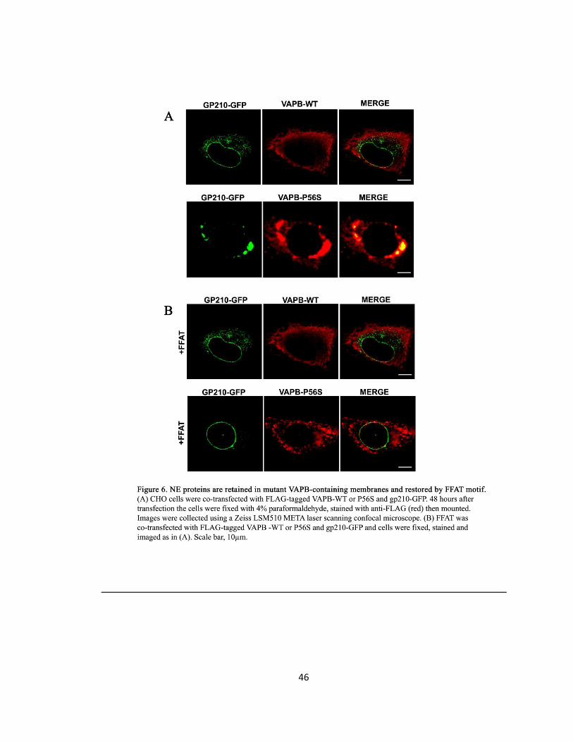
46
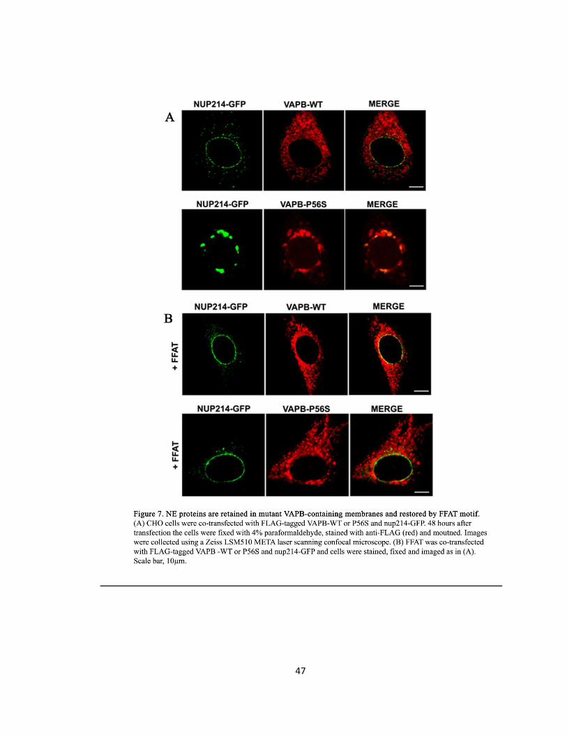
47
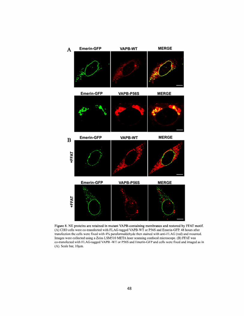
48
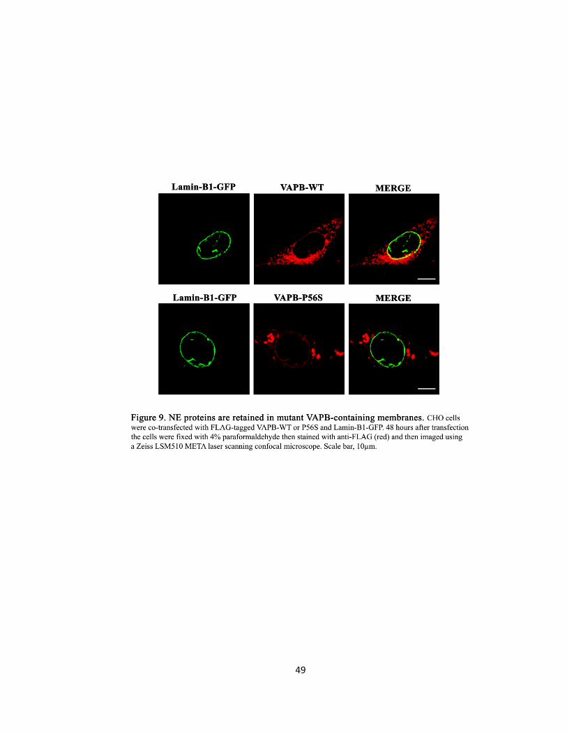
49
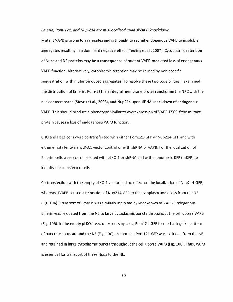
50
Emerin, Pom-121, and Nup-214 are mis-localized upon siVAPB knockdown
Mutant VAPB is prone to aggregates and is thought to recruit endogenous VAPB to insoluble
aggregates resulting in a dominant negative effect (Teuling et al., 2007). Cytoplasmic retention
of Nups and NE proteins may be a consequence of mutant VAPB-mediated loss of endogenous
VAPB function. Alternatively, cytoplasmic retention may be caused by non-specific
sequestration with mutant-induced aggregates. To resolve these two possibilities, I examined
the distribution of Emerin, Pom-121, an integral membrane protein anchoring the NPC with the
nuclear membrane (Stavru et al., 2006), and Nup214 upon siRNA knockdown of endogenous
VAPB. This should produce a phenotype similar to overexpression of VAPB-P56S if the mutant
protein causes a loss of endogenous VAPB function.
CHO and HeLa cells were co-transfected with either Pom121-GFP or Nup214-GFP and with
either empty lentiviral pLKO.1 vector control or with shRNA of VAPB. For the localization of
Emerin, cells were co-transfected with pLKO.1 or shRNA and with monomeric RFP (mRFP) to
identify the transfected cells.
Co-transfection with the empty pLKO.1 vector had no effect on the localization of Nup214-GFP,
whereas siVAPB caused a relocation of Nup214-GFP to the cytoplasm and a loss from the NE
(Fig. 10A). Transport of Emerin was similarly inhibited by knockdown of VAPB. Endogenous
Emerin was relocated from the NE to large cytoplasmic puncta throughout the cell upon siVAPB
(Fig. 10B). In the empty pLKO.1 vector expressing cells, Pom121-GFP formed a ring-like pattern
of punctate spots around the NE (Fig. 10C). In contrast, Pom121-GFP was excluded from the NE
and retained in large cytoplasmic puncta throughout the cell upon siVAPB (Fig. 10C). Thus, VAPB
is essential for transport of these Nups to the NE.
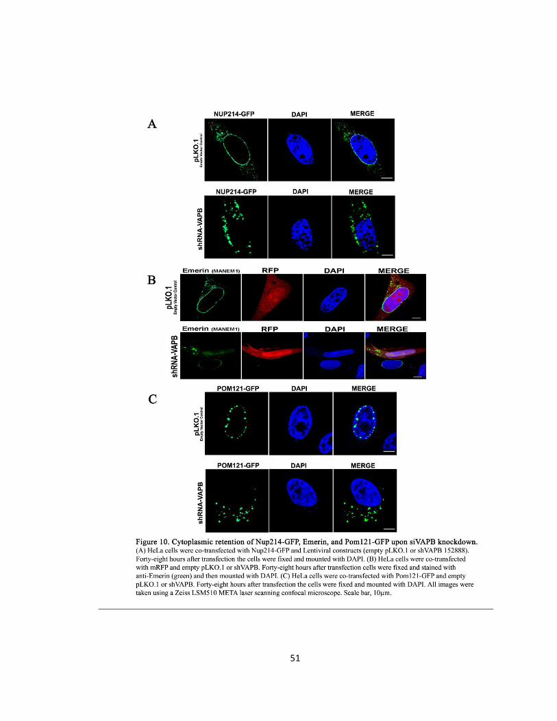
51
52
Characterizing the compartment in which INM and NPC proteins are retained
VAPB is localized to the ERGIC
It has been shown that anterograde ER-to-Golgi transport of VSVG is unaffected upon siVAPB
knockdown (Tran et al., 2012). Since the obtained results in this thesis demonstrated that Nups
and INM proteins are retained in the cytoplasm upon knockdown of VAPB, this suggests that
their transport is not via anterograde routes. Therefore, I examined retrograde transport routes.
To determine the cytoplasmic compartment in which Nups and NE proteins are retained in and
to identify if this compartment contains VAPB, I examined an organelle of the early secretory
pathway that displays retrograde movement: The Endoplasmic Reticulum Golgi Intermediate
Compartment (ERGIC). To address if this compartment contains VAPB, CHO and HeLa cells were
co-transfected with ERGIC-53-GFP and VAPB-WT.
Only cells with low level of VAPB expression were examined to avoid over-expression artifacts. I
found that VAPB-WT co-localized extensively with ERGIC-53-GFP cytoplasmic puncta (Fig. 11).
The Golgi ribbons lacked VAPB (Fig. 12); thus, VAPB resides primarily at the ERGIC. When
examined with intensity correlation analysis excluding the Golgi, VAPB-WT and ERGIC-53
showed a mean Pearson’s correlation of 0.73 ± 0.02 (n=20). VAPB-WT and ERGIC-53 also
showed a high Mander’s overlap coefficient of 0.79 ± 0.02 (n=20). These high correlations
suggest that VAPB primarily resides at the ERGIC.
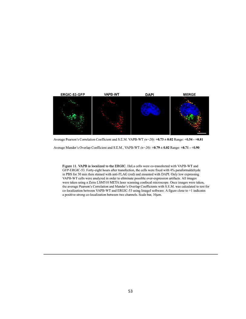
53
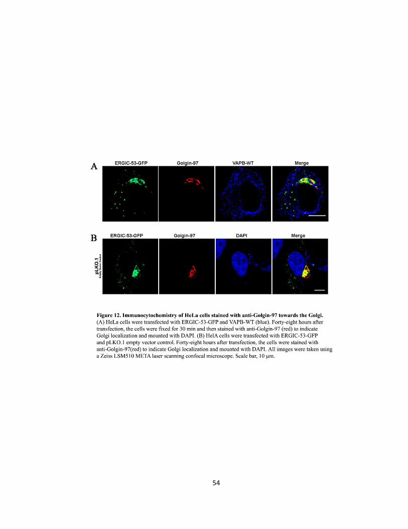
54
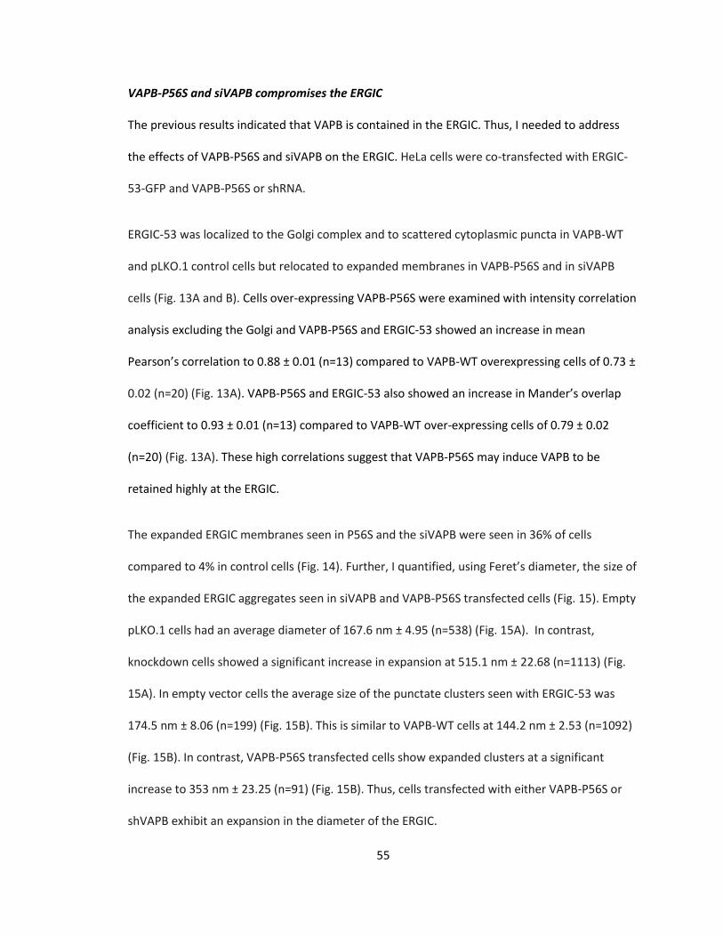
55
VAPB-P56S and siVAPB compromises the ERGIC
The previous results indicated that VAPB is contained in the ERGIC. Thus, I needed to address
the effects of VAPB-P56S and siVAPB on the ERGIC. HeLa cells were co-transfected with ERGIC-
53-GFP and VAPB-P56S or shRNA.
ERGIC-53 was localized to the Golgi complex and to scattered cytoplasmic puncta in VAPB-WT
and pLKO.1 control cells but relocated to expanded membranes in VAPB-P56S and in siVAPB
cells (Fig. 13A and B). Cells over-expressing VAPB-P56S were examined with intensity correlation
analysis excluding the Golgi and VAPB-P56S and ERGIC-53 showed an increase in mean
Pearson’s correlation to 0.88 ± 0.01 (n=13) compared to VAPB-WT overexpressing cells of 0.73 ±
0.02 (n=20) (Fig. 13A). VAPB-P56S and ERGIC-53 also showed an increase in Mander’s overlap
coefficient to 0.93 ± 0.01 (n=13) compared to VAPB-WT over-expressing cells of 0.79 ± 0.02
(n=20) (Fig. 13A). These high correlations suggest that VAPB-P56S may induce VAPB to be
retained highly at the ERGIC.
The expanded ERGIC membranes seen in P56S and the siVAPB were seen in 36% of cells
compared to 4% in control cells (Fig. 14). Further, I quantified, using Feret’s diameter, the size of
the expanded ERGIC aggregates seen in siVAPB and VAPB-P56S transfected cells (Fig. 15). Empty
pLKO.1 cells had an average diameter of 167.6 nm ± 4.95 (n=538) (Fig. 15A). In contrast,
knockdown cells showed a significant increase in expansion at 515.1 nm ± 22.68 (n=1113) (Fig.
15A). In empty vector cells the average size of the punctate clusters seen with ERGIC-53 was
174.5 nm ± 8.06 (n=199) (Fig. 15B). This is similar to VAPB-WT cells at 144.2 nm ± 2.53 (n=1092)
(Fig. 15B). In contrast, VAPB-P56S transfected cells show expanded clusters at a significant
increase to 353 nm ± 23.25 (n=91) (Fig. 15B). Thus, cells transfected with either VAPB-P56S or
shVAPB exhibit an expansion in the diameter of the ERGIC.
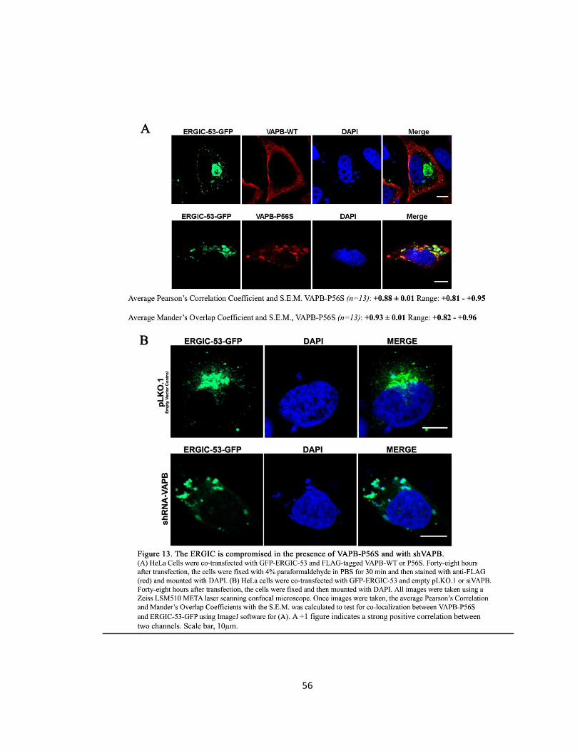
56

57
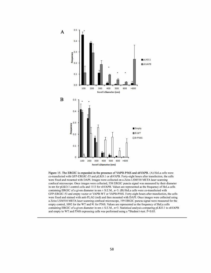
58
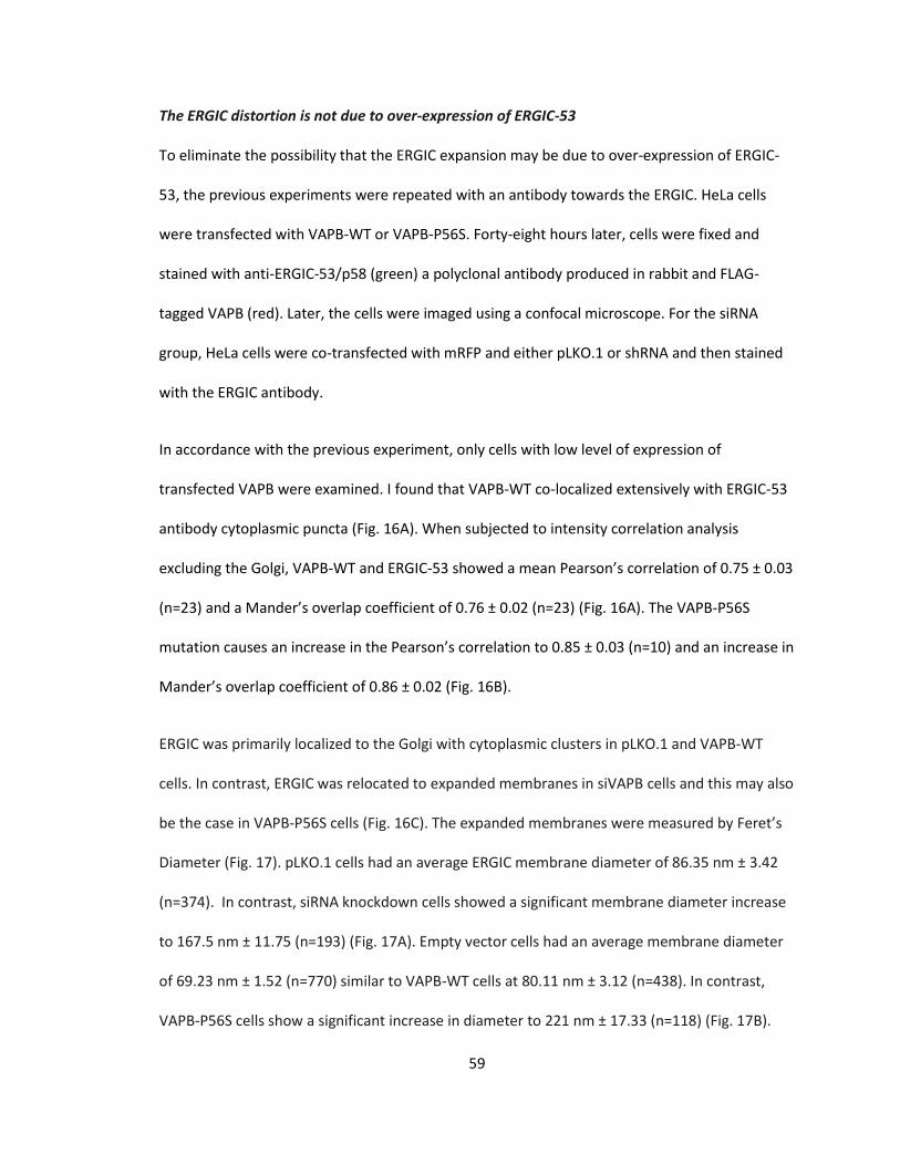
59
The ERGIC distortion is not due to over-expression of ERGIC-53
To eliminate the possibility that the ERGIC expansion may be due to over-expression of ERGIC-
53, the previous experiments were repeated with an antibody towards the ERGIC. HeLa cells
were transfected with VAPB-WT or VAPB-P56S. Forty-eight hours later, cells were fixed and
stained with anti-ERGIC-53/p58 (green) a polyclonal antibody produced in rabbit and FLAG-
tagged VAPB (red). Later, the cells were imaged using a confocal microscope. For the siRNA
group, HeLa cells were co-transfected with mRFP and either pLKO.1 or shRNA and then stained
with the ERGIC antibody.
In accordance with the previous experiment, only cells with low level of expression of
transfected VAPB were examined. I found that VAPB-WT co-localized extensively with ERGIC-53
antibody cytoplasmic puncta (Fig. 16A). When subjected to intensity correlation analysis
excluding the Golgi, VAPB-WT and ERGIC-53 showed a mean Pearson’s correlation of 0.75 ± 0.03
(n=23) and a Mander’s overlap coefficient of 0.76 ± 0.02 (n=23) (Fig. 16A). The VAPB-P56S
mutation causes an increase in the Pearson’s correlation to 0.85 ± 0.03 (n=10) and an increase in
Mander’s overlap coefficient of 0.86 ± 0.02 (Fig. 16B).
ERGIC was primarily localized to the Golgi with cytoplasmic clusters in pLKO.1 and VAPB-WT
cells. In contrast, ERGIC was relocated to expanded membranes in siVAPB cells and this may also
be the case in VAPB-P56S cells (Fig. 16C). The expanded membranes were measured by Feret’s
Diameter (Fig. 17). pLKO.1 cells had an average ERGIC membrane diameter of 86.35 nm ± 3.42
(n=374). In contrast, siRNA knockdown cells showed a significant membrane diameter increase
to 167.5 nm ± 11.75 (n=193) (Fig. 17A). Empty vector cells had an average membrane diameter
of 69.23 nm ± 1.52 (n=770) similar to VAPB-WT cells at 80.11 nm ± 3.12 (n=438). In contrast,
VAPB-P56S cells show a significant increase in diameter to 221 nm ± 17.33 (n=118) (Fig. 17B).
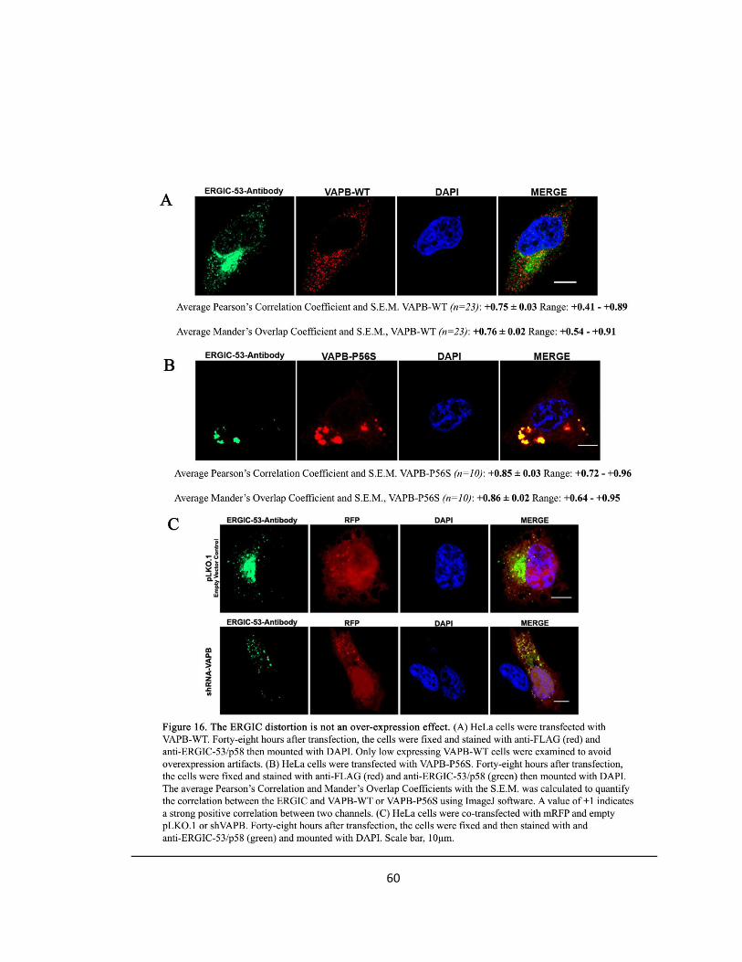
60
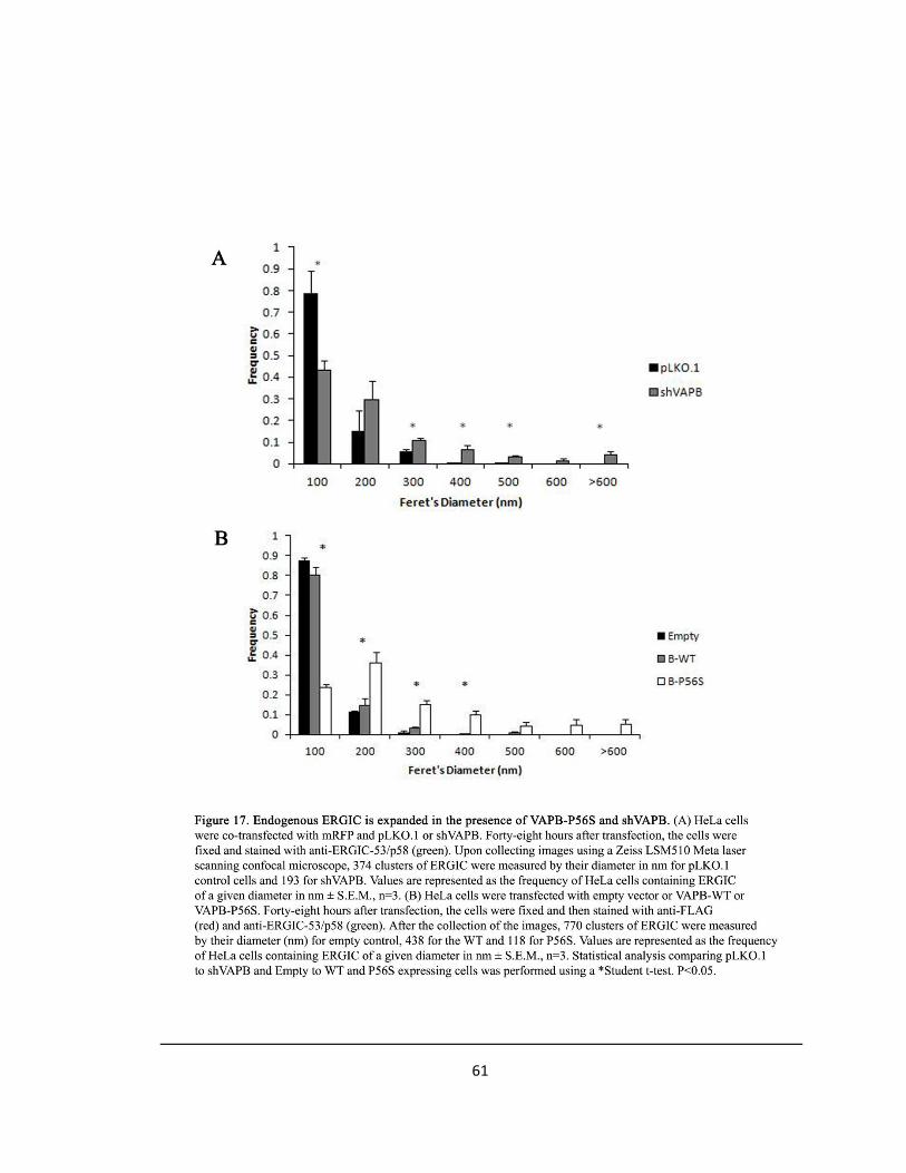
61
62
Emerin, Mab414 and Nup214-RFP Co-localize with ERGIC-53
Previous results indicated that VAPB co-localized with the ERGIC. The results also indicated that
the ERGIC is expanded in the presence of P56S and upon siRNA knockdown of endogenous
VAPB. I hypothesize that the expansion may be the result of accumulated proteins transported
to and retained at the ERGIC due to loss of VAPB retrograde transport. To address if Nups are
transported to the ERGIC, I co-transfected cells with either pLKO.1 and ERGIC-53-GFP and
stained for Mab414 or for Emerin or co-transfected cells with Nup214-RFP to undergo a
correlation analysis with ERGIC-53.
ERGIC-53 primarily resides at the Golgi (Fig. 12); however, there are punctate inclusions located
in the cytoplasm. These punctate inclusions are believed to be the ERGIC where proteins are
transported. Emerin, Nup214 and Mab414 were tested to see if they co-localized with these
punctate inclusions in the cytoplasm. When subjected to intensity correlation analysis excluding
the Golgi, Emerin and ERGIC-53 showed an average Pearson’s correlation coefficient of 0.49 ±
0.02 (n=24) and a Mander’s overlap coefficient of 0.68 ± 0.02 (n=24) (Fig. 18A). Nup214-RFP
average a Person’s correlation coefficient of 0.47 ± 0.04 (n=18) and a Mander’s overlap
coefficient of 0.79 ± 0.03 (n=18) (Fig. 19A). Lastly, Mab414 averaged a Pearson’s correlation
coefficient of 0.57 ± 0.04 (n=17) and a Mander’s overlap coefficient of 0.73 ± 0.03(n=17) (Fig.
20A). These figures indicate that NE proteins and Nups are transported to the ERGIC via
vesicular transport prior to the NE.
Furthermore, the ERGIC-53-GFP, Emerin and Mab414 punctate inclusions in the cytoplasm were
measured by their Feret’s diameter in nm for pLKO.1 control expression cells. Punctate inclusion
that were stained specifically with endogenous Emerin had an average diameter of 100.8 nm ±
1.68 (n=1524) (Fig. 21A). Mab414 stained pLKO.1 control cells had cytoplasmic inclusions with
63
an average diameter of 128 nm ± 2.3 (1683) (Fig. 21B). These figures are very close the normal
diameter of the ERGIC compartment previously examined under VAPB-WT and ERGIC-53
conditions (see Fig. 15 or 17).
Emerin, Mab414 and Nup-214 are retained at the ERGIC upon shVAPB
The previous results indicated that certain Nups and INM proteins are transported to the ERGIC.
This raised the question: Can this be the compartment in which the proteins are retained in
because of the loss of VAPB? To verify that Nups are retained at the ERGIC in a VAPB-
dependent manner, I stained VAPB knockdown cells with Emerin or Mab414 or co-transfected
cells with Nup214-RFP to determine if these proteins are retained at the expanded ERGIC
membranes.
Emerin is localized to the NE and had small cytoplasmic puncta in control cells, but is relocated
to the expanded ERGIC along with ERGIC-53 in siVAPB cells (Fig. 18B). When subjected to
intensity correlation analysis excluding the Golgi, Emerin and ERGIC-53 showed an average
Pearson’s correlation coefficient of 0.89 ± 0.01 (n=30) and a Mander’s overlap coefficient of 0.91
± 0.01 (n=30) (Fig. 18B). Nup214 was also seen in small cytoplasmic puncta in control cells and
localized to the NE, but was relocated to the expanded ERGIC upon siVAPB (Fig. 19B). Nup214-
RFP averaged a Pearson’s correlation coefficient of 0.88 ± 0.01 (n=21) and a Mander’s overlap
coefficient of 0.94 ± 0.01 (n=21) (Fig. 19B). Mab414 was localized to the rim of the NE and in
small cytoplasmic puncta in control cells, but was retained in expanded ERGIC-containing
membranes upon siVAPB (Fig. 20B). Mab414 averaged a Pearson’s correlation of 0.79 ± 0.02
(n=14) and a Mander’s overlap coefficient of 0.83 ± 0.01 (n=14) (Fig. 20B). Emerin, Nup214 and
Mab414 all showed an increase in correlation with the ERGIC upon knockdown of endogenous
VAPB compared to control cells. Thus, NE proteins and Nups transit through the ERGIC and are
retained in the ERGIC resulting in the accumulation of proteins, the expansion of the ERGIC and
64
an increase in correlation. VAPB is essential in NE and NPC protein transport to the NE.
Furthermore, under knockdown of endogenous VAPB the ERGIC-53-GFP, Emerin and Mab414
punctate inclusions in the cytoplasm illustrated a shift towards larger ERGIC-53 puncta based on
their Feret’s diameter. Punctate inclusion that were stained specifically with endogenous Emerin
under shVAPB conditions had an average diameter of 191.17 nm ± 3.98 (n=1206) compared to
control cells with 100.8 nm ± 1.68 (n=1524) (Fig. 21A). Mab414 stained shVAPB control cells
averaged a diameter of cytoplasmic inclusions of 182.62 nm ± 5.09 (n=825) compared to control
cells with 128 nm ± 2.3 (1683) (Fig. 21B). These figures are very close the expanded diameter
obtained previously under VAPB-P56S or shVAPB and ERGIC-53 conditions (see Fig. 15 or 17).
Thus, quantitation of Emerin and Mab414 cytoplasmic puncta indicated a shift towards larger
puncta in knockdown cells (Fig. 21). Together, this indicates that NE and transmembrane Nups
transit through ERGIC, and VAPB is required for final transport to the NE.
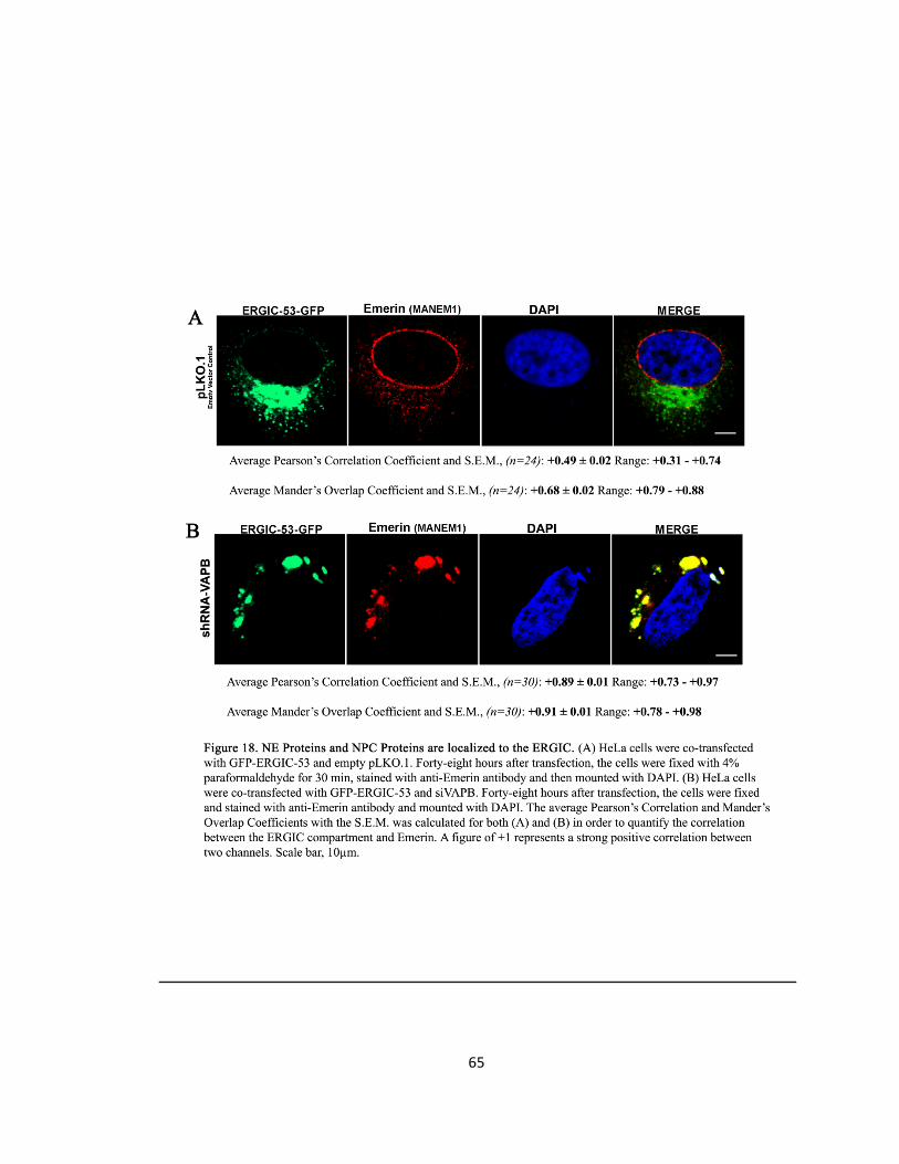
65
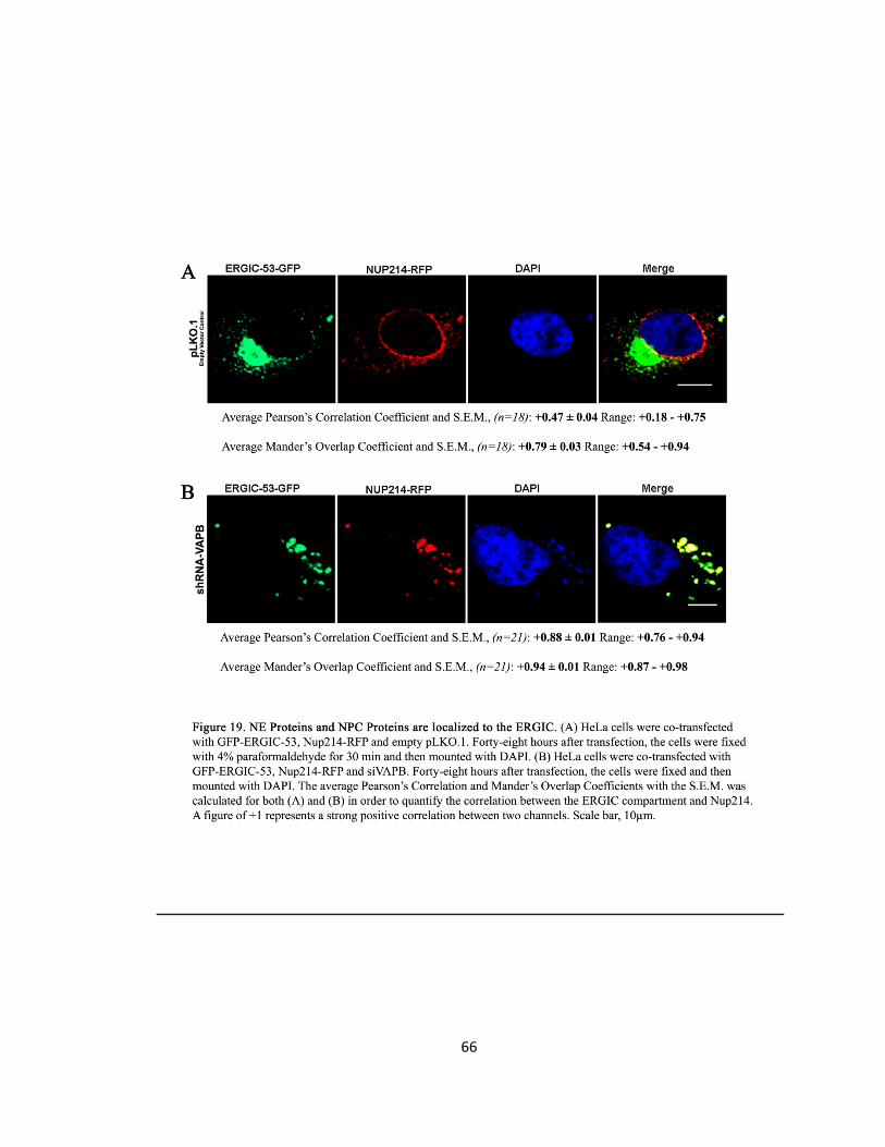
66
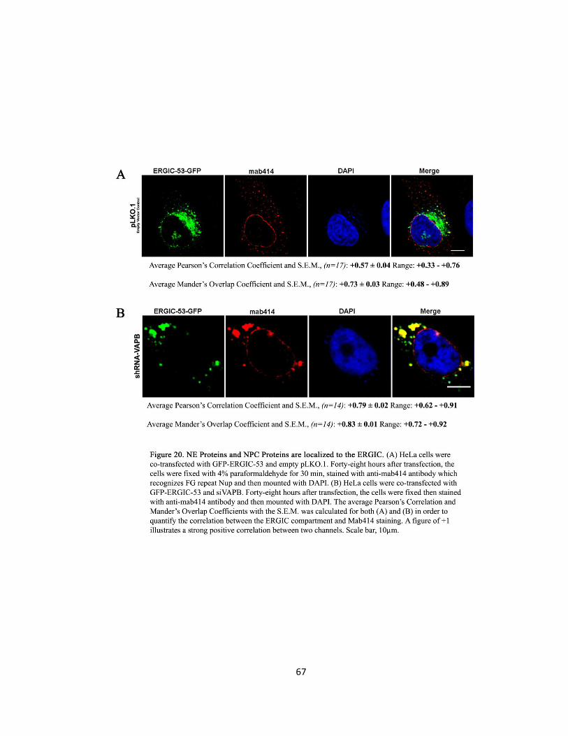
67
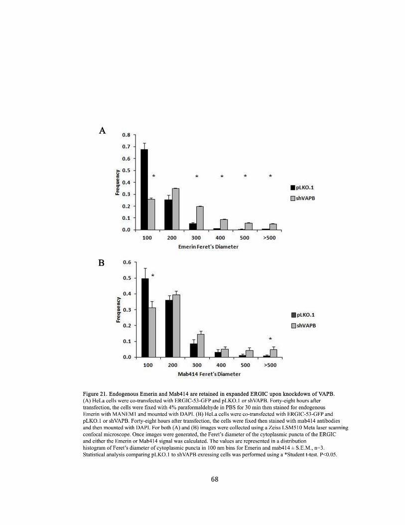
68
69
ERGIC-53 Accumulates in the ERGIC at 15°C and Retains Emerin
Incubation at 15°C is known to reversibly accumulate ERGIC-53 in the ERGIC by blocking
retrograde transport to the ER (Hauri et al., 2000; Ben-Tekaya et al 2005). Thus, I hypothesize
that if NE proteins are transported via vesicular transport to the ERGIC and then to the NE, NE
proteins will be retained in expanded ERGIC membranes due to lack of retrograde transport at
15°C. Cells were transfected with ERGIC-53-GFP and forty-eight hours later were transferred
from 37°C to 15°C from 0-8 hours. At each hour, cells were fixed and stained for endogenous
emerin. Since the ERGIC will expanded at 15°C, I propose that Emerin should be retained at the
ERGIC.
In control cells at time 0 hours and 37°C, ERGIC-53 localized to the Golgi and in punctate staining
throughout the cytoplasm. Emerin formed a ring-like pattern along the NE (Fig. 22). In contrast,
at 15°C for 4 hours, Emerin was retained in dilated cytoplasmic membranes and excluded from
the NE (Fig. 22). The ERGIC was also seen to lose the cytoplasmic puncta throughout the cell and
formed expanded membrane compartments in the cytoplasm that also contained Emerin (Fig.
22). This suggests that Emerin is transported from the ER to the ERGIC and then to the NE by
vesicular transport that requires VAPB.
This set of data illustrates Emerins transport route independent of VAPB-P56S and knockdown
of endogenous VAPB effects. This clearly depicts that Emerin is transported to the ERGIC before
the NE. Due to the loss of retrograde transport at 15°C, Emerin is retained at the ERGIC. Thus,
VAPB-P56S or shVAPB loss of VAPB retrograde transport may be the reason why Emerin is
trapped at the ERGIC.
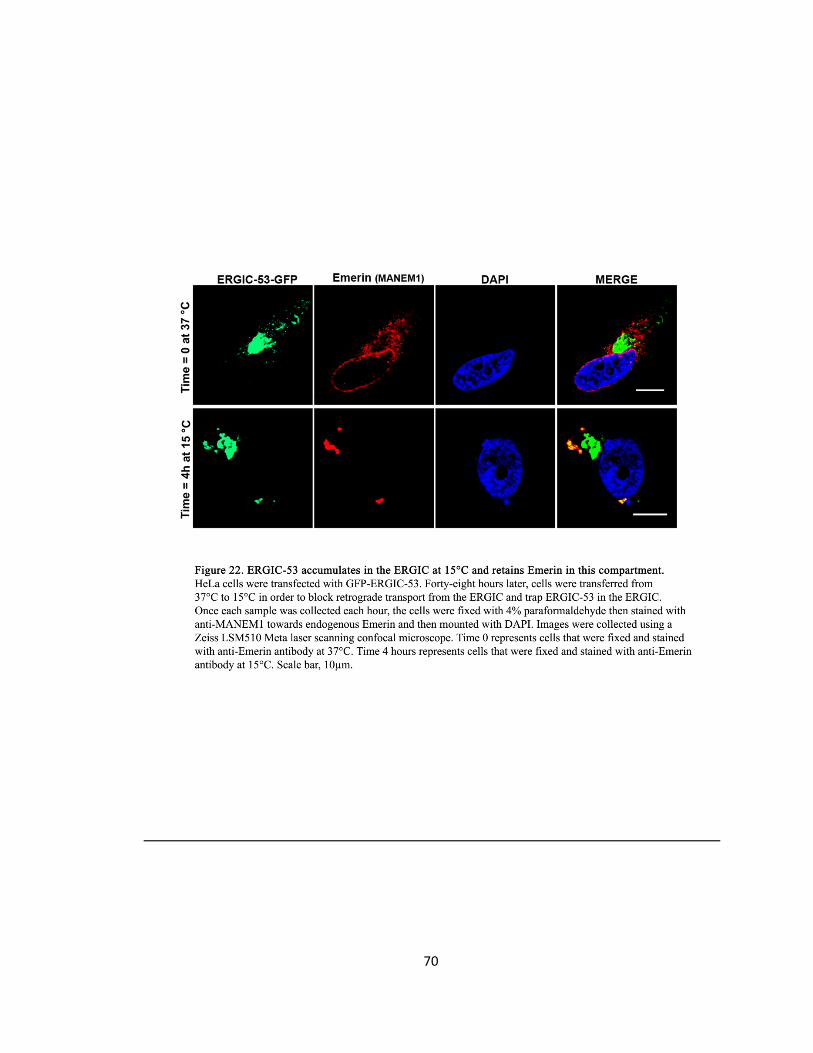
70
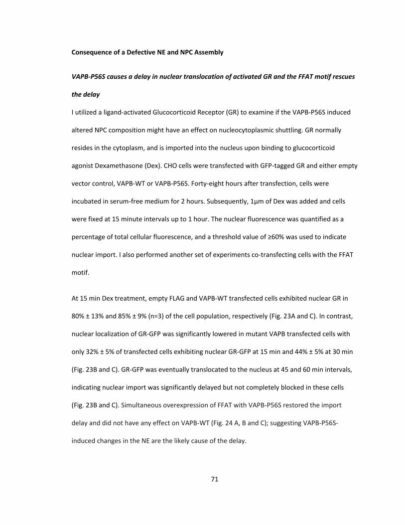
71
Consequence of a Defective NE and NPC Assembly
VAPB-P56S causes a delay in nuclear translocation of activated GR and the FFAT motif rescues
the delay
I utilized a ligand-activated Glucocorticoid Receptor (GR) to examine if the VAPB-P56S induced
altered NPC composition might have an effect on nucleocytoplasmic shuttling. GR normally
resides in the cytoplasm, and is imported into the nucleus upon binding to glucocorticoid
agonist Dexamethasone (Dex). CHO cells were transfected with GFP-tagged GR and either empty
vector control, VAPB-WT or VAPB-P56S. Forty-eight hours after transfection, cells were
incubated in serum-free medium for 2 hours. Subsequently, 1µm of Dex was added and cells
were fixed at 15 minute intervals up to 1 hour. The nuclear fluorescence was quantified as a
percentage of total cellular fluorescence, and a threshold value of ≥60% was used to indicate
nuclear import. I also performed another set of experiments co-transfecting cells with the FFAT
motif.
At 15 min Dex treatment, empty FLAG and VAPB-WT transfected cells exhibited nuclear GR in
80% ± 13% and 85% ± 9% (n=3) of the cell population, respectively (Fig. 23A and C). In contrast,
nuclear localization of GR-GFP was significantly lowered in mutant VAPB transfected cells with
only 32% ± 5% of transfected cells exhibiting nuclear GR-GFP at 15 min and 44% ± 5% at 30 min
(Fig. 23B and C). GR-GFP was eventually translocated to the nucleus at 45 and 60 min intervals,
indicating nuclear import was significantly delayed but not completely blocked in these cells
(Fig. 23B and C). Simultaneous overexpression of FFAT with VAPB-P56S restored the import
delay and did not have any effect on VAPB-WT (Fig. 24 A, B and C); suggesting VAPB-P56S-
induced changes in the NE are the likely cause of the delay.
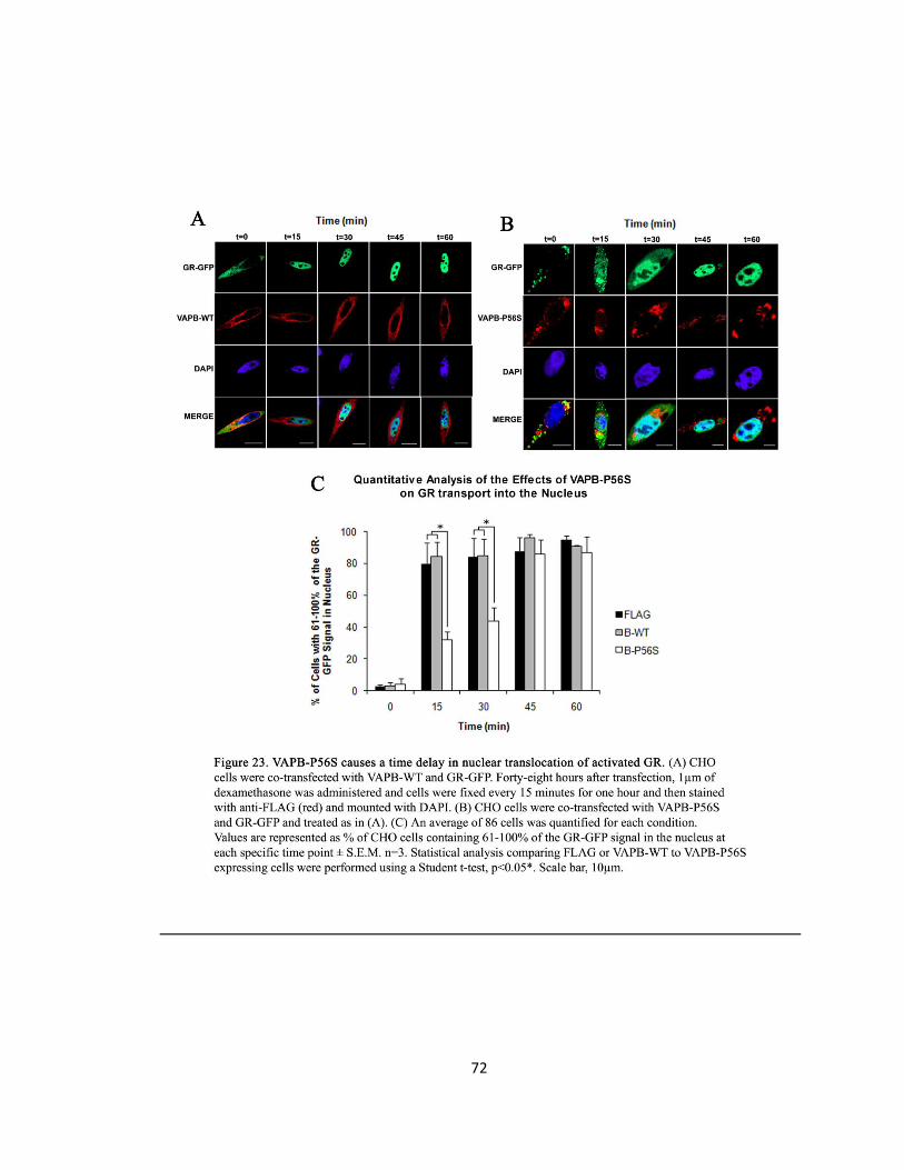
72
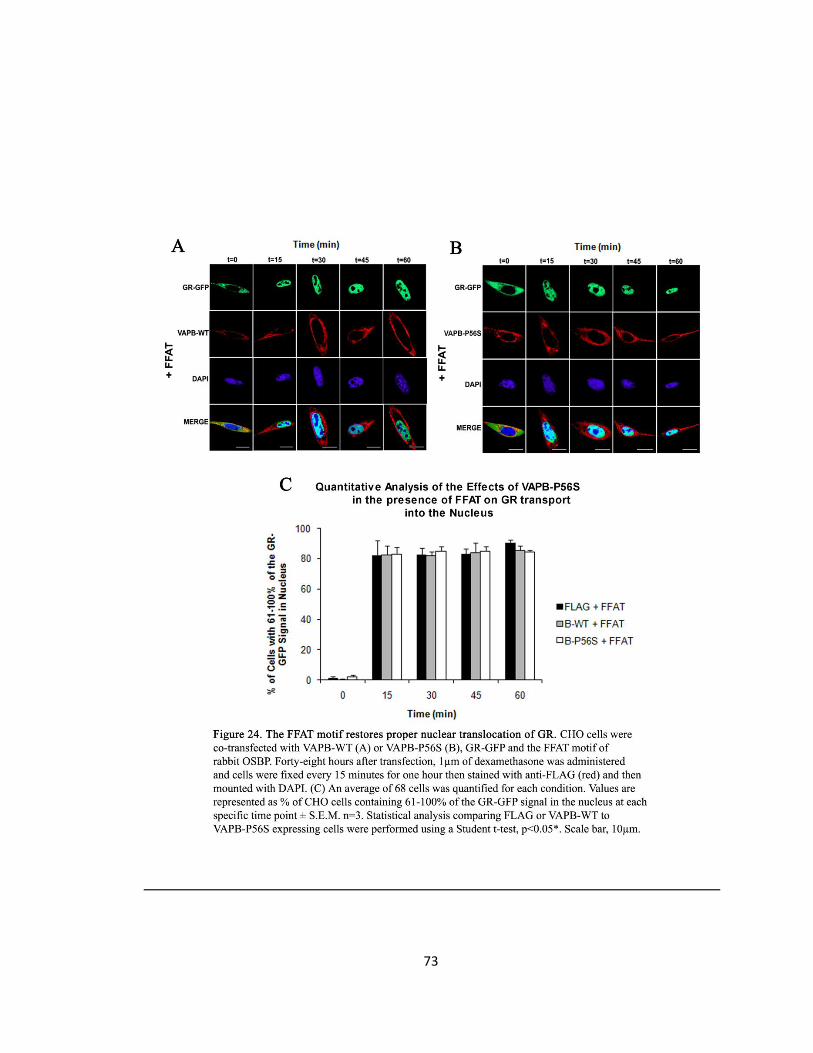
73
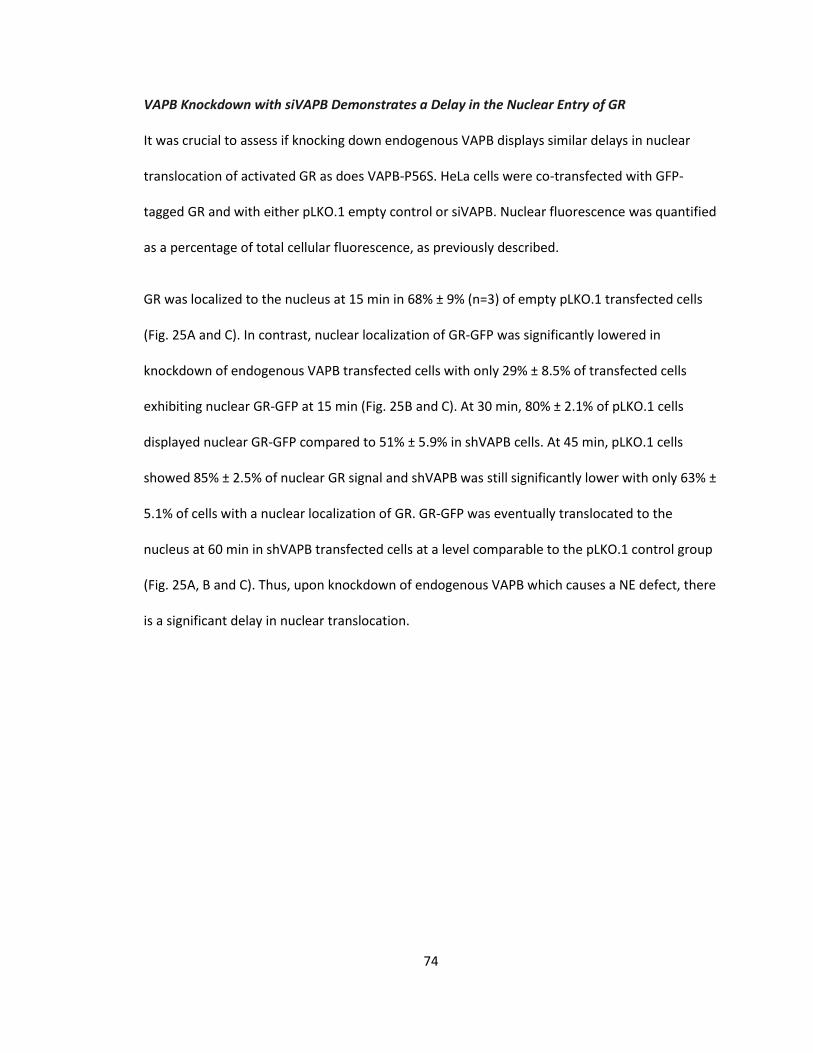
74
VAPB Knockdown with siVAPB Demonstrates a Delay in the Nuclear Entry of GR
It was crucial to assess if knocking down endogenous VAPB displays similar delays in nuclear
translocation of activated GR as does VAPB-P56S. HeLa cells were co-transfected with GFP-
tagged GR and with either pLKO.1 empty control or siVAPB. Nuclear fluorescence was quantified
as a percentage of total cellular fluorescence, as previously described.
GR was localized to the nucleus at 15 min in 68% ± 9% (n=3) of empty pLKO.1 transfected cells
(Fig. 25A and C). In contrast, nuclear localization of GR-GFP was significantly lowered in
knockdown of endogenous VAPB transfected cells with only 29% ± 8.5% of transfected cells
exhibiting nuclear GR-GFP at 15 min (Fig. 25B and C). At 30 min, 80% ± 2.1% of pLKO.1 cells
displayed nuclear GR-GFP compared to 51% ± 5.9% in shVAPB cells. At 45 min, pLKO.1 cells
showed 85% ± 2.5% of nuclear GR signal and shVAPB was still significantly lower with only 63% ±
5.1% of cells with a nuclear localization of GR. GR-GFP was eventually translocated to the
nucleus at 60 min in shVAPB transfected cells at a level comparable to the pLKO.1 control group
(Fig. 25A, B and C). Thus, upon knockdown of endogenous VAPB which causes a NE defect, there
is a significant delay in nuclear translocation.
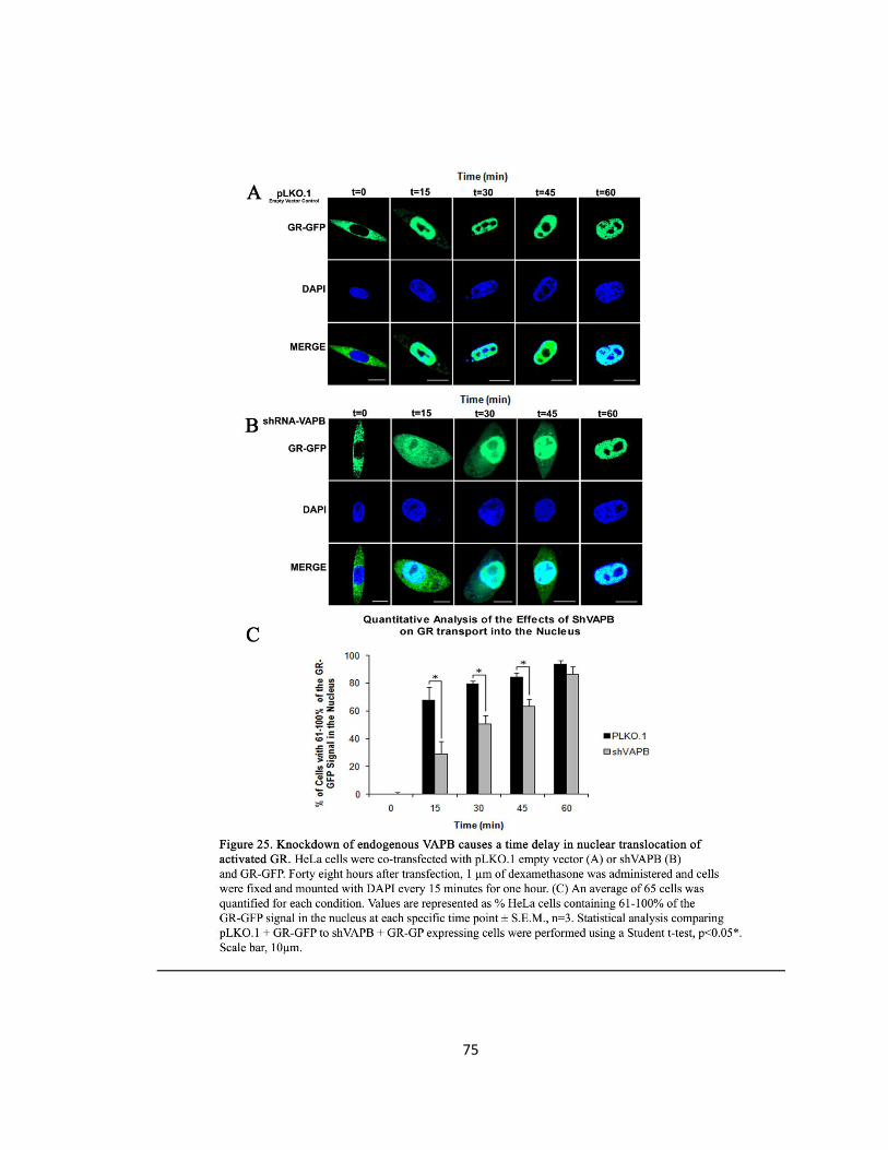
75
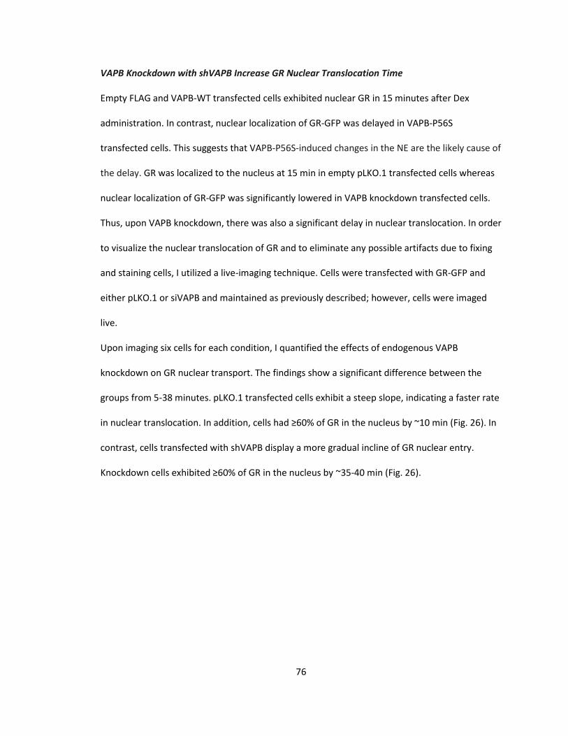
76
VAPB Knockdown with shVAPB Increase GR Nuclear Translocation Time
Empty FLAG and VAPB-WT transfected cells exhibited nuclear GR in 15 minutes after Dex
administration. In contrast, nuclear localization of GR-GFP was delayed in VAPB-P56S
transfected cells. This suggests that VAPB-P56S-induced changes in the NE are the likely cause of
the delay. GR was localized to the nucleus at 15 min in empty pLKO.1 transfected cells whereas
nuclear localization of GR-GFP was significantly lowered in VAPB knockdown transfected cells.
Thus, upon VAPB knockdown, there was also a significant delay in nuclear translocation. In order
to visualize the nuclear translocation of GR and to eliminate any possible artifacts due to fixing
and staining cells, I utilized a live-imaging technique. Cells were transfected with GR-GFP and
either pLKO.1 or siVAPB and maintained as previously described; however, cells were imaged
live.
Upon imaging six cells for each condition, I quantified the effects of endogenous VAPB
knockdown on GR nuclear transport. The findings show a significant difference between the
groups from 5-38 minutes. pLKO.1 transfected cells exhibit a steep slope, indicating a faster rate
in nuclear translocation. In addition, cells had ≥60% of GR in the nucleus by ~10 min (Fig. 26). In
contrast, cells transfected with shVAPB display a more gradual incline of GR nuclear entry.
Knockdown cells exhibited ≥60% of GR in the nucleus by ~35-40 min (Fig. 26).
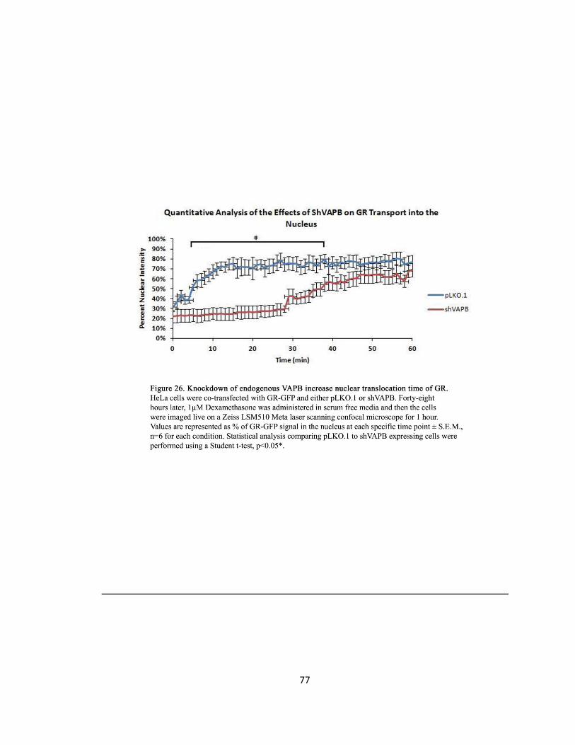
77
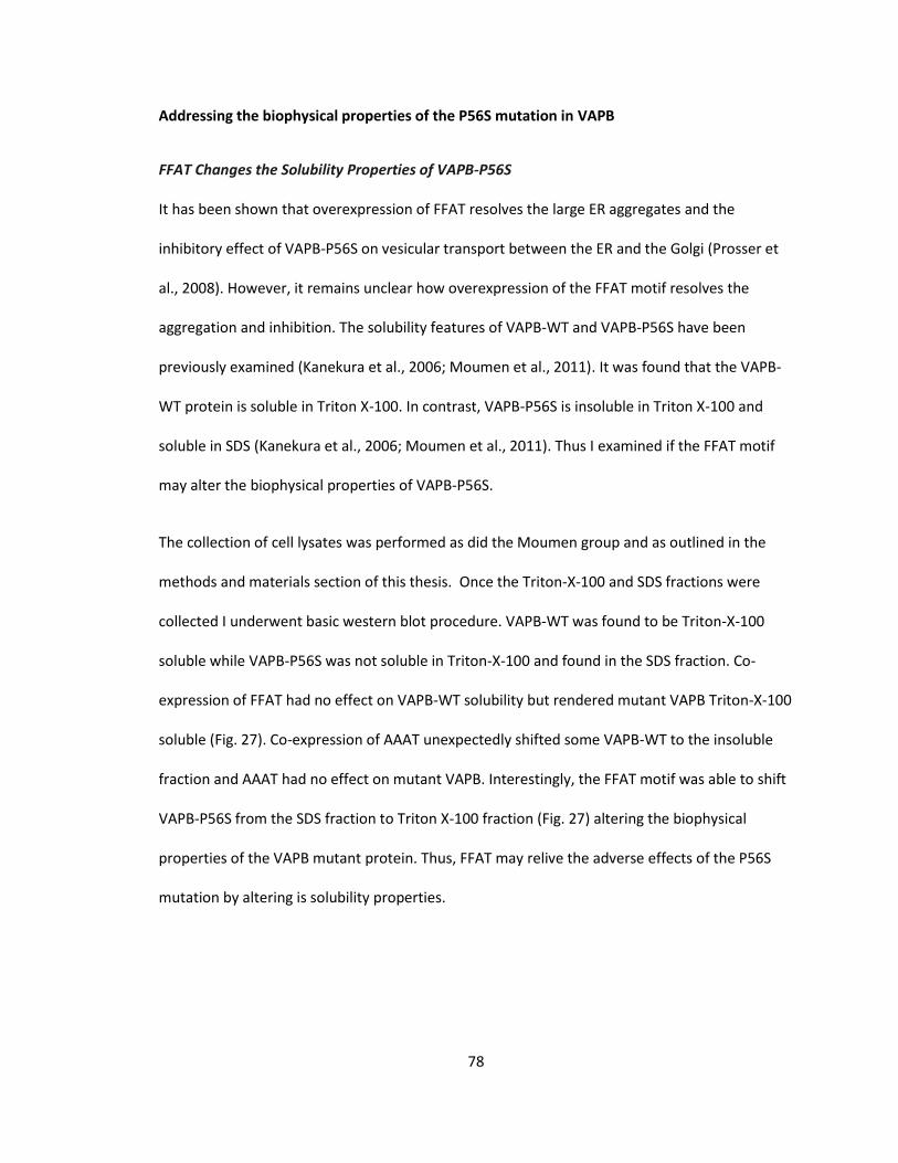
78
Addressing the biophysical properties of the P56S mutation in VAPB
FFAT Changes the Solubility Properties of VAPB-P56S
It has been shown that overexpression of FFAT resolves the large ER aggregates and the
inhibitory effect of VAPB-P56S on vesicular transport between the ER and the Golgi (Prosser et
al., 2008). However, it remains unclear how overexpression of the FFAT motif resolves the
aggregation and inhibition. The solubility features of VAPB-WT and VAPB-P56S have been
previously examined (Kanekura et al., 2006; Moumen et al., 2011). It was found that the VAPB-
WT protein is soluble in Triton X-100. In contrast, VAPB-P56S is insoluble in Triton X-100 and
soluble in SDS (Kanekura et al., 2006; Moumen et al., 2011). Thus I examined if the FFAT motif
may alter the biophysical properties of VAPB-P56S.
The collection of cell lysates was performed as did the Moumen group and as outlined in the
methods and materials section of this thesis. Once the Triton-X-100 and SDS fractions were
collected I underwent basic western blot procedure. VAPB-WT was found to be Triton-X-100
soluble while VAPB-P56S was not soluble in Triton-X-100 and found in the SDS fraction. Co-
expression of FFAT had no effect on VAPB-WT solubility but rendered mutant VAPB Triton-X-100
soluble (Fig. 27). Co-expression of AAAT unexpectedly shifted some VAPB-WT to the insoluble
fraction and AAAT had no effect on mutant VAPB. Interestingly, the FFAT motif was able to shift
VAPB-P56S from the SDS fraction to Triton X-100 fraction (Fig. 27) altering the biophysical
properties of the VAPB mutant protein. Thus, FFAT may relive the adverse effects of the P56S
mutation by altering is solubility properties.
79
80
DISCUSSION
Characterizing the Functional Role of VAPB in Transport of INM and NPC Proteins
Gp210, Nup214, and Emerin are mis-localized in VAPB-P56S Transfected Cells
The current study follows the discovery that overexpression of VAPB-P56S results in the
separation of the INM and ONM of the NE (Tran et al., 2012). EM images of CHO cells
transfected with VAPB-P56S show enlarged ER structures and a nucleus with areas of separation
between the ONM and INM of the NE (see Fig. 5). This led me to believe there is a NE defect and
suggests there may be a defect in the transport of proteins required for proper NPC assembly.
Since the NPC structurally contributes to the NE bilayers close apposition, deterioration in NPC
assembly and recruitment of proteins can ultimately lead to the separation and dilation of the
NE bilayers. This suggests that the mechanism responsible for the loss of motor neurons could
be due to defects at the NE.
EM images revealed that overexpression of VAPB-P56S, among its other functional deficits,
induces a change in the structure of the NE resulting in the separation of the INM and ONM.
This separation could be the result of the mis-localization of proteins in the nuclear lamina on
the INM or from the transport defects of Nups needed to anchor the NPC to the NE to hold the
tight apposition between the lipid bilayers. These questions were examined by employing
fluorescent microscopy to test the localization of NPC and NE proteins in VAPB-P56S expressing
cells. The findings in this thesis suggest that VAPB is involved in NPC and NE protein transport
from the ER to the NE and the FFAT motif can counteract the mutant’s adverse effects by
restoring proper protein localization. Gp210 has been proposed to play a role in fusing the ONM
and INM at the pore. To date, Gp210, Pom121 and Ndc1 are the three integral membrane
proteins of the NPC which anchor the NPC to the NE (Antonin et al., 2005). Gp210 is a type 1
integral protein of the NPC with a single membrane spanning region close to the C-terminus of
81
the protein (Antonin et al., 2005). This study showed that in VAPB-WT expressing cells Gp210
forms a ring-like pattern encompassing the rim of the NE as well as localized to scattered puncta
(see Fig. 6A). In contrast, Gp210 was excluded from the NE and retained in mutant VAPB dilated
membranes in the cytoplasm (Fig. 6A). These results are consistent with the claims that a defect
in nuclear envelope has been reported in sporadic ALS cases (Kinoshita et al., 2009). The loss of
the integral membrane protein of the NPC, Gp210, may result in the dilation of the NE and be
manifested by irregular nuclear contours.
The separation of the INM and ONM and the expansion of ER tubules in mutant VAPB
expressing cells could affect insertion of NPCs leading to a decrease in the number of NPCs on
the NE. The loss of NPC from the NE could result in the specific death of non-dividing cells such
as motor neurons in ALS8. Hetzer’s group examined the rejuvenation of NPC proteins and their
study proposed that in non-dividing cells the scaffold nucleoporins (Nups) which make up the
nuclear pore are downregulated and do not renew (D’Angelo et al., 2009). On the other hand,
peripheral Nups can be exchanged in time (D’Angelo et al., 2009). This implies that the vitality of
Nups localized to the scaffold of the nuclear pore is crucial for the survival and proper function
of the NPC. Since scaffold proteins do not renew, they are more prone to oxidative damage
(D’Angelo et al., 2009). This can result in the loss of the specific permeability of the NE and can
induce cytoplasmic proteins to leak into the nucleus (D’Angelo et al., 2009). If this mechanism
leads to cell death in ALS8 due to the loss of NPC integrity then the leakiness of the nucleus can
result in a toxic gain of function.
To determine if other Nups are affected upon expressing VAPB-P56S, I examined Nup214, a Nup
confined to the cytoplasmic face of the NPC (Xu et al., 2009). In a similar fashion to Gp210,
Nup214-GFP was localized to the NE in empty vector and VAPB-WT transfected cells, but
82
sequestered in mutant VAPB containing aggregates (see Fig 7A). Gp210 and Nup214 are
retained in the cytoplasmic foci along with aggregated mutant VAPB, suggesting mutant VAPB
inhibits their transport to the NE.
To determine if proteins associated with the architectural and structure support system of the
NE are affected, I examined the localization of Emerin and Lamin-b1 in cells overexpressing
VAPB-P56S. Lamin-B1 stabilizes the NE and anchors nuclear membrane proteins to the scaffold
of the INM (Holaska et al., 2006). Emerin is an integral protein of the INM expressed in almost all
human cells (Holaska et al., 2006). Emerin binds to Lamins, anchoring Emerin for architectural,
gene regulator and nuclear assembly roles (Holaska et al., 2006). Emerin binds to and co-
localizes with A and B-type Lamins in the INM (Holaska et al., 2006). I was able to show that
Emerin-GFP forming a ring-like pattern along the rim of the NE in both control and VAPB-WT
overexpressing cells (Fig 8A). In contrast, Emerin was retained in dilated cytoplasmic
membranes and excluded from the NE in mutant VAPB overexpressing cells (Fig. 8A). This effect
is not cell line specific as this was seen in CHO and HeLa cells. Lamin-B1, however, was not
affected by the cytoplasmic aggregates created by overexpressing VAPB-P56S (see Fig. 9). In
VAPB-WT and control cells, Lamin-B1 formed a stable ring-like pattern around the NE which was
not distorted by mutant VAPB (see Fig. 9). Emerin is retained in the INM in part by binding to
Lamins in the nuclear lamina. Thus, Lamin-B1 was examined to address whether Emerin
transport was affected or if its retention to the Lamin scaffold was altered causing Emerin to
accumulate in the cytoplasm upon overexpression of VAPB-P56S (Vaughan et al., 2001).
Examining the distribution of Lamin-B1 showed no change in distribution pattern or evidence of
nuclear deformation. Therefore, the loss of Emerin from the INM is likely due to disruption of
transport to the INM rather than loss of retention at the INM.
83
The loss of NPC proteins and INM proteins suggests that NPC production and insertion into the
NE is decreased. In addition, the lack of newly produced scaffold Nups and the lack of transport
of newly synthesized peripheral Nups from the ER to the NE in motor neurons in VAPB-P56S
expressing cells predicts that these cells would have less NPCs. VAPB-P56S and knockdown of
VAPB illustrates that VAPB is vital in the transport of Nups and NE proteins from the ER to the
NE. Without VAPB assisted transport, the already inserted NPCs would not be able to synthesize
new proteins and with time, age-deterioration of the NPC will make neuronal cells vulnerable to
protein damage and cell death (D’Angelo et al., 2009). This can explain the specific target
towards motor neurons and the late-onset nature of ALS8.
Leaky nuclear pores could contribute to cell death as suggested by D’Angelo et al. (2009) which
showed how aging accelerated the leakiness of the nucleus (D’Angelo et al., 2009). This study
examined the effect of oxidative damage and age-related deterioration on NPC. They utilized
the environmental toxin Paraquat to induce oxidative damage by increase the amount of
reactive oxygen species (ROS) in Caenorhabditis elegans for six days (D’Angelo et al., 2009). They
found that paraquat treated worms had greater amounts of the 70 kDa dextran in their nucleus
compared to controls and dextran is too great a size to transport through an intact NPC. They
also showed that the nuclear permeability of older worms was deteriorating compared to
younger worms. In addition, this permeability deterioration was accelerated with paraquat
treatment (D’Angelo et al., 2009). Age-related deterioration of NPCs leads to the loss of the NPC
selective permeability barrier (D’Angelo et al., 2009). Altogether, expression of VAPB-P56S
altered NE morphology and NPC composition affecting its permeability and can explain for the
preferential vulnerability of motor neurons to premature death and the late-onset of ALS8.
84
The FFAT Motif Rescues Mis-Localized NE and NPC Proteins of VAPB-P56S
The simultaneous overexpression of the FFAT-motif with mutant VAPB resolved the abnormal
ER morphology and restored ER-Golgi trafficking (Prosser et al., 2008). Thus, I addressed
whether this could resolve the effects of the mutation on Nups and NE proteins. The formation
of the ER expanded tubules that trapped Gp210, Nup214 and Emerin may be the result of
increased lipid synthesis at the ER (Prosser et al., 2008). Increased lipid synthesis recruits
endogenous FFAT-motif-containing lipid proteins to the ER by VAPB-P56S which increased the
interaction of these FFAT containing proteins with the MSP domain of VAPB. Increased
interaction with no proper regulation of lipids at the ER can induce theses expanded ER tubules
seen in cells overexpressing VAPB-P56S (Prosser et al., 2008). This rationale is based on studies
illustrating the interaction between the MSP domain of VAP with the FFAT motif of lipid-binding
proteins (Loewen and Levine, 2005). The role of this interaction is to maintain and regulate lipids
at the ER (Wyles et al., 2002). Studies have shown that VAPB-P56S does not hinder FFAT binding
to the MSP domain of SCS2, the yeast orthologue of VAP. SCS2-P51S, a mutation that manifests
itself like P56S in human VAPB, is able to interact with the FFAT motif of Opi1p (Loewen and
Levine, 2005). Thus, since VAPB-P56S can interact with FFAT containing proteins and this
interaction may be increased, the lipid composition at the ER may not be properly regulated.
Upon co-transfection of the FFAT motif with VAPB-P56S the adverse effects of mutant VAPB are
resolved. This suggests that exogenous expression of the FFAT motif may outcompete
endogenous FFAT-containing lipids and therefore restore proper lipid composition of the ER by
reducing the amount of FFAT-motif containing lipid proteins interacting with VAPB at the ER.
Normal nuclear localization of both Gp210-GFP and Nup214-GFP were restored upon co-
overexpression of the FFAT fragment with VAPB-P56S (see Fig. 6B and Fig 7B, respectively). Co-
overexpression of FFAT also restored Emerin localization to the NE in mutant VAPB co-
85
overexpressing cells (see Fig. 8B). Thus, the FFAT motif can resolve the formation of VAPB-
induced aggregates and relieve the transport defect of these Nups and Emerin. This may suggest
that, exogenous FFAT motifs binds to the MSP domain of VAPB-P56S and outcompetes
endogenous FFAT motif-containing lipid proteins.
Altogether, overexpression of VAPB-P56S expanded ER tubules and blocked transport of Nups
and INM proteins suggesting that VAPB-P56S alters ER protein trafficking. Co-expression of the
FFAT motif resolved ER tubule expansion and corrected the localization of NPC and NE proteins
to the NE. The alteration in NE morphology induced by VAPB-P56S has not been documented in
previous studies, and suggested that VAPB-P56S alters functions associated with the NE and
NPC.
Emerin, Pom-121, and Nup-214 are Mis-Localized upon siVAPB Knockdown
Mutant VAPB is prone to aggregates and can recruit endogenous VAPB to insoluble aggregates
resulting in a dominant negative effect (Teuling et al., 2007). To determine whether transport
defect is due to loss of VAPB function and to exclude non-specific sequestration of NE proteins
with aggregated mutant VAPB, I examined the distribution of Emerin, Pom121 and Nup214
upon siRNA knockdown of endogenous VAPB. Since similar phenotypes were observed in siRNA
cells as in overexpressing VAPB-P56S, this suggests that the P56S mutation causes a loss of
endogenous VAPB function.
Knockdown of endogenous VAPB with siVAPB caused Nup214-GFP to relocate to the cytoplasm
and was lost from the NE (see Fig. 10A). Transport of Emerin was similarly inhibited by
knockdown of VAPB with endogenous Emerin relocated from the NE to large cytoplasmic puncta
throughout the cell (see Fig. 10B). In addition, Pom121-GFP was also excluded from the NE and
86
retained in large cytoplasmic puncta upon siVAPB (see Fig. 10C). This demonstrates that VAPB is
essential for transport of these Nups. Altogether, the accumulation of Nup214-GFP, Pom121-
GFP and Emerin in siVAPB suggests that VAPB function is required for transport of pre-
assembled NPC to the NE. This is consistent with the view that mutant VAPB acts in a dominant
negative manner. Loss of NE protein localization is not simply due to inadvertent sequestration
of Nups with protein aggregates but likely a consequence of loss of endogenous VAPB in mutant
VAPB overexpressing cells. Thus, VAPB clearly plays an essential role in Nup and NE protein’s
transport to the NE.
Characterizing the Compartment in which INM and NPC Proteins are Retained
VAPB is Localized to the ERGIC
VAPA and VAPB have been implicated in intracellular trafficking of cargo proteins. The major
objective of this study is to characterize if VAPB plays a role in the transport of NPC and NE
proteins. My previous results have suggested that VAPB-P56S induces expanded ER tubules that
block the transport of Gp210 and Nup214 to the NPC. Also, VAPB-P56S interferes with the
transport of Emerin to the INM. The results indicate that these proteins are not sequestered
into VAPB-P56S-induced aggregates; however, they are retained in the cytoplasm due to loss of
VAPB function. This loss of VAPB function was illustrated through knocking down the synthesis
of VAPB with siRNA. In order to clarify where Nups and INM proteins are retained in the
cytoplasm I examined compartments of the early secretory pathway that hold both anterograde
and retrograde transport routes. Dr. Ngsee and Wendy Zhang showed that anterograde ER-to-
Golgi transport of VSVG is unaffected upon siVAPB knockdown (Tran et al., 2012). This suggests
that VAPB does not regulate anterograde transport of proteins from the ER. Thus, in order to
determine the compartment in which Nups and NE proteins are retained in and to identify if this
87
compartment contains VAPB, I examined an organelle that displays retrograde movement: The
Endoplasmic Reticulum Golgi Intermediate Compartment (ERGIC). Firstly, I addressed if this
compartment contains VAPB. The results suggest that VAPB-WT co-localized extensively with
ERGIC-53-GFP cytoplasmic puncta (see Fig. 11) and the Golgi ribbons lacked VAPB (see Fig. 12)
indicating that VAPB resides primarily at the ERGIC. Intensity correlation analysis showed that
VAPB co-localizes with the ERGIC suggesting that VAPB may function in retrograde transport of
proteins from the ERGIC to the ER or the NE.
VAPB-P56S and siVAPB Compromises the ERGIC
To further understand the effects of VAPB on the ERGIC, VAPB-P56S and siVAPB were expressed
in cells with ERGIC-53-GFP. Both VAPB-P56S expression and knocking down endogenous VAPB
resulted in expanded ERGIC membranes with a greater diameter compared to control cells. Cells
transfected with either VAPB-P56S or shVAPB exhibit an expansion in the diameter of the ERGIC.
The results indicated that the expansions of the ERGIC may be due to the accumulation of cargo
at the ERGIC that is not transported due to loss of VAPB-assisted retrograde transport.
Emerin, Mab414 and Nup214-RFP Correlate with the ERGIC and are Retained at the ERGIC upon
shVAPB
The previous results indicated that VAPB co-localizes with the ERGIC and the ERGIC is expanded
upon overexpression of VAPB-P56S and siRNA knockdown of endogenous VAPB. From this I
inferred that the expansion may be due to an accumulation of proteins transported to the ERGIC
and then are retained at the ERGIC in the absence of VAPB. Thus, VAPB is likely to function in
the retrograde transport of proteins from the ERGIC.
Emerin, Nup214 and Mab414 were tested to see if they co-localized with expanded ERGIC
88
membranes. Emerin, Nup214 and Mab414 staining showed an increase in both Pearson’s
correlation and Mander’s overlap coefficient when subjected to VAPB-P56S or knockdown of
VAPB compared to VAPB-WT or controls. These figures support the hypothesis that NE proteins
and Nups transit through the ERGIC via vesicular transport prior to the NE. In addition, the
cytoplasmic aggregates were extremely expanded in diameter indicating that Nups and NE
proteins transported to the ERGIC were retained at the ERGIC in a VAPB-dependent manner.
These proteins were seen to be relocated from the NE to the ERGIC-expanded membranes in
VAPB-P56S overexpressing cells and siVAPB. These results support the view that NE proteins and
Nups transit through the ERGIC and VAPB plays a role in the retrograde trafficking of these
proteins from the ERGIC to the NE. This is consistent with an earlier study on VAPB by Elazar’s
group. They demonstrated that addition of antibodies against ERG30 (now known as VAPB)
accumulated COPI-coated vesicles. COPI-coated vesicles transport retrograde cargo from the
Golgi complex to the ER (Soussan et al., 1999). Altogether, this indicates that NE proteins and
Nups transit through the ERGIC and VAPB is required for final transport to the NE.
Lateral Diffusion or Vesicular Transport
Cytoplasmic retention of Nups and Emerin in mutant VAPB aggregates is not due to inadvertent
sequestration with insoluble aggregates since siRNA knockdown of endogenous VAPB also
results in their cytoplasmic retention. Transport of Nups and INM proteins have been proposed
to be targeted to the NE through lateral diffusion along the ER membrane and the NE due to the
connection between the ER and the ONM (Ostlund et al., 1999).However, given that VAP-WT
resides primarily at the ERGIC and siVAPB knockdown results in expansion of the ERGIC and
retention of Nup and Emerin at the ERGIC, I suggests that these NE membrane proteins do not
reach the NE by lateral diffusion through the interconnecting ER and ONM network, but transit
89
through ERGIC by vesicular transport. Once the proteins reach the ERGIC they are then
transported to the NE. Transport from these ERGIC foci is clearly dependent on VAPB. However,
with loss of function either through siRNA knockdown or the dominant negative effect of
mutant VAPB overexpression, inhibiting proteins from exiting the ERGIC causes the ERGIC to
expand. While the mechanism by which VAPB facilitates this retrograde transport step remains
to be determined, deterioration of the NE and NPC is a consequence of disrupting this transport
step and may contribute to age-dependent onset of the disease.
Nup214 – A Soluble Protein Synthesized in the Cytosol
Studies are trying to address how and where the NPCs are assembled. There are two models
that propose how NPCs are assembled. According to the insertion model, presence of the NE is
required for the assembly of the NPC because after NPCs are formed they are inserted into the
NE (Lu et al., 2011). In the pre-pore model, Nups are recruited to the chromatin and then
assemble into substructures on regions devoid of NE and then the remaining Nups are recruited
after the NE forms (Lu et al., 2011). Although I did not test either model, the obtained results
may be helpful in addressing where NPCs are assembled. Nup214 is a soluble protein
synthesized in the cytosol and this site of production gives Nup214 the ability to be recruited to
the NE immediately after synthesis. However, my results indicated this is not the case. I showed
that Nup214 shows a high Pearson’s correlation with the ERGIC. In addition, Nup214 is retained
in the VAPB-P56S and siVAPB-induced expanded ERGIC in a similar fashion to Mab414 and
Emerin. Thus, Nup214 is recruited to the ERGIC with other Nups prior to transport to the NE. I
suggest that NPCs may be partially assembled at the ERGIC prior to transport to the NE.
According to the two models of NPC assembly, the results obtained may support the insertion
model which suggests that NPCs are assembled in the presence of an existing NE. CHO cells and
90
HeLa cells with intact NE were examined and Nup214 along with Gp210 was synthesized. Thus,
it may be possible that NPCs are assembled at the ERGIC and then transported to the NE for
their insertion into the NE.
Consequence of a Defected NE and NPC assembly
One of the characteristics of SALS and FALS is the mislocalization of the nuclear protein TAR-
DNA-binding protein-43 (TDP-43) to the cytoplasm (Anagnostou et al., 2008). A recent study
demonstrated that transgenic mice expressing human VAPB-P56S in the brain and spinal cord
regions of the nervous system showed TDP-43 localized in the cytoplasm as early as 18 months
of age (Tudor et al., 2010). TDP-43 localized in the cytoplasm instead of the nucleus suggests
there is a functional defect in the permeability of the NE. The previous results from this thesis
suggest that NPC and NE proteins are not properly transported to the NE and are retained at the
ERGIC due to the loss of VAPB retrograde transport. The loss of proper NPC and NE protein
transport may induce a leaky nucleus which may explain TDP-43 redistribution.
Nucleocytoplasmic shuttling is crucial in establishing cellular function and cell viability (Zhang, et
al., 2006). Since TDP-43 is mis-localized, a dysfunction in nucleocytoplasmic shuttling may be the
consequence of a defect in NPC assembly in ALS8.
Through utilizing the ligand-activated glucocorticoid receptor, I showed that an altered NPC
composition induced by VAPB-P56S overexpression had an effect on nucleocytoplasmic
shuttling. Upon Dex administration, the activated GR-GFP translocated into the nucleus within
15 minutes in VAPB-WT cells (see Fig. 23A and C). In contrast, nuclear localization of GR-GFP was
significantly delayed in VAPB-P56S overexpressing cells with GR-GFP nuclear translocation at 45
and 60 min intervals. This suggests that nuclear import was significantly delayed but not
completely blocked in VAPB-P56S overexpressing cells (see Fig. 23B and C). Simultaneous
91
overexpression of FFAT with mutant VAPB restored the import delay (see Fig. 24 A, B and C);
suggesting mutant VAPB-induced changes are the likely cause of the delay. Further, knocking
down the synthesis of VAPB also delayed the nuclear translocation of activated GR-GFP up to 60
min compared to empty pLKO.1 transfected cells exhibiting nuclear GR tranloscation at 15 min
(see Fig. 25A, B and C). These results suggest that upon knockdown of endogenous VAPB a
defect in NE structure and NPC composition is induced. Subsequently, the rate of
nucleocytoplasmic shuttling maybe significantly reduced due to a reduction in the amount of
functional NPCs at the NE. These results may suggest that TDP-43 is redistributed from the
nucleus and GR-GFP is delayed entry into the nucleus because of a deficit in NE and NPC
assembly.
Kusaka’s group investigated the nucleocytoplasmic transport system in ALS (Zhang et al., 2006).
Their results support my results that activated GR is delayed in entry to the nucleus in VAPB-
P56S cells and in knockdown of VAPB expressing cells. This delay could be caused in ALS8 by
disruption of the insertion of functional NPCs into the NE causing activated GR to be delayed in
the cytoplasm.
Initially, it was thought that the delay in nuclear GR translocation could be cause by the mis-
targeting of activated GR to pre-assembled NPC retained in the ERGIC expanded membranes. In
the current thesis cytoplasmic accumulation of activated GR-GFP was visualized in mutant VAPB
transfected cells at 15 and 30 min time intervals. Empty vector and VAPB-WT co-transfected
cells showed GR nuclear fluorescence 15 min after Dex treatment. Cells with GR aggregates
lacked a nuclear signal, suggesting that GR was mis-targeted. In order to test if the GR
aggregates observed is due to the recruitment of activated GR-GFP to Nups retained in the
mutant VAPB-induced dilated ERGIC membranes, antibodies towards Nups containing FG
92
repeats, for instance Mab414, in VAPB-P56S transfected cells should be examined at 15 min and
30 min after Dex treatment. If the distribution of GR and Mab414 co-localized then it may be
plausible that GR is mis-targeted to pre-assembled Nups in the cytoplasm. However, through
examination in this current thesis no co-localization of Nups with GR-GFP aggregates in the
cytoplasm was seen. Further, previously I assumed the aggregates formed by the VAP-P56S
mutation recruits GR into these aggregates. However, this is not likely the case because
knockdown of endogenous VAPB induces a time delay without the formation of aggregates.
Lastly, I propose that the time delay could be due to a lower density of properly assembled and
inserted NPC in the NE. The widening of the perinuclear space caused by the separation of the
ONM and INM and the loss of surface area on the nucleus could affect insertion of NPCs leading
to a decrease in NPCs on the NE. NPC consists of approximately 2-3000 pores. VAPB-P56S and
shVAPB may reduce the number of functional pores at the NE, thus resulting in a time delay in
nuclear translocation. As the cell ages, the disease progresses and degeneration becomes
extremely evident. Subsequently, the cells may die due to loss of proper NPC activity. This effect
could explain the vulnerability in non-dividing cells such as motor neurons. From my results, it is
clear there is a compromise in the transport of NPC proteins and in the assembly of NPCs.
Therefore, I propose this is a likely mechanism for the time delay in nuclear translocation of
activated GR. This alteration in the distribution of NPCs influences nucleocytoplasmic shuttling
and may be a leading cause of pathogenesis in ALS8.
93
Addressing the Biophysical Properties of the P56S Mutation in VAPB
The simultaneous over-expression of the FFAT-motif with mutant VAPB resolves the abnormal
ER morphology and restores ER-Golgi trafficking (Prosser et al., 2008). Co-overexpression of the
FFAT fragment counteracts the effects of mutant VAPB and interferes with mutant VAPB
aggregate formation (Prosser et al., 2008). The current thesis showed that the FFAT-motif
resolves the effects of the mutation on Nups and NE proteins. The normal localization of both
Gp210 and Nup214 were restored upon co-overexpression of the FFAT fragment with VAPB-
P56S (see Fig. 6B and Fig 7B, respectively). Co-overexpressoin of FFAT and VAPB-P56S also
restored Emerin to the NE (see Fig. 8B). The FFAT motif also restored nuclear translocation of
activated GR-GFP in VAPB-P56S overexpressing cells. Thus, the FFAT motif resolves the
formation of VAPB-P56S aggregates and relieves the transport defect of Nups and NE proteins.
Raoul’s group examined the solubility properties of VAPB-WT and VAPB-P56S through
differential detergent extraction followed by western blotting (Moumen, et al., 2011). VAPB-
P56S resulted in dense and insoluble cytosolic VAPB aggregates (Kanekura et al., 2006;
Moumen, et al., 2011). VAPB-P56S was only found in the SDS-soluble fractions whereas the WT
protein was found only in the Triton-X 100 fraction (Moumen, et al., 2011). Consistently, this
thesis observed the same results. In order to examine if the FFAT motif has an influence on the
solubility properties of the mutant VAPB protein, HeLa cells were co-transfected with the FFAT
motif and differential detergent extraction was performed. Strikingly, the results obtained
showed that co-overexpression of VAPB-P56S and the FFAT motif were found in the Triton-X
soluble fraction (see Fig. 27). How the FFAT motif relieves the defects of VAPB-P56S is currently
debatable. It is known that VAPB-P56S does not directly affect the binding of FFAT (Loewen and
Levine, 2005). VAPB-P56S also does not cause a complete loss of function of the VAPB protein
94
(Chai et al., 2008). It has been suggested that FFAT binding may induce a conformational
changes that reduces mutant VAPB aggregate formation and may restore wild-type function
(Tran et al., 2012). My results indicate that binding of the FFAT motif to VAPB-P56S may induce a
conformational change in VAPB-P56S resulting in its solubility in the Triton-X-100 fraction similar
to the properties of the VAPB-WT protein. This conformational change may restore VAPB
function. Inducing this change in the proteins solubility properties may explain why the
simultaneous over-expression of the FFAT-motif with mutant VAPB resolves the abnormal ER
morphology and restores the trafficking deficits of NPC and NE proteins.
95
REFERENCES
Amarilio, R., Ramachandra, S., Sabanay, H. and Lev, S. (2005). Differential regulation of
endoplasmic reticulum structure through VAP-Nir protein interaction. J. Biol. Chem.
280:5934-5944.
Anagnostou, G., Akbar, M. T., Paul, P., Angelinetta, C., Steiner, T. J. and de belleroche, J. (2008).
Vesicle associated membrane protein B (VAPB) is decreased in ALS spinal cord.
Neurobiol Aging.
Antonin, W., Franz, C., Haselmann, U., Antony, C., Mattaj, I. W. (2005). The Integral Membrane
Nucleoporin pom121 Functionally Links Nuclear Pore Complex Assembly and Nuclear
Envelope Formation. Mol. Cell. 17:83-92.
Baker, A.M.E., Roberts, T.M., and Stewart, M. (2002). 2.6 Å resolution crystal structure of helices
of the motile major sperm protein (MSP) of Caenorhabditis elegans. J. Mol. Biol.
319:491-499.
Ben-Tekaya H, Miura K, Pepperkok R, Hauri HP (2005) Live imaging of bidirectional traffic from the ERGIC. J. Cell Sci. 118(Pt 2):357-367
Boillee, S., Vande Velde, C. and Cleveland, D.W. (2006). ALS: a disease of motor neurons and
their nonneuronal neighbors. Neuron 52:39-59
Brickner, J.H., and P. Walter. 2004. Gene recruitment of the activated INO1 locus to the nuclear
membrane. PLoS Biol. 2:e342.
Burke, B. and Stewart, C.L. 2006. The laminopathies: the functional architecture of the nucleus
and its contribution to disease. Annu Rev Genomics Hum Genet 7:369-405.
Chai, A., Withers, J., Koh, Y. H., Parry, K., Bao, H., Zhang, B., Budnik, V. and Pennetta, G. (2008). hVAPB, the causative gene of a heterogeneous group of motor neuron diseases in humans, is functionally interchangeable with its Drosophila homologue DVAP-33A at the neuromuscular junction. Hum. Mol. Genet. 17, 266-280.
Cozzolino, M., Ferri, A. And Carri, M.T. (2008). Amyotrophic lateral sclerosis: from current
developments in the laboratory to clinical implications. Antioxid Redox Signal 10:405-
443.
D’Angelo, M.A., Raices, M., Panowski, S.H. and Hetzer, M.W. (2009). Age-dependent
deterioration of nuclear pore complexes causes a loss of nuclear integrity in postmitotic
cells. Cell 136:284-295.
96
Hamamoto, L., Nishimura, Y., Okamoto, T., Aizaki, H., Liu, M., Mori, Y., Abe, T., Suzuki, T., Lai, M.
M., Miyamura, T. et al. (2005). Human VAP-B is involved in hepatitis C virus replication
through interaction with NS5A and NS5B. J. Virol 79:13473-13482.
Hauri HP, Kappeler F, Andersson H, Appenzeller C. (2000) ERGIC-53 and traffic in the secretory pathway. J. Cell Sci. 113(Pt 4):587-596
Holaska, J. M., Wilson, K. L. (2006). Multiple roles for emerin: implications for Emery-Dreifuss
muscular dystrophy. Anat Rec A Discov Mol Cell Evol Biol. 288(7):676-680.
Kaiser, S.E., Brickner, J.H., Reilein, A.R., Fenn, T.D., Walter, P. and Brunger, A.T. (2005). Structural
basis of FFAT motif-mediated ER targeting. Structure 13:1035-1045.
Kanekura, K., Nishimoto, I., Aiso, S. and Matsuoka, M. (2006). Characterization of amyotrophic
lateral sclerosis-linked P56S mutation of vesicle-associated membrane protein-
associated protein B (VAPB/ALS8). J. Biol. Chem. 281 :30223-30233.
Kanekura, K., Suzuki, H., Aiso, S. and Matsuoka, M. (2009). ER stress and unfolded protein
response in amyotrophic lateral sclerosis. Mol Neurobiol. 39:81-89.
Kinoshita Y, Ito H, Hirano A, Fujita K, Wate R, Nakamura M, Kaneko S, Nakano S, Kusaka H (2009) Nuclear contour irregularity and abnormal transporter protein distribution in anterior horn cells in amyotrophic lateral sclerosis. J Neuropathol Exp Neurol 68:1184-1192.
Kukihara, H., Moriishi, K., Taguwa, S., Tani, H., Abe, T., Mori, Y., Suzuki, T., Fukuhara, T.,
Taketomi, A., Maehara, Y. et al. (2009). Human VAP-C negatively regulates hepatitis C
virus propagation. J. Virol. 83:7959-7969.
Lev, S., Ben Halevy, D., Peretti, D. and Dahan, N. (2008). The VAP protein family: from cellular
functions to motor neuron disease. Trends Cell Biol. 18:282-290.
Loewen, C. J. and Levine, T. P. (2005). A highly conserved binding site in vesicle-associated
membrane protein-associated protein (VAP) for the FFAT motif of lipid-binding proteins.
J. Biol. Chem. 280:14097-14104.
Loewen, C. J., Roy, A. and Levine, T. P. (2003). A conserved ER targeting motif in three families of
lipid binding proteins and in Opi1p binds VAP. EMBO J. 22:2025-2035.
Lu L, Ladinsky MS, Kirchhausen T (2011) Formation of the postmitotic nuclear envelope from extended ER cisternae precedes nuclear pore assembly. J Cell Biol 194(3):425-440.
Moumen A, Virard I, Raoul C (2011) Accumulation of wildtype and ALS-linked mutated VAPB
impairs activity of the proteasome. PLoS One 6(10):e26066
97
Meyer, J. S., Quenzer, L. F. (2005). Psychopharmacology : Drugs, the Brain, and Behavior. Sinauer
Associates Inc. Sunderland, Massachusetts USA.
Nishimura, Y., Hayashi, M., Inada, H. and Tanaka, T. (1999). Molecular cloning and
characterization of mammalian homologues of vesicle-associated membrane protein-
associated (VAMP-associated) proteins. Biochem Biophys Res. Commun. 254:21-26.
Nishimura, A.L., Mitne-Neto, M., Silva, H. C., Richieri-Costa, A., Middleton, S., Cascio, D., Kok, F.,
Oliveira, J. R., Gillingwater, T., Webb, J. et al. (2004). A mutation in the vesicle-trafficking
protein VAPB causes late-onset spinal muscular atrophy and amyotrophic lateral
sclerosis. Am. J. Hum. Genet. 75:822-831.
Nishimura, A. L., Al-Chalabi, A. and Zatz, M. (2005). A common founder for amyotrophic lateral
sclerosis type 8 (ALS8) in the Brazilian population. Hum. Genet. 118:499-500.
Olkkonen, V. M. and Levine, T. P. (2004). Oxysterol binding proteins: in more than one place at
one time? Biochem. Cell Biol. 82:87-98.
Olkkonen, V. M., Johansson, M., Suchanek, M., Yan, D., Hynynen, R., Ehnholm, C., Jauhiainen,
M., Thiele, C. and Lehto, M. (2006). The OSBP-related proteins (ORPs): global sterol
sensors for co-ordination of cellular lipid metabolism, membrane trafficking and
signaling processes? Biochem. Soc. Trans. 34:389-391.
Ostlund, C., Ellenberg, J., Hallberg, E., Lippincott-Schwartz, J., and Worman, H. J. (1999).
Intracellular trafficking of emerin, the Emery-Dreifuss muscular dystrophy protein. J. Cell
Sci. 112:1709-1719.
Perry, R. J. and Ridgway, N. D. (2006). Oxysterol-binding protein and vesicle-associated
membrane protein-associated protein are required for sterol-dependent activation of
the ceramide transport protein. Mol. Biol. Cell. 17:2604-2616.
Prosser, D. C., Tran, D., Gougeon, P.Y., Verly, C. and Ngsee, J.K. (2008). FFAT rescues VAPA-
mediated inhibition of ER-to-Golgi transport and VAPB-mediated ER aggregation. J. Cell
Sci. 121:3052-3061.
Prosser, D. C. (2008). Novel links between protein sorting and cytoskeletal dynamics in the
secretory pathway. ProQuest Dissertions and Theses.
Shaw, P. J., (2005). Molecular and cellular pathways of neurodegeneration in motor neurone
disease. J. Neurol. Neurosurg. Psychiatry. 76:1046-1057.
98
Skehel, P. A., Martin, K. C., Kandel, E. R., Bartsch, D. (1995). A VAMP-binding protein from
Aplysia required for neurotransmitter release. Science. 269:1580-1583.
Skehel, P. A., Fabian-Fine, R. and Kandel, E. R. (2000). Mouse VAP33 is associated with the
endoplasmic reticulum and microtubules. Proc. Natl. Acad. Sci. USA. 97:1101-1106
Soussan, L., Burakov, D., Daniels, M.P., Toister-Achituv, M., Porat, A., Yarden, Y. and Elazar, Z.
(1999). ERG30, a VAP-33-related protein, functions in protein transport mediated by
COPI vesicles. J. Cell. Biol. 146:301-311.
Stavru F, Nautrup-Pedersen G, Cordes VC, Görlich D (2006) Nuclear pore complex assembly and maintenance in POM121- and gp210-deficient cells. J Cell Biol 173(4):477-483
Teuling, E., Ahmed, S., Haasdijk, E., Demmers, J., Steinmetz, M. O., Akhmanova, A., Jaarsma, D.
and Hoogenraad, C. C., (2007). Motor neuron disease-associated mutant vesicle
membrane protein-associated protein (VAP) B recruits wild-type VAPs into endoplasmic
reticulum-derived tubular aggregates. J. Neurosci. 27:9801-9815.
Tran, D., Chalhoub, A., Schooley, A., Zhang, W., Ngsee, J. K. (2012). Amyotrophic Lateral Sclerosis
Mutant VAPB Causes a Nuclear Envelope Defect. J. Cell Sci.
Tsuda, T., S. Munthasser, P.E. Fraser, M.E. Percy, I. Rainero, G. Vaula, L. Pinessi, L. Bergamini, G.
Vignocchi, D.R.C. McLachlan, W.G. Tatton, and P. St George-Hyslop. (1994) Analysis of
the functional effects of a mutation in SOD1 associated with familial amyotrophic lateral
sclerosis. Neuron. 13:727-736.
Tudor, E. L., Galtrey, C. M., Perkinton, M.S., Lau, K. F., de Vos, K. J., Mitchell, J.C., Ackerley, S.,
Hortobagyi, T., Vamos, E., Leigh, P. N. et al. (2010). Amyotrophic lateral sclerosis mutant
vesicle-associated membrane protein-associated protein-B transgenic mice develop
TAR-DNA-binding protein-43 pathology. Neuroscience.
Vaughan, A., M. Alvarez-Reyes, J.M. Bridger, J.L. Broers, F.C. Ramaekers, M. Wehnert, G.E.
Morris, W.G.F. Whitfield, and C.J. Hutchison. 2001. Both emerin and lamin C depend on
lamin A for localization at the nuclear envelope. J Cell Sci. 114:2577-90.
Weir, M. L., Xie, H., Klip, A. and Trimble, W. S. (2001). VAP-A binds promiscuously to both v- and
tSNAREs. Biochem Biophys Res Commun. 286:616-621.
Wyles, J. P., McMaster, C. R. and Ridgway, N. D. (2002). Vesicle-associated membrane protein-
associated protein-A (VAP-A) interacts with the oxysterol-binding protein to modify
export from the endoplasmic reticulum. J.Biol Chem. 277:29908-29918.
99
Xu, S., Powers, M. A. (2009). Nuclear Pore Proteins and Cancer. Seminars in Cell &
Developmental Biology. 20:620-630.
Zhang, J., Ito, H., Wate, R., Ohnishi, S., Nakano, S., Kusaka, H. (2006). Altered distribution of
nucleocytoplasmic transport-related proteins in the spinal cord of a mouse model of
amyotrophic lateral sclerosis. Acta Neuropathol. 112(6):673-680.
Zuleger N, Kelly DA, Richardson AC, Kerr AR, Goldberg MW, Goryachev AB, Schirmer EC (2011) System analysis shows distinct mechanisms and common principles of nuclear protein dynamics. J. Cell Biol. 193(1):109-123


![NFL Football & Amyotrophic Lateral Sclerosis [ALS]](https://static.fdocuments.us/doc/165x107/559430511a28ab4c3d8b4747/nfl-football-amyotrophic-lateral-sclerosis-als.jpg)